marcescens - Oxford Academic
22
Published online 10 December 2021 Nucleic Acids Research, 2022, Vol. 50, No. 1 127–148 https://doi.org/10.1093/nar/gkab1186 PsrA is a novel regulator contributes to antibiotic synthesis, bacterial virulence, cell motility and extracellular polysaccharides production in Serratia marcescens Xuewei Pan 1 , Mi Tang 1 , Jiajia You 1 , Tolbert Osire 1 , Changhao Sun 1 , Weilai Fu 1,2 , Ganfeng Yi 2 , Taowei Yang 1,* , Shang-Tian Yang 3 and Zhiming Rao 1,* 1 Key Laboratory of Industrial Biotechnology of the Ministry of Education, Laboratory of Applied Microorganisms and Metabolic Engineering, School of Biotechnology, Jiangnan University, Wuxi 214122, China, 2 Fujian Dabeinong Aquatic Sci. & Tech. Co., Ltd., Zhangzhou 363500, China and 3 Department of Chemical and Biomolecular Engineering, The Ohio State University, Columbus, OH 43210, USA Received October 01, 2020; Revised November 13, 2021; Editorial Decision November 16, 2021; Accepted December 03, 2021 ABSTRACT Serratia marcescens is a Gram-negative bacterium of the Enterobacteriaceae family that can produce numbers of biologically active secondary metabo- lites. However, our understanding of the regulatory mechanisms behind secondary metabolites biosyn- thesis in S. marcescens remains limited. In this study, we identified an uncharacterized LysR family tran- scriptional regulator, encoding gene BVG90 12635, here we named psrA, that positively controlled prodi- giosin synthesis in S. marcescens. This phenotype corresponded to PsrA positive control of transcrip- tional of the prodigiosin-associated pig operon by directly binding to a regulatory binding site (RBS) and an activating binding site (ABS) in the promoter region of the pig operon. We demonstrated that L- proline is an effector for the PsrA, which enhances the binding affinity of PsrA to its target promot- ers. Using transcriptomics and further experiments, we show that PsrA indirectly regulates pleiotropic phenotypes, including serrawettin W1 biosynthesis, extracellular polysaccharide production, biofilm for- mation, swarming motility and T6SS-mediated an- tibacterial activity in S. marcescens. Collectively, this study proposes that PsrA is a novel regulator that contributes to antibiotic synthesis, bacterial viru- lence, cell motility and extracellular polysaccharides production in S. marcescens and provides important clues for future studies exploring the function of the PsrA and PsrA-like proteins which are widely present in many other bacteria. INTRODUCTION Serratia marcescens, a Gram-negative rod-shaped bac- terium of the Enterobacteriaceae family, is found in a wide range of ecological niches like soil, water, plants, insects, and foods. S. marcescens can produce many high-value sec- ondary metabolites like prodigiosin (1), althiomycin (2), ser- ratamolide (3), acetoin (4) and 2,3-butanediol (5). Prodi- giosin (PG), a red linear tripyrrole pigment and the most prominent member of the prodiginine family, has received widespread attention for its reported antimalarial, antibac- terial, antifungal, antiprotozoal and immunosuppressant activities (1). Previous studies on the metabolic regula- tion network of prodigiosin biosynthesis in S. marcescens have revealed that pigABCDEFGHIJKLMN genes were in- volved in the biosynthetic pathway of prodigiosin synthe- sis (Supplementary Figure S1) (1). Also, it is reported that prodigiosin biosynthesis in S. marcescens was widely regu- lated by a number of transcription factors including positive regulators EepR (6), PigP (7), GumB (8) and RbsR (9), and negative regulators MetR (10), SpnR (11), CopA (12), CRP (13), HexS (14), RssB (15), RcsB (16,17) and SmaR (18). Unfortunately, although several regulator-encoding genes have been shown to be essential for prodigiosin synthesis in S. marcescens, our knowledge of the regula- tory mechanisms behind prodigiosin production and the functions of these regulators in S. marcescens is still limited. S. marcescens JNB5-1 is a prodigiosin production strain isolated from soil samples (10). In this study, a Tn5G trans- poson library of strain JNB5-1 was constructed to identify the genes required for prodigiosin synthesis, and the LysR type regulator PsrA encoding gene psrA (BVG90 12635) was confirmed to positively regulate prodigiosin production in strain JNB5-1. The mechanism for positive regulation * To whom correspondence should be addressed. Tel: +1 86 510 85916881; Email: [email protected] Correspondence may also be addressed to Taowei Yang. Email: [email protected] C The Author(s) 2021. Published by Oxford University Press on behalf of Nucleic Acids Research. This is an Open Access article distributed under the terms of the Creative Commons Attribution-NonCommercial License (http://creativecommons.org/licenses/by-nc/4.0/), which permits non-commercial re-use, distribution, and reproduction in any medium, provided the original work is properly cited. For commercial re-use, please contact [email protected] Downloaded from https://academic.oup.com/nar/article/50/1/127/6459098 by guest on 11 August 2022
-
Upload
khangminh22 -
Category
Documents
-
view
1 -
download
0
Transcript of marcescens - Oxford Academic

Published online 10 December 2021 Nucleic Acids Research, 2022, Vol. 50, No. 1 127–148https://doi.org/10.1093/nar/gkab1186
PsrA is a novel regulator contributes to antibioticsynthesis, bacterial virulence, cell motility andextracellular polysaccharides production in SerratiamarcescensXuewei Pan1, Mi Tang1, Jiajia You1, Tolbert Osire1, Changhao Sun1, Weilai Fu1,2,Ganfeng Yi2, Taowei Yang1,*, Shang-Tian Yang3 and Zhiming Rao 1,*
1Key Laboratory of Industrial Biotechnology of the Ministry of Education, Laboratory of Applied Microorganisms andMetabolic Engineering, School of Biotechnology, Jiangnan University, Wuxi 214122, China, 2Fujian DabeinongAquatic Sci. & Tech. Co., Ltd., Zhangzhou 363500, China and 3Department of Chemical and BiomolecularEngineering, The Ohio State University, Columbus, OH 43210, USA
Received October 01, 2020; Revised November 13, 2021; Editorial Decision November 16, 2021; Accepted December 03, 2021
ABSTRACT
Serratia marcescens is a Gram-negative bacteriumof the Enterobacteriaceae family that can producenumbers of biologically active secondary metabo-lites. However, our understanding of the regulatorymechanisms behind secondary metabolites biosyn-thesis in S. marcescens remains limited. In this study,we identified an uncharacterized LysR family tran-scriptional regulator, encoding gene BVG90 12635,here we named psrA, that positively controlled prodi-giosin synthesis in S. marcescens. This phenotypecorresponded to PsrA positive control of transcrip-tional of the prodigiosin-associated pig operon bydirectly binding to a regulatory binding site (RBS)and an activating binding site (ABS) in the promoterregion of the pig operon. We demonstrated that L-proline is an effector for the PsrA, which enhancesthe binding affinity of PsrA to its target promot-ers. Using transcriptomics and further experiments,we show that PsrA indirectly regulates pleiotropicphenotypes, including serrawettin W1 biosynthesis,extracellular polysaccharide production, biofilm for-mation, swarming motility and T6SS-mediated an-tibacterial activity in S. marcescens. Collectively, thisstudy proposes that PsrA is a novel regulator thatcontributes to antibiotic synthesis, bacterial viru-lence, cell motility and extracellular polysaccharidesproduction in S. marcescens and provides importantclues for future studies exploring the function of thePsrA and PsrA-like proteins which are widely presentin many other bacteria.
INTRODUCTION
Serratia marcescens, a Gram-negative rod-shaped bac-terium of the Enterobacteriaceae family, is found in a widerange of ecological niches like soil, water, plants, insects,and foods. S. marcescens can produce many high-value sec-ondary metabolites like prodigiosin (1), althiomycin (2), ser-ratamolide (3), acetoin (4) and 2,3-butanediol (5). Prodi-giosin (PG), a red linear tripyrrole pigment and the mostprominent member of the prodiginine family, has receivedwidespread attention for its reported antimalarial, antibac-terial, antifungal, antiprotozoal and immunosuppressantactivities (1). Previous studies on the metabolic regula-tion network of prodigiosin biosynthesis in S. marcescenshave revealed that pigABCDEFGHIJKLMN genes were in-volved in the biosynthetic pathway of prodigiosin synthe-sis (Supplementary Figure S1) (1). Also, it is reported thatprodigiosin biosynthesis in S. marcescens was widely regu-lated by a number of transcription factors including positiveregulators EepR (6), PigP (7), GumB (8) and RbsR (9), andnegative regulators MetR (10), SpnR (11), CopA (12), CRP(13), HexS (14), RssB (15), RcsB (16,17) and SmaR (18).
Unfortunately, although several regulator-encodinggenes have been shown to be essential for prodigiosinsynthesis in S. marcescens, our knowledge of the regula-tory mechanisms behind prodigiosin production and thefunctions of these regulators in S. marcescens is still limited.
S. marcescens JNB5-1 is a prodigiosin production strainisolated from soil samples (10). In this study, a Tn5G trans-poson library of strain JNB5-1 was constructed to identifythe genes required for prodigiosin synthesis, and the LysRtype regulator PsrA encoding gene psrA (BVG90 12635)was confirmed to positively regulate prodigiosin productionin strain JNB5-1. The mechanism for positive regulation
*To whom correspondence should be addressed. Tel: +1 86 510 85916881; Email: [email protected] may also be addressed to Taowei Yang. Email: [email protected]
C© The Author(s) 2021. Published by Oxford University Press on behalf of Nucleic Acids Research.This is an Open Access article distributed under the terms of the Creative Commons Attribution-NonCommercial License(http://creativecommons.org/licenses/by-nc/4.0/), which permits non-commercial re-use, distribution, and reproduction in any medium, provided the original workis properly cited. For commercial re-use, please contact [email protected]
Dow
nloaded from https://academ
ic.oup.com/nar/article/50/1/127/6459098 by guest on 11 August 2022

128 Nucleic Acids Research, 2022, Vol. 50, No. 1
of prodigiosin production by PsrA protein was explored.Moreover, our study investigated the regulatory role of thePsrA protein in cell motility, biofilm formation, extracellu-lar polysaccharide production, serrawettin W1 biosynthe-sis, and type VI secretion system (T6SS) meditated antibac-terial activity in strain JNB5-1. Our data showed a novelregulator PsrA, which is important for various cellular pro-cesses in S. marcescens.
MATERIALS AND METHODS
Bacterial strains and growth conditions
Serratia marcescens JNB5-1 is a prodigiosin producingstrain isolated from soil sample (10). Mutant SK8-37 isa prodigiosin production mutant isolated from a Tn5Gtransposon insertion mutant library of strain JNB5-1. Es-cherichia coli DH5� and S17-1 were used for plasmid con-struction and E. coli BL21 (DE3) was used for protein ex-pression. For E. coli, bacterial cultures were incubated inLB medium at 37◦C and for S. marcescens and Enterobac-ter cloacae, strains were incubated in LB medium at 30◦C.Whenever necessary, appropriate antibiotics were added atdefined concentrations for strains cultivation. For the culti-vation of E. coli strains, spectinomycin at 25 �g/ml, strep-tomycin at 25 �g/ml, kanamycin at 50 �g/ml, ampicillinat 50 �g/ml, or gentamicin at 10 �g/ml were used. Forthe cultivation of S. marcescens strains, spectinomycin at 50�g/ml, streptomycin at 50 �g/ml, ampicillin at 150 �g/ml,apramycin at 50 �g/ml, or gentamicin at 50 �g/ml wereused. Bacterial strains and plasmids used in this study arelisted in Table 1.
Identification of the Tn5G inserted gene in mutant SK8-37
A mutant strain SK8-37, with significantly decreased prodi-giosin production, was isolated from a transposon Tn5G in-sertion mutant library of strain JNB5-1 conducted by themethod described previously (10). Inverse PCR was per-formed to identify Tn5G insertion site in mutant SK8-37 as described previously (19). In brief, genomic DNAof mutant SK8-37 was digested completely by the restric-tion nuclease TaqI, and subjected to self-ligation. The lig-ated molecules were then amplified using the primers OTn1and OTn2 (Supplementary Table S1), and amplified PCRproduct was cloned into pMD18T vector for sequencing.The obtained sequences were analyzed by searching theNCBI GenBank database (https://www.ncbi.nlm.nih.gov/)to identify the insertion site. The domains of the regu-latory protein PsrA (BVG90 12635) was identified usingthe online software CD-search (https://www.ncbi.nlm.nih.gov/Structure/cdd/wrpsb.cgi). For complementation exper-iment, the intact psrA gene was amplified using the primerpairs PsrA-F1 and PsrA-R1 listed in Supplementary Ta-ble S1 to construct the recombinant plasmid pXW1903 andtransformed into the mutant SK8-37 by electroporation.
Construction of psrA and tssE deletion mutants
The psrA and tssE genes of strain JNB5-1 were deleted bythe method described previously (10). Briefly, the upstreamand downstream DNA fragments of psrA gene or tssE gene
and DNA fragments of aacC3 resistance gene were ampli-fied by PCR method. The aacC3 gene was then integratedinto the middle of the upstream and downstream fragmentsof the psrA gene or the tssE gene by overlap extension PCR.The obtained PCR products were cloned into the pUTKmvector. The resulted plasmids were then transformed into E.coli S17-1, and introduced into the strain JNB5-1 by conju-gation to knock out the psrA gene and tssE gene, respec-tively. These deletions removed M1 to Q295 of the total 299amino acids in PsrA, and P6 to K188 of the total 191 aminoacids in TssE. The psrA gene deleted mutant �PsrA andtssE gene deleted mutant �TssE were confirmed by PCRand DNA sequencing analysis. Primers used for gene dele-tion are listed in Supplementary Table S1 in the supplemen-tal material.
Prodigiosin production assays
The method for the determination of prodigiosinyield of strains JNB5-1, SK8-37, SK8-37/pXW1903,SK8-37/pUCP18, �PsrA, �PsrA/pXW1903 and�PsrA/pUCP18 was by acidified ethanol and absorbancemeasurement as previously described (10,20). Briefly, aftercollecting samples at time intervals of 4, 6, 8, 10, 12 and24 h, the amount of prodigiosin produced by differentstrains was calculated according to the standard curve:Y = 1.1936X − 0.001. (Y indicates the wavelength ofsamples measured at A534 after the fermentation brothwas dissolved in acid ethanol at pH 3.0; X indicates theamount of prodigiosin produced by the strains, for which 1unit equals 10 mg/liter). Relative prodigiosin production ofdifferent strains was calculated per cell as A534/OD600 × 50,where OD600 is the optical density at 600 nm. Experimentswere independently replicated three times.
Growth curve assays
To analyze the growth curve of strains JNB5-1, SK8-37and SK8-37/pXW1803, the exponential-phase cells (OD600of 0.6) of these three strains were inoculated in fresh LBmedium at 3% inoculation volume. Optical densities of cul-tures were measured at 600 nm wavelength at time intervalsof 0, 2, 4, 6, 8, 10, 12, 24, 36 and 48 h, and the growthcurves were plotted by the values of A600 versus the incuba-tion time. Experiments were independently replicated threetimes.
Real-time quantitative PCR (RT-qPCR) assay
RT-qPCR assay was performed as described previously(10). In brief, to assess the expression levels of prodi-giosin synthesis-related genes, cell motility-related genes,biofilm formation-related genes, extracellular polysaccha-ride production-related genes, serrawettin W1 synthesis-related genes, and T6SS-related genes, 1-ml of the culturesof strains JNB5-1 and SK8-37 were collected after 12 h ofshake flask fermentation with an OD600 of 6.0. The col-lected cells were then subjected to total RNA extraction us-ing an RNAprep pure Cell/Bacteria Kit (Tiangen). Aftertreating the total bacterial RNA with DNase I (Promega),the concentration and quantity of the total bacterial RNA
Dow
nloaded from https://academ
ic.oup.com/nar/article/50/1/127/6459098 by guest on 11 August 2022
Nucleic Acids Research, 2022, Vol. 50, No. 1 129
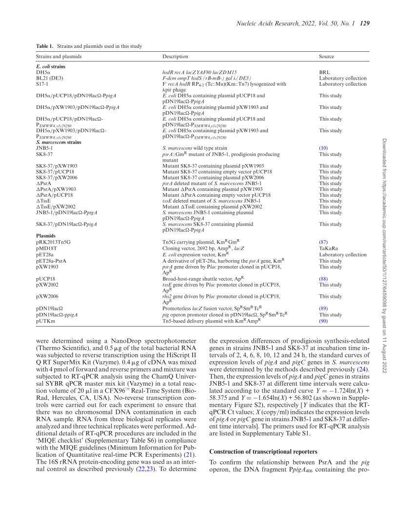
Table 1. Strains and plasmids used in this study
Strains and plasmids Description Source
E. coli strainsDH5� hsdR recA lacZYAF80 lacZDM15 BRLBL21 (DE3) F-dcm ompT hsdS (rB-mB-) gal λ(DE3) Laboratory collectionS17-1 F–recA hsdR RP4-2 (Tc::Mu)(Km::Tn7) lysogenized with
�pir phageLaboratory collection
DH5�/pUCP18/pDN19lac�-PpigA E. coli DH5� containing plasmid pUCP18 andpDN19lac�-PpigA
This study
DH5�/pXW1903/pDN19lac�-PpigA E. coli DH5� containing plasmid pXW1903 andpDN19lac�-PpigA
This study
DH5�/pUCP18/pDN19lac�-PSMWW4 v1c29280
E. coli DH5� containing plasmid pUCP18 andpDN19lac�-PSMWW4 v1c29280
This study
DH5�/pXW1903/pDN19lac�-PSMWW4 v1c29280
E. coli DH5� containing plasmid pXW1903 andpDN19lac�-PSMWW4 v1c29280
This study
S. marcescens strainsJNB5-1 S. marcescens wild type strain (10)SK8-37 psrA::GmR mutant of JNB5-1, prodigiosin producing
mutantThis study
SK8-37/pXW1903 Mutant SK8-37 containing plasmid pXW1903 This studySK8-37/pUCP18 Mutant SK8-37 containing empty vector pUCP18 This studySK8-37/pXW2006 Mutant SK8-37 containing plasmid pXW2006 This study�PsrA psrA deleted mutant of S. marcescens JNB5-1 This study�PsrA/pXW1903 Mutant �PsrA containing plasmid pXW1903 This study�PsrA/pUCP18 Mutant �PsrA containing empty vector pUCP18 This study�TssE tssE deleted mutant of S. marcescens JNB5-1 This study�TssE/pXW2002 Mutant �TssE containing plasmid pXW2002 This studyJNB5-1/pDN19lac�-PpigA S. marcescens JNB5-1 containing plasmid
pDN19lac�-PpigAThis study
SK8-37/pDN19lac�-PpigA S. marcescens SK8-37 containing plasmidpDN19lac�-PpigA
This study
PlasmidspRK2013Tn5G Tn5G carrying plasmid, KmRGmR (87)pMD18T Cloning vector, 2692 bp, AmpR, lacZ TaKaRapET28a E. coli expression vector, KmR Laboratory collectionpET28a-PsrA A derivative of pET-28a, harboring the psrA gene, KmR This studypXW1903 psrA gene driven by Plac promoter cloned in pUCP18,
ApRThis study
pUCP18 Broad-host-range shuttle vector, ApR (88)pXW2002 tssE gene driven by Plac promoter cloned in pUCP18,
ApRThis study
pXW2006 rhs2 gene driven by Plac promoter cloned in pUCP18,ApR
This study
pDN19lac� Promoterless lacZ fusion vector, SpRSmRTcR (89)pDN19lac�-ppigA pig operon promoter cloned in pDN19lac�, SpRSmRTcR This studypUTKm Tn5-based delivery plasmid with KmRAmpR (90)
were determined using a NanoDrop spectrophotometer(Thermo Scientific), and 0.5 �g of the total bacterial RNAwas subjected to reverse transcription using the HiScript IIQ RT SuperMix Kit (Vazyme). 0.4 �g of cDNA was mixedwith 4 pmol of forward and reverse primers and mixture wassubjected to RT-qPCR analysis using the ChamQ Univer-sal SYBR qPCR master mix kit (Vazyme) in a total reac-tion volume of 20 �l in a CFX96™ Real-Time System (Bio-Rad, Hercules, CA, USA). No-reverse transcription con-trols were carried out for each experiment to ensure thatthere was no chromosomal DNA contamination in eachRNA sample. RNA from three biological replicates wereanalyzed and three technical replicates were performed. Ad-ditional details of RT-qPCR procedures are included in the‘MIQE checklist’ (Supplementary Table S6) in compliancewith the MIQE guidelines (Minimum Information for Pub-lication of Quantitative real-time PCR Experiments) (21).The 16S rRNA protein-encoding gene was used as an inter-nal control as described previously (22,23). To determine
the expression differences of prodigiosin synthesis-relatedgenes in strains JNB5-1 and SK8-37 at incubation time in-tervals of 2, 4, 6, 8, 10, 12 and 24 h, the standard curves ofexpression levels of pigA and pigC genes in S. marcescenswere determined by the methods described previously (24).Then, the expression levels of pigA and pigC genes in strainsJNB5-1 and SK8-37 at different time intervals were calcu-lated according to the standard curve Y = −1.724ln(X) +58.375 and Y = −1.654ln(X) + 56.802 (as shown in Supple-mentary Figure S2), respectively [Y indicates that the RT-qPCR Ct values; X (copy/ml) indicates the expression levelsof pigA or pigC gene in strains JNB5-1 and SK8-37 at differ-ent time intervals]. The primers used for RT-qPCR analysisare listed in Supplementary Table S1.
Construction of transcriptional reporters
To confirm the relationship between PsrA and the pigoperon, the DNA fragment PpigA406 containing the pro-
Dow
nloaded from https://academ
ic.oup.com/nar/article/50/1/127/6459098 by guest on 11 August 2022
130 Nucleic Acids Research, 2022, Vol. 50, No. 1
moter region of the pig operon from positions -368 to + 38with respect to the transcriptional start site (TSS) of pigoperon as reported previously (15) was amplified with theprimers PigA-F3 and PigA-R3 listed in SupplementaryTable S1. The 406-bp-long PCR product was then clonedinto lacZ-containing plasmid pDN19lac� to obtain therecombinant plasmid pDN19lac�-PpigA406. The resultingrecombinant plasmid pDN19lac�-PpigA406 was thenintroduced into strains JNB5-1, SK8-37, DH5�/pUCP18or DH5�/pXW1903 by electroporation. The expres-sion levels of prodigiosin synthesis-related genes wereassessed by measuring �-galactosidase activities of thetransformants. To identify the DNA sequences necessaryfor PsrA binding, a transcriptional fusion reporter geneexpression of the pig operon was constructed by cloningthe DNA fragments PpigA281 (DNA fragment containingthe regulatory binding site (RBS) and activator binding site(ABS) spanning nucleotide positions −243 to + 38 relativeto the TSS of the pig operon), PpigA281 (A–90A–89A–88)(carrying mutation in RBS of positions TGT), PpigA281(A–90A–89) (carrying mutation in RBS of positions TG),PpigA281 (G–76G–75G–74) (carrying mutation in RBS ofpositions ACA), PpigA281 (G–75G–74) (carrying mutation inRBS of positions CA), PpigA281 (G–44G–43G–42) (carryingmutation in ABS of positions ACT), PpigA281 (G–44G–43)(carrying mutation in ABS of positions AC), PpigA281(C–30C–29C–28) (carrying mutation in ABS of positionsAGT), PpigA281 (C–29C–28) (carrying mutation in ABSof positions GT), PpigA281 (T–96T–95T–94) (carrying mu-tation of positions CAA), and PpigA281 (G–47G–46G–45)(carrying mutation of positions ATT) upstream of lacZ,respectively, with the primers listed in SupplementaryTable S1 in the supplemental material, and �-galactosidaseactivity in the strain JNB5-1 was determined. To analyzethe possible effectors of PsrA, different concentrationsof L-proline (0, 1.0, 2.0, and 3.0 mM), 2-Octenal (0, 1.0,2.0, and 3.0 mM), L-serine (0, 1.0, 2.0 and 3.0 mM) andS-adenosylmethionine (0, 1.0, 2.0 and 3.0 mM) were addedto LB medium, respectively, and then the �-galactosidaseassays were determined in S. marcescens JNB5-1 carryingthe PpigA281-lacZ. To determine whether L-proline hasa direct effect on PsrA binding to the pig operon andthe SMWW4 v1c29280 gene, different concentrationsof L-proline (0, 1.0, 2.0 and 3.0 mM) were added to theLB medium, and then the �-galactosidase assays weredetermined in E. coli DH5�/pUCP18/pDN19lac�-PpigA, DH5�/pXW1903/pDN19lac�-PpigA,DH5�/pUCP18/pDN19lac�-PSMWW4 v1c29280, andDH5�/pXW1903/pDN19lac�-PSMWW4 v1c29280strains,respectively. The �-galactosidase activity was deter-mined by SDS- and chloroform-permeabilized cellsusing o-nitrophenyl-�-D-galactopyranoside as thesubstrate, and the results were expressed in Millerunits with formula �-galactosidase activity = [1000 ×(A420 − 1.75 × A550)]/(V × T × A600) as described previ-ously (25–28) (A420 indicates the absorbance of the yellowo-nitrophenol; A550 indicates the scatter from cell debris,which, when multiplied by 1.75 approximates the scatterobserved at 420 nm; T indicates reaction time in minutes; Vindicates volume of culture assayed in milliliters; and A600reflects cell density). The experiments were independently
replicated three times and Student’s t test was used forstatistical analysis.
Production and purification of the PsrA protein
For the production and purification of the PsrA protein,the psrA gene was amplified with the primer pairs PsrA-F2 and PsrA-R2 listed in Supplementary Table S1. ThePCR product was then cloned into the pET28a plasmidto obtain recombinant plasmid pET28a-PsrA and elec-troporated into E. coli BL21(DE3) to construct recombi-nant strain E. coli BL21(DE3)/pET28-PsrA for overexpres-sion of a His-tagged PsrA. The single colonies of E. coliBL21(DE3)/pET28-PsrA were grown overnight at 37◦C inLB medium with the addition of 50 �g/ml kanamycin. Onemilliliter of the overnight culture was then inoculated into100 ml of LB medium and cultured at 37◦C until the celldensity reached 0.8 at OD600. PsrA expression was inducedat 16◦C for 10 h by the addition of 0.5 mM isopropyl-D-1-thiogalactopyranoside (IPTG). The cells were harvested bycentrifuging at 6000 × g for 10 min at 4◦C and washed twicewith phosphate-buffered saline (pH 7.4). Pellets were resus-pended in phosphate-buffered saline (pH 7.4) and disruptedby sonication to obtain the soluble intracellular proteins.The cellular lysate was centrifuged at 16 000 × g for 30 minat 4◦C to remove bacterial debris, and the supernatant wasloaded onto a 1-ml HisTrap HP column on an AKTA puri-fier system (GE Healthcare, Sweden) with binding buffer A(20 mM Tris–HCl buffer and 500 mM sodium chloride, pH7.4). Proteins were eluted with buffer B (20 mM Tris–HClbuffer, 500 mM sodium chloride, and 700 mM imidazole,pH 7.4), and the eluted fractions were pooled for sodiumdodecyl sulfate-polyacrylamide gel electrophoresis (SDS-PAGE) analysis. A Bradford protein assay kit was used todetermine protein concentration of PsrA.
To determine the predominant oligomeric state of PsrAprotein in solution, the native molecular weight of PsrAprotein was determined using a gel filtration column (Su-perdex 200 10/300 GL; GE Healthcare) as described previ-ously (29). The eluent buffer used for determination of PsrAdata was buffer C (20 mM sodium phosphate and 500 mMsodium chloride, pH 7.4), and the flow rate was 0.5 ml/min.
Electrophoretic mobility shift assay (EMSA)
EMSA was performed as described previously (30). Toprove whether PsrA directly binds to the pig operon, a 406-bp-long DNA fragments PpigA406 containing the promoterregion of the pig operon from positions −368 to + 38 withrespect to the TSS of pig operon was amplified with theprimers PigA-F3 and PigA-R3 listed in Supplementary Ta-ble S1. For the analysis of PsrA binding sites in the pro-moter region of the pig operon, a set of truncated DNAfragments PpigA281 (281-bp-long DNA fragment contain-ing the RBS and ABS of PsrA spanning nucleotide posi-tions −243 to + 38 relative to the TSS of the pig operon),PpigA134 (134-bp-long DNA fragment containing the RBSand ABS of PsrA spanning nucleotide positions −96 to + 38relative to the TSS of the pig operon), PpigA62 (62-bp-longDNA fragment without the RBS and ABS of PsrA span-ning nucleotide positions −24 to + 38 relative to the TSS of
Dow
nloaded from https://academ
ic.oup.com/nar/article/50/1/127/6459098 by guest on 11 August 2022
Nucleic Acids Research, 2022, Vol. 50, No. 1 131
the pig operon), PpigA73 (73-bp-long DNA fragment con-taining the RBS and ABS of PsrA spanning nucleotide po-sitions −96 to −24 relative to the TSS of the pig operon),PpigA170 (170-bp-long DNA fragment containing the RBSof PsrA spanning nucleotide positions −243 to −74 rela-tive to the TSS of the pig operon), PpigA169 (169-bp-longDNA fragment without the RBS and ABS of PsrA span-ning nucleotide positions −243 to −75 relative to the TSSof the pig operon), PpigA82 (82-bp-long DNA fragmentcontaining the ABS of PsrA spanning nucleotide positions−44 to + 38 relative to the TSS of the pig operon), andPpigA81 (81-bp-long DNA fragment without the RBS andABS of PsrA spanning nucleotide positions −43 to + 38relative to the TSS of the pig operon) were amplified bythe primer pairs listed in Supplementary Table S1 in thesupplemental material. To verify the PsrA-binding sites inthe genome of S. marcescens WW4 (NC 020211.1), differ-ent DNA fragments containing the promoter regions ofthe cytR, SMWW4 v1c21000, phoH, SMWW4 v1c48120,hemN, btuB, SMWW4 v1c09530, cueR, flhDC, swrW andSMWW4 v1c29280 genes were amplified by PCR methodwith the primers listed in Supplementary Table S1. Then,the purified DNA fragments were incubated with serialdilutions of the PsrA proteins in 20-�l reaction mixturescontaining 10-�l EMSA binding buffer (40 mM Tris–HCl,4 mM MgCl2, 100 mM NaCl, 10% glycerol, 2 mM DTT,0.2 mg/ml BSA and 1 mM EDTA) at room temperature for30 min. Electrophoresis was carried out on the 5% nativePAGE gels, and the gels were stained with ethidium bro-mide to visualize DNA bands.
Isothermal titration calorimetry
ITC experiments were performed on a MicroCal PEAQ-ITC microcalorimeter (Malvern, UK) as described previ-ously (31). For determination of PsrA binding isotherms forthe effectors, 30 �M PsrA solution was titrated with 0.3 mML-proline, L-serine or S-adenosylmethionine in 50 mM PBS(pH 7.4). The potential effector (2 �l) was injected 19 timesinto the 0.2-ml cell with stirring at 350 rpm at 25◦C. Bothprotein and effectors solutions were centrifuged for 5 minat 5000 × g. The binding isotherm was calculated from rawdata and fitted to a one-site binding model utilizing Micro-Cal PEAQ-ITC analysis software. The data were derivedfrom three parallel assays.
Transcriptome sequencing and data analysis
To analyze the effect of PsrA on the cellular processes in S.marcescens, three individuals of JNB5-1 and SK8-37 cellswere grown to OD600 of 5.0 before collecting. One milliliterof the collected cells was quickly frozen in liquid nitro-gen, treated with RNAprep pure kit (Tiangen) to extracttotal bacterial RNA, and delivered to Genewiz (Genewiz,Suzhou, China) in dry ice for transcriptome resequencinganalysis. The integrity, concentration and quantity of thebacterial RNA were determined using a NanoDrop spec-trophotometer (Thermo Scientific), a Bioanalyzer 2100 sys-tem (Agilent) and a 1% agarose gel. 1 �g total RNA withRIN value above 6.5 was used for following library prepa-ration. With Ribo-Zero rRNA Removal Kit (Illumina, San
Diego, CA, USA), the total bacterial RNA was subjectedto rRNA removing to obtain mRNA. The ribosomal de-pleted RNA was then fragmented and reverse-transcribed.ProtoScript II Reverse Transcriptase with random primersand Actinomycin D were used to synthesize the first strandcDNA. The second-strand cDNA was synthesized usingSecond Strand Synthesis Enzyme Mix (includes dACG-TP/dUTP). The purified double-stranded cDNA by beadswas then treated with End Prep Enzyme Mix to repair bothends and add a dA-tailing in one reaction, followed by aT-A ligation to add adaptors to both ends. Size selectionof Adaptor-ligated DNA was then performed using beads,and fragments of about 420 bp (with the approximate insertsize of 300 bp) were recovered. The dUTP-marked secondstrand was digested with Uracil-Specific Excision Reagentenzyme. Each sample was then amplified by PCR for 13cycles using P5 and P7 primers, with both primers carry-ing sequences, which can anneal with flow cell to performbridge PCR and P7 primer carrying a six-base index allow-ing for multiplexing. The PCR products were cleaned upusing beads, validated using a Qsep100 (Bioptic, Taiwan,China), and quantified by Qubit3.0 Fluorometer (Invitro-gen, Carlsbad, CA, USA). Then sequencing was carried outusing a 2 × 150 paired-end (PE) configuration; image anal-ysis and base calling were conducted by the HiSeq ControlSoftware (HCS) + OLB + GAPipeline-1.6 (Illumina) on theHiSeq instrument. The sequences were processed and ana-lyzed by GENEWIZ (Genewiz, Suzhou, China).
For annotation, the genome of S. marcescens WW4(NC 020211.1) was used as reference. The significant dif-ferentially expressed genes were determined between strainsSK8-37 and JNB5-1 using the DESeq software, with thestandards of P-value ≤0.05, and fold change |log2Ratio| ≥1.KEGG B class database was used to classify the genes withsignificantly differential expression based on their func-tions using the tool Omicshare (https://www.omicshare.com/tools/Home/Soft/pathwaygsea).
Motility assays
For swimming and swarming motility assays, one microliterof the exponential-phase cells (OD600 of 0.6) of JNB5-1,SK8-37, �PsrA and SK8-37/pXW1903 strains was spottedonto 0.3% and 0.5% semi-solid LB medium, respectively.After incubation at 30◦C for 24 h, diameter of the swimmingand swarming zones of these four strains were measured.Experiments were independently replicated three times.
Biofilm formation assay
Biofilm formation assay was performed as describedpreviously (24,32). Briefly, 20 �l exponential culture(OD600 = 0.6) was transferred into a 96-well microtiter plate(Corning, NY, USA) containing 200 �l fresh 3-fold dilutedLB medium. Then, the cultures in the microplates were re-moved and gently washed three times with water after in-cubation at 30◦C for 48 h without shaking. The biofilm inthe plates was stained with 0.1% crystal violet solution for15 min at room temperature. 95% ethanol was used to ex-tract the crystal violet and the biofilm amounts were mea-sured by the optical densities at 590 nm wavelength using a
Dow
nloaded from https://academ
ic.oup.com/nar/article/50/1/127/6459098 by guest on 11 August 2022
132 Nucleic Acids Research, 2022, Vol. 50, No. 1
BioTek Epoch2 microplate reader. Experiments were inde-pendently replicated three times.
Extracellular polysaccharide quantitation
Extracellular polysaccharide produced by strains JNB5-1,SK8-37, �PsrA and SK8-37/pXW1903 was determined bya method described previously with slight modification (8).In brief, bacterial cultures of the indicated strains were in-cubated in LB medium for 18 h, and then centrifuged at 12000 rpm for 15 min to harvest the bacterial cells. The pel-leted cells were resuspended in PBS solution (30 ml) and1% Zwittergent 3–14 citric acid solution (100 mM, pH 2.0,6 ml) and incubated at 50◦C for 20 min. The supernatantcells were transferred into 50-ml centrifugal bottles aftercentrifugation at 12 000 rpm for 30 min, and four volumesof cold ethanol (−20◦C) were added to each sample andplaced at −20◦C overnight. After overnight precipitation,the samples were centrifuged at 14 000 rpm and 4◦C for 45min and the supernatants were discarded. The exopolysac-charides in the sediment of each sample were air-dried in achemical fume hood, and the exopolysaccharides weighed.Experiments were independently replicated three times.
Haemolytic activity assay
Haemolytic activity assays were performed as previously de-scribed with slight modification (33,34). Briefly, 5 �l expo-nential culture (OD600 = 0.6) of strains JNB5-1, SK8-37,�PsrA and SK8-37/pXW1903 were plated on blood agarand the hemolysis zones visualized. Experiments were in-dependently replicated three times.
Antibacterial competition assay
Antibacterial competition assays were performed as de-scribed previously (35,36). Briefly, the attacker strain S.marcescens and the target strain E. coli MC4100 or E. cloa-cae ATCC13047 were grown overnight, normalized to anOD600 of 0.5, and mixed at a ratio of attacker/target of 5:1(the control mixture contained a 5:1 ratio of sterile LB totarget). 25 �l of this mixture was then spotted onto a pre-warmed agar plate and incubated for 4 h at 37◦C (for E. coliused as the target strain) or 30◦C (for E. cloacae used as thetarget strain). After cells were recovered from the spot andresuspended in 1 ml LB broth, the surviving target cells wereenumerated by serial dilution (10-fold) and viable counts onstreptomycin-supplemented media. The recovery of viablecells is reported as the total number recovered per cocul-ture spot. Control group consisted of sterile LB mixed withtarget bacteria at a ratio of 5:1. Experiments were indepen-dently replicated four times.
Identification of PsrA-binding sites
PsrA-binding sites were identified by searching DNA mo-tif in the genome sequence of S. marcescens WW4 (https://www.ncbi.nlm.nih.gov/nuccore/NC 020211.1). The palin-dromic nucleotide sequences 5′-TGTN11ACA-3′ (RBS)and 5′-ACTN11AGT-3′ (ABS) were used as query for se-quence similarity searching against the genome of WW4(NC 020211.1) using an online tool (http://meme-suite.org/tools/mast).
Phylogenetic tree construction of psrA-like genes and PsrA-like proteins
The homologs of psrA gene in S. marcescens were ana-lyzed by searching database Nucleotide collection (nr/nt)using Megablast. PSI-BLAST in NCBI was used to searchnon-redundant protein sequence (nr) database and analyzethe distribution of PsrA homologues in other bacteria. Thephylogenetic trees of the psrA-like genes and PsrA-like pro-teins were generated using BLAST pairwise alignments inNCBI and online software iTOL (http://itol.embl.de/) wasused to modify the phylogenetic trees.
Statistical analysis
Student’s test or one-way ANOVA was used for compar-ing statistical difference between the groups of experimen-tal data. Experiments were independently replicated at leastthree times.
RESULTS
Identification of a prodigiosin synthesis activator PsrA
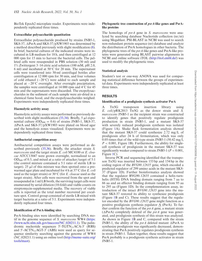
A Tn5G transposon insertion library usingE. coli/pRK2013 Tn5G as the donor strain and S.marcescens JNB5-1 as the recipient strain was constructedto identify genes that positively regulate prodigiosinproduction in strain JNB5-1, and a mutant SK8-37with severely reduced prodigiosin synthesis was isolated(Figure 1A). Shake flask fermentation analysis showedthat the mutant SK8-37 could synthesize 2.72 mg/L ofprodigiosin after 24 h of fermentation, which was only0.05-times that of the wild-type strain JNB5-1 (50.42 mg/l,P < 0.001, Figure 1B). Furthermore, the ability for single-cell synthesis of prodigiosin in the mutant SK8-37 wassignificantly weaker compared to strain JNB5-1 (P < 0.001,Figure 1C).
Inverse PCR and sequencing identified that the transpo-son Tn5G was inserted between 153 bp and 154 bp in thecoding region of the BVG90 12635 gene, which encoded apredicted regulator of 299 amino acids in the mutant SK8-37 (Figure 1D). Further bioinformatics analysis showedthat the regulator BVG90 12635 contained a helix-turn-helix (HTH) DNA binding domain ranging from 7 aa to66 aa and an effector binding domain ranging from 95 aato 295 aa (Figure 1D). In the complementation assay, in-troduction of the intact BVG90 12635 gene into the mu-tant SK8-37 restored its ability to synthesize prodigiosin(Figure 1B and C). These results suggest that the regula-tor encoded by the BVG90 12635 gene might function as apositive prodigiosin synthesis regulator A (PsrA). To fur-ther confirm the function of the psrA gene, a mutant strain(�PsrA) completely deleted of the psrA gene was gener-ated, and prodigiosin synthesis of this strain was analyzed.As shown in Figure 1B and C, compared with the strainJNB5-1, the ability of the psrA deleted mutant �PsrA tosynthesize prodigiosin was significantly decreased, demon-strating that PsrA positively regulates prodigiosin synthesisin strain JNB5-1. Taken together, these results suggest thatPsrA probably is a prodigiosin synthesis activator in strainJNB5-1.
Dow
nloaded from https://academ
ic.oup.com/nar/article/50/1/127/6459098 by guest on 11 August 2022

Nucleic Acids Research, 2022, Vol. 50, No. 1 133
Figure 1. Regulator PsrA positively regulates prodigiosin production in S. marcescens. (A) A prodigiosin producing mutant SK8-37 was identified byTn5G transposon insertion mutation. (B) Prodigiosin production analysis of strains JNB5-1, SK8-37, SK8-37/pXW1903, SK8-37/pUCP18, �PsrA,�PsrA/pXW1903 and SK8-37/pUCP18. JNB5-1 is a wild-type S. marcescens, SK8-37 is a psrA disrupted mutant, �PsrA is a psrA deleted mutant,SK8-37/pXW1903 and �PsrA/pXW1903 are psrA complemented strains, and SK8-37/pUCP18 and �PsrA/pUCP18 are recombinant strains withempty vector pUCP18. (C) Analysis of prodigiosin levels in JNB5-1, SK8-37, SK8-37/pXW1903, SK8-37/pUCP18, �PsrA, �PsrA/pXW1903, and SK8-37/pUCP18 strains. (D) The genetic loci identified in mutant SK8-37. The upper panel indicates the genetic map of the disrupted gene BVG90 12635 (psrA)and its surrounding genes. Tn5G insertion site is denoted by black arrow points. The middle panel represents the domain organization of the BVG90 12635protein. The lower panel indicates that BVG90 12635 is a LysR family transcriptional regulator. For B and C, the experiments were independently repli-cated three times. Error bars indicate standard deviations. One-way analysis of variance (ANOVA) was used to examine the mean differences between thedata groups. ****P < 0.001; ns, no significance difference.
PsrA controls prodigiosin production via direct transcrip-tional regulation of the pigA-pigN operon
To investigate how PsrA activates prodigiosin productionin S. marcescens, the cell growth of strains JNB5-1 (wild-type strain), SK8-37 (psrA disrupted mutant), and SK8-37/pXW1903 (complemented strain) were determined, andresults showed that there was no significant difference in cellgrowth between these three strains (Figure 2A). This resultsuggests that PsrA activation of prodigiosin production inS. marcescens was probably not related to biomass and thesignificantly decreased production of prodigiosin in mutantSK8-37 was possibly due to the lower expression levels ofthe prodigiosin related pig gene cluster.
The enzymes for prodigiosin production in S. marcescensare encoded by genes pigABCDEFGHIJKLMN, a totalof 14 genes (Figure 2B, and Supplementary Figure S1).Among them, pigB, pigD and pigE genes use 2-octenalas the substrate to synthesize 2-methyl-3-n-amyl-pyrrole(MAP), while pigA, pigF, pigG, pigH, pigI, pigJ, pigM andpigN genes use the L-proline as the substrate to synthesize4-methoxy-2,2′-bipyrrole-5-carbaldehyde (MBC). Finally,the pigC gene encodes terminal condensing enzyme thatcondenses both MAP and MBC to prodigiosin (Supple-mentary Figure S1) (1). Hence, to reveal how PsrA regulates
prodigiosin synthesis in S. marcescens, Real-time quantita-tive PCR (RT-qPCR) was carried out to analyze the expres-sion levels of pigA and pigC genes in strains JNB5-1 andSK8-37 at incubation time intervals of 2, 4, 6, 8, 10, 12 and24 h. These two genes displayed significantly lower expres-sion levels in the seven incubation time intervals in the psrAdisrupted strain SK8-37 than in the parent strain JNB5-1(Figure 2B, and Supplementary Figure S3). Further, RT-qPCR was used to analyze the expression levels of pigA-BCDEFGHIJKLMN genes in strains JNB5-1 and SK8-37at the stationary phase (OD600 of 6.0) at which prodigiosinbegan to be produced in large quantities. As shown in Fig-ure 2C, the expression levels of these 14 genes were down-regulated by 14.69-fold to 51.18-fold in mutant SK8-37,suggesting that PsrA positively regulates prodigiosin syn-thesis related genes in S. marcescens. This conclusion wasfurther verified by a transcriptional lacZ reporter fusion bycloning the 406-bp-long promoter region of the pig operonupstream of the lacZ gene, and the expression of the piggene cluster was determined by measurement of cellular �-galactosidase activity. Results showed that the transcriptionof the pig operon exhibited significantly lower expressionlevels in the absence of PsrA with the P values lower than0.005 (Figure 2D, P < 0.005).
Dow
nloaded from https://academ
ic.oup.com/nar/article/50/1/127/6459098 by guest on 11 August 2022

134 Nucleic Acids Research, 2022, Vol. 50, No. 1
Figure 2. PsrA regulates the prodigiosin synthesis genes directly. (A) Growth curves of JNB5-1, SK8-37, and SK8-37/pXW1903 strains. (B) RT-qPCRanalysis of the expression level of the pigA gene in the strains SK8-37 and JNB5-1 at time intervals of 2, 4, 6, 8, 10, 12 and 24 h. (C) RT-qPCR analysis ofthe expression level of pigABCDEFGHIJKLMN genes in the strains SK8-37 and JNB5-1 at an OD600 of 6.0. (D) Analysis of �-galactosidase activity ofstrains JNB5-1 (PsrA+), SK68 (PsrA-), DH5�/pXW1903 (PsrA+) and DH5�/pUCP18 (PsrA-) harboring the PpigA-lacZ reporter fusion at an OD600 of5.0. (E) EMSA for PsrA protein binding to the promoter region of the pig operon. (F) The regulatory network of PsrA controls prodigiosin synthesis inS. marcescens. For A−D, the experiments were independently replicated three times. Error bars indicated standard deviations. Student’s t test was used toexamine the mean differences between the data groups. ***P < 0.005.
To further explore whether PsrA directly controls the ex-pression of the pig gene cluster, then influence prodigiosinproduction in strain JNB5-1, heterogeneous expression reg-ulation was investigated in E. coli. Results showed that thetranscription fusion PpigA-lacZ showed significantly higherexpression level in the presence of PsrA, suggesting thatPsrA probably directly controls the expression of the pigA-
BCDEFGHIJKLMN genes (Figure 2D, P < 0.005). Thisdata was consistent with the result of EMSA test that PsrAcould directly bind to the promoter region of pig operon,a 406-bp-long DNA fragment (PpigA406) between positions-368 and + 38 relative to the TSS of pig operon, which wasamplified using primers PigA-F3 and PigA-R3 (Figure 2E).Taken together, our study supports model that PsrA prob-
Dow
nloaded from https://academ
ic.oup.com/nar/article/50/1/127/6459098 by guest on 11 August 2022
Nucleic Acids Research, 2022, Vol. 50, No. 1 135
ably controls prodigiosin production via direct transcrip-tional regulation of the pigA-pigN operon (Figure 2F).
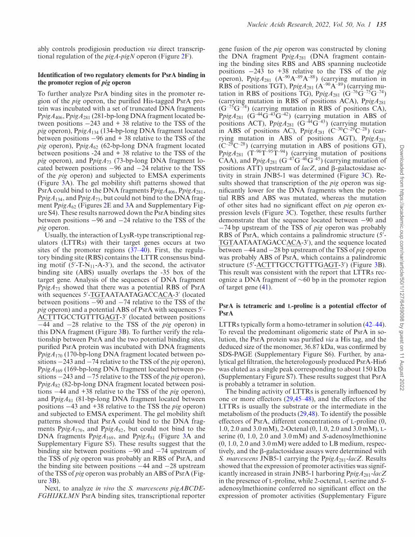
Identification of two regulatory elements for PsrA binding inthe promoter region of pig operon
To further analyze PsrA binding sites in the promoter re-gion of the pig operon, the purified His-tagged PsrA pro-tein was incubated with a set of truncated DNA fragmentsPpigA406, PpigA281 (281-bp-long DNA fragment located be-tween positions −243 and + 38 relative to the TSS of thepig operon), PpigA134 (134-bp-long DNA fragment locatedbetween positions −96 and + 38 relative to the TSS of thepig operon), PpigA62 (62-bp-long DNA fragment locatedbetween positions -24 and + 38 relative to the TSS of thepig operon), and PpigA73 (73-bp-long DNA fragment lo-cated between positions −96 and −24 relative to the TSSof the pig operon) and subjected to EMSA experiments(Figure 3A). The gel mobility shift patterns showed thatPsrA could bind to the DNA fragments PpigA406, PpigA281,PpigA134, and PpigA73, but could not bind to the DNA frag-ment PpigA62 (Figures 2E and 3A and Supplementary Fig-ure S4). These results narrowed down the PsrA binding sitesbetween positions −96 and −24 relative to the TSS of thepig operon.
Usually, the interaction of LysR-type transcriptional reg-ulators (LTTRs) with their target genes occurs at twosites of the promoter regions (37–40). First, the regula-tory binding site (RBS) contains the LTTR consensus bind-ing motif (5′-T-N11-A-3′), and the second, the activatorbinding site (ABS) usually overlaps the -35 box of thetarget gene. Analysis of the sequences of DNA fragmentPpigA73 showed that there was a potential RBS of PsrAwith sequences 5′-TGTAATAATAGACCACA-3′ (locatedbetween positions −90 and −74 relative to the TSS of thepig operon) and a potential ABS of PsrA with sequences 5′-ACTTTGCCTGTTTGAGT-3′ (located between positions−44 and −28 relative to the TSS of the pig operon) inthis DNA fragment (Figure 3B). To further verify the rela-tionship between PsrA and the two potential binding sites,purified PsrA protein was incubated with DNA fragmentsPpigA170 (170-bp-long DNA fragment located between po-sitions −243 and −74 relative to the TSS of the pig operon),PpigA169 (169-bp-long DNA fragment located between po-sitions −243 and −75 relative to the TSS of the pig operon),PpigA82 (82-bp-long DNA fragment located between posi-tions −44 and +38 relative to the TSS of the pig operon),and PpigA81 (81-bp-long DNA fragment located betweenpositions −43 and +38 relative to the TSS the pig operon)and subjected to EMSA experiment. The gel mobility shiftpatterns showed that PsrA could bind to the DNA frag-ments PpigA170, and PpigA82, but could not bind to theDNA fragments PpigA169, and PpigA81 (Figure 3A andSupplementary Figure S5). These results suggest that thebinding site between positions −90 and −74 upstream ofthe TSS of pig operon was probably an RBS of PsrA, andthe binding site between positions −44 and −28 upstreamof the TSS of pig operon was probably an ABS of PsrA (Fig-ure 3B).
Next, to analyze in vivo the S. marcescens pigABCDE-FGHIJKLMN PsrA binding sites, transcriptional reporter
gene fusion of the pig operon was constructed by cloningthe DNA fragment PpigA281 (DNA fragment contain-ing the binding sites RBS and ABS spanning nucleotidepositions −243 to +38 relative to the TSS of the pigoperon), PpigA281 (A–90A–89A–88) (carrying mutation inRBS of positions TGT), PpigA281 (A–90A–89) (carrying mu-tation in RBS of positions TG), PpigA281 (G–76G–75G–74)(carrying mutation in RBS of positions ACA), PpigA281(G–75G–74) (carrying mutation in RBS of positions CA),PpigA281 (G–44G–43G–42) (carrying mutation in ABS ofpositions ACT), PpigA281 (G–44G–43) (carrying mutationin ABS of positions AC), PpigA281 (C–30C–29C–28) (car-rying mutation in ABS of positions AGT), PpigA281(C–29C–28) (carrying mutation in ABS of positions GT),PpigA281 (T–96T–95T–94) (carrying mutation of positionsCAA), and PpigA281 (G–47G–46G–45) (carrying mutation ofpositions ATT) upstream of lacZ, and �-galactosidase ac-tivity in strain JNB5-1 was determined (Figure 3C). Re-sults showed that transcription of the pig operon was sig-nificantly lower for the DNA fragments when the poten-tial RBS and ABS was mutated, whereas the mutationof other sites had no significant effect on pig operon ex-pression levels (Figure 3C). Together, these results furtherdemonstrate that the sequence located between −90 and−74 bp upstream of the TSS of pig operon was probablyRBS of PsrA, which contains a palindromic structure (5′-TGTAATAATAGACCACA-3′), and the sequence locatedbetween −44 and −28 bp upstream of the TSS of pig operonwas probably ABS of PsrA, which contains a palindromicstructure (5′-ACTTTGCCTGTTTGAGT-3′) (Figure 3B).This result was consistent with the report that LTTRs rec-ognize a DNA fragment of ∼60 bp in the promoter regionof target gene (41).
PsrA is tetrameric and L-proline is a potential effector ofPsrA
LTTRs typically form a homo-tetramer in solution (42–44).To reveal the predominant oligomeric state of PsrA in so-lution, the PsrA protein was purified via a His tag, and thededuced size of the monomer, 36.87 kDa, was confirmed bySDS-PAGE (Supplementary Figure S6). Further, by ana-lytical gel filtration, the heterologously produced PsrA-His6was eluted as a single peak corresponding to about 150 kDa(Supplementary Figure S7). These results suggest that PsrAis probably a tetramer in solution.
The binding activity of LTTRs is generally influenced byone or more effectors (29,45–48), and the effectors of theLTTRs is usually the substrate or the intermediate in themetabolism of the products (29,48). To identify the possibleeffectors of PsrA, different concentrations of L-proline (0,1.0, 2.0 and 3.0 mM), 2-Octenal (0, 1.0, 2.0 and 3.0 mM), L-serine (0, 1.0, 2.0 and 3.0 mM) and S-adenosylmethionine(0, 1.0, 2.0 and 3.0 mM) were added to LB medium, respec-tively, and the �-galactosidase assays were determined withS. marcescens JNB5-1 carrying the PpigA281-lacZ. Resultsshowed that the expression of promoter activities was signif-icantly increased in strain JNB5-1 harboring PpigA281-lacZin the presence of L-proline, while 2-octenal, L-serine and S-adenosylmethionine conferred no significant effect on theexpression of promoter activities (Supplementary Figure
Dow
nloaded from https://academ
ic.oup.com/nar/article/50/1/127/6459098 by guest on 11 August 2022

136 Nucleic Acids Research, 2022, Vol. 50, No. 1
Figure 3. Identification of the binding motif and effector of PsrA. (A) Mapping of the PsrA-binding sequences by EMSA assay. DNA fragments withthe PsrA-regulatory binding site (RBS) or PsrA-activating binding site (ABS) showed positive bindings (blue color). DNA fragments with the defectivePsrA-RBS and PsrA-ABS showed no bindings (gray color). Arabic numerals represent the distances (bp) upstream from the TSS of the pig operon. (B)The organization of the upstream region of the pig operon is shown. The −10 box and −35 box of the pig operon are underlined. The start codon ATG andTSS of pig operon are in red, italic and bold. TSS is denoted by bent arrows and + 1, and the direction of the arrow indicates the direction of gene transcrip-tion. The putative PsrA-RBS with sequences 5′-TGTAATAATAGACCACA-3′ and putative PsrA-ABS with sequences 5′-ACTTTGCCTGTTTGAGT-3′are boxed, and the palindromes are in bold. (C) In vivo analysis of the PsrA RBS and ABS motifs using site-directed mutagenesis of the promoter re-gion of pig operon. The transcription fusions PpigA281-lacZ, PpigA281 (A–90A–89A–88)-lacZ, PpigA281 (A–90A–89)-lacZ, PpigA281 (G–76G–75G–74)-lacZ,PpigA281 (G–75G–74)-lacZ, PpigA281 (G–44G–43G–42)-lacZ, PpigA281 (G–44G–43)-lacZ, PpigA281 (C–30C–29C–28)-lacZ, PpigA281 (C–29C–28)-lacZ, PpigA281(T–96T–95T–94)-lacZ and PpigA281 (G–47G–46G–45)-lacZ were used in the S. marcescens strain JNB5-1. PpigA281: DNA fragment containing PsrA-RBSand ABS. PpigA281 (A–90A–89A–88): PpigA281 carrying mutation in positions TGT. PpigA281 (A–90A–89): PpigA281 carrying mutation in positions TG.PpigA281 (G–76G–75G–74): PpigA281 carrying mutation in positions ACA. PpigA281 (G–75G–74): PpigA281 carrying mutation in positions CA. PpigA281(G–44G–43G–42): PpigA281 carrying mutation in positions ACT. PpigA281 (G–44G–43): PpigA281 carrying mutation in positions AC. PpigA281 (C–30C–29C–28):PpigA281 carrying mutation in positions AGT. PpigA281 (C–29C–28): PpigA281 carrying mutation in positions GT. PpigA281 (T–96T–95T–94): PpigA281 car-rying mutation in positions CAA. PpigA281 (G–47G–46G–45): PpigA281 carrying mutation in positions ATT. (D) Direct binding of L-proline to the solublePsrA protein was measured by isothermal titration microcalorimetry (ITC). L-proline exhibited binding affinity of KD = 1.36 × 10⁻⁵ M to PsrA. (E andF) ITC analysis revealed that PsrA could not directly binding to the L-serine (E) and S-adenosylmethionine (F). ITC experiments were performed on a Mi-croCal PEAQ-ITC microcalorimeter. For C, the experiment was independently replicated three times. Error bars indicated standard deviations. Student’st test was used to examine the mean differences between the data groups. ****P < 0.001; ns: no significant difference.
Dow
nloaded from https://academ
ic.oup.com/nar/article/50/1/127/6459098 by guest on 11 August 2022

Nucleic Acids Research, 2022, Vol. 50, No. 1 137
S8). This result indicates that L-proline is a possible effec-tor of PsrA.
To further confirm that the effector of PsrA is the L-proline, the binding of possible effectors with PsrA wastested using isothermal titration calorimetry (ITC). Asshown in Figure 3D−F, the ITC assays revealed an activeinteraction between PsrA and L-proline but not with L-serine and S-adenosylmethionine. Furthermore, to deter-mine whether L-proline affects the DNA binding ability ofPsrA, we performed EMSA assay with the promoter re-gion of the pig operon. Results showed that supplemen-tation of 0.20 �M L-proline to PsrA protein exhibited anincreased promoter binding ability (Supplementary FigureS4A and B). This data was consistent with the result thatL-proline could activate transcription of the pig operonwhich was confirmed by determination of �-galactosidaseactivities derived from the PpigA-lacZ reporter gene fu-sion in DH5�/pXW1903/pDN19lac�-PpigA strain (Sup-plementary Figure S4C, P < 0.01). Also, the direct ef-fect of L-proline on PsrA binding to the promoter re-gion of the SMWW4 v1c29280 gene, containing the PsrA-RBS was assessed by EMSAs and �-galactosidase activ-ity assay. Results showed that supplementation of 0.20�M L-proline to 5.30 �M PsrA protein resulted in un-detectable free DNA fragment, whereas in the absence ofL-proline, free DNA fragment remained with the sameamount of PsrA (Supplementary Figure S9). This resultwas further supported by the fact that L-proline couldrepress transcription of the SMWW4 v1c29280 gene inthe DH5�/pXW1903/pDN19lac�-PSMWW4 v1c29280 straincarrying the intact psrA gene (Supplementary Figure S9,P < 0.05). Collectively, these data suggest that L-proline isan effector of PsrA.
Transcriptome analysis reveals PsrA as a pleiotropic regula-tor
More and more regulators have now been experimen-tally proved to play important roles in various bacterialmetabolic pathways (24,49–52). As a transcription regu-lator of LysR family encoded in bacteria, PsrA may alsoplay important regulatory role in the cellular metabolism.To analyze the effect of PsrA in the cellular processes inS. marcescens, RNAseq analysis was performed to eval-uate gene expression differences between wild-type strainJNB5-1 and psrA disrupted mutant SK8-37. Comparativetranscriptome data showed that the expression levels of60 genes were significantly upregulated (log2FC ≥ 1, P-value < 0.05), while 558 genes were significantly down-regulated (log2FC ≤ −1, P-value < 0.05) when compar-ing the psrA disrupted mutant strain SK8-37 to its parentstrain JNB5-1 (Figure 4A, Supplementary Table S2, S3 andS7). According to the annotation of KEGG B Class, these618 significantly upregulated or downregulated genes wereclassified into 20 major cellular processes, including globaland overview maps, membrane transport, and amino acidmetabolism (Figure 4B). Further, these 618 genes could begrouped into several major metabolic pathways, such as reg-ulator proteins, flagellar assembly, two-component system,prodigiosin synthesis, biofilm formation, and ribosome pro-teins (Figure 4C). Altogether, these results suggest that reg-
ulator PsrA possibly functions as a key regulator control-ling versatile cellular functions in strain JNB5-1.
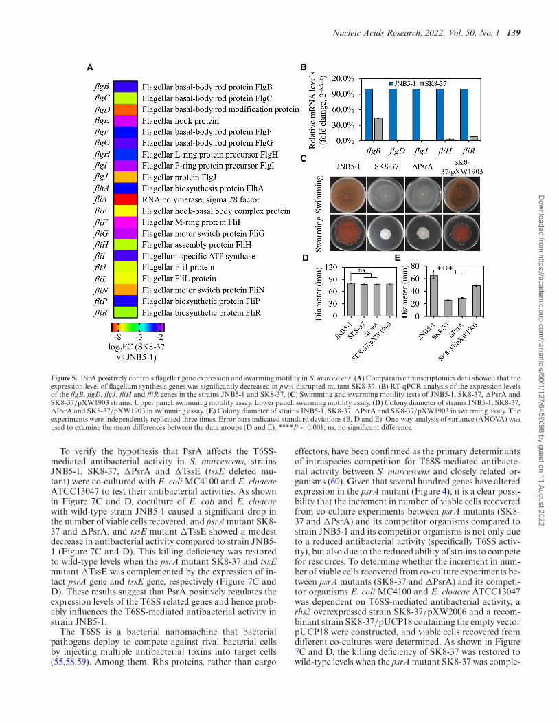
PsrA positively controls swarming motility
Swimming and swarming are two modes of motilities foundin S. marcescens, and it is reported that multiple cellularsystems were involved in these two motilities, especially thepresence of functional flagellar and type IV pili (10,53).Transcriptome analysis showed that the transcription offlgB, flgC, flgD, flgE, flgF, flgG, flgH, flgI, flgJ, flhA, fliA,fliE, fliF, fliG, fliH, fliI, fliJ, fliN, flip and fliR genes involvedin functional flagellar synthesis was significantly decreasedin the psrA disrupted mutant SK8-37 (log2FC ≤ −1, P-value < 0.05, Figure 5A, Supplementary Table S3 and S7).This data was further confirmed by RT-qPCR analysis, withthe flgB, flgD, flgJ, fliH and fliR genes showed significantlydecreased levels of expression in the mutant SK8-37 thanthat of the parental strain JNB5-1 (Figure 5B), suggestingthat PsrA probably positively regulates cell motility in S.marcescens.
To further explore the role of PsrA in cell motilityin S. marcescens, swarming and swimming motility as-say was conducted in JNB5-1, SK8-37, �PsrA and SK8-37/pXW1903 strains. Results showed that when testing forswimming motility with 0.3% semi-solid agarose plates, nosignificant difference was observed among JNB5-1, SK8-37, �PsrA and SK8-37/pXW1903, suggesting PsrA proba-bly had no effect on bacterial swimming motility (Figure5C and D). For swarming test, absence of PsrA (strainsSK8-37 and �PsrA) resulted in a smaller swarming zonecompared to parental strain JNB5-1 and complementarystrain SK8-37/pXW1903, indicating that PsrA probablypositively influences the swarming motility (Figure 5C, andE).
PsrA regulates biofilm formation, extracellular polysaccha-ride production, and serrawettin W1 biosynthesis
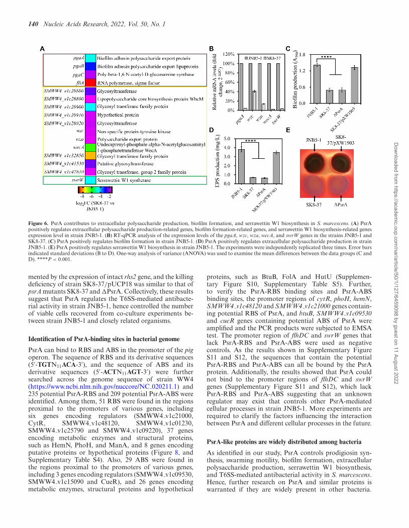
Biofilm formation, extracellular polysaccharide produc-tion, and serrawettin W1 biosynthesis are other threeimportant cellular processes in S. marcescens and theyare regulated by multiple genes. Transcriptome data high-lighted that the transcription of biofilm formation-relatedgenes pgaA, pgaB, pgaC, and fliA, extracellular polysac-charide production-related genes SMWW4 v1c28880,SMWW4 v1c28890, SMWW4 v1c28900,SMWW4 v1c28910, SMWW4 v1c28920, wzc, wza,wecA, SMWW4 v1c32850, SMWW4 v1c41530 andSMWW4 v1c47610, and serrawettin W1-related geneswrW were dramatically downregulated in the psrA dis-rupted mutant SK8-37 (log2FC ≤ −1, P-value < 0.05,Figure 6A, Supplementary Tables S3 and S7). To furtherverify the transcriptome data, the expression patterns ofpgaA, wzc, wza, wecA and swrW genes in strains JNB5-1and SK8-37 were validated by RT-qPCR. The transcrip-tion levels of these five genes were reduced by 7.22-timesto 44.05-times in the mutant SK8-37 compared to thewild-type strain JNB5-1 (Figure 6B). Taken together, thesedata suggest that PsrA probably plays an important role inbiofilm formation, extracellular polysaccharide production,and serrawettin W1 biosynthesis in S. marcescens.
Dow
nloaded from https://academ
ic.oup.com/nar/article/50/1/127/6459098 by guest on 11 August 2022

138 Nucleic Acids Research, 2022, Vol. 50, No. 1
Figure 4. Transcriptome analysis of strains JNB5-1 and psrA disrupted mutant SK8-37. (A) Genome-wide analysis of gene expression differences betweenpsrA disrupted mutant SK8-37 and wild-type strain JNB5-1 at an OD600 of 6.0. X-axis represents the logarithmic transformation value of gene expressionlevels in strain JNB5-1. Y-axis represents log2-transformed value of gene expression change folds between strains SK8-37 and JNB5-1. Genes belongingto different pathways are represented by different colored shapes as indicated. Others represent genes not belonging to the indicated pathways. (B) Basedon the KEGG B class, significant differentially expressed genes between strains SK8-37 and JNB5-1 were classified into different cellular processes. (C)Expression profiles of the genes belonging to the indicated metabolic pathways.
The effect of PsrA on biofilm formation and extracellu-lar polysaccharide production was evaluated further, andthe results indeed showed that the psrA mutants SK8-37(psrA disrupted mutant) and �PsrA (psrA deleted mu-tant) produced much less biofilm and extracellular polysac-charide than strain JNB5-1 or SK8-37/pXW1903 (Fig-ure 6C and D), indicating that PsrA positively influ-ences the biofilm formation and extracellular polysac-charide production in S. marcescens. Hemolysis, an in-direct measurement of serrawettin W1 production, wasused in serrawettin W1 biosynthesis analysis in strainsJNB5-1, SK8-37, �PsrA and SK8-37/pXW1903, and re-sults showed that the psrA mutants SK8-37 and �PsrAwere defective in hemolysis (Figure 6E), suggesting thatPsrA positively regulates the serrawettin W1 biosynthesis inS. marcescens.
PsrA required for T6SS-mediated antibacterial activity
The type VI secretion system (T6SS) is widely distributedin Gram-negative bacteria and bacteria can use them asweapons against prokaryotic or eukaryotic target cellswith diverse effectors (54–57). Transcriptome data showedthat lots of the T6SS-related genes, including tssABCE-FGHKLM, tagF, hcp, fha, pppA, tagJ, ppkA, vgrG2 andrhs2 genes were significantly downregulated in the psrA dis-rupted mutant SK8-37 compared to wild-type strain JNB5-1 (log2FC ≤ −1, P-value < 0.05, Figure 7A, SupplementaryTables S3 and S7). This result was further verified by RT-qPCR analysis, with the expression levels of the tssE, hcp,vgrG2, tssB and tssC genes reduced by 2.65-times to 14.38-times in the mutant SK8-37 compared to the strain JNB5-1(Figure 7B). These results suggest that PsrA might affectT6SS-mediated antibacterial activity in strain JNB5-1.
Dow
nloaded from https://academ
ic.oup.com/nar/article/50/1/127/6459098 by guest on 11 August 2022
Nucleic Acids Research, 2022, Vol. 50, No. 1 139
Figure 5. PsrA positively controls flagellar gene expression and swarming motility in S. marcescens. (A) Comparative transcriptomics data showed that theexpression level of flagellum synthesis genes was significantly decreased in psrA disrupted mutant SK8-37. (B) RT-qPCR analysis of the expression levelsof the flgB, flgD, flgJ, fliH and fliR genes in the strains JNB5-1 and SK8-37. (C) Swimming and swarming motility tests of JNB5-1, SK8-37, �PsrA andSK8-37/pXW1903 strains. Upper panel: swimming motility assay. Lower panel: swarming motility assay. (D) Colony diameter of strains JNB5-1, SK8-37,�PsrA and SK8-37/pXW1903 in swimming assay. (E) Colony diameter of strains JNB5-1, SK8-37, �PsrA and SK8-37/pXW1903 in swarming assay. Theexperiments were independently replicated three times. Error bars indicated standard deviations (B, D and E). One-way analysis of variance (ANOVA) wasused to examine the mean differences between the data groups (D and E). ****P < 0.001; ns, no significant difference.
To verify the hypothesis that PsrA affects the T6SS-mediated antibacterial activity in S. marcescens, strainsJNB5-1, SK8-37, �PsrA and �TssE (tssE deleted mu-tant) were co-cultured with E. coli MC4100 and E. cloacaeATCC13047 to test their antibacterial activities. As shownin Figure 7C and D, coculture of E. coli and E. cloacaewith wild-type strain JNB5-1 caused a significant drop inthe number of viable cells recovered, and psrA mutant SK8-37 and �PsrA, and tssE mutant �TssE showed a modestdecrease in antibacterial activity compared to strain JNB5-1 (Figure 7C and D). This killing deficiency was restoredto wild-type levels when the psrA mutant SK8-37 and tssEmutant �TssE was complemented by the expression of in-tact psrA gene and tssE gene, respectively (Figure 7C andD). These results suggest that PsrA positively regulates theexpression levels of the T6SS related genes and hence prob-ably influences the T6SS-mediated antibacterial activity instrain JNB5-1.
The T6SS is a bacterial nanomachine that bacterialpathogens deploy to compete against rival bacterial cellsby injecting multiple antibacterial toxins into target cells(55,58,59). Among them, Rhs proteins, rather than cargo
effectors, have been confirmed as the primary determinantsof intraspecies competition for T6SS-mediated antibacte-rial activity between S. marcescens and closely related or-ganisms (60). Given that several hundred genes have alteredexpression in the psrA mutant (Figure 4), it is a clear possi-bility that the increment in number of viable cells recoveredfrom co-culture experiments between psrA mutants (SK8-37 and �PsrA) and its competitor organisms compared tostrain JNB5-1 and its competitor organisms is not only dueto a reduced antibacterial activity (specifically T6SS activ-ity), but also due to the reduced ability of strains to competefor resources. To determine whether the increment in num-ber of viable cells recovered from co-culture experiments be-tween psrA mutants (SK8-37 and �PsrA) and its competi-tor organisms E. coli MC4100 and E. cloacae ATCC13047was dependent on T6SS-mediated antibacterial activity, arhs2 overexpressed strain SK8-37/pXW2006 and a recom-binant strain SK8-37/pUCP18 containing the empty vectorpUCP18 were constructed, and viable cells recovered fromdifferent co-cultures were determined. As shown in Figure7C and D, the killing deficiency of SK8-37 was restored towild-type levels when the psrA mutant SK8-37 was comple-
Dow
nloaded from https://academ
ic.oup.com/nar/article/50/1/127/6459098 by guest on 11 August 2022
140 Nucleic Acids Research, 2022, Vol. 50, No. 1
Figure 6. PsrA contributes to extracellular polysaccharide production, biofilm formation, and serrawettin W1 biosynthesis in S. marcescens. (A) PsrApositively regulates extracellular polysaccharide production-related genes, biofilm formation-related genes, and serrawettin W1 biosynthesis-related genesexpression level in strain JNB5-1. (B) RT-qPCR analysis of the expression levels of the pgaA, wzc, wza, wecA, and swrW genes in the strains JNB5-1 andSK8-37. (C) PsrA positively regulates biofilm formation in strain JNB5-1. (D) PsrA positively regulates extracellular polysaccharide production in strainJNB5-1. (E) PsrA positively regulates serrawettin W1 biosynthesis in strain JNB5-1. The experiments were independently replicated three times. Error barsindicated standard deviations (B to D). One-way analysis of variance (ANOVA) was used to examine the mean differences between the data groups (C andD). ****P < 0.001.
mented by the expression of intact rhs2 gene, and the killingdeficiency of strain SK8-37/pUCP18 was similar to that ofpsrA mutants SK8-37 and �PsrA. Collectively, these resultssuggest that PsrA regulates the T6SS-mediated antibacte-rial activity in strain JNB5-1, hence controlled the numberof viable cells recovered from co-culture experiments be-tween strain JNB5-1 and closely related organisms.
Identification of PsrA-binding sites in bacterial genome
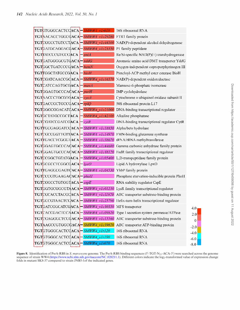
PsrA can bind to RBS and ABS in the promoter of the pigoperon. The sequence of RBS and its derivative sequences(5′-TGTN11ACA-3′), and the sequence of ABS and itsderivative sequences (5′-ACTN11AGT-3′) were furthersearched across the genome sequence of strain WW4(https://www.ncbi.nlm.nih.gov/nuccore/NC 020211.1) and235 potential PsrA-RBS and 209 potential PsrA-ABS wereidentified. Among them, 51 RBS were found in the regionsproximal to the promoters of various genes, includingsix genes encoding regulators (SMWW4 v1c21000,CytR, SMWW4 v1c48120, SMWW4 v1c01230,SMWW4 v1c25790 and SMWW4 v1c09220), 37 genesencoding metabolic enzymes and structural proteins,such as HemN, PhoH, and ManA, and 8 genes encodingputative proteins or hypothetical proteins (Figure 8, andSupplementary Table S4). Also, 29 ABS were found inthe regions proximal to the promoters of various genes,including 3 genes encoding regulators (SMWW4 v1c09530,SMWW4 v1c15090 and CueR), and 26 genes encodingmetabolic enzymes, structural proteins and hypothetical
proteins, such as BtuB, FolA and HutU (Supplemen-tary Figure S10, Supplementary Table S5). Further,to verify the PsrA-RBS binding sites and PsrA-ABSbinding sites, the promoter regions of cytR, phoH, hemN,SMWW4 v1c48120 and SMWW4 v1c21000 genes contain-ing potential RBS of PsrA, and btuB, SMWW4 v1c09530and cueR genes containing potential ABS of PsrA wereamplified and the PCR products were subjected to EMSAtest. The promoter region of flhDC and swrW genes thatlack PsrA-RBS and PsrA-ABS were used as negativecontrols. As the results shown in Supplementary FigureS11 and S12, the sequences that contain the potentialPsrA-RBS and PsrA-ABS can all be bound by the PsrAprotein. Additionally, the results showed that PsrA couldnot bind to the promoter regions of flhDC and swrWgenes (Supplementary Figure S11 and S12), which lackPsrA-RBS and PsrA-ABS suggesting that an unknownregulator may exist that controls other PsrA-mediatedcellular processes in strain JNB5-1. More experiments arerequired to clarify the factors influencing the interactionbetween PsrA and different cellular processes in the future.
PsrA-like proteins are widely distributed among bacteria
As identified in our study, PsrA controls prodigiosin syn-thesis, swarming motility, biofilm formation, extracellularpolysaccharide production, serrawettin W1 biosynthesis,and T6SS-mediated antibacterial activity in S. marcescens.Hence, further research on PsrA and similar proteins iswarranted if they are widely present in other bacteria.
Dow
nloaded from https://academ
ic.oup.com/nar/article/50/1/127/6459098 by guest on 11 August 2022

Nucleic Acids Research, 2022, Vol. 50, No. 1 141
Figure 7. PsrA required for T6SS-mediated antibacterial activity in S. marcescens. (A) PsrA positively regulates T6SS-related genes expression level in strainJNB5-1. (B) RT-qPCR analysis of the expression levels of the tssE, hcp, vgrG2, tssB and tssC genes in the strains JNB5-1 and SK8-37. (C) Recovery ofviable E. coli MC4100 cells after coculture with the indicated S. marcescens strains for 4 h at 37◦C, with an initial ratio of 5:1 (attacker/target). (D) Recoveryof viable E. cloacae ATCC13047 cells after coculture with the indicated S. marcescens strains for 4 h at 30◦C, with an initial ratio of 5:1 (attacker/target).For C and D, the control mixture contained a 5:1 ratio of sterile LB to target. JNB5-1 is a wild-type S. marcescens, SK8-37 is a psrA disrupted mutant,�PsrA is a psrA deleted mutant, SK8-37/pXW1903 is a psrA complemented strain, SK8-37/pUCP18 is a recombinant strain with empty vector pUCP18,SK8-37/pXW2006 is a recombinant strain with plasmid pXW2006, �TssE is a tssE deleted mutant, and �TssE/pXW2002 is a recombinant strain withplasmid pXW2002. Plasmid pXW1903, pXW2002, and pXW2006 carries intact psrA, tssE and rhs2 gene, respectively. The experiments were independentlyreplicated four times. Error bars indicated standard deviations (B−D).
Homologs of psrA gene were searched in S. marcescens,and found that the psrA gene was highly conserved in S.marcescens with similarities ranging 92.33–99.56% and thestrains containing psrA gene were found worldwide (Figure9A). Also, homologs of PsrA protein were searched withan E-values lower than 1E-121, and result showed that thetop 1000 proteins show similarities ranging 59.86–100% andare widely found in the groups of Serratia sp., Klebsiellasp., Cronobacter sp., Pantoea sp., Erwinia sp., Pseudomonassp. and Enterobacter sp. (Figure 9B). The fact that the PsrA-like proteins are highly homologous suggests that PsrA or
PsrA-like proteins in S. marcescens and other bacteria prob-ably control similar cellular processes as in strain JNB5-1.
DISCUSSION
S. marcescens, a part of the Enterobacteriaceae family of eu-bacteria, is found in a wide range of ecological niches andcan produce many high-value secondary metabolites likeprodigiosin (1), althiomycin (2), serratamolide (3). Besidesthe well-studied bacterial genes involved in the metabolicpathway of the secondary metabolites, a number of tran-
Dow
nloaded from https://academ
ic.oup.com/nar/article/50/1/127/6459098 by guest on 11 August 2022
142 Nucleic Acids Research, 2022, Vol. 50, No. 1
Figure 8. Identification of PsrA-RBS in S. marcescens genome. The PsrA-RBS binding sequences (5′-TGT-N11-ACA-3′) were searched across the genomesequence of strain WW4 (https://www.ncbi.nlm.nih.gov/nuccore/NC 020211.1). Different colors indicate the log2-transformed value of expression changefolds in mutant SK8-37 compared to strain JNB5-1of the indicated genes.
Dow
nloaded from https://academ
ic.oup.com/nar/article/50/1/127/6459098 by guest on 11 August 2022

Nucleic Acids Research, 2022, Vol. 50, No. 1 143
Figure 9. PsrA-like proteins are widely distributed among bacteria. (A) Phylogenetic tree of the psrA-like genes of S. marcescens with similarities rangingfrom 92.33–99.56%. (B) Phylogenetic tree of the top 1000 homologous proteins of PsrA with the E-values lower than 1E−121. Orange triangle stands forthe PsrA protein of strain JNB5-1. The scale bar represents the number of substitutions per site. Colored rectangles stand for different species as indicated.
scriptional regulator-encoding genes that play importantroles in secondary metabolites synthesis in S. marcescenshave also been investigated, such as regulator MetR (10),SpnR (11), CopA (12), CRP (13), HexS (14), RssB (15),RcsB (16,17), SmaR (18), EepR (6), PigP (7), GumB (8),RbsR (9), and RpoS (61) mediates prodigiosin produc-tion and regulator GumB (8), EepR (6,34), PigP (62) andHexS (7) regulation of serratamolide synthesis. However,our understanding of the regulatory mechanisms behindsecondary metabolites synthesis in S. marcescens is still lim-ited. In this study, an uncharacterized LysR family regula-tor PsrA was confirmed to function as a prodigiosin acti-vator for the first time by directly binding to the promoterregion of the prodigiosin synthesis related pig operon fortranscriptional activation (Figures 1 and 2).
Usually, the interaction between LysR-type transcrip-tional regulators (LTTRs) and their target genes occurs atthe regulatory binding site (RBS) and the activator bind-ing site (ABS). The RBS is generally centered at posi-tion −65 relative to the TSS of the activated promoter,containing the LTTR consensus binding motif (5′-T-N11-A-3′). The ABS usually overlap the −35 box of the tar-
get gene and often possesses no conserved sequence motif(42,43,63). Moreover, active complex formation of a LTTRprotein is a complex process, which includes the follow-ing steps: (i) perception signal via the effector binding do-main at the C-terminal of LTTRs, (ii) oligomerization viathe DNA binding and amino acids involved in dimeriza-tion, (iii) binding to the promoter region of the target DNAvia the helix-turn-helix motif at the N-terminal of LTTRs,(iv) DNA bending during higher ordered complex forma-tion and (v) transcriptional activation of the target geneby the interaction of the formed complex with the RNApolymerase (42). In this study, we demonstrated that PsrAbinds to the promoter region of the pig operon includingboth RBS and ABS (Figures 3A−C, Supplementary Fig-ure S4, and S5). The sequences located between −90 and−74 bp upstream of the TSS of pig operon was probablyRBS of PsrA, which contains a palindromic structure (5′-TGTAATAATAGACCACA-3′). The sequences containinga palindromic structure (5′-ACTTTGCCTGTTTGAGT-3′) located between −44 and −28 bp upstream of the TSSof pig operon was probably ABS of PsrA (Figure 3B). In-terestingly, unlike the regulator AlsR of B. subtilis (26), the
Dow
nloaded from https://academ
ic.oup.com/nar/article/50/1/127/6459098 by guest on 11 August 2022

144 Nucleic Acids Research, 2022, Vol. 50, No. 1
Figure 10. Proposed model of PsrA mediated regulatory pathways in strain JNB5-1. The regulatory pathways include the cellular processes and the genesdirectly or indirectly influenced by the PsrA regulator. PsrA is an important regulator controls many cellular processes including prodigiosin synthesis,serrawettin W1 biosynthesis, extracellular polysaccharide (EPS) production, biofilm formation, swarming motility and T6SS-mediated antibacterial activityin strain JNB5-1. Black arrows represent direct regulatory relationships between PsrA and the target genes. Blue arrows represent indirect regulatoryrelationships between PsrA and the target genes. PsrA controls prodigiosin synthesis of strain JNB5-1 by directly binding to the PsrA-RBS and PsrA-ABSin the promoter region of the pig operon. L-proline is the effector of PsrA. PsrA controls other cellular processes of strain JNB5-1 by indirectly influencethe expression levels of genes involve in different cellular processes. FlgB, FlgD, FlgJ, FliR and FliH are key enzymes in flagellum synthesis and swarmingmotility. SwrW is a key enzyme in serrawettin W1 biosynthesis. VgrG2, TssE, TssB, Hcp and TssC are key enzymes in T6SS-mediated antibacterial activity.PgaA, PgaB, PgaC and FliA are key enzymes in biofilm formation. Wza, Wzc and WecA are key enzymes in extracellular polysaccharide production.
regulator CysB of Salmonella enterica serovar Typhimurium(64), and the regulator LigR of Sphingobium sp. strainSYK-6 (47) binding to target DNA which resulted in theformation of three retarded complexes, PsrA binding re-sults in the concentration-dependent generation of com-plexes with different migration velocities (SupplementaryFigures S2E and S4). There are several possible expla-nations for this, such as multiple binding sites, differentoligomeric states of PsrA and DNA deformations. More ex-periments are needed to further understand the underlyingmechanisms.
To efficiently activate the transcription of correspondingtarget genes and hence influence cellular processes in bacte-rial, the binding activity of LTTRs to its target gene is gen-erally influenced by one or more effectors. For example, ac-etate is the effector of AlsR and activates the transcriptionof the alsSD operon for acetoin formation in Bacillus sub-tilis (26,38), glutarate is the effector of GcdR and controlsglutarate catabolism in Pseudomonas putida KT2440 (29),hydroxyhydroquinone or possibly 2,3,5-trihydroxybenzoateis the effector of DbdR and influences expression of the en-tire 3,5-DHB anaerobic degradation pathway in Thaueraaromatica (48), benzoate and cis,cis-muconate are the effec-tors of BenM and control aromatic compound degradationin Acinetobacter sp. ADP1 (45), 1-Naphthol is the effectorof McbG to activate mcbBCDEF cluster transcription andinvolved in the upstream pathway of carbaryl degradationin Pseudomonas sp. XWY-1 (65), and shikimate, the effec-tor of QuiR induces transcriptional activation of protocat-echuate biosynthesis genes and modulates the conversionof quinate and shikimate metabolites to protocatechuate(66). In this study, through �-galactosidase activity analy-sis, isothermal titration calorimetry (ITC), and EMSAs, wefound that L-proline is the effector of PsrA (Figure 3D, Sup-
plementary Figures S4A−C, S8 and S9). Interaction withL-proline molecules increases the binding of PsrA to itstarget DNA, resulting in upregulation of the expression ofthe prodigiosin synthesis related pigABCDEFGHIJKLMNgenes in strain JNB5-1 (Supplementary Figure S8).
LTTRs are the large family of transcriptional regula-tors in bacteria, and a number of this family regulatorsare involved in the control of a series of core cellularprocesses in different microorganisms, such as the synthe-sis of virulence factors, the control of the central carboncatabolism, the influence of expression of antibiotic resis-tance, and the induction of aromatic compound metab-olization (42,43,63,67–69). In this study, using transcrip-tomics and further experiments, PsrA, was demonstrated tobe a global regulator, which influences prodigiosin synthe-sis (Figure 1A−C), swarming motility (Figure 5C and E),biofilm formation (Figure 6C), extracellular polysaccharideproduction (Figure 6D), serrawettin W1 biosynthesis (Fig-ure 6E), and T6SS-mediated antibacterial activity (Figure7) in S. marcescens for the first time. Additionally, the re-sults showed that there were no PsrA-binding sites in thepromoter region of these cellular processes control genes(Figure 8, and Supplementary Figure S10, and Supplemen-tary Tables S4 and S5), and PsrA could not bind to the pro-moter regions of flhDC and swrW genes (SupplementaryFigure S11 and S12) suggesting that PsrA may indirectlycontrols the cellular processes of swarming motility, extra-cellular polysaccharide production, biofilm formation, ser-rawettin W1 biosynthesis, and T6SS-mediated antibacterialactivity in strain JNB5-1 (Figure 10).
The presence of functional flagellar contributes toswarming motility in bacteria. Using transcriptomics andRT-qPCR analysis, we found that PsrA positively regulatedthe transcription of flagellar synthesis flgBCDEFGHIJ,
Dow
nloaded from https://academ
ic.oup.com/nar/article/50/1/127/6459098 by guest on 11 August 2022

Nucleic Acids Research, 2022, Vol. 50, No. 1 145
flhA and fliAEFGHIJNPR genes in strain JNB5-1 (Fig-ure 5A and B). Additionally, in this work, data showedthat PsrA positively regulates swarming motility in strainJNB5-1 (Figure 5C and E). Taken together, this work showsthat PsrA plays an important role in swarming motility inS. marcescens, probably by controlling the transcription offlagellar synthesis-related genes (Figure 10).
A variety of transcription regulators have been identifiedplaying important roles in biofilm synthesis in different mi-croorganisms, such as the transcription regulator SrpA (24),BsrA (30), PyeR (70), AmzR (71), BswR (72) and FleQ(73) in Pseudomonas aeruginosa, the regulator RydC (74)and LrhA (75) in E. coli, the regulator SinR (76) in B. sub-tilis, the regulator CytR (77) in Vibrio cholerae, and theregulator Fur (78) in Yersinia pestis. In S. marcescens, afew regulators have been reported to be involved in biofilmformation, including positive regulators MetR (10), OxyR(79) and RpoS (61), and negative regulators GumB (8) andCpxR (80). In this study, PsrA was identified as a novelregulator which positively regulates biofilm formation inS. marcescens. When psrA was disrupted, the ability ofS. marcescens to synthesize biofilm was significantly de-creased (Figure 6D). Transcriptomics, RT-qPCR analysisand bioinformatics analysis showed that the way PsrA af-fected the formation of biofilm was likely by indirectly reg-ulating the expression of the biofilm formation related genes(Figures 6A, B and 10).
Type VI secretion systems (T6SSs) are nanomachineswidely used by Gram-negative bacteria to deliver toxic ef-fector proteins into neighbouring cells and provide a com-petitive advantage in mixed bacterial populations. Num-bers of transcription regulators have been identified to playimportant roles in T6SS-meditated antibacterial activity indifferent bacteria, such as the regulators SrpA (24), SuhB(52), CueR (81), AnvM (82) and AmrZ (83) in P. aerugi-nosa, the regulators BaeSR (84) and HpaR (85) in E. coli,and the regulator QstR (86) in V. cholerae. In S. marcescens,only regulator RcsB (36) has been identified to be impor-tant for T6SS-mediated antibacterial activity. In this work,using transcriptomics, RT-qPCR analysis and further ex-periments, we show that PsrA, indirectly regulates T6SS-meditated antibacterial activity in S. marcescens (Figures7 and 10).
In summary, this work describes a novel regulator PsrA,which regulates prodigiosin synthesis, swarming motility,biofilm formation, extracellular polysaccharide production,serrawettin W1 biosynthesis, and T6SS-mediated antibacte-rial activity in S. marcescens (Figure 10). Further research isneeded to reveal the roles played by other PsrA or PsrA-likeproteins, which are widely present in bacteria.
DATA AVAILABILITY
The raw data of RNA-seq has been deposited in the GEOdatabase of NCBI with the accession number GSE158829.
SUPPLEMENTARY DATA
Supplementary Data are available at NAR Online.
FUNDING
National Natural Science Foundation of China [31870066,32100055, 21778024]; National Key Research and Devel-opment Program of China [2021YFC2100900]; NaturalScience Foundation of Jiangsu Province [BK20210464];Project funded by China Postdoctoral Science Foundation[2021M691280]; Jiangsu Planned Projects for PostdoctoralResearch Funds [2021K296B]; Ningxia Hui AutonomousRegion’s Flexible introduction project [KJT201701]; Na-tional first-class discipline program of Light Industry Tech-nology and Engineering [LITE2018-06]; Program of In-troducing Talents of Discipline to Universities [111-2-06];Fundamental Research Funds for the Central Universities[JUSRP12119]. Funding for open access charge: NationalNatural Science Foundation of China [31870066].Conflict of interest statement. None declared.
REFERENCES1. Williamson,N.R., Fineran,P.C., Leeper,F.J. and Salmond,G.P. (2006)
The biosynthesis and regulation of bacterial prodiginines. Nat. Rev.Microbiol., 4, 887–899.
2. Gerc,A.J., Stanley-Wall,N.R. and Coulthurst,S.J. (2014) Role of thephosphopantetheinyltransferase enzyme, PswP, in the biosynthesis ofantimicrobial secondary metabolites by Serratia marcescens Db10.Microbiology, 160, 1609–1617.
3. Webber,M.A., Shanks,R.M.Q., Stella,N.A., Lahr,R.M., Wang,S.,Veverka,T.I., Kowalski,R.P. and Liu,X. (2012) Serratamolide is ahemolytic factor produced by Serratia marcescens. PLoS One, 7,e36398.
4. Gao,S., Guo,W., Shi,L., Yu,Y., Zhang,C. and Yang,H. (2014)Characterization of acetoin production in a budC gene disruptedmutant of Serratia marcescens G12. J. Ind. Microbiol. Biotechnol., 41,1267–1274.
5. Rao,B., Zhang,L.Y., Sun,J., Su,G., Wei,D., Chu,J., Zhu,J. and Shen,Y.(2012) Characterization and regulation of the 2,3-butanediol pathwayin Serratia marcescens. Appl. Microbiol. Biotechnol., 93, 2147–2159.
6. Shanks,R.M., Stella,N.A., Lahr,R.M., Aston,M.A., Brothers,K.M.,Callaghan,J.D., Sigindere,C. and Liu,X. (2017) Suppressor analysis ofeepR mutant defects reveals coordinate regulation of secondarymetabolites and serralysin biosynthesis by EepR and HexS.Microbiology, 163, 280–288.
7. Shanks,R.M., Lahr,R.M., Stella,N.A., Arena,K.E., Brothers,K.M.,Kwak,D.H., Liu,X. and Kalivoda,E.J. (2013) A Serratia marcescensPigP homolog controls prodigiosin biosynthesis, swarming motilityand hemolysis and is regulated by cAMP-CRP and HexS. PLoS One,8, e57634.
8. Stella,N.A., Brothers,K.M., Callaghan,J.D., Passerini,A.M.,Sigindere,C., Hill,P.J., Liu,X., Wozniak,D.J. and Shanks,R.M.Q.(2018) An IgaA/UmoB family protein from Serratia marcescensregulates motility, capsular polysaccharide biosynthesis, andsecondary metabolite production. Appl. Environ. Microbiol., 84,e02575-17.
9. Lee,C.M., Monson,R.E., Adams,R.M. and Salmond,G.P.C. (2017)The LacI-family transcription factor, RbsR, is a pleiotropic regulatorof motility, virulence, siderophore and antibiotic production, gasvesicle morphogenesis and flotation in serratia. Front. Microbiol., 8,1678.
10. Pan,X., Sun,C., Tang,M., You,J., Osire,T., Zhao,Y., Xu,M.,Zhang,X., Shao,M., Yang,S. et al. (2020) LysR-Type transcriptionalregulator MetR controls prodigiosin production, methioninebiosynthesis, cell motility, H2O2 tolerance, heat tolerance, andexopolysaccharide synthesis in Serratia marcescens. Appl. Environ.Microbiol., 86, e02241-19.
11. Horng,Y.T., Deng,S.C., Daykin,M., Soo,P.C., Wei,J.R., Luh,K.T.,Ho,S.W., Swift,S., Lai,H.C. and Williams,P. (2002) The LuxR familyprotein SpnR functions as a negative regulator of N-acylhomoserine
Dow
nloaded from https://academ
ic.oup.com/nar/article/50/1/127/6459098 by guest on 11 August 2022
146 Nucleic Acids Research, 2022, Vol. 50, No. 1
lactone-dependent quorum sensing in Serratia marcescens. Mol.Microbiol., 45, 1655–1671.
12. Williamson,N.R., Simonsen,H.T., Harris,A.K., Leeper,F.J. andSalmond,G.P. (2006) Disruption of the copper efflux pump (CopA) ofSerratia marcescens ATCC 274 pleiotropically affects coppersensitivity and production of the tripyrrole secondary metabolite,prodigiosin. J. Ind. Microbiol. Biotechnol., 33, 151–158.
13. Stella,N.A. and Shanks,R.M. (2014) Cyclic-AMP inhibition offimbriae and prodigiosin production by Serratia marcescens isstrain-dependent. Arch. Microbiol., 196, 323–330.
14. Stella,N.A., Fender,J.E., Lahr,R.M., Kalivoda,E.J. and Shanks,R.M.(2012) The LysR transcription factor, HexS, is required for glucoseinhibition of prodigiosin production by Serratia marcescens. AdvMicrobiol., 2, 511–517.
15. Horng,Y.T., Chang,K.C., Liu,Y.N., Lai,H.C. and Soo,P.C. (2010) TheRssB/RssA two-component system regulates biosynthesis of thetripyrrole antibiotic, prodigiosin, in Serratia marcescens. Int. J. Med.Microbiol., 300, 304–312.
16. Brothers,K.M., Callaghan,J.D., Stella,N.A., Bachinsky,J.M.,AlHigaylan,M., Lehner,K.L., Franks,J.M., Lathrop,K.L., Collins,E.,Schmitt,D.M. et al. (2019) Blowing epithelial cell bubbles withGumB: ShlA-family pore-forming toxins induce blebbing and rapidcellular death in corneal epithelial cells. PLoS Pathog., 15, e1007825.
17. Pan,X.W., Tang,M., You,J.J., Liu,F., Sun,C.H., Osire,T., Fu,W.L.,Yi,G.F., Yang,T.W., Yang,S.T. et al. (2021) Regulator RcsB controlsprodigiosin synthesis and various cellular processes in Serratiamarcescens JNB5-1. Appl. Environ. Microb., 87, e02052-20.
18. Coulthurst,S.J., Williamson,N.R., Harris,A.K., Spring,D.R. andSalmond,G.P. (2006) Metabolic and regulatory engineering ofSerratia marcescens: mimicking phage-mediated horizontalacquisition of antibiotic biosynthesis and quorum-sensing capacities.Microbiology, 152, 1899–1911.
19. Wang,J., Mushegian,A., Lory,S. and Jin,S. (1996) Large-scaleisolation of candidate virulence genes of Pseudomonas aeruginosa byin vivo selection. Proc. Natl. Acad. Sci. U.S.A., 93, 10434–10439.
20. Kalivoda,E.J., Stella,N.A., Aston,M.A., Fender,J.E., Thompson,P.P.,Kowalski,R.P. and Shanks,R.M. (2010) Cyclic AMP negativelyregulates prodigiosin production by Serratia marcescens. Res.Microbiol., 161, 158–167.
21. Bustin,S.A., Benes,V., Garson,J.A., Hellemans,J., Huggett,J.,Kubista,M., Mueller,R., Nolan,T., Pfaffl,M.W., Shipley,G.L. et al.(2009) The MIQE guidelines: Minimum information for publicationof quantitative real-time PCR experiments. Clin. Chem., 55, 611–622.
22. Wilf,N.M., Williamson,N.R., Ramsay,J.P., Poulter,S., Bandyra,K.J.and Salmond,G.P. (2011) The RNA chaperone, Hfq, controls twoluxR-type regulators and plays a key role in pathogenesis andproduction of antibiotics in Serratia sp. ATCC 39006. Environ.Microbiol., 13, 2649–2666.
23. Brothers,K.M., Stella,N.A., Romanowski,E.G., Kowalski,R.P. andShanks,R.M.Q. (2015) EepR mediates secreted-protein production,desiccation survival, and proliferation in a corneal infection model.Infect. Immun., 83, 4373–4382.
24. You,J., Sun,L., Yang,X., Pan,X., Huang,Z., Zhang,X., Gong,M.,Fan,Z., Li,L., Cui,X. et al. (2018) Regulatory protein SrpA controlsphage infection and core cellular processes in Pseudomonasaeruginosa. Nat. Commun., 9, 1846.
25. Zhang,W.M., Zhang,J.J., Jiang,X., Chao,H.J. and Zhou,N.Y. (2015)Transcriptional activation of multiple operons involved inpara-Nitrophenol degradation by Pseudomonas sp. Strain WBC-3.Appl. Environ. Microb., 81, 220–230.
26. Fradrich,C., March,A., Fiege,K., Hartmann,A., Jahn,D. andHartig,E. (2012) The transcription factor AlsR binds and regulatesthe promoter of the alsSD operon responsible for acetoin formationin Bacillus subtilis. J. Bacteriol., 194, 1100–1112.
27. Xia,Y.S., Wang,D., Pan,X.L., Xia,B., Weng,Y.D., Long,Y.Q., Ren,H.,Zhou,J.Y., Jin,Y.X., Bai,F. et al. (2020) TpiA is a key metabolicenzyme that affects virulence and resistance to aminoglycosideantibiotics through CrcZ in Pseudomonas aeruginosa. Mbio, 11,e02079-19.
28. Miller,J.H. (1972) In: Experiments in molecular genetics. Cold SpringHarbor Laboratory Press, NY.
29. Zhang,M., Kang,Z., Guo,X., Guo,S., Xiao,D., Liu,Y., Ma,C.,Gao,C. and Xu,P. (2019) Regulation of glutarate catabolism by GntR
family regulator CsiR and LysR family regulator GcdR inPseudomonas putida KT2440. mBio, 10, e01570-19.
30. Yang,X., Zhang,Z., Huang,Z., Zhang,X., Li,D., Sun,L., You,J.,Pan,X. and Yang,H. (2019) A putative LysR-type transcriptionalregulator inhibits biofilm synthesis in Pseudomonas aeruginosa.Biofouling, 35, 541–550.
31. Xu,M., Tang,M., Chen,J., Yang,T., Zhang,X., Shao,M., Xu,Z. andRao,Z. (2020) PII signal transduction protein GlnK alleviatesfeedback inhibition of N-acetyl-l-glutamate kinase by l-arginine inCorynebacterium glutamicum. Appl. Environ. Microbiol., 86,e00039-20.
32. Djordjevic,D., Wiedmann,M. and McLandsborough,L.A. (2002)Microtiter plate assay for assessment of Listeria monocytogenesbiofilm formation. Appl. Environ. Microbiol., 68, 2950–2958.
33. Di Venanzio,G., Stepanenko,T.M. and Garcia Vescovi,E. (2014)Serratia marcescens ShlA pore-forming toxin is responsible for earlyinduction of autophagy in host cells and is transcriptionally regulatedby RcsB. Infect. Immun., 82, 3542–3554.
34. Stella,N.A., Lahr,R.M., Brothers,K.M., Kalivoda,E.J., Hunt,K.M.,Kwak,D.H., Liu,X. and Shanks,R.M. (2015) Serratia marcescensCyclic AMP receptor protein controls transcription of EepR, a novelregulator of antimicrobial secondary metabolites. J. Bacteriol., 197,2468–2478.
35. Murdoch,S.L., Trunk,K., English,G., Fritsch,M.J., Pourkarimi,E.and Coulthurst,S.J. (2011) The opportunistic pathogen Serratiamarcescens utilizes type VI secretion to target bacterial competitors.J. Bacteriol., 193, 6057–6069.
36. Lazzaro,M., Feldman,M.F. and Garcia Vescovi,E. (2017) Atranscriptional regulatory mechanism finely tunes the firing of type VIsecretion system in response to bacterial enemies. mBio, 8, e00559-17.
37. Koentjoro,M.P., Adachi,N., Senda,M., Ogawa,N. and Senda,T.(2018) Crystal structure of the DNA-binding domain of theLysR-type transcriptional regulator CbnR in complex with a DNAfragment of the recognition-binding site in the promoter region.FEBS J., 285, 977–989.
38. Hartig,E., Fradrich,C., Behringer,M., Hartmann,A.,Neumann-Schaal,M. and Jahn,D. (2018) Functional definition of thetwo effector binding sites, the oligomerization and DNA bindingdomains of the Bacillus subtilis LysR-type transcriptional regulatorAlsR. Mol. Microbiol., 109, 845–864.
39. Porrua,O., Garcia-Jaramillo,M., Santero,E. and Govantes,F. (2007)The LysR-type regulator AtzR binding site: DNA sequences involvedin activation, repression and cyanuric acid-dependent repositioning.Mol. Microbiol., 66, 410–427.
40. Parsek,M.R., Kivisaar,M. and Chakrabarty,A.M. (1995) DifferentialDNA bending introduced by the Pseudomonas putida LysR-typeregulator, CatR, at the plasmid-borne pheBA and chromosomalcatBC promoters. Mol. Microbiol., 15, 819–828.
41. Shin,M., Rumi,O., Naoto,O., Takamasa,N., Kiyotaka,M. andToshiya,S. (2003) Crystal structure of a full-length LysR-typetranscriptional regulator, CbnR: Unusual combination of twosubunit forms and molecular bases for causing and changing DNAbend. J. Mol. Biol., 328, 555–566.
42. Schell,M.A. (1993) Molecular biology of the LysR family oftranscriptional regulators. Annu. Rev. Microbiol., 47, 597–626.
43. Tropel,D. and van der Meer,J.R. (2004) Bacterial transcriptionalregulators for degradation pathways of aromatic compounds.Microbiol. Mol. Biol. Rev., 68, 474–500.
44. Choi,H., Kim,S., Mukhopadhyay,P., Cho,S., Woo,J., Storz,G. andRyu,S.E. (2001) Structural basis of the redox switch in the OxyRtranscription factor. Cell, 105, 103–113.
45. Bundy,B.M., Collier,L.S., Hoover,T.R. and Neidle,E.L. (2002)Synergistic transcriptional activation by one regulatory protein inresponse to two metabolites. Proc. Natl. Acad. Sci. U.S.A., 99,7693–7698.
46. Craven,S.H., Ezezika,O.C., Haddad,S., Hall,R.A., Momany,C. andNeidle,E.L. (2009) Inducer responses of BenM, a LysR-typetranscriptional regulator from Acinetobacter baylyi ADP1. Mol.Microbiol., 72, 881–894.
47. Kamimura,N., Takamura,K., Hara,H., Kasai,D., Natsume,R.,Senda,T., Katayama,Y., Fukuda,M. and Masai,E. (2010) Regulatorysystem of the protocatechuate 4,5-cleavage pathway genes essentialfor lignin downstream catabolism. J. Bacteriol., 192, 3394–3405.
Dow
nloaded from https://academ
ic.oup.com/nar/article/50/1/127/6459098 by guest on 11 August 2022
Nucleic Acids Research, 2022, Vol. 50, No. 1 147
48. Pacheco-Sanchez,D., Molina-Fuentes,A., Marin,P., Diaz-Romero,A.and Marques,S. (2019) DbdR, a new member of the LysR family oftranscriptional regulators, coordinately controls four promoters inthe Thauera aromatica AR-1 3,5-Dihydroxybenzoate anaerobicdegradation pathway. Appl. Environ. Microbiol., 85, e02295-18.
49. Xie,J.L., Qin,L., Miao,Z., Grys,B.T., Diaz,J.C., Ting,K., Krieger,J.R.,Tong,J., Tan,K., Leach,M.D. et al. (2017) The Candida albicanstranscription factor Cas5 couples stress responses, drug resistanceand cell cycle regulation. Nat. Commun., 8, 499.
50. Potts,A.H., Vakulskas,C.A., Pannuri,A., Yakhnin,H., Babitzke,P.and Romeo,T. (2017) Global role of the bacterial post-transcriptionalregulator CsrA revealed by integrated transcriptomics. Nat.Commun., 8, 1596.
51. Sause,W.E., Balasubramanian,D., Irnov,I., Copin,R., Sullivan,M.J.,Sommerfield,A., Chan,R., Dhabaria,A., Askenazi,M., Ueberheide,B.et al. (2019) The purine biosynthesis regulator PurR moonlights as avirulence regulator in Staphylococcus aureus. Proc. Natl. Acad. Sci.U.S.A., 116, 13563–13572.
52. Li,K., Xu,C., Jin,Y., Sun,Z., Liu,C., Shi,J., Chen,G., Chen,R., Jin,S.and Wu,W. (2013) SuhB is a regulator of multiple virulence genes andessential for pathogenesis of Pseudomonas aeruginosa. mBio, 4,e00419-13.
53. Minamino,T. and Imada,K. (2015) The bacterial flagellar motor andits structural diversity. Trends Microbiol., 23, 267–274.
54. Mariano,G., Trunk,K., Williams,D.J., Monlezun,L., Strahl,H.,Pitt,S.J. and Coulthurst,S.J. (2019) A family of Type VI secretionsystem effector proteins that form ion-selective pores. Nat. Commun.,10, 5484.
55. Trunk,K., Peltier,J., Liu,Y.C., Dill,B.D., Walker,L., Gow,N.A.R.,Stark,M.J.R., Quinn,J., Strahl,H., Trost,M. et al. (2018) The type VIsecretion system deploys antifungal effectors against microbialcompetitors. Nat Microbiol, 3, 920–931.
56. Filloux,A., Hachani,A. and Bleves,S. (2008) The bacterial type VIsecretion machine: yet another player for protein transport acrossmembranes. Microbiology (Reading), 154, 1570–1583.
57. Gallique,M., Bouteiller,M. and Merieau,A. (2017) The type VIsecretion system: a dynamic system for bacterial communication?Front Microbiol, 8, 1454.
58. Cianfanelli,F.R., Juliana,A.D., Guo,M., Virginia,D., Matthias,T.,Coulthurst,S.J. and Eric,C.J.P.P. (2016) VgrG and PAAR proteinsdefine distinct versions of a functional type VI secretion system.PLoS Pathog., 12, e1005735.
59. Maximilian,J.F, Katharina,T., Juliana Alcoforado,D., Manman,G.,Matthias,T. and Sarah,J.C. (2013) Proteomic identification of novelsecreted antibacterial toxins of the Serratia marcescens type VIsecretion system. Mol. Cell. Proteomics, 12, 2735–2749.
60. Alcoforado Diniz,J. and Coulthurst,S.J. (2015) Intraspeciescompetition in Serratia marcescens is mediated by type VI-SecretedRhs effectors and a conserved effector-associated accessory protein.J. Bacteriol., 197, 2350–2360.
61. Qin,H., Liu,Y., Cao,X., Jiang,J., Lian,W., Qiao,D., Xu,H. and Cao,Y.(2020) RpoS is a pleiotropic regulator of motility, biofilm formation,exoenzymes, siderophore and prodigiosin production, and trade-offduring prolonged stationary phase in Serratia marcescens. PLoS One,15, e0232549.
62. Tanikawa,T., Nakagawa,Y. and Matsuyama,T. (2006) Transcriptionaldownregulator hexS controlling prodigiosin and serrawettin W1biosynthesis in Serratia marcescens. Microbiol. Immunol., 50,587–596.
63. Maddocks,S.E. and Oyston,P.C.F. (2008) Structure and function ofthe LysR-type transcriptional regulator (LTTR) family proteins.Microbiology (Reading), 154, 3609–3623.
64. Hryniewicz,M.M. and Kredich,N.M. (1994) Stoichiometry ofbinding of CysB to the cysJIH, cysK, and cysP promoter regions ofSalmonella typhimurium. J. Bacteriol., 176, 3673–3682.
65. Ke,Z., Zhou,Y., Jiang,W., Zhang,M., Wang,H., Ren,Y., Qiu,J.,Cheng,M. and Hong,Q. (2021) McbG, a LysR family transcriptionalregulator, activates the mcbBCDEF gene cluster involved in theupstream pathway of carbaryl degradation in Pseudomonas sp. strainXWY-1. Appl. Environ. Microbiol., 87, e02970-20.
66. Prezioso,S.M., Xue,K., Leung,N., Gray-Owen,S.D. andChristendat,D. (2018) Shikimate induced transcriptional activation ofprotocatechuate biosynthesis genes by QuiR, a LysR-Type
transcriptional regulator, in Listeria monocytogenes. J. Mol. Biol.,430, 1265–1283.
67. Peck,M.C., Fisher,R.F., Bliss,R. and Long,S.R. (2013) Isolation andcharacterization of mutant Sinorhizobium meliloti NodD1 proteinswith altered responses to luteolin. J. Bacteriol., 195, 3714–3723.
68. Mandal,R.S., Ta,A., Sinha,R., Theeya,N., Ghosh,A., Tasneem,M.,Bhunia,A., Koley,H. and Das,S. (2016) Ribavirin suppresses bacterialvirulence by targeting LysR-type transcriptional regulators. Sci. Rep.,6, 39454.
69. Sperandio,B., Gautier,C., Pons,N., Ehrlich,D.S., Renault,P. andGuedon,E. (2010) Three paralogous LysR-type transcriptionalregulators control sulfur amino acid supply in Streptococcus mutans.J. Bacteriol., 192, 3464–3473.
70. Mac Aogain,M., Mooij,M.J., McCarthy,R.R., Plower,E., Wang,Y.P.,Tian,Z.X., Dobson,A., Morrissey,J., Adams,C. and O’Gara,F. (2012)The non-classical ArsR-family repressor PyeR (PA4354) modulatesbiofilm formation in Pseudomonas aeruginosa. Microbiology(Reading), 158, 2598–2609.
71. Jones,C.J., Ryder,C.R., Mann,E.E. and Wozniak,D.J. (2013) AmrZmodulates Pseudomonas aeruginosa biofilm architecture by directlyrepressing transcription of the psl operon. J. Bacteriol., 195,1637–1644.
72. Wang,C., Ye,F., Kumar,V., Gao,Y.G. and Zhang,L.H. (2014) BswRcontrols bacterial motility and biofilm formation in Pseudomonasaeruginosa through modulation of the small RNA rsmZ. NucleicAcids Res., 42, 4563–4576.
73. Baraquet,C., Murakami,K., Parsek,M.R. and Harwood,C.S. (2012)The FleQ protein from Pseudomonas aeruginosa functions as both arepressor and an activator to control gene expression from the peloperon promoter in response to c-di-GMP. Nucleic Acids Res., 40,7207–7218.
74. Bordeau,V. and Felden,B. (2014) Curli synthesis and biofilmformation in enteric bacteria are controlled by a dynamic small RNAmodule made up of a pseudoknot assisted by an RNA chaperone.Nucleic Acids Res., 42, 4682–4696.
75. Blumer,C., Kleefeld,A., Lehnen,D., Heintz,M., Dobrindt,U.,Nagy,G., Michaelis,K., Emody,L., Polen,T., Rachel,R. et al. (2005)Regulation of type 1 fimbriae synthesis and biofilm formation by thetranscriptional regulator LrhA of Escherichia coli. Microbiology, 151,3287–3298.
76. Kearns,D.B., Chu,F., Branda,S.S., Kolter,R. and Losick,R. (2005) Amaster regulator for biofilm formation by Bacillus subtilis. Mol.Microbiol., 55, 739–749.
77. Haugo,A.J. and Watnick,P.I. (2002) Vibrio cholerae CytR is arepressor of biofilm development. Mol. Microbiol., 45, 471–483.
78. Sun,F., Gao,H., Zhang,Y., Wang,L., Fang,N., Tan,Y., Guo,Z., Xia,P.,Zhou,D. and Yang,R. (2012) Fur is a repressor of biofilm formationin Yersinia pestis. PLoS One, 7, e52392.
79. Shanks,R.M., Stella,N.A., Kalivoda,E.J., Doe,M.R., O’Dee,D.M.,Lathrop,K.L., Guo,F.L. and Nau,G.J. (2007) A Serratia marcescensOxyR homolog mediates surface attachment and biofilm formation.J. Bacteriol., 189, 7262–7272.
80. Bruna,R.E., Molino,M.V., Lazzaro,M., Mariscotti,J.F. and GarciaVescovi,E. (2018) CpxR-Dependent thermoregulation of Serratiamarcescens PrtA metalloprotease expression and its contribution tobacterial biofilm formation. J. Bacteriol., 200, e00006-18.
81. Han,Y., Wang,T., Chen,G., Pu,Q., Liu,Q., Zhang,Y., Xu,L., Wu,M.and Liang,H. (2019) A Pseudomonas aeruginosa type VI secretionsystem regulated by CueR facilitates copper acquisition. PLoSPathog., 15, e1008198.
82. Zhang,Y., Zhou,C.M., Pu,Q., Wu,Q., Tan,S., Shao,X., Zhang,W.,Xie,Y., Li,R., Yu,X.J. et al. (2019) Pseudomonas aeruginosaregulatory protein AnvM controls pathogenicity in anaerobicenvironments and impacts host defense. mBio, 10, e01362-19.
83. Allsopp,L.P., Wood,T.E., Howard,S.A., Maggiorelli,F., Nolan,L.M.,Wettstadt,S. and Filloux,A. (2017) RsmA and AmrZ orchestrate theassembly of all three type VI secretion systems in Pseudomonasaeruginosa. Proc. Natl. Acad. Sci. U.S.A., 114, 7707–7712.
84. Hu,L., Wang,C., Lu,W., Lu,H., Chen,H. and Tan,C. (2020) BaeSRactivates type VI secretion system expression in porcineextra-intestinal pathogenic Escherichia coli to enhance bacterialresistance to zinc stress. Microb. Pathog., 147 , 104357.
85. Wang,Z., Wang,T., Cui,R., Zhang,Z., Chen,K., Li,M., Hua,Y.,Gu,H., Xu,L., Wang,Y. et al. (2020) HpaR, the repressor of aromatic
Dow
nloaded from https://academ
ic.oup.com/nar/article/50/1/127/6459098 by guest on 11 August 2022
148 Nucleic Acids Research, 2022, Vol. 50, No. 1
compound metabolism, positively regulates the expression of T6SS4to resist oxidative stress in Yersinia pseudotuberculosis. FrontMicrobiol, 11, 705.
86. Jaskolska,M., Stutzmann,S., Stoudmann,C. and Blokesch,M. (2018)QstR-dependent regulation of natural competence and type VIsecretion in Vibrio cholerae. Nucleic Acids Res., 46, 10619–10634.
87. Nunn,D.N. and Lory,S. (1992) Components of the protein-excretionapparatus of Pseudomonas aeruginosa are processed by the type IVprepilin peptidase. Proc. Natl. Acad. Sci. U.S.A., 89, 47–51.
88. Schweizer,H.P. (1991) Escherichia-Pseudomonas shuttle vectorsderived from pUC18/19. Gene, 97, 109–121.
89. Totten,P.A. and Lory,S. (1990) Characterization of the type a flagellingene from Pseudomonas aeruginosa PAK. J. Bacteriol., 172,7188–7199.
90. Herrero,M., de Lorenzo,V. and Timmis,K.N. (1990) Transposonvectors containing non-antibiotic resistance selection markers forcloning and stable chromosomal insertion of foreign genes ingram-negative bacteria. J. Bacteriol., 172, 6557–6567.
Dow
nloaded from https://academ
ic.oup.com/nar/article/50/1/127/6459098 by guest on 11 August 2022