cvn254.pdf - Oxford Academic
10
Pharmacological activation of the prostaglandin E 2 receptor EP4 improves cardiac function after myocardial ischaemia/reperfusion injury Keiichi Hishikari 1 , Jun-ichi Suzuki 1,2 *, Masahito Ogawa 1 , Kazuya Isobe 1 , Teisuke Takahashi 3 , Michihito Onishi 3 , Kiyoshi Takayama 3 , and Mitsuaki Isobe 1 1 Department of Cardiovascular Medicine, Tokyo Medical and Dental University, 1-5-45 Yushima, Bunkyo, Tokyo 113-8519, Japan; 2 Department of Advanced Clinical Science and Therapeutics, Graduate School of Medicine, University of Tokyo, 7-3-1 Hongo, Bunkyo, Tokyo 113-8655, Japan; and 3 Central Research Institute, Taisho Pharmaceutical Co., Ltd, 1-403 Yoshino, Kita, Saitama 331-9530, Japan Received 30 January 2008; revised 10 September 2008; accepted 15 September 2008; online publish-ahead-of-print 18 September 2008 Time for primary review: 28 days Aims Increased expression of several subtypes of prostaglandin E 2 receptors (EP1–4) has recently been described in clinical and experimental myocardial ischaemia/reperfusion (I/R) injury. However, their pathophysiological significance in I/R remains obscure. Thus, we determined whether the activation of the prostanoid receptor, EP4, suppresses myocardial I/R injury. Methods and results To analyse the role of EP4, we administered an EP4 selective agonist (EP4RAG, 1 or 3 mg/kg) or vehicle to rats with myocardial I/R injury. After 7 days of reperfusion, I/R rats exhibited left ventricular (LV) dilatation and contractile dysfunction with myocyte hypertrophy and interstitial fibro- sis. EP4RAG significantly reduced infarction area/ischaemic myocardium (72.4 + 0.7 vs. 23.3 + 0.6%; P , 0.05) and improved LV contraction and dilatation compared with that of the vehicle. EP4RAG also attenuated the recruitment of inflammatory cells, especially macrophages, and interstitial fibrosis in hearts. Monocyte chemoattractant protein (MCP)-1 and other cytokines were increased in both non- ischaemic (area not at risk, ANAR) and ischaemic (area at risk, AAR) myocardium; however, western blot analysis and RNase protection assay showed that EP4RAG suppressed these changes. Gelatin zymo- graphy revealed EP4RAG significantly reduced matrix metalloproteinase-2 and -9 activities in both ANAR and AAR. Chemoattractant assay demonstrated that EP4RAG suppressed the migration of cytokine- stimulated macrophages and decreased the level of MCP-1 production in the supernatant (587.3 + 55.3 vs. 171.5 + 47.5 pg/mL; P , 0.05). Conclusion The data suggest that the EP4 agonist is effective for attenuation of I/R injury by suppres- sing MCP-1 and the infiltration of inflammatory cells, especially macrophages. KEYWORDS Prostaglandins; Inflammation; Reperfusion injury 1. Introduction Ischaemia/reperfusion (I/R) is a common antecedent event that predisposes a patient to congestive heart failure. Loss of cardiac function following I/R occurs in the context of myocyte death and interstitial fibrosis and is referred to ventricular remodelling. 1 Recent studies have demonstrated that inflammatory responses may cause myocardial damage and fibrosis, leading to progressive impairment of cardiac function. 2 Specifically, macrophages are important mediators in I/R pathogenesis. 3 Activated macrophages release various cytokines, such as tumour necrosis factor (TNF)-a, interleukin (IL)-1b, -6, and -12, interferon-g, che- mokines like monocyte chemoattractant protein (MCP)-1, 4 matrix metalloproteinase (MMP), 5 and transforming growth factors-b 6 through an autocrine/paracrine mechanism. This is possibly representing a vicious cycle operative in the development of myocardial remodelling. Considering the harmful infiltration of macrophages, inhibition of the production of MCP-1 is very important, and to prevent the deposition of fibroblasts, reduction of the production of MMPs is also effective to attenuate left ventricular (LV) remodelling after I/R. Prostaglandin E 2 (PGE 2 ) is produced during inflammatory responses and controls a variety of both innate and adaptive immunity through four different receptor subtypes (EP1–4) with various signalling cascades. 7 However, the precise roles of each receptor have not yet been elucidated. The use of gene-targeted mice and a selective agonist/antagon- ist responsible for each receptor has gradually revealed that * Corresponding author. Tel: þ81 3 5800 9116; fax: 81 3 5800 9182. E-mail address: [email protected] Published on behalf of the European Society of Cardiology. All rights reserved. & The Author 2008. For permissions please email: [email protected]. Cardiovascular Research (2009) 81, 123–132 doi:10.1093/cvr/cvn254 Downloaded from https://academic.oup.com/cardiovascres/article/81/1/123/275632 by guest on 07 February 2022
-
Upload
khangminh22 -
Category
Documents
-
view
2 -
download
0
Transcript of cvn254.pdf - Oxford Academic
Pharmacological activation of the prostaglandinE2 receptor EP4 improves cardiac function aftermyocardial ischaemia/reperfusion injury
Keiichi Hishikari1, Jun-ichi Suzuki1,2*, Masahito Ogawa1, Kazuya Isobe1, Teisuke Takahashi3,Michihito Onishi3, Kiyoshi Takayama3, and Mitsuaki Isobe1
1Department of Cardiovascular Medicine, Tokyo Medical and Dental University, 1-5-45 Yushima, Bunkyo, Tokyo 113-8519,Japan; 2Department of Advanced Clinical Science and Therapeutics, Graduate School of Medicine, University of Tokyo, 7-3-1Hongo, Bunkyo, Tokyo 113-8655, Japan; and 3Central Research Institute, Taisho Pharmaceutical Co., Ltd, 1-403 Yoshino, Kita,Saitama 331-9530, Japan
Received 30 January 2008; revised 10 September 2008; accepted 15 September 2008; online publish-ahead-of-print 18 September 2008
Time for primary review: 28 days
Aims Increased expression of several subtypes of prostaglandin E2 receptors (EP1–4) has recently beendescribed in clinical and experimental myocardial ischaemia/reperfusion (I/R) injury. However, theirpathophysiological significance in I/R remains obscure. Thus, we determined whether the activationof the prostanoid receptor, EP4, suppresses myocardial I/R injury.Methods and results To analyse the role of EP4, we administered an EP4 selective agonist (EP4RAG, 1 or3 mg/kg) or vehicle to rats with myocardial I/R injury. After 7 days of reperfusion, I/R rats exhibited leftventricular (LV) dilatation and contractile dysfunction with myocyte hypertrophy and interstitial fibro-sis. EP4RAG significantly reduced infarction area/ischaemic myocardium (72.4+0.7 vs. 23.3+0.6%;P , 0.05) and improved LV contraction and dilatation compared with that of the vehicle. EP4RAG alsoattenuated the recruitment of inflammatory cells, especially macrophages, and interstitial fibrosis inhearts. Monocyte chemoattractant protein (MCP)-1 and other cytokines were increased in both non-ischaemic (area not at risk, ANAR) and ischaemic (area at risk, AAR) myocardium; however, westernblot analysis and RNase protection assay showed that EP4RAG suppressed these changes. Gelatin zymo-graphy revealed EP4RAG significantly reduced matrix metalloproteinase-2 and -9 activities in both ANARand AAR. Chemoattractant assay demonstrated that EP4RAG suppressed the migration of cytokine-stimulated macrophages and decreased the level of MCP-1 production in the supernatant (587.3+55.3 vs. 171.5+47.5 pg/mL; P , 0.05).Conclusion The data suggest that the EP4 agonist is effective for attenuation of I/R injury by suppres-sing MCP-1 and the infiltration of inflammatory cells, especially macrophages.
KEYWORDSProstaglandins;
Inflammation;
Reperfusion injury
1. Introduction
Ischaemia/reperfusion (I/R) is a common antecedent eventthat predisposes a patient to congestive heart failure. Lossof cardiac function following I/R occurs in the context ofmyocyte death and interstitial fibrosis and is referred toventricular remodelling.1 Recent studies have demonstratedthat inflammatory responses may cause myocardial damageand fibrosis, leading to progressive impairment of cardiacfunction.2 Specifically, macrophages are importantmediators in I/R pathogenesis.3 Activated macrophagesrelease various cytokines, such as tumour necrosis factor(TNF)-a, interleukin (IL)-1b, -6, and -12, interferon-g, che-mokines like monocyte chemoattractant protein (MCP)-1,4
matrix metalloproteinase (MMP),5 and transforming growthfactors-b6 through an autocrine/paracrine mechanism.This is possibly representing a vicious cycle operative inthe development of myocardial remodelling. Consideringthe harmful infiltration of macrophages, inhibition of theproduction of MCP-1 is very important, and to prevent thedeposition of fibroblasts, reduction of the production ofMMPs is also effective to attenuate left ventricular (LV)remodelling after I/R.
Prostaglandin E2 (PGE2) is produced during inflammatoryresponses and controls a variety of both innate and adaptiveimmunity through four different receptor subtypes (EP1–4)with various signalling cascades.7 However, the preciseroles of each receptor have not yet been elucidated. Theuse of gene-targeted mice and a selective agonist/antagon-ist responsible for each receptor has gradually revealed that* Corresponding author. Tel: þ81 3 5800 9116; fax: 81 3 5800 9182.
E-mail address: [email protected]
Published on behalf of the European Society of Cardiology. All rights reserved. & The Author 2008.For permissions please email: [email protected].
Cardiovascular Research (2009) 81, 123–132doi:10.1093/cvr/cvn254
Dow
nloaded from https://academ
ic.oup.com/cardiovascres/article/81/1/123/275632 by guest on 07 February 2022
each receptor functions via a distinct signal cascade andplays a unique role in a variety of disease conditions, includ-ing rheumatoid arthritis8 and tumour growth-associatedangiogenesis.9 It has recently been shown that PGE2 protectsthe reperfused myocardium from ischaemic injury via itsreceptor EP4, suggesting a particular role of this subtypefor cardio protection.10 Nevertheless, it is unknown howPGE2 protects myocardium, and to what cells PGE2 actson. Nowadays, the close relationships between I/R injuryand inflammatory responses by macrophages are reported,3
and a previous study also revealed that PGE2 suppressedchemokine production in macrophages through EP4.11 Wehypothesized that cardioprotective effect of PGE2 throughEP4 would relate to suppression of infiltration and chemo-kine production in macrophages. Therefore, the purpose ofthis study was to determine whether EP4 selective agonistsignalling can inhibit the infiltration of macrophages, pro-duction of MCP-1, deposition of fibroblasts throughreduction of the production of MMPs, and can attenuatethe progressive LV dysfunction in a rat I/R model using aselective agonist of EP4, EP4RAG.
2. Methods
2.1 Rat myocardial ischaemia/reperfusioninjury models
Eight- to 10-week-old male Sprague–Dawley rats were used in thisexperiment. Rats were anaesthetized with 40 mg/kg sodium pento-barbital intraperitoneally immediately before operation. Rats wereintubated orally with a polyethylene tube for artificial respiration(SN-480-7, Shinano, Tokyo, Japan). The left anterior descending cor-onary artery was visualized using a microscopy and ligated with 6-0silk suture. Myocardial ischaemia was confirmed by epicardial cya-nosis and wall asynergy. After 30 min of coronary artery occlusion,reperfusion was made by loosening the suture and verified visually.The chest wall and the skin were then closed with 3-0 silk suture.12
Animals used in this study were maintained in accordance with theGuide for the Care and Use of Laboratory Animals published by theUS National Institute of Health (NIH Publication No. 85-23, revised1996).
2.2 Treatment protocols
A novel EP4 selective agonist (EP4RAG), (2E)-17,18,19,20-tetranor-16-(3-biphenyl)-2,3,13,14-tetradehydro-PGE1 (MolecularWeight: 446.55) was kindly provided by Taisho PharmaceuticalCo., Ltd. The EP4RAG selectively bound to mouse EP4 receptorwith a Ki value of 2.8 nM, compared with PGE2. The compoundhardly bound to other mouse receptors EP1, EP2, and EP3 with aKi value of .1000, 357.0, and 491.0, respectively. The serum phar-macokinetic parameters of EP4RAG were obtained after subcu-taneous injection of EP4RAG (1 mg/kg, reconstituted in PBS) intomale rats. Maximum plasma concentration (Cmax) was 414.41+55.31 ng/mL, and half-life (t1/2) was 0.64+0.30 h. Area underthe concentration–time curve (AUCinf) was 272.26+35.11 ng h/mL. The trough levels were ,10 nM. Dosage regimen of EP4RAG inanimal experiments was determined based on pharmacokineticparameters.
The animals were assigned randomly to one of three treatmentgroups (n ¼ 6 each), as follows: (a) subcutaneous injection ofEP4RAG (3 mg/kg) 15 min before reperfusion and every 12 h to day7; (b) subcutaneous injection of EP4RAG (1 mg/kg) 15 min beforereperfusion and every 12 h to day 7; (c) subcutaneous injection ofPBS 15 min before reperfusion and every 12 h to day 7. To clarifythe dose-dependent responses of the in vivo normal rat hearts of
the drug, we administered PBS, 1 or 3 mg/kg EP4RAG to the ratswithout I/R injury (n ¼ 3 each).
2.3 Echocardiographic measurements
Rats were anaesthetized mildly with sodium pentobarbital (40 mg/kg). Trans-thoracic echocardiography was performed with a com-mercially available ultrasound equipment (Nemio, Toshiba, Tokyo,Japan) before the operation, immediately, 1, 4, and 7 days afterreperfusion. A 7 MHz annular array transducer was used. Heartswere imaged in the two-dimensional mode in short-axis views atthe level of papillary muscle. M-mode views were used to measurethe LV dimensions according to the American Society for Ecocardio-graphy leading edge method.13 Left ventricular end-diastolic dimen-sion (LVDd) and end-systolic dimension (LVDs), and fractionalshortening (%FS ¼ [(LVDd 2 LVDs)/LVDd] � 100) were calculatedfrom the M-mode recordings.
2.4 Haemodynamic measurements
Blood pressure and heart rate of all rats were evaluated on days 0,1, 4, and 7. Blood pressure (systolic, diastolic, and mean pressure)was measured in conscious rats by using a tail-cuff system(BP-98A, Softron Co., Tokyo, Japan).14 Before the study wasinitiated, rats were adapted to the apparatus for at least 5 days.
2.5 Measurement of area at risk and infarct size
At day 7 after reperfusion, the anaesthetized rats were intubatedand thoracotomy was repeated (n ¼ 6 in each group). Immediatelyafter harvesting, heart weights were measured. The left anteriordescending artery was religated tightly and Evans Blue dye (1 mLof 2.0% solution) was infused via an inferior vena cava to determinethe non-ischaemic zone (area not at risk, ANAR). Hearts were thensliced transversely into four slices and incubated in 2.0% triphenyltetrazolium chloride (TTC) (Sigma, Tokyo, Japan) for 15 min at378C to verify the viable and necrotic area in the ischaemic myocar-dium (area at risk, AAR) as described earlier.15 Each slice wasweighed and photographed, and the areas of infarction, AAR, andleft ventricle were evaluated using computer-assisted planimetry(Scion Image b4.0.2) by observers blinded to the treatment proto-col. The volumes of each area were determined by the followingprocess: volume of AREA ¼ (A1 �Wt1) þ (A2 �Wt2) þ (A3 �Wt3) þ (A4 �Wt4), where A is the percentage of each area by pla-nimetry from subscripted numbers 1–4 indicating sections and Wt isthe weight of the same numbered sections.12
2.6 Pathology
To perform myocardial pathology of reperfused myocardium, rathearts were cut at the level of the papillary muscles and harvestedin 10% formalin solution (n ¼ 6 in each group). We obtained fourtransverse sections per heart for histopathological examination.Apex, mid-ventricular, and basal level slices were stained with HEand Mallory. The number of infiltrating cells in myocardium wascounted as the sum of the cell counts on three fields at �400 mag-nification in the HE staining.16 The area of myocardium and sur-rounding tissue affected by I/R (consisting of inflammatory cells,myocardial necrosis, and fibroblast) was determined with acomputer-assisted analyser (Scion Image beta 4.0.2) in the Mallorystaining. The area ratio (affected/LV as a percentage) was calcu-lated as described previously.17 Values for three ventricularregions were averaged for each heart, and the mean percentageof affected area for each group was calculated. All data wereanalysed in a blind fashion by two independent investigators andaveraged.
K. Hishikari et al.124
Dow
nloaded from https://academ
ic.oup.com/cardiovascres/article/81/1/123/275632 by guest on 07 February 2022
2.7 Immunohistochemistry
To perform immunohistochemistry of reperfused myocardium, rathearts were cut at the level of the papillary muscles and frozen inOCT compound (Sakura Finetek, Tokyo, Japan). Each section(5 mm) was then incubated with anti-rat antibodies to CD68 (ED1,AbD serotec, Oxford, UK), CD4 (OX-35), CD8 (OX-8) (PharMingen,San Diego, CA), MCP-1 (R17), MMP-2 (H-76), or MMP-9 (H-129)(Santa Cruz Biotechnology, Santa Cruz, CA, USA) (each at 1–10 mg/mL) for 8 h at 48C and then with histofine simple stain rat(Nichirei, Tokyo, Japan). Finally, each section was reacted withAEC matrix solution for 5–20 min. Immunostained type- and class-matched non-immune PBS was used as the negative controls foreach antibody. Intensity of expression was scored as follows: 0, novisible staining; 1, few cells with faint staining; 2, few cells withmoderate staining; 3, some cells with moderate staining; 4,diffuse cells with moderate staining; or 5, diffuse and intense stain-ing. Scores of two independent reviewers were averaged.18
2.8 Western blot analysis
Proteins were separated by SDS–PAGE, transferred to a nitrocellu-lose membrane, and incubated with monoclonal antibodies toMCP-1 (Santa Cruz Biotechnology) or to actin (MAB3128, Millipore,Billerica, MA, USA) at 48C overnight. The membranes were incu-bated with a secondary antibody (Amersham Biosciences, Piscat-away, NJ, USA) for 2 h and developed with ECL reagent(Amersham Biosciences). Enhanced chemiluminescence wasdetected with an LAS-1000 (Fujifilm, Tokyo, Japan).12
2.9 Zymography
Zymography was performed using myelin basic protein (MBP) poly-merized in an SDS gel as the substrate. Cell supernatants were sep-arated by SDS–PAGE, and following the renaturation of the proteinswith 2.5% Triton X-100, the gels were incubated for 22 h in a buffercontaining Tris–HCl (50 mmol/l), CaCl2 (5 mmol/L), and NaN3
(0.02%). Gels were then stained with Coomassie Blue and MBPdegradation assessed densitometrically.18
2.10 Ribonuclease protection assay
The harvested hearts were homogenized in Trizol reagent (LifeTechnologies, Grand Island, NY, USA) and frozen at 2808C. CytokinemRNA expression was measured by (ribonuclease protection assay)RPA using rCK1 template (Riboquany kit, PharMingen). Levels ofmRNA expression were quantified and normalized to GAPDH mRNAusing densitometry.19
2.11 THP-1 cell culture
The human monocytic cell line THP-1 was obtained from theAmerican Type Culture Collection. THP-1 cells were maintained inRPMI-1640 supplemented with 10% foetal bovine serum (Sigma, StLouis, MO, USA), L-glutamine (200 mmol/L), non-essential aminoacids, and 1 mmol /L sodium pyruvate. THP-1 cells (2 � 106 cells/10 mm dish) were differentiated by stimulation with PMA (50 ng/mL, final concentration) for 1 day to obtain a macrophage-likephenotype that closely resembles human monocyte-derivedmacrophages.20
2.12 Migration assay of macrophage
Monocyte chemotaxis was measured using a 6-well Micro Chemo-taxis Chamber. First, THP-1 macrophages were cultured for 15 minin the absence (control) or presence of EP4RAG (10, 50, and100 nM). After incubation, the medium with 1 � 104cells/mL(defined as conditioned medium) was transferred to the lowerchamber of the Micro Chemotaxis Chamber. The lower and upperchambers were separated by a polycarbonate membrane with apore size of 8 mm (Coster). An aliquot of THP-1 monocyte cell
suspension (3.5 � 105 cells/mL) was added to the upper chamber,and IL-1b (10 ng/mL) was added, after that, the cells wereallowed to transmigrate for 24 h. After transmigration, the surfaceof the membrane facing the THP-1 cell suspension was scrapedand washed three times according to the manufacturer’s instruc-tions. The migrated cells, on the side of the membrane facing theconditioned medium, were fixed and then counted. The number ofcells was counted under a light microscope at a magnification of�400. At least five fields in each well were counted. There werethree repeats for each experimental condition. The extent of cellmigration was expressed as the number of cells per field.21
2.13 Enzyme-linked immunosorbent assayof monocyte chemoattractant protein-1
The concentrations of MCP-1 protein in the upper supernatants weredetermined by ELISA (BioSource, Camarillo, CA, USA) according tothe manufacturer’s instructions.12
2.14 Statistical analysis
Values are given as mean+SD. Groups were compared withScheffe’s ANOVA (Stat View, SAS Institute, Inc.). We used atwo-way multiple ANOVA for statistical analyses of haemodynamicsin Tables 1 and 2. We used Student’s t-test for comparisonsbetween two groups in the second experiment. Differences wereconsidered statistically significant at a value of P , 0.05.
3. Results
3.1 EP4RAG prevented myocardial dysfunctionafter infarction
M-mode echocardiogram of a normal rat heart showed goodLV contraction without any impaired wall movement. Thevehicle-treated rat heart on day 7 after reperfusionshowed that the wall motion in antero-lateral region of LVwas decreased remarkably. However, EP4RAG (3 mg/kg)improved the regional wall motion in rats 7 days afterreperfusion. Serial changes in %FS are presented in Table 1.There was no difference in %FS at baseline and at immedi-ately after I/R between the EP4RAG- and the vehicle-treatedgroups. However, 7 days after reperfusion, %FS of theEP4RAG-treated groups was dose-dependently improvedcompared with the vehicle-treated groups (Table 1; seeSupplementary material online, Data 1). EP4RAG (3 mg)did not affect the %FS in the animals without I/R injury(58+1% before the injection of EP4RAG, and 57+1% 1 dayafter the injection of EP4RAG).
3.2 Blood pressure and heart rate
There was no significant difference in heart rate among thethree groups, but mean blood pressure of the EP4RAG-treated groups were dose-dependently improved comparedwith the vehicle-treated groups on 7 days after reperfusion(Table 1). EP4RAG (3 mg) did not statistically affect meanblood pressure (91.6+3 mmHg before the injection ofEP4RAG, and 74.1+3 mmHg 1 day after the injection ofEP4RAG) and heart rates (445+3 b.p.m. before the injec-tion of EP4RAG, and 438+9 b.p.m. 1 day after the injectionof EP4RAG) in the animals without I/R injury.
EP4 receptor activation improves myocardial ischaemia 125
Dow
nloaded from https://academ
ic.oup.com/cardiovascres/article/81/1/123/275632 by guest on 07 February 2022
3.3 EP4RAG reduced ischaemia/reperfusion-induced myocardial damage
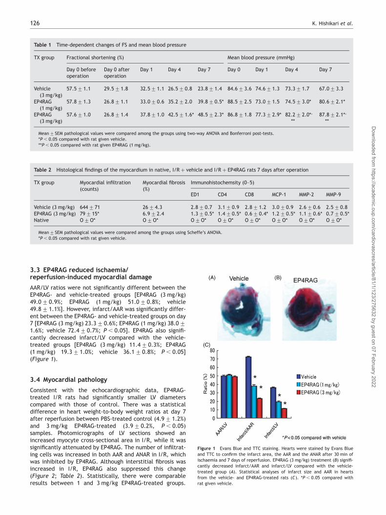
AAR/LV ratios were not significantly different between theEP4RAG- and vehicle-treated groups [EP4RAG (3 mg/kg)49.0+0.9%; EP4RAG (1 mg/kg) 51.0+0.8%; vehicle49.8+1.1%]. However, infarct/AAR was significantly differ-ent between the EP4RAG- and vehicle-treated groups on day7 [EP4RAG (3 mg/kg) 23.3+0.6%; EP4RAG (1 mg/kg) 38.0+1.6%; vehicle 72.4+0.7%; P , 0.05]. EP4RAG also signifi-cantly decreased infarct/LV compared with the vehicle-treated groups [EP4RAG (3 mg/kg) 11.4+0.3%; EP4RAG(1 mg/kg) 19.3+1.0%; vehicle 36.1+0.8%; P , 0.05](Figure 1).
3.4 Myocardial pathology
Consistent with the echocardiographic data, EP4RAG-treated I/R rats had significantly smaller LV diameterscompared with those of control. There was a statisticaldifference in heart weight-to-body weight ratios at day 7after reperfusion between PBS-treated control (4.9+1.2%)and 3 mg/kg EP4RAG-treated (3.9+0.2%, P , 0.05)samples. Photomicrographs of LV sections showed anincreased myocyte cross-sectional area in I/R, while it wassignificantly attenuated by EP4RAG. The number of infiltrat-ing cells was increased in both AAR and ANAR in I/R, whichwas inhibited by EP4RAG. Although interstitial fibrosis wasincreased in I/R, EP4RAG also suppressed this change(Figure 2; Table 2). Statistically, there were comparableresults between 1 and 3 mg/kg EP4RAG-treated groups.
Table 1 Time-dependent changes of FS and mean blood pressure
TX group Fractional shortening (%) Mean blood pressure (mmHg)
Day 0 beforeoperation
Day 0 afteroperation
Day 1 Day 4 Day 7 Day 0 Day 1 Day 4 Day 7
Vehicle(3 mg/kg)
57.5+1.1 29.5+1.8 32.5+1.1 26.5+0.8 23.8+1.4 84.6+3.6 74.6+1.3 73.3+1.7 67.0+3.3
EP4RAG(1 mg/kg)
57.8+1.3 26.8+1.1 33.0+0.6 35.2+2.0 39.8+0.5* 88.5+2.5 73.0+1.5 74.5+3.0* 80.6+2.1*
EP4RAG(3 mg/kg)
57.6+1.0 26.8+1.4 37.8+1.0 42.5+1.6* 48.5+2.3* 86.8+1.8 77.3+2.9* 82.2+2.0*,
**87.8+2.1*,
**
Mean+ SEM pathological values were compared among the groups using two-way ANOVA and Bonferroni post-tests.*P , 0.05 compared with rat given vehicle.**P , 0.05 compared with rat given EP4RAG (1 mg/kg).
Table 2 Histological findings of the myocardium in native, I/R þ vehicle and I/R þ EP4RAG rats 7 days after operation
TX group Myocardial infiltration(counts)
Myocardial fibrosis(%)
Immunohistochemsity (0–5)
ED1 CD4 CD8 MCP-1 MMP-2 MMP-9
Vehicle (3 mg/kg) 644+71 26+4.3 2.8+0.7 3.1+0.9 2.8+1.2 3.0+0.9 2.6+0.6 2.5+0.8EP4RAG (3 mg/kg) 79+15* 6.9+2.4 1.3+0.5* 1.4+0.5* 0.6+0.4* 1.2+0.5* 1.1+0.6* 0.7+0.5*Native O+O* O+O* O+O* O+O* O+O* O+O* O+O* O+O*
Mean+ SEM pathological values were compared among the groups using Scheffe’s ANOVA.*P , 0.05 compared with rat given vehicle.
Figure 1 Evans Blue and TTC staining. Hearts were stained by Evans Blueand TTC to confirm the infarct area, the AAR and the ANAR after 30 min ofischaemia and 7 days of reperfusion. EP4RAG (3 mg/kg) treatment (B) signifi-cantly decreased infarct/AAR and infarct/LV compared with the vehicle-treated group (A). Statistical analyses of infarct size and AAR in heartsfrom the vehicle- and EP4RAG-treated rats (C). *P , 0.05 compared withrat given vehicle.
K. Hishikari et al.126
Dow
nloaded from https://academ
ic.oup.com/cardiovascres/article/81/1/123/275632 by guest on 07 February 2022

However, there was a difference between the results ofcontrol and 1 mg/kg EP4RAG.
3.5 Myocardial immunohistochemistry
The immunoreactive staining for EP4 receptor was barelydetectable in the LV obtained from native rats. In contrast,in post-I/R animals on day 7, the expression of EP4 receptorwas clearly increased in AAR in LV (Supplementary materialonline, Data 2). In the vehicle-treated I/R rats, theexpression of MCP-1 and severe myocardial macrophageinfiltration was observed at day 7. However, EP4RAG signifi-cantly attenuated the expression of MCP-1 and infiltration ofmacrophages. T lymphocytes (CD4 and CD8), to which MCP-1can be also chemotactic, were detected in the I/R rats,which were also reduced by EP4RAG. MMP-2 and -9 wereexpressed in both LV of EP4RAG- and vehicle-treatedgroups on day 7, but the expression was much stronger invehicle-treated groups (Figure 3; Table 2). Although therewere comparable results between 1 and 3 mg/kg EP4RAG-treated groups, there was a difference between controland 1 mg/kg EP4RAG.
3.6 Myocardial expression of cytokines,chemokines and matrix metalloproteinases
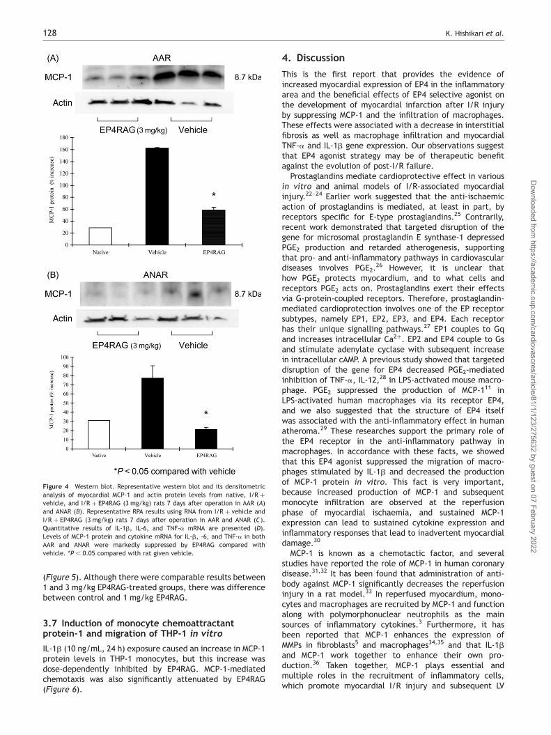
MCP-1 levels were significantly higher in I/R than those innative animals. However, EP4RAG attenuated the increasein MCP-1 expression in both AAR and ANAR (Figure 4A andB). EP4RAG also attenuated inflammatory cytokines, includ-ing TNF-a, IL-1b, and IL-6, induced by I/R (Figure 4C and D).On day 7 after I/R, LV gelatinolytic activity of pro- andactive-MMP-2 and -9 appeared in both EP4RAG- and vehicle-treated groups, but EP4RAG-treated groups showed signifi-cantly weaker activity not only in AAR but also in ANAR
Figure 2 HE and Masson-trichrome staining. Low-power (A, B, E, and F) andhigh-power (C, D, G, and H ) photomicrographs of HE-stained (A through D)and Masson-trichrome-stained (E through H) LV cross-sections obtained fromI/R (A, C, E, and G), I/R þ EP4RAG (3 mg/kg) (B, D, F, and H) rats 7 daysafter operation. EP4RAG-treated I/R rats had significantly smaller LV diam-eters compared with those of vehicle. EP4RAG attenuated infiltration ofinflammatory cells and LV fibrosis (D and H) compared with vehicle (C andG). Scale bars: 2 mm (A, B, E, and F) and 100 mm (C, D, G, and H).
Figure 3 Inflammatory cell infiltration. Representative immunohistochem-ical findings of myocardium of the I/R rats at day 7. Left panels show myocar-dium from vehicle-treated rats at day 7; right panels are from those treatedwith EP4RAG (3 mg/kg) at day 7. ED1 (A and B), CD4 (C and D), CD8 (E and F),MCP-1 (G and H ), MMP-2 (I and J ), and MMP-9 (K and L) staining. Increasednumbers of ED1-, CD4-, and CD8-positive cells were observed in vehicle-treated rats, while EP4RAG suppressed the cell numbers significantly.MCP-1, MMP-2, and -9 were strongly expressed on the infiltrating cells invehicle-treated rats, but these changes were attenuated in EP4RAG-treatedrats. Scale bars: 50 mm.
EP4 receptor activation improves myocardial ischaemia 127
Dow
nloaded from https://academ
ic.oup.com/cardiovascres/article/81/1/123/275632 by guest on 07 February 2022
(Figure 5). Although there were comparable results between1 and 3 mg/kg EP4RAG-treated groups, there was differencebetween control and 1 mg/kg EP4RAG.
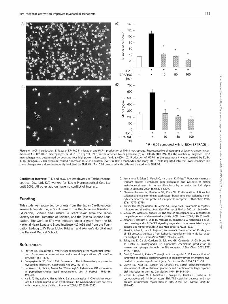
3.7 Induction of monocyte chemoattractantprotein-1 and migration of THP-1 in vitro
IL-1b (10 ng/mL, 24 h) exposure caused an increase in MCP-1protein levels in THP-1 monocytes, but this increase wasdose-dependently inhibited by EP4RAG. MCP-1-mediatedchemotaxis was also significantly attenuated by EP4RAG(Figure 6).
4. Discussion
This is the first report that provides the evidence ofincreased myocardial expression of EP4 in the inflammatoryarea and the beneficial effects of EP4 selective agonist onthe development of myocardial infarction after I/R injuryby suppressing MCP-1 and the infiltration of macrophages.These effects were associated with a decrease in interstitialfibrosis as well as macrophage infiltration and myocardialTNF-a and IL-1b gene expression. Our observations suggestthat EP4 agonist strategy may be of therapeutic benefitagainst the evolution of post-I/R failure.
Prostaglandins mediate cardioprotective effect in variousin vitro and animal models of I/R-associated myocardialinjury.22–24 Earlier work suggested that the anti-ischaemicaction of prostaglandins is mediated, at least in part, byreceptors specific for E-type prostaglandins.25 Contrarily,recent work demonstrated that targeted disruption of thegene for microsomal prostaglandin E synthase-1 depressedPGE2 production and retarded atherogenesis, supportingthat pro- and anti-inflammatory pathways in cardiovasculardiseases involves PGE2.
26 However, it is unclear thathow PGE2 protects myocardium, and to what cells andreceptors PGE2 acts on. Prostaglandins exert their effectsvia G-protein-coupled receptors. Therefore, prostaglandin-mediated cardioprotection involves one of the EP receptorsubtypes, namely EP1, EP2, EP3, and EP4. Each receptorhas their unique signalling pathways.27 EP1 couples to Gqand increases intracellular Ca2þ. EP2 and EP4 couple to Gsand stimulate adenylate cyclase with subsequent increasein intracellular cAMP. A previous study showed that targeteddisruption of the gene for EP4 decreased PGE2-mediatedinhibition of TNF-a, IL-12,28 in LPS-activated mouse macro-phage. PGE2 suppressed the production of MCP-111 inLPS-activated human macrophages via its receptor EP4,and we also suggested that the structure of EP4 itselfwas associated with the anti-inflammatory effect in humanatheroma.29 These researches support the primary role ofthe EP4 receptor in the anti-inflammatory pathway inmacrophages. In accordance with these facts, we showedthat this EP4 agonist suppressed the migration of macro-phages stimulated by IL-1b and decreased the productionof MCP-1 protein in vitro. This fact is very important,because increased production of MCP-1 and subsequentmonocyte infiltration are observed at the reperfusionphase of myocardial ischaemia, and sustained MCP-1expression can lead to sustained cytokine expression andinflammatory responses that lead to inadvertent myocardialdamage.30
MCP-1 is known as a chemotactic factor, and severalstudies have reported the role of MCP-1 in human coronarydisease.31,32 It has been found that administration of anti-body against MCP-1 significantly decreases the reperfusioninjury in a rat model.33 In reperfused myocardium, mono-cytes and macrophages are recruited by MCP-1 and functionalong with polymorphonuclear neutrophils as the mainsources of inflammatory cytokines.3 Furthermore, it hasbeen reported that MCP-1 enhances the expression ofMMPs in fibroblasts5 and macrophages34,35 and that IL-1band MCP-1 work together to enhance their own pro-duction.36 Taken together, MCP-1 plays essential andmultiple roles in the recruitment of inflammatory cells,which promote myocardial I/R injury and subsequent LV
Figure 4 Western blot. Representative western blot and its densitometricanalysis of myocardial MCP-1 and actin protein levels from native, I/R þvehicle, and I/R þ EP4RAG (3 mg/kg) rats 7 days after operation in AAR (A)and ANAR (B). Representative RPA results using RNA from I/R þ vehicle andI/R þ EP4RAG (3 mg/kg) rats 7 days after operation in AAR and ANAR (C).Quantitative results of IL-1b, IL-6, and TNF-a mRNA are presented (D).Levels of MCP-1 protein and cytokine mRNA for IL-b, -6, and TNF-a in bothAAR and ANAR were markedly suppressed by EP4RAG compared withvehicle. *P , 0.05 compared with rat given vehicle.
K. Hishikari et al.128
Dow
nloaded from https://academ
ic.oup.com/cardiovascres/article/81/1/123/275632 by guest on 07 February 2022

remodelling. Therefore, the suppression of MCP-1expression by EP4 agonist is an effective way to preventharmful inflammatory cell accumulation in reperfused myo-cardium. In addition, recent studies have revealed thatMCP-1 may exert other biological activities than recruitingmonocytes/macrophages, because the expression of MCP-1does not always correlate with the extent of their infiltra-tion.7 Therefore, the inhibition of MCP-1 by EP4 agonistshown in this study might not be attributable solely to itschemoattractant properties on inflammatory cells. In fact,EP4RAG reduced the expression of TNF-a in the post-I/Rhearts in both AAR and ANAR. TNF-a has profound and wide-ranging effects that can initiate a cascade of myocyte hyper-trophy and apoptosis. Therefore, a proposed mechanism ofEP4RAG for reverse LV remodelling is related to the attenu-ation of this cytokine after I/R.
MCP-1 can also directly stimulate collagen production viaupregulation of IL-1b expression in cardiac fibroblasts.Therefore, the inhibition of interstitial fibrosis in I/R byEP4RAG could be attributable to the attenuation of IL-1b.However, we cannot exclude the possibility that the longer-term inhibition of MCP-1 signalling may enhance LV remodel-ling and even cause cardiac rupture. Although we did notexperience cardiac rupture in our series of rat experiment,we apparently need additional studies to clarify thiscrucial issue.
Adding to the fact that EP4RAG suppressed the expressionof MCP-1 and infiltration of inflammatory cells including
macrophages, it also attenuated the expression and activi-ties of MMP-2 and -9 in both AAR and ANAR. Sustainedexpression of MMP-2 and -9 leads to the myocardial fibrosis,LV enlargement, and the resultant LV failure,37 so this effectof EP4RAG is noteworthy. Cytokines including TNF-a andIL-1b evoke a secondary MCP-1 production from othertypes of cells, including cardiac myocytes and fibroblasts,and establish a positive loop, amplifying and sustaining theproinflammatory response. Thus, there is an intimate linkbetween MCP-1 and cytokines, and MMPs in the heartsthrough an autocrine/paracrine mechanism, possibly repre-senting a vicious cycle implicated in myocardial failure.Therefore, an inhibition of MCP-1 signalling by EP4RAG hasa great potential for breaking the vicious feedback loop ofinflammation and may attenuate the development of myo-cardial remodelling. Considering these cardioprotectiveeffects of PGE2 via its receptor EP4, EP4 agonist may beuseful for the treatment of cardiac dysfunction after myo-cardial infarction.
This study has a limitation. We administered EP4RAG15 min before reperfusion and every 12 h for 7 days.Because clinical pre-treatment of patients prior to theonset of ischaemia may be difficult to achieve, furthertreatment protocols are needed.
In conclusion, the results of the present studydemonstrate a robust cardioprotective effect againstmyocardial reperfusion injury by pre-treatment andcontinued treatment with the selective EP4 agonist. This
Figure 4 Continued
EP4 receptor activation improves myocardial ischaemia 129
Dow
nloaded from https://academ
ic.oup.com/cardiovascres/article/81/1/123/275632 by guest on 07 February 2022

cardioprotective effect is based on the suppression ofmacrophage infiltration and macrophage-derived MCP-1.It makes great sense that activation of EP4 results inreduction of necrotic area as well as in preservation ofcardiac physiological function. Although further investi-gations are required to evaluate the anti-leucocytetherapy, EP4RAG could potentially become a part of strategyto prevent the I/R injury.
Supplementary material
Supplementary material is available at CardiovascularResearch online.
Acknowledgements
We thank Libby for his encouragement and mentorship. We thank MsNoriko Tamura and Ms Yasuko Matsuda for their excellent assistance.
Figure 5 Zymography. Gelatin zymography of heart from native, I/R þ vehicle and I/R þ EP4RAG (3 mg/kg) rats 7 days after operation in AAR (A) and ANAR (B).Relative intensity of MMP-2 (pro- and active forms) and -9 are shown. In accordance with the LV fibrosis, vehicle-treated rats showed the high activity of MMP-2and -9, but EP4RAG suppressed these changes. *P , 0.05 compared with rat given vehicle.
K. Hishikari et al.130
Dow
nloaded from https://academ
ic.oup.com/cardiovascres/article/81/1/123/275632 by guest on 07 February 2022
Conflict of interest: T.T. and M.O. are employees of Taisho Pharma-ceutical Co., Ltd. K.T. worked for Taisho Pharmaceutical Co., Ltd,until 2006. All other authors have no conflict of interest.
Funding
This study was supported by grants from the Japan CardiovascularResearch Foundation, a Grant-in-Aid from the Japanese Ministry ofEducation, Science and Culture, a Grant-in-Aid from the JapanSociety for the Promotion of Science, and the Takeda Science Foun-dation. The work on EP4 was initiated under a grant from the USNational Heart Lung and Blood Institute HL34636 and from the Foun-dation Leducq to Dr Peter Libby, Brigham and Women’s Hospital andthe Harvard Medical School.
References1. Pfeffer MA, Braunwald E. Ventricular remodeling after myocardial infarc-
tion. Experimental observations and clinical implications. Circulation1990;81:1161–1172.
2. Frangogiannis NG, Smith CW, Entman ML. The inflammatory response inmyocardial infarction. Cardiovasc Res 2002;53:31–47.
3. Herskowitz A, Choi S, Ansari AA, Wesselingh S. Cytokine mRNA expressionin postischemic/reperfused myocardium. Am J Pathol 1995;146:419–428.
4. Nanki T, Nagasaka K, Hayashida K, Saita Y, Miyasaka N. Chemokines regu-late IL-6 and IL-8 production by fibroblast-like synoviocytes from patientswith rheumatoid arthritis. J Immunol 2001;167:5381–5385.
5. Yamamoto T, Eckes B, Mauch C, Hartmann K, Krieg T. Monocyte chemoat-tractant protein-1 enhances gene expression and synthesis of matrixmetalloproteinase-1 in human fibroblasts by an autocrine IL-1 alphaloop. J Immunol 2000;164:6174–6179.
6. Gharaee-Kermani M, Denholm EM, Phan SH. Costimulation of fibroblastcollagen and transforming growth factor beta1 gene expression by mono-cyte chemoattractant protein-1 via specific receptors. J Biol Chem 1996;271:17779–17784.
7. Breyer RM, Bagdassarian CK, Myers SA, Breyer MD. Prostanoid receptors:subtypes and signaling. Annu Rev Pharmacol Toxicol 2001;41:661–690.
8. McCoy JM, Wicks JR, Audoly LP. The role of prostaglandin E2 receptors inthe pathogenesis of rheumatoid arthritis. J Clin Invest 2002;110:651–658.
9. Amano H, Hayashi I, Endo H, Kitasato H, Yamashita S, Maruyama T et al.Host prostaglandin E(2)-EP3 signaling regulates tumor-associated angio-genesis and tumor growth. J Exp Med 2003;197:221–232.
10. Xiao CY, Yuhki K, Hara A, Fujino T, Kuriyama S, Yamada Tet al. Prostaglan-din E2 protects the heart from ischemia-reperfusion injury via its recep-tor subtype EP4. Circulation 2004;109:2462–2468.
11. Takayama K, Garcia-Cardena G, Sukhova GK, Comander J, Gimbrone MAJr, Libby P. Prostaglandin E2 suppresses chemokine production inhuman macrophages through the EP4 receptor. J Biol Chem 2002;277:44147–44154.
12. Onai Y, Suzuki J, Kakuta T, Maejima Y, Haraguchi G, Fukasawa H et al.Inhibition of IkappaB phosphorylation in cardiomyocytes attenuates myo-cardial ischemia/reperfusion injury. Cardiovasc Res 2004;63:51–59.
13. Litwin SE, Katz SE, Morgan JP, Douglas PS. Serial echocardiographicassessment of left ventricular geometry and function after large myocar-dial infarction in the rat. Circulation 1994;89:345–354.
14. Suzuki J, Ogawa M, Futamatsu H, Kosuge H, Tanaka H, Isobe M. Acyclooxygenase-2 inhibitor alters Th1/Th2 cytokine balance and sup-presses autoimmune myocarditis in rats. J Mol Cell Cardiol 2006;40:688–695.
Figure 6 MCP-1 production. Efficacy of EP4RAG in migration and MCP-1 production of THP-1 macrophage. Representative photographs of lower chamber in con-dition of 1 � 104 THP-1 macrophages/mL (IL-1b, 10 ng/mL, 24 h) in the absence (A) or presence (B) of EP4RAG (100 nM). (C) The number of migrated THP-1macrophages was determined by counting four high-power microscope fields (�400). (D) Production of MCP-1 in the supernatant was estimated by ELISA.IL-1b (10 ng/mL, 24 h) exposure caused a increase in MCP-1 protein levels in THP-1 monocytes and many THP-1 cells migrated into the lower chamber, butthese changes were dose-dependently inhibited by EP4RAG. *P , 0.05 compared with cells not treated with EP4RAG.
EP4 receptor activation improves myocardial ischaemia 131
Dow
nloaded from https://academ
ic.oup.com/cardiovascres/article/81/1/123/275632 by guest on 07 February 2022
15. Vivaldi MT, Kloner RA, Schoen FJ. Triphenyltetrazolium staining of irre-versible ischemic injury following coronary artery occlusion in rats. AmJ Pathol 1985;121:522–530.
16. Yamashiro S, Takeya M, Kuratsu J, Ushio Y, Takahashi K, Yoshimura T.Intradermal injection of monocyte chemoattractant protein-1 inducesemigration and differentiation of blood monocytes in rat skin. Int ArchAllergy Immunol 1998;115:15–23.
17. Suzuki J, Ogawa M, Sagesaka YM, Isobe M. Tea catechins attenuate ven-tricular remodeling and graft arterial diseases in murine cardiac allo-grafts. Cardiovasc Res 2006;69:272–279.
18. Cheung PY, Sawicki G, Wozniak M, Wang W, Radomski MW, Schulz R.Matrix metalloproteinase-2 contributes to ischemia-reperfusion injuryin the heart. Circulation 2000;101:1833–1839.
19. Futamatsu H, Suzuki J, Kosuge H, Yokoseki O, Kamada M, Ito H et al.Attenuation of experimental autoimmune myocarditis by blocking acti-vated T cells through inducible costimulatory molecule pathway. Cardio-vasc Res 2003;59:95–104.
20. Gianturco SH, Ramprasad MP, Lin AH, Song R, Bradley WA. Cellularbinding site and membrane binding proteins for triglyceride-rich lipopro-teins in human monocyte-macrophages and THP-1 monocytic cells.J Lipid Res 1994;35:1674–1687.
21. Zhu P, Ding J, Zhou J, Dong WJ, Fan CM, Chen ZN. Expression of CD147 onmonocytes/macrophages in rheumatoid arthritis: its potential role inmonocyte accumulation and matrix metalloproteinase production.Arthritis Res Ther 2005;7:R1023–R1033.
22. Jugdutt BI, Hutchins GM, Bulkley BH, Becker LC. Dissimilar effects ofprostacyclin, prostaglandin E1, and prostaglandin E2 on myocardialinfarct size after coronary occlusion in conscious dogs. Circ Res 1981;49:685–700.
23. Woditsch I, Schror K. Prostacyclin rather than endogenous nitric oxide is atissue protective factor in myocardial ischemia. Am J Physiol 1992;263:H1390–H1396.
24. Schror K, Woditsch I. Endogenous prostacyclin preserves myocardial func-tion and endothelium-derived nitric oxide formation in myocardial ische-mia. Agents Actions Suppl 1992;37:312–319.
25. Hohlfeld T, Meyer-Kirchrath J, Vogel YC, Schror K. Reduction of infarctsize by selective stimulation of prostaglandin EP(3)receptors in thereperfused ischemic pig heart. J Mol Cell Cardiol 2000;32:285–296.
26. Wang M, Zukas AM, Hui Y, Ricciotti E, Pure E, FitzGerald GA. Deletion ofmicrosomal prostaglandin E synthase-1 augments prostacyclin andretards atherogenesis. Proc Natl Acad Sci USA 2006;103:14507–14512.
27. Narumiya S, Sugimoto Y, Ushikubi F. Prostanoid receptors: structures,properties, and functions. Physiol Rev 1999;79:1193–1226.
28. Nataraj C, Thomas DW, Tilley SL, Nguyen MT, Mannon R, Koller BH et al.Receptors for prostaglandin E(2) that regulate cellular immune responsesin the mouse. J Clin Invest 2001;108:1229–1235.
29. Takayama K, Sukhova GK, Chin MT, Libby P. A novel prostaglandin E recep-tor 4-associated protein participates in antiinflammatory signaling. CircRes 2006;98:499–504.
30. Hayashidani S, Tsutsui H, Shiomi T, Ikeuchi M, Matsusaka H, Suematsu N etal. Anti-monocyte chemoattractant protein-1 gene therapy attenuatesleft ventricular remodeling and failure after experimental myocardialinfarction. Circulation 2003;108:2134–2140.
31. de Lemos JA, Morrow DA, Sabatine MS, Murphy SA, Gibson CM, Antman EMet al. Association between plasma levels of monocyte chemoattractantprotein-1 and long-term clinical outcomes in patients with acute coron-ary syndromes. Circulation 2003;107:690–695.
32. Matsumori A, Furukawa Y, Hashimoto T, Yoshida A, Ono K, Shioi T et al.Plasma levels of the monocyte chemotactic and activating factor/mono-cyte chemoattractant protein-1 are elevated in patients with acute myo-cardial infarction. J Mol Cell Cardiol 1997;29:419–423.
33. Ono K, Matsumori A, Furukawa Y, Igata H, Shioi T, Matsushima K et al. Pre-vention of myocardial reperfusion injury in rats by an antibody againstmonocyte chemotactic and activating factor/monocyte chemoattractantprotein-1. Lab Invest 1999;79:195–203.
34. Stuve O, Chabot S, Jung SS, Williams G, Yong VW. Chemokine-enhancedmigration of human peripheral blood mononuclear cells is antagonizedby interferon beta-1b through an effect on matrix metalloproteinase-9.J Neuroimmunol 1997;80:38–46.
35. Okuma T, Terasaki Y, Kaikita K, Kobayashi H, Kuziel WA, Kawasuji M et al.C-C chemokine receptor 2 (CCR2) deficiency improves bleomycin-inducedpulmonary fibrosis by attenuation of both macrophage infiltration andproduction of macrophage-derived matrix metalloproteinases. J Pathol2004;204:594–604.
36. Damas JK, Aukrust P, Ueland T, Odegaard A, Eiken HG, Gullestad L et al.Monocyte chemoattractant protein-1 enhances and interleukin-10 sup-presses the production of inflammatory cytokines in adult rat cardiomyo-cytes. Basic Res Cardiol 2001;96:345–352.
37. Ducharme A, Frantz S, Aikawa M, Rabkin E, Lindsey M, Rohde LE et al.Targeted deletion of matrix metalloproteinase-9 attenuates left ventri-cular enlargement and collagen accumulation after experimental myo-cardial infarction. J Clin Invest 2000;106:55–62.
K. Hishikari et al.132
Dow
nloaded from https://academ
ic.oup.com/cardiovascres/article/81/1/123/275632 by guest on 07 February 2022