biolreprod0039.pdf - Oxford Academic
18
BIOLOGY OF REPRODUCTION 16, 39-56 (1977) 39 Control of Parturition in Man G. C. LIGGINS, CHRISTINE S. FORSTER, SUSAN A. GRIEVES and A. L. SCHWARTZ Postgraduate School of Obstetrics and Gynaecology, University of Auckland, Auckland, New Zealand ABSTRACT Human pregnancy differs from that of many other species in showing no abrupt changes in maternal levels of estrogen and progesterone at the start of labor. The fetus appears to play a relatively minor role in initiating parturition since mean pregnancy length is not markedly affected by major disorders of the fetal hypothalamus, pituitary or adrenals. Human pregnancy cannot be induced by estrogen treatment and corticosteroids are ineffective except in women beyond term. Prostaglandins are released into the maternal circulation and amniotic fluid during labor but there is no unequivocal evidence of their involvement in initiation of labor. However, there is circumstantial evidence favoring a local mechanism involving the fetal membranes and deciduum that controls prostaglandin release. These tissues contain glycerophospholipids enriched with arachidonic acid in the sn-2 position, phospholipase A2 activity and prostaglandin synthetase. The local mechanism is readily activated by local trauma, It is ptoposed that the onset of labor is mainly the outcome of a genetically-determined maturational event in the amnion and/or chorion. The fetus itself and the mother may modulate, but rarely control, the time of birth. INTRODUCTION Despite the advances that have been made in recent years in the understanding of the physi- ology of the initiation of labor in various species, notably some of the ruminants and rodents, the mechanism in man remains enig- matic. Knowledge is fragmentary and not yet capable of being synthesized into a usable hypothesis incorporating each facet. It is clear that maintenance of human pregnancy is dependent on placental hormones and that the corpus luteum serves no useful purpose after the second month of pregnancy (Csapo et al., 1973). Thus luteolysis, which has an indispensi- ble function in parturition in species such as the goat (Currie et al., 1973), rabbit (Fraenkl, 1905), and mouse (Harris, 1927), is not a component of the human system. Some species, of which the sheep and cow have been studied intensively, share with man an independence from corpus luteum function but differ from man in demonstrating readily measurable changes in placental hormone metabolism be- fore labor starts. Man, along with nonhuman primates and guinea pigs (Heap and Deansley, 1966) appear to belong to a select group of ‘placenta-dependent’ mammals in which labor starts without the levels of hormones in the maternal circulation showing any clear indica- tion of altered placental hormone metabolism. In general, the reaction of investigators to the unaltered hormone levels in the maternal circulation at the start of labor has been either disbelieving (‘evidence will be found if it is looked for correctly’) or negative (‘maternal hormone levels do not reflect placental hor- mone metabolism’). Little attention has been paid so far to the possibility that women begin labor in the absence of any abrupt change in the placental production of steroid or other hormones and that changes in target tissue responses rather than production may be the more important. This omission is rather surpris- ing in view of the remarkable efficacy in inducing labor of quite minor local mechanical stimuli (e.g. stripping membranes) that would be unlikely to disturb placental function. The dominant role of the fetus in initiating parturition in the sheep (a ‘placenta-dependent’ species) and the goat (a ‘corpus luteum-depen- dent’ species) has encouraged investigations to determine the extent to which the concept of fetal control of labor can be extrapolated to other species. This approach has been fruitful in some species but in the nonhuman primate as is apparent from the material presented by Dr. Lanman and in man as reviewed below, it seems that the part played by the fetus (as distinct from the conceptus as a whole) and in particu- Downloaded from https://academic.oup.com/biolreprod/article/16/1/39/2768197 by guest on 02 February 2022
-
Upload
khangminh22 -
Category
Documents
-
view
0 -
download
0
Transcript of biolreprod0039.pdf - Oxford Academic

BIOLOGY OF REPRODUCTION 16, 39-56 (1977)
39
Control of Parturition in Man
G. C. LIGGINS, CHRISTINE S. FORSTER, SUSAN A. GRIEVES
and A. L. SCHWARTZ
Postgraduate School of Obstetrics and Gynaecology,
University of Auckland,
Auckland, New Zealand
ABSTRACT
Human pregnancy differs from that of many other species in showing no abrupt changes in
maternal levels of estrogen and progesterone at the start of labor. The fetus appears to play a
relatively minor role in initiating parturition since mean pregnancy length is not markedly affected
by major disorders of the fetal hypothalamus, pituitary or adrenals. Human pregnancy cannot be
induced by estrogen treatment and corticosteroids are ineffective except in women beyond term.
Prostaglandins are released into the maternal circulation and amniotic fluid during labor but thereis no unequivocal evidence of their involvement in initiation of labor. However, there is
circumstantial evidence favoring a local mechanism involving the fetal membranes and deciduum
that controls prostaglandin release. These tissues contain glycerophospholipids enriched witharachidonic acid in the sn-2 position, phospholipase A2 activity and prostaglandin synthetase. The
local mechanism is readily activated by local trauma, It is ptoposed that the onset of labor ismainly the outcome of a genetically-determined maturational event in the amnion and/or chorion.
The fetus itself and the mother may modulate, but rarely control, the time of birth.
INTRODUCTION
Despite the advances that have been made in
recent years in the understanding of the physi-
ology of the initiation of labor in various
species, notably some of the ruminants and
rodents, the mechanism in man remains enig-
matic. Knowledge is fragmentary and not
yet capable of being synthesized into a usable
hypothesis incorporating each facet. It is
clear that maintenance of human pregnancy is
dependent on placental hormones and that the
corpus luteum serves no useful purpose after
the second month of pregnancy (Csapo et al.,
1973). Thus luteolysis, which has an indispensi-
ble function in parturition in species such as the
goat (Currie et al., 1973), rabbit (Fraenkl,
1905), and mouse (Harris, 1927), is not a
component of the human system. Some species,
of which the sheep and cow have been studied
intensively, share with man an independence
from corpus luteum function but differ from
man in demonstrating readily measurable
changes in placental hormone metabolism be-
fore labor starts. Man, along with nonhuman
primates and guinea pigs (Heap and Deansley,
1966) appear to belong to a select group of
‘placenta-dependent’ mammals in which labor
starts without the levels of hormones in the
maternal circulation showing any clear indica-
tion of altered placental hormone metabolism.
In general, the reaction of investigators to
the unaltered hormone levels in the maternal
circulation at the start of labor has been either
disbelieving (‘evidence will be found if it is
looked for correctly’) or negative (‘maternal
hormone levels do not reflect placental hor-
mone metabolism’). Little attention has been
paid so far to the possibility that women begin
labor in the absence of any abrupt change in
the placental production of steroid or other
hormones and that changes in target tissue
responses rather than production may be the
more important. This omission is rather surpris-
ing in view of the remarkable efficacy in
inducing labor of quite minor local mechanical
stimuli (e.g. stripping membranes) that would
be unlikely to disturb placental function.
The dominant role of the fetus in initiating
parturition in the sheep (a ‘placenta-dependent’
species) and the goat (a ‘corpus luteum-depen-
dent’ species) has encouraged investigations to
determine the extent to which the concept of
fetal control of labor can be extrapolated to
other species. This approach has been fruitful in
some species but in the nonhuman primate as is
apparent from the material presented by Dr.
Lanman and in man as reviewed below, it seems
that the part played by the fetus (as distinct
from the conceptus as a whole) and in particu-
Dow
nloaded from https://academ
ic.oup.com/biolreprod/article/16/1/39/2768197 by guest on 02 February 2022
40 LIGGINS ET AL.
lar by the fetal hypothalamic-pituitary-adrenal
system is a relatively minor one. The void left
by so relegating the human fetus has yet to be
filled by an alternative dominant system. Per-
haps the onset of labor is truly controlled by a
‘complex multifactorial system’ in which no
factor is dominant and each factor acts by itself
so weakly as to be unidentifiable in isolation.
Such a concept has little to commend it as a
working hypothesis to be tested by investiga-
tion.
This paper reviews the present state of
knowledge of the physiology of the onset of
labor in man and considers possible mechanisms
that are consistent with available evidence.
THE ROLE OF THE HUMAN FETUS
Labor occurs spontaneously in the absence
of normal function of the fetal hypothalamic-
pituitary-adrenal system but the length of
pregnancy is likely to be disordered. In preg-
nancies complicated by anencephaly, the mean
pregnancy length in patients in whom there is
no polyhydramnios and no obstretic interfer-
ence is approximately 40 weeks but the range is
very wide (Honnebier and Swaab, 1973). Pro-
longed pregnancy has long been recognized as
having a striking association with anencephaly
but the fact that premature delivery is equally
common has been noted only recently (Milic
and Adamsons 1969). Generally, fetuses with
adrenal hypoplasia but no other malformations
are born at, or close to, term (Liggins, 1974). A
family has been described in which siblings with
adrenal hypoplasia were born at 42-43 weeks
whereas unaffected siblings were delivered at
term (O’Donohoe and Holland, 1968).
A prospective study of women at risk of
premature labor revealed that a rise in plasma
estradiol-1713 preceded the onset of labor in
those women delivering prematurely (Tamby
Raja et al., 1974; Tamby Raja et al., 1975). A
study of the adrenal weights in a group of
premature infants dying soon after birth sug-
gests that accelerated growth of the adrenals
may be the cause of the rise in estrogen;
Anderson et al. (1971) found that the adrenals
were heavier when premature labor had no
recognizable cause than when it followed a
complication of pregnancy such as placenta
previa or placental abruption.
A number of studies have measured cortico-
steroids in cord blood in an attempt to deter-
mine whether there is a prepartum rise in fetal
levels. There is a progressive rise in plasma total
corticosteroid (Smith and Shearman, 1974) and
cortisol (Murphy and Diez d’Aux, 1972) with
advancing gestational age at birth during the
last month of pregnancy and a similar rise
occurs in cortisol levels in amniotic fluid
(Murphy et a!., 1975; Fend and Tulchinsky,
1975). However it is less certain that a sharp
rise in fetal cortisol levels preceeds the onset of
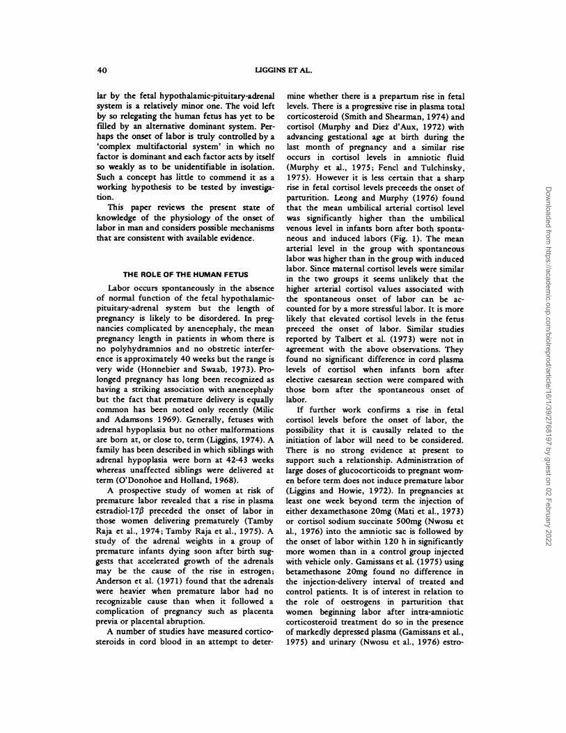
parturition. Leong and Murphy (1976) found
that the mean umbilical arterial cortisol level
was significantly higher than the umbilical
venous level in infants born after both sponta-
neous and induced labors (Fig. 1). The mean
arterial level in the group with spontaneous
labor was higher than in the group with induced
labor. Since maternal cortisol levels were similar
in the two groups it seems unlikely that the
higher arterial cortisol values associated with
the spontaneous onset of labor can be ac-
counted for by a more stressful labor. It is more
likely that elevated cortisol levels in the fetus
preceed the onset of labor. Similar studies
reported by Talbert et al. (1973) were not in
agreement with the above observations. They
found no significant difference in cord plasma
levels of cortisol when infants born after
elective caesarean section were compared with
those born after the spontaneous onset of
labor.
If further work confirms a rise in fetal
cortisol levels before the onset of labor, the
possibility that it is causally related to the
initiation of labor will need to be considered.
There is no strong evidence at present to
support such a relationship. Administration of
large doses of glucocorticoids to pregnant wom-
en before term does not induce premature labor
(Liggins and Howie, 1972). In pregnancies at
least one week beyond term the injection of
either dexamethasone 20mg (Mati et al., 1973)
or cortisol sodium succinate 500mg (Nwosu et
al., 1976) into the amniotic sac is followed by
the onset of labor within 120 h in significantly
more women than in a control group injected
with vehicle only. Gamissans et al. (1975) using
betamethasone 20mg found no difference in
the injection-delivery interval of treated and
control patients. It is of interest in relation to
the role of oestrogens in parturition that
women beginning labor after intra-amniotic
corticosteroid treatment do so in the presence
of markedly depressed plasma (Gamissans et al.,
1975) and urinary (Nwosu et al., 1976) estro-
Dow
nloaded from https://academ
ic.oup.com/biolreprod/article/16/1/39/2768197 by guest on 02 February 2022

ELECTIVEARTERIAL VENOUS ARTERIAL VENOUS
i�:iii�50
25
0
MEAN 78.6 61.9
SD 27.5 27.8
SE 4.1 4.1
N 45 46
58.4 50.6
25.8 22.1
6.9
14 14
105.0 97.0
20.5 39.4
5.9 1.0 19.8
4 4
PARTURITION IN MAN 41
150
CORDBLOOD
CORTISOL 125
at DELIVERY
ng/ml 100
75
SPONTANEOUS INDUCED
after SRMARTERIAL VENOUS
FIG. 1. Cord serum cortisol levels at delivery. SRM = spontaneous rupture of membranes. (From Leong and
Murphy (1976) with permission of the Editor of the American Journal of Obstetrics and Gynecology).
gen levels (Fig. 2). The enormous dosage of
corticosteroids given intra-amniotically, the in-
consistent effect in inducing postterm labor and
the absence of a response to treatment before
term all suggest that cortisol is unlikely to serve
as a physiological triggering mechanism in
human labor. Possibly cortisol shares in a more
complex endocrine trigger but is ineffective
alone.
The case for an involvement of fetal cortisol
in parturition would be strengthened by the
demonstration of a means by which the action
of cortisol could be mediated. There is no
evidence of an action of cortisol on the human
placenta similar to that seen in sheep. Maternal
pregnanediol excretion is not lowered by corti-
costeroid treatment and estrogen levels are
depressed (Oakey, 1970) rather than elevated as
they are in sheep. The cause of the lowered
estrogen production lies in a diminished secre-
tion of dehydroepiandrosterone sulfate by the
fetal adrenal (Simmer et al., 1974) whereas
placental metabolism of steroids is unaffected
by corticosteroid treatment.
The extremely low rate of estrogen produc-
tion associated with placental sulfatase defi-
ciency is associated with prolonged pregnancy
and failure to respond to induction of labor in
primigravidas but multiparas may begin labor
spontaneously at or before term (France et al.,
1973). The problems in sulfatase-deficient pri-
migravid pregnancy appear to be associated
with the state of the cervix which is small, hard
and closed, resembling a nonpregnant cervix.
It is conceivable that the human adrenal
stimulates labor through the combined effects
of estrogen and cortisol since increased adrenal
activity should lead to enhanced secretion of
both cortisol and estrogen precursors. If such
were the case, treatment with estrogens or
cortisol alone might well be ineffective in
inducing labor. We have compared the effective-
ness in inducing labor of corticosteroids alone
with corticosteroids combined with estrogen. In
a controlled trial, women at or beyond 41
weeks of pregnancy were injected intra-amnioti-
cally with either dexamethasone 20mg, dexa-
methasone 20mg plus estradiol-17j3 20mg, or
vehicle. Significantly more women started labor
with 80 h in the group treated with both
steroids, but, nevertheless, the response was
inconsistent and by 120 h only 70 percent of
Dow
nloaded from https://academ
ic.oup.com/biolreprod/article/16/1/39/2768197 by guest on 02 February 2022

550 30 -
500’ 25-
Artei
Artery
1 SPONTANEOUS LABOR
20
15Vein
‘PM
#{149}‘�-$ MR
0 2 4
100 10
50 5 -
0--
FIG. 3. Oxytocin (white bars) and vasopressin
(shaded bars) concentrations in the umbilical artery
and vein at the time of delivery in 38 women. (FromChard (1973) with permission of the Editor of
Memoirs of the Society for Endocrinology).
25
(I)
0� 20
LUzz 5
U
0
E IC
E
0
� 5.I-C,,LU
0
FIG. 2. Urinary estriol levels after intraamnioticinstillation of 500mg cortisol sodium succinate. (FromNwosu et al. (1976) with permission of the Editor of
Obstetrics and Gynecology).
the women had delivered (Liggins, unpublished
observations).
Hormones other than those arising from the
fetal adrenal cortex may have functions in
parturition. Chard (1973) described high immu-
no-reactive oxytocin levels in cord blood at
delivery compared to those in the maternal
circulation and noted an umbilical arterio-
venous difference, levels in the umbilical vein
being the higher (Fig. 3). Arginine vasopressin
also is elevated in cord blood during labor
(Chard, 1973). Disordered pregnancy length
associated with anencephaly could be explained
by deficiency of the posterior lobe hormones.
However, present evidence does not favor either
fetal oxytocin or arginine vasopressin being
involved in parturition. The concentrations of
both hormones increase in fetal blood through-
out the course of labor and there is a lack of
evidence of increased levels at the time when
labor starts. Furthermore, the human hemo-
chorial placenta presents conceptual difficulties
in postulating a route by which a hormone in
the fetal circulation could reach the myome-
42 LIGGINS ET AL.
NO. OF DAYS AFTERCORTISOL INSTILLATION
450 -
E ‘E
0 .0
0. 0.
.E 150 -c
0>
trium without first passing through the mater-
nal heart and lungs after entering the blood of
the choriodecidual space. There are similar
problems in assigning a role to arginine vasoto-
cm, an octapeptide found in fetal rats (Swaab,
1976). In addition, preliminary observations
have failed to detect significant amounts of
arginine vasotocin in the human fetus at term
(Chard, 1976).
Notwithstanding these reservations about
the likelihood of fetal hormones other than
those secreted by the placenta entering the
maternal circulation in amounts that could have
actions on the myometrium, the possibility that
fetal hormones could act on maternal tissues
through a route other than the blood stream
should not be overlooked. For example, consid-
eration will be given in a subsequent section of
this paper to hormones in amniotic fluid that
might influence the metabolism of fetal mem-
branes and decidua.
PROSTAG LANDI NS
There is abundant evidence that prostaglan-
dins have an important place in the physiology
of human labor but as yet the question of
whether prostaglandins are directly concerned
Dow
nloaded from https://academ
ic.oup.com/biolreprod/article/16/1/39/2768197 by guest on 02 February 2022

E
0.
C-U-0
0
V
V
-S
0)
0
C”
1000
900
80
700
60
50
40
300
20
100
-4/4-6 2
W..ks batonpartunitlon
*2 2-5 5-8 a9 cm
C.rvical dilatation duringlabor
FIG. 4. Plasma levels of 15-keto-13,14-dihydro-
PGF2a during pregnancy and labor in the human.
(From Green et a!. (1974) with permission of theEditor of the American Journal of Obstetrics and
Gynecology).
PARTURITION IN MAN 43
with the initiation of labor remains unresolved.
Other speakers in this symposium have pre-
sented the evidence which shows that in many
species increased synthesis and release of pros-
taglandins preceed labor and probably play an
important part in its initiation. No such una-
nimity of opinion exists amongst human re-
productive physiologists. Some physiologists
liken the initiation of labor to a see-saw on one
end of which is progesterone and on the other,
prostaglandin (Csapo, 1973). The concentration
of prostaglandin in the myometrium is envis-
aged as constant; prostaglandin-induced smooth
muscle activation is prevented by progesterone.
When the influence of progesterone is removed,
prostaglandin expresses its presence and labor
starts. Other physiologists consider the synthe-
sis and release of prostaglandins to be sup-
pressed, probably in part by progesterone, until
labor is about to start when prostaglandins are
synthesized in rapidly increasing amounts and
activate the myometrium regardless of its hor-
monal status (Liggins, 1973). Although in
species such as the sheep, the synthesis and
release of prostaglandin F2a (PGF2a) is known
to be stimulated by estrogen and inhibited by
progesterone (see preceeding paper by Dr.
Thorburn) there is little evidence of these
effects in women. The infusion of estradiol-1713
into normal women at term stimulates uterine
activity but there is no measurable increase in
PGF in peripheral blood (Larsen et al., 1973).
There are at least two reasons why such
experiments are difficult to interpret. First,
increased release of prostaglandin near the
target organ may be sufficient to induce a
response yet not to elevate levels in peripheral
blood to the point where detection is possible
by radioimmunoassay. Second, the role of
highly potent intermediates in prostaglan din
synthesis has yet to be determined. For the
same reasons, interpretation of evidence relat-
ing to the place of prostaglandins in the
initiation of labor is unsatisfactory.
Intuitively, one might well feel that prosta-
glandins play an important part in initiating
labor. Not only is prostaglandin treatment able
to stimulate labor or abortion that mimics the
spontaneous event but prostaglandin inhibitors
such as aspirin and indomethacin delay both
the progress of induced mid-trimester abortions
(Waltman et al., 1973) and the onset of labor at
term (Lewis and Schulman, 1973). However,
the third piece of evidence neended to establish
prostaglandins as strong contenders for the
place as the major stimulus to labor is missing;
it has yet to be shown that the onset of labor is
immediately preceeded either by increased re-
lease of prostaglandins or by enhanced respon-
siveness of the uterine tissues to prostaglandins.
Detection of PGF2� or PGE2 in peripheral
blood is a formidable task because of the low
levels likely to be present. It has been estimated
from studies of the kinetics of prostaglandin
metabolism (Granstrom, 1972) and of produc-
tion rates (Samuelsson, 1973) that the expected
concentration of PGF2a during pregnancy is
about 2 pg/mI, a level well below the limits of
sensitivity of radioimmunoassays and bioassays.
Metabolites of PGF2� which have relatively
long half-lives in plasma and which are not
formed during collection of blood samples offer
a better prospect of studying changes in the
rate of release of PGF2a. Green et al. (1974)
assayed the major metabolite, 15-keto-13,14-
dihydro-PGF2 �, by GLC-mass spectrometry
and found a small increase in peripheral plasma
levels close to term and a sharp increase during
active labor (Fig. 4). The concentrations of
PGF2� and PGE2 in amniotic fluid show a
similar pattern (Salmon and Amy, 1973; Keirse
et a!., 1974). In none of these studies has a rise
in levels been clearly identified close to the
onset of labor. The evidence that most strongly
supports prepartum release of PGF2a was
reported by Hillier et a!. (1974) who found
Dow
nloaded from https://academ
ic.oup.com/biolreprod/article/16/1/39/2768197 by guest on 02 February 2022
120
*6
12
8
cEstradiol
I
IProgeaterone
100
7-6 5 4 3 2 1 LabourWCSKS b�F0Rf LAaOUP.
FIG. 5. Mean levels ±SEM of plasma progesteroneand estradiol in 33 primigravidas during the 7 weeks
before the spontaneous onset of labor and in the
second stage of labor. (From Turnbull et a!. (1974)
with permission of the Editor of Lancet).
44 LIGGINS ET AL.
significantly higher concentrations of PGF2a in
amniotic fluid obtained in early spontaneous
labor than in early induced labor although
uterine activity in the latter group was greater.
The site of increased prostaglandin produc-
tion in labor is uncertain. Evidence that it is
released from the contracting myometrium is
conflicting. Reports that the concentration of
PGF in peripheral blood of women in spontane-
ous labor shows peaks corresponding in time
with uterine contractions (Sharma et al., 1973;
Challis et al., 1974) were not confirmed in a
similar study in which 15-keto-13,14 dihydro-
PGF2a was measured (Gret�n et al., 1974).
Assays of prostaglandins or their metabolites in
serial samples of uterine vein blood obtained at
caesarean section in laboring women have not
been reported. As regards the initiation of
labor, a myometrial source dependent on con-
tractions for generation of prostaglandins is
unsatisfactory for obvious reasons. Deciduum is
favored as a source of prostaglandins because it
may have the potential for prostaglandin release
independently of uterine contractions. The
possible role of decidua and fetal membranes in
prostaglandin synthesis will be considered in a
subsequent section of this paper.
Not only the rate of synthesis but also the
rate of degradation could influence levels of
prostaglandin in tissues. The enzyme 15-hy-
droxydehydrogenase is present in human pla-
centa and chorionic membrane and it has been
suggested that a high rate of degradation of
prostaglandins by these tissues may have a
physiological role in maintaining pregnancy
(Keirse and Turnbull, 1976; Keirse et al., 1976;
Keirse et al., 1974). However, there is no
evidence of decreased activity at the onset of
labor as would be expected if the enzyme
contributed significantly to uterine activation.
MYOMETRIUM AND CERVIX
The successful accomplishment of the first
stage of labor is dependent not only on the
quality of myometrial contractions but also on
the capacity of the cervix to distend sufficient-
ly to allow the passage of the conceptus. In
labor at term it is usual that these two
components act in concert to the extent that
dilatation of the cervix has usually been consid-
ered to be a consequence of myometrial activ-
ity alone. At the same time it has been clearly
recognized that the cervix undergoes consider-
able changes in its physical properties prior to
the onset of labor and that labor in the absence
of these changes is likely to be protracted or
unsuccessful. Since these cervical changes pre-
ceed active labor it is reasonable to consider
them as part of the initiation process.
Activation of the Myometrium
The factors responsible for uterine activation
are unknown. Indeed, the controversy is not
yet resolved as to whether activation represents
a release from inhibitory influences or a re-
sponse to one or more stimuli. There is no
convincing evidence in women of withdrawal of
an inhibitor before labor starts. Equally, how-
ever, there is no convincing evidence of release
of a uterine stimulant. On the whole, present
opinion tends to favor a uterine stimulant
mainly, perhaps, because of current interest in
the prostaglandins.
Uterine Inhibitors
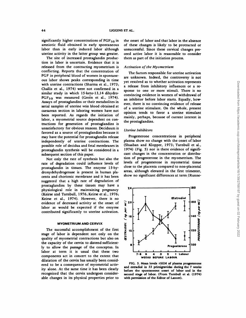
Progesterone concentrations in peripheral
plasma show no change with the onset of labor
(Shaaban and Klopper, 1973; Turnbull et al.,
1974) (Fig. 5) nor is there evidence of signifi-
cant changes in the concentration or distribu-
tion of progesterone in the myometrium. The
levels of progesterone in myometrial tissue
close to the placenta compared to non-placental
areas, although elevated in the first trimester,
show no significant differences at term (Runne-
140 ‘-
*20
Dow
nloaded from https://academ
ic.oup.com/biolreprod/article/16/1/39/2768197 by guest on 02 February 2022

PARTURITION IN MAN 45
baum and Zander, 1971). Further studies,
particularly of progesterone receptor-site con-
centrations in various parts of the uterine
muscle, are needed before the possibility of
changes in local concentrations of progesterone
in the myometrium can be dismissed. But even
were local changes observed, their significance
would be impossible to assess in the absence of
evidence of a direct inhibitory influence of
progesterone on the human myometrium in
vivo. There has been a consistent failure of
attempts to inhibit uterine activity with large
doses of progesterone or medroxyprogesterone
whether administered intramuscularly, intrave-
nously, intraamniotically or intramyometrially
(Hendricks et a!., 1961; Wood et al., 1963).
This failure is understandable if progesterone
receptors are fully saturated at normal plasma
progesterone concentrations but it follows from
this that a reduced influence of progesterone on
the myometrium will occur only when the
concentration of receptors falls or when proges-
terone is displaced from the binding sites;
neither of these changes has been described.
Interest is now turning to progesterone binding
in uterine tissues other than myometrium
where progesterone may take part in the
regulation of prostaglandin biosynthesis
(Schwarz et al., 1974).
Relaxin has passed through a long phase of
obscurity since clinical traits of porcine relaxin
in women in labor failed to show consistent
effects. For several reasons, previous studies
cannot be regarded as evidence that relaxin is of
no importance in women. First, relaxin may be
species-specific and porcine relaxin may be
inactive in man (Bryant, 1972). Second, many
of the clinical studies used doses that may have
been inadequate (Slate and Mengert, 1960).
Third, the method of quantifying responses in
women in labor may have been inappropriate if
relaxin has little effect (as is the case with the
guinea pig) on the responsiveness of the myo-
metrium to agents such as oxytocin and prosta-
glandin (Porter, 1972). Renewed interest stems
from the development of a radioimmunoassay
for human relaxin (Bryant, 1972) and it may
now be possible to establish whether relaxin
plays any part in the physiology of human
parturition.
Some of the synthetic betamimetic amines
such as ritodrine and salbutamol are remarkably
effective in inhibiting labor (Landesman et al.,
1971; Liggins and Vaughan, 1973) but there is
no evidence that the uterus normally is strongly
inhibited by endogenous cateholamines. The
catecholamine content of the pregnant uterus is
relatively low (Cha et al., 1965). Hypertensive
pregnant women treated with large doses of the
beta blocker, propranolol, are not prone to
premature labor (G. C. Liggins, unpublished
observations). Assays of urinary catecholamines
have not revealed changes at the onset of labor
although there is probably an increase in
excretion of norepinephrine during labor
(Goodall and Diddle, 1971; Kudo and Rouse,
1970). There is insufficient information at
present to define the role of the nervous system
and of catecholamines in human parturition. It
seems unlikely that they play a major part in
the initiation process but it is possible that
neural mechanisms modulate uterine activity to
the extent of influencing both the time of day
when labor starts and the intensity of uterine
contractions.
Uterine Stimulants
Evidence for the involvement of oxytocin in
human parturition is conflicting. On the one
hand, sensitive radioimmunoassays detect oxy-
tocin in peripheral blood in the early first stage
of labor in only about 10 percent of patients
and measurements of intramammary pressure
usually show no changes during labor (Cobo,
1968; Chard, 1973). The frequency of detec-
tion of oxytocin by radioimmunoassay in-
creases as labor progresses and is maximal in the
second stage. On the other hand, intravenous
alcohol inhibits premature labor when the
membranes are intact and the cervix is less than
3 cm dilated (Fuchs et a!., 1967). The effect of
ethanol has been attributed to inhibition of
oxytocin release from the posterior pituitary.
This proposal is supported by the observation
that there was parallelism in oxytocin dose-
response curves obtained before and during
ethanol infusion in five out of ten women at
term (Mantell and Liggins, 1970). The sensitiv-
ity of existing assays of oxytocin is insufficient
to examine the effects of ethanol by direct
means. Present evidence does not suggest that
oxytocin in the maternal circulation is impor-
tant in initiating labor but it is consistent with a
role in the maintenance of established labor.
There may be an interaction of prostaglandin
and oxytocin since there is a progressive en-
hancement of oxytocin response during pro-
longed intravenous infusion of subthreshold
doses of PGF2a (Hutton and Liggins, unpub-
Dow
nloaded from https://academ
ic.oup.com/biolreprod/article/16/1/39/2768197 by guest on 02 February 2022

46 LIGGINS ET AL.
CGalactosamine is contained only in chondroitins and dermatan sulfate.
lished observations).
The acute effects of estrogen on the human
myometrium are the subject of controversy.
Jarvinen et a!. (1965), Pinto et al. (1966) and
Larsen et a!. (1973) were able to stimulate
uterine contractions but not labor by adminis-
tering large doses of estrogen to women at
term. These reports together with the finding
that estrogen treatment caused a marked in-
crease in uterine activity in a castrated woman
(Moawad and Bengtsson, 1968) suggested that
estrogen is oxytocic. Similar observations were
made in vitro using rats or rabbits pretreated
with estrogen (Melton and Saldivar, 1966;
Csapo, 1969). The effect of estrogen appears to
be to enhance propogation of electrical im-
pulses over the muscle. However, estrogen
applied directly to rat uterine strips in an organ
bath abolishes electrical and mechanical activity
(Melton and Saldivar, 1966). These apparently
paradoxical results are explained in part by the
effects of estrogen in reversing castration
smooth muscle atrophy but an apparently
oxytocic action in vivo remains unexplained. In
view of recent work linking prostaglandin
synthesis with estrogen, consideration should
be given to the possibility that the effects of
estrogen on propogation and contractility are
mediated by prostaglandins.
Uterine stimulating effects of prostaglandins
are considered elsewhere. Of the various other
biological agents with oxytocic activities such
as serotonin, norepinephrine, kinins and angio-
tensin II, none has been implicated in the
initiation of labor.
Cervix
In the last few days or weeks of pregnancy
the cervix changes from a long, firm, closed
structure to one that is short, soft and distensi-
ble. The structural changes that accompany the
altered macroscopic features have been de-
scribed by Danforth et al. (1974). Compared to
the non-pregnant cervix, the connective tissue
of the postpartum cervix shows features that
are consistent with disolution of the collagen
framework. There is a slight increase in water
content and a marked decline in the content of
collagen and glycoprotein (Table 1). In addition
to an absolute loss of collagen, the bundles are
widely separated and probably are permitted to
slide upon one another (Fig. 6). Marked
changes are found in the ground substance of
the postpartum cervix. The amount of gly-
cosaminoglycans is greatly increased and their
composition is changed. The nonpregnant cer-
vix contains little uronic acid-free glycosamino-
glycan (usually considered to be keratin sul-
phate) whereas the postpartum cervix contains
a substantial amount. Glycoproteins are mark-
edly reduced. In addition, Danforth et al.
(1974) found in the dilated cervix a consider-
able quantity of an unidentified material, possi-
bly a glycosaminoglycan, which is almost ab-
sent from the nonpregnant cervix.
Neither the source of the proteases and
collagenases that must be responsible for the
altered collagen structure of the softened cervix
nor the stimulus to their activity is known. In
mice, treatment with estradiol or relaxin in-
duces loosening and scattering of the collagen
fibrils (Leppi and Kennison, 1971). In women,
there is some indirect evidence to suggest a
relationship between estrogen and cervical soft-
ening. Prelabor cervical changes are normally
occurring at a time when plasma estradiol is
rising to maximal levels (Turnbull et al., 1974)
(Fig. 5). In sulfatase deficient pregnancies with
very low estrogen levels, the cervix may remain
small, firm and closed (France et al., 1973). In
the sheep, local application (Liggins et al.,
1976) or arterial infusion (Fitzpatrick, 1976) of
PGF21� can cause softening and dilatation of
TABLE 1. Composition of glycosaminoglycans in human cervix (micromoles per gram of dry weight of defattedtissues). From Danforth et al. (1974).
Cervix
Hexo-
saminesaUronic
acidbGalacto-
saminecKeratin
sulfate
Nonpregnant 14.6 10.7 9.9 3.9
Pregnant 38.9 7.7 17.5 31.2
aHexosamine is contained in all glycosaminoglycans.
b�ronic acid is contained in all glycosazninoglycans except keratin sulfate.
Dow
nloaded from https://academ
ic.oup.com/biolreprod/article/16/1/39/2768197 by guest on 02 February 2022

FIG. 6. Cervix approximately 1cm above plane of external os. Stained with Mulligan’s triichrome stain forcollagen. On left, nonpregnant. On right, immediately postpartum. X 835. (From Danforth et a!. (1960) with
permission of the Editor of the American Journal of Obstetrics and Gynecology).
PARTURITION IN MAN 47
the cervix and in women there are reports of
‘ripening’ of the cervix by means of prostaglan-
din treatment (Calder and Embrey, 1973; Weiss
et al., 1975). In the latter studies, no attempt
was made to assess the contribution that
enhanced uterine activity might have made to
the cervical changes. It is difficult to separate
cervical and myometrial effects of prostaglan-
dins since the continued administration of small
doses of prostaglandins usually leads to an
increase in uterine activity. However, in one
study of 20 women at term in whom uterine
activity was monitored during intravenous infu-
sion of subthreshold doses of PGF2�, marked
cervical changes were observed in the absence
of uterine contractions in three patients (Lig-
gins et al., 1976).
Certain clinical conditions suggest that myo-
metrial activity and cervical changes can be
dissociated under certain abnormal circum-
stances. On the one hand, strong uterine con-
tractions may cause complete annular detach-
ment of an unyielding cervix (Jeffcoate, 1952).
On the other hand, the ‘incompetent’ cervix
may dilate more or less completely in the
absence of contractions.
Although we are almost completely ignorant
of the physiological control of the pregnant
cervix, two principles are clear. First, that
cervical softening in pre!abor must be explained
in biochemical terms. And second, that an
hypothesis for the initiation of human labor is
incomplete unless it includes a satisfactory
explanation for the structural changes in the
cervix.
Mid-trimester Abortion
Some insight into the mechanisms of labor
at term can be gained from induced midtri-
mester abortion in which the whole process of
parturition is accomplished within 24 h at a
time when pregnancy is at its most stable. A
variety of techniques are effective in inducing
abortion, a notable exception being oxytocin
which fails to induce abortion unless given in
massive doses late in the midtrimester (Burnhill
et a!., 1962). Mechanical (bougies and bags),
Dow
nloaded from https://academ
ic.oup.com/biolreprod/article/16/1/39/2768197 by guest on 02 February 2022
48 LIGGINS ET AL.
physical (hypertonic solutions of sodium chlo-
ride, urea or glucose), chemical (dilute formalin
and Uter’s paste), bacteriological (inadvertent
amnionitis) and pharmacological (prostaglan-
dins) agents placed intraovularly or extraovu-
larly cause abortion with great consistency
(see Gustavii, 1973 for review). Furthermore,
the uterus of patients who fail to abort after
such interventions is usually found to have
become more responsive to oxytocin to which
it responds with cyclical activity and abor-
tion.
There is no agreement on the mechanisms
involved in the initiation of induced abortions.
Rapid expansion of amniotic fluid volume
(Csapo, 1969) fetal death (Kov#{225}cs, 1970) and
placental damage (Weist et al., 1970) have all
been proposed. Degenerative changes in the
placenta in saline-induced abortions has been
described (Wynn, 1965; Jakobovits et al., 1970)
and may be the basis of the modest fall in the
concentration of progesterone in maternal plas-
ma at the time when uterine activity is
established some hours after injection of the
hypertonic solution (Csapo et al., 1969). Pla-
cental damage and a fall in progesterone pro-
duction are less likely to result from methods
of induction that invade only the extraovular
space. In these circumstances, as well as when
hyperosmolar or irritant solutions are placed in
the amniotic sac, the tissues most vulnerable to
damage are the fetal membranes and the decid-
uum. Marked degenerative changes in decidua
have been described after intraamniotic injec-
tions of hyperosmolar saline (Gustavii, 1973;
Vassilakos et al., 1973).
Gustavii (1973) suggested that the various
methods of inducing abortion had in common
as their mechanism of action the release of
prostaglandins from the decidua. Experiments
designed to substantiate the role of prosta-
glandin release have had a measure of suc-
cess. Aspirin significantly extends the injec-
tion-abortion interval in nulliparous women
treated with intraamniotic urea (Niebyl et al.,
1976) and indomethacin has a similar effect
in saline-induced abortion (Waltman et al.,
1973).
Induced abortion cannot be regarded as an
experimental model for spontaneous parturi-
tion at term. Nevertheless it serves a useful
function in drawing attention to the possible
importance of a local intrauterine system that
may provoke parturition without fetal or pla-
cental participation.
DECIDUA AND FETAL MEMBRANES
Decidua and fetal membranes probably are
able to synthesize prostaglandins and may be
the source of prostaglandins that accumulate in
amniotic fluid during labor. Sykes et al. (1975)
found a low rate of synthesis of PGF and PGE
when fresh deciduum or myometrium was
incubated in vitro in a medium containing
reduced glutathione. Addition of arachidonic
acid or phospholipase A2 (E.C.3.1.1.4) to the
incubates increased PG production, particularly
that of PGE. Keirse and Turnbull (1976)
demonstrated synthesis of PGE during incuba-
tion of chorion but not of amnion. Chorionic
membrane usually includes some decidual tissue
(Gustavii and Brunk, 1972) which could be the
site of prostaglandin production attributed to
chorion. Further studies comparing the rates of
decidual and chorionic production of prosta-
glandins under identical conditions are needed
to resolve this question. The use of decidua-
free chorion such as can be obtained from
binovular twin pregnancies will be desirable.
The fetal membranes and deciduum are
uniquely suited to prostaglandin production
since all of these tissues contain glycerophos-
pholipids that are highly enriched with arachi-
donic acid, the obligatory precursor of PGF2a
and PGE2. Schwarz et al. (1975) noted that the
20 percent of the total fatty acid content of
chorioamnion was arachidonic acid compared
with 0.4 percent in maternal peritoneum. Dc-
ciduum is similarly enriched with arachidonic
acid. The arachidonic acid content of the fetal
membranes at term could meet many times
over all the substrate requirements for prosta-
glandin synthesis in labor (Schwarz et a!.,
1975).
The incorporation into amnion phospho-
lipids of arachidonic acid relative to palmitic
acid has been studied in vitro by Schwartz et al.
(1976b). Palmitic acid is the most abundant
fatty acid of amniotic fluid and is not a
precursor of prostaglandins. They found that
arachidonic acid incorporated into phospho-
lipids is contained mainly (53 percent) in
lecithin and to a lesser extent (27 percent) in
phosphatidyl ethanolamine. This distribution is
similar to that of lecithin and phosphatidyl
ethanolamine in term amnion (Pritchard et a!.,
1968). However, whereas the relative incorpora-
tion of palmitic acid and arachidonic acid into
phosphatidyl ethanolamine is in accord with
fatty acid composition, as described by Robert-
Dow
nloaded from https://academ
ic.oup.com/biolreprod/article/16/1/39/2768197 by guest on 02 February 2022

PARTURITION IN MAN 49
‘Mean percent arachidonic acid ± SD.
son and Sprecher (1968), the incorporation of
arachidonic acid into lecithin greatly exceeds
the amount expected from the latter’s composi-
tion. This suggests that the turnover of arachi-
donic acid relative to palmitic acid is high in
lecithin. The factors that regulate fatty acid
turnover in fetal membranes and cause incorpo-
ration of arachidonic acid in preference to
other unsaturated fatty acids are unknown.
Hormones contained in the amniotic fluid need
to be considered in this regard. Cortisol (Joli-
vet, 1972) prolactin and vasopressin (Manku et
a!., 1975) are all present in amniotic fluid and
can induce marked changes in the permeability
to water of the fetal membranes. In addition,
amniotic fluid contains oxytocin and conju-
gated oestrogen. The membranes contain sulfat-
ase activity that could liberate unconjugated
oestrogens in the membranes and deciduum
(Warren and Timberlake, 1962).
The pattern of fatty acid content of, and
incorporation into, fetal membranes at various
stages of pregnancy has not been described.
However, the fatty acid composition of phos-
pholipids in amniotic fluid described by Das et
a!. (1975) suggests that specific incorporation
of arachidonic acid occurs late in pregnancy
(Table 2).
The means of liberating arachidonic acid
from storage in phospholipids must be available
in a tissue that is to serve as an effective source
of substrate for prostaglandin synthesis. Since
arachidonic acid is contained mainly in the
2-acyl position of glycerophospholipids, a phos-
pholipase A2 is required for hydrolysis. Term
amnion (Grieves and Liggins, 1976), chorio-
amnion and deciduum (Schultz et a!., 1975)
contain phospholipase A2 activity that has
specificity for phospholipids containing arachi-
donic acid (Schultz et a!., 1975). The specific
activities in the various tissues was compared by
Grieves and Liggins (1976) who found the
greatest activities in deciduum and amnion; the
activities of chorion and myometrium were
substantially less (Fig. 7). They found no
differences between tissues obtained before and
during labor.
In many tissues, phospholipases are lyso-
somal enzymes and their activity depends on
release from the lysosome. This led Gustavii
(1972) to propose a ‘lysosomal theory’ of
parturition in which a key role in the initiation
of parturition is attributed to lysosomes of the
deciduum. According to Gustavii, the deciduum
becomes rich in lysosomes which are main-
tained in a stable state by the presence of
stabilizers particularly progesterone. The onset
of labor or abortion is precipitated by labiizing
influences that cause leakage of lysosomal
enzymes, including phospholipase A2, into the
cytoplasm. The increased activity of phospho-
lipase A2 causes accelerated deacylation of
phospholipids at the sn-2 position which leads
in turn to the release of fatty acids including
arachidonic acid. Since the synthesis of prosta-
glandins probably is not rate-limited by prosta-
glandin synthetase (Pace-Asciak and Wolfe,
1970), an increase in arachidonic levels results
in increased prostaglandin production. The
prostaglandin released from the deciduum is
postulated as diffusing partly into the myome-
trium which it activates, and into the amniotic
fluid where a rise in concentration is observed.
Evidence is accumulating to support this
hypothesis. The lysosomes of decidual cells are
unusually fragile and leak their contents when
subjected in vitro to slight physical stresses
(such as hypo- or hyperosmolar conditions)
that have no discernible effect on other tissues
(Brunk and Gustavii, 1973). Within two hours
of injection of hypertonic saline into the
amniotic sac, marked degenerative changes that
preceed any fall in maternal progesterone levels
are observed in the decidua parietalis (Vassi-
lakos et al., 1974). Even an isotonic solution
(0.9 percent NaCI) induces abortion when
TABLE 2. Arachidonic acid composition of lecithin and sphingomyelin of human amniotic fluid at differentperiods of gestation (Das et al., 1975).
Phospholipid
Duration o f pregnancy
18-22 wk 27-3 3 wk 34-40 wk Term labor
Lecithin 6.3 ± 0.1’ 11.5 ± 0.2 0.8 ± 0.01 10.6 ±0.1
Sphingomyelin 3.3 ± 0.07 1.5 ± 0.0 1.7 ± 0.06 30.4 ± 4.2
Dow
nloaded from https://academ
ic.oup.com/biolreprod/article/16/1/39/2768197 by guest on 02 February 2022

DECIDUA AMNION CHORION MYOMETRIUM35
30
25
20
15
10
5
FIG. 7. Activity in human tissues of phospholipase A2 according to pH. The tissues were obtained atcaesarean section during labor. Phosphatidyl ethanolamune containing t� HI -arachidonic acid at the sn-2 position
was incubated at 37#{176}Cwith crude lysosomal fractions. The amount of substrate hydrolyzed was calculated fromthe ratio of total counts in the reaction mixture to the counts in the purified fatty acid fraction. o-o 15
mm incubation; S-#{149} 2 h incubation.
45678 678 4 5 6 7 8
pH45678
50 LIGGINS ET AL.
z
I-
0
0
In
0
0>.
I
Id
I-In
In
injected outside the membranes (Gustavii,
1974). Decidual cells obtained at elective cae-
sarean section at term show marked degener-
ative changes (Fig. 8) and signs of release of the
lysosomal enzyme, acid phosphatase, into the
cytoplasm (Gustavii, 1975). Thus, the first step
in the biosynthesis of prostaglandins-the re-
lease of phospholipase A2 from lysosomes-is
active prior to the onset of labor or abortion.
There is evidence also that the next step-the
release of free arachidonic acid-is enhanced
during labor. MacDonald et al. (1974) com-
pared the concentration of free arachidonic
acid in amniotic fluid from women before and
during labor. They found a fourfold increase of
both arachidonic acid and PGF. Other fatty
acids increased less strikingly. The third step-
conversion of arachidonic acid to PGF2 a-has
been demonstrated by MacDonald et al. (1974).
Injection of 1.2g of potassium arachidonate
into the amniotic sac induces abortion whereas
oleate is inactive. The arachidonate-induced
abortion is prevented by simultaneous ingestion
of aspirin. These workers suggest that the large
dose of arachidonate is needed because of
metabolism of PGF2a by the fetal membranes.
Finally, the last step-activation of the myome-
trium by prostaglandin from within the
uterus-is a well known effect of intra-amniotic
or extra-ovular administration of PGF2a or
PGE2.
Not only the deciduum but also the fetal
membranes have features that make them par-
ticularly well suited to take part in the biosyn-
thesis of prostaglandins. As already described,
there are some reservations about the metabolic
activities of the chorion because of its struc-
tural inhomogeneity but amnion is readily
obtainable without contamination by other
tissues. Studies of the amnion show it to be
incapable of prostaglandin synthesis or degrada-
tion (Keirse and Turnbill, 1976) but in other
respects it has several similarities to deciduum.
The amnion, unlike the chorion, is an unusually
fragile tissue, undergoing marked cell lysis
during brief incubation (1 h) in 0.9 percent
NaCI (Schwartz et al., 1976a). To maintain the
structural and metabolic integrity of the am-
nion, incubation media closely resembling
amniotic fluid are required (Fig. 9). Despite the
small fraction of the amnion that is cellular, it
is a highly metabolically active tissue (Schwartz
et a!., 1976a), the rate of glucose utilization
exceeding that of muscle (Baltzan et al., 1962),
Dow
nloaded from https://academ
ic.oup.com/biolreprod/article/16/1/39/2768197 by guest on 02 February 2022

FIG. 8. Decidual cells stained with hematoxylin and eosin. On left, tissue from midpregnancy. On right,
tissue from term pregnancy showing cellular degeneration. X910. (From Gustavii (1975) with permission of the
Editor of the British Journal of Obstetrics and Gynaecology).
PARTURITION IN MAN 51
red blood cells (Jandl, 1965) and fetal brain
(Adam et al., 1975). The activity of phospho-
lipase A2 in amnion is equal to that of decidua
and greatly exceeds that of chorion and myo-
metrium (Grieves and Liggins, 1976). Some of
the amnion glycerophospholipids, especially
phosphatidyl ethanolamine, are highly enriched
in arachidonic acid (Robertson and Sprecher,
1968) and the turnover of arachidonic acid
relative to palmitic acid in lecithin is rapid
(Schwartz et al., 1976b). These characteristics
of amnion suggest that it has a considerable
potential for generation of arachidonic acid
although it lacks the capacity to convert arachi-
donic acid to prostaglandins.
The properties of fetal membranes, de-
ciduum and myometrium that are pertinent to
prostaglandin synthesis are summarized in Ta-
ble 3. When considered together, they suggest
that the fetal membranes and deciduum may
function as a unit, the membranes serving as a
substantial source of arachidonic acid for pros-
taglandin synthesis in the deciduum and possi-
bly the chorion.
The factors promoting selective incorpora-
tion of arachidonic into glycerophospholipids
and, more importantly, the factors stimulating
release of arachidonic acid are unknown. Gus-
tavii (1975) suggested that progesterone stabi-
lizes lysosomes thereby preventing release of
phospholipase A2. Schwarz et al. (1974) inves-
tigated progesterone binding in subcellular frac-
tions of homogenates of chorioamnion and
found evidence consistent with Gustavii’s pro-
posal. Labeled progesterone was found in high-
est concentration in lysosomal fractions and
was not extractable with high concentrations of
salt. Moreover, they found that the cytosol
contains a progesterone-binding protein in low
concentration in fetal membranes before 38weeks of pregnancy but in much higher concen-
tration at term. It differs from cytosol proges-
terone receptors and transcortin. This led
Schwarz et a!. (1974) to propose that a specific
progesterone binding protein appears in the
cytosol of the fetal membranes near term and
competes with lysosomes for progesterone. As a
consequence, the lysosomes become more un-
Dow
nloaded from https://academ
ic.oup.com/biolreprod/article/16/1/39/2768197 by guest on 02 February 2022

52 LIGGINS ET AL.
I’4.
4
A4
I
tt
I
pa
�Sykes et al. (1975).
e
$4
FIG. 9. Amniotic membrane obtained at caesarean section at term and stained with hematoxylin and eosin.On left, tissue fixed immediately. On right, tissue incubated at 37#{176}Cfor 2 h in 0.9% sodium chloride solution
before fixation. X400.
stable and their contents leak out. Thus, in
effect, there is a local withdrawal of progester-
one in the membranes independent of maternal
plasma levels. The cause for the rapid accumula-
tion of progesterone binding protein is un-
known.
TABLE 3. Comparison of factors related to prostaglandin production in uterine tissues.
Myo-Amnion Chorion Deciduum metrium
Tissue ‘fragility’Phospholipase activity
b�d
�c#{247}�d
b
�d
Enrichment of phospholipids with arachidonic acid
Prostaglandin synthesis
Prostaglandin degradation
++a,e
-�‘
�f
++?e
�pf++f
#{247}+e
�g
�g
p
�g
�g
aSchwartz et al. (1976).
bSChwattz and Liggins, unpublished observations.
CBk and Gustavii (1973).
dGi and Liggins (1976).
eShwa� et al. (1975).
�Keirse and Turnbull (1976).
Dow
nloaded from https://academ
ic.oup.com/biolreprod/article/16/1/39/2768197 by guest on 02 February 2022
PARTURITION IN MAN 53
TWO MECHANISMS OF LABOR?
The forgoing discussion raises the question
of whether there might, in any given species, be
two mechanisms of labor-one in which the
fetal adrenal is important and another which
depends on an interaction between fetal mem-
branes and deciduum. According to species, one
or other system might be dominant. In some
species such as sheep, the fetal role is dominant
but in others such as nonhuman primates and
humans, the latter system might be the more
important. The local ‘membrane mechanism’ is
seen as being a biological clock, able to deter-
mine a rather crude species-specific life span to
pregnancy. Superimposed on the ‘membrane
mechanism’ is a more refined system (the ‘fetal
adrenal mechanism’) that adds greater accuracy
to the timing of pregnancy duration. It should
be added that yet another system (the ‘mater-
nal mechanism’) may further refine the time of
onset of labor. The three mechanisms might
respectively determine the week, the day and
the hours of birth.
It is possible that the membrane mechanism
is merely a part of the fetal adrenal mechanism.
On the other hand it might represent a matura-
tional event, timed by the same sort of geneti-
cally-controlled clock that operates in maturing
fetal organs. The rapid, precisely-timed appear-
ance of an enzyme in, say, fetal liver is readily
accepted as a normal part of maturation. Why
not in fetal membranes? Maternal and fetal
hormones might do no more than wind up the
clock.
A local membrane mechanism that is not
necessarily dependent on extraneous hormonal
influences is consistent with findings in several
species. Reference has been made already to
human anencephalic pregnancy in which mean
gestation length is approximately 40 weeks but
the range is very wide. A similar observation
was reported by Novy (1976) who hypophysec-
tomized fetal rhesus monkey and found that a
third were born prematurely, a third were born
at term and the remaining third were born
beyond term. A local mechanism is consistent
also with widely spaced births of human twins
in bicornuate uteri and with the observation
that when premature labor is induced by fetal
infusion of ACTH in sheep with twin lambs, the
infused fetus invariably is born first (Liggins et
a!., 1976). Asymmetrical delivery was induced
in rabbits by Costa and Csapo (1959) bydislocating one placenta in one horn of animals
ovariectomized on Day 25; administration of
oxytocin 9 h later caused delivery of all the
fetuses in the horn containing the dislocated
placenta whereas pregnancy was maintained in
the other horn. Fetectomy experiments also are
possibly relevant to this question. Dr. Lanman
has described in his paper the results of his
fetectomy experiments in rhesus monkeys. In
the mouse, Newton (1935) fetectomized 20
animals in groups of five on Days 12, 13, 14and 15 of pregnancy and observed parturition
on Days 16-23. Twelve rats fetectomized be-
tween Days 9 and 13 delivered the placenta
8-12 days later (Selye et al., 1935). The
consistent feature of these experiments is that
pregnancy continues for some time after fetec-
tomy. Delivery occurs very roughly at term but
with a wide range compared to intact preg-
nancies.
Labor in human pregnancy may be initiated
mainly by a genetically-controlled maturational
signal arising in the fetal membrane and ex-
pressing itself through an interaction with the
deciduum; fetal and maternal hormone may
play a lesser, dispensible part.
ACKNOWLEDGMENT
The work was supported by the New ZealandMedical Research Council.
REFERENCES
Adam, P. A. J., R�ihil, N. C. IL, Raihala, E. L andKekomaki, M. P. (1975). Oxidation and glucose
and I)-13-OH-butyrate in the early human fetalbrain. Acta Paediat. Scand. 64, 17-24.
Anderson, A. B. M., Laurence, K. M., Davies, K.,Campbell, H. and Turnbull, A. C. (1971). Fetaladrenal weight and the cause of premature deliveryin human pregnancy. J. Obstet. Gynaec. Br. Com-monw. 78,481-488.
Baltzan, M. A., Andres, P. and Cader, G. (1962).
Heterogeneity of forearm metabolism with specialreference to free fatty acids. J. (un. Invest. 41,116-125.
Brunk, U. and Gustavii, B. (1973). Lability of human
decidual cells. In vitro effects of autolysis andosmotic stress. Am. J. Obstet. Gynec. 115,81 1-816.
Bryant, G. D. (1972). The detection of relaxin inporcine, ovine and human plasma by radioimmuno-assay. Endocrinology 91, 1113-1117.
Burnhill, M. S., Gaines, J. A. and Guttmacher, A. F.
(1962). Concentrated oxytocin solution for thera-peutic interruption of midtriinester pregnancy.Obstet. Gynec. 20, 94-100.
Calder, A. and Embrey, M. P. (1973). Prostagiandins
and the unfavourable cervix. Lancer, 2, 1322-1323.
Cha, K. S., Lee, W. C., Rudzik, A. and Miller, J. W.
(1965). A comparison of the catecholamine con-centrations of uteri from several species and the
Dow
nloaded from https://academ
ic.oup.com/biolreprod/article/16/1/39/2768197 by guest on 02 February 2022

54 LIGGINS ET AL.
alterations which occur during pregnancy. J.Pharm. Exp. Ther. 148, 9-13.
Challis, J. K. G., Osathanodh, R., Ryan, K. J. andTulchinsky, D. (1974). Maternal and fetal plasmaprostaglandin levels at vaginal delivery and caesar-
ean section. Prostagiandins 6, 281-286.
Chard, T. (1973). The posterior pituitary and the
induction of labor. Mem. Soc. Endocrinol. 20,
6 1-76.
Chard, T. (1976). The fetal hypothalamus and poste-
rior pituitary in the initiation of labor. In ‘TheFetus and Birth’. Ciba Foundation Symposium No.
47 (M. O’Connor and J. Knight, eds.). Elsevier/
Excerpta Medica, Amsterdam (in press).
Cobo, E. (1968). Uterine and milk-ejecting activities
during human labor. J. AppI. Physiol. 24, 3 17-323.
Costa, L. M. and Csapo, A. (1959). Asymmetricaldelivery in rabbits. Nature 184, 144-146.
Csapo, A. 1. (1969). The four direct regulatory factors
of myometrial function. In ‘Progesterone: its regu-
latory effect on the myometrium’. Ciba Founda-
tion Study Group No. 34 (G. E. W. Wolstenholme
and J. Knight, eds) pp. 13-42. Little, Brown and
Co., Boston.
Csapo, A. 1. (1973). The regulatory interplay of
progesterone and prostaglandin F20 in the control
of the pregnant uterus. In ‘Uterine contractions-side effects of steroidal contraceptives (J. B.
Josimovich, ed.) pp. 223-255. Wiley-Interscience,New York.
Csapo, A. I., Pulkkinen, M. 0. and Weist, W. G.
(1973). Effects of luteectomy and progesteronereplacement therapy in early pregnant patients. J.Obstet. Gynec. 115, 759-765.
Currie, W. B., Wong, M. S. F., Cox, R. I. andThorbum, G. D. (1973). Spontaneous or dexa-methasone induced parturition in the sheep and
goat: changes in plasma concentrations of maternalprostaglandin F and foctal oestrogen sulphate.Mem. Soc. Endocrinol. 20, 95-118.
Danforth, D. N., Buckingham, J. C. and Roddick, J.
W. (1960). Connective tissue changes incident tocervical effacement. Am. J. Obstet. Gynec. 80,
93 9-94 5.Das, S. K., Foster, I-I. W., Adhikary, P. K., Mody, B. B.
and Bhattacharyya, D. K. (1975). Gestationalvariation of fatty acid composition of human
amniotic fluid lipids. Obstet. Gynec. 45, 425-432.
Fend, M.de M. and Tulchinsky, D. (1975). Total
cortisol in amniotic fluid and fetal lung matura-tion. New Eng. J. Med. 292, 133-136.
Fitzpatrick, R. J. (1976). Effects of prostaglandin F�0on the cervix of the pregnant sheep. In ‘The Fetus
and Birth’. Ciba Foundation Symposium No. 47
(M. O’Connor and J. Knight, eds.) Elsevier/Excerpta Medica, Amsterdam (in press).
Franenkl, L. (1905). Die funktion des corpus luteum.Arch. Gynaek. 68, 438-535.
France, J. T., Seddon, R. J. and Liggins, G. C. (1973).
A study of a pregnancy with low oestrogenproduction due to placental sulfatase deficiency. J.Clin. Endocrinol. Metab. 36, 1-9.
Fuchs, F., Fuchs, A. R., Poblete, V. F. and Risk, A.
(1967). Effect of ethanol on threatened premature
labor. Am. J. Obstet. Gynec. 99, 627-637.
Gamissans, 0., Pujol-Ainat, P., Davi, E., P#{233}rez-Picanol,E. and Wilson, G. K. (1975). The effect of
intra-amniotic administration of $3-metasone on theonset of labor in human pregnancy. Acta Endo-
crinol (Kbh) Suppl. 199, 95.
Goodall, M. and Diddle, A. W. (1971). Epinephrineand norepinephrine in pregnancy. Am. J. Obstet.Gynec. 111, 896-904.
Gramstrom, E. (1972). On the metabolism of prosta-
glandin F20 in female subjects. Structures of two
metabolites in blood. Europ. J. Biochem. 27,462-468.
Green, K., Bygdeman, M., Toppozada, M. and Wiquist,
N. (1974). The role of prostaglandin F20 in human
parturition. Endogenous plasma levels of 1 5-keto-
13, 14-dehydro-PGF20 during labor. Am. J. Oh-
stet. Gynec. 120, 25-31.
Grieves, S. A. and Liggins, G. C. (1976). Phospholipase
A activity in human and ovine uterine tissues.
Prostaglandins 12, 229-241.
Gustavii, B. (1972). Labor: a delayed menstruation?
Lancet 2, 1149-1150.
Gustavii, B. (1973). Studies on the mode of action of
intra-amniotically and extra-amniotically injectedhypertonic saline in therapeutic abortion. Acta
Obstet. Gynec. Scand. Suppl. 25, 5-22.Gustavii, B. (1974). Sweeping of the fetal membranes
by a physiologic saline solution:effects on decidualcells. Am. J. Obstet. Gynec. 120, 531-536.
Gustavii, B. (1975). Release of lysosomal acid phos-
phatase into the cytoplasm of decidual cells beforethe onset of labor in humans. Br. J. Obstet.
Gynaec. 82, 177-181.
Gustavii, B., and Brunk, U. (1972). A histological
study of the effect on the placenta of intra-amnio-tically and extra-amniotically injected hypertonic
saline in therapeutic abortion. Acts Obstet. Gynec.
Scand. 51, 121-125.
Harris, R. G. (1927). Effect of bilateral ovariectomyupon the duration of pregnancy in mice. Anat.
Rec. 37, 126.I-leap, R. B. and Deanesly, K. (1966). Progesterone in
systemic blood and placentac of intact and ovariec-tomized pregnant guinea-pip. J. Endocrinol. 34,417-423.
Hendricks, C. H., Brenner, W., Gabel, R. and Kerenyi,
T. (1961). The effect of progesterone administered
intraamniotically in late human pregnancy. InBrook Lodge Symposium: Progesterone (A. C.Barnes, ed.) pp. 53-64. Brook Lodge Press, Au-
gusta, Michian.
Hillier, K., Calder, A. A. and Embrey, M. P. (1974).
Prostaglandin F20 and parturition. Concentrations
in amniotic fluid and plasma in spontaneousdelivery and induced labors. J. Obstet. Gynec. Br.
Commonw. 81, 257-263.
Honnebier, W. J. and Swaab, D. F. (1973). The
influence of anencephaly upon intrauterine growth
of fetus and placenta and upon gestation length.Br. J. Obstet. Gynaec. 80, 577-588.
Jakobovits, A., Traub, A., Farkas, M. and Morway, J.(1970). The effect of intra-amniotic injection ofhypertonic saline on the structure and endocrine
function of the human placenta. lnt. J. Gynaec.Obstet. 8, 497-506.
Jandl, J. H. (1965). Leaky red cells. Blood 26,36 7-3 82.
Jilrvinen, P., Lukkainen, T. and Vaisto, L. (1965). Theeffect of oestrogen treatment on myometrial activ-
Dow
nloaded from https://academ
ic.oup.com/biolreprod/article/16/1/39/2768197 by guest on 02 February 2022

PARTURITION IN MAN 55
ity late in pregnancy. Acta Obstet. Gynec. Scand.
44, 258-264.Jeffcoate, T. N. A. (1952). Annular detachment of the
cervix. J. Obstet. Gynaec. Br. Emp. 59, 327-335.
Jolivet, A. (1972). Cortisol in amniotic fluid and
human parturition. In Abstracts of 4th mt. Cong.
Horm. Steroids. (K. 0. Scow, ed.) p. 367. ExcerptsMedics, Amsterdam.
Keirse, M. J. N. C., Flint, A. P. F. and Tumbull, A. C.
(1974). F Prostagiandins in amniotic fluid during
pregnancy and labor. J. Obstet. Gynaec. Br. Com-monw. 81, 131-135.
Keirse, M. J. N. C., Hicks, B. IL and Turnbull, A. C.
(1976). Prostaglandin dehydrogenase in the pla-
centa before and after the onset of labor. Br. J.Obstet. Gynaec. 83, 152-155.
Keirse, M. J. N. C. and Turnbull, A. C. (1976). Thefetal membranes as a possible source of amniotic
fluid prostaglandin. Br. J. Obstet. Gynaec. 83,146-151.
Kov&ks, L., Resch, B., Sc#{246}ll�si, J. and Herczeg, J.(1970). The role of fetal death in the process of
therapeutic abortion induced by intra-amniotic
injection of hypertonic saline. J. Obstet. Gynaec.
Br. Commonw. 77, 1132-1136.
Kudo, T. and Roux, J. F. (1970). Catecholamine
excretion in the urine of term pregnant women and
the newborn. J. Reprod. Med. 4, 237-242.
Landesman, R., Wilson, K. H., Courinho, E. M, Klima,
I. M. and Marcus, R. S. (1971). The relaxant action
of ritodrine, a sympathomimetic amine, on the
uterus during term labor. Am. J. Obstet. Gynec.
110, 111-114.
Larsen, J. W., Hanson, T. M., CaIdwell, B. V. and
Speroff, L. (1973). The effect of estradiol infusion
on uterine activity and peripheral levels of prosta-
glandin F and progesterone. Am. J. Obstet. Gynec117, 276-279.
Leong, M. K. H. and Murphy, B. E. P. (1976). Cortisol
levels in maternal venous and umbilical cord
arterial and venous serum at vaginal delivery. Am.
J. Obstet. Gynec. 124, 471-473.
Leppi, T. J. and Kinnison, P. A. (1971). THe
connective tissue ground substance in the mouse
uterine cervix: an electron microscopic histochemi-
cal study. Anat. Rec. 170, 97-117.
Lewis, IL B. and Schulman, J. D. (1973). Influence of
acetylsalicylic acid, an inhibitor of prostaglandin
synthesis, on duration of human gestation and
labor. Lancet 2, 1159-1 161.
Liggins, G. C. (1973). Hormonal interaction in the
mechanism of parturition. Mem. Soc. Endocrinol.
20, 119-139.
Liggins, G. C. (1974). The influence of the fetal
hypothalamus and pituitary on growth. In ‘Size at
Birth’. Ciba Foundation Symposium No. 27 (K.
Elliott and J. Knight, eds.) pp. 165-183. Elsevier/
Excerpta Medica, Amsterdam.
Liggins, G. C., Grieves, S., Forster, C., Knox, B. S. and
Fitzpatrick, K. J. (1976). Prostaglandins in parturi-
tion. In ‘Obstetric and Gynaecological uses ofProstaglandins’ (S. M. M. Karim, ed.) pp. 22-30.
Medical and Technical Publishing Co., Lancaster,
England.
Liggins, G. C. and Howie, K. N. (1972). A controlled
trial of antepartum glucocorticoid treatment for
prevention of the respiratory distress syndrome in
premature infants. Pediatrics 50, 5 15-525.
Liggins, G. C. and Vaughan, G. S. (1973). Intravenousinfusion of salbutamol in the management of
premature labor. J. Obstet. Gynaec. Br. Com-
monw. 80, 29-33.
MacDonald, P. C., Schultz, F. M., Duenhoelter, J. H.,
Gant, N. F., Jimenez, J. M., Pritchard, J. A.,Porter, J. C. and Johnston, J. M. (1974). Initiation
of human parturition. i. Mechanism of action of
arachidonic acid. Obstet. Gynec. 44, 629-636.
Manku, M. S., Mtabaji, J. P. and Horrobin, D. F.
(1975). Effect of cortisol, prolacun and ADH on
the amniotic membrane. Nature 258, 78-80.
Mantell, C. D. and Liggins, G. C. (1970). The effect of
ethanol on the response of the myometrium tooxytocin in pregnant women at term. J. Obstet.Gynaec. Br. Commonw. 77, 976-981.
Mati, J. K. B., Horrobin, D. F. and Bramley, P. S.
(1973). Induction of labor in sheep and in humans
by single doses of corticosteroids. Br. Med. J. 2,
149-15 1.
Melton, C. E. and Saldivar, J. T. (1966). Effect of
estrogen and progesterone in impulse conductionin rat myometrium. Amer. J. Physiol. 211,
835-843.
Milic, A. B. and Adamsons, K. (1969). The relation-
ship between anencephaly and prolonged preg-
nancy. Br. J. Obstet. Gynaec. 76, 102-111.
Moawad, A. H. and Bengtsson, L. P. (1968). In vivostudies of the motility patterns of the non-preg-
nant human uterus. ii. The effect of estrogen on
myometrial action. Acts Obstet. Gynec. Scand. 47,
22 5-2 3 2.
Murphy, B. E. P. and Diez d’Aux, K. C. (1972).
Steroid levels in human fetus: cortisol and corti-
sone. J. din. Endocrinol. Metab. 35, 678-683.
Murphy, B. E. P., Patrick, J. and Denton, R. L.
(1975). Cortisol in amniotic fluid during human
gestation. J. din. Endocrinol. Metab. 40, 164-167.
Newton, W. H. (1935). “Pseudo-parturition” in the
mouse and the relation of the placenta to post-partum oestrus. J. Physiol. 84, 196-207.
Niebyl, J. K., Blake, D. A., Burnett, L S. and King, T.
M. (1976). The influence of aspirin on the course
of induced midtrunester abortion. Am. J. Obstet.Gynec. 124, 607-610.
No��’, M. J. (1976). Endocrine and pharmacologicalfactors which modify the onset of labor in rhesus
monkeys. In ‘The Fetus and Birth’. Ciba Founda-tion Symposium No. 47 (M. O’Connor and J.Knight, eds.) Elsevier/Excerpta Medica, Amster-
dam (in press).
Nwosu, U. C., Wallach, E. E. and Bologness, K. J.(1976). Initiation of labor by intraamniotic corti-
sol instillation in prolonged human pregnancy.Obstet. Gynec. 47, 137-142.
Oakey, R. E. (1970). The interpretation of urinary
oestrogen and pregnanediol excretion in pregnant
women receiving corticosteroids. J. Obstet.Gynaec. Br. Commonw. 77, 922-927.
O’Donohoe, N. V. and Holland, P. D. J. (1968).
Familial congenital adrenal hypoplasia. Arch. Dis.
Childh. 43, 717-723.
Pace-Asciak, C. and Wolfe, L. 5. (1970). Biosynthesis
of prostaglandins E2 and F2-alpha from tritium-labeled arachidonic acid by rat stomach homoge-
nates. Biochim. Biophys. Acts 218, 539-542.
Dow
nloaded from https://academ
ic.oup.com/biolreprod/article/16/1/39/2768197 by guest on 02 February 2022

56 LIGGINS ET AL.
Pinto, K. M., Leon, C., Mazzocco, N. and Scasserra, V.
C. (1967). Action of estradiol-17� at term and at
onset of labor. Am. J. Obstet. Gynec. 98, 540-546.Porter, D. G. (1972). Myometrium of the guinea pig:
the probable importance of relaxin. J. Reprod.Fertil. 28, 9-14.
Pritchard, E. T., Armstrong, W. D. and Wilt, J. C.(1968). Examination of lipids from amnion,
chorion and vernix. Am. J. Obstet. Gynec. 100,
289-298.Robertson, A. F. and Sprecher, H. (1968). A review of
human placental lipid metabolism and transport.Acts Paediat. Scand. Suppl. 183, 1-18.
Kunnebaum, B. and Zander, J. (1971). Progesterone
and 20o-dihydroprogesterone in human myome-trium during pregnancy. Acta Endocrinol. 66,
Suppl 150,1-18.
Salmon, J. A. and Amy, J. J. (1973). Levels of
prostaglandin F in amniotic fluid during pregnancyand labor. Prostaglandins 4, 523-533.
Samuelsson, B. (1973). Quantitative aspects of prosta-
glandin synthesis in man. In Advances in Bio-sciences 9, International Conference on Prostaglan-
dins (S. Bergetrom and S. Bernhaed, eds) p. 7.Pergamon Press, Oxford.
Schultz, F. M., Schwarz, B. E., MacDonald, P. C. and
Johnston, J. M. (1975). Initiation of humanparturition ii. Identification of phospholipase A2
in fetal chorioamnion and uterine decidua. Am. J.Obstet. Gynec. 123, 650-653.
Schwartz, A. L., Forster, C. S., Smith, P. A. andLiggins G. C. (1976a). Human amnion metabolism
i. In vitro maintenance (In preparation).
Schwartz, A. L., Forster, C. S., Smith, P. A. andLiggins, G. C. (1976b). Human amnion metabo-lism. ii. Incorporation of fatty acids into tissue
phospholipids in vitro. (In preparation).Schwarz, B. E., Schultz, F. M., MacDonald, P. C. and
Johnston, J. M. (1975). Initiation of humanparturition iii. Fetal membrane content of prosta-
glandin E2 and F precursor. Obstet. Gy nec. 46,564-568.
Schwarz, B. E., Vanatta, P., Siiteri, P. K. and
MacDonald, P. C. (1974). Steroid receptors in
human fetal membranes. Gynec. Invest. 5, 21.
Selye, H., Coup, J. B. and Thompson, J. B. (1935).Endocrine interrelations during pregnancy. Endo-
crinolo�’ 19, 151-159.
Shaaban, M. M. and Klopper, A. (1973). Changes inunconjugated oestrogens and progesterone concen-
tration in plasma at the approach of labor. J.Obstet. Gynaec. Br. Commonw. 80, 210-217.
Sharma, S. C., Hibbard, B. M., Hamlett, J. D. and
Fitzpatrick, K. J. (1973). Prostaglandin F20 con-centrations in peripheral blood during the first
stage of normal labor. Br. Med. J. 1, 709-711.
Simmer, H. H., Tulchinsky, D., Gold, E. M., Frank-land, M, Griepel, M. and Gold, A. 5. (1974). On
the regulation of estrogen production by cortisol
and ACTH in human pregnancy at term. Am. J.Obstet. Gynec. 119, 283-296.
Slate, W. G. and Mengert, W. (1960). Effect of the
relaxing hormone on the laboring human uterus.
Obstet. Gynec. 15, 409-414.Smith, I. D. and Shearman, R. P. (1974). Fetal plasma
steroids in relation to parturition. J. Obstet.Gynaec. Br. Commonw. 81, 11-15.
Swaab, D. F. (1976). The fetal hypothalamus and
pituitary and the onset and course of parturition.In ‘The Fetus and Birth’. Ciba Foundation Sym-posium No. 47 (M. O’Connor and J. Knight, eds.)
Elsevier/Excerpta Medics, Amsterdam (in press).
Sykes, J. A. C., Williams, K. I. and Rogers, A. F.
(1975). Prostaglandin production and metabolismby homogenates of pregnant human deciduum and
myometrium. J. Endocrinol 64, 18P-19P.
Talbert, L M., Easterling, W. E. and Potter, H. D.
(1973). Maternal and fetal plasma levels of adrenal
corticoids in spontaneous vaginal delivery andcaesarean section. Am. J. Obstet. Gynec. 117,554-559.
Tamby Ka ja, K. L, Anderson, A. B. M. and Turnbuil,
A. C. (1974). Endocrine changes in prematurelabor. Br. Med. J. 2, 67-71.
Tamby Ra is, K. L, Tumbull, A. C. and Ratnam, S. S.
(1975). Predictive ocstradiol surge of premature
labor and its suppression by glucocorticoids. Aust.New Zeal. J. Obstet. Gynaec. 15, 191-203.
Turnbull, A. C., Patten, P. T., Flint, A. P. F., Keirse,
M. J. N. C., Jeremy, J. Y. and Anderson, A. B. M.
(1974). Significant fall in progesterone and rise in
oestradiol levels in human peripheral plasma before
Onset of labor. Lancet 1, 101-104.Vassilakos, P., Wyss, R. and Stastny, J. (1974).
Decidual changes during hypertonic saline-inducedabortion. Am. J. Obstet. Gynec. 119, 889-894.
Waltman, R., Tricomi, V. and Palav, A. (1973).
Aspirin and indomethacin-effect on instillation-abortion time of mid-trimester hypertonic saline
induced abortion. Prostaglandins 3, 47-58.Warren, J. C. and Timnberlake, C. E. (1962). Steroid
sulfatase in the human placenta. J. dIm. Endo-
crinol. Metab. 22, 1148-1153.Weiss, K. K., Teiani, N., Israeli, I., Evans, M. I.,
Bhakthavathsalan, A. and Mann, L. (1975). Prim-
ing of the uterine cervix with oral prostaglandin E2
in the term multigravida. Obstet. Gynec. 46,
181-184.Weist, W. G., Pulkkinen, M. 0., Sauvage, J. and Csapo,
A. (1970). Plasma progesterone levels during sa-line-induced abortion. J. din. Endocrinol. Metab.
30, 774-777.Wood, C., Elstein, M. and Pinkerton, J. H. (1963). The
effect of progestagens upon uterine activity. J.Obstet. Gynaec, Br. Commonw. 70, 839-846.
Wynn, R. M. (1965). Electron microscopic contribu-
tions to placental physiology. J. Obstet. Gynaec.
Br. Commonw. 72, 9 55-963.
Dow
nloaded from https://academ
ic.oup.com/biolreprod/article/16/1/39/2768197 by guest on 02 February 2022