evz072.pdf - Oxford Academic
11
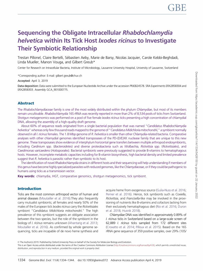
Sequencing the Obligate Intracellular Rhabdochlamydia helvetica within Its Tick Host Ixodes ricinus to Investigate Their Symbiotic Relationship Trestan Pillonel, Claire Bertelli, S ebastien Aeby, Marie de Barsy, Nicolas Jacquier, Carole Kebbi-Beghdadi, Linda Mueller, Manon Vouga, and Gilbert Greub* Center for Research on Intracellular Bacteria, Institute of Microbiology, Lausanne University Hospital, University of Lausanne, Switzerland *Corresponding author: E-mail: [email protected] Accepted: April 3, 2019 Data deposition: Data were submitted to the European Nucleotide Archive under the accession PRJEB24578. SRA Experiments ERX2858304 and ERX2858303. Assembly GCA_901000775. Abstract The Rhabdochlamydiaceae family is one of the most widely distributed within the phylum Chlamydiae, but most of its members remain uncultivable. Rhabdochlamydia 16S rRNA was recently reported in more than 2% of 8,534 pools of ticks from Switzerland. Shotgun metagenomics was performed on a pool of five female Ixodes ricinus ticks presenting a high concentration of chlamydial DNA, allowing the assembly of a high-quality draft genome. About 60% of sequence reads originated from a single bacterial population that was named “Candidatus Rhabdochlamydia helvetica” whereas only few thousand reads mapped to the genome of “Candidatus Midichloria mitochondrii,” a symbiont normally observed in all I. ricinus females. The 1.8 Mbp genome of R. helvetica is smaller than other Chlamydia-related bacteria. Comparative analyses with other chlamydial genomes identified transposases of the PD-(D/E)XK nuclease family that are unique to this new genome. These transposases show evidence of interphylum horizontal gene transfers between multiple arthropod endosymbionts, including Cardinium spp. (Bacteroidetes) and diverse proteobacteria such as Wolbachia, Rickettsia spp. (Rickettsiales), and Caedimonas varicaedens (Holosporales). Bacterial symbionts were previously suggested to provide B-vitamins to hematophagous hosts. However, incomplete metabolic capacities including for B-vitamin biosynthesis, high bacterial density and limited prevalence suggest that R. helvetica is parasitic rather than symbiotic to its host. The identification of novel Rhabdochlamydia strains in different hosts and their sequencing will help understanding if members of this genus have become highly specialized parasites with reduced genomes, like the Chlamydiaceae, or if they could be pathogenic to humans using ticks as a transmission vector. Key words: chlamydia, HGT, comparative genomics, shotgun metagenomics, tick symbiont. Introduction Ticks are the most common arthropod vector of human and animal diseases (Moutailler et al. 2016).They also frequently carry mutualist symbionts; all females and nearly 50% of the males of the European tick Ixodes ricinus carry the Rickettsiales symbiont “Candidatus Midichloria mitochondrii.” The high prevalence of this symbiont suggests an obligate association between the two species, but the role of the symbiont in the biology of I. ricinus remains unknown (Ahantarig et al. 2013; Moutailler et al. 2016). As confirmed by whole genome se- quencing, ticks are incapable of de novo heme synthesis and acquire heme from exogenous source (Gulia-Nuss et al. 2016; Perner et al. 2016). Hence, tick symbionts such as Coxiella, Rickettsia, and Francisella-like may be involved in the provi- sioning of nutrients like B-vitamins and cofactors lacking from their exclusively hematophagous diet (Rio et al. 2016; Duron et al. 2018; Husnik 2018). Chlamydiae DNA was identified in approximately 0.89% of I. ricinus ticks in Switzerland based on a large-scale screen of 62,889 I. ricinus ticks sampled from 172 different sites (Croxatto et al. 2014; Pilloux et al. 2015). Based on the 16S rRNA gene sequence of 359 positive samples, over 29% (105/ ß The Author(s) 2019. Published by Oxford University Press on behalf of the Society for Molecular Biology and Evolution. This is an Open Access article distributed under the terms of the Creative Commons Attribution License (http://creativecommons.org/licenses/by/4.0/ ), which permits unrestricted reuse, distribution, and reproduction in any medium, provided the original work is properly cited. 1334 Genome Biol. Evol. 11(4):1334–1344. doi:10.1093/gbe/evz072 Advance Access publication April 4, 2019 GBE Downloaded from https://academic.oup.com/gbe/article/11/4/1334/5428150 by guest on 22 January 2022
-
Upload
khangminh22 -
Category
Documents
-
view
1 -
download
0
Transcript of evz072.pdf - Oxford Academic
Sequencing the Obligate Intracellular Rhabdochlamydia
helvetica within Its Tick Host Ixodes ricinus to Investigate
Their Symbiotic Relationship
Trestan Pillonel, Claire Bertelli, S�ebastien Aeby, Marie de Barsy, Nicolas Jacquier, Carole Kebbi-Beghdadi,Linda Mueller, Manon Vouga, and Gilbert Greub*
Center for Research on Intracellular Bacteria, Institute of Microbiology, Lausanne University Hospital, University of Lausanne, Switzerland
*Corresponding author: E-mail: [email protected]
Accepted: April 3, 2019
Data deposition: Data were submitted to the European Nucleotide Archive under the accession PRJEB24578. SRA Experiments ERX2858304 and
ERX2858303. Assembly GCA_901000775.
Abstract
The Rhabdochlamydiaceae family is one of the most widely distributed within the phylum Chlamydiae, but most of its members
remain uncultivable. Rhabdochlamydia 16S rRNA was recently reported in more than 2% of 8,534 pools of ticks from Switzerland.
Shotgun metagenomics was performed on a pool of five female Ixodes ricinus ticks presenting a high concentration of chlamydial
DNA, allowing the assembly of a high-quality draft genome.
About 60% of sequence reads originated from a single bacterial population that was named “Candidatus Rhabdochlamydia
helvetica” whereasonly fewthousandreadsmapped to thegenomeof“CandidatusMidichloriamitochondrii,” a symbiontnormally
observed in all I. ricinus females. The 1.8 Mbp genome of R. helvetica is smaller than other Chlamydia-related bacteria. Comparative
analyses with other chlamydial genomes identified transposases of the PD-(D/E)XK nuclease family that are unique to this new
genome. These transposases show evidence of interphylum horizontal gene transfers between multiple arthropod endosymbionts,
including Cardinium spp. (Bacteroidetes) and diverse proteobacteria such as Wolbachia, Rickettsia spp. (Rickettsiales), and
Caedimonas varicaedens (Holosporales). Bacterial symbionts were previously suggested to provide B-vitamins to hematophagous
hosts. However, incomplete metabolic capacities including for B-vitamin biosynthesis, high bacterial density and limited prevalence
suggest that R. helvetica is parasitic rather than symbiotic to its host.
The identification of novel Rhabdochlamydia strains in different hosts and their sequencing will help understanding if members of
thisgenushavebecomehighly specializedparasiteswith reducedgenomes, like theChlamydiaceae, or if theycouldbepathogenic to
humans using ticks as a transmission vector.
Key words: chlamydia, HGT, comparative genomics, shotgun metagenomics, tick symbiont.
Introduction
Ticks are the most common arthropod vector of human and
animal diseases (Moutailler et al. 2016).They also frequently
carry mutualist symbionts; all females and nearly 50% of the
males of the European tick Ixodes ricinus carry the Rickettsiales
symbiont “Candidatus Midichloria mitochondrii.” The high
prevalence of this symbiont suggests an obligate association
between the two species, but the role of the symbiont in the
biology of I. ricinus remains unknown (Ahantarig et al. 2013;
Moutailler et al. 2016). As confirmed by whole genome se-
quencing, ticks are incapable of de novo heme synthesis and
acquire heme from exogenous source (Gulia-Nuss et al. 2016;
Perner et al. 2016). Hence, tick symbionts such as Coxiella,
Rickettsia, and Francisella-like may be involved in the provi-
sioning of nutrients like B-vitamins and cofactors lacking from
their exclusively hematophagous diet (Rio et al. 2016; Duron
et al. 2018; Husnik 2018).
Chlamydiae DNA was identified in approximately 0.89% of
I. ricinus ticks in Switzerland based on a large-scale screen of
62,889 I. ricinus ticks sampled from 172 different sites
(Croxatto et al. 2014; Pilloux et al. 2015). Based on the 16S
rRNA gene sequence of 359 positive samples, over 29% (105/
� The Author(s) 2019. Published by Oxford University Press on behalf of the Society for Molecular Biology and Evolution.
This is an Open Access article distributed under the terms of the Creative Commons Attribution License (http://creativecommons.org/licenses/by/4.0/), which permits unrestricted reuse,
distribution, and reproduction in any medium, provided the original work is properly cited.
1334 Genome Biol. Evol. 11(4):1334–1344. doi:10.1093/gbe/evz072 Advance Access publication April 4, 2019
GBED
ownloaded from
https://academic.oup.com
/gbe/article/11/4/1334/5428150 by guest on 22 January 2022

359) belonged to the Rhabdochlamydiaceae family and exhib-
ited high amounts of chlamydial DNA (up to 8� 106 copies/ml,
see Pilloux et al. 2015). Additional studies reported the pres-
ence of Rhabdochlamydia spp. in various tick populations
around the world (Hokynar et al. 2016; Burnard et al. 2017).
However, no Rhabdochlamydia isolate has been cultivated
from ticks yet. The two other described species, were also
identified in arthropods, suggesting that Rhabdochlamydia
spp. may infect a wide range of arthropods. “Candidatus
Rhabdochlamydia porcellionis” was isolated from hepato-
pancreatic cells of the terrestrial crustacean Porcellio scaber
(Kostanjsek 2004) and “Candidatus Rhabdochlamydia
crassificans” was identified in the cockroach Blatta orientalis
(Corsaro et al. 2007). Up to now, only “Candidatus
Rhabdochlamydia porcellionis” could be cultured in SF-9 cells
(Sixt et al. 2013). Both R. porcellionis and R. crassificans spe-
cies exhibited a particular cell morphology; elementary bodies
presented a five layered cell wall and oblong translucent struc-
tures in the cytoplasm (Corsaro et al. 2007; Kostanjsek 2004).
Older records describe very similar bacteria with pentalaminar
cell walls infecting the spider Pisaura mirabilis (Morel 1978),
the scorpion Buthus occitanus (Morel 1976), and the midge
larvae Chironomus dorsalis (Morel 1980). No hybridization
between the DNA from the scorpion and the midge larvae
parasites could be observed and Guanine–Cytosine (GC)-con-
tent and genome size estimations were significantly different
(1,550 kb [kilobases], 41% GC vs. 2,650 kb, 36.7% GC, re-
spectively) for these two putative chlamydia-related bacteria
of arthropods, suggesting that they likely belong to different
genera (Frutos et al. 1989, 1994). The proposition to classify
these bacteria in the genus Porochlamydia (Morel 1976) was
not recognized and Porochlamydia were considered to be
part of the Rickettsiella genus (Fournier and Raoult 2005).
More recent metagenomics analyses of the spider
Oedothorax gibbosus microbiome revealed a high prevalence
of Rhabdochlamydia spp. in one spider population
(Vanthournout and Hendrickx 2015), suggesting that these
so-called “Porochlamydia” might in fact represent two mem-
bers of the Rhabdochlamydiaceae family.
If R. porcellionis and R. crassificans were shown to be det-
rimental to their host (Radek 2000; Kostanj�sek and Pirc Marolt
2015), the nature of the relationship between
Rhabdochlamydia spp. and ticks, as well as their pathogenicity
toward humans, remain to be investigated. In the present
study, we bypassed the difficult step of cell-culture by sequenc-
ing the genome of a Rhabdochlamydia within its host I. ricinus,
allowing us to identify distinctive features and to learn about
the biology of this largely unknown chlamydial family.
Materials and methods
Genome Sequencing, Assembly, and Annotation
A sample with five female ticks from Lucerne area
(Switzerland) in the work of Pilloux et al. (2015) was selected
for high throughput sequencing. Genomic DNA was
extracted and purified using Wizard Genomic DNA purifica-
tion kit (Promega, Duebendorf, Switzerland). Genomic librar-
ies were constructed using Nextera XT library kit (Illumina),
normalized on beads and pooled into a single library for 150
base pairs (bp) paired-end sequencing using the MiSeq
(Illumina, San Diego, CA).
Illumina reads were filtered and trimmed with fastq-mcf
1.1.2 (Aronesty 2013), keeping 150 bp reads with an average
Phred quality score higher than 30 (–max-ns 1 -l 150 -L 150 –
qual-mean 30). Filtered reads were assembled with Edena 3
(Hernandez et al. 2008) and metaSPAdes 3.10.1 (Nurk et al.
2017) with k-mer or overlap size ranging from 51 to 127. In
addition, reads that did not map on published genome as-
semblies (GCA_000973045.1 and GCF_000208615.1) from
the ticks I. ricinus and Ixodes scapularis (Pagel Van Zee et al.
2007; Quillery et al. 2014) were assembled using MaSuRCA
2.1.9 (Zimin et al. 2013) and Velvet 1.2.09 (Zerbino and Birney
2008). Scaffolds were blasted against a database including
the I. ricinus and I. scapularis genome assemblies with
BlastN (e-value� 1e�5, identity � 80%) to remove contami-
nants. Scaffolds that did not show significant similarity to
Ixodes sequences were further investigated as follows: (1)
Coding sequences were predicted with prodigal 2.6 (param-
eters: -c -m -g 11 -p meta -f sco -q) (Hyatt et al. 2010). (2) Each
predicted coding sequences (CDS) was assigned to a taxo-
nomic rank with Kaiju 1.6.2 (Menzel et al. 2016) and the
proGenomes database (Mende et al. 2017). (3) A bacterial
phylum was assigned to each scaffold based on the most
frequent phylum assigned to its predicted CDSs. The assembly
graph produced by metaSPAdes was visualized with Bandage
(Wick et al. 2015) in order to investigate the connectivity of
scaffolds assigned to the Chlamydiae phylum. Finally, 8 assem-
blies from the 4 assemblers presenting between 107 and 66
scaffolds were compared and combined with Consed
(Gordon et al. 1998). The combined assembly was split in
case of disagreement between assembly methods. Only scaf-
folds larger than 1,000 bp exhibiting no significant similarity
to Ixodes sequences, consistent GC-content and sequencing
depth were retained for further analyses. The completeness of
the final genome assembly was estimated based on the iden-
tification of 104 nearly universal single-copy genes with
CheckM (Parks et al. 2015).
Raw reads were mapped to the assembled
Rhabdochlamydia helvetica genome as well as to I.
scapularis, I. ricinus, and “Candidatus Midichloria
mitochondrii” (GCA_000973045.2, GCF_000208615.1, and
GCF_000219355.1). The mapping was done with BWA
mem version 0.7.17 (Li and Durbin 2009) with a minimum
seed length of 23 and all 4 genomes combined into a single
fasta file.
Scaffolds were reordered based on the reference genome
of Simkania negevensis Z (NC_015713) using Mauve v. 2.3.1
(Darling et al. 2004). Genome annotation was performed
Sequencing the Obligate Intracellular Rhabdochlamydia helvetica GBE
Genome Biol. Evol. 11(4):1334–1344 doi:10.1093/gbe/evz072 Advance Access publication April 4, 2019 1335
Dow
nloaded from https://academ
ic.oup.com/gbe/article/11/4/1334/5428150 by guest on 22 January 2022

using Prokka v. 1.10 (Seemann 2014). Domains were anno-
tated using InterProScan 5.18-57 (Jones et al. 2014).
Predicted coding sequences were compared with the COG
database (Galperin et al. 2014) using BlastP 2.2.28þ(Camacho et al. 2009) with an e-value cutoff of 1e�5, and
to the RefSeq database release 79 (Haft et al. 2018) using
PLAST 2.3.1 (parameters: -a 8 -M BLOSUM62 -e 1e-3 -G 11 -E
1 -force-query-order 1000 -max-hit-per-query 100 -max-hsp-
per-hit 1) (Van Nguyen and Lavenier 2009). The coding den-
sity was calculated based on predicted open reading frames
(ORF), or published annotations. For draft assemblies, only
contigs larger than 10 kb were used to estimate the coding
density.
Confirmation of Tick Species from Genomic Data
To confirm that the host species was I. ricinus, raw reads were
mapped on the 18S rRNA sequence of 82 different tick spe-
cies (merged into a single fasta file, supplementary table S1,
Supplementary Material online). Mapped reads were
extracted and remapped individually on each 18S rRNA se-
quence and inspected with IGV (Robinson et al. 2011).
Metabolism and Respiratory Chains Annotation
Metabolic pathways and modules were investigated using the
Kyoto Encyclopedia of Genes and Genomes (KEGG) database
(Ogata et al. 1999). KEGG orthology assignments were done
using the GhostKOALA web server (Kanehisa et al. 2016).
Mapping between KEGG orthologs and pathways/modules
was done using the KEGG API (http://www.kegg.jp/kegg/
docs/keggapi.html; last accessed April 10, 2019). Specific fo-
cus was made on basic energy metabolism, amino acids,
cofactors, and vitamins biosynthesis. Transporters were anno-
tated using gBlast3 from the transporter classification data-
base (Saier et al. 2016), with an e-value cutoff of 10�5 and
50% coverage of both query and hit. When proteins of in-
terest could not be identified, pseudogenes remnants or
unpredicted ORFs were searched in raw contigs of the
Rhabdochlamydia assembly using TBlastN and in the raw
reads using Diamond (Buchfink et al. 2015).
Cluster of Orthologs and Phylogenetic Reconstructions
Protein sequences of 61 PVC (Planctomycetes,
Verrucomicrobia, and Chlamydiae) superphylum strains, in-
cluding 31 members of the Chlamydiales order, were down-
loaded from RefSeq (supplementary table S2, Supplementary
Material online) and grouped into orthologous groups using
OrthoFinder 0.4.0 with default parameters (Emms and Kelly
2015). The complete orthology table is reported in supple-
mentary table S3 (Supplementary Material online). The ge-
nome assemblies of the “Candidatus Limichlamydia” strain
SM23_39 (marine sediments metagenome) and
“Candidatus Hydrochlamydia” strain Ga0074140 (drinking
water treatment plant metagenome), were not considered
for genome content analyses. Unannotated genome assem-
blies were annotated with Prokka v.1.10. In addition, all pre-
dicted protein sequences were annotated using the same
tools as for R. helvetica (see above). Protein identities were
calculated based on multiple sequence alignments built using
MAFFT 7.058b (Katoh and Standley 2013). Gaps were not
considered in pairwise identity calculations. Genome maps
were generated using Circos (Krzywinski et al. 2009). A phy-
logenetic tree was reconstructed based on 172 conserved
single-copy proteins with RAxML 8.2.0 (Stamatakis 2014) us-
ing the LG substitution matrix (PROTGAMMALG) with distinct
partitions for each of the 172 alignments and 100 rapid boot-
strap replicates. This model was preferred based on previous
experience with similar data sets (Pillonel et al. 2015). Figures
with phylogenies and associated data were drawn with the
Python package ete2 (Huerta-Cepas et al. 2010).
Proteins harboring a PD-(D/E)XK nuclease family transpo-
sase domain (Pfam accession PF12784) were searched in
6661 reference and representative genomes (downloaded
from RefSeq, September 2017) with hmmsearch (HMMER
v. 3.1b2 [Eddy 2011] with “–—cut_tc” parameter). The
1,911 identified proteins were further filtered based on sim-
ilarity to at least 1 of the 6 R. helvetica homologs (filtered with
BlastP v. 2.7.1 with an e-value of 1e�5 and 80% of hsp query
coverage) and clustered at 90% identity with cd-hit v. 4.6 (Fu
et al. 2012) to reduce redundancy. The phylogeny was recon-
structed with FastTree v.2.1.9 (Price et al. 2010) double pre-
cision with default parameters.
Results and Discussion
Sequencing the Endosymbiont Genome within Its TickHost
Shotgun metagenomics of the tick pool DNA produced 25.7
million paired-ends reads in two sequencing runs. Among 80
tick species, I. ricinus and I. scapularis recruited the highest
number of reads by mapping on the 18S rRNA (respectively
985 and 835). Because some regions of the 18S rRNA are
strictly identical between ticks species, all recruited reads were
mapped again individually on I. scapularis and I. ricinus
sequences. The single base of 18S rRNA sequence enabling
to distinguish both species was identical to I. ricinus variant,
confirming that all five ticks in the pool indeed belonged to
I. ricinus species. No other polymorphism could be identified
except for one position exhibiting two populations of reads.
Sixteen million high-quality reads were used to assemble
the genome de novo. Most raw reads mapped either to the
R. helvetica assembly (59.96%) or to one of the two Ixodes
genomes (40.03%). Only 8,087 reads mapped to “Ca. M.
mitochondrii” genome. Scaffolds assembled de novo were
carefully investigated to remove contaminants such as
Ixodes sequences (low sequencing depth, GC-content
>40%) (supplementary fig. S1, Supplementary Material
Pillonel et al. GBE
1336 Genome Biol. Evol. 11(4):1334–1344 doi:10.1093/gbe/evz072 Advance Access publication April 4, 2019
Dow
nloaded from https://academ
ic.oup.com/gbe/article/11/4/1334/5428150 by guest on 22 January 2022

online). All scaffolds larger than 12.5 kb were classified as
belonging to the Chlamydiae phylum (Most of them have a
median depth >1600x and a GC content<40%) (supplemen-
tary fig. S1, Supplementary Material online). Chlamydial scaf-
folds formed a highly interconnected network of about
1.88 Mbp in the assembly graph (supplementary fig. S2,
Supplementary Material online). The final manually curated
assembly of 38 scaffolds, comprising 1,830,543 bp, was esti-
mated to be complete (104/104 CheckM markers identified)
and exhibited no detectable contamination (no duplicated
marker gene). The cumulative GþC skew of the reordered
assembly exhibited the typical inverted “V” shape of bacterial
genomes (supplementary fig. S3, Supplementary Material on-
line). Altogether, these results indicate that Rhabdochlamydia
DNA was largely dominant in the pool of ticks sequenced, and
only few sequences from other bacterial species were
obtained, allowing to assemble a high-quality draft genome.
A putative 23,934 bp circular plasmid was identified thanks to
its uniformly lower sequencing depth as compared with the
rest of the assembly (supplementary fig. S4, Supplementary
Material online). It encodes a homolog of the plasmid inte-
grase pGP8-D commonly found in chlamydial plasmids.
A New Species of the Rhabdochlamydia Genus
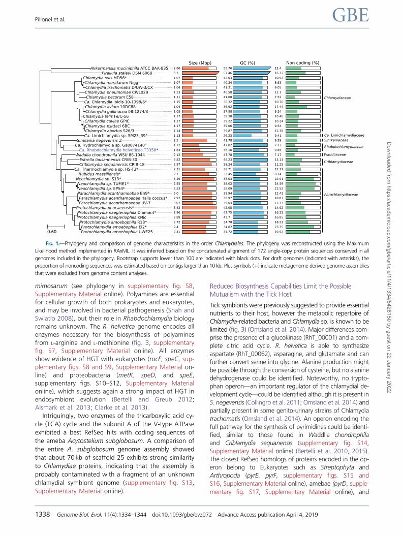
The 16S ribosomal sequence exhibited 97.88% and 97.98%
identity with R. porcellionis (AY223862) and R. crassificans
(AY928092), respectively. The conservation of this single
gene is not sufficient to accurately classify Chlamydiae at
the genus and species level (Pillonel et al. 2015). However,
given the level of sequence divergence (>2%) and the lack of
other Rhabdochlamydia genomes available, we considered
this strain as a new Candidatus species named “Candidatus
Rhabdochlamydia helvetica”. It exhibited 91.78% and
88.93% sequence identity respectively with the 16S and
23S rRNA of S. negevensis strain Z (supplementary table S4,
Supplementary Material online). The reconstruction of the
Chlamydiae phylogeny based on a concatenated set of 172
conserved protein sequences (fig. 1, supplementary table S5,
Supplementary Material online) as well as the high number of
orthologous protein families (supplementary figs. S4 and S5,
Supplementary Material online) confirmed that S. negevensis
is the closest sequenced relative of R. helvetica.
Horizontal Gene Transfers Likely Occurred with OtherSymbionts and Eukaryotic Hosts
Over 62.8% of the 1,717 predicted proteins of R. helvetica
exhibited a best RefSeq hit within the Chlamydiae phylum
(Fig. 2). Comparative analyses of 31 chlamydial genomes
from 6 families (supplementary table S1, Supplementary
Material online, excluding the 2 metagenome-assembled
genomes) revealed that 322 (18%) R. helvetica CDS exhibited
no ortholog in other chlamydial genomes (supplementary fig.
S4, Supplementary Material online, purple CDS). Among
these, 73 had significant hits in RefSeq database (supplemen-
tary table S6, Supplementary Material online). Six are PD-(D/
E)XK nuclease family transposases exhibiting more than 60%
sequence identity with proteins of Occidentia massiliensis, a
soft tick symbiont from the Rickettsiaceae family
(Mediannikov et al. 2014). The transposase phylogeny includ-
ing homologs identified in 6,661 RefSeq reference and rep-
resentative genomes (supplementary fig. S6, Supplementary
Material online) showed that the 6 R. helvetica homologs are
monophyletic and cluster with phylogenetically diverse intra-
cellular bacteria belonging to Bacteroidetes (Cardinium spp.)
and Proteobacteria in the orders Rickettsiales (Wolbachia,
Rickettsia, Orientia, and Occidentia spp.), Holosporales
(Caedimonas varicaedens), and Myxococcales
(Pajaroellobacter abortibovis). Highly similar sequences were
also identified in two arthropod genomes (Cimex lectularius
and Vollenhovia emeryi). This could represent an example of
interkingdom horizontal gene transfer (HGT), but might also
result from the contamination of the two arthropod genomes
with DNA sequences of colonizing bacteria. Indeed, C. lectu-
larius was shown to carry Wolbachia symbionts (Rasgon and
Scott 2004). Interphylum transfer of transposases is not un-
common, particularly in case of shared habitats (Hooper et al.
2009), and was already observed in arthropod symbionts
(Duron 2013). Several other proteins specific to R. helvetica
showed significant sequence similarity to other obligate endo-
symbionts (supplementary table S6, Supplementary Material
online). These results suggest the occurrence of HGTs with
other bacteria sharing a similar niche, in this case ticks and
other arthropods, as previously described for Parachlamydia
and other ameba-infecting microorganisms (Gimenez et al.
2011).
Seventy-eight R. helvetica proteins exhibited significant
similarity to eukaryotic sequences (fig. 2, supplementary table
S7, Supplementary Material online). These include known
cases of chlamydial HGT such as multiple enzymes involved
in the metabolism of C5 isoprenoid (ispD, ispG, ispE) (Lange
et al. 2000), menaquinone (menA, menD), uridine mono-
phosphate (pyrE, pyrF, discussed below), heme (hemY), and
Pimeloyl-ACP (fabI, fabF) (Collingro et al. 2011). As previously
shown (Schmitz-Esser et al. 2010), proteins with bacteria-host
interaction domains such as ankyrin repeats, ubiquitin prote-
ase or BTB/POZ domains also exhibit high proportions of
BLAST hits in eukaryotic sequences.
Among the 78 proteins sharing similarity with eukary-
otic sequences (fig. 2), 17 proteins are unique to R. hel-
vetica. A “methyl-accepting chemotaxis” domain-
containing protein presented more than 50% amino
acid identity with arthropod sequences. Two lysine/orni-
thine decarboxylases (including a partial 145 amino-acids
sequence) participating to the polyamine biosynthesis
pathway (supplementary fig. S7, Supplementary
Material online) exhibited 60.25% identity with a se-
quence from the common house spider Stegodyphus
Sequencing the Obligate Intracellular Rhabdochlamydia helvetica GBE
Genome Biol. Evol. 11(4):1334–1344 doi:10.1093/gbe/evz072 Advance Access publication April 4, 2019 1337
Dow
nloaded from https://academ
ic.oup.com/gbe/article/11/4/1334/5428150 by guest on 22 January 2022
mimosarum (see phylogeny in supplementary fig. S8,
Supplementary Material online). Polyamines are essential
for cellular growth of both prokaryotes and eukaryotes,
and may be involved in bacterial pathogenesis (Shah and
Swiatlo 2008), but their role in Rhabdochlamydia biology
remains unknown. The R. helvetica genome encodes all
enzymes necessary for the biosynthesis of polyamines
from L-arginine and L-methionine (fig. 3, supplementary
fig. S7, Supplementary Material online). All enzymes
show evidence of HGT with eukaryotes (rocF, speC, sup-
plementary figs. S8 and S9, Supplementary Material on-
line) and proteobacteria (metK, speD, and speE,
supplementary figs. S10–S12, Supplementary Material
online), which suggests again a strong impact of HGT in
endosymbiont evolution (Bertelli and Greub 2012;
Alsmark et al. 2013; Clarke et al. 2013).
Intriguingly, two enzymes of the tricarboxylic acid cy-
cle (TCA) cycle and the subunit A of the V-type ATPase
exhibited a best RefSeq hits with coding sequences of
the ameba Acytostelium subglobosum. A comparison of
the entire A. subglobosum genome assembly showed
that about 70 kb of scaffold 25 exhibits strong similarity
to Chlamydiae proteins, indicating that the assembly is
probably contaminated with a fragment of an unknown
chlamydial symbiont genome (supplementary fig. S13,
Supplementary Material online).
Reduced Biosynthesis Capabilities Limit the PossibleMutualism with the Tick Host
Tick symbionts were previously suggested to provide essential
nutrients to their host, however the metabolic repertoire of
Chlamydia-related bacteria and Chlamydia sp. is known to be
limited (fig. 3) (Omsland et al. 2014). Major differences com-
prise the presence of a glucokinase (RhT_00001) and a com-
plete citric acid cycle. R. helvetica is able to synthesize
aspartate (RhT_00062), asparagine, and glutamate and can
further convert serine into glycine. Alanine production might
be possible through the conversion of cysteine, but no alanine
dehydrogenase could be identified. Noteworthy, no trypto-
phan operon—an important regulator of the chlamydial de-
velopment cycle—could be identified although it is present in
S. negevensis (Collingro et al. 2011; Omsland et al. 2014) and
partially present in some genito-urinary strains of Chlamydia
trachomatis (Omsland et al. 2014). An operon encoding the
full pathway for the synthesis of pyrimidines could be identi-
fied, similar to those found in Waddlia chondrophila
and Criblamydia sequanensis (supplementary fig. S14,
Supplementary Material online) (Bertelli et al. 2010, 2015).
The closest RefSeq homologs of proteins encoded in the op-
eron belong to Eukaryotes such as Streptophyta and
Arthropoda (pyrE, pyrF, supplementary figs. S15 and
S16, Supplementary Material online), amebae (pyrD, supple-
mentary fig. S17, Supplementary Material online), and
Size (Mbp) GC (%) Non coding (%)Akkermansia muciniphila ATCC BAA-835 2.66 55.76 12.4
Pirellula staleyi DSM 6068 6.2 57.46 16.32 Chlamydia suis MD56* 1.07 42.03 10.92 Chlamydia muridarum Nigg 1.07 40.34 8.63 Chlamydia trachomatis D/UW-3/CX 1.04 41.31 9.05 Chlamydia pneumoniae CWL029 1.23 40.58 12.1 Chlamydia pecorum E58 1.11 41.08 7.52
Ca. Chlamydia ibidis 10-1398/6* 1.15 38.32 10.76 Chlamydia avium 10DC88 1.04 36.92 17.44 Chlamydia gallinacea 08-1274/3 1.05 37.88 9.24
Chlamydia felis Fe/C-56 1.17 39.38 10.46 Chlamydia caviae GPIC 1.17 39.22 10.24 Chlamydia psittaci 6BC 1.17 39.06 9.54 Chlamydia abortus S26/3 1.14 39.87 12.38 Ca. Limichlamydia sp. SM23_39+ 1.13 26.23 9.41
Simkania negevensis Z 2.5 41.78 9.39 Ca. Hydrochlamydia sp. Ga0074140+ 1.72 47.82 7.73
Ca. Rhabdochlamydia helveticae T3358* 1.83 36.16 9.65 Waddlia chondrophila WSU 86-1044 2.12 43.78 7.17
Estrella lausannensis CRIB-30 2.82 48.23 13.11 Criblamydia sequanensis CRIB-18 2.97 38.24 11.25
Ca. Thermochlamydia sp. HS-T3* 2.31 38.71 12.17 Rubidus massiliensis* 2.7 32.45 8.74
Neochlamydia sp. S13* 3.19 38.03 22.91 Neochlamydia sp. TUME1* 2.55 38.02 24.59 Neochlamydia sp. EPS4* 2.53 38.09 23.52 Parachlamydia acanthamoebae Bn9* 3.0 38.94 12.42 Parachlamydia acanthamoebae Halls coccus* 2.97 38.97 10.87 Parachlamydia acanthamoebae UV-7 3.07 39.03 12.33
Protochlamydia phocaeensis* 3.42 42.05 14.92 Protochlamydia naegleriophila Diamant* 2.96 42.75 16.22 Protochlamydia naegleriophila KNic 2.89 42.7 16.85 Protochlamydia amoebophila R18* 2.72 34.78 18.23 Protochlamydia amoebophila EI2* 2.4 34.82 23.35 Protochlamydia amoebophila UWE25 2.41 34.72 19.92 0.60
Chlamydiaceae
Simkaniaceae
Rhabdochlamydiaceae
Criblamydiaceae
Waddliaceae
Parachlamydiaceae
Ca. Limichlamydiaceae
FIG. 1.—Phylogeny and comparison of genome characteristics in the order Chlamydiales. The phylogeny was reconstructed using the Maximum
Likelihood method implemented in RAxML. It was inferred based on the concatenated alignment of 172 single-copy protein sequences conserved in all
genomes included in the phylogeny. Bootstrap supports lower than 100 are indicated with black dots. For draft genomes (indicated with asterisks), the
proportion of noncoding sequences was estimated based on contigs larger than 10 kb. Plus symbols (þ) indicate metagenome derived genome assemblies
that were excluded from genome content analyses.
Pillonel et al. GBE
1338 Genome Biol. Evol. 11(4):1334–1344 doi:10.1093/gbe/evz072 Advance Access publication April 4, 2019
Dow
nloaded from https://academ
ic.oup.com/gbe/article/11/4/1334/5428150 by guest on 22 January 2022

(a)
(b)
FIG. 2.—(a) Protein homologs to the R. helvetica proteome in the National Center for Biotechnology Information (NCBI) RefSeq database. The inner gray
circle (1) reports the localization of genes encoded on leading and lagging strands. The second circle (2) reports the conservation of the protein families in the
61 genomes included in the comparison (supplementary table S2, Supplementary Material online). Protein families conserved in <25 genomes are
highlighted in red. The following circles report the taxonomic classification of the top 100 NCBI RefSeq hits of each R. helvetica protein: circle (3): number
of chlamydial (blue) and other bacteria (orange) hits; circle (4): number of eukaryotic hits (green); circle (5): number of archaeal hits (pink). The “two outer
circles” report (6) the GC-skew along the genome and (7) the localization of gaps in the genome assembly. (b) Summary of RefSeq hits taxonomy. Left part:
consensus taxonomy of the top 100 best RefSeq hits (majority rule) and taxonomy of the best RefSeq hit of each R. helvetica protein. Right part: number of
proteins with significant similarity to eukaryotic sequences.
Sequencing the Obligate Intracellular Rhabdochlamydia helvetica GBE
Genome Biol. Evol. 11(4):1334–1344 doi:10.1093/gbe/evz072 Advance Access publication April 4, 2019 1339
Dow
nloaded from https://academ
ic.oup.com/gbe/article/11/4/1334/5428150 by guest on 22 January 2022

gamma-proteobacteria such as Rickettsiella spp., obligate
intracellular bacteria infecting a wide range of arthropods
(pyrC, pyrB, carB, carA, supplementary figs. S18–S21,
Supplementary Material online), suggesting that the operon
may have been acquired through HGTs. Biosynthesis of vita-
mins and cofactors is restricted to heme and menaquinone
(Figure 3), suggesting that R. helvetica could participate to the
supply—albeit limited—of some essential factors to its host
like other symbionts of blood-feeding parasite (Leclerque
2008; Duron et al. 2018; Husnik 2018). However, R. helvetica
is not able to synthetize quite a number of vitamins and
cofactors, limiting its possible mutualistic interactions with
the host, and rather suggesting a pathogenic behavior.
Hallmarks of Chlamydial Parasitism
As mentioned in a previous publication and like all Chlamydiae
sequenced to date, the genome of R. helvetica encodes
homologs of the type three secretion system (T3SS) apparatus
and chaperones (Pillonel et al. 2018). Homologs of several
known T3SS effectors such as NUE, pkn5, and Lda2 were
also identified (see supplementary table S8, Supplementary
Material online, for detailed list of identified loci). Like its clos-
est sequenced relative S. negevensis, R. helvetica does not
encode a homolog of the protease-like activity factor
(CPAF), a well-known chlamydial virulence factor. R. helvetica
encodes about half as many transporters as S. negevensis
(supplementary table S9, Supplementary Material online). It
does not have a homolog of the sulfate permease (family
2.A.53) present in most chlamydial genomes nor the
sodium-driven transporters found in Chlamydiaceae such as
the anion/Na+ symporter SodiTl (2.A.47, CT_204), the amino
acid symporters BrnQ (2.A.26, CT_554), and TnaT (CT_231).
The loss of such transporters might be linked to the loss of the
complex 1 of the respiratory chain involved in Naþ gradient
generation. No homolog of the Naþ/Hþ antiporter NhaE
(2.A.111: CT_805) was identified, but R. helvetica has a
CPA2 family Naþ/Hþ antiporter homolog absent from the
Adenine ribonucleotide biosynthesis (M00049)
Alanine, aspartate and glutamate metabolism (map00250)
Arginine and proline metabolism (map00220)
Arginine biosynthesis (map00330)
Biotin biosynthesis (M00123)
Cobalamin biosynthesis (M00122)
Coenzyme A biosynthesis (M00120)
Cysteine and methionine metabolism (map00270)
Glycine, serine and threonine metabolism (map00260)
Guanine ribonucleotide biosynthesis (M00050)
Heme biosynthesis (M00121)
Histidine metabolism (map00340) Lysine biosynthesis (map00300)
Lysine degradation (map00310)
Menaquinone biosynthesis (M00116)
NAD biosynthesis (M00115)
Pantothenate biosynthesis (M00119)
Phenylalanine metabolism (map00360)
Phenylalanine, tyrosine and tryptophan biosynthesis (map00400)
Spermidine biosynthesis (M00133)
Putrescine biosynthesis (M00134)
Pyrimidine deoxyribonuleotide biosynthesis (M00053) Pyrimidine ribonucleotide biosynthesis (M00052)
Inosine monophosphate biosynthesis (M00048) 9 10 - - - - - - - - - - - - - 1 1 1 2 - - - 1 - - - - - - - - - - - -
Riboflavin biosynthesis (M00125)
Tetrahydrofolate biosynthesis (M00126)
Thiamine biosynthesis (M00127)
Tryptophan metabolism (map00380)
Ubiquinone biosynthesis (M00117)
Uridine monophosphate biosynthesis (M00051)
Valine, leucine and isoleucine biosynthesis (map00290)
Valine, leucine and isoleucine degradation (map00280)
Akkermansia m
uciniphila ATCC BAA-835
5
16
9
12
4
5
4
17
19
5
7
2 12
3
7
5
5
-
3
1
-
5 3
5
7
5
19
1
7
10
4
Pirellula staleyi DSM
6068
5
19
12
11
4
1
4
21
23
5
10
5 9
4
1
5
4
1
4
1
-
6 3
5
6
3
20
4
7
10
7
Chlamydia suis M
D56
2
1
-
-
1
-
1
3
9
2
9
1 8
2
1
-
-
-
1
-
-
6 3
5
2
-
8
3
-
1
3
Chlamydia m
uridarum N
igg
3
1
-
-
1
-
1
3
7
5
9
1 8
2
1
-
-
-
1
-
-
6 3
5
2
-
6
3
-
1
3
Chlamydia trachom
atis D/U
W-3/CX
3
1
-
-
1
-
1
3
9
3
9
1 8
2
1
-
-
-
1
-
-
6 3
5
2
-
9
3
-
1
3
Chlamydia pneum
oniae CWL029
3
2
1
1
4
-
1
3
7
4
9
1 8
2
1
-
-
-
1
-
-
6 3
5
1
-
7
3
1
1
3
Chlamydia pecorum
E58
3
1
-
-
4
-
1
3
9
5
9
1 7
2
1
-
-
-
1
-
-
6 3
5
-
-
12
3
1
1
3
Ca. Chlamydia ibidis 10-1398/6
3
1
-
-
4
-
1
3
7
3
9
1 8
2
1
-
-
-
1
-
-
6 3
5
1
1
8
3
1
1
3
Chlamydia avium
10DC88
3
1
-
-
4
-
1
4
7
3
9
1 7
2
1
-
-
-
1
-
-
6 3
5
1
-
8
3
1
1
3
Chlamydia gallinacea 08-1274/3
3
1
-
-
4
-
1
3
7
3
8
1 8
2
1
-
-
-
1
-
-
6 3
5
1
-
8
3
1
1
3
Chlamydia felis Fe/C-56
3
1
-
-
4
-
1
3
9
5
9
1 8
2
1
-
-
-
1
-
-
6 3
5
1
2
13
3
1
1
3
Chlamydia caviae G
PIC
3
1
-
-
1
-
1
3
9
5
9
- 8
2
1
-
-
-
-
-
-
6 3
5
2
2
12
3
1
1
3
Chlamydia psittaci 6BC
3
1
-
-
4
-
1
3
7
5
9
1 8
2
1
-
-
-
1
-
-
6 3
5
1
2
8
3
1
1
3
Chlamydia abortus S26/3
3
1
-
-
4
-
1
2
7
3
9
- 8
2
1
-
-
-
-
-
-
6 3
5
1
2
6
3
1
2
3
Ca. Limichlam
ydia sp. SM23_39
3
5
1
3
1
-
1
2
6
3
2
- 7
-
-
2
-
-
-
-
-
4 2
-
1
-
6
-
-
1
3
Simkania negevensis Z
5
9
6
2
1
-
1
6
12
4
9
2 8
4
9
5
1
-
2
2
-
6 3
1
1
-
17
1
-
3
4
Ca. Hydrochlam
ydia sp. Ga0074140
5
11
3
3
-
-
2
4
13
4
10
2 8
2
8
2
-
-
1
-
-
6 3
1
1
-
7
2
5
1
3
Ca. Rhabdochlamydia helvetica T3358
5
8
5
3
-
-
1
7
8
4
11
1 8
2
6
2
1
-
1
2
2
6 3
-
1
-
7
2
4
2
4
Waddlia chondrophila W
SU 86-1044
5
12
8
3
-
1
1
9
18
5
9
1 8
6
7
3
-
-
-
-
1
6 3
5
5
-
7
1
5
2
11
Estrella lausannensis CRIB-30
4
10
10
3
4
1
1
9
16
3
10
4 8
5
7
2
-
-
1
2
-
6 3
2
2
-
7
1
2
2
8
Criblamydia sequanensis CRIB-18
3
11
12
2
4
1
1
7
19
3
10
4 8
6
8
2
-
-
1
1
-
6 3
2
4
-
8
4
5
2
9
Ca. Thermochlam
ydia sp. HS-T3
3
7
8
3
-
-
2
7
16
3
9
3 8
3
7
3
-
-
1
-
-
5 3
5
4
-
8
1
-
-
3 Rubidus m
assiliensis
5
10
11
3
4
1
2
7
14
5
10
3 8
4
7
3
-
-
1
-
-
6 3
5
4
-
7
1
-
1
7 N
eochlamydia sp. S13
4
2
1
-
-
1
1
3
9
3
9
- 8
1
7
2
-
-
-
-
-
6 3
1
3
-
6
1
-
1
5
Neochlam
ydia sp. TUM
E1 4
2
1
-
-
1
1
4
9
3
9
- 8
1
6
2
-
-
-
-
-
6 3
1
3
-
5
-
-
1
5
Neochlam
ydia sp. EPS4
3
2
1
-
-
1
1
4
9
3
9
- 8
1
7
2
-
-
-
-
-
6 3
1
3
-
5
1
-
1
5
Parachlamydia acantham
oebae Bn9
3
10
12
4
4
1
1
7
16
4
11
4 8
5
8
3
-
-
1
2
-
7 3
5
4
1
7
1
1
1
7
Parachlamydia acantham
oebae Halls coccus
3
8
12
3
4
1
1
7
16
4
11
4 8
5
8
3
-
-
1
2
-
7 3
5
4
1
7
1
1
1
7
Parachlamydia acantham
oebae UV-7
3
9
13
3
4
1
1
7
16
4
11
4 8
5
8
3
-
-
1
2
-
7 3
5
4
1
7
1
1
1
7
Protochlamydia phocaeensis
3
11
13
5
-
1
1
8
21
3
10
4 8
3
7
3
-
-
1
1
-
6 3
5
4
1
13
2
1
3
3
Protochlamydia naegleriophila D
iamant
3
8
12
3
4
1
2
10
19
3
10
2 8
2
8
3
1
-
1
1
-
6 3
5
4
1
7
2
-
3
3
Protochlamydia naegleriophila KN
ic
3
8
12
3
4
1
2
10
18
3
10
2 8
2
8
3
1
-
1
1
-
6 3
5
4
1
7
2
-
3
3
Protochlamydia am
oebophila R18
3
6
2
2
-
1
1
5
14
3
11
- 8
3
8
-
-
-
-
-
-
7 3
5
4
-
6
1
1
-
1
Protochlamydia am
oebophila EI2
3
6
2
2
-
1
1
5
13
3
10
- 8
2
7
-
-
-
-
-
-
6 3
5
4
-
6
1
1
-
1
Protochlamydia am
oebophila UW
E25
3
6
2
2
-
1
1
4
14
3
11
- 8
3
8
-
-
-
-
-
-
7 3
5
4
-
6
1
1
-
1
0.60
Pyrimidines
Purines
Amino acids
Cofactors and vitamins
Polyamines
FIG. 3.—Comparative analysis of cofactors, vitamins, amino acids, polyamines, and nucleotides metabolism. Each row reports the number of unique
KEGG ortholog(s) for the corresponding module/pathway. NAD: nicotinamide adenine dinucleotide.
Pillonel et al. GBE
1340 Genome Biol. Evol. 11(4):1334–1344 doi:10.1093/gbe/evz072 Advance Access publication April 4, 2019
Dow
nloaded from https://academ
ic.oup.com/gbe/article/11/4/1334/5428150 by guest on 22 January 2022

Chlamydiaceae. These transporters involved Naþ tolerance,
pH homeostasis, tolerance to alkali, and fluctuations in osmo-
larity are infrequent in intracellular symbionts and pathogens
(Krulwich et al. 2009). Some oligopeptide and amino acid
transporters were identified in R. helvetica; A D-alanine/glycine
sodium symporter (GlyP), an alanine sodium symporter
(AgcS), two putative proton/glutamate-aspartate symporters
(family 2.A.23), and an active oligopeptide transporter
OppABCDF operon. The 13 periplasmic solute-binding pro-
teins (OppA) constitute the largest group of paralogs in the R.
helvetica genome.
Phosphorylated glucose can be imported through the
UhpC transporter, a highly conserved transporter encoded
in all sequenced chlamydia genomes (2.A.1.4.6). Like in other
Chlamydiae, no phosphoenolpyruvate transport system was
found in R. helvetica. Multiple cofactor and vitamin transport-
ers were identified, including putative biotin transporters with
low similarity to Vibrio cholerae, and three transporters with
low similarity to riboflavin transporters from Ochrobactrum
anthropi and V. cholerae (22%–28% identity). An S-adeno-
sylmethionine transporter and a two-partner secretion operon
with low similarity to a heme/hemopexin transport system
(huxA/huxB) from Haemophilus influenzae were also identi-
fied (supplementary table S8, Supplementary Material online).
Seven ATP/ADP antiporter homologs were identified. One
homolog (RhT_00329) is most closely related to S. negevensis
SnNTT1 and C. trachomatis Npt1 which were shown to trans-
port adenosine triphosphate (Adenosine diphosphate) and
ATP/NAD, respectively (Knab et al. 2011; Fisher et al. 2013).
Another homolog (RhT_01503) is more closely related to
SnNTT2, a guanine nucleotide/ATP/Hþ transporter (Knab
et al. 2011). Three subsequent paralogs (RhT_00750,
RhT_00751, RhT_00752) are phylogenetically most closely re-
lated to SnNTT4, whose substrate remains undetermined. The
last putative ATP/ADP antiporters (RhT_00462, RhT_00945)
are distant from all transporters characterized so far.
In summary, despite the presence of a larger metabolic
repertoire than Chlamydiaceae, Rhabdochlamydia is probably
highly dependent on its host, thus questioning the mutualistic
or parasitic nature of the relationship with its host.
Rhabdochlamydia helvetica Membrane Proteins
The membrane of Chlamydia is thought to be stabilized by
extensive disulfide bonds between the major outer mem-
brane protein (MOMP) and two cysteine-rich proteins OmcB
and OmcA (Hatch 1996). These three abundantly expressed
proteins are the major constituents of the chlamydial outer
membrane complex that also contains porins and polymor-
phic membrane proteins (Pmps) (Birkelund et al. 2009; Liu
et al. 2010). The outer membrane protein composition of
Chlamydia-related bacteria is very divergent, probably reflect-
ing differences in their ecological niches, but all of them, with
the notable exception of S. negevensis, contain OmcB, OmcA,
and other cysteine-rich proteins (Collingro et al. 2011; Rusconi
et al. 2013; Aistleitner et al. 2015). OmcB and OmcA could
not be retrieved in the genome of R. helvetica that also en-
code few cysteine-rich membrane proteins of the OmpA fam-
ily compared with W. chondrophila (n¼ 11) or Estrella
lausannensis (n¼ 10) (Bertelli et al. 2010, 2015). However,
the R. helvetica genome encodes one homolog of MOMP
(RhT_00042) and five MOMP-like proteins (RhT_00150,
RhT_00151, RhT_00153, RhTp_00022, RhT_01103). These
latter have a 1.4%–2.7% higher cysteine content than the
37 S. negevensis MOMP and MOMP-like proteins (<1.1%
cysteine) (Aistleitner et al. 2015). Like S. negevensis (Vouga
et al. 2016), the membrane of R. helvetica might not be sta-
bilized by a network of highly cross-linked proteins hence
conserving more flexibility.
Chlamydiaceae bacteria express between 9 and 21 Pmps
autotransporters that share little sequence similarity but play
important roles in adhesion via conserved multiple repeats of
the tetrapeptide motifs GGA(ILV) and FXXN (Molleken et al.
2010; Becker and Hegemann 2014). The R. helvetica genome
contains three Simkania PmpB homologs predicted to form a
beta barrel structure in the outer membrane (RhT_00712,
RhT_00713, and RhT_01040), but only RhT_01040 contains
the GGAI sequence and none displays a FXXN motif.
However, R. helvetica possesses one additional protein
(RhT_00552) with an autotransporter beta-domain displaying
similar structural features as the Pmp-like protein of W. chon-
drophila (wcw_0271); a signal peptide, a passenger domain,
and a beta-barrel C-terminal domain. This protein containing
five FXXN repeats and one repeat of the GGA(ILV) motif in the
passenger domain could, by similarity with wcw_0271, be
implicated in adhesion to the host cells (Kebbi-Beghdadi
et al. 2015).
Conclusion
The 1.83 Mb genome of R. helvetica sequenced directly from
a pool of ticks provides the first molecular data to investigate
the biology of this widespread genus (Lagkouvardos et al.
2014) infecting a wide range of arthropods (Kostanjsek
2004; Corsaro et al. 2007; Pilloux et al. 2015;
Vanthournout and Hendrickx 2015; Cooling et al. 2017).
Evidence of multiple HGTs between Rhabdochlamydia,
arthropods and several lineages of arthropod symbionts sug-
gests that Rhabdochlamydiaceae are widely distributed ar-
thropod parasites and highlights the importance of
nonvertical inheritance in the evolution of symbionts and par-
asites. Although arthropods are frequently engaged in mutu-
alistic associations with bacteria (Moran et al. 2008), the
R. helvetica genome only shows reduced metabolic capacities
limiting any potential mutualism. In contrast, it encodes sev-
eral ATP/ADP transporters and multiple transporters of vita-
mins, cofactors, amino acids, and oligopeptides. Considering
that the 2.65 Gb genome of I. scapularis (Cramaro et al. 2017)
Sequencing the Obligate Intracellular Rhabdochlamydia helvetica GBE
Genome Biol. Evol. 11(4):1334–1344 doi:10.1093/gbe/evz072 Advance Access publication April 4, 2019 1341
Dow
nloaded from https://academ
ic.oup.com/gbe/article/11/4/1334/5428150 by guest on 22 January 2022
is over 1,000-fold larger than the R. helvetica genome and
that 60% percent of the raw reads obtained by shotgun
metagenomics belonged to R. helvetica, the bacterial density
was likely extremely high and not without consequences for
the host. Altogether, the limited metabolic capacities, high
bacterial density and low prevalence suggest that R. helvetica
is rather parasitic to its host.
Data Availability
The genome assembly of R. helvetica as well as raw sequenc-
ing reads are available under the European Nucleotide Archive
project accession PRJEB24578.
Acknowledgments
This project was funded by the budget of the Center for
Research on Intracellular Bacteria. The salary of some of the
authors (Carole Kebbi-Beghdadi, Marie de Barsy and Manon
Vouga) are from diverse Swiss National Science Foundation
grants.
Supplementary Material
Supplementary data are available at Genome Biology and
Evolution online.
Literature CitedAhantarig A, Trinachartvanit W, Baimai V, Grubhoffer L. 2013. Hard ticks
and their bacterial endosymbionts (or would be pathogens). Folia
Microbiol. 58(5):419–428.
Aistleitner K, et al. 2015. Conserved features and major differences in the
outer membrane protein composition of Chlamydiae. Environ
Microbiol. 17(4):1397–1413.
Alsmark C, et al. 2013. Patterns of prokaryotic lateral gene transfers
affecting parasitic microbial eukaryotes. Genome Biol. 14(2):R19.
Aronesty, E. 2013. Comparison of sequencing utility programs. Open
Bioinforma J 7:1–8.
Becker E, Hegemann JH. 2014. All subtypes of the Pmp adhesin family are
implicated in chlamydial virulence and show species-specific function.
MicrobiologyOpen 3(4):544–556.
Bertelli C, et al. 2015. Sequencing and characterizing the genome of
Estrella lausannensis as an undergraduate project: training students
and biological insights. Front Microbiol. 6:101.
Bertelli C, et al. 2010. The Waddlia genome: a window into chlamydial
biology. PloS One 5(5):e10890.
Bertelli C, Greub G. 2012. Lateral gene exchanges shape the genomes of
amoeba-resisting microorganisms. Front Cell Infect Microbiol. 2, doi:
10.3389/fcimb.2012.00110.
Birkelund S, et al. 2009. Analysis of proteins in Chlamydia trachomatis L2
outer membrane complex, COMC. FEMS Immunol Med Microbiol.
55(2):187–195.
Buchfink B, Xie C, Huson DH. 2015. Fast and sensitive protein alignment
using DIAMOND. Nat Methods 12(1):59–60.
Burnard D, et al. 2017. Novel Chlamydiales genotypes identified in ticks
from Australian wildlife. Parasit Vectors 10(1):46.
Camacho C, et al. 2009. BLASTþ: architecture and applications. BMC
Bioinformatics 10(1):421.
Clarke M, et al. 2013. Genome of Acanthamoeba castellanii highlights
extensive lateral gene transfer and early evolution of tyrosine kinase
signaling. Genome Biol. 14(2):R11.
Collingro A, et al. 2011. Unity in variety—the pan-genome of the
Chlamydiae. Mol Biol Evol. 28(12):3253–3270.
Cooling M, Gruber MAM, Hoffmann BD, S�ebastien A, Lester PJ. 2017. A
metatranscriptomic survey of the invasive yellow crazy ant,
Anoplolepis gracilipes, identifies several potential viral and bacterial
pathogens and mutualists. Insectes Soc. 64(2):197–207.
Corsaro D, et al. 2007. “Candidatus Rhabdochlamydia crassificans”, an
intracellular bacterial pathogen of the cockroach Blatta orientalis
(Insecta: Blattodea). Syst Appl Microbiol. 30(3):221–228.
Cramaro WJ, Hunewald OE, Bell-Sakyi L, Muller CP. 2017. Genome scaf-
folding and annotation for the pathogen vector Ixodes ricinus by ultra-
long single molecule sequencing. Parasit Vectors 10(1):71.
Croxatto A, et al. 2014. Presence of Chlamydiales DNA in ticks and fleas
suggests that ticks are carriers of Chlamydiae. Ticks Tick-Borne Dis
5(4):359–365.
Darling ACE, Mau B, Blattner FR, Perna NT. 2004. Mauve: multiple align-
ment of conserved genomic sequence with rearrangements. Genome
Res. 14(7):1394–1403.
Duron O. 2013. Lateral transfers of insertion sequences between
Wolbachia, Cardinium and Rickettsia bacterial endosymbionts.
Heredity 111(4):330–337.
Duron O, et al. 2018. Tick-bacteria mutualism depends on B vitamin syn-
thesis pathways. Curr Biol. 28(12):1896–1902.e5.
Eddy SR. 2011. Accelerated profile HMM searches. PLoS Comput Biol.
7(10):e1002195.
Emms DM, Kelly S. 2015. OrthoFinder: solving fundamental biases in
whole genome comparisons dramatically improves orthogroup infer-
ence accuracy. Genome Biol. 16:157.
Fisher DJ, Fern�andez RE, Maurelli AT. 2013. Chlamydia trachomatis trans-
ports NAD via the Npt1 ATP/ADP translocase. J Bacteriol.
195(15):3381–3386.
Fournier PE, Raoult D. 2005. Genus II. Rickettsiella Philip 1956, 267AL.
In:Garrity G, Brenner DJ, Krieg NR, Staley JR. editors. Bergey’s
Manual of Systematic Bacteriology. Springer US. Vol. 2, p. 241–247.
Frutos R, Federici BA, Revet B, Bergoin M. 1994. Taxonomic studies of
Rickettsiella, Rickettsia, and Chlamydia using genomic DNA. J Invertebr
Pathol. 63(3):294–300.
Frutos R, Pages M, Bellis M, Roizes G, Bergoin M. 1989. Pulsed-field gel
electrophoresis determination of the genome size of obligate intracel-
lular bacteria belonging to the genera Chlamydia, Rickettsiella, and
Porochlamydia. J Bacteriol. 171(8):4511–4513.
Fu L, Niu B, Zhu Z, Wu S, Li W. 2012. CD-HIT: accelerated for clustering the
next-generation sequencing data. Bioinformatics 28(23):3150–3152.
Galperin MY, Makarova KS, Wolf YI, Koonin EV. 2014. Expanded micro-
bial genome coverage and improved protein family annotation in the
COG database. Nucleic Acids Res. 43(D1):D261–D269.
Gimenez G, et al. 2011. Insight into cross-talk between intra-amoebal
pathogens. BMC Genomics 12:542.
Gordon D, Abajian C, Green P. 1998. Consed: a graphical tool for se-
quence finishing. Genome Res. 8(3):195–202.
Gulia-Nuss M, et al. 2016. Genomic insights into the Ixodes scapularis tick
vector of Lyme disease. Nat Commun. 7:10507.
Haft DH, et al. 2018. RefSeq: an update on prokaryotic genome annota-
tion and curation. Nucleic Acids Res. 46(D1):D851–D860.
Hatch TP. 1996. Disulfide cross-linked envelope proteins: the functional
equivalent of peptidoglycan in Chlamydiae? J Bacteriol. 178(1):1–5.
Hernandez D, Francois P, Farinelli L, Østeras M, Schrenzel J. 2008. De novo
bacterial genome sequencing: millions of very short reads assembled
on a desktop computer. Genome Res. 18(5):802–809.
Hokynar K, et al. 2016. Chlamydia-like organisms (CLOs) in Finnish Ixodes
ricinus ticks and human skin. Microorganisms 4(3):28.
Pillonel et al. GBE
1342 Genome Biol. Evol. 11(4):1334–1344 doi:10.1093/gbe/evz072 Advance Access publication April 4, 2019
Dow
nloaded from https://academ
ic.oup.com/gbe/article/11/4/1334/5428150 by guest on 22 January 2022
Hooper SD, Mavromatis K, Kyrpides NC. 2009. Microbial co-habitation
and lateral gene transfer: what transposases can tell us. Genome
Biol. 10(4):R45.
Huerta-Cepas J, Dopazo J, Gabald�on T. 2010. ETE: a python environment
for tree exploration. BMC Bioinformatics 11:24.
Husnik F. 2018. Host-symbiont-pathogen interactions in blood-feeding
parasites: nutrition, immune cross-talk and gene exchange.
Parasitology 145(10):1294–1303.
Hyatt D, et al. 2010. Prodigal: prokaryotic gene recognition and translation
initiation site identification. BMC Bioinformatics 11:119.
Jones P, et al. 2014. Sequence analysis InterProScan 5: genome-scale pro-
tein function classification. Bioinformatics 30(9):1236–1240.
Kanehisa M, Sato Y, Morishima K. 2016. BlastKOALA and GhostKOALA:
KEGG tools for functional characterization of genome and metage-
nome sequences. J Mol Biol. 428(4):726–731.
Katoh K, Standley DM. 2013. MAFFT multiple sequence alignment soft-
ware version 7: improvements in performance and usability. Mol Biol
Evol. 30(4):772–780.
Kebbi-Beghdadi C, et al. 2015. OmpA family proteins and Pmp-like auto-
transporter: new adhesins of Waddlia chondrophila. Pathog Dis. doi:
10.1093/femspd/ftv035.
Knab S, Mushak TM, Schmitz-Esser S, Horn M, Haferkamp I. 2011.
Nucleotide parasitism by Simkania negevensis (Chlamydiae). J
Bacteriol. 193(1):225–235.
Kostanjsek R. 2004. “Candidatus Rhabdochlamydia porcellionis”, an intra-
cellular bacterium from the hepatopancreas of the terrestrial isopod
Porcellio scaber (Crustacea: Isopoda). Int J Syst Evol Microbiol.
54(2):543–549.
Kostanj�sek R, Pirc Marolt T. 2015. Pathogenesis, tissue distribution and
host response to Rhabdochlamydia porcellionis infection in rough
woodlouse Porcellio scaber. J Invertebr Pathol. 125:56–67.
Krulwich TA, Hicks DB, Ito M. 2009. Cation/proton antiporter comple-
ments of bacteria: why so large and diverse? Mol Microbiol.
74(2):257–260.
Krzywinski M, et al. 2009. Circos: an information aesthetic for comparative
genomics. Genome Res. 19(9):1639–1645.
Lagkouvardos I, et al. 2014. Integrating metagenomic and amplicon data-
bases to resolve the phylogenetic and ecological diversity of the
Chlamydiae. ISME J. 8(1):115–125.
Lange BM, Rujan T, Martin W, Croteau R. 2000. Isoprenoid biosynthesis:
the evolution of two ancient and distinct pathways across genomes.
Proc Natl Acad Sci U S A. 97(24):13172–13177.
Leclerque A. 2008. Whole genome-based assessment of the taxonomic
position of the arthropod pathogenic bacterium Rickettsiella grylli.
FEMS Microbiol Lett. 283(1):117–127.
Li H, Durbin R. 2009. Fast and accurate short read alignment with
Burrows–Wheeler transform. Bioinformatics 25:1754–1760.
Liu X, Afrane M, Clemmer DE, Zhong G, Nelson DE. 2010. Identification of
Chlamydia trachomatis outer membrane complex proteins by differ-
ential proteomics. J Bacteriol. 192(11):2852–2860.
Marreiros BC, et al. 2016. Exploring membrane respiratory chains. Biochim
Biophys Acta 1857(8):1039–1067.
Mediannikov O, et al. 2014. High quality draft genome sequence and
description of Occidentia massiliensis gen. nov., sp. nov., a new mem-
ber of the family Rickettsiaceae. Stand Genomic Sci. 9:9.
Mende DR, et al. 2017. proGenomes: a resource for consistent functional
and taxonomic annotations of prokaryotic genomes. Nucleic Acids
Res. 45(D1):D529–D534.
Menzel P, Ng KL, Krogh A. 2016. Fast and sensitive taxonomic classifica-
tion for metagenomics with Kaiju. Nat Commun. 7:11257.
Molleken K, Schmidt E, Hegemann JH. 2010. Members of the Pmp protein
family of Chlamydia pneumoniae mediate adhesion to human cells via
short repetitive peptide motifs. Mol Microbiol. 78(4):1004–1017.
Moran NA, McCutcheon JP, Nakabachi A. 2008. Genomics and evolution
of heritable bacterial symbionts. Annu Rev Genet. 42(1):165–190.
Morel G. 1978. Isolement de deux chlamydiales (rickettsies) chez un arach-
nide: l’araign�ee Pisaura mirabilis Cl [Two Chlamydiales (Rickettsias) in
an Arachnida: the spider Pisaura mirabilis Cl]. Experientia
34(3):344–346.
Morel G. 1976. Studies on Porochlamydia buthi g. n., sp. n., an intracellular
pathogen of the scorpion Buthus occitanus. J Invertebr Pathol.
28(2):167–175.
Morel G. 1980. Surface projections of a chlamydia-like parasite of midge
larvae. J Bacteriol. 144(3):1174–1175.
Moutailler S, et al. 2016. Co-infection of ticks: the rule rather than the
exception. PLoS Negl Trop Dis. 10(3):e0004539.
Nurk S, Meleshko D, Korobeynikov A, Pevzner PA. 2017. metaSPAdes: a
new versatile metagenomic assembler. Genome Res. 27(5):824–834.
Ogata H, et al. 1999. KEGG: Kyoto Encyclopedia of Genes and Genomes.
Nucleic Acids Res. 27(1):29–34.
Omsland A, Sixt BS, Horn M, Hackstadt T. 2014. Chlamydial metabolism
revisited: interspecies metabolic variability and developmental stage-
specific physiologic activities. FEMS Microbiol Rev. 38(4):779–801.
Pagel Van Zee J, et al. 2007. Tick genomics: the Ixodes genome project and
beyond. Int J Parasitol. 37(12):1297–1305.
Parks DH, Imelfort M, Skennerton CT, Hugenholtz P, Tyson GW. 2015.
CheckM: assessing the quality of microbial genomes recovered from
isolates, single cells, and metagenomes. Genome Res.
25(7):1043–1055.
Perner J, et al. 2016. Acquisition of exogenous haem is essential for tick
reproduction. eLife. 5:e12318.
Pillonel T, Bertelli C, Greub G. 2018. Environmental metagenomic assem-
blies reveal seven new highly divergent chlamydial lineages and hall-
marks of a conserved intracellular lifestyle. Front Microbiol. 9.
Pillonel T, Bertelli C, Salamin N, Greub G. 2015. Taxogenomics of the order
Chlamydiales. Int J Syst Evol Microbiol. 65(4):1381–1393.
Pilloux L, et al. 2015. The high prevalence and diversity of Chlamydiales
DNA within Ixodes ricinus ticks suggest a role for ticks as reservoirs and
vectors of Chlamydia-related bacteria. Appl Environ Microbiol.
81(23):8177–8182.
Price MN, Dehal PS, Arkin AP. 2010. FastTree 2—approximately
maximum-likelihood trees for large alignments. PloS One 5(3):e9490.
Quillery E, Quenez O, Peterlongo P, Plantard O. 2014. Development of
genomic resources for the tick Ixodes ricinus: isolation and character-
ization of single nucleotide polymorphisms. Mol Ecol Resour.
14(2):393–400.
Radek R. 2000. Light and electron microscopic study of a Rickettsiella
species from the cockroach Blatta orientalis. J Invertebr Pathol.
76(4):249–256.
Rasgon JL, Scott TW. 2004. Phylogenetic characterization of Wolbachia
symbionts infecting Cimex lectularius L. and Oeciacus vicarius Horvath
(Hemiptera: Cimicidae). J Med Entomol. 41(6):1175–1178.
Rio RVM, Attardo GM, Weiss BL. 2016. Grandeur alliances: symbiont met-
abolic integration and obligate arthropod hematophagy. Trends
Parasitol. 32(9):739–749.
Robinson JT, et al. 2011. Integrative genomics viewer. Nat Biotechnol.
29(1):24–26.
Rusconi B, et al. 2013. Crescent and star shapes of members of the
Chlamydiales order: impact of fixative methods. Antonie Van
Leeuwenhoek 104:521–532.
Saier MH, et al. 2016. The transporter classification database (TCDB): re-
cent advances. Nucleic Acids Res. 44(D1):D372–D379.
Schmitz-Esser S, et al. 2010. The genome of the amoeba symbiont
“Candidatus Amoebophilus asiaticus” reveals common mechanisms
for host cell interaction among amoeba-associated bacteria. J
Bacteriol. 192(4):1045–1057.
Sequencing the Obligate Intracellular Rhabdochlamydia helvetica GBE
Genome Biol. Evol. 11(4):1334–1344 doi:10.1093/gbe/evz072 Advance Access publication April 4, 2019 1343
Dow
nloaded from https://academ
ic.oup.com/gbe/article/11/4/1334/5428150 by guest on 22 January 2022

Seemann T. 2014. Prokka: rapid prokaryotic genome annotation.
Bioinformatics 30:2068–2069.
Shah P, Swiatlo E. 2008. A multifaceted role for polyamines in bacterial
pathogens. Mol Microbiol. 68(1):4–16.
Sixt BS, Kostanj�sek R, Mustedanagic A, Toenshoff ER, Horn M. 2013.
Developmental cycle and host interaction of Rhabdochlamydia porcel-
lionis, an intracellular parasite of terrestrial isopods. Environ Microbiol.
15(11):2980–2993.
Stamatakis A. 2014. RAxML version 8: a tool for phylogenetic analysis
and post-analysis of large phylogenies. Bioinformatics
30(9):1312–1313.
Van Nguyen H, Lavenier D. 2009. PLAST: parallel local alignment search
tool for database comparison. BMC Bioinformatics 10:329.
Vanthournout B, Hendrickx F. 2015. Endosymbiont dominated bacterial
communities in a dwarf spider. PloS One 10(2):e0117297.
Vouga M, Baud D, Greub G. 2016. Simkania negevensis, an insight into the
biology and clinical importance of a novel member of the Chlamydiales
order. Crit Rev Microbiol. doi: 10.3109/1040841X.2016.1165650.
Wick RR, Schultz MB, Zobel J, Holt KE. 2015. Bandage: interactive visual-
ization of de novo genome assemblies. Bioinformatics 31:3350–3352.
Zerbino DR, Birney E. 2008. Velvet: algorithms for de novo short read
assembly using de Bruijn graphs. Genome Res. 18(5):821–829.
Zimin AV, et al. 2013. The MaSuRCA genome assembler. Bioinformatics
29:2669–2677.
Associate editor: Richard Cordaux
Pillonel et al. GBE
1344 Genome Biol. Evol. 11(4):1334–1344 doi:10.1093/gbe/evz072 Advance Access publication April 4, 2019
Dow
nloaded from https://academ
ic.oup.com/gbe/article/11/4/1334/5428150 by guest on 22 January 2022