dgab792.pdf - Oxford Academic
11
The Journal of Clinical Endocrinology & Metabolism, 2022, Vol. 107, No. 3, e1009–e1019 https://doi.org/10.1210/clinem/dgab792 Clinical Research Article ISSN Print 0021-972X ISSN Online 1945-7197 Printed in USA https://academic.oup.com/jcem e1009 © The Author(s) 2021. Published by Oxford University Press on behalf of the Endocrine Society. This is an Open Access article distributed under the terms of the Creative Commons Attribution-NonCommercial-NoDerivs licence (https://creativecommons.org/licenses/by-nc-nd/4.0/), which permits non-commercial reproduction and distribution of the work, in any medium, provided the original work is not altered or transformed in any way, and that the work is properly cited. For commercial re-use, please contact [email protected] Clinical Research Article No Evidence of Long-Term Disruption of Glycometabolic Control After SARS-CoV-2 Infection Andrea Laurenzi, 1 Amelia Caretto, 1 Chiara Molinari, 1 Alessia Mercalli, 1 Raffaella Melzi, 1 Rita Nano, 1 Cristina Tresoldi, 2 Patrizia Rovere Querini, 3,4 Fabio Ciceri, 4,5 Vito Lampasona, 1 Emanuele Bosi, 1,4 Marina Scavini, 1 and Lorenzo Piemonti 1,4 1 San Raffaele Diabetes Research Institute, IRCCS Ospedale San Raffaele, Milan, Italy; 2 Molecular Hematology Unit, IRCCS Ospedale San Raffaele, Milan, Italy; 3 Unit of Internal Medicine and Endocrinology, IRCCS Ospedale San Raffaele, Milan, Italy; 4 Università Vita-Salute San Raffaele, Milan, Italy; and 5 Hematology and Bone Marrow Transplantation Unit, IRCCS Ospedale San Raffaele, Milan, Italy ORCiD numbers: 0000-0002-6073-5098 (A. Laurenzi); 0000-0003-1153-6700 (A. Caretto); 0000-0002-2346-3759 (C. Molinari); 0000-0003-4654-5770 (A. Mercalli); 0000-0003-1511-8266 (R. Melzi); 0000-0003-4520-8481 (R. Nano); 0000-0001-7711-5621 (C. Tresoldi); 0000-0003-2615-3649 (P. Rovere Querini); 0000-0003-0873-0123 (F. Ciceri); 0000-0001-5162-8445 (V. Lampasona); 0000-0002-2371-2495 (E. Bosi); 0000-0002-7983-6905 (M. Scavini); 0000-0002-2172-2198 (L. Piemonti). Abbreviations: ACE2, angiotensin converting enzyme-2; COVID-19, coronavirus disease 2019; CRP, C-reactive protein; DFG, fasting glucose in the diabetes range; FBG, fasting blood glucose; HbA1c, glycated hemoglobin; HR, hazard ratio; IFG, impaired fasting glucose; LDH, lactate dehydrogenase; SARS-CoV-2, severe acute respiratory syndrome coronavirus 2; rtPCR, reverse-transcriptase polymerase chain reaction; TMPRSS2, transmembrane serine protease 2. Received: 30 August 2021; Editorial Decision: 25 October 2021; First Published Online: 29 October 2021; Corrected and Typeset: 19 November 2021. Abstract Purpose: To assess whether dysglycemia diagnosed during severe acute respiratory syndrome coronavirus 2 pneumonia may become a potential public health problem after resolution of the infection. In an adult cohort with suspected coronavirus disease 2019 (COVID-19) pneumonia, we integrated glucose data upon hospital admission with fasting blood glucose (FBG) in the year prior to COVID-19 and during postdischarge follow-up. Methods: From February 25 to May 15, 2020, 660 adults with suspected COVID-19 pneumonia were admitted to the San Raffaele Hospital (Milan, Italy). Through structured interviews/ medical record reviews, we collected demographics, clinical features, and laboratory tests upon admission and additional data during hospitalization or after discharge and in the previous year. Upon admission, we classified participants according to American Diabetes Association criteria as having (1) preexisting diabetes, (2) newly diagnosed diabetes, (3) hyperglycemia not in the diabetes range, or (4) normoglycemia. Downloaded from https://academic.oup.com/jcem/article/107/3/e1009/6414404 by guest on 12 July 2022
-
Upload
khangminh22 -
Category
Documents
-
view
3 -
download
0
Transcript of dgab792.pdf - Oxford Academic
The Journal of Clinical Endocrinology & Metabolism, 2022, Vol. 107, No. 3, e1009–e1019https://doi.org/10.1210/clinem/dgab792
Clinical Research Article
ISSN Print 0021-972X ISSN Online 1945-7197Printed in USA
https://academic.oup.com/jcem e1009© The Author(s) 2021. Published by Oxford University Press on behalf of the Endocrine Society.This is an Open Access article distributed under the terms of the Creative Commons Attribution-NonCommercial-NoDerivs licence (https://creativecommons.org/licenses/by-nc-nd/4.0/), which permits non-commercial reproduction and distribution of the work, in any medium, provided the original work is not altered or transformed in any way, and that the work is properly cited. For commercial re-use, please contact [email protected]
Clinical Research Article
No Evidence of Long-Term Disruption of Glycometabolic Control After SARS-CoV-2 InfectionAndrea Laurenzi,1 Amelia Caretto,1 Chiara Molinari,1 Alessia Mercalli,1 Raffaella Melzi,1 Rita Nano,1 Cristina Tresoldi,2 Patrizia Rovere Querini,3,4 Fabio Ciceri,4,5 Vito Lampasona,1 Emanuele Bosi,1,4 Marina Scavini,1 and Lorenzo Piemonti1,4
1San Raffaele Diabetes Research Institute, IRCCS Ospedale San Raffaele, Milan, Italy; 2Molecular Hematology Unit, IRCCS Ospedale San Raffaele, Milan, Italy; 3Unit of Internal Medicine and Endocrinology, IRCCS Ospedale San Raffaele, Milan, Italy; 4Università Vita-Salute San Raffaele, Milan, Italy; and 5Hematology and Bone Marrow Transplantation Unit, IRCCS Ospedale San Raffaele, Milan, Italy
ORCiD numbers: 0000-0002-6073-5098 (A. Laurenzi); 0000-0003-1153-6700 (A. Caretto); 0000-0002-2346-3759 (C. Molinari); 0000-0003-4654-5770 (A. Mercalli); 0000-0003-1511-8266 (R. Melzi); 0000-0003-4520-8481 (R. Nano); 0000-0001-7711-5621 (C. Tresoldi); 0000-0003-2615-3649 (P. Rovere Querini); 0000-0003-0873-0123 (F. Ciceri); 0000-0001-5162-8445 (V. Lampasona); 0000-0002-2371-2495 (E. Bosi); 0000-0002-7983-6905 (M. Scavini); 0000-0002-2172-2198 (L. Piemonti).
Abbreviations: ACE2, angiotensin converting enzyme-2; COVID-19, coronavirus disease 2019; CRP, C-reactive protein; DFG, fasting glucose in the diabetes range; FBG, fasting blood glucose; HbA1c, glycated hemoglobin; HR, hazard ratio; IFG, impaired fasting glucose; LDH, lactate dehydrogenase; SARS-CoV-2, severe acute respiratory syndrome coronavirus 2; rtPCR, reverse-transcriptase polymerase chain reaction; TMPRSS2, transmembrane serine protease 2.
Received: 30 August 2021; Editorial Decision: 25 October 2021; First Published Online: 29 October 2021; Corrected and Typeset: 19 November 2021.
Abstract
Purpose: To assess whether dysglycemia diagnosed during severe acute respiratory syndrome coronavirus 2 pneumonia may become a potential public health problem after resolution of the infection. In an adult cohort with suspected coronavirus disease 2019 (COVID-19) pneumonia, we integrated glucose data upon hospital admission with fasting blood glucose (FBG) in the year prior to COVID-19 and during postdischarge follow-up.Methods: From February 25 to May 15, 2020, 660 adults with suspected COVID-19 pneumonia were admitted to the San Raffaele Hospital (Milan, Italy). Through structured interviews/ medical record reviews, we collected demographics, clinical features, and laboratory tests upon admission and additional data during hospitalization or after discharge and in the previous year. Upon admission, we classified participants according to American Diabetes Association criteria as having (1) preexisting diabetes, (2) newly diagnosed diabetes, (3) hyperglycemia not in the diabetes range, or (4) normoglycemia.
Dow
nloaded from https://academ
ic.oup.com/jcem
/article/107/3/e1009/6414404 by guest on 12 July 2022
e1010 The Journal of Clinical Endocrinology & Metabolism, 2022, Vol. 107, No. 3
FBG prior to admission and during follow-up were classified as normal or impaired fasting glucose and fasting glucose in the diabetes range.Results: In patients with confirmed COVID (n = 589), the proportion with preexisting or newly diagnosed diabetes, hyperglycemia not in the diabetes range and normoglycemia was 19.6%, 6.7%, 43.7%, and 30.0%, respectively. Patients with dysglycemia associated to COVID-19 had increased markers of inflammation and organs’ injury and poorer clinical outcome compared to those with normoglycemia. After the infection resolved, the prevalence of dysglycemia reverted to preadmission frequency.Conclusions: COVID-19–associated dysglycemia is unlikely to become a lasting public health problem. Alarmist claims on the diabetes risk after COVID-19 pneumonia should be interpreted with caution.
Key Words: COVID-19, diabetes, humans
Individuals with either type 1 or type 2 diabetes are more likely to develop severe coronavirus disease 2019 (COVID-19) and to die from severe acute respiratory syndrome coronavirus 2 (SARS-CoV-2) infection than people who do not have diabetes (1-7). Moreover, acute metabolic de-compensation of preexisting diabetes is a well-recognized complication of SARS-CoV-2 infection (8,9). Furthermore, isolated cases of new-onset diabetes have been reported during COVID-19 (10-16), suggesting the possibility that SARS-CoV-2 might exert direct cytotoxicity against beta cells with a diabetogenic effect (17,18). Intriguingly, in vitro studies reported that the putative receptor angio-tensin converting enzyme-2 (ACE2) and the effector pro-tease transmembrane serine protease 2 (TMPRSS2), 2 factors associated with SARS-CoV-2 infection (19), are expressed in isolated human islets and in pancreatic endo-crine cells derived from human pluripotent stem cells (20,21). This would be consistent with the observation that, using both pseudoviral particles and live SARS-CoV-2, pancreatic alpha and beta cells appear permissive to SARS-CoV-2 infection (21,22). Finally, the existence of a SARS-CoV-2-induced beta-cell transdifferentiation was recently suggested (23). However, other studies have disproved a significant expression of ACE2 and TMPRSS2 in native human pancreatic islet beta cells from normal donors, sug-gesting that a direct diabetogenic effect of SARS-CoV-2 via ACE2 and TMPRSS2 is unlikely (24,25). Despite these discrepancies, all studies agreed that SARS-CoV-2 can be detected in the pancreas where it may cause inflamma-tion and indirectly affect beta cells. Consistently, pancre-atic enlargement, abnormal amylase or lipase levels, and pancreatitis were described in critically ill COVID-19 pa-tients (26-28). However, the evidence that different pan-creatic tissues are susceptible to SARS-CoV-2 infection does not automatically imply that SARS-CoV-2 infection will have a permanent effect on glucose homeostasis after infection resolution or will trigger a permanent diabetes.
Recently, Montefusco et al (29) reported that among pa-tients with new-onset hyperglycemia at hospital admission for COVID-19, persistent hyperglycemia continued to be observed in the 6 months after resolution of SARS-CoV-2 infection in 20 out 57 of patients (35.1%), and overt dia-betes was diagnosed in 1 out of 57 of patients (1.6%), sug-gesting the persistence of aberrant glycometabolic control long after recovery from COVID-19. Using a classification of dysglycemia associated with SARS-CoV-2 infection that differs from that of Montefusco et al (29), we previously reported in a cohort of patients with confirmed COVID-19 that only a small percentage of patients without preexisting diabetes maintained or achieved aberrant glycometabolic control during follow-up, possibly unrelated to the SARS-CoV-2 infection (30). To address the discrepancy between these 2 studies, we analyzed glucose data at the time of admission in a cohort of 660 adult cases with suspected COVID-19. We classified their dysglycemia according to the criteria used by Montefusco et al (29) and assessed whether this persists throughout follow-up or reverts when the viral infection resolves.
Materials and Methods
Study Population and Data Sources
The study population consisted of 660 adults (aged ≥18 years) with suspected COVID-19 pneumonia admitted between February 25 and May 15, 2020, to the emergency or clinical departments of the IRCCS San Raffaele Hospital (Milan, Italy) and for whom a serum sample was stored in our institutional biobank (31). This series of patients is part of an institutional clinical–biological cohort (COVID-BioB; ClinicalTrials.gov Identifier: NCT04318366) of patients with COVID-19 (32). The Institutional Review Board (protocol number 34/int/2020) approved the study. Informed consent was obtained by all partici-pants according to Institutional Review Board guidelines.
Dow
nloaded from https://academ
ic.oup.com/jcem
/article/107/3/e1009/6414404 by guest on 12 July 2022

The Journal of Clinical Endocrinology & Metabolism, 2022, Vol. 107, No. 3 e1011
A confirmed COVID-19 case was defined as previously de-scribed (33). Briefly, a patient with SARS-CoV-2 positive reverse-transcriptase polymerase chain reaction (rtPCR) from a nasal/throat swab and signs, symptoms, and radio-logical findings suggestive of COVID-19 pneumonia was classified as positive (n = 558). In case of multiple (at least 2) SARS-CoV-2 negative rtPCR in the presence of radio-logical findings suggestive of COVID-19 pneumonia, subjects were classified as having a confirmed infection if they were positive for immunoglobulin M/immunoglobulin G against SARS-CoV-2 spike protein (34) (n = 28). SARS-CoV-2 infection was excluded in subjects with multiple (at least 2) SARS-CoV-2 negative rtPCR and negative for im-munoglobulin M/immunoglobulin G against SARS-CoV-2 spike protein (n = 74). Demographic information, clinical features, and laboratory tests were obtained within 72 h from admission. Data were collected as previously de-scribed (33) during hospitalization or after discharge from both structured baseline patient interviews and hospital paper or electronic medical records. We also reviewed the electronic medical records in the year prior to the hospital admission for COVID-19.
Laboratory Variables
Routine blood tests encompassed serum biochemistry [including renal function and lactate dehydrogenase (LDH)], complete blood counts with differential, inflam-mation markers [C-reactive protein (CRP), ferritin, inter-leukin-6 (IL-6)], and D-dimer.
Definition of Glycinemic Alteration
Fasting blood glucose (FBG) were measured in each patient at different time points after admission to the emergency room and during hospitalization. A mean of 4 ± 1.6 (SE) time points per patients were available. In the year prior to hospital admission, we retrieved FBG for 234 out of the 660 patients in our cohort. Glycated hemoglobin (HbA1c) levels during in hospital stay or in the year prior to hos-pital admission were also abstracted, where available. We also recorded FBG, body mass index (BMI), and HbA1c during the postdischarge follow-up (ie, at the 1-, 3-, 6-, and 12-month outpatient visits), where available. Regarding glucose treatment modalities applied during the hospi-talization period, as no specific guidelines were available during the first wave, patients were treated as suggested by American Diabetes Association (ADA) Standards of Medical Care in Diabetes—2020 (35): insulin therapy was initiated for treatment of persistent hyperglycemia starting at a threshold ≥180 mg/dL (10.0 mmol/L). Once insulin therapy was started, a target glucose range of 140 to 180 mg/dL (7.8–10.0 mmol/L) was recommended.
We stratified glucose abnormalities at the time of hospi-talization using the criteria recently reported by Montefusco et al (29). Briefly, study participants were defined as having (1) preexisting diabetes if prior to hospital admission for COVID-19 they had a documented diagnosis of diabetes or were prescribed diabetes medications; (2) newly diag-nosed diabetes if they had a negative history of diabetes, no prescription of diabetes medications, and a FBG during hospitalization, in the absence of infusions of dextrose, of 7.0 mmol/L or higher (ADA criteria); (3) hyperglycemia not in the diabetes range if they had random blood glucose levels between 100 and 199 mg/dL or 2 FBG >100 mg/dL and <126 mg/dL; or (4) normoglycemia if they had no his-tory of diabetes and had normal glucose levels according to the ADA criteria. FBG recorded before admission and during the follow-up were classified as normal fasting glu-cose, impaired fasting glucose (IFG), or fasting glucose in the diabetes range (DFG) according to ADA criteria.
Statistical Analysis
Categorical variables are reported as frequency or per-centage, continuous variables as median with interquartile range, or mean with SD. Categorical variables were com-pared using Chi-square or Fisher’s exact test, as appro-priate. Continuous variables were compared using paired/unpaired t-test, analysis of variance (Bonferroni post hoc test), Mann-Whitney test, Kruskal-Wallis test (Dunn’s post hoc test), and Wilcoxon, as appropriate. Spearman correl-ation was performed to assess the association between gly-cemia and biochemistry variables. The time to event was estimated according to Kaplan-Meier, from the date of symptom onset to the date of the event or of last follow-up visit, whichever occurred first. We used univariate and multivariate Cox proportional hazards models to study the association between patient characteristics with time to ad-verse outcome (a composite endpoint of admission to in-tensive care unit or death, whichever occurred first). The effect estimates were reported as hazard ratios (HRs) with the corresponding 95% CI estimated according to the Wald approximation. Two-tailed P-values are reported, with P-value < 0.05 indicating statistical significance. All CIs are 2-sided and not adjusted for multiple testing unless other-wise specified. Statistical analyses were performed with SPSS 24 (SPSS Inc./IBM) and GraphPad Prism version 5.04.
Results
Glucose Categories at the Time of Admission for Suspected COVID-19 Pneumonia
From February 25 to May 15, 2020, 660 adults with sus-pected COVID-19 pneumonia were enrolled in our in-stitutional clinical–biological cohort (COVID-BioB).
Dow
nloaded from https://academ
ic.oup.com/jcem
/article/107/3/e1009/6414404 by guest on 12 July 2022

e1012 The Journal of Clinical Endocrinology & Metabolism, 2022, Vol. 107, No. 3
A diagnosis of COVID-19 was confirmed in 586 out of 660 cases (88.8%, COVID cohort), while the SARS-CoV-2 infection was excluded in the remaining 74 cases (5.1%, No-COVID cohort). In the COVID cohort, 154 out of 586 patients (26.3%) presented with increased blood glu-cose levels (Fig. 1A). One hundred and fifteen out of 154 (19.6%) had preexisting diabetes, while a new diagnosis of diabetes was made in the remaining 39 patients (6.7%) during their in hospital stay. Hyperglycemia not in the dia-betes range was observed during hospitalization in 256 out of 586 subjects (43.7%), without preexisting or newly diag-nosed diabetes, while the remaining 176 patients (30%) had normal blood glucose levels throughout their hospital stay. Among the 74 patients in whom COVID-19 was not confirmed, 17 (23.0%) had diabetes, either preexisting dia-betes (n = 10, 13.5%) or newly diagnosed diabetes (n = 7, 9.5%); 15 (20.2%) had hyperglycemia not in the diabetes range; and 42 (56.8%) were normoglycemics throughout their hospital stay (Fig. 1A). Demographic and clinical char-acteristics of our study population are reported in Table 1, where we combined patients with preexisting diabetes and newly diagnosed diabetes. As expected, mean HbA1c levels were significantly higher in patients with diabetes com-pared to patients with hyperglycemia not in the diabetes range or normoglycemia but not different between patients with hyperglycemia and normoglycemia (Fig. 1B). On the other hand, mean FGB and peak blood glucose levels during hospital stay were different between groups, with the highest levels in patients with diabetes (Fig. 1C). Upon admission patients with diabetes exhibited significantly higher white blood cell count [7.9 (5.8-11.7) vs 6.4 (4.7-9) × 109/L, P < 0.0001], neutrophil-to-lymphocyte ratio [3.32 (0.72-6) vs 2.33 (0.64-3.87), P < 0.0001], serum creatinine [1.1 (0.86-1.5) vs 0.92 (0.77-1.14) mg/dL, P < 0.0001], LDH [380 (301-524) vs 277 (229-380) U/L, P < 0.0001], CRP [84.3 (27.1-168) vs 48.7 (14-123.3) mg/L, P < 0.001], ferritin [1058 (546-1662) vs 929 (385-929) mcg/L, P < 0.001], and D-dimer [1.79 (0.94-3.81) vs 0.88 (0.4-1.81) mcg/L, P < 0.0001] levels compared to patients with normoglycemia (Fig. 2). Generally, patients with hypergly-cemia not in the diabetes range showed intermediate levels between patients with diabetes and patients with normo-glycemia (Fig. 2). IL-6 levels were not different between groups. In the overall population, Spearman’s correlation showed negligible or weak correlation, although statistic-ally significant, between mean FBG or peak glycemia values during hospitalization and the biochemistry variables ana-lyzed [Supplementary Figure 1 (36)]. Glucose abnormal-ities were strongly associated with an increased risk of adverse clinical outcome, as defined by composite endpoint of admission to intensive care unit or death, whichever oc-curred first (Fig. 1D-1E, Table 1). Sex- and age-adjusted
Cox proportional hazards model showed an increased risk for the adverse clinical outcome in patients with diabetes compared to those with normoglycemia [HR 3.28 (95% CI 2.04-5.3); P < 0.001] or hyperglycemia not in the diabetes range [HR 1.59, (95% CI 1.14-2.2); P = 0.006]. Moreover, an increased risk of adverse clinical outcome was evident also for patients with hyperglycemia not in the diabetes range compared to normoglycemics ones [HR 2.16 (95% CI 1.35-3.4); P = 0.001]. A multivariate analysis, including BMI, creatinine hypertension, cardiovascular disease, and known markers of diseases severity at admission (LDH, CRP, white blood cell), confirmed diabetes as an inde-pendent predictor of adverse clinical outcome (Fig. 1E). Finally, patients with diabetes and hyperglycemia not in the diabetes range had a longer hospital stay compared to pa-tients with normoglycemia (Fig. 1F).
FBG Prior to COVID-19 and After Hospital Discharge, by the Glucose Categories at the Time of Hospitalization for COVID-19
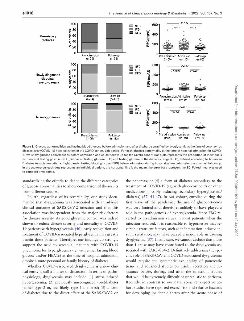
The median follow-up time after symptoms onset was 215 (95% CI 208-222) and 198 (95% CI 110-285) days for the COVID and No-COVID cohorts, respectively. Among discharged patients, 355 out of 475 in the COVID cohort [74.7%, median time after discharge 6 (3-6) months] and 23 out of 65 in the No-COVID cohort [35.4%, median time after discharge 6 (3-12) months] had at least 1 FBG during postdischarge follow-up. Moreover, our retrospective ab-straction from electronic medical records provided FBG values in the year prior to hospital admission for COVID-19 for 199 out of 586 (34%) and 35 out of 74 (47.3%) pa-tients in the COVID and No-COVID cohorts, respectively. In the COVID cohort, we analyzed FBG prior to COVID-19 hospitalization and after hospital discharge in the dif-ferent glucose categories at the time of hospitalization for COVID-19 (Fig. 3). Among 115 patients with preexisting diabetes, in the year prior to admission FBG was available for 50 subjects (43.5%) and during follow-up for 63 out of 76 surviving patients (82.1%). As expected, most pa-tients showed DFG both before and after SARS-CoV-2 in-fection, without significant changes during the time. During the follow-up, no diabetic patients required drug treatment escalation. A marginal trend in increasing FBG was evi-dent during hospitalization [from 137 (116-172) mg/dL to 150 (115-230) mg/dL, P = 0.11], followed by return to preinfection levels during follow-up [134 (108-169) mg/dL, P = 0.84]. Among 295 patients with newly diagnosed dia-betes or hyperglycemia not in the diabetes range, FBG prior to COVID-19 was available for 84 patients (28.5%) and FBG during follow-up for 178 out of the 238 surviving pa-tients (74.8%). Of note, in this group in 33.3% of patients
Dow
nloaded from https://academ
ic.oup.com/jcem
/article/107/3/e1009/6414404 by guest on 12 July 2022

The Journal of Clinical Endocrinology & Metabolism, 2022, Vol. 107, No. 3 e1013
IFG/DFG was already present prior to SARS-CoV-2 in-fection. A significant increase of FBG was evident during hospitalization [from 95 (85-107) to 102 mg/dL (93-115), P = 0.003] followed by return to preinfection levels during follow-up [97.5 (90-107) mg/dL; P = 0.24 vs preinfection levels, P = 0.004 vs hospitalization]. Among the 176 patients with normoglycemia during hospitalization, FBG prior to COVID-19 was available for 65 subjects (36.9%) and glu-cose measurements during follow-up for 112 out of the 161 surviving patients (69.6%). In this group a significant decrease of FBG was evident during hospitalization [from 91 (86-96.5) to 85 (78-89) mg/dL, P < 0.001] followed by a return to preinfection levels during follow-up [94 (88-100) mg/dL; P = 0.72 vs preinfection levels, P < 0.001 vs hospitalization]. A reduction in BMI from baseline was re-ported at the last observation in patients with newly diag-nosed diabetes or hyperglycemia not in the diabetes range during hospitalization (Fig. 4). On the contrary, a modest increase in BMI during follow-up was evident in patients
with normoglycemia during hospitalization. HbA1c did not change significantly during the observation period in all 3 groups (Fig. 4). The same analysis was performed also in the No-COVID cohort with similar results, even if the low number of patients in this cohort does not allow to draw definite conclusions [Supplementary Figure 2 (36)].
Discussion
Whether dysglycemia associated with SARS-CoV-2 in-fection should be considered a specific clinical entity, and whether it persists or reverts when the viral infection re-solves is still a matter of discussion. To address this issue, we studied a cohort of 621 adults patients with suspected COVID-19 pneumonia, assessing dysglycemia not only at the time of hospitalization but also in the year prior to COVID-19 hospitalization and during postdischarge follow-up, when FBG were available. Our study generated several interesting findings.
Figure 1. Dysglycemia associated with severe acute respiratory syndrome coronavirus 2 (SARS-CoV-2) infection. We analyzed a series of 586 cases with confirmed coronavirus disease 2019 (COVID-19; COVID cohort) and 74 cases in which SARS-CoV-2 infection was excluded (No-COVID cohort). (A) Glucose abnormalities at hospital admission. Bar plots represent the proportion of individuals with diabetes (either preexisting or newly diagnosed), hyperglycemia not in the diabetes range and normoglycemia in the COVID-19 cohort and No-COVID cohort. (B and C) Mean glycated hemoglobin levels and mean peak blood glucose levels were summarized for patients with diabetes (either preexisting or newly diagnosed), hyperglycemia not in the diabetes range, and normoglycemia. Scatterplots show the mean ± SD; the error bars represent the SD, and each dot represents an individual patient. (D) Kaplan-Meyer time-to-event analysis for survival without adverse clinical outcome in the three patient groups. Log-rank (Mantel-Cox) test. (E) Forest plot of the hazard ratio for diseases severity (composite endpoint of admission to the intensive care unit or death, whichever occurred first) in the 3 patient groups. Sex-and age-adjusted multivariate Cox proportional hazards model including body mass index, creatinine, hyperten-sion, cardiovascular disease, lactate dehydrogenase, C-reactive protein, and white blood cells. (F) Time to hospital discharge in the 3 patient groups. Scatterplots show the mean ± SD; the error bars represent the SD, with each dot representing an individual patient. (B, C, and E) One-way analysis of variance with Bonferroni correction.
Dow
nloaded from https://academ
ic.oup.com/jcem
/article/107/3/e1009/6414404 by guest on 12 July 2022

e1014 The Journal of Clinical Endocrinology & Metabolism, 2022, Vol. 107, No. 3
Tab
le 1
. D
emo
gra
ph
ics
and
clin
ical
ch
arac
teri
stic
s o
f th
e st
ud
y p
op
ula
tio
n, s
trat
ified
by
glu
cose
cat
ego
ries
at
the
tim
e o
f ad
mis
sio
n f
or
susp
ecte
d C
OV
ID-1
9 p
neu
mo
nia
CO
VID
coh
ort
No-
CO
VID
coh
ort
A
llD
iabe
tes
Hyp
ergl
ycem
iaN
orm
ogly
cem
iaP
Mis
sing
P v
s C
OV
ID
N58
615
425
617
6
74
M
/F39
1/19
597
/78
185/
7198
/78
0.00
10
41/3
30.
069
Age
, yea
rs64
± 1
4.5
68.3
± 1
2.4a
,b63
.6 ±
14
61.1
± 1
6<0
.001
062
± 1
80.
16B
MI,
kg/m
228
± 5
.32
29.7
± 6
a ,b
27.6
± 4
.327
.2 ±
5.8
<0.0
0172
24.7
± 3
.90.
006
Eur
opea
ns49
6 (8
4.6)
136(
88.3
)21
2 (8
2.8)
148
(84.
1)0.
40
63 (
85.1
)0.
86H
yper
tens
ion,
n (
%)
288
(49.
2)10
7 (6
9)11
9 (4
7)62
(35
)<0
.001
125
(33
.8)
0.01
3C
AD
, n (
%)
85 (
14.5
)38
(25
)28
(11
)19
(11
)<0
.001
113
(17
.6)
0.49
AC
E/A
RB
, n (
%)
164
(28.
3)61
(40
)68
(27
)35
(20
)<0
.001
718
(25
)0.
58St
eroi
d th
erap
y du
ring
hos
pita
lizat
ion
112
(19.
1)31
(20.
1)55
(21
.5)
26 (
14.8
)0.
640
——
Swab
neg
ativ
izat
ion,
day
s 40
(38
.2-4
1.7)
41 (
38-4
4)40
(37
-43)
37 (
33-4
1)0.
491
——
Dea
th, n
(%
)11
1 (1
8.9)
50 (
32.5
)46
(18
)15
(8.
5)<0
.001
09
(12.
2)0.
2In
tens
ive
care
uni
t, n
(%)
93 (
15.9
)35
(23
)48
(19
)10
(5.
7)<0
.001
00
(0)
<0.0
01M
ean
fast
ing
glyc
emia
, mg/
dL11
3.6
± 43
.516
2.5
± 57
a,b
104
± 15
c84
.5 ±
9<0
.001
010
2 ±
320.
042
Max
fas
ting
gly
cem
ia, m
g/dL
137.
2 ±
6821
1.3
± 92
a,b
125.
1 ±
22c
89.8
± 8
<0.0
010
113
± 66
0.00
3H
bA1c
, mm
ol/m
ol45
.7 ±
19
53 ±
23a,
b38
± 7
36 ±
5<0
.001
484
41 ±
80.
37W
hite
blo
od c
ount
, ×10
9 /L7
(5.1
-9.8
)7.
6 (5
.8-1
1.7)
a,b
6.9
(5.1
-9.6
)6.
4 (4
.7-9
)<0
.001
09.
1 (6
.9-1
1.4)
<0.0
01R
ed b
lood
cou
nt, m
illio
n/m
m3
4.45
(4.
01-4
.9)
4.4
(3.9
-5)
4.5
(4.1
-4.9
)4.
6 (4
.2-5
.0)
0.24
04.
5 (4
.1-4
.8)
0.52
Hem
oglo
bin,
g/d
L13
(11
.5-1
4.4)
12.8
(11
.3-1
4.4)
13.3
(12
-14.
3)13
(11
.5-1
4.4)
0.15
013
.4 (
12.1
-14.
5)0.
16H
emat
ocri
t, %
38.9
5 (3
5-42
.3)
39.1
(34
.8-4
3.5)
39.2
(35
.7-4
2.2)
38.9
(34
.5-4
2.7)
0.56
039
.45
(35.
9-43
.1)
0.15
Plat
elet
cou
nt, ×
109 /L
232
(170
-309
.5)
236
(175
-314
)23
2 (1
71-3
14)
228
(169
-301
)0.
650
239.
5 (1
72.7
-305
)0.
99N
eutr
ophi
ls, ×
109 /L
5.2
(35-
7.9)
5.8
(4.1
-8.8
)a 5.
2 (3
.6-7
.7)
4.7
(3-6
.2)
<0.0
019
6.35
(4.
6-9.
3)0.
14Ly
mph
ocyt
es, ×
109 /L
1 (0
.7-1
4)1
(0.7
-1.4
)0.
9 (0
.7-1
.2)c
1.2
(0.7
-1.5
)0.
003
161.
4 (0
.9-2
)0.
50M
onoc
ytes
, ×10
9 /L0.
5 (0
.3-0
.7)
0.5
(0.3
-0.8
)0.
4 (0
.3-0
.6)
0.5
(0.4
-0.7
)0.
079
160.
7 (0
.5-0
.8)
0.49
Pati
ents
wit
h a
FBG
ava
ilabl
e in
the
yea
r pr
ior
to a
dmis
sion
, n (
%)
199
(34)
56 (
36.4
)78
(30
.5)
65 (
37)
——
35 (
47.3
)—
Pati
ents
dis
char
ged
aliv
e w
ith
at le
ast
1 FB
G a
t fo
llow
-up,
n (
%)
355
(74.
3)83
(79
.8)
160
(76.
2)11
2 (6
9.6)
——
23 (
35.4
)—
Abb
revi
atio
ns: A
CE
/AR
B, a
ngio
tens
in c
onve
rtin
g en
zym
e/an
giot
ensi
n-re
cept
or b
lock
ers;
BM
I, bo
dy m
ass
inde
x; C
AD
, cor
onar
y ar
tery
dis
ease
; FB
G, f
asti
ng b
lood
glu
cose
; CO
VID
-19,
cor
onav
irus
dis
ease
201
9; M
/F, m
ale/
fem
ale.
a Post
hoc
ana
lysi
s: P
< 0
.05
diab
etes
vs
norm
ogly
cem
ia.
b Post
hoc
ana
lysi
s: P
< 0
.05
diab
etes
vs
hype
rgly
cem
ia.
c Post
hoc
ana
lysi
s: P
< 0
.05
hype
rgly
cine
mic
vs
norm
ogly
cem
ia.
Dow
nloaded from https://academ
ic.oup.com/jcem
/article/107/3/e1009/6414404 by guest on 12 July 2022

The Journal of Clinical Endocrinology & Metabolism, 2022, Vol. 107, No. 3 e1015
First, the dysglycemia associated with COVID-19 (ie, newly diagnosed diabetes or hyperglycemia not in the dia-betes range) resolved in the majority of patients after the SARS-CoV-2 infection resolved. A similar behavior was also evident in the No-COVID cohort, and it is, there-fore, reasonable to speculate that reversible transient fac-tors, such as inflammation-induced insulin resistance, may be causing dysglycemia in patients with pneumonia from SARS-CoV-2 or other pathogens (37). This hypothesis is supported by the association between biochemical markers of inflammation during hospitalization and the degree of dysglycemia. Therefore, this finding does not support the persistence of a long-term disruption of glycometabolic control after SARS-CoV-2 infection, as claimed by other authors (29). One possible explanation for this discrep-ancy is a potential assessment bias (ie, how patients with dysglycemia are identified and classified) in observational studies conducted under emergency conditions. Obviously, a biased stratification of the groups leads to biased conclu-sions, independently by the quality of data and the statis-tical analysis performed. A negative history of diabetes or prediabetes cannot rule out the presence of undiagnosed diabetes. Thanks to the access, although retrospective, to FBG values in the year prior to hospitalization for COVID-19, in our cohort we could document that DFG or IFG were often preexistent in patients that at hospital admission for
suspected COVID-19 were classified as normoglycemics or with newly diagnosed diabetes or with hyperglycemia not in the diabetes range. Moreover, the prevalence of IFG, DFG, and FBG, as well as median HbA1c and BMI before and 6 months after SARS-CoV-2 infection (ie, outside an acute illness) were similar in the 3 groups.
Second, the prevalence of preexisting diabetes was similar in patients of the COVID and No-COVID co-horts, while the prevalence of newly diagnosed diabetes and hyperglycemia not in the diabetes range was higher in the COVID-19 cohort. This suggest that diabetes per se, as risk factor, is not unique to COVID-19. On the other hand, the well-known high level of inflammatory stress associ-ated with the acute phase of COVID-19 pneumonia may account for the excess difference in newly diagnosed dia-betes and hyperglycemia not in the diabetes range between the COVID and No-COVID cohort. Third, the prevalence of the dysglycemia associated with SARS-CoV-2 infec-tion may vary significantly according to the criteria used to define glucose abnormalities. In this study, overt hyper-glycemia was present in 43.7% of patients during hospi-talization. This prevalence is comparable to that reported by Montefusco et al (29) (not unexpectedly, having used the same classification criteria) but certainly higher than that previously reported by us (38) or by other groups (39) using other classification. This underlines the need for
Figure 2. Biochemistry at admission according to glucose abnormalities. Routine blood tests encompassed serum biochemistry [including serum creatinine and lactate dehydrogenase (LDH)], complete blood count with differential, inflammation markers [C-reactive protein (CRP), ferritin, inter-leukin-6 (IL-6)], and D-dimer. Scatterplots show the median; the error bars represent the interquartile range, and each dot represents an individual patient. Kruskal-Wallis with Dunn’s correction was used for comparison between groups.
Dow
nloaded from https://academ
ic.oup.com/jcem
/article/107/3/e1009/6414404 by guest on 12 July 2022
e1016 The Journal of Clinical Endocrinology & Metabolism, 2022, Vol. 107, No. 3
standardizing the criteria to define the different categories of glucose abnormalities to allow comparison of the results from different studies.
Fourth, regardless of its reversibility, our study docu-mented that dysglycemia was associated with an adverse clinical outcome of SARS-CoV-2 infection and that this association was independent from the major risk factors for disease severity. As good glycemic control was indeed shown to reduce disease severity and mortality in COVID-19 patients with hyperglycemia (40), early recognition and treatment of COVID-associated hyperglycemia may greatly benefit these patients. Therefore, our findings do strongly support the need to screen all patients with COVID-19 pneumonia for hyperglycemia (ie, with either fasting blood glucose and/or HbA1c) at the time of hospital admission, despite a mute personal or family history of diabetes.
Whether COVID-associated dysglycemia is a new clin-ical entity is still a matter of discussion. In terms of patho-physiology, dysglycemia may include (1) stress-induced hyperglycemia; (2) previously unrecognized (pre)diabetes (either type 2 or, less likely, type 1 diabetes); (3) a form of diabetes due to the direct effect of the SARS-CoV-2 on
the pancreas; or (4) a form of diabetes secondary to the treatment of COVID-19 (eg, with glucocorticoids or other medications possibly inducing secondary hyperglycemia/diabetes) (37, 41-47). In our cohort, enrolled during the first wave of the pandemic, the use of glucocorticoids was very limited and, therefore, unlikely to have played a role in the pathogenesis of hyperglycemia. Since FBG re-verted to preadmission values in most patients when the infection resolved, it is reasonable to hypothesize that re-versible transient factors, such as inflammation-induced in-sulin resistance, may have played a major role in causing dysglycemia (37). In any case, we cannot exclude that more than 1 cause may have contributed to the dysglycemia as-sociated with SARS-CoV-2. Definitively addressing the spe-cific role of SARS-CoV-2 in COVID-associated dysglycemia would require the systematic availability of pancreatic tissue and advanced studies on insulin secretion and re-sistance before, during, and after the infection, studies that would be extremely difficult or unrealistic to perform. Recently, in contrast to our data, some retrospective co-hort studies have reported excess risk and relative hazards for developing incident diabetes after the acute phase of
Figure 3. Glucose abnormalities and fasting blood glucose before admission and after discharge stratified by dysglycemia at the time of coronavirus disease 2019 (COVID-19) hospitalization in the COVID cohort. Left panels: For each glucose abnormality at the time of hospital admission for COVID-19 we show glucose abnormalities before admission and at last follow-up for the COVID cohort. Bar plots represents the proportion of individuals with normal fasting glucose (NFG), impaired fasting glucose (IFG) and fasting glucose in the diabetes range (DFG), defined according to American Diabetes Association criteria. Right panels: fasting blood glucose (FBG) before admission, during hospitalization (admission), and at last follow-up. In the scatterplots each dots represents an individual patient, the horizontal line is the mean, the error bars represent the SD. Paired t-test was used to compare time points.
Dow
nloaded from https://academ
ic.oup.com/jcem
/article/107/3/e1009/6414404 by guest on 12 July 2022

The Journal of Clinical Endocrinology & Metabolism, 2022, Vol. 107, No. 3 e1017
SARS-CoV-2 infection in adults (48,49). Significant risk differences for type 2 diabetes after SARS-CoV-2 infection ranged from 0.47 to 0.82 per 100 people, and HRs ranged from 1.39 to 1.83. The major limitation of these studies is that the incident new cases in the postacute phase may be just the identification of a preexisting condition that was simply undiagnosed before. In fact, epidemiological evi-dence in many countries suggests that for any 2 patients with diagnosed diabetes there might be 1 with undiagnosed diabetes. Moreover, those studies identified an excess risk for diabetes that was not unique to SARS-CoV-2, since it was also observed with other serious viral infections. For example, compared to individuals who were hospitalized with seasonal influenza, patients who were hospitalized for COVID-19 did not have a significantly higher burden of type 2 diabetes, while they had a significant trend to a lower burden of type 1 diabetes (48,49).
Our study has some limitations. First, our cohort con-sists of hospitalized patients with COVID-19, excluding asymptomatic or paucisymptomatic patients who were treated at home. Second, preadmission and postdischarge FBG were not available for all patients, and we cannot ex-clude a selection bias. Third, death might be an important competing risk (in the sense that prevent us from appreci-ating diabetes cases in patients who are deceased), and we
did not perform a competing risk analysis. Fourth, we only systematically assessed FBG, and we acknowledge that more specific markers of beta cell function, such as serum insulin and or C-peptide levels, would have provided rele-vant information. Fifth, this has been a single-center study, covering mainly Europeans. Consequently, the conclusion should be limited to Europeans, and we cannot exclude that in other ethnic groups the COVID-19 encounter may led to a different clinical outcome in view of different beta cell functional capacity (ie, Asians as compared to Europeans). Finally, a 6-month follow-up may be too short to unveil a direct diabetogenic effects of the SARS-CoV-2 virus. In fact, there may be a delay between immunologic factors or infection damages and the onset of diabetes. It will be, therefore, crucial to conduct long-term follow-up studies in these patients.
Conclusions
COVID-associated dysglycemia is a complication of SARS-CoV-2 infection, and this clinical entity still needs to be ad-equately characterized in relationship with preexisting (pre)diabetes or glucose intolerance. It is clear from our study that patients with COVID-associated dysglycemia had in-creased levels of inflammatory markers and indicators of
Figure 4. Body mass index (BMI) and glycated hemoglobin at admission and after discharge stratified by dysglycemia at the time of coronavirus disease 2019 (COVID-19) hospitalization in the COVID cohort. BMI and glycated hemoglobin at hospital admission and at last follow-up. In the scatterplots each dots represents an individual patient, the horizontal line is the mean; the error bars represent the SD. Paired t-test was used to compare time points.
Dow
nloaded from https://academ
ic.oup.com/jcem
/article/107/3/e1009/6414404 by guest on 12 July 2022
e1018 The Journal of Clinical Endocrinology & Metabolism, 2022, Vol. 107, No. 3
organ injuries and showed a poorer clinical outcome. This strongly support the need to screen all patients COVID-19 pneumonia for hyperglycemia at the time of admission, despite a mute personal or family history of diabetes, and treat their hyperglycemia promptly to achieve and main-tain a good glycemic control during hospitalization. On the other hand, our data do not support the persistence of a long-term disruption of glycometabolic control after SARS-CoV-2 infection. Large epidemiological studies in the next years will be required to clarify whether COVID-19 induce permanent diabetes. However, at this time, any alarmist claim on an increased risk of diabetes after SARS-CoV-2 infection should be interpreted with caution.
AcknowledgmentsThis work was funded by Program Project COVID-19 OSR-
UniSR and Ministero della Salute (COVID-2020-12371617).Author Contributions: A.L.: data curation, investigation, writing
(review and editing); A.C.: data curation, investigation, writing (re-view and editing); C.M.: data curation, investigation, writing (review and editing); A.M.: investigation; R.M.: investigation; R.N.: inves-tigation; C.T.: investigation; P.R.Q.: data curation; F.C.: funding acquisition; V.L.: resources, formal analysis; E.B.: writing (review and editing); M.S.: resources, formal analysis, writing (review and editing); and L.P.: conceptualization, methodology, formal analysis, writing (original draft). L.P. is the guarantor of this work and, as such, had full access to all the data presented in the study and takes responsibility for the integrity of the data and the accuracy of the data analysis. The final manuscript has been read and approved by all named authors.
Additional InformationCorrespondence: Lorenzo Piemonti, MD, Diabetes Research Insti-
tute, San Raffaele Scientific Institute, Via Olgettina 60, 20132 Milan, Italy. Email: [email protected].
Disclosures: The authors have no conflict of interest to disclose in relation to the topic of this manuscript.
Data Availability: Some or all data sets generated during and/or analyzed during the current study are not publicly available but are available from the corresponding author on reasonable request.
References 1. Mantovani A, Byrne CD, Zheng MH, Targher G. Diabetes as a
risk factor for greater COVID-19 severity and in-hospital death: a meta-analysis of observational studies. Nutr Metab Cardiovasc Dis. 2020;30(8):1236-1248.
2. Hussain S, Baxi H, Chand Jamali M, Nisar N, Hussain MS. Burden of diabetes mellitus and its impact on COVID-19 pa-tients: a meta-analysis of real-world evidence. Diabetes Metab Syndr. 2020;14(6):1595-1602.
3. Figliozzi S, Masci PG, Ahmadi N, et al. Predictors of adverse prognosis in COVID-19: a systematic review and meta-analysis. Eur J Clin Invest. 2020;50(10):e13362.
4. Aggarwal G, Lippi G, Lavie CJ, Henry BM, Sanchis-Gomar F. Diabetes mellitus association with coronavirus disease 2019
(COVID-19) severity and mortality: a pooled analysis. J Diabetes. 2020;12(11):851-855.
5. Lee MH, Wong C, Ng CH, Yuen DC, Lim AY, Khoo CM. Effects of hyperglycaemia on complications of COVID-19: a meta-analysis of observational studies. Diabetes Obes. Metab. 2021;23(1):287-289.
6. Fu EL, Janse RJ, de Jong Y, et al. Acute kidney injury and kidney replacement therapy in COVID-19: a systematic review and meta-analysis. Clin Kidney J. 2020;13(4):550-563.
7. Piazza G, Campia U, Hurwitz S, et al. Registry of arterial and venous thromboembolic complications in patients with COVID-19. J Am Coll Cardiol. 2020;76(18):2060-2072.
8. Li J, Wang X, Chen J, Zuo X, Zhang H, Deng A. COVID-19 infection may cause ketosis and ketoacidosis. Diabetes Obes Metab. 2020;22(10):1935-1941.
9. Li J, Wang X, Chen J, Zuo X, Zhang H, Deng A. COVID-19 infection may cause ketosis and ketoacidosis. Diabetes Obes Metab. 2020;22(10):1935-1941.
10. Chee YJ, Ng SJH, Yeoh E. Diabetic ketoacidosis precipitated by Covid-19 in a patient with newly diagnosed diabetes mellitus. Diabetes Res Clin Pract. 2020;164:108166.
11. Goldman N, Fink D, Cai J, Lee YN, Davies Z. High prevalence of COVID-19-associated diabetic ketoacidosis in UK secondary care. Diabetes Res Clin Pract. 2020;166:108291.
12. Hollstein T, Schulte DM, Schulz J, et al. Autoantibody-negative insulin-dependent diabetes mellitus after SARS-CoV-2 infection: a case report. Nat Metab. 2020;2(10):1021-1024.
13. Rafique S, Ahmed FW. A case of combined diabetic ketoacidosis and hyperosmolar hyperglycemic state in a patient with COVID-19. Cureus. 2020;12(7):e8965.
14. Unsworth R, Wallace S, Oliver NS, et al. New-onset type 1 dia-betes in children during COVID-19: multicenter regional find-ings in the U.K. Diabetes Care. 2020;43(11):e170-e171.
15. Heaney AI, Griffin GD, Simon EL. Newly diagnosed diabetes and diabetic ketoacidosis precipitated by COVID-19 infection. Am J Emerg Med. 2020;38(11):2491.e3-2491.e4.
16. Soliman AT, Al-Amri M, Alleethy K, Alaaraj N, Hamed N, De Sanctis V. Newly-onset type 1 diabetes mellitus precipi-tated by COVID-19 in an 8-month-old infant. Acta Biomed. 2020;91(3):e2020046.
17. Marchand L, Pecquet M, Luyton C. Type 1 diabetes onset trig-gered by COVID-19. Acta Diabetol. 2020;57(10):1265-1266.
18. Liang X, Xu J, Xiao W, Shi L, Yang H. The association of dia-betes with COVID-19 disease severity: evidence from adjusted effect estimates. Hormones (Athens). 2021;20(2):409-414.
19. Hoffmann M, Kleine-Weber H, Schroeder S, et al. SARS-CoV-2 cell entry depends on ACE2 and TMPRSS2 and is blocked by a clinically proven protease inhibitor. Cell. 2020;181(2):271-280.e8.
20. Fignani D, Licata G, Brusco N, et al. SARS-CoV-2 receptor angio-tensin I-converting enzyme type 2 (ACE2) is expressed in human pancreatic β-cells and in the human pancreas microvasculature. Front Endocrinol (Lausanne). 2020;11:596898.
21. Yang L, Han Y, Nilsson-Payant BE, et al. A human pluripotent stem cell-based platform to study SARS-CoV-2 tropism and model virus infection in human cells and organoids. Cell Stem Cell. 2020;27(1):125-136.e7.
22. Müller JA, Groß R, Conzelmann C, et al. SARS-CoV-2 infects and replicates in cells of the human endocrine and exocrine pan-creas. Nat Metab. 2021;3(2):149-165.
Dow
nloaded from https://academ
ic.oup.com/jcem
/article/107/3/e1009/6414404 by guest on 12 July 2022
The Journal of Clinical Endocrinology & Metabolism, 2022, Vol. 107, No. 3 e1019
23. Tang X, Uhl S, Zhang T, et al. SARS-CoV-2 infection induces beta cell transdifferentiation. Cell Metab. 2021;33(8):1577-1591.e7.
24. Coate KC, Cha J, Shrestha S, et al; HPAP Consortium. SARS-CoV-2 cell entry factors ACE2 and TMPRSS2 are expressed in the microvasculature and ducts of human pancreas but are not enriched in β cells. Cell Metab. 2020;32(6):1028-1040.e4.
25. Kusmartseva I, Wu W, Syed F, et al. Expression of SARS-CoV-2 entry factors in the pancreas of normal organ donors and indi-viduals with COVID-19. Cell Metab. 2020;32(6):1041-1051.e6.
26. Liu F, Long X, Zhang B, Zhang W, Chen X, Zhang Z. ACE2 expression in pancreas may cause pancreatic damage after SARS-CoV-2 infection. Clin Gastroenterol Hepatol. 2020;18(9):2128-2130.e2.
27. Zhu L, She ZG, Cheng X, et al. Association of blood glucose control and outcomes in patients with COVID-19 and pre-existing type 2 diabetes. Cell Metab. 2020;31(6):1068-1077.e3.
28. Akarsu C, Karabulut M, Aydin H, et al. Association between acute pancreatitis and COVID-19: could pancreatitis be the missing piece of the puzzle about increased mortality rates? J Invest Surg. Published online November 2, 2020. Doi:10.1080/08941939.2020.1833263
29. Montefusco L, Nasr MB, D’Addio F, et al. Acute and long-term disruption of glycometabolic control after SARS-CoV-2 infec-tion. Nat Metab. 2021;3:774-785.
30. Molinari C, Laurenzi A, Caretto A, et al. Dysglycemia after COVID-19 pneumonia: a six-month cohort study. Acta Diabetol. 2021;58(11):1481-1490.
31. Rovere-Querini P, Tresoldi C, Conte C, et al. Biobanking for COVID-19 research. Panminerva Med. Published online October 19, 2020. Doi:10.23736/S0031-0808.20.04168-3
32. Ciceri F, Castagna A, Rovere-Querini P, et al. Early predictors of clinical outcomes of COVID-19 outbreak in Milan, Italy. Clin Immunol. 2020;217:108509.
33. Molinari C, Laurenzi A, Caretto A, et al. Dysglycemia after COVID-19 pneumonia: a six-month cohort study. Acta Diabetol. 2021;58(11):1481-1490.
34. Secchi M, Bazzigaluppi E, Brigatti C, et al. COVID-19 sur-vival associates with the immunoglobulin response to the SARS-CoV-2 spike receptor binding domain. J Clin Invest. 2020;130(12):6366-6378.
35. American Diabetes Association. 15. Diabetes care in the hos-pital: standards of medical care in diabetes—2020. Diabetes Care. 2020;43(supp 1):S193-S202.
36. Laurenzi A, Caretto A, Molinari C, et al. Data from: no evi-dence of long-term disruption of glycometabolic control after SARS-CoV-2 infection. Posted September 29, 2021. FigShare. doi:10.6084/m9.figshare.16543146.v2
37. Sathish T, Tapp RJ, Cooper ME, Zimmet P. Potential metabolic and inflammatory pathways between COVID-19 and new-onset diabetes. Diabetes Metab. 2021;47(2):101204.
38. Lampasona V, Secchi M, Scavini M, et al. Antibody response to multiple antigens of SARS-CoV-2 in patients with diabetes: an ob-servational cohort study. Diabetologia. 2020;63(12):2548-2558.
39. Sathish T, Kapoor N, Cao Y, Tapp RJ, Zimmet P. Proportion of newly diagnosed diabetes in COVID-19 patients: a sys-tematic review and meta-analysis. Diabetes Obes Metab. 2021;23(3):870-874.
40. Sardu C, D’Onofrio N, Balestrieri ML, et al. Outcomes in patients with hyperglycemia affected by COVID-19: can we do more on glycemic control? Diabetes Care. 2020;43(7):1408-1415.
41. Boddu SK, Aurangabadkar G, Kuchay MS. New onset dia-betes, type 1 diabetes and COVID-19. Diabetes Metab Syndr. 2020;14(6):2211-2217.
42. Papachristou S, Stamatiou I, Stoian AP, Papanas N. New-onset diabetes in COVID-19: time to frame its fearful symmetry. Diabetes Ther. 2021;12(2):461-464.
43. Unsworth R, Wallace S, Oliver NS, et al. New-onset type 1 dia-betes in children during COVID-19: multicenter regional find-ings in the U.K. Diabetes Care. 2020;43(11):e170-e171.
44. Nassar M, Nso N, Baraka B, et al. The association between COVID-19 and type 1 diabetes mellitus: A systematic review. Diabetes Metab Syndr. 2021;15(1):447-454.
45. Apicella M, Campopiano MC, Mantuano M, Mazoni L, Coppelli A, Del Prato S. COVID-19 in people with diabetes: understanding the reasons for worse outcomes. Lancet Diabetes Endocrinol. 2020;8(9):782-792.
46. Smith SM, Boppana A, Traupman JA, et al. Impaired glu-cose metabolism in patients with diabetes, prediabetes, and obesity is associated with severe COVID-19. J Med Virol. 2021;93(1):409-415.
47. Wang J, Meng W. COVID-19 and diabetes: the contributions of hyperglycemia. J Mol Cell Biol. 2020;12(12):958-962.
48. Daugherty SE, Guo Y, Heath K, et al. Risk of clinical sequelae after the acute phase of SARS-CoV-2 infection: retrospective co-hort study. BMJ. 2021;373:n1098.
49. Al-Aly Z, Xie Y, Bowe B. High-dimensional characterization of post-acute sequelae of COVID-19. Nature. 2021;594(7862):259-264.
Dow
nloaded from https://academ
ic.oup.com/jcem
/article/107/3/e1009/6414404 by guest on 12 July 2022