pbac004.pdf - Oxford Academic
11
Precision Clinical Medicine, 2022, 5: pbac004 DOI: 10.1093/pcmedi/pbac004 Advance access publication date: 3 February 2022 Review Tumorigenicity risk of iPSCs in vivo: nip it in the bud Chaoliang Zhong 1,§ , Miao Liu 1,§ , Xinghua Pan 2,3, * and Haiying Zhu 1, * 1 Department of Cell Biology, Naval Medical University, Shanghai 200433, China 2 Department of Biochemistry and Molecular Biology, School of Basic Medical Sciences, and Guangdong Provincial Key Laboratory of Single Cell Technology and Application, Southern Medical University, Guangzhou 510515, China 3 Shenzhen Bay Laboratory, Shenzhen 518032, China ∗ Correspondence: Xinghua Pan, [email protected]; Haiying Zhu, [email protected] § Chaoliang Zhong and Miao Liu contributed equally to the work. Abstract In 2006,Takahashi and Yamanaka first created induced pluripotent stem cells from mouse fibroblasts via the retroviral introduction of genes encoding the transcription factors Oct3/4, Sox2, Klf44, and c-Myc. Since then, the future clinical application of somatic cell reprogramming technology has become an attractive research topic in the field of regenerative medicine. Of note, considerable interest has been placed in circumventing ethical issues linked to embryonic stem cell research. However, tumorigenicity, immunogenicity, and heterogeneity may hamper attempts to deploy this technology therapeutically.This review highlights the progress aimed at reducing induced pluripotent stem cells tumorigenicity risk and how to assess the safety of induced pluripotent stem cells cell therapy products. Keywords: induced pluripotent stem cells (iPSCs), tumorigenicity, regenerative medicine, reprogramming transcription factors, chemical-induced reprogramming, drug-inducible suicide system Introduction Induced pluripotent stem cells (iPSCs), characterized by self- renewal and multiple differentiation potential, have been ex- plored and applied in regenerative therapy, disease modeling, drug toxicity evaluation, and developmental biology. To date, transcrip- tion factor (TF)-mediated and chemical inductive reprogramming have been the strategies of choice to obtain iPSCs. 1–4 Specifi- cally, in TF-mediated reprogramming, the Yamanaka factors, Oct- 4, Sox2, c-Myc, and Klf4 (OSMK), or alternative TFs (Table 1), are introduced into somatic cells with viral or non-viral vectors. In the chemical inductive reprogramming strategy, somatic cells are induced to become PSC through small molecules, cytokines, or growth factors. 4–6 Romanazzo et al. summarized the merits and demerits of these reprogramming strategies and pointed out that the TFs used in the transgenic strategy lead to random epigenetic events, the activation of various pluripotent genes (i.e. Oct-4 and c-Myc), and genomic instability [e.g. chromosomal aberrations, copy number variations (CNVs), and single nucleotide variants (SNVs)]. 7 Such events were identified predominantly in iPSCs com- pared with embryonic stem cells (ESCs), suggesting a potential link between iPSCs and tumorigenicity. Compared with the trans- genic strategy, chemical induction seems to decrease the possi- bility of tumorigenesis. However, the lower reprogramming effi- ciency and the induction system’s complexity are still obstacles to the clinical application of chemical induction. Additionally, the heterogeneity of the cellular product derived from iPSCs for clinical therapy could lead to potential tumori- genicity risks in vivo. Theoretically, regardless of the strategy used to generate iPSCs, it may be impossible to avoid their hetero- geneity due to the heterogeneity of the original somatic cells. Specifically, the established iPSC lines may still contain somatic cells and partially reprogrammed cells similar to iPSCs. Thus, the final cell preparation obtained may be a mixture of target cells for therapeutic use, the residual undifferentiated iPSCs, or partially differentiated iPSCs with teratoma-forming potential. 18 The complicated in vivo environment makes cell integration and reproduction uncontrolled. Hence, decreasing the heterogene- ity and increasing the controllability of iPSCs and cell prepa- ration through isolation, purification, amplification, and mod- ification of stem cells are necessary to decrease the tumori- genicity potential. Significant progress has been made over the last two decades in this research field, although many problems remain. Finally, we focused on the current discussion of the safety assessment of cell therapy products (CTP) of iPSCs, including genome integrity, heterogeneity, and in vivo tumorigenicity. Al- though there are no mandatory provisions issued yet, the evalua- tion of iPSC genome integrity is recommended as one of the most important items because it presents a close association with the tumorigenicity of the iPSC products. 19 To date, many alternative methods for checking genetic mutations have become available. However, the cost of the procedure, the complexity of results inter- pretation, and the workload of data analysis have to be considered when the practical methods are considered. 20 Optimizing the cocktail of reprogramming factors As shown in Table 1, the past decade has seen the establishment of several combinations of TFs that can efficiently reprogram somatic cells based on the Yamanaka factors. Of these, c-Myc is the most controversial TF.It is well known that c-Myc is a proto- oncogene, encoding the family of beta helix–loop–helix/leucine zinc finger TFs, 21 and its deregulated expression occurs in a Received: December 1, 2021. Revised: January 18, 2022. Accepted: January 23, 2022 C The Author(s) 2022. Published by Oxford University Press on behalf of the West China School of Medicine & West China Hospital of Sichuan University. This is an Open Access article distributed under the terms of the Creative Commons Attribution License (https://creativecommons.org/licenses/by/4.0/), which permits unrestricted reuse, distribution, and reproduction in any medium, provided the original work is properly cited. Downloaded from https://academic.oup.com/pcm/article/5/1/pbac004/6521459 by guest on 10 July 2022
-
Upload
khangminh22 -
Category
Documents
-
view
1 -
download
0
Transcript of pbac004.pdf - Oxford Academic
Precision Clinical Medicine, 2022, 5: pbac004
DOI: 10.1093/pcmedi/pbac004Advance access publication date: 3 February 2022
Review
Tumorigenicity risk of iPSCs in vivo: nip it in the budChaoliang Zhong1,§, Miao Liu1,§, Xinghua Pan2,3,* and Haiying Zhu1,*
1Department of Cell Biology, Naval Medical University, Shanghai 200433, China2Department of Biochemistry and Molecular Biology, School of Basic Medical Sciences, and Guangdong Provincial Key Laboratory of Single Cell Technology andApplication, Southern Medical University, Guangzhou 510515, China3Shenzhen Bay Laboratory, Shenzhen 518032, China∗Correspondence: Xinghua Pan, [email protected]; Haiying Zhu, [email protected]§Chaoliang Zhong and Miao Liu contributed equally to the work.
Abstract
In 2006, Takahashi and Yamanaka first created induced pluripotent stem cells from mouse fibroblasts via the retroviral introductionof genes encoding the transcription factors Oct3/4, Sox2, Klf44, and c-Myc. Since then, the future clinical application of somatic cellreprogramming technology has become an attractive research topic in the field of regenerative medicine. Of note, considerable interesthas been placed in circumventing ethical issues linked to embryonic stem cell research. However, tumorigenicity, immunogenicity, andheterogeneity may hamper attempts to deploy this technology therapeutically. This review highlights the progress aimed at reducinginduced pluripotent stem cells tumorigenicity risk and how to assess the safety of induced pluripotent stem cells cell therapy products.
Keywords: induced pluripotent stem cells (iPSCs), tumorigenicity, regenerative medicine, reprogramming transcription factors,chemical-induced reprogramming, drug-inducible suicide system
IntroductionInduced pluripotent stem cells (iPSCs), characterized by self-renewal and multiple differentiation potential, have been ex-plored and applied in regenerative therapy, disease modeling, drugtoxicity evaluation, and developmental biology. To date, transcrip-tion factor (TF)-mediated and chemical inductive reprogramminghave been the strategies of choice to obtain iPSCs.1–4 Specifi-cally, in TF-mediated reprogramming, the Yamanaka factors, Oct-4, Sox2, c-Myc, and Klf4 (OSMK), or alternative TFs (Table 1), areintroduced into somatic cells with viral or non-viral vectors. Inthe chemical inductive reprogramming strategy, somatic cells areinduced to become PSC through small molecules, cytokines, orgrowth factors.4–6 Romanazzo et al. summarized the merits anddemerits of these reprogramming strategies and pointed out thatthe TFs used in the transgenic strategy lead to random epigeneticevents, the activation of various pluripotent genes (i.e. Oct-4 andc-Myc), and genomic instability [e.g. chromosomal aberrations,copy number variations (CNVs), and single nucleotide variants(SNVs)].7 Such events were identified predominantly in iPSCs com-pared with embryonic stem cells (ESCs), suggesting a potentiallink between iPSCs and tumorigenicity. Compared with the trans-genic strategy, chemical induction seems to decrease the possi-bility of tumorigenesis. However, the lower reprogramming effi-ciency and the induction system’s complexity are still obstaclesto the clinical application of chemical induction.
Additionally, the heterogeneity of the cellular product derivedfrom iPSCs for clinical therapy could lead to potential tumori-genicity risks in vivo. Theoretically, regardless of the strategy usedto generate iPSCs, it may be impossible to avoid their hetero-geneity due to the heterogeneity of the original somatic cells.Specifically, the established iPSC lines may still contain somaticcells and partially reprogrammed cells similar to iPSCs. Thus,
the final cell preparation obtained may be a mixture of targetcells for therapeutic use, the residual undifferentiated iPSCs, orpartially differentiated iPSCs with teratoma-forming potential.18
The complicated in vivo environment makes cell integration andreproduction uncontrolled. Hence, decreasing the heterogene-ity and increasing the controllability of iPSCs and cell prepa-ration through isolation, purification, amplification, and mod-ification of stem cells are necessary to decrease the tumori-genicity potential. Significant progress has been made over thelast two decades in this research field, although many problemsremain.
Finally, we focused on the current discussion of the safetyassessment of cell therapy products (CTP) of iPSCs, includinggenome integrity, heterogeneity, and in vivo tumorigenicity. Al-though there are no mandatory provisions issued yet, the evalua-tion of iPSC genome integrity is recommended as one of the mostimportant items because it presents a close association with thetumorigenicity of the iPSC products.19 To date, many alternativemethods for checking genetic mutations have become available.However, the cost of the procedure, the complexity of results inter-pretation, and the workload of data analysis have to be consideredwhen the practical methods are considered.20
Optimizing the cocktail of reprogrammingfactorsAs shown in Table 1, the past decade has seen the establishmentof several combinations of TFs that can efficiently reprogramsomatic cells based on the Yamanaka factors. Of these, c-Myc isthe most controversial TF. It is well known that c-Myc is a proto-oncogene, encoding the family of beta helix–loop–helix/leucinezinc finger TFs,21 and its deregulated expression occurs in a
Received: December 1, 2021. Revised: January 18, 2022. Accepted: January 23, 2022C© The Author(s) 2022. Published by Oxford University Press on behalf of the West China School of Medicine & West China Hospital of Sichuan University. Thisis an Open Access article distributed under the terms of the Creative Commons Attribution License (https://creativecommons.org/licenses/by/4.0/), whichpermits unrestricted reuse, distribution, and reproduction in any medium, provided the original work is properly cited.
Dow
nloaded from https://academ
ic.oup.com/pcm
/article/5/1/pbac004/6521459 by guest on 10 July 2022

2 | Precis Clin Med, 2022, 5: pbac004
Table 1. Representative cocktail of TFs or TF-chemicals applied in the generation of iPSCs.
TFs Chemicals Efficiency Original cell Ref.
OSKM / 0.02% Human dermal fibroblasts 8
OSNL / 0.022% IMR90 fetal fibroblasts 2
OK & L-Myc / 0.016% MEF 9
OKM / 0.001%–0.002% Mouse neural progenitor cells 10
OSE / ∼50% of OSKM MEF 11
OSK VPA 0.05% MEF 6
OSK / <0.001% Human dermal fibroblasts 12
OSNL / 0.01% Human newborn foreskinfibroblasts
2
OSK VPA 1.1% Human neonatal foreskinfibroblasts
13
OS VPA 0.004% Human neonatal dermalfibroblasts
13
OK RepSox 0.05% MEF 14
OSK CHIR99021 0.2%–0.4% MEF 15
O VPA, HIR99021, RepSox 0.3% MEF 16
SKM VPA, SB431542 0.02% MEF 17
OSNL: Oct4, Sox2, Nanog, Lin28; OSE: Oct4, Sox2, Esrrb; OSK: Oct4, Sox2, Klf4; OS: Oct4, Sox2; OK: Oct4, Klf4; O: Oct4; SKM: Sox2, Klf4, c-Myc; VPA: valproic acid;MEF: mouse embryonic fibroblasts.
wide range of human cancers, which leads to the discussionabout the connection between c-Myc and iPSCs tumorigenicity.Hence, some researchers prepared iPSCs without c-Myc-basedcell therapy to explore whether the absence of exogenous c-Myccan reduce iPSCs tumorigenic capacity without influencing thepluripotency.9, 12, 22 For example, Li et al. reported that 3-geneiPSCs (without c-Myc) differentiate into functional hepatocytesafter receiving proper differentiation stimuli. iPSCs and iPSC-derived hepatocytes could decrease the thioacetamide-inducedhepatic necrosis of mice and restore liver function in mice withlethal acute hepatic failure after undergoing intravenous orintrasplenic transplantation. This study highlights the poten-tial of iPSCs, without c-Myc, in diminishing the incidence oftumorigenesis of cell transplantation.23
Alternatively, it is reported that c-Myc paralogs, such as L-Mycand N-Myc can be used to reduce teratoma in iPSCs.21 Nakagawaet al. chose L-Myc and c-Myc mutants (W136E c-Myc mutant anddN2 c-Myc mutant) to balance the efficiency and safety in re-programming. Notably, compared to the mice derived from Myc-minus iPSCs, those from L-Myc iPSCs did not present higher tu-morigenicity. Importantly, although the mice from L-Myc-iPSCsexhibited slightly higher mortality, these alternatives to the Mycfamily are worth considering when obtaining iPSCs with non/lowteratogenicity from human somatic cells.9
In addition, some chemicals can replace specific TFs to helpimprove reprogramming efficiency,14–17 but the systematic stud-ies on the tumorigenicity of iPSCs derived from different cock-tails are not sufficient. Pushp et al. argued that cocktails con-taining TFs and small molecules are better in reprogrammingefficiency than chemical cocktails alone, exhibiting less tumori-genicity than TFs only.24 The optimized formulas are listed in Ta-ble 1. For instance, Maherali et al. demonstrated that ALK4/5/7inhibitor SB-431542 replacing exogenous c-Myc improved the re-programming efficiency of mouse embryonic fibroblasts (MEF).25
Huangfu and colleagues discovered that valproic acid (VPA), a his-tone deacetylase inhibitor, and DNA methyltransferase inhibitorsfacilitate MEF and primary human fibroblast reprogramming pro-cesses.26,13 Also, either RepSox or CHIR99021 could substitute forSox2 and c-Myc,14,15 and a combination of Oct4, VPA, CHIR99021,and RepSox could induce reprogramming of MEF to form alkaline
phosphatase (AP)-positive clones.16 Unfortunately, there was noarticle showing results regarding the tumorigenicity of iPSCs de-rived from different cocktails.
Strategy of chemical-inducedreprogrammingStudies have shown that cell-fate reprogramming is compara-bly successful through the use of chemicals instead of conven-tional gene transformation.4–6,27–33As listed in Table 2, Deng andhis team reported a series of significant achievements.4,28,34,35
Specifically, they obtained PSCs, named CiPSCs (chemically in-duced PSCs),4,28 through chemical inductive reprogramming, butalso obtained transdifferentiated cells directly from original so-matic cells, known as direct reprogramming or lineage reprogram-ming.28,34 In addition, recent reports demonstrated that biochem-ical signals (e.g. cytokines, growth factors, extracellular matrix(ECM) proteins, and small molecules) directly generated the targetterminally differentiated cells from original somatic cells withoutmaking iPSCs in advance.7 Li et al. reported that the chemically in-duced extra-embryonic endoderm (XEN)-like cells obtained fromMEF can be induced and reprogrammed directly to functionalneurons and hepatocytes, bypassing the pluripotent state.36 Suchfindings might lead to a decreased tumorigenesis risk of iPSCs.However, a related systematic study on the underlying mecha-nism and tumorigenesis risk has not been reported to date. More-over, unlike TFs, chemicals are mostly synthetic with clear tar-gets for regulating biological activities, primarily through recep-tors and enzymes. More studies are needed to develop chemi-cals for reprogramming cell fate as reliably and rationally as TFsbut without safety concerns. Chen et al. summarized the smallmolecule combinations that have been demonstrated with suf-ficient efficiency in somatic cell reprogramming and lineage re-programming.6 It is worth noting that there are a few chemicalsthat promote reprogramming effectively through epigenetic mod-ifications, such as histone deacetylase inhibitor TSA, SAHA andVPA, DNMT (DNA methyltransferase) inhibitor 5-AZA and RG108,histone methyltransferase G9a (Bix-01294) and H3K36 demethy-lase vitamin C.6 Fu et al. found that crotonic acid facilitated
Dow
nloaded from https://academ
ic.oup.com/pcm
/article/5/1/pbac004/6521459 by guest on 10 July 2022
How to decrease tumorigenicity risk of iPSCs | 3
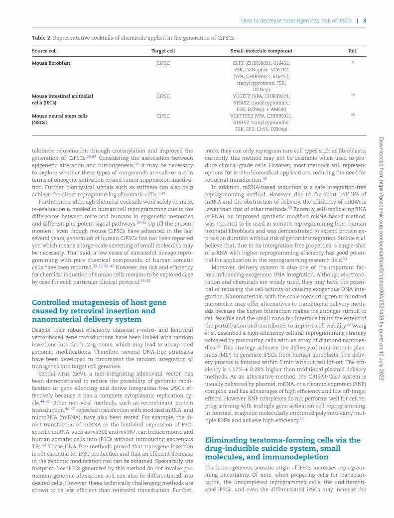
Table 2. Representative cocktails of chemicals applied in the generation of CiPSCs.
Source cell Target cell Small-molecule compound Ref.
Mouse fibroblast CiPSC C6FZ (CHIR99021, 616452,FSK, DZNep) or VC6TFZ(VPA, CHIR99021, 616452,
tranylcypromine, FSK,DZNep)
4
Mouse intestinal epithelialcells (IECs)
CiPSC VC6TFZ (VPA, CHIR99021,616452, tranylcypromine,
FSK, DZNep) + AM580
28
Mouse neural stem cells(NSCs)
CiPSC VC6TFE5Z (VPA, CHIR99021,616452, tranylcypromine,FSK, EPZ, Ch55, DZNep)
28
telomere rejuvenation through crotonylation and improved thegeneration of CiPSCs.29,37 Considering the association betweenepigenetic alteration and tumorigenesis,38 it may be necessaryto explore whether these types of compounds are safe or not interms of oncogene activation or/and tumor suppression inactiva-tion. Further, biophysical signals such as stiffness can also helpachieve the direct reprogramming of somatic cells.7,39
Furthermore, although chemical cocktails work safely on mice,re-evaluation is needed in human cell reprogramming due to thedifferences between mice and humans in epigenetic memoriesand different pluripotent signal pathways.30–32 Up till the presentmoment, even though mouse CiPSCs have advanced in the lastseveral years, generation of human CiPSCs has not been reportedyet, which means a large-scale screening of small molecules maybe necessary. That said, a few cases of successful lineage repro-gramming with pure chemical compounds, of human somaticcells have been reported.33,35, 40–42 However, the risk and efficiencyfor chemical induction of human cells remains to be explored caseby case for each particular clinical protocol.39,43
Controlled mutagenesis of host genecaused by retroviral insertion andnanomaterial delivery systemDespite their robust efficiency, classical γ -retro- and lentiviralvector-based gene transductions have been linked with randominsertions into the host genome, which may lead to unexpectedgenomic modifications. Therefore, several DNA-free strategieshave been developed to circumvent the random integration oftransgenes into target cell genomes.
Sendai-virus (SeV), a non-integrating adenoviral vector, hasbeen demonstrated to reduce the possibility of genomic modi-fication or gene silencing and derive integration-free iPSCs ef-fectively because it has a complete cytoplasmic replication cy-cle.44,45 Other non-viral methods, such as recombinant proteintransduction,46,47 repeated transfection with modified mRNA, andmicroRNA (miRNA), have also been tested. For example, the di-rect transfection of miRNA or the lentiviral expression of ESC-specific miRNA, such as mir302 and mir367, can induce mouse andhuman somatic cells into iPSCs without introducing exogenousTFs.48 These DNA-free methods proved that transgene insertionis not essential for iPSC production and that an efficient decreasein the genomic modification risk can be obtained. Specifically, thefootprint-free iPSCs generated by this method do not involve per-manent genomic alterations and can also be differentiated intodesired cells. However, these technically challenging methods areshown to be less efficient than retroviral transduction. Further-
more, they can only reprogram rare cell types such as fibroblasts;currently, this method may not be desirable when used to pro-duce clinical-grade cells. However, most methods still representoptions for in vitro biomedical applications, reducing the need forretroviral transduction.49
In addition, mRNA-based induction is a safe integration-freereprogramming method. However, due to the short half-life ofmRNA and the obstruction of delivery, the efficiency of mRNA islower than that of other methods.50 Recently, self-replicating RNA(srRNA), an improved synthetic modified mRNA-based method,was reported to be used in somatic reprogramming from humanneonatal fibroblasts and was demonstrated to extend protein ex-pression duration without risk of genomic integration. Steinle et al.believe that, due to its intergration-free properties, a single-shotof srRNA with higher reprogramming efficiency has good poten-tial for application in the reprogramming research field.51
Moreover, delivery system is also one of the important fac-tors influencing exogenous DNA integration. Although electropo-ration and chemicals are widely used, they may have the poten-tial of reducing the cell activity or causing exogenous DNA inte-gration. Nanomaterials, with the scale measuring ten to hundrednanometer, may offer alternatives to tranditional delivery meth-ods because the higher interaction makes the stronger stimuli tocell feasible and the small nano-bio interface limits the extent ofthe perturbation and contributes to improve cell viability.52 Wanget al. described a high-efficiency cellular reprogramming strategyachieved by puncturing cells with an array of diamond nanonee-dles.53 This strategy achieves the delivery of mini-intronic plas-mids (MIP) to generate iPSCs from human fibroblasts. The deliv-ery process is finished within 5 min without cell lift-off. The effi-ciency is 1.17% ± 0.28% higher than traditional plasmid deliverymethods. As an alternative method, the CRISPR/Cas9 system isusually delivered by plasmid, mRNA, or a ribonucleoprotein (RNP)complex, and has advantages of high efficiency and low off-targeteffects. However, RNP complexes do not performs well for cell re-programming with multiple gene activation cell reprogramming.In contrast, magnetic molecularly imprinted polymers carry mul-tiple RNPs and achieve high efficiency.54
Eliminating teratoma-forming cells via thedrug-inducible suicide system, smallmolecules, and immunodepletionThe heterogeneous somatic origin of iPSCs increases reprogram-ming uncertainty. Of note, when preparing cells for transplan-tation, the uncompleted reprogrammed cells, the undifferenti-ated iPSCs, and even the differentiated iPSCs may increase the
Dow
nloaded from https://academ
ic.oup.com/pcm
/article/5/1/pbac004/6521459 by guest on 10 July 2022

4 | Precis Clin Med, 2022, 5: pbac004
Figure 1. Mechanism of iCASP9’s safety switch. The AAVS1 TALEN and the donor constructs are delivered to cells via plasmid transfection. Then, theantibiotic-resistant cells are selected. Next, the administration of APs leads to the dimerization of FKBP12-F36V. As a result, caspase9 and thedownstream effector caspases, such as caspase-3 and caspase-7, are activated, which leads to cell apoptosis.
oncogenic potential of therapeutic cells. This observation is a con-sequence of genetic or epigenetic aberrations from cellular repro-gramming or prolonged cell culture.26,55,56 Therefore, for safe clin-ical application of iPSCs, it is essential to eliminate these cells dur-ing cell preparation. To this end, suicide gene technology is cur-rently widely used to improve the safety of stem cell-based ther-apy. Specifically, the approach is to selectively eliminate aberranttherapeutic cells by activating a highly efficient safety switch. Todate, three main suicide strategies have been developed.
One of these strategies is to use an anti-CD20 monoclonalantibody to induce antibody-dependent cytotoxicity, thus killingthe cells expressing the B-specific human CD20 gene exoge-nously. For example, Inrona et al. achieved the elimination ofCD3 + CD20 + human T cells by adding monoclonal, chimericanti-CD20 IgG1(kappa) Rituximab antibody (Roche) in the pres-ence of complement. However, to date, no experiments have beenperformed applying CD20 to the iPSCs field.57
The second strategy is to use a metabolic suicide gene, herpessimplex virus thymidine kinase (HSV-TK) gene, and its prodrug,ganciclovir (GCV). The product of GCV yielded through phospho-rylation by HSV-TK incorporates into replicating DNA, causing cellapoptosis. HSV-TK has been tested in human iPSCs as a suicidegene system.58–60 Studies have demonstrated that the HSV-TK-expressing cells can be eliminated both in vitro and in vivo withhigh specificity and efficiency.59–62 However, as Kimura et al. in-sisted, the HSV-TK system cannot sufficiently shrink the iPSC-derived teratomas in vivo.58 Furthermore, as a potent cytotoxicantiviral drug, GCV often unavoidably kills transplanted cells ex-pressing the HSV-TK suicide gene system when used to treat her-pes virus infections.63, 64
The most recent suicide gene system, inducible caspase-9(iCASP9), highlights the potential of iPSC-based regenerative ther-apy with improved safety.65 Its operating principle is to substi-tute the caspase recruitment domain of pro-apoptotic caspase-9with a mutated dimerizer drug-binding domain from the humanFK506-binding protein (FKBP12-F36V).66,67AP1903 (aka rimiducid),a chemical inducer, binds to the F36V mutation with high affin-ity. Consequently, the dimerization of F36V, and the activation of
caspase-9 and downstream effector caspases, such as caspase-3 and 7, occur (Fig. 1). Therefore, upon adding AP1903 in themedium, the iCASP9 system can induce apoptosis.67 This systemis effective in lentivirus-infected T cells and human iPSCs.68,69
However, the nonspecific lentivirus-mediated genomic integrationmay lead to oncogenic, genetic changes, or unexpected silenc-ing.70
To overcome the random genomic integration, the iCASP9-based lentiviral vector’s genomic integration technique was im-proved to precisely introduce the gene into the genomic safe har-bor AAVS1 locus. This resulted in the CAG promoter activatingstrong and stable expression of iCASP9. The AAVS1 locus resides inintron 1 of the PPP1R12C gene on human chromosome 19; this lo-cus is widely used as an ideal and stable genome safe harbor site.71
The small molecule AP1903 could cause iCASP9 dimerization andcell apoptosis,72,73 eliminating iPSCs and iPSC-derived cells thatintegrated iCASP9. Of note, studies have shown that the iPSC-derived teratomas shrink dramatically upon applying AP1903.63
As an improved application in eliminating residual undifferen-tiated PCSs, Wu et al. selected the SOX2 locus as a safe harbor ofthe iCASP9 gene from three candidates, OCT4, Nanog, and SOX2.74
Specifically, the SOX2 gene had a lower risk of an off-target effectthan the other two loci. Compared with the AAVS1 locus system,the SOX2iCASP9 system can precisely eliminate the iPSCs withoutaffecting iPSC-derived cells without SOX2 expression. However,the SOX2iCASP9 system cannot be used to eliminate undifferenti-ated iPSCs mixed with cell types expressing high levels of SOX2,such as neural progenitor cells75 and liver progenitor cells.76 Incontrast with that, NANOG expresses in rare differentiated lin-eages and has been applied as a safe harbor site by Martin etal. They engineered H9 hPSCs carrying three safeguard systems,NANOGiCasp9, ACTBTK, and ACTBOiCasp9 and demonstrated theirefficiency in ablating undesirable cell populations upon smallmolecule (AP20187 and/or AP21967) administration both in vitroand in vivo.18
In addition, Lee et al. created another suicide system with a cy-tosine deaminase (CD) gene inserted within episomal vectors. CDconverts non-toxic 5-fluorocytosine into 5-fluorouracil, which can
Dow
nloaded from https://academ
ic.oup.com/pcm
/article/5/1/pbac004/6521459 by guest on 10 July 2022
How to decrease tumorigenicity risk of iPSCs | 5
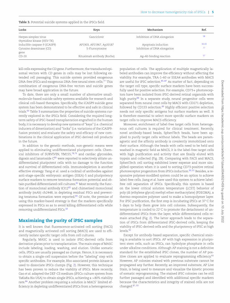
Table 3. Potential suicide systems applied in the iPSCs field.
Locks Keys Mechanism Ref.
Herpes simplex virusthymidine kinase (HSV-TK)
Ganciclovir Inhibition of DNA elongation 58
Inducible caspase-9 (iCASP9) AP1903, AP21967, Ap20187 Apoptosis induction 18,63
Cytosine deaminase (CD)gene
5-Flurocytosine Inhibition of DNA elongation 77
CD-20 Rituximab antibody (Roche) Ag–Ab binding reaction 57
kill cells expressing the CD gene. Furthermore, the transduced epi-somal vectors with CD genes in cells may be lost following ex-tended cell passaging. This suicide system provided exogenousDNA-free iPSCs and exogenous DNA-free neural stem cells.77 Thiscombination of exogenous DNA-free vectors and suicide genesmay have broad application in the future.
To date, there are only a small number of alternative small-molecule-based suicide safety systems available for research andclinical cell-based therapies. Specifically, the iCASP9 suicide genesystem has been demonstrated to be effective and safe in clinicaltrials.66 Table 3 summarizes the properties of suicide systems cur-rently explored in the iPSCs field. Considering the required long-term safety of iPSC-based transplantation engrafted in the humanbody, it is necessary to develop new systems of “keys” (i.e. chemicalinducers of dimerization) and “locks” (i.e. variations of the iCASP9-fusion protein) and evaluate the safety and efficacy of new com-binations in the clinical application of iPSC-derived cell productsin the future.
In addition to the genetic methods, non-genetic means wereapplied to eliminating undifferentiated pluripotent cells. Chem-ical inhibitors of SURVIVIN, YM155,78,79 and cardiac glycosides,digoxin and lanatoside C80 were reported to selectively ablate un-differentiated pluripotent cells with no damage to the functionand survival of differentiated cells. Immunodepletion is anothereffective strategy. Tang et al. used a cocktail of antibodies againstanti-stage-specific embryonic antigen (SSEA)-5 and pluripotencysurface markers to remove teratoma-formation potential and ob-tain purified differentiated cell cultures.81 Most recently, the func-tion of monoclonal antibody K31282 and chimerised monoclonalantibody (mAb) ch2448, in depleting residual PSCs and prevent-ing teratoma formation were reported.83 Of note, the premise ofusing this marker-based strategy is that the markers specificallyexpressed in PSCs so as to avoid killing differentiated cells whileremoving undifferentiated PSCs.18
Maximizing the purity of iPSC samplesIt is well known that fluorescence-activated cell sorting (FACS)and magnetically activated cell sorting (MACS) are used to effi-ciently isolate specific target cells from cell cultures.
Regularly, MASC is used to isolate iPSC-derived cells fromderivation plates prior to transplantation. The main steps of MASCinclude labeling, loading, washing, and elution. Unlike somaticcells, iPSCs are usually passaged as clumps. Hence, it is necessaryto obtain a single-cell suspension before the “labeling” step withspecific antibodies. For example, Rho-associated protein kinase isused to dissociate iPSCs clumps (Fig. 2). However, this techniquehas been proven to reduce the viability of iPSCs. More recently,Gao et al. adapted the DEF-CS medium (iPSCs culture system fromTaKaRa Bio USA) to obtain single iPSCs, achieving a >80% viabilityrate.84 Another problem requiring a solution is MACS’ limited ef-ficiency in depleting undifferentiated iPSCs from a heterogeneous
population of cells. The application of multiple magnetically la-beled antibodies can improve the efficiency without affecting theviability. For example, TRA-1–60 or SSEA4 antibodies with MACSare useful for iPSC selection.85–87 As matter of fact, depending onthe target cell type, specific surface markers have been success-fully used for positive selection. For example, CD73+ photorecep-tors have been isolated from iPSC-derived retinal organoids withhigh purity.88 In a separate study, neural progenitor cells wereseparated from neural crest cells by MACS with CD271 depletion,followed by CD133 selection.89 Highly efficient positive selectionneeds not only specific antigens but surface markers as well. Itis therefore essential to select more specific surface markers ontarget cells to improve MACS efficiency.
Moreover, enrichment of label-free target cells from heteroge-nous cell cultures is required for clinical treatment. Recently,novel antibody-based beads, SpheriTech beads, have been ap-plied to purify target cells without labels. The beads are param-agnetic and the affinity antibody is covalently immobilized ontotheir surface. Although the beads with cells need to be held andwashed in magnetic field as MACS, it is the label-free target cellswith high purification and activity that are finally eluted withtrypsin and collected (Fig. 2B). Comparing with FACS and MACS,SpheriTech cell sorting exhibited lower expense and more sim-plified operation when it is used to sorting CD73-positive retinalphotoreceptor progenitors from iPSCs induction.90,91 Besides, a re-sponsive polymer-modified system could be an option to achievelabel-free cells. Jiang et al. have used this system to achieve label-free cell separation of iPSCs. Specifically, this system is basedon the lower critical solution temperature (LCST) behavior ofpoly (di (ethylene glycol) methyl ether methacrylate) (PDEGMA), athermo-responsive polymer used to make a homopolymer layer.For iPSC purification, the first step is incubating iPSCs at 37◦C for5 days to help them grow into cell colonies. Subsequently, thetemperature is cooled to 22◦C to promote the detachment of un-differentiated iPSCs from the layer, while differentiated cells re-main attached (Fig. 2). The latter approach leads to the separa-tion of iPSCs from differentiated iPSC-derived cells, keeping theviability of iPSC-derived cells and the pluripotency of iPSC at highlevels.92
Except for antibody-based separation, specific chemical stain-ing is available to sort iPSCs. AP with high expression in pluripo-tent stem cells, such as iPSCs, can hydrolyze phosphate in cellsunder alkaline conditions. Although AP staining is not a definitivestandard for the established iPSC clones, the number of AP pos-itive clones are applied to evaluate reprogramming efficiency.93
However, AP colonies stained with previous substrate cannot bepropagated any further. Recently, an improved substrate, AP LiveStain, is being used to measure and visualize the kinetic processof somatic reprogramming. The stained iPSC colonies can be stillfurther passaged and identified with additional specific markersbecause the characteristics and integrity of stained cells are notchanged.87, 93
Dow
nloaded from https://academ
ic.oup.com/pcm
/article/5/1/pbac004/6521459 by guest on 10 July 2022

6 | Precis Clin Med, 2022, 5: pbac004
Figure 2. Schematic diagram of methods for purifying iPSC samples. (A) LCST behavior of PDEGMA. iPSCs are cultured on the polymer at 37◦C for 5days and grow into colonies on the layer. By cooling the temperature to 22◦C, iPSC colonies detach from the layer while differentiated cells remain onthe layer. (B) After incubation with SpheriTech beads coated with affinity antibody, the target cells in a hetergenous cell suspension bind to the beads.Label-free target cells are eluted from the beads by trypsin and affinity antibodies still stay attached to the beads. (C) Compared with SpheriTechbeads, antibodies or magnetic particles attached to the target cells may influence the cells for positive selection by MACS. HCS: heterogeneous cellsuspension; TCs: target cells.
Figure 3. Strategies for reducing tumorigenesis risk.
Dow
nloaded from https://academ
ic.oup.com/pcm
/article/5/1/pbac004/6521459 by guest on 10 July 2022

How to decrease tumorigenicity risk of iPSCs | 7
Assessment of the quality of iPSCs beforeclinical useIt is realized that the safety and efficacy of iPSCs or iPSCs-derivedcells must be evaluated before they can be used in clinical treat-ment. With an in-depth understanding of the proliferation anddifferentiation characteristics of iPSCs in vitro, researchers haveput forward their own views regarding quality control and relatedmethods.
Colter et al. noticed that the complexity of iPSC production anddownstream inductive differentiation processes brought aboutuncertainty from processing to clinical effect, including low qual-ity, heterogeneity and other problems. Therefore, the productionand application of stem cells should rely on more rigorous andeffective quality control of molecular and cell characteristics. Itis necessary to establish a systematic evaluation of the variabil-ity between different batches of products and complete datasetscombined with relevant computational methods. These effortswill help to develop a practical model and improve the robustnessof the production process.20
Assou et al. believe that, compared with ESCs, iPSCs need ad-ditional mandatory quality control because of the possibly ac-quired genetic changes, such as aneuploidy and oncogene mu-tations (such as TP53), which directly affect the safety and ef-ficacy of iPSCs and derivative products.19 Therefore, genome in-tegrity testing should be used as a routine test item, and kary-otype analysis should be a standard method of evaluation.19
Thus, it is logically reasonable that mutation screening shouldbe applied systematically and corresponding judgment standardsshould be established. Concerning the methods, short tandem re-peats (STR) analysis can be considered as an essential indicatorof genomic integrity in addition to karyotype. Indeed, STR analy-sis is also recommended as a mandatory item to indicate the ge-nomic integrity of iPSC establishment.94 It is emphasized that theSTR profile of qualified iPSCs should be established in early pas-sages and must match that of the cell donor.94 The ANSI/ATCCASN-0002–2011 standard for the authentication of human celllines requires at least eight core STR loci with an 80% thresh-old match. Kerrigan et al. increased the number of STR loci to15 and Taylor believed 16 STR specific sites may be necessaryfor the plain identification even if iPSCs are generated from au-tologous somatic cells.95 Besides, fluorescence in situ hybridiza-tion (FISH), array comparative genetic hybridization (aCGH), andother microarray approaches, such as quantitative PCR (qPCR),SNP arrays, digital drop PCR (ddPCR), and next generation se-quencing (NGS) were also used to assess insertion and dele-tion (indel), CNV, and SNV. Baker et al. evaluated a set of CNVand SNV determination methods for advantages and disadvan-tages.96 It is proposed that PCR technology and FISH technol-ogy are more suitable for detecting known frequent small frag-ment mutations, while aCGH and NGS fit more for large fragmentanalysis.97
However, when comprehensively considering cost, workload ofdata analysis, and complexity of result interpretation, ddPCR iscommonly applied at present, and has high accuracy and rela-tively low cost compared with aCGH, NGS, and FISH technologies.Therefore, when evaluationg tumorigenicity of iPSCS and iPSCs-derived cells, the ddPCR of the known oncogene mutations andchosen targeted sequencing could be set as the top priority inanalysis. Of course, because NGS is the whole genome at single-base resolution and can detect most genetic anomalies, depend-ing on the sequencing depth, it may be considered when a highstandard is preferred. However, it will be difficult for high-depth
NGS to become a routine means of quality control before the se-quencing cost is reduced to an affordable price.19,98,99
Rehakova et al. proposed a set of mandatory criteria and “forinformation only” tests, including differentiation, genetic stabil-ity, identity, vector clearance, morphology, pluripotency, robust-ness, viability, and histocompatibility, and proposed a set of testingmethods corresponding to the current good manufacturing prac-tices and regulations about production of clinical-grade iPSC andESC lines.94 In addition, with regard to the detection of iPSC het-erogeneity, flow cytometry analysis is a reliable method becauseit is timesaving and robust, and the results are quantitative andcomparable among different laboratories. Baghbaderani et al. ar-gued that for iPSCs, the release criteria for clinical use shouldcontain >70% of cells positive for SSEA4, OCT3/4, TRA-1–60, andTRA-1–81 and < 5% of cells negative for CD34+.100 In addition,because single cell RNA-seq showed its extreme power in eluci-dating the heterogeneity of cellular population,101 exampled byhematopoitic stem cells and mesenchymal stem cells, it may beapplied in evaluation of purifity of iPSCs and iPSCs derivativeswhen a protocol is set up for a clinical product.102, 103
Besides the in vitro assessment items mentioned above, it issuggested that in vivo tumorigenicity be included, because cellu-lar behavior in the engrafted site may be one of the most directpieces of evidence to confirm the clinical usability of an iPSCs celltherapy product (CTPs). However, it is difficult to standardize theexperimental conditions, such as the selection of animal model,the number of inoculated cells, the study duration, and the site oftransplantation. For instance, immunocompromised mice are se-lected to test tumorigenicity, but there is still no recognized stan-dard on the number of animals and the controls that are requiredto demonstrate that the CTP is unlikely to form a tumor. With re-gard to the site of transplantation, CTPs are now inoculated intothe clinical equivalent site intended for patients via the clinicalroute, though its theoretical foundation is still debated. Moreover,the reduced lifespan of the test animal compared to humans alsoconstitutes a limitation for the long-term observation of tumorformation.104 Therefore, Sato et al. pointed out that although cus-tomized assays have been established to test different productson a case-by-case basis, it is essential to reach a global consensuson the standard of the test approach so that it can be applied toany relevant CTP.105
ConclusionsThis review focused on current progress to diminish the tumori-genesis risk of iPSC technology, including reducing the potentialfor tumorigenicity and promoting the killing of abnormal cells, asshown in Fig. 3. These efforts and further pursuits outlined hereare critical for the clinic application of iPSCs technology.
A reduction in the tumorigenicity potential of the produced iP-SCs can be obtained by optimizing the cocktail of reprogrammingfactors, using the strategy of chemical inductive reprogramming,purifying cells by FACS and MACS, and applying exogenous DNA-free vectors. Although the traditional MACS purification strat-egy is firmly established, a wider variety of iPSC surface mark-ers, apparatuses, and protocols remain to be explored, such asSpheriTech beads, a novel label-free affinity purification method.In addition, interdisciplinary contributions like polymer materialsand nanotechnology have shown potential to separate iPSCs andtarget cells, such as PDEGAM.
Currently, it is demonstrated that the suicide systems havevery good application potential in effectively eliminating ab-normal cells with teratogenicity in cell preparations of iPSCs.
Dow
nloaded from https://academ
ic.oup.com/pcm
/article/5/1/pbac004/6521459 by guest on 10 July 2022
8 | Precis Clin Med, 2022, 5: pbac004
However, for improvement of specificity and efficiency, futurestudies are needed to design and develop new “keys” and “locks”and select specific safeguard sites for exogenous insertion.
Finally, systematic analysis of iPSC tumorigenicity, focusing onthe mechanisms, is currently lacking. However, it represents anessential characteristic of iPSCs technology. Therefore, a standardof evaluation for cell preparation tumorigenicity in clinical ap-plications should be established in the future, in which the ge-netic integrity validation of iPSCs and derivations is essential. Inparticular, assessment of safety seems to be more essential asmore and more effective techniques, such as the CRISPR-dCas9platform,106,107 are applied to fix the genetic defects of iPSCs de-rived from patients for subsequent therapy with the iPSCs CTPsin clinic.
AcknowledgementsWe thank the National Natural Science Foundation of China(grants No. 32171387 and 32071452), Shenzhen Bay LaboratoryOpen Program (grant No. SZBL2020090501003), and the Pearl RiverTalents Program Local Innovative and Research Teams (grant No.2017BT01S131).
Author contributionsC.Z. and M.L. collected and organized the data and wrote therough copy. X.P. and H.Z. conceived the topic and revised themanuscript.
Conflict of interestNone declared. In addition, as an Associate Editor of Precision Clin-ical Medicine, the corresponding author Xinghua Pan was blindedfrom reviewing and making decisions on this manuscript.
References1. Takahashi K, Yamanaka S. Induction of pluripotent stem cells
from mouse embryonic and adult fibroblast cultures by definedfactors. Cell 2006;126:663–76. doi: 10.1016/j.cell.2006.07.024.
2. Yu J, Vodyanik MA, Smuga-Otto K, et al. Induced pluripo-tent stem cell lines derived from human somatic cells. Science2007;318:1917–20. doi:10.1126/science.1151526.
3. Omole AE, Fakoya AOJ. Ten years of progress and promise ofinduced pluripotent stem cells: historical origins, characteris-tics, mechanisms, limitations, and potential applications. PeerJ2018;6:e4370. doi: 10.7717/peerj.4370.
4. Hou P, Li Y, Zhang X, et al. Pluripotent stem cells induced frommouse somatic cells by small-molecule compounds. Science2013;341:651–4. doi:10.1126/science.1239278.
5. Li X, Xu J, Deng H. Small molecule-induced cellular fate repro-gramming: promising road leading to Rome. Curr Opin Genet Dev2018;52:29–35. doi: 10.1016/j.gde.2018.05.004.
6. Chen G, Guo Y, Li C, et al. Small Molecules that Promote Self-Renewal of Stem Cells and Somatic Cell Reprogramming. StemCell Rev Rep 2020;16:511–23. doi: 10.1007/s12015-020-09965-w.
7. Romanazzo S, Lin K, Srivastava P, et al. Targeting cellplasticity for regeneration: From in vitro to in vivo re-programming. Adv Drug Deliv Rev 2020;161-162:124–44. doi:10.1016/j.addr.2020.08.007.
8. Takahashi K, Tanabe K, Ohnuki M, et al. Induction of pluripotentstem cells from adult human fibroblasts by defined factors. Cell2007;131:861–72. doi: 10.1016/j.cell.2007.11.019.
9. Nakagawa M, Takizawa N, Narita M, Ichisaka T, YamanakaS. Promotion of direct reprogramming by transformation-deficient Myc. Proc Natl Acad Sci 2010;107:14152–7. doi:10.1073/pnas.1009374107.
10. Eminli S, Utikal J, Arnold K, et al. Reprogramming of neural pro-genitor cells into induced pluripotent stem cells in the absenceof exogenous Sox2 expression. Stem Cells 2008;26:2467–74. doi:10.1634/stemcells.2008-0317.
11. Feng B, Jiang J, Kraus P, et al. Reprogramming of fibroblasts intoinduced pluripotent stem cells with orphan nuclear receptorEsrrb. Nat Cell Biol 2009;11:197–203. doi: 10.1038/ncb1827.
12. Nakagawa M, Koyanagi M, Tanabe K, et al. Generation ofinduced pluripotent stem cells without Myc from mouseand human fibroblasts. Nat Biotechnol 2008;26:101–6. doi:10.1038/nbt1374.
13. Huangfu D, Osafune K, Maehr R, et al. Induction of pluripotentstem cells from primary human fibroblasts with only Oct4 andSox2. Nat Biotechnol 2008;26:1269–75. doi: 10.1038/nbt.1502.
14. Ichida JK, Blanchard J, Lam K, et al. A Small-Molecule In-hibitor of Tgf-β Signaling Replaces Sox2 in Reprogram-ming by Inducing Nanog. Cell Stem Cell 2009; 5:491–503. doi:10.1016/j.stem.2009.09.012.
15. Li W, Zhou HY, Abujarour R, et al. Generation of Human In-duced Pluripotent Stem Cells in the Absence of Exogenous-Sox2. Stem Cells 2009; 27:2992–3000. doi: 10.1002/stem.240.
16. Li Y, Zhang Q, Yin X, et al. Generation of iPSCs from mouse fi-broblasts with a single gene, Oct4, and small molecules. Cell Res2011; 21: 196–204. doi: 10.1038/cr.2010.142.
17. Tan F, Qian C, Tang K, et al. Inhibition of transforming growthfactor β (TGF-β) signaling can substitute for Oct4 proteinin reprogramming and maintain pluripotency. J Biol Chem2015;290:4500–11. doi: 10.1074/jbc.M114.609016.
18. Martin RM, Fowler JL, Cromer MK, et al. Improving the safetyof human pluripotent stem cell therapies using genome-edited orthogonal safeguards. Nat Commun 2020;11:2713–9. doi:10.1038/s41467-020-16455-7.
19. Assou S, Bouckenheimer J, De Vos J. Concise Review: Assess-ing the Genome Integrity of Human Induced Pluripotent StemCells: What Quality Control Metrics? Stem Cells 2018;36:814–21.doi: 10.1002/stem.2797.
20. Colter J, Murari K, Biernaskie J, Kallos MS. Induced pluripotencyin the context of stem cell expansion bioprocess development,optimization, and manufacturing: a roadmap to the clinic. NPJRegen Med 2021;6:72. doi: 10.1038/s41536-021-00183-7.
21. Maali A, Maroufi F, Sadeghi F, et al. Induced pluripotent stemcell technology: trends in molecular biology, from genetics toepigenetics. Epigenomics 2021;13:631–47. doi: 10.2217/epi-2020-0409.
22. Wernig M, Meissner A, Cassady JP, et al. c-Myc is dispensablefor direct reprogramming of mouse fibroblasts. Cell Stem Cell2008;2:10–2. doi:10.1016/j.stem.2007.12.001.
23. Li HY, Chien Y, Chen YJ, et al. Reprogramming induced pluripo-tent stem cells in the absence of c-Myc for differentiationinto hepatocyte-like cells. Biomaterials 2011;32:5994–6005. doi:10.1016/j.biomaterials.2011.05.009.
24. Pushp P, Nogueira DES, Rodrigues CAV, et al. A Concise Re-view on Induced Pluripotent Stem Cell-Derived Cardiomy-ocytes for Personalized Regenerative Medicine. Stem Cell Rev Rep2021;17:748–76. doi: 10.1007/s12015-020-10061-2.
25. Maherali N, Hochedlinger K. Tgfbeta signal inhibition cooper-ates in the induction of iPSCs and replaces Sox2 and cMyc. CurrBiol 2009;19:1718–23. doi: 10.1016/j.cub.2009.08.025.
Dow
nloaded from https://academ
ic.oup.com/pcm
/article/5/1/pbac004/6521459 by guest on 10 July 2022
How to decrease tumorigenicity risk of iPSCs | 9
26. Huangfu D, Maehr R, Guo W, et al. Induction of pluripotentstem cells by defined factors is greatly improved by small-molecule compounds. Nat Biotechnol 2008;26:795–7. doi:10.1038/nbt1418.
27. Cao S, Yu S, Chen Y, et al. Chemical reprogramming of mouseembryonic and adult fibroblast into endoderm lineage. J BiolChem 2017;292:19122–32. doi: 10.1074/jbc.M117.812537.
28. Ye J, Ge J, Zhang X, et al. Pluripotent stem cells induced frommouse neural stem cells and small intestinal epithelial cellsby small molecule compounds. Cell Res 2016;26:34–45. doi:10.1038/cr.2015.142.
29. Fu H, Tian CL, Ye X, et al. Dynamics of Telomere Rejuvenationduring Chemical Induction to Pluripotent Stem Cells. Stem CellReports 2018;11:70–87. doi: 10.1016/j.stemcr.2018.05.003.
30. Papp B, Plath K. Epigenetics of reprogramming toinduced pluripotency. Cell 2013;152:1324–43. doi:10.1016/j.cell.2013.02.043.
31. Scesa G, Adami R, Bottai D. iPSC Preparation and EpigeneticMemory: Does the Tissue Origin Matter? Cells 2021;10:1470.doi:10.3390/cells10061470.
32. Kim Y, Jeong J, Choi D. Small-molecule-mediated reprogram-ming: a silver lining for regenerative medicine. Exp Mol Med2020;52:213–26. doi:10.1038/s12276-020-0383-3.
33. Cheng L, Hu W, Qiu B, et al. Generation of neural progenitorcells by chemical cocktails and hypoxia. Cell Res 2014;24:665–79. doi:10.1038/cr.2014.32.
34. Ma Y, Xie H, Du X, et al. In vivo chemical reprogram-ming of astrocytes into neurons. Cell Discov 2021;7:12. doi:10.1038/s41421-021-00243-8.
35. Cao N, Huang Y, Zheng J, et al. Conversion of human fibrob-lasts into functional cardiomyocytes by small molecules. Sci-ence 2016;352:1216–20. doi: 10.1126/science.aaf1502.
36. Li X, Liu D, Ma Y, et al. Direct Reprogramming of Fibrob-lasts via a Chemically Induced XEN-like State. Cell Stem Cell2017;21:264–73 e7. doi:10.1016/j.stem.2017.05.019.
37. Wang F, Yin Y, Ye X, et al. Molecular insights into theheterogeneity of telomere reprogramming in inducedpluripotent stem cells. Cell Res 2012;22:757–68. doi:10.1038/cr.2011.201.
38. Flavahan WA, Gaskell E, Bernstein BE. Epigenetic plastic-ity and the hallmarks of cancer. Science 2017;357:eaal2380.doi:10.1126/science.aal2380.
39. Takeda Y, Harada Y, Yoshikawa T, et al. Chemical compound-based direct reprogramming for future clinical applications.Biosci Rep 2018;38:BSR20171650. doi: 10.1042/BSR20171650.
40. Hu W, Qiu B, Guan W, et al. Direct Conversion of Normaland Alzheimer’s Disease Human Fibroblasts into NeuronalCells by Small Molecules. Cell Stem Cell 2015;17:204–12. doi:10.1016/j.stem.2015.07.006.
41. Zhang L, Yin JC, Yeh H, et al. Small Molecules Efficiently Re-program Human Astroglial Cells into Functional Neurons. CellStem Cell 2015;17:735–47. doi: 10.1016/j.stem.2015.09.012.
42. Kim Y, Kang K, Lee SB, et al. Jeong J, Choi D. Smallmolecule-mediated reprogramming of human hepatocytesinto bipotent progenitor cells. J Hepatol 2019;70:97–107. doi:10.1016/j.jhep.2018.09.007.
43. Yuan ZD, Zhu WN, Liu KZ, et al. Small Molecule Epigenetic Mod-ulators in Pure Chemical Cell Fate Conversion. Stem Cells Int2020;2020:8890917. doi: 10.1155/2020/8890917.
44. Bitzer M, Armeanu S, Lauer UM, et al. Sendai virus vectors as anemerging negative-strand RNA viral vector system. J Gene Med2003;5:543–53. doi: 10.1002/jgm.426.
45. Tan GW, Kondo T, Imamura K, et al. Simple derivation ofskeletal muscle from human pluripotent stem cells usingtemperature-sensitive Sendai virus vector. J Cell Mol Med2021;25:9586–96. doi: 10.1111/jcmm.16899.
46. Zhou H, Wu S, Joo JY, et al. Generation of induced pluripo-tent stem cells using recombinant proteins. Cell Stem Cell2009;4:381–4. doi:10.1016/j.stem.2009.04.005.
47. Nordin F, Ahmad RNR, Farzaneh F. Transactivator protein:An alternative for delivery of recombinant proteins for saferreprogramming of induced Pluripotent Stem Cell. Virus Res2017;235:106–14. doi: 10.1016/j.virusres.2017.04.007.
48. Anokye-Danso F, Trivedi CM, Juhr D, et al. Highly efficientmiRNA-mediated reprogramming of mouse and human so-matic cells to pluripotency. Cell Stem Cell 2011;8:376–88. doi:10.1016/j.stem.2011.03.001.
49. Borgohain MP, Haridhasapavalan KK, Dey C, et al. An In-sight into DNA-free Reprogramming Approaches to GenerateIntegration-free Induced Pluripotent Stem Cells for ProspectiveBiomedical Applications. Stem Cell Rev Rep 2019;15:286–313. doi:10.1007/s12015-018-9861-6.
50. Wang AYL. Application of Modified mRNA in Somatic Repro-gramming to Pluripotency and Directed Conversion of CellFate. Int J Mol Sci 2021;22:8148. doi: 10.3390/ijms22158148.
51. Steinle H, Weber M, Behring A, et al. Generation of iPSCs byNonintegrative RNA-Based Reprogramming Techniques: Bene-fits of Self-Replicating RNA versus Synthetic mRNA. Stem CellsInt 2019;2019:7641767. doi: 10.1155/2019/7641767.
52. Tay A, Melosh N. Nanostructured Materials for Intra-cellular Cargo Delivery. Acc Chem Res 2019;52:2462–71.doi:10.1021/acs.accounts.9b00272.
53. Wang Y, Wang Z, Xie K, et al. High-Efficiency Cellular Repro-gramming by Nanoscale Puncturing. Nano Lett 2020;20:5473–81. doi:10.1021/acs.nanolett.0c01979.
54. Lee MH, Lin CC, Thomas JL, et al. Cellular reprogrammingwith multigene activation by the delivery of CRISPR/dCas9ribonucleoproteins via magnetic peptide-imprinted chi-tosan nanoparticles. Mater Today Bio 2021;9:100091. doi:10.1016/j.mtbio.2020.100091.
55. Lister R, Pelizzola M, Kida YS, et al. Hotspots of aberrant epige-nomic reprogramming in human induced pluripotent stemcells. Nature 2011;471:68–73. doi: 10.1038/nature09798.
56. Gore A, Li Z, Fung HL, et al. Somatic coding mutations inhuman induced pluripotent stem cells. Nature 2011;471:63–7.doi:10.1038/nature09805.
57. Introna M, Barbui AM, Bambacioni F, et al. Genetic modifica-tion of human T cells with CD20: a strategy to purify andlyse transduced cells with anti-CD20 antibodies. Hum Gene Ther2000;11:611–20. doi: 10.1089/10430340050015798.
58. Kimura Y, Shofuda T, Higuchi Y, et al. Human Genomic SafeHarbors and the Suicide Gene-Based Safeguard System foriPSC-Based Cell Therapy. Stem Cells Transl Med 2019;8:627–38.doi: 10.1002/sctm.18-0039.
59. Liang Q, Monetti C, Shutova MV, et al. Linking a cell-divisiongene and a suicide gene to define and improve cell therapysafety. Nature 2018;563:701–4. doi: 10.1038/s41586-018-0733-7.
60. Iwasawa C, Tamura R, Sugiura Y, et al. Increased Cytotoxicity ofHerpes Simplex Virus Thymidine Kinase Expression in HumanInduced Pluripotent Stem Cells. Int J Mol Sci 2019;20:E810. doi:10.3390/ijms20040810.
61. Sułkowski M, Konieczny P, Chlebanowska P, et al. Introduc-tion of Exogenous HSV-TK Suicide Gene Increases Safetyof Keratinocyte-Derived Induced Pluripotent Stem Cells by
Dow
nloaded from https://academ
ic.oup.com/pcm
/article/5/1/pbac004/6521459 by guest on 10 July 2022

10 | Precis Clin Med, 2022, 5: pbac004
Providing Genetic “Emergency Exit” Switch. Int J Mol Sci2018;19:E197. doi: 10.3390/ijms19010197.
62. Sawdon AJ, Zhang J, Peng S, et al. Polymeric Nanovectors In-corporated with Ganciclovir and HSV-tk Encoding Plasmidfor Gene-Directed Enzyme Prodrug Therapy. Mol Basel Switz2021;26:1759. doi: 10.3390/molecules26061759.
63. Shi ZD, Tchao J, Wu L, et al. Precision installation of a highlyefficient suicide gene safety switch in human induced pluripo-tent stem cells. Stem Cells Transl Med 2020;9:1378–88. doi:10.1002/sctm.20-0007.
64. Jones BS, Lamb LS, Goldman F, et al. Improving the safety ofcell therapy products by suicide gene transfer. Front Pharmacol2014;5:254. doi:10.3389/fphar.2014.00254.
65. Dahlke J, Schott JW, Vollmer Barbosa P, et al. Efficient GeneticSafety Switches for Future Application of iPSC-Derived CellTransplants. J Pers Med 2021;11:565. doi: 10.3390/jpm11060565.
66. Zhou X, Naik S, Dakhova O, et al. Serial Activation of theInducible Caspase 9 Safety Switch After Human StemCell Transplantation. Mol Ther 2016;24:823–31. doi:10.1038/mt.2015.234.
67. Zhou X, Brenner MK. Improving the safety of T-Cell therapiesusing an inducible caspase-9 gene. Exp Hematol 2016;44:1013–9.doi:10.1016/j.exphem.2016.07.011.
68. Yagyu S, Hoyos V, Del Bufalo F, et al. An Inducible Caspase-9Suicide Gene to Improve the Safety of Therapy Using HumanInduced Pluripotent Stem Cells. Mol Ther 2015;23:1475–85. doi:10.1038/mt.2015.100.
69. Mashima H, Zhang R, Kobayashi T, et al. Improved safetyof induced pluripotent stem cell-derived antigen-presentingcell-based cancer immunotherapy. Mol Ther Methods Clin Dev2021;21:171–9. doi: 10.1016/j.omtm.2021.03.002.
70. Yagyu S, Hoyos V, Del Bufalo F, et al. Multiple mechanismsdetermine the sensitivity of human-induced pluripotent stemcells to the inducible caspase-9 safety switch. Mol Ther MethodsClin Dev 2016;3:16003. doi: 10.1038/mtm.2016.3.
71. Sadelain M, Papapetrou EP, Bushman FD. Safe harbours for theintegration of new DNA in the human genome. Nat Rev Cancer2011;12:51–8. doi: 10.1038/nrc3179.
72. Clackson T, Yang W, Rozamus LW, et al. Redesigning an FKBP-ligand interface to generate chemical dimerizers with novelspecificity. Proc Natl Acad Sci U S A 1998;95:10437–42. doi:10.1073/pnas.95.18.10437.
73. Amatya C, Pegues MA, Lam N, et al. Development ofCAR T Cells Expressing a Suicide Gene Plus a ChimericAntigen Receptor Targeting Signaling Lymphocytic-ActivationMolecule F7. Mol Ther J Am Soc Gene Ther 2021;29:702–17. doi:10.1016/j.ymthe.2020.10.008.
74. Wu Y, Chang T, Long Y, Huang H, et al. Using GeneEditing to Establish a Safeguard System for PluripotentStem-Cell-Based Therapies. iScience 2019;22:409–22. doi:10.1016/j.isci.2019.11.038.
75. Avilion AA, Nicolis SK, Pevny LH, et al. Multipotent cell lineagesin early mouse development depend on SOX2 function. GenesDev 2003;17:126–40. doi: 10.1101/gad.224503.
76. Sherwood RI, Chen TY, Melton DA. Transcriptional dynam-ics of endodermal organ formation. Dev Dyn 2009;238:29–42.doi:10.1002/dvdy.21810.
77. Lee M, Ha J, Son YS, et al. Efficient exogenous DNA-free re-programming with suicide gene vectors. Exp Mol Med 2019;51:1–12. doi:10.1038/s12276-019-0282-7.
78. Lee MO, Moon SH, Jeong HC, et al. Inhibition of pluripo-tent stem cell-derived teratoma formation by small
molecules. Proc Natl Acad Sci U S A 2013;110:E3281–90.doi: 10.1073/pnas.1303669110.
79. Kang SJ, Park YI, Hwang SR, et al. Hepatic population derivedfrom human pluripotent stem cells is effectively increasedby selective removal of undifferentiated stem cells usingYM155. Stem Cell Res Ther 2017;8:78. doi: 10.1186/s13287-017-0517-2.
80. Lin YT, Wang CK, Yang SC, et al. Elimination of undifferenti-ated human embryonic stem cells by cardiac glycosides. Sci Rep2017;7:5289. doi:10.1038/s41598-017-05616-2.
81. Tang C, Lee AS, Volkmer JP, et al. An antibody against SSEA-5 glycan on human pluripotent stem cells enables removalof teratoma-forming cells. Nat Biotechnol 2011;29:829–34. doi:10.1038/nbt.1947.
82. Park J, Lee DG, Lee NG, et al. Monoclonal antibody K312-based depletion of pluripotent cells from differentiated stemcell progeny prevents teratoma formation. BMB Rep 2021:5405.
83. Tan HL, Tan BZ, Goh WXT, et al. In vivo surveillance and elimi-nation of teratoma-forming human embryonic stem cells withmonoclonal antibody 2448 targeting annexin A2. Biotechnol Bio-eng 2019;116:2996–3005. doi: 10.1002/bit.27135.
84. Gao X, Sprando RL, Yourick JJ. A Rapid and Highly Effi-cient Method for the Isolation, Purification, and Passaging ofHuman-Induced Pluripotent Stem Cells. Cell Reprogramming2018;20:282–8. doi: 10.1089/cell.2018.0022.
85. Yang W, Liu Y, Slovik KJ, et al. Generation of iPSCs as a PooledCulture Using Magnetic Activated Cell Sorting of Newly Repro-grammed Cells. PLoS One 2015;10:e0134995. doi: 10.1371/jour-nal.pone.0134995.
86. Fong CY, Peh GSL, Gauthaman K, et al. Separation of SSEA-4 and TRA-1-60 labelled undifferentiated human embry-onic stem cells from a heterogeneous cell population us-ing magnetic-activated cell sorting (MACS) and fluorescence-activated cell sorting (FACS). Stem Cell Rev Rep 2009;5:72–80. doi:10.1007/s12015-009-9054-4.
87. Quintanilla RH, Asprer J, Sylakowski K, et al. Kinetic Measure-ment and Real Time Visualization of Somatic Reprogramming.J Vis Exp JoVE 2016;54190. doi: 10.3791/54190.
88. Gagliardi G, Ben M’Barek K, Chaffiol A, et al. Characterizationand Transplantation of CD73-Positive Photoreceptors Isolatedfrom Human iPSC-Derived Retinal Organoids. Stem Cell Rep2018;11:665–80. doi: 10.1016/j.stemcr.2018.07.005.
89. Bowles KR, Tcw J, Qian L, et al. Reduced variability of neuralprogenitor cells and improved purity of neuronal cultures us-ing magnetic activated cell sorting. PLoS One 2019;14:e0213374.doi: 10.1371/journal.pone.0213374.
90. Weil BD, Jenkins MJ, Uddin S, et al. An integrated experimentaland economic evaluation of cell therapy affinity purificationtechnologies. Regen Med 2017;12:397–417. doi: 10.2217/rme-2016-0156.
91. González F, Boué S, Izpisúa Belmonte JC. Methods for makinginduced pluripotent stem cells: reprogramming à la carte. NatRev Genet 2011;12:231–42. doi: 10.1038/nrg2937.
92. Jiang S, Müller M, Schönherr H. Propagation and Purifica-tion of Human Induced Pluripotent Stem Cells with Selec-tive Homopolymer Release Surfaces. Angew Chem Int Ed Engl2019;58:10563–6. doi: 10.1002/anie.201903299.
93. Singh U, Quintanilla RH, Grecian S, et al. Lakshmipathy U.Novel live alkaline phosphatase substrate for identification ofpluripotent stem cells. Stem Cell Rev Rep 2012;8:1021–9. doi:10.1007/s12015-012-9359-6.
Dow
nloaded from https://academ
ic.oup.com/pcm
/article/5/1/pbac004/6521459 by guest on 10 July 2022

How to decrease tumorigenicity risk of iPSCs | 11
94. Rehakova D, Souralova T, Koutna I. Clinical-Grade HumanPluripotent Stem Cells for Cell Therapy: Characterization Strat-egy. Int J Mol Sci 2020;21:2435. doi: 10.3390/ijms21072435.
95. Kerrigan L, Nims RW. Authentication of human cell-basedproducts: the role of a new consensus standard. Regen Med2011;6:255–60. doi:10.2217/rme.11.5.
96. Baker D, Hirst AJ, Gokhale PJ, et al. Detecting Genetic Mosaicismin Cultures of Human Pluripotent Stem Cells. Stem Cell Rep2016;7:998–1012. doi: 10.1016/j.stemcr.2016.10.003.
97. Mak ACY, Lai YYY, Lam ET, et al. Genome-Wide Structural Vari-ation Detection by Genome Mapping on Nanochannel Arrays.Genetics 2016;202:351–62. doi: 10.1534/genetics.115.183483.
98. Bai Q, Desprat R, Klein B, Lemaître JM, et al. Embryonicstem cells or induced pluripotent stem cells? A DNAintegrity perspective. Curr Gene Ther 2013;13:93–8. doi:10.2174/1566523211313020003.
99. Oliveira PH, da Silva CL, Cabral JMS. Concise review: Genomicinstability in human stem cells: current status and future chal-lenges. Stem Cells 2014;32:2824–32. doi: 10.1002/stem.1796.
100. Baghbaderani BA, Tian X, Neo BH, et al. cGMP-ManufacturedHuman Induced Pluripotent Stem Cells Are Available for Pre-clinical and Clinical Applications. Stem Cell Rep 2015;5:647–59.doi: 10.1016/j.stemcr.2015.08.015.
101. Zhang X, Marjani SL, Hu Z, et al. Single-Cell Sequencing forPrecise Cancer Research: Progress and Prospects. Cancer Res2016;76:1305–12. doi: 10.1158/0008-5472.CAN-15-1907.
102. Yang J, Tanaka Y, Seay M, et al. Single cell transcrip-tomics reveals unanticipated features of early hematopoi-etic precursors. Nucleic Acids Res 2017;45:1281–96. doi:10.1093/nar/gkw1214.
103. Qu R, He K, Fan T, et al. Single-cell transcriptomic sequencinganalyses of cell heterogeneity during osteogenesis of humanadipose-derived mesenchymal stem cells. Stem Cells Dayt Ohio2021;39:1478–88. doi: 10.1002/stem.3442.
104. Takei Y, Morioka M, Yamashita A, et al. Quality assessment testsfor tumorigenicity of human iPS cell-derived cartilage. Sci Rep2020;10:12794. doi: 10.1038/s41598-020-69641-4.
105. Sato Y, Bando H, Di Piazza M, et al. Tumorigenicity assess-ment of cell therapy products: The need for global consen-sus and points to consider. Cytotherapy 2019;21:1095–1111. doi:10.1016/j.jcyt.2019.10.001.
106. Liu XS, Wu H, Krzisch M, et al. Rescue of Fragile X SyndromeNeurons by DNA Methylation Editing of the FMR1 Gene. Cell2018;172:979–92 e6. doi: 10.1016/j.cell.2018.01.012.
107. Gjaltema RAF, Rots MG. Advances of epigenetic editing. CurrOpin Chem Biol 2020;57:75–81. doi: 10.1016/j.cbpa.2020.04.020.
Dow
nloaded from https://academ
ic.oup.com/pcm
/article/5/1/pbac004/6521459 by guest on 10 July 2022