Accelerating Vascularization in Polycaprolactone Scaffolds by Endothelial Progenitor Cells
12
Accelerating Vascularization in Polycaprolactone Scaffolds by Endothelial Progenitor Cells Shivani Singh, Ph.D., 1 Benjamin M. Wu, D.D.S., Ph.D., 1,2 and James C.Y. Dunn, M.D., Ph.D. 1,3 Vascularization is a major challenge in tissue engineering. The purpose of this study is to expedite the formation of blood vessels in porous polycaprolactone (PCL) scaffolds by the delivery of endothelial progenitor cells (EPCs). To establish a pro-angiogenic and pro-vasculogenic microenvironment, we employed EPCs seeded in PCL scaffold with surface-immobilized heparin and vascular endothelial growth factor (VEGF). EPCs seeded on scaffolds with VEGF exhibited phosphorylation of the receptor. After 7 days of subcutaneous implantation in immunodeficient mice, heparin-immobilized PCL scaffolds with VEGF induced significantly high density of blood vessel formation. The anastomosis of EPC-derived vessels with the host circulatory system was evident by the presence of murine erythrocytes in the lumen of human-CD31 positive vessels. A more uniform distribution of blood vessels was achieved within 2-mm thick scaffolds by seeding an optimal density of EPCs. The seeding of a higher density of EPC resulted in an increase in apoptosis and a concomitant decline in blood vessel formation at the scaffold’s inner core. When co-seeded with other cells, the EPCs maintained the ability to accelerate vessel formation. The excessive expansion of EPCs in vitro was associated with a decline in their in vivo vasculogenic potential. EPCs accelerated the vascularization of heparin-immobilized PCL scaffolds in the presence of VEGF. Introduction T he delay in the blood vessel formation is a bottleneck for tissue-engineering scaffolds of larger dimensions. In the absence of the vessel network during early stages, the limited diffusion of nutrients restricts the cell survival to the periphery of implanted scaffolds. The in-growth of host vessels can be improved by the delivery of angiogenic growth factors or gene-encoding angiogenic factors. 1,2 However, angiogenesis alone is not adequate to rapidly vascularize thicker scaffolds. An attractive option to aug- ment scaffold vascularization is the delivery of endothelial cells (ECs). Scaffolds seeded with human umbilical vein EC (HUVEC) and human microvasculature ECs were able to form vessels in the implanted scaffolds. 3–5 The major im- pediments in clinical use of adult ECs are their insufficient supply, limited in vitro expansion, and in vivo vessel- formation potential. Embryonic and adult stem cells have been recognized as a promising source of ECs, as they can differentiate into ECs in vitro. 6,7 ECs derived from human embryonic stem cells and bone marrow stromal cells formed vessels in tissue-engineered scaffolds in vivo. Nonetheless, the application of stem cells for therapeutic purposes is subject to attaining thorough understanding and control over the differentiation process. More recently, the vessel-forming endothelial progenitor cell (EPC) population has been identified in the human umbilical cord and circulating adult blood. 8,9 EPCs ex- pressed vascular cell surface markers such as CD31, vWF, and KDR and lacked hematopoietic specific CD45 and CD14 antigens. 10,11 EPCs showed robust proliferation, self-renewal, and superior vessel-forming ability compared with the ma- tured ECs. 11,12 EPCs are clinically safer to use than embry- onic stem cells, as they differentiate exclusively into the endothelial lineage. The process of blood vessel formation from the undifferentiated ECs is defined as vasculogenesis. There are two common strategies to vascularize scaffolds using EC precursors. The in vitro vasculogenesis involves prevascularization of the scaffold before implantation so that the preformed graft vessels could readily anastomose with the recipient vessels. 4,5 The second approach entails im- plantation of scaffolds seeded with EPCs to induce in vivo vasculogenesis. Other researchers have tested the latter ap- proach in scaffolds based on collagen 10,13 and Matrigel. 9,14,15 In this study, we used human umbilical cord-derived EPCs to induce in vivo vasculogenesis in porous poly- caprolactone (PCL) scaffolds in 7 days. The distribution of the vessels formed by EPCs was assessed as a function of the scaffold thickness. We also evaluated the effect of heparin immobilization and vascular endothelial growth factor Departments of 1 Bioengineering, 2 Material Science and Engineering, and 3 Surgery, University of California, Los Angeles, California. TISSUE ENGINEERING: Part A Volume 17, Numbers 13 and 14, 2011 ª Mary Ann Liebert, Inc. DOI: 10.1089/ten.tea.2010.0708 1819
-
Upload
independent -
Category
Documents
-
view
3 -
download
0
Transcript of Accelerating Vascularization in Polycaprolactone Scaffolds by Endothelial Progenitor Cells

Accelerating Vascularization in Polycaprolactone Scaffoldsby Endothelial Progenitor Cells
Shivani Singh, Ph.D.,1 Benjamin M. Wu, D.D.S., Ph.D.,1,2 and James C.Y. Dunn, M.D., Ph.D.1,3
Vascularization is a major challenge in tissue engineering. The purpose of this study is to expedite the formationof blood vessels in porous polycaprolactone (PCL) scaffolds by the delivery of endothelial progenitor cells(EPCs). To establish a pro-angiogenic and pro-vasculogenic microenvironment, we employed EPCs seeded inPCL scaffold with surface-immobilized heparin and vascular endothelial growth factor (VEGF). EPCs seeded onscaffolds with VEGF exhibited phosphorylation of the receptor. After 7 days of subcutaneous implantation inimmunodeficient mice, heparin-immobilized PCL scaffolds with VEGF induced significantly high density ofblood vessel formation. The anastomosis of EPC-derived vessels with the host circulatory system was evident bythe presence of murine erythrocytes in the lumen of human-CD31 positive vessels. A more uniform distributionof blood vessels was achieved within 2-mm thick scaffolds by seeding an optimal density of EPCs. The seedingof a higher density of EPC resulted in an increase in apoptosis and a concomitant decline in blood vesselformation at the scaffold’s inner core. When co-seeded with other cells, the EPCs maintained the ability toaccelerate vessel formation. The excessive expansion of EPCs in vitro was associated with a decline in their in vivovasculogenic potential. EPCs accelerated the vascularization of heparin-immobilized PCL scaffolds in thepresence of VEGF.
Introduction
The delay in the blood vessel formation is a bottleneckfor tissue-engineering scaffolds of larger dimensions. In
the absence of the vessel network during early stages, thelimited diffusion of nutrients restricts the cell survival to theperiphery of implanted scaffolds. The in-growth of hostvessels can be improved by the delivery of angiogenicgrowth factors or gene-encoding angiogenic factors.1,2
However, angiogenesis alone is not adequate to rapidlyvascularize thicker scaffolds. An attractive option to aug-ment scaffold vascularization is the delivery of endothelialcells (ECs). Scaffolds seeded with human umbilical vein EC(HUVEC) and human microvasculature ECs were able toform vessels in the implanted scaffolds.3–5 The major im-pediments in clinical use of adult ECs are their insufficientsupply, limited in vitro expansion, and in vivo vessel-formation potential. Embryonic and adult stem cells havebeen recognized as a promising source of ECs, as they candifferentiate into ECs in vitro.6,7 ECs derived from humanembryonic stem cells and bone marrow stromal cells formedvessels in tissue-engineered scaffolds in vivo. Nonetheless,the application of stem cells for therapeutic purposes issubject to attaining thorough understanding and controlover the differentiation process.
More recently, the vessel-forming endothelial progenitorcell (EPC) population has been identified in the humanumbilical cord and circulating adult blood.8,9 EPCs ex-pressed vascular cell surface markers such as CD31, vWF,and KDR and lacked hematopoietic specific CD45 and CD14antigens.10,11 EPCs showed robust proliferation, self-renewal,and superior vessel-forming ability compared with the ma-tured ECs.11,12 EPCs are clinically safer to use than embry-onic stem cells, as they differentiate exclusively into theendothelial lineage. The process of blood vessel formationfrom the undifferentiated ECs is defined as vasculogenesis.There are two common strategies to vascularize scaffoldsusing EC precursors. The in vitro vasculogenesis involvesprevascularization of the scaffold before implantation so thatthe preformed graft vessels could readily anastomose withthe recipient vessels.4,5 The second approach entails im-plantation of scaffolds seeded with EPCs to induce in vivovasculogenesis. Other researchers have tested the latter ap-proach in scaffolds based on collagen10,13 and Matrigel.9,14,15
In this study, we used human umbilical cord-derivedEPCs to induce in vivo vasculogenesis in porous poly-caprolactone (PCL) scaffolds in 7 days. The distribution ofthe vessels formed by EPCs was assessed as a function of thescaffold thickness. We also evaluated the effect of heparinimmobilization and vascular endothelial growth factor
Departments of 1Bioengineering, 2Material Science and Engineering, and 3Surgery, University of California, Los Angeles, California.
TISSUE ENGINEERING: Part AVolume 17, Numbers 13 and 14, 2011ª Mary Ann Liebert, Inc.DOI: 10.1089/ten.tea.2010.0708
1819

(VEGF) delivery with the aim of improving blood vesselformation by the EPCs. We examined the optimal EPCsseeding density and the effect of passaging on vessel-formingability of EPCs.
Materials and Methods
Animals
Eight-week-old nonobese diabetic/severe combined im-munodeficient mice (NOD-SCID; Charles River Labora-tories) were used as recipients for scaffolds with EPCs.Autoclaved cages with irradiated diet and acidified waterwere used to house NOD-SCID mice. Transgenic mice strainFVB.Cg-Tg (GFPU) 5 Nagy/J ( Jackson Laboratory) weremaintained as a breeding colony and used as donors forhepatocytes and bone marrow stromal cells. The animalswere maintained and handled in compliance with the insti-tutional regulations established and approved by the AnimalResearch Committee at the University of California, LosAngeles.
Culture of EPCs
EPCs isolated from human umbilical cord blood werekindly provided by Dr. Mervin C. Yoder, Indiana University,Indianapolis. The EPCs were cultured in EBM-2 media(Lonza) supplemented with EGM-2-SingleQuots and 10%fetal bovine serum (FBS). The cells were maintained at 37�Cin a humidified incubator containing 10% carbon dioxide,and the culture medium was changed every 2–3 days. Thecells were trypsinized on reaching confluency and were re-plated for the next passage.
Isolation of hepatocytes and bone marrow stromal cells
Two-month-old transgenic mice were used for the isola-tion of green fluorescent protein (GFP)-positive primary he-patocytes and bone marrow stromal cells. The hepatocyteswere harvested by a modified two-step perfusion proce-dure.16 Briefly, the liver was perfused successively with twosolutions via portal vein. The first solution, containing20 mM HEPES, 7 mM KCl, 142 mM NaCl, and 0.1 mM EDTAat pH 7.4 and 37�C, was perfused for 10 min at 3 mL/min.The second solution, containing collagenase type IV (0.5 mg/mL; Sigma) in 20 mM HEPES, 20 mM CaCl2, 7 mM KCl,and 142 mM NaCl at pH 7.4 and 37�C, was perfused tillthe liver was sufficiently disrupted. The excised liverwas placed in cold Dulbecco’s modified Eagle’s medium(DMEM) containing 10% fetal bovine serum on ice. Theliver capsule was removed gently to free the hepatocytesand was filtered through 70 mm nylon mesh. The cells werecentrifuged at 50 g for 2 min at 4�C and the cell pellet wasre-suspended in Percoll (1.05 g/mL; Sigma) and Hank’sbalanced salt solution. The solution was centrifuged at 50 gfor 5 min. The obtained cell pellet was re-suspended in coldDMEM containing 10% FBS and used immediately for theexperiments.
The bone marrow stromal cells were harvested from thetibia and femur by flushing the marrow plug with DMEMand 10% FBS under sterile conditions. The cell suspensionwas filtered through 70mm nylon mesh and plated. The cellswere maintained at 37�C in a humidified incubator, and theculture media was changed after 3 days. The cells were
cultured for *20 days, and the confluent cells were trypsi-nized immediately before seeding on the scaffold.
Preparation of scaffold
Porous PCL (Lactel Absorbable Polymers) scaffolds wereprepared by solvent casting and particulate leaching tech-nique. Briefly, PCL (10% w/w) was dissolved in a mixedsolution of chloroform and methanol (65 chloroform: 35methanol w/w). Sieved sucrose particles of 212–300 mm di-ameter were added to the PCL solution. The ratio of su-crose:PCL was 96:4 (w/w). The blend was briefly mixed toform homogeneous slurry and was cast into Teflon moldswith inner diameter of 6 mm and height of 2 mm. Scaffoldswere freeze dried, and the sucrose was leached out in deio-nized water. The scaffolds were sterilized in 70% ethanol for30 min and washed several times with phosphate-bufferedsaline (PBS).
The heparin was immobilized on the surface of the porousPCL scaffold by cross-linking heparin using carbodiimidereaction. The chemicals 2-(N-Morpholino)ethanesulfonic acid(MES), N-(3-Dimethylaminopropyl)-N’-ethylcarbodiimidehydrochloride (EDC), N-hydroxysuccinimide (NHS), andunfractionated heparin sodium salt derived from porcineintestinal mucosa were purchased from Sigma. The scaffoldswere saturated with 0.05 M MES buffer (pH 5.5) for 15 minbefore the reaction. Subsequently, the scaffolds were im-mersed in freshly prepared solution of heparin (1% w/v),0.5 M EDC, and 0.5 M NHS in MES buffer and briefly vor-texed. After 15 h of reaction at room temperature, the scaf-folds were extensively washed with distilled water toremove the byproducts.
The human rhVEGF165 (R&D Systems) was loaded on thescaffold by applying a solution of 10mg of growth factor in60 mL PBS in three aliquots onto the scaffold. The VEGF-loaded scaffold was allowed to dry in laminar hood for40 min at room temperature and immersed overnight in 1 mLof PBS at 4�C to remove freely diffusible VEGF.
In vitro bioactivity of scaffolds
The scaffolds were assessed for their ability to phosphor-ylate the receptor for VEGF, VEGFR2, in the seeded EPCs.Scaffolds loaded with 1mg of VEGF were washed severaltimes with either PBS or heparin/PBS solution (1 mg/mL)for 1 h followed by several washes with PBS. Confluent EPCsin a T75 flask were incubated overnight in serum-free EBM-2basal media. The cells were trypsinized and were suspendedin serum-free media containing 0.1 mM Na3VO4 (Sigma) toinhibit phosphatase activity while centrifuging at 1200 rpmfor 5 min. The cell pellet was re-suspended in serum-freemedia, and *1 million cell/50 mL were seeded in the scaf-folds for 5 min at 37�C. The negative and positive controlswere EPCs incubated without VEGF and with 100 ng ofsoluble VEGF, respectively. The reaction was stopped byadding 1 mL of cold solution of 0.2 mM Na3VO4 in PBS toeach group. The cells were collected by centrifuging at1200 rpm for 5 min, and the obtained cell pellet was lysedwith RIPA buffer supplemented with Halt protease andphosphatase inhibitor (Thermo Scientific) on ice for 10 min.The lysate was centrifuged at 14,000 g for 15 min, andthe supernatant was collected. The lysate proteins wereseparated by SDS-PAGE and transferred to nitrocellulose
1820 SINGH ET AL.

membrane for detection of phosphorylated VEGFR2 andVEGFR2 using anti-phospho-VEGFR2 (Tyr1175) and anti-VEGFR2 (Cell Signaling).
Scaffold implantation
The scaffolds were seeded with a specified number ofEPCs suspended in 50 mL media and incubated for 2 h at37�C in a humidified incubator before implantation. For co-seeding experiments, the heparin-modified PCL scaffoldsloaded with 10 mg of VEGF were used. There were 500,000hepatocytes seeded alone or along with an equal number ofEPCs in 50mL of media. 100,000 bone marrow stromal cellswere seeded alone or along with 500,000 EPCs.
Mice were anesthetized with isofluorane, and a 1-cm-longdorsal midline incision was made to create a subcutaneouspocket. The scaffold was inserted in the pocket, and the skinwas sutured with 3–0 Vicryl (Ethicon). One scaffold wasimplanted in each mouse. The animals were sacrificed at 7days postimplantation. The scaffolds were harvested, fixedin 10% formalin overnight, and processed for histology.
Immunohistochemistry and analysis
The in vitro cultured EPCs were stained with live/deadstain (Invitrogen). The cells, after being fixed with 10%formalin for 10 min, were stained for hematoxylin and eosinor with mouse anti-von Willebrand factor (vWF) anti-body (DAKO), followed by a fluorescein isothiocyanate-conjugated goat anti-mouse secondary antibody ( JacksonImmunoResearch Laboratories).
The paraffin-embedded scaffolds were cut into 6-mm sec-tions and were stained with the mouse anti-human CD31antibody (DAKO) and biotinylated horse anti-mouse sec-ondary antibody (antibody detail; Vector Laboratories). Thestaining was visualized with a diaminobenzidine substrate(Vector Laboratories) at 200 · magnification. The bloodvessel density and the area occupied by vessels within thescaffolds were measured using ImageScope software (AperioTechnologies). The DNA fragmentation in apoptotic cellswas visualized with Terminal deoxynucleotidyl transferase-mediated deoxyuridine triphosphate nick end labeling (TU-NEL) assay using the ApopTag fluorescein detection kit(Chemicon).
For multiple immunostaining, the agarose-embeddedscaffolds were cut with a Vibratome (Ted Pella) into 200-mmsections17 and stained with anti-mouse CD31 antibody (BDBiosciences), anti-human CD31 antibody, and anti-GFP an-tibody (Abcam), combined with fluorescent secondary anti-bodies ( Jackson ImmunoResearch Laboratories). Thesamples were visualized on a Zeiss Axioskop microscope(Thornwood).
Statistics
The Student’s t-test was used to evaluate the differencebetween the groups. The data were considered statisticallysignificant when p < 0.05.
Results
To enhance angiogenesis, VEGF was preloaded on PCLscaffolds with or without heparin immobilization. The bio-activity of the VEGF in scaffolds was confirmed by VEGFR2
phosphorylation in the seeded EPCs. The VEGF-loadedscaffolds, after washing away the unbound growth factorwith PBS, were able to phosphorylate VEGFR2 in EPCsseeded onto the scaffold (Fig. 1). Moreover, after eluting theadsorbed VEGF on scaffolds with soluble heparin, the VEGFremaining on scaffolds still exhibited the ability to phos-phorylate VEGFR2.
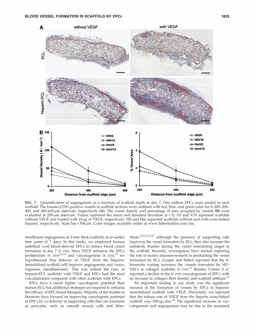
EPCs were seeded in PCL scaffolds with or without VEGFand heparin. Scaffolds seeded with 1 million EPCs weresubcutaneously implanted in NOD-SCID mice and were re-trieved after 7 days. Macroscopically, the heparin-PCL scaf-fold loaded with 10mg of VEGF exhibited intense rednessgrossly, indicating the presence of red blood cells within thescaffold (Fig. 2). The increase in angiogenesis and vasculo-genesis in heparin-PCL scaffolds with VEGF was confirmedby double-immunostaining with anti-mouse CD31 and anti-human CD31 antibodies (Fig. 3). Moreover, the EPCs formeddenser vessels in the regions of increased angiogenesis. Theextent of vascularization was quantified by measuring thetotal vessel density and the percentage area occupied byblood vessels (Fig. 4). The heparin-PCL scaffolds with VEGFpossessed significantly higher density of EPC-derived vesselsthan other scaffolds. There was a threefold increase in vesseldensity in heparin-PCL scaffolds with VEGF as comparedwith PCL scaffolds without heparin and VEGF, and a> twofold increase in vessel density was observed comparedwith PCL scaffolds with either VEGF or heparin (Fig. 5). Theheparin-PCL scaffolds with VEGF exhibited improved an-giogenesis and vasculogenesis. The anastomosis between thehost vessels and EPC-derived vessels was evident by thepresence of red blood cells in human CD31-positive vesselsand the close proximity of mouse and human cells-derivedvessels (Fig. 6). The spatial distribution of the EPC-derivedvessels was quantified as a function of scaffold thickness atintervals of 200mm from the scaffold-tissue interface (Fig. 7).Most of the vessels formed by EPCs were restricted to theperiphery of the scaffold.
FIG. 1. VEGFR2 phosphorylation in EPCs. The trypsinizedEPCs were seeded in the scaffold for 5 min and assessed forthe VEGFR2 phosphorylation. Lane 1: trypsinized EPCs asnegative control; lane 2: trypsinized EPCs exposed to solubleVEGF in PBS as positive control; lane 3: cells seeded in PCLscaffold; lane 4: cells seeded in PCL scaffold loaded withVEGF and prewashed with PBS; lane 5: cells seeded in PCLscaffold loaded with VEGF and prewashed with solubleheparin and PBS; lane 6: cells seeded in heparin-PCL scaf-fold; lane 7: cells seeded in heparin-PCL scaffold loaded withVEGF and prewashed with PBS; lane 8: cells seeded inheparin-PCL scaffold loaded with VEGF and prewashedwith soluble heparin and PBS. PCL, polycaprolactone; EPC,endothelial progenitor cell; VEGF, vascular endothelialgrowth factor; PBS, phosphate-buffered saline.
BLOOD VESSEL FORMATION IN SCAFFOLD BY EPCS 1821
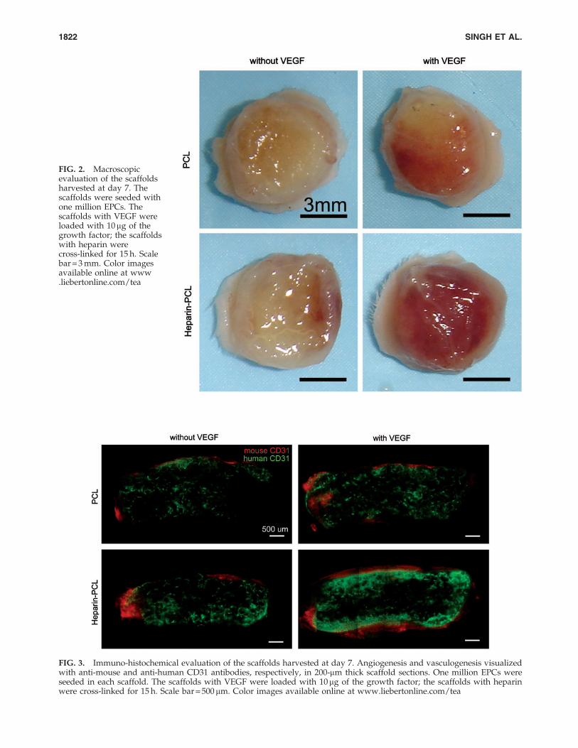
FIG. 3. Immuno-histochemical evaluation of the scaffolds harvested at day 7. Angiogenesis and vasculogenesis visualizedwith anti-mouse and anti-human CD31 antibodies, respectively, in 200-mm thick scaffold sections. One million EPCs wereseeded in each scaffold. The scaffolds with VEGF were loaded with 10mg of the growth factor; the scaffolds with heparinwere cross-linked for 15 h. Scale bar = 500 mm. Color images available online at www.liebertonline.com/tea
FIG. 2. Macroscopicevaluation of the scaffoldsharvested at day 7. Thescaffolds were seeded withone million EPCs. Thescaffolds with VEGF wereloaded with 10mg of thegrowth factor; the scaffoldswith heparin werecross-linked for 15 h. Scalebar = 3 mm. Color imagesavailable online at www.liebertonline.com/tea
1822 SINGH ET AL.

To achieve uniform vessel formation in heparin-PCLscaffolds with VEGF, we assessed the effect of varying theEPCs seeding density. The scaffolds seeded with 105, 106, or107 EPCs in 50mL of the media were implanted in NOD-
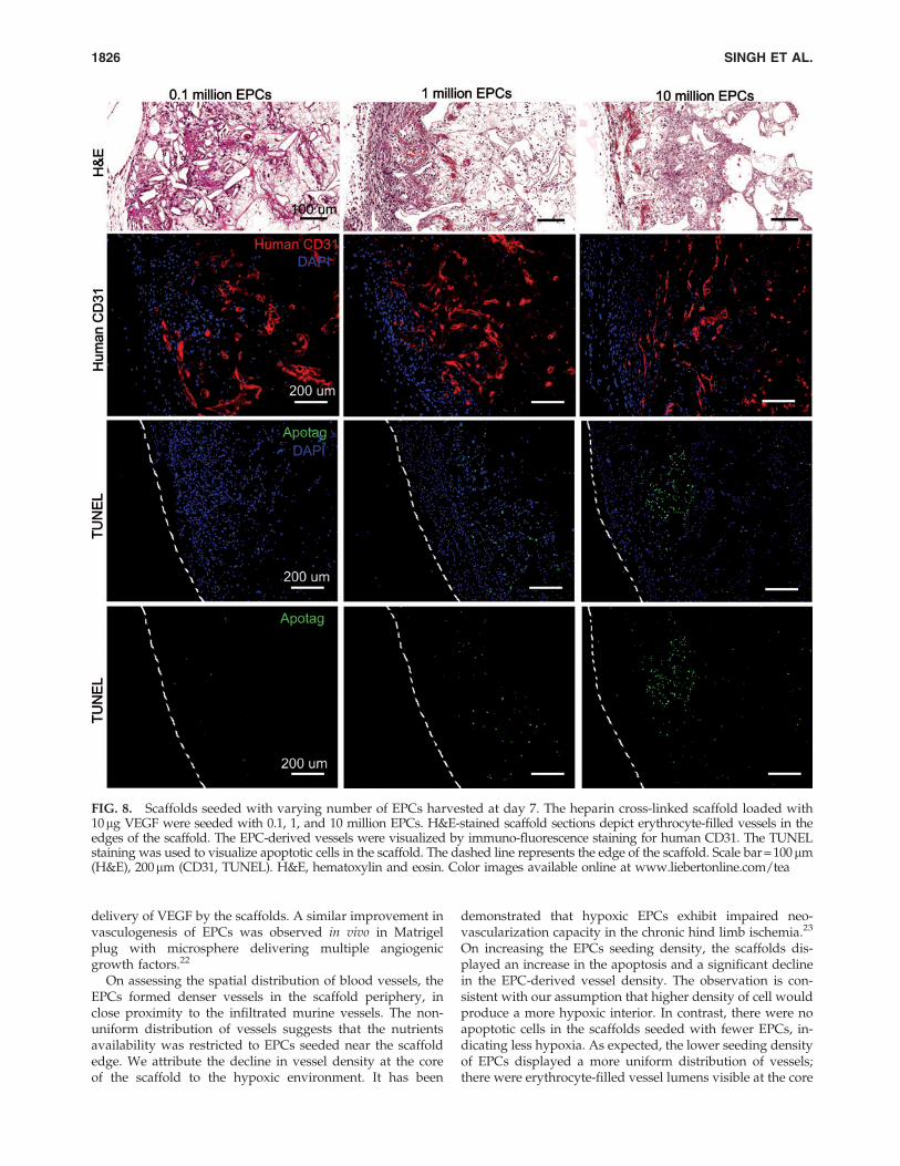
SCID mice. After 7 days in vivo, the scaffolds were assessedfor vasculogenesis and apoptosis using anti-human CD31and TUNEL staining, respectively (Fig. 8). The periphery ofscaffolds displayed more apoptotic cells as the EPC seeding
FIG. 4. Immuno-histochemical evaluation of the scaffolds harvested at day 7. Angiogenesis and vasculogenesis quantified bydiaminobenzidine staining of the scaffold sections with anti-mouse CD31 (A) and anti-human CD31 (B) antibodies, respectively.One million EPCs were seeded in each scaffold. The scaffolds with VEGF were loaded with 10mg of the growth factor. Thescaffolds with heparin were cross-linked for 15 h. Scale bar = 100mm. Color images available online at www.liebertonline.com/tea
BLOOD VESSEL FORMATION IN SCAFFOLD BY EPCS 1823

density increased. There were less apoptosis and moreerythrocyte-filled vessels in scaffolds seeded with 105 EPCs.The core of the scaffold had human-CD31 positive vesselswhen fewer EPCs were initially seeded (Fig. 9). In contrast,the core of the scaffold seeded with more EPCs lacked vessellumens and showed cellulardebris with hematoxylin andeosin stain. The dead cells caused nonspecific adsorption ofanti-human CD31 antibody. The quantification of human-CD31 positive vessels in the scaffolds demonstrated signifi-cantly higher vessel density in scaffolds seeded with 105 and106 EPCs when compared with 107 EPCs (Fig. 10). There wasno significant difference in overall vessel density betweenseeding with 105 and 106 EPCs; however, a more uniformspatial distribution of vessels was observed in scaffoldsseeded with 105 EPCs.
We tested whether the EPCs maintained the vasculogen-esis potential in the presence of other seeded cells. Theheparin-PCL scaffolds with 10 mg of VEGF and 105 EPCs
were co-seeded with GFP-positive primary hepatocytes orbone marrow stromal cells and were implanted in NOD-SCID mice. After 1 week, the retrieved implants weresectioned and immunostained for anti-human CD31 andanti-GFP antibodies. The results indicated that the EPCscontinued to form vessels in the presence of other cell types;however, the vessels were confined to the edge of scaffolds(Fig. 11).
To examine the effect of the in vitro expansion of EPCs ontheir in vivo vaculogenic potential, the EPCs were maintainedin culture for 90 days. The EPCs at passage 13 looked mor-phologically distinct from the cells at passage 5 (Fig. 12). TheEPCs at a lower passage number had several large clusters ofcobble-stone shaped cells that stained intensely with anti-vWF antibody. In contrast, EPCs at a higher passage numberhad few anti-vWF positive cells inter-dispersed among pre-dominantly spindle-shaped cells. When scaffolds seededwith high passage number EPCs were implanted in vivo for 1week, there was a significant decline in anti-human CD31positive vessels as compared with those seeded with EPCs atan earlier passage number.
Discussion
In our previous study, PCL scaffolds with surface cross-linked heparin were shown to have an increase in VEGF-mediated angiogenesis after 14 days of implantation.18
However, even under the most optimal conditions, there was
FIG. 5. Quantification of vessels in the scaffold harvestedon day 7. One million EPCs were seeded in each scaffold.The average vessel density (A) and the percentage of scaffoldarea occupied by vessels (B) were quantified using anti-mouse CD31 and anti -human CD31 antibodies, respectively.Values represent the mean and standard deviation (n = 3). V0and V10 represent scaffold without VEGF and loaded with10 mg of VEGF, respectively. H0 and Hm represent scaffoldswithout and with cross-linked heparin, respectively.$,*p < 0.0005, #p < 0.005, + p < 0.05.
FIG. 6. Anastomoses between the EPC-derived vessels andmurine vessels in the scaffolds harvested at day 7. Theheparin cross-linked scaffold loaded with 10mg of VEGF andseeded with one million EPCs displays human CD31-positive vessels filled with murine erythrocytes. Doubleimmuno-fluorescence staining of 200-mm thick section showsthe mouse and human CD31-positive vessel network in closeproximity. Scale bar = 50 mm. Color images available online atwww.liebertonline.com/tea
1824 SINGH ET AL.
insufficient angiogenesis in 2-mm thick scaffolds at an earliertime point of 7 days. In this study, we employed humanumbilical cord blood-derived EPCs to induce blood vesselformation at day 7 in vivo. Since VEGF enhances the EPCsproliferation in vitro11,14 and vasculogenesis in vivo,19 wehypothesized that delivery of VEGF from the heparin-immobilized scaffold will improve angiogenesis and vascu-logenesis, simultaneously. This was indeed the case, asheparin-PCL scaffolds with VEGF and EPCs had the mostvascularization compared with other scaffolds with EPCs.
EPCs have a much higher vasculogenic potential thanmature ECs, but additional strategies are required to enhancethe efficacy of EPC-based therapies. Majority of the studies inliterature have focused on improving vasculogenic potentialof EPCs by co-delivery of supporting cells that can functionsas pericytes, such as smooth muscle cells and fibro-
blasts.3,9,14,15,20 Although the presence of supporting cellsimproves the vessel formation by ECs, they also increase themetabolic burden during the vessel remodeling stages inthe scaffold. Recently, investigators have started exploringthe role of matrix microenviroment in modulating the vesselformation by ECs. Cooper and Sefton reported that the fi-bronectin coating increases the vessels formation by HU-VECs in collagen scaffolds in vivo.21 Besides, Critser et al.reported a decline in the in vivo vasculogenesis of EPCs withan increase in collagen fiber density and scaffold stiffness.13
An important finding in our study was the significantincrease in the formation of vessels by EPCs in heparin-immobilized scaffold with VEGF. Previously, we reportedthat the release rate of VEGF from the heparin cross-linkedscaffold was 100 ng/day.18 The significant increase in vas-culogenesis and angiogenesis may be due to the sustained
FIG. 7. Quantification of angiogenesis as a function of scaffold depth at day 7. One million EPCs were seeded in eachscaffold. The human CD31-positive vessels in scaffold sections were outlined with red, blue, and green color for 0–200, 200–400, and 400–600mm intervals, respectively (A). The vessel density and percentage of area occupied by vessels (B) wereevaluated at 200-mm intervals. Values represent the mean and standard deviation (n = 3). V0 and V10 represent scaffoldswithout VEGF and loaded with 10 mg of VEGF, respectively. H0 and Hm represent scaffolds without and with cross-linkedheparin, respectively. Scale bar = 500 mm. Color images available online at www.liebertonline.com/tea
BLOOD VESSEL FORMATION IN SCAFFOLD BY EPCS 1825
delivery of VEGF by the scaffolds. A similar improvement invasculogenesis of EPCs was observed in vivo in Matrigelplug with microsphere delivering multiple angiogenicgrowth factors.22
On assessing the spatial distribution of blood vessels, theEPCs formed denser vessels in the scaffold periphery, inclose proximity to the infiltrated murine vessels. The non-uniform distribution of vessels suggests that the nutrientsavailability was restricted to EPCs seeded near the scaffoldedge. We attribute the decline in vessel density at the coreof the scaffold to the hypoxic environment. It has been
demonstrated that hypoxic EPCs exhibit impaired neo-vascularization capacity in the chronic hind limb ischemia.23
On increasing the EPCs seeding density, the scaffolds dis-played an increase in the apoptosis and a significant declinein the EPC-derived vessel density. The observation is con-sistent with our assumption that higher density of cell wouldproduce a more hypoxic interior. In contrast, there were noapoptotic cells in the scaffolds seeded with fewer EPCs, in-dicating less hypoxia. As expected, the lower seeding densityof EPCs displayed a more uniform distribution of vessels;there were erythrocyte-filled vessel lumens visible at the core
FIG. 8. Scaffolds seeded with varying number of EPCs harvested at day 7. The heparin cross-linked scaffold loaded with10mg VEGF were seeded with 0.1, 1, and 10 million EPCs. H&E-stained scaffold sections depict erythrocyte-filled vessels in theedges of the scaffold. The EPC-derived vessels were visualized by immuno-fluorescence staining for human CD31. The TUNELstaining was used to visualize apoptotic cells in the scaffold. The dashed line represents the edge of the scaffold. Scale bar = 100mm(H&E), 200mm (CD31, TUNEL). H&E, hematoxylin and eosin. Color images available online at www.liebertonline.com/tea
1826 SINGH ET AL.
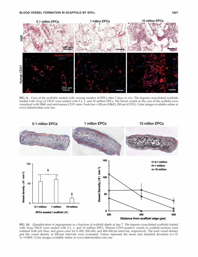
FIG. 9. Core of the scaffolds seeded with varying number of EPCs after 7 days in vivo. The heparin cross-linked scaffoldsloaded with 10 mg of VEGF were seeded with 0.1, 1, and 10 million EPCs. The blood vessels in the core of the scaffold werevisualized with H&E and anti-human CD31 stain. Scale bar = 100 mm (H&E), 200 mm (CD31). Color images available online atwww.liebertonline.com/tea
FIG. 10. Quantification of angiogenesis as a function of scaffold depth at day 7. The heparin cross-linked scaffolds loadedwith 10 mg VEGF were seeded with 0.1, 1, and 10 million EPCs. Human CD31-positive vessels in scaffold sections wereoutlined with red, blue, and green color for 0–200, 200–400, and 400–600 mm intervals, respectively. The total vessel densityand the vessel density at 200-mm intervals were evaluated. Values represent the mean and standard deviation (n = 3).$p < 0.0005. Color images available online at www.liebertonline.com/tea
BLOOD VESSEL FORMATION IN SCAFFOLD BY EPCS 1827
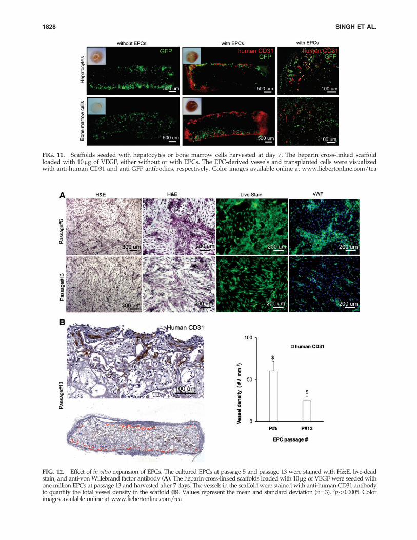
FIG. 11. Scaffolds seeded with hepatocytes or bone marrow cells harvested at day 7. The heparin cross-linked scaffoldloaded with 10mg of VEGF, either without or with EPCs. The EPC-derived vessels and transplanted cells were visualizedwith anti-human CD31 and anti-GFP antibodies, respectively. Color images available online at www.liebertonline.com/tea
FIG. 12. Effect of in vitro expansion of EPCs. The cultured EPCs at passage 5 and passage 13 were stained with H&E, live-deadstain, and anti-von Willebrand factor antibody (A). The heparin cross-linked scaffolds loaded with 10mg of VEGF were seeded withone million EPCs at passage 13 and harvested after 7 days. The vessels in the scaffold were stained with anti-human CD31 antibodyto quantify the total vessel density in the scaffold (B). Values represent the mean and standard deviation (n = 3). $p < 0.0005. Colorimages available online at www.liebertonline.com/tea
1828 SINGH ET AL.

of the 2-mm thick scaffold. To test the clinical applicability ofEPCs, they were co-seeded with hepatocytes or bone marrowcells. The vessel formation by EPCs was limited to the scaf-fold edges at day 7. Implantation of scaffolds for longer timepoints would be required to evaluate the contribution ofEPCs in liver and bone regeneration. A strategy to minimizehypoxia in the scaffold core could improve efficacy of EPCsdelivery in large-sized scaffolds. The oxygen-generatingscaffolds reported by Oh et al. may improve the vessel for-mation by the EPCs.24 Alternatively, it could be possible todelay the onset of hypoxia in the scaffold by pre-vascularization with EPCs before implantation. It has beenshown that scaffolds prevasculariized with HUVEC formedvessels much earlier than non-prevascularized scaffolds.4
We observed a significant decline in the in vivo vesselforming ability of EPCs at a higher passage after 60 days ofin vitro expansion. Our result suggests that the EPCs undergocell maturation after extended in vitro culture. Others havenoticed a decline in the EPCs growth kinetics and prolifer-ative response toward VEGF in vitro at a high passagenumber.14 There was an eightfold decline in the vessel den-sity in the implanted Matrigel between the EPCs at passage 3and passage 12. We employed human umbilical cord blood-derived EPCs in our current studies. However, the humanadult peripheral blood would be a valuable source for au-tologous EPCs in clinical applications. Melero-Martin et al.reported a comparable in vivo vasculogenic potential of theperipheral blood and cord blood-derived EPCs.14
In summary, our results demonstrate that the vessel-forming ability of EPCs was significantly improved by thedelivery of VEGF from scaffolds with immobilized heparin.In future, the strategies to reduce hypoxia in scaffold duringthe EPC vasculogenesis should be implemented for moreuniform vascularization.
Acknowledgments
This work was funded by the National Institutes of HealthR01 DK083319 and the Fubon Foundation.
Disclosure Statement
No competing financial interests exist.
References
1. Richardson, T.P., Peters, M.C., Ennett, A.B., and Mooney,D.J. Polymeric system for dual growth factor delivery. NatBiotechnol 19, 1029, 2001.
2. Mao, Z., Shi, H., Guo, R., Ma, L., Gao, C., Han, C., and Shen,J. Enhanced angiogenesis of porous collagen scaffolds byincorporation of TMC/DNA complexes encoding vascularendothelial growth factor. Acta Biomater 5, 2983, 2009.
3. Levenberg, S., Rouwkema, J., Macdonald, M., Garfein, E.S.,Kohane, D.S., Darland, D.C., Marini, R., van Blitterswijk,C.A., Mulligan, R.C., D’Amore, P.A., and Langer, R. En-gineering vascularized skeletal muscle tissue. Nat Biotechnol23, 879, 2005.
4. Chen, X., Aledia, A.S., Ghajar, C.M., Griffith, C.K., Putnam,A.J., Hughes, C.C., and George, S.C. Prevascularization of afibrin-based tissue construct accelerates the formation offunctional anastomosis with host vasculature. Tissue EngPart A 15, 1363, 2009.
5. Montano, I., Schiestl, C., Schneider, J., Pontiggia, L., Lu-ginbuhl, J., Biedermann, T., Bottcher-Haberzeth, S., Braziu-lis, E., Meuli, M., and Reichmann, E. Formation of humancapillaries in vitro: the engineering of prevascularized ma-trices. Tissue Eng Part A 16, 269, 2010.
6. Levenberg, S., Golub, J.S., Amit, M., Itskovitz-Eldor, J., andLanger, R. Endothelial cells derived from human embryonicstem cells. Proc Natl Acad Sci USA 99, 4391, 2002.
7. Valarmathi, M.T., Davis, J.M., Yost, M.J., Goodwin, R.L., andPotts, J.D. A three-dimensional model of vasculogenesis.Biomaterials 30, 1098, 2009.
8. Mead, L.E., Prater, D., Yoder, M.C., and Ingram, D.A. Isola-tion and characterization of endothelial progenitor cells fromhuman blood. Curr Protoc Stem Cell Biol 6, 2C.1.1, 2008.
9. Fedorovich, N.E., Haverslag, R.T., Dhert, W.J., and Alblas, J.The role of endothelial progenitor cells in prevascularizedbone tissue engineering: development of heterogeneousconstructs. Tissue Eng Part A 16, 2355, 2010.
10. Yoder, M.C., Mead, L.E., Prater, D., Krier, T.R., Mroueh,K.N., Li, F., Krasich, R., Temm, C.J., Prchal, J.T., and Ingram,D.A. Redefining endothelial progenitor cells via clonalanalysis and hematopoietic stem/progenitor cell principals.Blood 109, 1801, 2007.
11. Ingram, D.A., Mead, L.E., Tanaka, H., Meade, V., Fenoglio,A., Mortell, K., Pollok, K., Ferkowicz, M.J., Gilley, D., andYoder, M.C. Identification of a novel hierarchy of endothe-lial progenitor cells using human peripheral and umbilicalcord blood. Blood 104, 2752, 2004.
12. Decaris, M.L., Lee, C.I., Yoder, M.C., Tarantal, A.F., andLeach, J.K. Influence of the oxygen microenvironment on theproangiogenic potential of human endothelial colony form-ing cells. Angiogenesis 12, 303, 2009.
13. Critser, P., Kreger, S., Voytik-Harbin, S., and Yoder, M.Collagen matrix physical properties modulate endothelialcolony forming cell-derived vessels in vivo. Microvasc Res80, 23, 2010.
14. Melero-Martin, J.M., Khan, Z.A., Picard, A., Wu, X., Par-uchuri, S., and Bischoff, J. In vivo vasculogenic potential ofhuman blood-derived endothelial progenitor cells. Blood109, 4761, 2007.
15. Melero-Martin, J.M., De Obaldia, M.E., Kang, S.Y., Khan,Z.A., Yuan, L., Oettgen, P., and Bischoff, J. Engineering ro-bust and functional vascular networks in vivo with humanadult and cord blood-derived progenitor cells. Circ Res 103,
194, 2008.16. Smith, M.K., Peters, M.C., Richardson, T.P., Garbern, J.C.,
and Mooney, D.J. Locally enhanced angiogenesis promotestransplanted cell survival. Tissue Eng 10, 63, 2004.
17. Lee, S., Jilani, S.M., Nikolova, G.V., Carpizo, D., and Iruela-Arispe, M.L. Processing of VEGF-A by matrix metallopro-teinases regulates bioavailability and vascular patterning intumors. J Cell Biol 169, 681, 2005.
18. Singh, S., Wu, B.M., and Dunn, J.C. The enhancement ofVEGF-mediated angiogenesis by polycaprolactone scaffoldswith surface cross-linked heparin. Biomaterials 32, 2059, 2011.
19. Li, B., Sharpe, E.E., Maupin, A.B., Teleron, A.A., Pyle, A.L.,Carmeliet, P., and Young, P.P. VEGF and PlGF promoteadult vasculogenesis by enhancing EPC recruitment andvessel formation at the site of tumor neovascularization.FASEB J 20, 1495, 2006.
20. Wu, X., Rabkin-Aikawa, E., Guleserian, K.J., Perry, T.E.,Masuda, Y., Sutherland, F.W., Schoen, F.J., Mayer, J.E. Jr.,and Bischoff, J. Tissue-engineered microvessels on three-dimensional biodegradable scaffolds using human endothelial
BLOOD VESSEL FORMATION IN SCAFFOLD BY EPCS 1829

progenitor cells. Am J Physiol Heart Circ Physiol 287,
H480, 2004.21. Cooper, T., and Sefton, M. Fibronectin coating of collagen
modules increases in vivo HUVEC survival and vessel for-mation in SCID mice. Acta Biomater 7, 1072, 2011.
22. Saif, J., Schwarz, T.M., Chau, D.Y., Henstock, J., Sami, P.,Leicht, S.F., Hermann, P.C., Alcala, S., Mulero, F., Shakesheff,K.M., Heeschen, C., and Aicher, A. Combination of injectablemultiple growth factor-releasing scaffolds and cell therapy asan advanced modality to enhance tissue neovascularization.Arterioscler Thromb Vasc Biol 30, 1897, 2010.
23. Marsboom, G., Pokreisz, P., Gheysens, O., Vermeersch, P.,Gillijns, H., Pellens, M., Liu, X., Collen, D., and Janssens, S.Sustained endothelial progenitor cell dysfunction afterchronic hypoxia-induced pulmonary hypertension. StemCells 26, 1017, 2008.
24. Oh, S.H., Ward, C.L., Atala, A., Yoo, J.J., and Harrison, B.S.Oxygen generating scaffolds for enhancing engineered tis-sue survival. Biomaterials 30, 757, 2009.
Address correspondence to:James C.Y. Dunn, M.D., Ph.D.
Department of BioengineeringUniversity of California
Box 709818, 10833 Le Conte Ave.Los Angeles, CA 90095
E-mail: [email protected]
Received: December 5, 2010Accepted: March 10, 2011
Online Publication Date: April 27, 2011
1830 SINGH ET AL.