Identification of Proteins Regulated by PACAP in PC12 Cells by 2D Gel Electrophoresis Coupled to...
9
NYAS nyas˙vip049 Dispatch: 5-29-2006 CE: IV Journal MSP No. No. of pages: 8 PE: Julia nyas˙vip˙049 NYAS2006.cls (1994/07/13 v1.2u Standard LaTeX document class) 5-29-2006 :1083 Identification of Proteins Regulated by PACAP in PC12 Cells by 2D Gel Electrophoresis Coupled to Mass Spectrometry ALEXIS LEBON, a,b DAMIEN SEYER, a,c PASCAL COSETTE, a,c LAURENT COQUET, a,c THIERRY JOUENNE, a,c PHILIPPE CHAN, a,b JEROME LEPRINCE, a,b ALAIN FOURNIER, d HUBERT VAUDRY, a,b BRUNO J. GONZALEZ, a,b AND DAVID VAUDRY a,b a European Institute for Peptide Research (IFRMP23), University of Rouen, 76821 Mont-Saint-Aignan, France b INSERM U413, Laboratory of Cellular and Molecular Neuroendocrinology, European Institute for Peptide Research, University of Rouen, 76821 Mont-Saint-Aignan, France c IBBR Group, CNRS UMR 6522, Proteomic Platform of the IFRMP23 d INRS-Institut Armand Frappier, University of Qu´ ebec, Pointe-Claire, Canada H9R 1G6 Q1 ABSTRACT: The rat pheochromocytoma PC12 cell line has been widely used as a model to study neuronal differentiation. In particular, after serum depletion, PC12 cells stop to proliferate and undergo apopto- sis. Under such conditions, treatment with pituitary adenylate cyclase- activating polypeptide (PACAP) promotes cell survival and induces neu- rite outgrowth. The identification of the proteins regulated by PACAP in PC12 cells under apoptotic conditions should provide valuable informa- tion concerning the mechanisms controlling neuronal cell survival and differentiation. To this aim, PC12 cells cultured in serum-free medium were treated with PACAP (10 −7 M), proteins were extracted, separated by two-dimensional gel electrophoresis (2-DE), and identified by MALDI- ToF mass spectrometry. The comparison between 16 2-DE-maps led to the characterization of 110 proteins regulated by PACAP among which 22 have been identified by automatic query of the Mascot, Aldente, and Profound servers with the ProGeR-CDD database. Seventy-six percent of these proteins, including the p17 subunit of caspase-3, the heat shock pro- tein hsp60, and the GTPase ran were found to be repressed whereas the others notably hsp27, tubulin -5, and calmodulin were overexpressed. Investigation of the putative functions indicated that some of the proteins Address for correspondence: Dr. Hubert Vaudry, INSERM U413, Laboratory of Cellular and Molec- ular Neuroendocrinology, European Institute for Peptide Research (IFRMP 23), University of Rouen, 76821 Mont-Saint-Aignan, France. Voice: +33 235-14-6624; fax: +33 235-14-6946. e-mail: [email protected] Ann. N.Y. Acad. Sci. xxxx: 1–8 (2006). C 2006 New York Academy of Sciences. doi: 10.1196/annals.1317.049 1
Transcript of Identification of Proteins Regulated by PACAP in PC12 Cells by 2D Gel Electrophoresis Coupled to...

NYAS nyas˙vip049 Dispatch: 5-29-2006 CE: IV
Journal MSP No. No. of pages: 8 PE: Julia
nyas˙vip˙049 NYAS2006.cls (1994/07/13 v1.2u Standard LaTeX document class) 5-29-2006 :1083
Identification of Proteins Regulated byPACAP in PC12 Cells by 2D GelElectrophoresis Coupled to MassSpectrometry
ALEXIS LEBON,a,b DAMIEN SEYER,a,c PASCAL COSETTE,a,c
LAURENT COQUET,a,c THIERRY JOUENNE,a,c PHILIPPE CHAN,a,b
JEROME LEPRINCE,a,b ALAIN FOURNIER,d HUBERT VAUDRY,a,b
BRUNO J. GONZALEZ,a,b AND DAVID VAUDRYa,b
aEuropean Institute for Peptide Research (IFRMP23), University of Rouen,76821 Mont-Saint-Aignan, FrancebINSERM U413, Laboratory of Cellular and Molecular Neuroendocrinology,European Institute for Peptide Research, University of Rouen, 76821Mont-Saint-Aignan, FrancecIBBR Group, CNRS UMR 6522, Proteomic Platform of the IFRMP23dINRS-Institut Armand Frappier, University of Quebec, Pointe-Claire, CanadaH9R 1G6 Q1
ABSTRACT: The rat pheochromocytoma PC12 cell line has been widelyused as a model to study neuronal differentiation. In particular, afterserum depletion, PC12 cells stop to proliferate and undergo apopto-sis. Under such conditions, treatment with pituitary adenylate cyclase-activating polypeptide (PACAP) promotes cell survival and induces neu-rite outgrowth. The identification of the proteins regulated by PACAP inPC12 cells under apoptotic conditions should provide valuable informa-tion concerning the mechanisms controlling neuronal cell survival anddifferentiation. To this aim, PC12 cells cultured in serum-free mediumwere treated with PACAP (10−7 M), proteins were extracted, separated bytwo-dimensional gel electrophoresis (2-DE), and identified by MALDI-ToF mass spectrometry. The comparison between 16 2-DE-maps led tothe characterization of 110 proteins regulated by PACAP among which22 have been identified by automatic query of the Mascot, Aldente, andProfound servers with the ProGeR-CDD database. Seventy-six percent ofthese proteins, including the p17 subunit of caspase-3, the heat shock pro-tein hsp60, and the GTPase ran were found to be repressed whereas theothers notably hsp27, tubulin �-5, and calmodulin were overexpressed.Investigation of the putative functions indicated that some of the proteins
Address for correspondence: Dr. Hubert Vaudry, INSERM U413, Laboratory of Cellular and Molec-ular Neuroendocrinology, European Institute for Peptide Research (IFRMP 23), University of Rouen,76821 Mont-Saint-Aignan, France. Voice: +33 235-14-6624; fax: +33 235-14-6946.
e-mail: [email protected]
Ann. N.Y. Acad. Sci. xxxx: 1–8 (2006). C© 2006 New York Academy of Sciences.doi: 10.1196/annals.1317.049
1

nyas˙vip˙049 NYAS2006.cls (1994/07/13 v1.2u Standard LaTeX document class) 5-29-2006 :1083
2 ANNALS NEW YORK ACADEMY OF SCIENCES
regulated by PACAP and identified in the present study could control cellsurvival or differentiation.
KEYWORDS: proteomics; PC12 cells; PACAP
INTRODUCTION
Pituitary adenylate cyclase-activating polypeptide (PACAP) was isolatedfrom the sheep hypothalamus on the basis of its capacity to stimulate theproduction of cAMP in rat anterior pituitary cells.1 PACAP and its recep-tors are widely expressed in the brain and peripheral organs where they exertmany biological activities.2 In particular, PACAP has been shown to act asa neurotrophic factor promoting cell differentiation during development andinhibiting apoptosis induced by neurotoxic agents.3,4,5
The rat pheochromocytoma PC12 cell line can easily be manipulated to studyneuronal differentiation and apoptosis.6,7 When cells are cultured with serum,addition of PACAP inhibits cell proliferation and induces neurite outgrowth.8,9
When cells are cultured in serum-free medium, PACAP promotes PC12 sur-vival and neuritogenesis.10 The effect of PACAP on neurite outgrowth requiresthe phosphorylation of the MAP kinases ERK1/211 but the downstream tar-gets controlling cell differentiation remain largely unknown. The aim of thepresent study was thus to identify new proteins regulated by PACAP in PC12cells using two-dimensional gel electrophoresis (2-DE) analysis coupled tomass spectrometry identification.
MATERIALS AND METHODS
PC12 cells were grown as previously reported.7 When cell density reached200,000 cells/mL, cultures were shifted to serum-free medium for 12 h andthen incubated in the absence or presence of 10−7 M PACAP38 for 9 h(FIG. 1 A). At the end of the treatment, cells were washed twice with PBSand the proteins were extracted according to two different protocols to max-imize the number of identified proteins. The first lysis buffer contained 1%Triton X-100, 50 mM Tris-HCl, and 10 mM EDTA. After sonication, the ho-mogenate was centrifuged (9,500 g, 4◦C, 15 min) and the proteins containedin the supernatant were precipitated by the addition of 10% trichloroaceticacid. The samples were centrifuged (9,500 g, 4◦C, 15 min) and the pellet waswashed three times with alcohol/ether. The extracted proteins were finally driedand kept frozen until use. The second extraction protocol was carried out ac-cording to the instructions provided with the Complete Mammalian ProteomeExtraction Kit (Calbiochem ). Q2
For 2-DE, proteins were resuspended in 1 mL isoelectric focusing (IEF)buffer containing 7 M urea, 2 M thiourea, 0.5% ampholines (pH 3.5-10), 2 mM

nyas˙vip˙049 NYAS2006.cls (1994/07/13 v1.2u Standard LaTeX document class) 5-29-2006 :1083
LEBON et al.: PROTEINS REGULATED BY PACAP IN PC12 CELLS 3
FIGURE 1. Identification process of proteins regulated by PACAP in PC12 cells. Cellswere incubated in the absence or presence of PACAP (10−7 M) for 9 h before proteinextraction (A). Proteins were separated according to their isoelectric point (B) and molecularweight (C) to generate 2D gels (D). The gels were digitized and analyzed with the PD-Questsoftware (E). After excision and tryptic digestion, a fingerprint of the proteins was obtainedby MALDI-ToF mass spectrometry (F) and the identification was conducted on severalproteomic servers (G).

nyas˙vip˙049 NYAS2006.cls (1994/07/13 v1.2u Standard LaTeX document class) 5-29-2006 :1083
4 ANNALS NEW YORK ACADEMY OF SCIENCES
tributyl phosphine, and 0.5% amidosulfobetaine 14 (ASB14). After quantifi-cation,6 200�g of solubilized proteins were separated in the first dimensionaccording to their isoelectric point (FIG. 1 B) along nonlinear immobilized pH-gradient strips (IPGS pH 3–10, 17 cm, Amersham Biosciences) in a Protean Q3
IEF-Cell (Bio-Rad). At the end of the first dimension, IPGS were reequili- Q4
brated (buffer containing 2% DTT and 2.5% iodoacetamide) and applied ontop of a 12.5% SDS polyacrylamide gel for the second dimension (FIG.1 C,Protean II Xi, Bio-Rad). At the end of the migration, gels were fixed overnightin a solution of 10% acetic acid, 30% ethanol, washed three times in water,twice in 10% ethanol, and sensitized in a 0.02% sodium thiosulfate solution for1 min. Finally, gels were stained in a 0.1% silver nitrate solution for 30 min andrevealed in the presence of 0.12% sodium carbonate, 0.004% formaldehyde,and 0.008% sodium thiosulfate. Revelation was stopped with an 8% aceticacid solution (FIG. 1 D).
Gels were digitized using a computer-assisted densitometer (GS-800, Bio-Rad). Spot detection and statistical analysis were performed with the PD-Quest7.01 software. For each experimental condition, spot quantities correspondedto the mean value of 4 independent gels (FIG. 1E). For protein identification,2-DE gels were performed by loading 500 �g proteins per gel and by usingcolloidal blue staining. Gels were fixed overnight in a solution containing50% ethanol and 1.4% orthophosphoric acid. After rinsing in water, the gelswere sensitized with 17% ammonium sulphate, 0.34% methanol, and 1.4%orthophosphoric acid. Staining was carried out for 3-4 days in the sensitizationsolution completed with 0.06% G 250 blue. Spots of interest were then excised(ProXcision, Perkin Elmer), gel plugs were dried using a Speed-Vac evaporator Q5
and subjected to trypsin (MultiProbe II, Perkin Elmer). Peptide fragmentswere expulsed from the gels with acetonitrile, spotted on a MALDI platein the presence of a cis-alpha cyano 4-hydroxy cinnamic acid matrix. Massspectrometry was carried out on a Voyager DEPro MALDI-ToF spectrometer(Applied Biosystems) in the reflectron mode (FIG. 1 F). Peptide fingerprints Q6
were matched against in silico digests of the SwissProt database using theMascot (www.matrixscience.com), Aldente (www.expasy.org), and Profound(http://prowl.rockefeller.edu) servers (FIG. 1 G).
RESULTS AND DISCUSSION
For each extraction condition, protein regulations were investigated from thedifferential analysis of four control and PACAP-treated gels. Statistical anal-ysis performed with the PD-Quest 7.01 software made it possible to identify110 proteins significantly regulated 2-fold or more after a 9-h treatment withPACAP. After staining with colloidal blue, 46 spots were excised and submit-ted to identification. MALDI-ToF mass spectra of the generated peptides wereuploaded in the ProGeR-CDD database (http://proger-cdd.crihan.fr) and 22 of

nyas˙vip˙049 NYAS2006.cls (1994/07/13 v1.2u Standard LaTeX document class) 5-29-2006 :1083
LEBON et al.: PROTEINS REGULATED BY PACAP IN PC12 CELLS 5
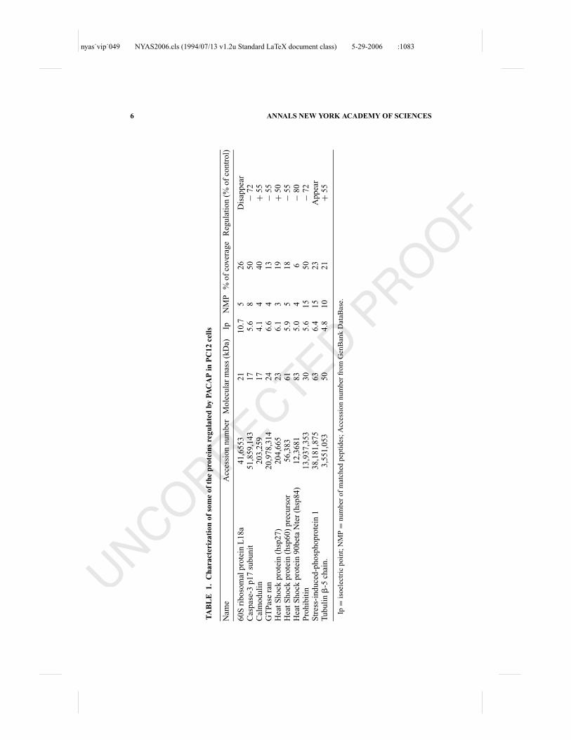
the proteins regulated by PACAP have already been identified. Ten of theseregulated proteins are listed in TABLE 1 .
One of the proteins repressed by PACAP is the p17 subunit of caspase-3.This protein, which plays a key role in apoptosis, has previously been shownto be inhibited by PACAP in cerebellar granule cells.12 Inhibition of the activeform of caspase-3 could be sufficient to protect PC12 cells from apoptosisinduced by serum privation. Two heat shock proteins, i.e. hsp60 and hsp90�,were also repressed by PACAP. It is interesting to note that hsp60 is inducedby neurotoxic agents, such as ethanol13 or lipopolysaccharide14 and repressedupon treatment with NGF.15 The expression level of hsp90� is significantlyhigher in human breast cancer cells than in normal tissues but the role ofthis protein in the control of tumor cell proliferation or differentiation remainsunclear.16 Functional investigations should be conducted to clarify the involve-ment of hsp60 and/or hsp90� proteins in the antiapoptotic and neurotrophiceffects of PACAP. Other proteins repressed by PACAP, such as the GTPaseran17,18,19 and prohibitin20,21 could also be involved in the control of PC12cells proliferation. However, there is still considerable controversy concerningthe role of these two proteins that can either induce or repress cell senescenceor migration depending on the cell type.
PACAP stimulated the expression of hsp27, which is known to protect PC12cells from apoptosis after heat shock and NGF withdrawal.22 Recently, hsp27has also been shown to inhibit c-Jun N-terminal kinase-induced neuronaldeath23 and to prevent cytochrome c release and caspase-3 activation inducedby 6-hydroxydopamine.24 On the basis of these observations, the possible in-volvement of hsp27 in the antiapoptotic effect of PACAP should be considered.Expression of the tubulin �-5 subunit was found to be increased by PACAPin PC12 cells, which corroborates previous microarray results where the genecoding for this protein was induced during cell differentiation.25 Another pro-tein that appears to be regulated by several neurotrophic factors, includingPACAP, during PC12 cell differentiation is the calcium-binding regulatory pro-tein calmodulin.26,27 This protein would at least participate to neuritogenesis asshown with cells treated with calmodulin antisense oligodeoxynucleotides28,29
probably by interacting with the MEK/ERK transduction pathway.For each of the identified proteins, various information including the puta-
tive functions and the availability of specific antibodies or siRNA have beenrepatriated in the ProGeR-CDD database. These data will now facilitate theinvestigation of the possible role of the proteins identified in the present studyin the antiapoptotic and neurotrophic activities of PACAP.
ACKNOWLEDGMENTS
This work was supported by INSERM (U 413), CNRS (UMR 6522), theEuropean Institute for Peptide Research (IFRMP23), the Association pour la
nyas˙vip˙049 NYAS2006.cls (1994/07/13 v1.2u Standard LaTeX document class) 5-29-2006 :1083
6 ANNALS NEW YORK ACADEMY OF SCIENCES
TA
BL
E1.
Cha
ract
eriz
atio
nof
som
eof
the
prot
eins
regu
late
dby
PAC
AP
inP
C12
cells
Nam
eA
cces
sio
nn
um
ber
Mo
lecu
lar
mas
s(k
Da)
IpN
MP
%o
fco
ver
age
Reg
ula
tio
n(%
of
con
tro
l)
60
Sri
bo
som
alp
rote
inL
18
a4
1,6
55
32
11
0.7
52
6D
isap
pea
rC
asp
ase-
3p
17
sub
un
it5
1,8
59
,14
31
75
.68
50
−7
2C
alm
od
uli
n2
03
,25
91
74
.14
40
+5
5G
TP
ase
ran
20
,97
8,3
14
24
6.6
41
3−
55
Hea
tS
ho
ckp
rote
in(h
sp2
7)
20
4,6
65
23
6.1
31
9+
50
Hea
tS
ho
ckp
rote
in(h
sp6
0)
pre
curs
or
56
,38
36
15
.95
18
−5
5H
eat
Sh
ock
pro
tein
90
bet
aN
ter
(hsp
84
)1
2,3
68
18
35
.04
6−
80
Pro
hib
itin
13
,93
7,3
53
30
5.6
15
50
−7
2S
tres
s-in
du
ced
-ph
osp
ho
pro
tein
13
8,1
81
,87
56
36
.41
52
3A
pp
ear
Tu
bu
lin
�-5
chai
n.
3,5
51
,05
35
04
.81
02
1+
55
Ip=
iso
elec
tric
po
int;
NM
P=
nu
mb
ero
fm
atch
edp
epti
des
;A
cces
sio
nn
um
ber
fro
mG
enB
ank
Dat
aBas
e.

nyas˙vip˙049 NYAS2006.cls (1994/07/13 v1.2u Standard LaTeX document class) 5-29-2006 :1083
LEBON et al.: PROTEINS REGULATED BY PACAP IN PC12 CELLS 7
Recherche sur le Cancer (to D.V.), an INSERM-FRSQ exchange program (toA.F. and H.V.), and the Conseil Regional de Haute-Normandie. H.V. is affiliatedprofessor at the INRS-Institut Armand Frappier, University of Quebec.
REFERENCES
1. MIYATA, A., A. ARIMURA, R.R. DAHL, et al. 1989. Isolation of a novel 38residue-hypothalamic polypeptide which stimulates adenylate cyclase in pitu-itary cels. Biochem. Biophys. Res. Commun. 164: 567–574.
2. ARIMURA, A., A. SOMOGYVARI-VIGH, K. MIZUNO, et al. 1991. Tissue distributionof PACAP as determined by RIA: highly abundant in the rat brain and testes.Endocrinology 129: 2787–2789.
3. VAUDRY, D., B.J. GONZALEZ, M. BASILLE, et al. 2000. Pituitary adenylate cyclase-activating polypeptide and its receptors: from structure to functions. Pharmacol.Rev. 52: 269–324.
4. VAUDRY, D., B.J. GONZALEZ, M. BASILLE, et al. 1999. Neurotrophic activity ofpituitary adenylate cyclase-activating polypeptide on rat cerebellar cortex duringdevelopment. Proc. Natl. Acad. Sci. USA 96: 9415–9420.
5. VAUDRY, D., A. FALLUEL-MOREL, S. LEUILLET, et al. 2003. Regulators of cerebellargranule cell development act through specific signaling pathways. Science 300:1532–1534.
6. VAUDRY, D., Y. CHEN, C.M. HSU & L.E. EIDEN. 2002. PC12 cells as a model tostudy the neurotrophic activities of PACAP. Ann. N. Y. Acad. Sci. 971: 491–496.
7. VAUDRY, D., P.J. STORK, P. LAZAROVICI & L.E. EIDEN. 2002. Signaling pathwaysfor PC12 cell differentiation: making the right connections. Science 296: 1648–1649.
8. DEUTSH, P.J. & Y. SUN. 1992. The 38-amino acid form of pituitary adenylatecyclase-activating polypeptide stimulates dual signalling cascades in PC12 cellsand promotes neurite outgrowth. J. Biol. Chem. 267: 5108–5113.
9. LAZAROVICI, P., H. JIANG & D. FINK. 1998. The 38-amino-acid form of pituitaryadenylate cyclase-activating polypeptide induces neurite outgrowth in PC12 cellsthat is dependent on protein kinase C and extracellular signal-regulated kinasebut no on protein kinase A, nerve growth factor receptor tyrosine kinase, p21(ras) G protein, and pp60 (c-src) cytoplasmic tyrosine kinase. Mol. Pharmacol.54: 547–558.
10. VAUDRY, D., Y. CHEN, A. RAVNI, et al. 2002. Analysis of the PC12 cell transcrip-tome after differentiation with pituitary adenylate cyclase activating polypeptide(PACAP). J. Neurochem. 83: 1272–1284.
11. BARRIE, A.P., A.M. CLOHESSY, C.S. BUENSUCESO, et al. 1997. Pituitary adenylylcyclase-activating peptide stimulates extracellular signal-regulated kinase 1 or 2(ERK1/2) activity in a Ras-independent, mitogen-activated protein Kinase 1 or2-dependent manner in PC12 cells. J. Biol. Chem. 272: 19666–19671.
12. VAUDRY, D., T.F. PAMANTUNG, M. BASILLE, et al. 2002. PACAP protects cerebellargranule neurons against oxidative stress-induced apoptosis. Eur. J. Neurosci. 15:1451–1460.
13. DWYER, D.S., Y. LIU & R.J. BRADLEY. 1999. An ethanol-sensitive variant of thePC12 neuronal cell line: sensitivity to alcohol is associated with increased celladhesion and decreased glucose accumulation. J. Cell. Physiol. 178: 93–101.

nyas˙vip˙049 NYAS2006.cls (1994/07/13 v1.2u Standard LaTeX document class) 5-29-2006 :1083
8 ANNALS NEW YORK ACADEMY OF SCIENCES
14. HUANG, Y.H., A.Y. CHANG, C.M. HUANG, et al. 2002. Proteomic analysis oflipopolysaccharide-induced apoptosis in PC12 cells. Proteomics 2: 1220–1228.
15. DWYER, D.S., Y. LIU, S. MIAO & R.J. BRADLEY. 1996. Neuronal differentiation inPC12 cells is accompanied by diminished inducibility of Hsp70 and Hsp60 inresponse to heat and ethanol. Neurochem. Res. 21: 659–666.
16. YANO, M., Z. NAITO, S. TANAKA & G. ASANO. 1996. Expression and roles of heatshock proteins in human breast cancer. Jpn. J. Cancer. Res. 87: 908–915.
17. FERRANDO-MAY, E., V. CORDES, I. BILLER-CKOVRIC, et al. 2001. Mediate nucle-oporin cleavage, but not early redistribution of nuclear transport factors andmodulation of nuclear permeability in apoptosis. Cell Death Differ. 8: 495–505.
18. KING, F.W. & E. SHTIVELMAN. 2004. Inhibition of nuclear import by the proapop-totic protein CC3. Mol. Cell. Biol. 24: 7091–7101.
19. SCREATON, R.A., S. KIESSLING, O.J. SANSOM, et al. 2003. Fas-associated deathdomain protein interacts with methyl-CpG binding domain protein 4: a potentiallink between genome surveillance and apoptosis. Proc. Natl. Acad. Sci. USA100: 5211–5216.
20. FUSARO, G., P. DASGUPTA, S. RASTOGI, et al. 2003. Prohibitin induces the transcrip-tional activity of p53 and is exported from the nucleus upon apoptotic signaling.J. Biol. Chem. 278: 47853–47861.
21. RAJALINGAM, K., C. WUNDER, V. BRINKMANN, et al. 2005. Prohibitin is required forRas-induced Raf-MEK-ERK activation and epithelial cell migration. Nat. CellBiol. 7: 837–843.
22. MEAROW, K.M., M.E. DODGE, M. RAHIMTULA & C. YEGAPPAN. 2002. Stress-mediated signaling in PC12 cells—the role of the small heat shock protein,Hsp27, and Akt in protecting cells from heat stress and nerve growth factorwithdrawal. J. Neurochem. 83: 452–462.
23. NAKAGOMI, S., Y. SUZUKI, K. NAMIKAWA, et al. 2003. Expression of the activatingtranscription factor 3 prevents c-Jun N-terminal kinase-induced neuronal deathby promoting heat shock protein 27 expression and Akt activation. J. Neurosci.23: 5187–5196.
24. GORMAN, A.M., E. SZEGEZDI, D.J. QUIGNEY & A. SAMALI. 2005. Hsp27 inhibits6-hydroxydopamine-induced cytochrome c release and apoptosis in PC12 cells.Biochem. Biophys. Res. Commun. 327: 801–810.
25. ISHIDO, M. & Y. MASUO. 2004. Transcriptome of pituitary adenylate cyclase-activating polypeptide-differentiated PC12 cells. Regul. Pept., 123: 15–21.
26. BAI, G. & B. WEISS. 1991. The increase of calmodulin in PC12 cells induced byNGF is caused by differential expression of multiple mRNAs for calmodulin. J.Cell. Physiol. 149: 414–421.
27. NATSUKARI, N., S.P. ZHANG, R.A. NICHOLS & B. WEISS. 1995. Immunocytochem-ical localization of calmodulin in PC12 cells and its possible interaction withhistones. Neurochem. Int. 26: 465–476.
28. HOU, W.F., S.P. ZHANG, G. DAVIDKOVA, et al. 1998. Effect of antisenseoligodeoxynucleotides directed to individual calmodulin gene transcripts on theproliferation and differentiation of PC12 cells. Antisense Nucleic Acid DrugDev. 8: 295–308.
29. TASHIMA, K., H. YAMAMOTO, C. SETOYAMA, et al. 1996. Overexpression ofCa2+/calmodulin-dependent protein kinase II inhibits neurite outgrowth ofPC12 cells. J. Neurochem. 66: 57–64.

nyas˙vip˙049 NYAS2006.cls (1994/07/13 v1.2u Standard LaTeX document class) 5-29-2006 :1083
Queries
Q1 Author: Please confirm affiliations and location for “ c”
Q2 Author: Please provide the location for the manufacturer “Calbiochem”
Q3 Author: Please provide the location for the manufacturer “AmershamBiosciences”
Q4 Author: Please provide the location for the manufacturer “Bio-Rad”
Q5 Author: Please provide the location for the manufacturer “Perkin Elmer”
Q6 Author: Please provide the location for the manufacturer “Applied Biosys-tems”