INTRODUCTION Denaturing gradient gel electrophoresis ...
16
Comparison of RNase A, a chemical cleavage and GC-clamped denaturing gradient gel electrophoresis for the detection of mutations in exon 9 of the human acid f-glucosidase gene Bimal D.M.Theophilus, Theresa Latham, Gregory A.Grabowskil and Frances I.Smith Department of Microbiology and Department of Pediatrics and IDivision of Medical and Molecular Genetics, Mount Sinai School of Medicine, New York, NY 10029, USA Received July 5, 1989; Revised and Accepted August 23, 1989 ABSTRACT Gaucher disease (GD), which results from mutations in the human acid ,3-glucosidase ((3-Glc) gene, was used as a model system to compare the utility of three methods capable of detecting single base substitutions. PCR-amplified f3-Glc exon 9 sequences of GD patients were screened for single base mutations by GC-clamped denaturing gradient gel electrophoresis (DGGE) and RNase A cleavage of RNA-DNA heteroduplexes, and by chemical (hydroxylamine/osmium tetroxide) cleavage of dsDNA heteroduplexes. PCR products showing abnormal behaviour were cloned and sequenced. Three new point mutations were detected by this strategy. A G to C (Asp" to His4) substitution was present in two Type 1 and one Type 3 GD patients; an A to T transversion (Asp' to Val4) was detected in only a single Type 3 individual, and a G to T mutation (Val394 to Leu394) was present in one Type 1 and one Type 3 patient. GD thus exhibits extensive molecular heterogeneity, with at least five single base mutations in f3-Glc exon 9. In every case verified by ASO hybridization, DGGE had correcdy identified the presence of the three new mutations, as well as the two previously described exon 9 mutations. In comparison, although RNase A and the chemical method were both able to detect some of these mutations, neither method reproducibly detected all of them. Additionally, DGGE was the only method that was able to reliably determine whether a given mutation was present homozygously or heterozygously. These results suggest that GC-clamped DGGE may be a more reliable and informative screening method for point mutation detection. INTRODUCTION As the number of disease-related, cloned genes increases, there will be increasing interest in methods to detect mutations found in patient material. Direct sequencing of isolated mutant clones, or of genetic material amplified by the polymerase chain reaction (PCR; 1), will be necessary as a final step to identify the exact base changes. However, rapid screening methods also are needed to roughly localize mutations, and to identify groups of mutants before selecting candidates to sequence. Additionally, future data obtained from the human genome project may lead to an explosion in the number of genes that are found to be tightly linked to given diseases. Rapid screening for polymorphisms/mutations within such candidate disease-causing genes of patients and their families would also be useful in establishing a relationship between orphan genes and diseases. Denaturing gradient gel electrophoresis (DGGE; 2) and RNase A cleavage (3) have been widely used to detect single base mutations. However, neither of these methods could detect all point mutations. In comparision, two recent reports suggested methods for detecting all possible single base mutations: the hydroxylamine/osmium tetroxide chemical cleavage method (4) and DGGE of PCR products which have been modified by GC-clamps (5). These methods have not been directly compared for their sensitivity, accuracy and © IRL Press Nucleic Acids Research Volume 17 Number 19 1989 7707
-
Upload
khangminh22 -
Category
Documents
-
view
1 -
download
0
Transcript of INTRODUCTION Denaturing gradient gel electrophoresis ...

Comparison of RNase A, a chemical cleavage and GC-clamped denaturing gradient gelelectrophoresis for the detection of mutations in exon 9 of the human acid f-glucosidase gene
Bimal D.M.Theophilus, Theresa Latham, Gregory A.Grabowskil and Frances I.Smith
Department of Microbiology and Department of Pediatrics and IDivision of Medical and MolecularGenetics, Mount Sinai School of Medicine, New York, NY 10029, USA
Received July 5, 1989; Revised and Accepted August 23, 1989
ABSTRACTGaucher disease (GD), which results from mutations in the human acid ,3-glucosidase ((3-Glc) gene,was used as a model system to compare the utility of three methods capable of detecting single basesubstitutions. PCR-amplified f3-Glc exon 9 sequences of GD patients were screened for single basemutations by GC-clamped denaturing gradient gel electrophoresis (DGGE) and RNase A cleavageofRNA-DNA heteroduplexes, and by chemical (hydroxylamine/osmium tetroxide) cleavage of dsDNAheteroduplexes. PCR products showing abnormal behaviour were cloned and sequenced. Three newpoint mutations were detected by this strategy. A G to C (Asp" to His4) substitution was presentin two Type 1 and one Type 3 GD patients; an A to T transversion (Asp' to Val4) was detectedin only a single Type 3 individual, and a G to T mutation (Val394 to Leu394) was present in oneType 1 and one Type 3 patient. GD thus exhibits extensive molecular heterogeneity, with at leastfive single base mutations in f3-Glc exon 9. In every case verified by ASO hybridization, DGGEhad correcdy identified the presence of the three new mutations, as well as the two previously describedexon 9 mutations. In comparison, although RNase A and the chemical method were both able todetect some of these mutations, neither method reproducibly detected all of them. Additionally, DGGEwas the only method that was able to reliably determine whether a given mutation was presenthomozygously or heterozygously. These results suggest that GC-clamped DGGE may be a morereliable and informative screening method for point mutation detection.
INTRODUCTIONAs the number of disease-related, cloned genes increases, there will be increasing interestin methods to detect mutations found in patient material. Direct sequencing of isolatedmutant clones, or of genetic material amplified by the polymerase chain reaction (PCR;1), will be necessary as a final step to identify the exact base changes. However, rapidscreening methods also are needed to roughly localize mutations, and to identify groupsof mutants before selecting candidates to sequence. Additionally, future data obtained fromthe human genome project may lead to an explosion in the number of genes that are foundto be tightly linked to given diseases. Rapid screening for polymorphisms/mutations withinsuch candidate disease-causing genes of patients and their families would also be usefulin establishing a relationship between orphan genes and diseases.
Denaturing gradient gel electrophoresis (DGGE; 2) and RNase A cleavage (3) have beenwidely used to detect single base mutations. However, neither of these methods could detectall point mutations. In comparision, two recent reports suggested methods for detectingall possible single base mutations: the hydroxylamine/osmium tetroxide chemical cleavagemethod (4) and DGGE of PCR products which have been modified by GC-clamps (5).These methods have not been directly compared for their sensitivity, accuracy and
© IRL Press
Nucleic Acids ResearchVolume 17 Number 19 1989
7707
Nucleic Acids Research
reproducibility in identifying single point mutations. For this purpose, Gaucher disease(GD) was chosen as a model system.
GD, the most prevalent lysosomal storage disease, results from inherited defects of acid3-glucosidase (f-Glc; EC 3.2.1.45, D-glucosyl-N-acylsphingosine glucohydrolase; 6).
Clinical features including hepatosplenomegaly and skeletal abnormalities are present tovarying extents, but the major types have been delineated by the absence (Type 1) orpresence and severity (Types 2 and 3) of neuronopathic symptoms. Type 1 GD has thehighest incidence within the Ashkenazi Jewish population, and Type 3 GD was first
described in the Swedish population. However, all Types of GD are panethnic. Molecularstudies indicate that GD results from single base substitutions in the,B-Glc gene, and thatheterogeneity of causative mutations exists both between and within the types (7, 8, 9,10, 11). To date, four allelic point mutations have been described. Two mutations [a Gto A transition in exon 5, Arg'20 to Gln'20 (9) and a C to G transversion in exon 9, Pro415to Arg415 (10)] were found in single families (11). In comparision, an A to G transitionin exon 9 [Asn370 to Ser370 (8)] occurred frequently but only in Type 1 GD patients ofvarying ethnicity (8, 11). Also, a T to C transition in exon (Leu444 to Pro44) was
present at high frequency in Type 2 and 3 GD patients, and occurred heterozygously atlow frequency in Type GD patients (7, 10, 11). Extensive molecular analyses of a large
GD population indicated that the genotype could be determined completely by the described
mutations in only about 30% of patients. The varying clinical phenotypes and ethnic origins
of these incompletely characterized patients indicated the existence of multiple additional
causal mutations in the f-Glc gene (11).
To further investigate the molecular heterogeneity of this model disease, 3-Glc exon
9 specific sequences of incompletely characterized patients were amplified by PCR, and
screened for the presence of single point mutations by RNase A, chemical cleavage and
DGGE. Exon 9 was selected for analysis because two of the four previously characterized
mutations were located in this exon, suggesting that it may code for an important structural
or catalytic domain. Three new mutations in this exon were detected and their frequencies
in the GD population were determined. The results of this screening provided for direct
comparisons of the efficiency of three different methods for the detection of both known
and unknown point mutations. DGGE of RNA-DNA heteroduplexes containing GC-rich
clamps was found to be more reliable at mutation detection than either RNase A analysis
(3) of RNA-DNA heteroduplexes or chemical cleavage (4) of ds DNA heteroduplexes.
MATERIALS AND METHODSMaterialsThe following reagents were obtained from commercial suppliers and used under therecommended conditions: restriction endonucleases, DNA polymerase I large fragment
(Klenow), T4 polynucleotide kinase, Thermus aquaticus (Taq) polymerase (New England
Biolabs, Beverly, MA); Taq polymerase (Perkin Elmer Cetus, Norwalk, CT); SI nuclease
(Sigma, St.Louis, MO); RNase A (Worthington, Freehold, NJ.); radiolabelled nucleotides
(New England Nuclear, Boston, MA); unlabelled nucleotides (Fisher, Springfield,
hydroxylamine, osmium tetroxide, piperidine, pyridine, diethylamine (Aldrich Chemical
Co., Milwaukee, WIS).Patient descriptionGenomic DNA from normal individuals and GD patients were extracted from fibroblast
or lymphoblastoid cell lines as described(11). The patient population has been described
7708
Nucleic Acids Research
in detail (11). All patient samples were obtained by informed consent, or assent of minors,according to institutional and NIH guidelines.Preparation ofprobes and primersOligonucleotides were synthesized on an Applied Biosystems 380B DNA Synthesizer. f-Glc specific RNA probes for RNase A cleavage analysis were transcribed from plasmidpIBI30-2257 that contained a ,B-Glc genomic insert between the T7 and T3 RNA polymerasepromoters [f-Glc bases #4729 to 6988; numbering throughout this manuscript is accordingto ref. 12; EMBL/Genbank Data Libraries Accession No. J03059]. pIBI30-2257 wasobtained by subcloning into the vector pIBI30 (IBI, New Haven, CT) the relevant SmaI/Sacdfragment from a ,B-Glc genomic clone (N3; 11). Transcription reactions were carried outin the presence of [a-32P]CTP as described (13). To produce a sense probe, thepIBI30-2257 was digested with EcoRI, and transcribed with T3 polymerase. For theantisense probe, pIBI30-2257 was digested with XbaI, and transcribed with T7 polymerase.DNA probes for the chemical cleavage method were produced from pIBI30-2257 (sense
probe) or from pIBI31-2257 (antisense probe). pIBI31-2257 was constructed in the sameway as pIBI30-2257, but contained the 3-Glc insert in the opposite orientation. Singlestranded copies of these constructs were obtained by superinfection of plasmid-containingbacteria with a helper phage, as described by the supplier (IBI). 50 pmol of the antisensestrand primer XBA3EX9 (5'-CCTCTAGAAA TGGGGGTGCC CGCCCTCCA-3', a29-mer oligonucleotide including a 5' XbaI site and 21 bases complementary to f3-Glcsequences #6009-6029) or the sense strand primer ECOR5EX9 (5'-GGGAATTCGTGTTGAGCCTT TGTCTCTT-3', a 28-mer containing an EcoRI site and 20 basescomplementary to f-Glc sequences #5800-5819), were annealed to 0.6 pmol of the single-stranded construct in 10 Al of 75 mM NaCl, 15 mM Tris-HCl pH7.5, 15 mM DTT, 15mM MgCl2, by boiling for 3 min followed by incubation at room temperature for 30 min.The primer was then extended for 30 min at room temperature in 30 Al of the above bufferthat contained the following additional reagents: 17 pmol each of [a-32P]dATP and[a-32P]dCTP, 17 ,tM each of dGTP and dTTP, 2.5 AM each of dATP and dCTP, andS units of Klenow polymerase. The reaction was stopped by addition of 50 Al 10mM EDTA,followed by phenol-chloroform (50:50, v/v) extraction and ethanol precipitation. The samplewas then digested with either 16 units of AluI to produce an antisense probe complementaryto f-Glc bases #5752 to 6029, or with 16 units of Stul to produce a sense probecomplementary to 3-Glc bases #5800 to 6039. The probes were purified by electrophoresisthrough a 6.5% polyacrylamide gel containing 7 M urea and, following autoradiography,the band of size corresponding to the probe was cut out and eluted.
For DGGE, an antisense probe containing 40 nt of GC-rich sequence at one end wastranscribed (13) by T7 polymerase from the EcoRI digested plasmid pIBI3l-CLEX9. Thisconstruct contains 3-Glc sequences #5800-6039 obtained from PCR-amplification of theexon 9 region of genomic clone N3 (11). The sense primer used was ECORSEX9 (seeabove) and the antisense primer was XBACL3EX9 (5'-GGTCTAGACC CCCGCCCCGCCCGCCGCGCC CCCCGAGCCC CCGCCCGCCC TGGTATGGAA TGGGGGTG-3',a 68-mer containing an XbaI site, a 40 base GC-rich sequence, and 20 bases complementaryto,B-Glc sequences #6020-6039). The PCR product then was digested with the restrictionenzymes EcoRI and XbaI, and ligated into EcoRI/XbaI digested pIBI31.Mutation notationPreviously, mutations were numbered according to the amino acid substitution that resultedand/or according to the position of the base change in the cDNA, where the A of the most
7709
Nucleic Acids Research
5' AUG was designated base # 1 (for example, ref. 9). A potential problem with thisnomenclature is that any mutations discovered in non-coding regions cannot be numberedusing this system. Since the entire 3-Glc gene has recently been sequenced (12), it is nowpossible to assign numbers to all point mutations. Thus, in this paper, mutations werenamed according to the number of the genomic nucleotide which was altered and theresultant base difference: 5841G (8); 5912T, 5957C, 5958T (this study); 5976G (10); 6433C(7).PCR conditionsFor exon 9 screening, PCR was used to amplify 3-Glc specific genomic DNA sequencesusing intron-specific primers whose 3' ends terminate between 5 and 20 nucleotides fromthe exon 9 boundaries. Patient genomic DNA (lsg) or plasmid DNA (SOng) was addedwith 50 pmol of each amplimer (ECOR5EX9 and XBA3EX9 for the RNase A and chemicalcleavage methods, or amplimers ECOR5EX9 and XCL3EX9 for DGGE) in 100 ,ul ofPCR buffer. The buffer used was as described by New England Biolabs (Beverly, MA),or from a GeneampT kit (Perkin-Elmer Cetus, Norwalk, CT). Denaturation was at 94°Cfor 7 mins, after which 2.5 units of Taq polymerase were added. The mixtures wereoverlayed with 100 Al of mineral oil (Sigma), and 30 cycles of amplification (30 sec at94°C followed by 5 min at 66°C) were carried out (DNA Thermal Cycler, Perkin Elmer-Cetus).For dot blot analyses, selective amplification of exon 9 structural gene-derived sequences
was accomplished as described (11). The sense amplimer was ECORDELEX9(5'-CCGAATTCTG AACCCCGAAG GAGGACC-3', a 27-mer containing an EcoRI siteand 20 bases complementary to bases # 5885-5904, which are deleted in the pseudogene)and the antisense amplimer was XBA3EX9 (see above).RNase A CleavageRNase A cleavage was performed as described (3). Briefly, the labelled RNA probe washybridized to denatured exon 9 PCR products and incubated in 10 mM Tris-HCl pH7.5,1 mM EDTA, 200 mM NaCl and 100 mM LiCl containing RNase A at 5 jtg/ml, 25 pg/mlor 80 Ag/ml for 1 hr at 25°C. The reaction was stopped by addition of SDS to 0.5% andproteinase K to 0.25 mg/ml followed by incubation for 15 min at 37°C. The samples thenwere extracted with phenol:chloroform (50:50 v/v), ethanol precipitated, resuspended inloading mix (14) boiled for 2 mins, and analyzed by electrophoresis in an 8% polyacrylamidegel containing 7 M urea. The conditions for RNase A cleavage were optimized using threeplasmids obtained from Dr. R. Myers that contained ,3-globin gene inserts: two plasmids,irsvwt2 and 7rsvo339, contained a normal and a mutant gene, respectively, and one plasmidwas an RNA transcription vector that contained a normal ,B-globin gene (pSP6105; ref.3).Chemical cleavageHeteroduplex formation, hydroxylamine and osmium tetroxide modification, piperidinecleavage and analysis on gels was as described by (4) with the following modifications.A 10-75 molar excess of unlabelled PCR product over labelled DNA probe was usedfor heteroduplex formation and about 18,000 cpm (range 6000-30,000 cpm) of thehybridization mix was distributed to each assay tube. Chemical reactions were performedin 2 jig sonicated salmon sperm DNA and heteroduplexes were reacted with 3.8 M (finalconcentration) hydroxylamine for 120 min at 37°C, or with 0.5% (final concentration)osmium tetroxide for 10 min at 37°C. Cleavage products were analyzed on 8%polyacrylamide gels containing 7 M urea.Denaturing gradient gel electrophoresis (DGGE),B-Glc exon 9 sequences were amplified using one primer that contained a GC clamp
7710
Nucleic Acids Research
(XCL3EX9, see above), hybridized with a complementary RNA probe, and treated withSI nuclease to remove unreacted probe as described previously (15). The resultingheteroduplexes then were analyzed by polyacrylamide gel electrophoresis (PAGE; 6.5%),the gel was autoradiographed, and the band corresponding to the structural gene was cutout and eluted. The gel purified sample was ethanol precipitated, and resuspended in water.Approximately 3,000 cpm per lane was subjected to PAGE (6.5 %) in a linearly increasinggradient of denaturant (100% denaturant = 7 M urea/60% (v/v) formamide). The designof the electrophoresis apparatus was as described (16) and the running buffer (40mM Trisacetate, 1 mM EDTA) was maintained at 55°C.Dot blot analysis by allele specific oligonucleotide (ASO) hybridizationASO hybridization procedures were as described ( 1). The ASOs used to detect the mutation5957C were 5'-AACGTGTCCTTGGTG-3' (normal) and 5'-AACGTGTGCTTGGTG-3'(mutant), using washing temperatures of 46°C and 52°C, respectively, in 4 x SSC. TheASOs used to detect the mutation 5958T were 5'-ACCAAGGACACGTTT-3' (normal)and 5'-ACCAAGGTCACGTTT-3' (mutant), using washing temperatures of 47°C. TheASOs used to detect the mutation 5812T were 5'-TTACGCACCCAATTG-3' (normal)and 5'-TTACGCAACCAATTG-3' (mutant), using washing temperatures of 44°C and42°C, respectively.
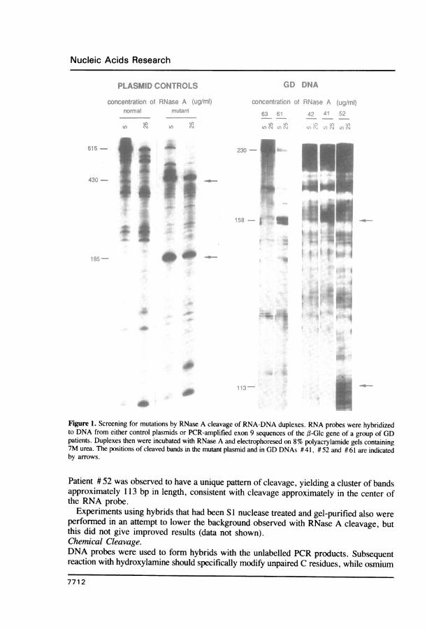
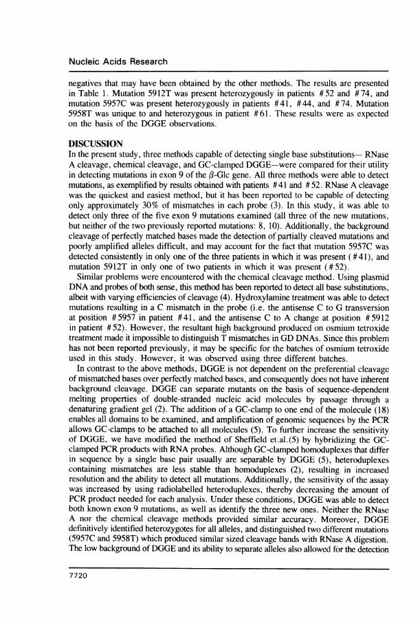
RESULTSRNase A CleavageRNA probes of normal sequence were hybridized to the products of PCR amplificationof,-Glc exon 9, and the resulting RNA-DNA duplexes were treated with RNase A, whichpreferentially cleaves the RNA strand at mismatched bases (3). Therefore, treatment ofa hybrid formed with normal sequences ideally results only in trimming of excessunhybridized probe and on electrophoresis produces only a single band equal to the lengthof the PCR product. In contrast, the presence of mutant sequences produces smallermolecular weight bands due to cleavage at mismatches. The conditions for RNase A cleavagewere optimized using plasmids obtained from Dr. R. Myers. Mismatched RNA-DNAhybrids produced by using these plasmids were easily cleaved at low concentrations ofRNase A (5 ug/ml; Fig. 1). However, it has been reported that some mismatches requirelonger incubation times or higher concentrations of RNase A (3). Therefore, all patientsamples were treated with three different concentrations of RNase A (5, 25, and 80 Ag/ml).Mutations detected generally were seen at all concentrations, although the 80 itg/ml resultedin very high background (data not shown).
Fig. 1 shows examples of results obtained for GD DNAs at the two lowest enzymeconcentrations in two different experiments, one using the antisense probe (DNAs #61and # 63), one using the sense probe (DNAs # 41, # 42, and # 52). Multiple backgroundbands which were characteristic for each probe were obtained at all RNase A concentrationsfor all GD DNAs. Typical background patterns for these probes are shown by DNAs # 42and # 63. However, samples #41 and # 61 clearly exhibit a new set of bands approximately158 bp in length, indicating the presence of a new mutation in exon 9 in at least one alleleof each of these patients. In both samples, a second lower band about 72 bp in lengthwould also be expected, but was not observed. Patient #41 is known to have an allelewith the exon 9 Type 1 mutation 5841G (11), but on comparision with DNAs known tobe negative for this mutation, no specific cleavage bands of the sizes predicted for thismutation (41 and 189 bp) were distinguishable on RNase A cleavage of this DNA (Fig. 1),or of other GD DNAs known to contain at least one copy of this mutation (data not shown).
7711
Nucleic Acids Research
-~~~~~A"
_ _.~~~A&
0i I:-10
*.....** r ~~~~~~~~~~~~~~~~~~~~~~~~~~~~~~~~~1:
Figure 1. Screening for mutations by RNase A cleavage of RNA-DNA duplexes. RNA probes were hybridizedto DNA from either control plasmids or PCR-amplified exon 9 sequences of the ,(-Glc gene of a group of GDpatients. Duplexes then were incubated with RNase A and electrophoresed on 8% polyacrylamide gels containing7M urea. The positions of cleaved bands in the mutant plasmid and in GD DNAs # 41, # 52 and # 61 are indicatedby arrows.
Patient # 52 was observed to have a unique pattern of cleavage, yielding a cluster of bandsapproximately 1 3 bp in length, consistent with cleavage approximately in the center ofthe RNA probe.
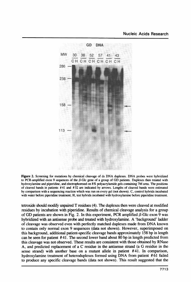
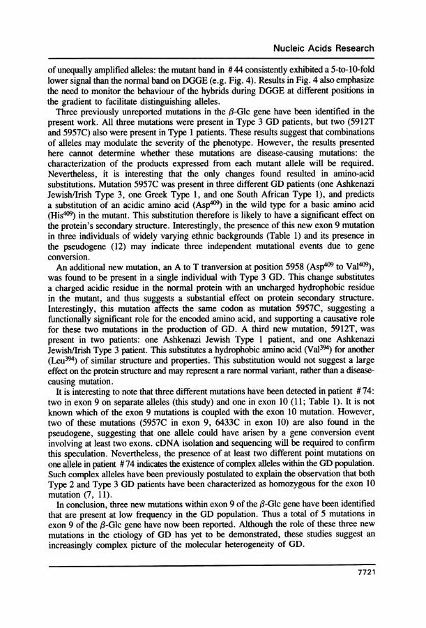
Experiments using hybrids that had been SI nuclease treated and gel-purified also wereperformed in an attempt to lower the background observed with RNase A cleavage, butthis did not give improved results (data not shown).Chemical Cleavage.DNA probes were used to form hybrids with the unlabelled PCR products. Subsequentreaction with hydroxylamine should specifically modify unpaired C residues, while osmium
7712
Nucleic Acids Research
GD DNA
MW 30 38 52 57 41 43
COH C H HC H CH OH286
238
158 -
113 -
Figure 2. Screening for mutations by chemical cleavage of ds DNA duplexes. DNA probes were hybridizedto PCR-amplified exon 9 sequences of the j3-Glc gene of a group of GD patients. Duplexes then treated withhydroxylamine and piperidine, and electrophoresed on 8% polyacrylamide gels containing 7M urea. The positionsof cleaved bands in patients #41 and #52 are indicated by arrows. Lengths of cleaved bands were estimatedby comparison with a sequencing reaction which was run on every gel (not shown). C, control hybrids incubatedwith water before piperidine treatment; H, test hybrids incubated with hydroxylamine before piperidine treatment.
tetroxide should modify unpaired T residues (4). The duplexes then were cleaved at modifiedresidues by incubation with piperidine. Results of chemical cleavage analysis for a groupof GD patients are shown in Fig. 2. In this experiment, PCR amplified f-Glc exon 9 washybridized with an antisense probe and treated with hydroxylamine. A 'background' ladderof cleavage was observed even with perfectly matched duplexes made from DNA knownto contain only normal exon 9 sequences (data not shown). However, superimposed onthis background, additional patient-specific cleavage bands approximately 158 bp in lengthcan be seen for patient # 41. The second lower band about 80 bp in length predicted fromthis cleavage was not observed. These results are consistent with those obtained by RNaseA, and predicted replacement of a C residue in the antisense strand (a G residue in thesense strand) with another base on a mutant allele in patient #41. In comparison,hydroxylamine treatment of heteroduplexes formed using DNA from patient # 61 failedto produce any specific cleavage bands (data not shown). This result suggested that the
7713
Nucleic Acids Research
A. MELTING MAPS OF EXON 9 (WITH AND WITHOUTGC-CLAMP)
95
92
89
a 86E
83
0 80
E 77
CY)c 7 4
E 7
68
0 30 60 90 120 150 180 210 240 270 300
base position_zi_rn
intron 8 exon 9 intron 9 GC-clamp(partial) (partial)
B. PERPENDICULAR DENATURING GRADIENT GEL ELECTROPHORESIS
% denaturant
10 20 30 40 50 60 70 80 90
I~~~ I
- _ with GC-clamp
_c-without GC-clamp0
-C
0
43%
7714
Nucleic Acids Research
exon 9 mutation in this patient detected by RNase A cleavage did not result from substitutionof a C residue in the antisense strand. Additionally, patient #52 DNA produced an
approximately 113 bp cleavage band, which was similar to the result obtained with RNaseA cleavage.
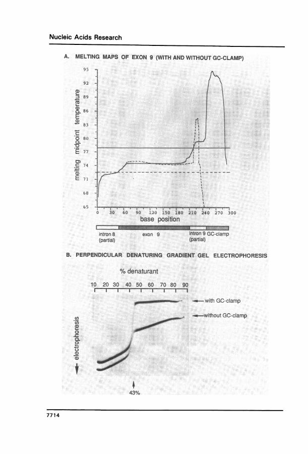
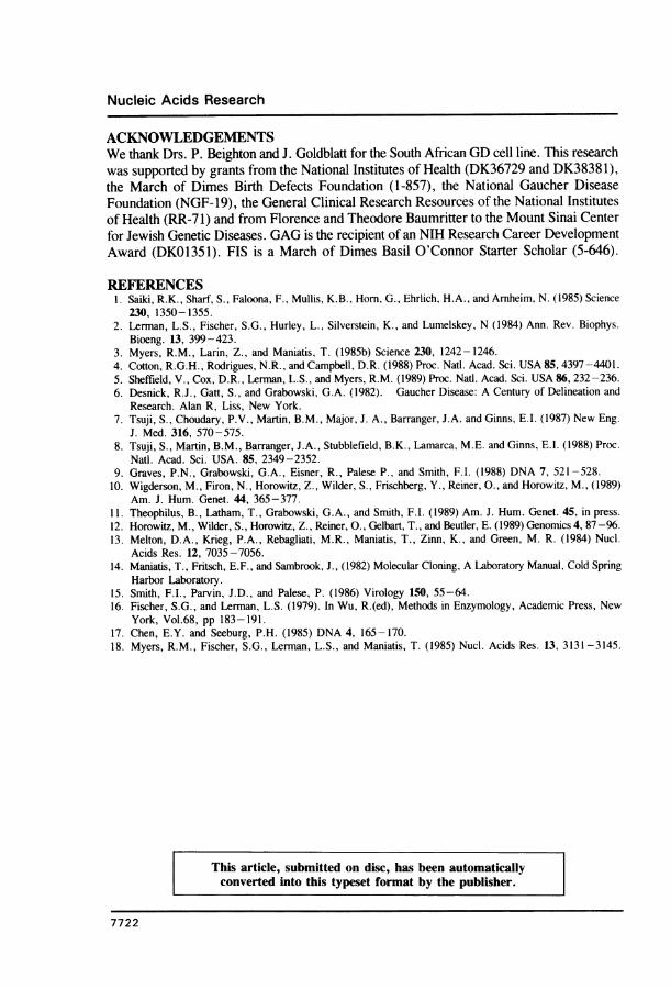
Similar experiments were conducted with osmium tetroxide, but high background cleavagewas obtained with three different batches of osmium tetroxide and over a wide range ofosmium tetroxide and piperidine concentrations. While it was possible to detect cleavageof T-mismatches produced using two different sequenced plasmid DNAs, the methodconsistently failed to detect a T-mismatch using a third plasmid DNA. Also, osmiumtetroxide treatment of genomic DNA led to high background cleavage, obscuring thedetection of any mutations (data not shown).Denaturing gradient gel electrophoresis (DGGE).The effect of the addition of a 40 bp GC-clamp on the expected melting progression alonga ds DNA PCR product was calculated as described (2; Fig. 3). The ,B-Glc sequences
spanned positions # 5781 to # 6020, and contained all of exon 9 as well as some flankingbases. For the molecule lacking a GC-clamp, strand dissociation was probable at a
temperature lower than that required for melting of the coding region. Therefore, for thisfragment there was no temperature at which a partially melted molecule was favored bythe equilibrium, and thus it could not be examined for base substitutions. In contrast, theaddition of a GC-clamp raised the temperature at which strand dissociation was probableto significantly above that at which the coding region melted, thereby converting thismolecule into two distinct domains. Consequently, the entire coding region became a lowmelting domain (LMD), which was amenable to analysis by DGGE. Previous analysesindicated that the same relative order of domains found in ds DNA molecules was alsofound in RNA-DNA molecules (15), suggesting that a GC-clamp would also convert an
RNA-DNA duplex into two melting domains.To obtain a GC-clamp at one end of the PCR product, PCRs were performed using
one primer that contains 40 bases of a GC-rich sequence, in addition to the 20 nucleotidescomplementary to the ,B-Glc gene. These products then were hybridized to RNA probesthat also contained these extra sequences. Analysis of these heteroduplexes by DGGEdistinguishes those containing single base mismatches in their LMDs by their altered meltingbehaviour. Mismatched duplexes are less stable than perfectly paired duplexes, and undergopartial melting, and thus retardation, at a lower concentration of denaturant than do perfectlymatched duplexes.
Electrophoresis on a perpendicular denaturing gradient gel of RNA-DNA homoduplexes,formed by hybridizing PCR products either with or without a GC-clamp to a radioactively
Figure 3. Theoretical and experimental analyses of the melting behaviour of homoduplexes. Panel A. Theoreticalmelting maps of PCR-amplified j3Glc exon 9 and its flanking regions. The abscissa represents positions alongthe PCR product, and the ordinate shows the temperature at which each base has an equal probability of helix
or random chain configuration. The position of the coding region is indicated in the box diagram below the maps.Melting maps of the PCR product either with (continuous lines) or without (hatched lines) a 40bp GC-clampat the 3'-end are shown. The temperature at which the dissociation constant for complete strand dissociation
of the PCR product is 10-7M is indicated by either a hatched line (unclamped) or a continuous line (GC-clamped).Panel B. Experimental melting patterns of homoduplexes after perpendicular DGGE. The mobilities of RNA-
DNA homoduplexes (either with or without a 4Obp GC-clamp) were examined on a denaturing 10-90% gradientperpendicular to the electric field. The samples were applied uniformly across the top of the gel. An arrow indicates
the midpoint of the transitions from duplexes to single stranded molecules.
7715
Nucleic Acids Research
PARALLEL DENATURING GRADIENT GEL ELECTROPHORESISA.
GD DNA27 41 55 61 4 31
5z9 57 CL c
.7 ~ ~ ~ ~~.CD
n1a ing#^~~~~~~~t
onger time ofelectrophoresis
GD DNA27 41 55 61 4 31
srun~~~~~~~~sa:ta
450'
B.GD DNA
63 74 41 44 61 55 52
CD
(059570oOO
0 55Cp8 T _
59." . ...s_,76Gq'/,0
a~~~~~~a
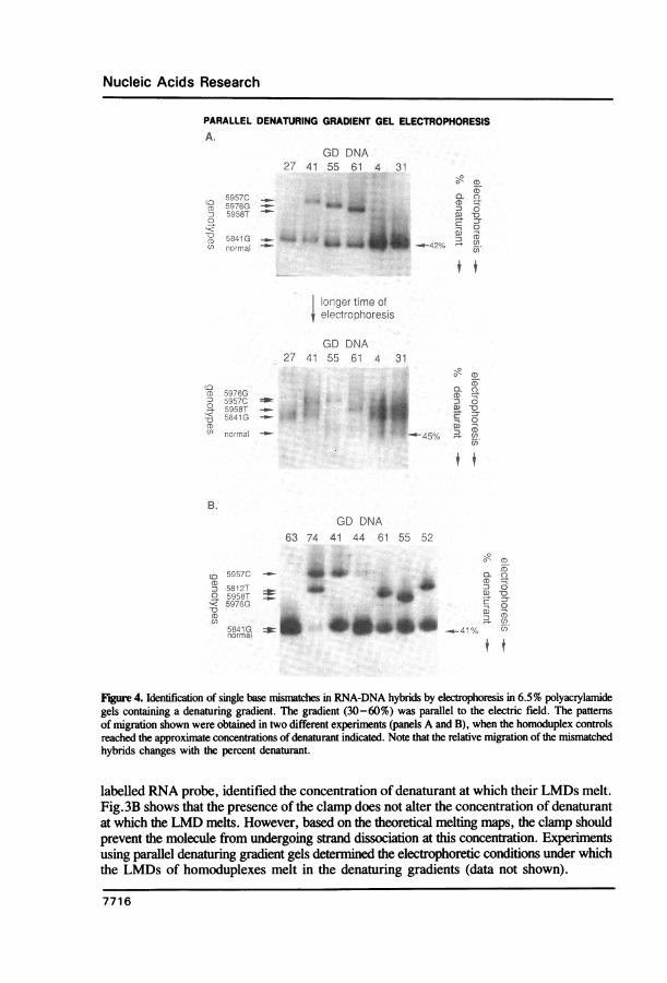
Figure 4. Identification of single base mismatches in RNA-DNA hybrids by electrophoresis in 6.5% polyacrylamidegels containing a denaturing gradient. The gradient (30-60%) was parallel to the electric field. The patternsof migration shown were obtained in two different experiments (panels A and B), when the homoduplex controlsreached the approximate concentrations of denaturant indicated. Note that the relative migration of the mismatchedhybrids changes with the percent denaturant.
labelled RNA probe, identified the concentration of denaturant at which their LMDs melt.Fig.3B shows that the presence of the clamp does not alter the concentration of denaturantat which the LMD melts. However, based on the theoretical melting maps, the clamp shouldprevent the molecule from undergoing strand dissociation at this concentration. Experimentsusing parallel denaturing gradient gels determined the electrophoretic conditions under whichthe LMDs of homoduplexes melt in the denaturing gradients (data not shown).
7716
Nucleic Acids Research
The same samples that had been analyzed by RNase A and chemical cleavage wereexamined by DGGE (Fig.4). DGGE in the absence of a GC-clamp failed to detect anymutations (data not shown). However, GC-clamped DGGE detected all previouslycharacterized mutations. Fig.4 shows a single retarded band for patient #27, who washomozygous for the exon 9 mutation 5841G. Two bands (one normal, one mutant) wereobserved for patient # 55, who was demonstrated to be heterozygous for the mutation 5976G(10). These results show that DGGE can clearly distinguish heterozygotes from homozygotesfor these alleles.
It should be noted that the highest melting domain (HMD) of the GC-clamped hybridmelted at concentrations of denaturant only slightly higher than those required to melt theLMD. Therefore, we were confident that the hybrids had reached the correct denaturantconcentration to melt the LMD only if two time points were analyzed on each gel: onewhich gave sharp bands due to retardation after melting of the LMD but before stranddissociation, and a slightly longer time point that exhibited blurry bands (resulting fromHMD strand dissociation), verifying that the normal controls had passed through theconcentration of denaturant sufficient to melt the HMD. Mutations that cause only a smallchange in stability of the heteroduplex would be detectable only during a very small windowof denaturant concentration. For example, the mutation in patient # 55 was clearly visibleover multiple time points, whereas that in patient #27 was visible only shortly beforestrand dissociation (Fig. 4). A further reason for examining the migration of bands atdifferent time points is that the relative migration of retarded species may change (Fig.4), increasing the likelihood of distinguishing between different mutations.
Analysis of GD DNA by GC-clamped DGGE also detected all mutations tentativelyidentified by RNase A or chemical cleavage screening. Fig. 4 shows that PCR productsderived from patient #61 showed two bands on DGGE: one with identical mobility tothe normal control indicating the presence of a normal sequence in exon 9 on one allele,and one whose mobility was retarded indicating the presence of a mutation in exon 9 onthe other allele. This mutation is labelled 5958T in Fig. 4. DNA from patient #41 showedtwo different retarded bands: one with a mobility identical to that seen with DNA whichwas homozygous for mutation 5841G, and one with a mobility not previously seen in anyDNA, suggesting the presence of a new mutation. This mutation is labelled 5957C in Fig.4. It should be noted that the indicative bands in patients #41 and #61 were retardedto different degrees, suggesting the presence of different mutations in these two patients.Similar analyses ofDNA from patient # 52 also identified two retarded bands: one consistentwith mutation 5841G, and a second with a new migration pattern. This latter findingsuggested the presence of a new mutation (labelled 5912T in Fig. 4).DGGE screening also indicated the presence of retarded bands in the PCR products
derived from patients # 44 and # 74 (Fig. 4). Two of these bands had mobilities similarto that of the most retarded band in patient #41 (mutation 5957C). It should be notedthat the signal obtained from the mutant band in patient #44 was consistently 5-to-0-foldlower than that obtained with the normally migrating band (Fig. 4). This probably reflectsinefficient PCR amplification of this allele. A second differently retarded band was seenin patient # 74, indicating that both alleles in this patient contained mutations in exon 9.This second band had similar mobility to that of the most retarded band seen in patient#52 (mutation 5912T; Fig. 4).Characterization of new exon 9 mutationsThe PCR products of exon 9 amplification of DNAs from patients # 41, # 52, # 61 and#74 were cloned directly into pIBI31, using the EcoRI and XbaI sites that had been
7717
Nucleic Acids Research
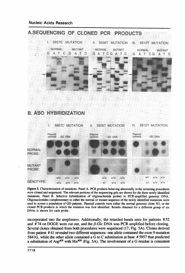
A.SEQUENCING OF CLONED PCR PRODUCTS
I. 5957C MUTATION I1. 5958T MUTATION III. 5912T MUTATION
NORMAL MUTANT NORMAL MUTANT NG A T C G AT G A T CG A T C G
...........
:_
- ' ' I _
NORMAL MUTANTA T CG A T C
B. ASO HYBRIDIZATION
I. 5957C MUTATION 11. 5958T MUTATION 111. 5912T MUTATION
Plasmid Plasmid Plasmidcontrols GD DNA controls GD DNA controls GD DNA
NORMALPROBE: - 'i i +PROBE:
* ~~~~~~~~~~~~~~~~~~~~~~~~~~~~.... _.:
+1+ +1-
+1+ +14. +1+
+1+ +1+ +1+
Figure 5. Characterization of mutations. Panel A. PCR products behaving abnormally in the screening procedureswere cloned and sequenced. The relevant portions of the sequencing gels are shown for the three newly identifiedmutations. Panel B. Selective hybridization of oligonucleotide probes to PCR-amplified genomic DNA.Oligonucleotides complementary to either the normal or mutant sequence of the newly identified mutations wereused to screen a population of GD patients. Plasmid controls were either the normal genomic clone N3, or thecloned PCR-products in which the mutation was first identified. Results obtained for a different group of sixDNAs is shown for each probe.
incorporated into the amplimers. Additionally, the retarded bands seen for patients # 52and # 74 on DGGE were cut out, and the ,B-Glc DNA was PCR amplified before cloning.Several clones obtained from both procedures were sequenced (17; Fig. 5A). Clones derivedfrom patient #41 revealed two different sequences: one allele contained the exon 9 mutation5841G, while the other allele contained a G to C substitution at base # 5957 that predicteda substitution of Asp4O9 with His*)9 (Fig. 5A). The involvement of a G residue is consistent
7718
GENOTYPE:
+1- +1+ +1
+1- +1+ +1+
Nucleic Acids Research
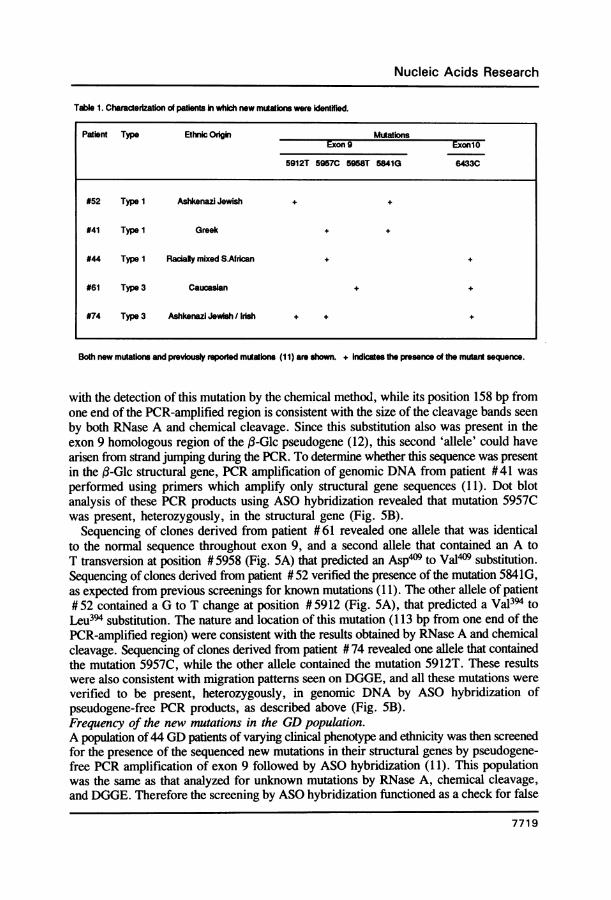
Table 1. Charaderization of patients in which new mutations were identHied.
Patient Type Ethnic Oigbin MutationExon 9 ExonlO
5912T 59570 5958T 5841G 64330
#52 Type I Ashkenazi Jewish + +
#41 Type 1 Greek + +
#44 Type 1 Racialy mixed S.African + +
#61 Type 3 Caucasian + +
#74 Type 3 Ashkenazi Jewish /Irish + + +
Both new mutations and previously repotted mutations (11) are shown. + Indicates the presence of the mutant sequence.
with the detection of this mutation by the chemical method, while its position 158 bp fromone end of the PCR-amplified region is consistent with the size of the cleavage bands seenby both RNase A and chemical cleavage. Since this substitution also was present in theexon 9 homologous region of the (3-Glc pseudogene (12), this second 'allele' could havearisen from strand jumping during the PCR. To determine whether this sequence was presentin the (3-Glc structural gene, PCR amplification of genomic DNA from patient # 41 wasperformed using primers which amplify only structural gene sequences (11). Dot blotanalysis of these PCR products using ASO hybridization revealed that mutation 5957Cwas present, heterozygously, in the structural gene (Fig. SB).
Sequencing of clones derived from patient #61 revealed one allele that was identicalto the normal sequence throughout exon 9, and a second allele that contained an A toT transversion at position # 5958 (Fig. SA) that predicted an Asp4'9 to Val409 substitution.Sequencing of clones derived from patient # 52 verified the presence of the mutation 5841G,as expected from previous screenings for known mutations (11). The other allele of patient#52 contained a G to T change at position #5912 (Fig. SA), that predicted a Val394 toLeu394 substitution. The nature and location of this mutation (113 bp from one end of thePCR-amplified region) were consistent with the results obtained by RNase A and chemicalcleavage. Sequencing of clones derived from patient # 74 revealed one allele that containedthe mutation 5957C, while the other allele contained the mutation 5912T. These resultswere also consistent with migration patterns seen on DGGE, and all these mutations wereverified to be present, heterozygously, in genomic DNA by ASO hybridization ofpseudogene-free PCR products, as described above (Fig. SB).Frequency of the new mutations in the GD population.A population of44 GD patients of varying clinical phenotype and ethnicity was then screenedfor the presence of the sequenced new mutations in their structural genes by pseudogene-free PCR amplification of exon 9 followed by ASO hybridization (11). This populationwas the same as that analyzed for unknown mutations by RNase A, chemical cleavage,and DGGE. Therefore the screening by ASO hybridization functioned as a check for false
7719
Nucleic Acids Research
negatives that may have been obtained by the other methods. The results are presentedin Table 1. Mutation 5912T was present heterozygously in patients #52 and #74, andmutation 5957C was present heterozygously in patients #41, #44, and # 74. Mutation5958T was unique to and heterozygous in patient #61. These results were as expectedon the basis of the DGGE observations.
DISCUSSIONIn the present study, three methods capable of detecting single base substitutions- RNaseA cleavage, chemical cleavage, and GC-clamped DGGE-were compared for their utilityin detecting mutations in exon 9 of the f-Glc gene. All three methods were able to detectmutations, as exemplified by results obtained with patients #41 and # 52. RNase A cleavagewas the quickest and easiest method, but it has been reported to be capable of detectingonly approximately 30% of mismatches in each probe (3). In this study, it was able todetect only three of the five exon 9 mutations examined (all three of the new mutations,but neither of the two previously reported mutations: 8, 10). Additionally, the backgroundcleavage of perfectly matched bases made the detection of partially cleaved mutations andpoorly amplified alleles difficult, and may account for the fact that mutation 5957C wasdetected consistently in only one of the three patients in which it was present (#41), andmutation 5912T in only one of two patients in which it was present (#52).
Similar problems were encountered with the chemical cleavage method. Using plasmidDNA and probes of both sense, this method has been reported to detect all base substitutions,albeit with varying efficiencies of cleavage (4). Hydroxylamine treatment was able to detectmutations resulting in a C mismatch in the probe (i.e. the antisense C to G transversionat position # 5957 in patient #41, and the antisense C to A change at position #5912in patient # 52). However, the resultant high background produced on osmium tetroxidetreatment made it impossible to distinguish T mismatches in GD DNAs. Since this problemhas not been reported previously, it may be specific for the batches of osmium tetroxideused in this study. However, it was observed using three different batches.
In contrast to the above methods, DGGE is not dependent on the preferential cleavageof mismatched bases over perfectly matched bases, and consequently does not have inherentbackground cleavage. DGGE can separate mutants on the basis of sequence-dependentmelting properties of double-stranded nucleic acid molecules by passage through adenaturing gradient gel (2). The addition of a GC-clamp to one end of the molecule (18)enables all domains to be examined, and amplification of genomic sequences by the PCRallows GC-clamps to be attached to all molecules (5). To further increase the sensitivityof DGGE, we have modified the method of Sheffield et.al.(5) by hybridizing the GC-clamped PCR products with RNA probes. Although GC-clamped homoduplexes that differin sequence by a single base pair usually are separable by DGGE (5), heteroduplexescontaining mismatches are less stable than homoduplexes (2), resulting in increasedresolution and the ability to detect all mutations. Additionally, the sensitivity of the assaywas increased by using radiolabelled heteroduplexes, thereby decreasing the amount ofPCR product needed for each analysis. Under these conditions, DGGE was able to detectboth known exon 9 mutations, as well as identify the three new ones. Neither the RNaseA nor the chemical cleavage methods provided similar accuracy. Moreover, DGGEdefinitively identified heterozygotes for all alleles, and distinguished two different mutations(5957C and 5958T) which produced similar sized cleavage bands with RNase A digestion.The low background of DGGE and its ability to separate alleles also allowed for the detection
7720
Nucleic Acids Research
of unequally amplified alleles: the mutant band in #44 consistently exhibited a 5-to-lO-foldlower signal than the normal band on DGGE (e.g. Fig. 4). Results in Fig. 4 also emphasizethe need to monitor the behaviour of the hybrids during DGGE at different positions inthe gradient to facilitate distinguishing alleles.Three previously unreported mutations in the g-Glc gene have been identified in the
present work. All three mutations were present in Type 3 GD patients, but two (5912Tand 5957C) also were present in Type patients. These results suggest that combinationsof alleles may modulate the severity of the phenotype. However, the results presentedhere cannot determine whether these mutations are disease-causing mutations: thecharacterization of the products expressed from each mutant allele will be required.Nevertheless, it is interesting that the only changes found resulted in amino-acidsubstitutions. Mutation 5957C was present in three different GD patients (one AshkenaziJewish/Irish Type 3, one Greek Type 1, and one South African Type 1), and predictsa substitution of an acidic amino acid (Asp4)9) in the wild type for a basic amino acid
(His409) in the mutant. This substitution therefore is likely to have a significant effect on
the protein's secondary structure. Interestingly, the presence of this new exon 9 mutationin three individuals of widely varying ethnic backgrounds (Table 1) and its presence inthe pseudogene (12) may indicate three independent mutational events due to gene
conversion.An additional new mutation, an A to T tranversion at position 5958 (Asp409 to Val409),
was found to be present in a single individual with Type 3 GD. This change substitutesa charged acidic residue in the normal protein with an uncharged hydrophobic residuein the mutant, and thus suggests a substantial effect on protein secondary structure.
Interestingly, this mutation affects the same codon as mutation 5957C, suggesting a
functionally significant role for the encoded amino acid, and supporting a causative rolefor these two mutations in the production of GD. A third new mutation, 5912T, was
present in two patients: one Ashkenazi Jewish Type 1 patient, and one Ashkenazi
Jewish/Irish Type 3 patient. This substitutes a hydrophobic amino acid (Val394) for another
(Leu394) of similar structure and properties. This substitution would not suggest a largeeffect on the protein structure and may represent a rare normal variant, rather than a disease-causing mutation.
It is interesting to note that three different mutations have been detected in patient #74:two in exon 9 on separate alleles (this study) and one in exon 10 (11; Table 1). It is not
known which of the exon 9 mutations is coupled with the exon 10 mutation. However,two of these mutations (5957C in exon 9, 6433C in exon 10) are also found in the
pseudogene, suggesting that one allele could have arisen by a gene conversion event
involving at least two exons. cDNA isolation and sequencing will be required to confirmthis speculation. Nevertheless, the presence of at least two different point mutations on
one allele in patient #74 indicates the existence of complex alleles within the GD population.Such complex alleles have been previously postulated to explain the observation that bothType 2 and Type 3 GD patients have been characterized as homozygous for the exon 10mutation (7, 11).
In conclusion, three new mutations within exon 9 of the,B-Glc gene have been identifiedthat are present at low frequency in the GD population. Thus a total of 5 mutations inexon 9 of the (3-Glc gene have now been reported. Although the role of these three new
mutations in the etiology of GD has yet to be demonstrated, these studies suggest an
increasingly complex picture of the molecular heterogeneity of GD.
7721
Nucleic Acids Research
ACKNOWLEDGEMENTSWe thank Drs. P. Beighton and J. Goldblatt for the South African GD cell line. This researchwas supported by grants from the National Institutes of Health (DK36729 and DK38381),the March of Dimes Birth Defects Foundation (1-857), the National Gaucher DiseaseFoundation (NGF-19), the General Clinical Research Resources of the National Institutesof Health (RR-7 1) and from Florence and Theodore Baumritter to the Mount Sinai Centerfor Jewish Genetic Diseases. GAG is the recipient of an NIH Research Career DevelopmentAward (DK01351). FIS is a March of Dimes Basil O'Connor Starter Scholar (5-646).
REFERENCES1. Saiki, R.K., Sharf, S., Faloona, F., Mullis, K.B., Horn, G., Ehrlich, H.A., and Arnheim, N. (1985) Science
230, 1350-1355.2. Lerman, L.S., Fischer, S.G., Hurley, L., Silverstein, K., and Lumelskey, N (1984) Ann. Rev. Biophys.
Bioeng. 13, 399-423.3. Myers, R.M., Larin, Z., and Maniatis, T. (1985b) Science 230, 1242-1246.4. Cotton, R.G.H., Rodrigues, N.R., and Campbell, D.R. (1988) Proc. Natl. Acad. Sci. USA 85, 4397-4401.5. Sheffield, V., Cox, D.R., Lerman, L.S., and Myers, R.M. (1989) Proc. Nati. Acad. Sci. USA 86, 232-236.6. Desnick, R.J., Gatt, S., and Grabowski, G.A. (1982). Gaucher Disease: A Century of Delineation and
Research. Alan R, Liss, New York.7. Tsuji, S., Choudary, P.V., Martin, B.M., Major, J. A., Barranger, J.A. and Ginns, E.I. (1987) New Eng.
J. Med. 316, 570-575.8. Tsuji, S., Martin, B.M., Barranger, J.A., Stubblefield, B.K., Lamarca, M.E. and Ginns, E.I. (1988) Proc.
NatI. Acad. Sci. USA. 85, 2349-2352.9. Graves, P.N., Grabowski, G.A., Eisner, R., Palese P., and Smith, F.I. (1988) DNA 7, 521-528.
10. Wigderson, M., Firon, N., Horowitz, Z., Wilder, S., Frischberg, Y., Reiner, O., and Horowitz, M., (1989)Am. J. Hum. Genet. 44, 365-377.
11. Theophilus, B., Latham, T., Grabowski, G.A., and Smith, F.I. (1989) Am. J. Hum. Genet. 45, in press.
12. Horowitz, M., Wilder, S., Horowitz, Z., Reiner, O., Gelbart, T., and Beutler, E. (1989) Genomics 4, 87-96.13. Melton, D.A., Krieg, P.A., Rebagliati, M.R., Maniatis, T., Zinn, K., and Green, M. R. (1984) Nucl.
Acids Res. 12, 7035 -7056.14. Maniatis, T., Fritsch, E.F., and Sambrook, J., (1982) Molecular Cloning, A Laboratory Manual, Cold Spring
Harbor Laboratory.15. Smith, F.I., Parvin, J.D., and Palese, P. (1986) Virology 150, 55-64.16. Fischer, S.G., and Lerman, L.S. (1979). In Wu, R.(ed), Methods in Enzymology, Academic Press, New
York, Vol.68, pp 183-191.17. Chen, E.Y. and Seeburg, P.H. (1985) DNA 4, 165-170.18. Myers, R.M., Fischer, S.G., Lerman, L.S., and Maniatis, T. (1985) Nucl. Acids Res. 13, 3131 -3145.
7722
This article, submitted on disc, has been automaticallyconverted into this typeset format by the publisher.