histo-physiological studies on the pituitary gland of some ...
98
Instructions for use Title HISTO-PHYSIOLOGICAL STUDIES ON THE PITUITARY GLAND OF SOME TELEOST FISHES, WITH SPECIAL REFERENCE TO THE CLASSIFICATION OF HORMONE-PRODUCING CELLS IN THE ADENOHYPOPHYSIS Author(s) NAGAHAMA, Yoshitaka Citation MEMOIRS OF THE FACULTY OF FISHERIES HOKKAIDO UNIVERSITY, 21(1), 1-63 Issue Date 1973-10 Doc URL http://hdl.handle.net/2115/21854 Type bulletin (article) File Information 21(1)_P1-63.pdf Hokkaido University Collection of Scholarly and Academic Papers : HUSCAP
-
Upload
khangminh22 -
Category
Documents
-
view
0 -
download
0
Transcript of histo-physiological studies on the pituitary gland of some ...

Instructions for use
TitleHISTO-PHYSIOLOGICAL STUDIES ON THE PITUITARY GLAND OF SOME TELEOST FISHES, WITHSPECIAL REFERENCE TO THE CLASSIFICATION OF HORMONE-PRODUCING CELLS IN THEADENOHYPOPHYSIS
Author(s) NAGAHAMA, Yoshitaka
Citation MEMOIRS OF THE FACULTY OF FISHERIES HOKKAIDO UNIVERSITY, 21(1), 1-63
Issue Date 1973-10
Doc URL http://hdl.handle.net/2115/21854
Type bulletin (article)
File Information 21(1)_P1-63.pdf
Hokkaido University Collection of Scholarly and Academic Papers : HUSCAP

HISTO·PHYSIOLOGICAL STUDIES ON THE PITUITARY GLAND OF
SOME TELEOST FISHES, WITH SPECIAL REFERENCE TO THE
CLASSIFICATION OF HORMONE-PRODUCING
CELLS IN THE ADENOHYPOPHYSIS
Yoshitaka NAGAHAMA
Faculty of Fi8heri88, Hokkaido Univer8ity, Hakodate, Japan
Contents page
I. Introduction...................................................... 2 II. Materials and Methods. . . .. .. . . .. .. .. . . . . . . .. . . .. .. .. .. .. .. . . . . . . .. . 4
1. Materials............................................... 4 2. Histological and cytological techniques ........................... .
III. Results and Discussion ........................................... . A. General morphology of the pituitary gland of teleostean fishes ....... .
1. Terminology ............................................... . 2. General morphology ......................................... .
5 6 6 6 7
B. Prolactin cells .................................................. 10 1. General morphology of prolactin cells .......................... 10 2. Prolactin cells of freshwater fishes . . . . . . . . . . . . . . . . . . . . . . . . . . . . 11 3. Prolactin cells of seawater fishes ............................ .. 11 4. Cytological changes of prolactin cells after the transfer of seawater
medaka to fresh water. . . . . . . . . . . . . . . . . . . . . . . . . . . . . . . . . . . . . . . . 12 5. Cytological changes of prolactin cells of salmonid fishes during the
life-cycle. . . . . . . . . . . . . . . . . . . . . . . . . . . . . . . . . . . . . . . . . . . . . . . . . . .. 13 (a) Chum salmon .. . . . . . . . . . . . . . . . . . . . . . . . . . . . . . . . . . . . . . . .. 13 (b) Kokanee salmon ........................................ 14
6. Discussion.......................... . . . . . . . . . . . • . . . . . . . . . • .. 14 C. Corticotrophs.................................................... 16
1. General morphology of corticotrophs .......................... 16 2. Cytological changes of corticotrophs of the goldfish after the
administration of metopirone (SU 4885) ........................ 17 3. Effects of ovariectomy on corticotrophs of the female goldfish .... 18 4. Cytological changes of corticotrophs of the chum salmon during
the life-cycle ................................................ 18 5. Cytological changes of corticotrophs after transfer of fishes from
fresh water to sea water ...................................... 19 6. Discussion.................................................. 19
D. Somatotrophs .....................•............................ 21 1. General morphology of somatotrophs .......................... 21 2. Effects of starvation on somatotrophs of the goldfish .... . . . . . . .. 21 3. Effects of gonadectomy and estrogen treatment on somatotrophs of
the goldfish . . . . . . . . . . . . . . . . . . . . . . . . . . . . . . . . . . . . . • . . . . . . . . .. 22 4. Cytological changes of somatotrophs of salmonid fishes during
the life-cycle ................................................ 23 (a) Chum salmon .......................................... 23 (b) Kokanee salmon ........................................ 23
- 1 -

Mem. Fac. Fish. Hokkaido Unlv.
5. Cytological changes of somatotrophs of the male eel matured by the injection of Synahorin ................................... .
6. Discussion ................................................. . E. Thyrotrophs ................................................... .
1. General morphology ofthyrotrophs ........................... . 2. Effects of thiourea on thyrotrophs of the goldfish and medaka ... .
(a) Goldfish ............................................. . (b) Medaka ............................................. .
3. Cytological changes of thycotrophs of the chum salmon during the life-cycle ................................................... .
4. Cytological ch'1nges of thyrotrophs of the male eel matured by the injection of Syn.ahorin when transferred from fresh water to sea water ............................................... .
5. Discussion ................................................. . F. GonadCltrophs ................................................. .
1. General morphology of gonadotrophs ......................... . 2. Cytological changes of pnadotrophs of the goldfish during the
reproductive cycle ......................................... . 3. Effects of ovariectomy on gonadotrophs of the female goldfish ... . 4. Effects of estrogen on gonadotrophs of the female goldfish ..... . 5. Effects of starvation on gonadotrophs of mature male goldfish ... . 6. Cytological changes of gonadotrophs of the eel after the Synahorin
treatment ................................................ . 7. Cytological changes of gonadotrophs of salmonid fishes during the
life-cycle ................................................... . (a) Chum salmon . . . . . . . . . . . . . . . . . . . . . . . . . . . . . .......... . (b) Kokanee salmon .. . .................................. .
8. Discussion ................................................. . G. The chromClphobe,_ meta-adenohypophysial cells, neurohypophysis and
capillaries ................................................. . 1. General mClrphology of chromophobes ......................... . 2. General m:Jrphology of the glandular cells of the meta-
adenohypophysis ........................................... . 3. General morphology of the neurohypophysis, and its relationship
to the glandular cells ......................................... . 4. Discussion ................................................. .
IV. Summary ......................................................... . References ....................................................... . Explanation of Plates ............................................. .
I. Introduction
[XXt, 1
24 24 26 26 26 27 27
27
28 29 31 31
31 32 33 34
34
35 35 36 37
39 39
40
41 42 44 45 55
It is well known that in most vertebrates the pituitary gland regulates other endocrine organs by secreting trophic hormones. At least six different hormones (prolactin, corticotrophin, somatotrophin, thyrotrophin, follicle stimulating hormone, and luteinizing hormone) have been isolated from the mammalian anterior pituitary gland. Many attempts have been made to correlate each of these hormones with one of the glandular cell types of this gland using the light microscope.I ) Notwithstanding these laborious works, the glandular cells secreting each hormone have not yet been identified with certainty, because of the small size of
- 2 -
<'

1973] Nagahama: Pituitary gland of BOme teleost fishes
the component cells and the complicated structure of the pituitary gland. Although the pituitary hormones of teleost fishes are not completely identical
with those of mammalian pituitaries,2) it is generally accepted that a prolactin, a corticotrophin, a somatotrophin, a thyrotrophin, one or two gonadotrophin(s) are also produced from the pituitary glands of teleost fishes. 3),4),5) As for the structure of teleost pituitaries, many light microscopical investigations have already been performed.3),4),5) From these findings, it is certain that the pituitary gland of teleost fishes consists of the adenohypophysis and the neurohypophysis, and that the adenohypophysis consists of three lobes, i.e. pro-adenohypophysis (rostral pars distalis), meso-adenohypophysis (proximal pars distalis) and meta-adenohypophysis (pars intermedia). Further, the region of adenohypophysis in teleostean pituitary, which includes pro- and meso-adenohypophysis, is believed to be functionally equivalent to the tetrapod anterior lobe. However, the identification of the glandular cells secreting each tropic hormone have so far been done only in a few teleost pituitaries at the level of the light microscope.
The electron microscope is far more advantageous for the identification of each cell type in the pituitary glands than the light microscope, because the former apparatus has extremely high resolving power which enables us to observe structural characteristics such as differences among secretory granules in the cells. Furthermore, the apparatus is very useful for the detection of fine changes in cell structure induced by various experiments which may help us to determine the function of a given cell type. Fernandez-Moran and Luft6) reported the first electron microscopic observations on the pituitary gland. Since then, many authors have investigated the fine structure of the glands of many higher vertebrates.7),8),9),10),l1)
In teleost pituitaries, the electron microscopical investigation was first carried out by Legait and Legait12) on the carp and trout organs. Then, Follenius,13) and Follenius and Porte14),15),16) observed the pituitary gland of many species of teleost fishes with the electron microscope. Further, the works by Knowles and Vollrath,1?) Follenius,18) Hopkins and Baker,19) and by Nagahama and Yamamot020),21) have been done on the ultrastructure of teleost pituitaries. From these results, five or six different hormone-secreting cell types have been identified in the pro- and mesoadenohypophysis. However, only a few studies on the ultrastructural changes of each cell type correlated with the physiological changes of the fish have been published; for instance, those associated with the reproductive cycle,22),23),24) the seasonal variation or life cycle25),26),27),28),29) and under the various experimental conditions. 30),31),32),33),34),35) At present, therefore, the information on the func
tional classification of cell types of the fish pituitary gland is still very fragmentary and inconclusive, and further experimental evidence needs to be accumulated. In the present study, the author deals with the fine structure of glandular cells of some teleost fishes and the detailed response of the cells to various experimental
-3-

Mem. Fac. Fish. Hokkaido Univ. [XXI, I
treatments. The goldfish and medaka were employed as experimental animals, because these fishes have very convenient size, and can be easily obtained and handled. In addition, the chum- and kokanee salmon and eel were also used in this study. Since some of them live only in fresh water and others migrate between the sea and the river, the studies of the pituitary gland of these fishes during their life cycle, present an interesting problem from an endocrinological point of view.
Before proceeding further, the author wishes to express his hearty thanks to Professor Kiichiro Yamamoto, Faculty of Fisheries, Hokkaido University, for his kind guidance during the course of the present study with important advice, helpful encouragement and valuable criticisms and for his kind reading of the manuscript. The author is also very grateful to Professors Hidejiro Niiyama and Juro Yamada, Faculty of Fisheries, Hokkaido University, who have interested in the present study and kindly offered much advice. The author is also indebted to Professor Howard A. Bern, and Mr. Richard S. Nishioka, Department of Zoology, University of California, for their valuable criticisms and suggestions in the preparation of the manuscript. A debt of gratitude is also owed to Assistant Professor Tatsuro Kubo, Assistant Professor Hiroya Takahashi, Mr. Kazunori Takano, Dr. Fumio Yamazaki, Mr. Hiroshi Onozato, Mr. Isao Oota and Mr. Osamu Hiroi, for their kind help and advice in the course of the present study. Thanks are also offered to Messrs. Y oshiaki Matsuda, Masao Shimomura, Takehiro Yahata, Kiyoshi Ishii and Seiichi Kasuga, for their friendly help in various ways in the course of the present study. For the collection of pituitary glands of chum- and kokanee salmon, the author is greatly indebted to Dr. Seizo Sano, Mr. Kazuhiko Nishino and Dr. Toyohiko Hikita, Hokkaido Salmon Hatchery, and to Messrs. Shigeru Hara and Yoshio Ishikawa, the Tokachi Branch of Hokkaido Salmon Hatchery and Mr. Tsuneo Nishiyama, Hokkaido University. Moreover, he is indebted to Dr. M.E. Zimmerlin, Ciba Co. Ltd., Takarazuka, Japan, for a generous supply of metopirone (SU 4:885).
II. Materials and Methods
1. Materials
The materials used in the present study are the goldfish (Carassius auratus), medaka (Oryzias latipes), eel (Anguilla japonica), and two kinds of salmonid fishes (Oncorhynchus keta and Oncorhynchus nerka).
The goldfish were restricted to the "Wakin" variety which had been stocked either in a large outdoor pool or in an aquarium set in a greenhouse in the campus of the Faculty of Fisheries, Hokkaido University. They were usually fed on commercial trout pellets. The wild type medaka were collected from a pond at Yunokawa Hot Spring, Hakodate, Hokkaido. They were cultured in an outdoor
- 4 -

1973] N agahama: Pituitary gland of some teleost fishes
pond on the campus until needed. The cultured eels were purchased from a commercial shop. In addition to these animals, several yellow or silver eels weighing about 500 g to 1,500 g were also obtained from the Mabuchi River, Aomori Prefecture, Japan. Both groups were kept in fresh water at about 20°C until needed. The kokanee salmon, the landlocked form of Oncorhynchus neJrka, from 4 months to 3 years in age and in various stages of sexual maturation were collected from outdoor ponds of the Chitose Branch and the Mori Branch of the Hokkaido Salmon Hatchery. They were composed of O-year-, one-year-, two-year- and three-year-old groups, cultured separately in outdoor ponds. Samplings were made in May, June, August, September, October, November and December of 1968, 1969, 1970 and 1971. In addition chum salmon of various ages were obtained. Two-year-old fish and two-month-old fish cultured in fresh water in the Nanae Fish-Culture Experimental Station, Hokkaido University, were sampled respectively in June of 1968 and in May, 1969. Those just after entering the sea were obtained in the coastal sea off Mori, southern Hokkaido, in July of 1969. Those in the period of feeding migration, which include both sexually immature and mature fish, were collected in the northern Pacific Ocean in July and August, 1968. Those on the route of the anadromous migration were captured at three points in the Tokachi River, eastern Hokkaido, during the periods from September to November in 1966 and 1967, at Atsunai, which is located at the coast about 20 Km distant from the mouth of the river, at Otsu near the mouth of the river, and at Chiyoda about 45 Km north of the mouth of the river. In addition, some naturally spawned fish were obtained from the spawning beds in the Yakumo River, southern Hokkaido, in December, 1967. Moreover, fish kept for several days after ovulation were collected from the stock raised in an outdoor pond of the Chitose Branch of the Hokkaido Salmon Hatchery in October of 1966.
2. Histological and cytological techniques
For the purpose of morphological investigations on the pituitary glands, fishes were killed by quick decapitation and their organs were rapidly removed.
For light microscopical observations, the pituitary glands were fixed for 24 hours with Bonin's, Zenker-formol, Ciaccio's, Susa's, Bonin-HoIland-sublimate and 10% formalin solutions. Then, serial sagittal sections were made at 4-8 micra in thickness from paraffin or "Tissuemat" embedded blocks. Carbowax embedded blocks were used also for histological analysis. Bouin-, Zenker-formol-, Susa- and Bonin-Holland-sublimate preparations were stained with Heidenhain-azan or Halmi's36) aldehyde fuchsin-light green-orange G for general histological observations. Similar preparations were also employed for the demonstration of polysaccharides by the periodic acid-Schiff reaction (PAS). The glands fixed with Ciaccio's fluid were stained with Sudan black B for lipids, l\!Qreover, the mercuric
~9~

Mem. Fac. Fish. Hokkaido Univ. [XXI,l
bromphenol blue (Hg-BPB) staining technique of Mazia et al.3?) was used for protein. The glands, which were fixed in 10% formalin and embedded in carbowax, were stained only with Sudan black B for the demonstration of lipids. In addition to these staining methods, MacConaill's lead hematoxylin method38)
was used in order to identify the corticotrophs from other cell types of the pituitary gland.
For electron microscopical observations, the pituitary glands were cut in small pieces and immersed in Millonig's solution for 2 hours. Some of them were also placed in 6.25% or 2% glutaraldehyde in 0.5M phosphate buffer for 1 hour and then immersed in Millonig's solution for 2 hours. Mter dehydration in graded ethanoIs, the organs were embedded in Epon epoxy resin mixture. 39) The sections were cut with glass knives on a Porter-Blum microtome at a thickness of about 500 to 800 A, and doubly stained with uranyl acetate and Karnovsky's lead method40) or Reynolds' lead citrate,41) and examined with a Hitachi HS-7 electron microscope. Thick sections of about 1 micron were cut for light microscopy by the same method as above, and stained with the methods of Richardson et al., (2)
Huber(3) or Yamamoto. (4)
III. Results and Discussion
A.. General morphology of the pituitary gland of teleostean fishes
1. Terminology
It is certain that the teleost pituitary is composed of two primary portions similar to that of other higher vertebrates: the adenohypophysis and neurohypophysis. The teleost adenohypophysis can be divided into three regions, mainly based on the component cell types. However, the relationships of the three regions vary among fishes. Therefore, the terminology applied to these three regions of the teleost adenohypophysis was in a stage of confusion for many years. Pickford8 ) in an attempt to clarify and standardize the terminology of the teleost pituitary has proposed "pro-adenohypophysis" for the pars anterior, pars follicularis or anterior glandular region, "meso-adenohypophysis" for the transitional lobe and "meta-adenohypophysis" for the pars intermedia or posterior glandular region. On the other hand, Gorbman(6
) pointed out that the most preferable nomenclature on the fish adenohypophysis was the one based on Green, (6) that the pro- and meso-adenohypophysis together are physiologically similar to the pars distalis of higher vertebrates and the meta-adenohypophysis to the pars intermedia of the latter, i.e. rostral pars distalis, proximal pars distalis and pars intermedia. In the present study, the author employs the terminology proposed by Pickford in order to avoid confusing the literature regarding the adenohypophysis in teleosts from that of the adenohypophysis in many higher vertebra.tes.
- 6 -

1973] Nagahama: Pituitary gland of some teleost fishes
2. General morphology
Results and Discussion
It is well known that the organization of teleost pituitary gland is largely variable among the species. (7),48) Furthermore, some investigators have demonstrated that the glands undergo changes in shape and proportion with the growth of fishes. (9),50),51) Figs. 1-10 indicate the organization of the pituitary glands of fishes investigated in the present study and the changes accompanying their growth. In adult fishes, the glands appear to be divided in general into two types according to their shape; the antero-posterior type (medaka (Fig. 2), eel (Fig. 3)) and the dorso-ventral type (goldfish (Fig. 1), salmon (Fig. 4)). In the juvenile fish, however, the gland of the dorso-ventral type generally resembles that of the antero-posterior type (Figs. 5 and 8).
The pituitary glands examined in this study could be divided into four regions, i.e. pro-, meso- and meta-adenohypophysis and neurohypophysis.
The pro-adenohypophysis occupies the anterior portion of the glands in the case of the antero-posterior type, while in the dorso-ventral type it occupies the antero-dorsal portion. The lobe generally contains two hormone-secreting cells, i.e. prolactin cells and corticotrophs. In a few fishes such as the goldfish and eel, however, in addition to these cells thyrotrophs can be found in the lobe. Moreover, in a few fishes such as the eel and salmonid fishes, this lobe shows a typical follicular arrangement. According to the review done by Ball and Baker,4) this follicular structure is often observed in the pro-adenohypophysis of some primitive fishes (isospondylous forms, salmonids, clupeoids, and apodes). Moreover, Sathyanesan 52) suggested that in certain clupeoid and salmonid fry, and in the adult Hilsa ilisha, the lumina of the rostral follicles communicate with a persistent orohypophysial duct which probably represents the cavity of embryonic Rathke's pouch. Unfortunately, however, detailed embryological studies on the fish pituitary have not been done in the present study. So, the significance of follicular structure in the pro-adenohypophysis remains still uncertain.
The meso-adenohypophysis takes a position in the central portion of the glands. This lobe generally contains three kinds of hormone-secreting cells, i.e.
thyrotrophs, somatotrophs and gonadotrophs. Likewise in the case of the proadenohypophysis, this lobe of a few fishes consists of many columns, separated by the well developed connective tissue. However, no lumen can be found in the lobe.
The meta-adenohypophysis occupies the ventro-posterior portion in the anteroposterior type, while in the dorso-ventral type it is located in the ventro-posterior portion. This lobe generally contains two types of glandular cells but their functional significance is still uncertain.
-7-

Mem. Fac. Fish. Hokkaido Univ.
Figs. 1-4 Diagrams of midsagittal section of some teleost pituitaries. . (Fig. 1, goldfish; Fig. 2, medaka; Fig. 3, eel; Fig. 4, chum salmon)
[XXI,l
o orEa) • Prolactin cell; @), Thyrotroph; 0, Corticotroph; e, Gonadotroph; @),
Soma~roph; ~ft~~:~~, Meta-cell;;;;;;;;, Neurohypophysis; III , Neurohypophysis. contain. ing much A-F positive material.
In the pituitary gland of the dorso-ventral type, the neurohypophysis in general forms the central axis and the three regions of the adenohypophysis are arranged around this axis. The most extensive ramification of the neurohypophysis is recognized in the ventro-posterior part of the glands. In the Case of the
1;1

1973] Nagahama: Pituitary gland of some teleost fishes
Figs. 5-10 Changes in the shape and structure of the fish pituitaries accompaning with their growth. [], meso-adenohypophysis.
Fig. 5 Diagram of the pituitary gland of the juvenile goldfish of about 30 days old. Fig. 6 Diagram of the pituitary gland of immature goldfish of about 60-80 days old. Fig. 7 Diagram of the pituitary gland of the adult goldfish.
glands of the antero-posterior type, the neurohypophysis occupies the dorsal half of posterior region of the gland, although their branches can be found penetrating into each lobe.
- 9 ~

Mem. Fac. Fish. Hokkaido Univ. [XXI,1
Fig. 8 Diagram of the pituitary gland of the juvenile chum salmon of about 2 months old.
Fig. 9 Diagram of the pituitary gland of sexually immature chum salmon caught in the northern Pacific Ocean.
Fig. 10 Diagram of the pituitary gland of sexually maturing chum salmon caught in the coastal sea.
B. Prolactin cells
1. General morphology of prolactin cells
The prolactin cells of fishes investigated In the present study, occupy the major part of the pro-adenohypophysis. The cells are elongated, sometimes polygonal in shape and are 7-15 p in diameter. The cells are stained with azocarmine
-10 -

1973) N'agahama: Pituitary gland of BOrne teieost J1shes
G and light green. Histochemical studies indicate that they are positive to HgBPB, while negative to the PAS-reaction, aldehyde fuchsin and Sudan black B. In the goldfish and medaka, the cells are found throughout the lobe and exhibit no particular arrangement (Figs. 11 and 12). On the other hand, in the eel and salmonid fishes, the cells are columnar in shape and are arranged in the form of follicles (Figs. 13 and 14). The cells encircling the lumen are joined with each other by tight- and desmosome-junctions (Figs. 16 and 17), and posess short microvilli and cilia extended into the lumen showing a typical 9+2 -fibril pattern (Fig. 18). Many lysosomal bodies which seem to correspond to aniline blue or aldehyde fuchsin positive materials observed with the light microscope, are often recognized in the follicular lumen (Figs. 19 and 20).
2. Prolactin cells of freshwater fishes
The goldfish, medaka and eel were used as materials. The prolactin cells are intensely stained with azocarmine G and light green (Figs. 11 and 13). In 0.5-I I-' sections, the cells contain a number of very fine granules stained strongly with methylene blue (Fig. 12). The nucleus situated in a central portion of the cell is prominent. Ultrastructurally, the prolactin cells of freshwater fish include a large number of dense, membrane-bound, round or oval secretory granules (Fig. 15). The diameter of the granules is 250--350 ml-' in goldfish and 200--300 ml-' in medaka. A well-developed rough endoplasmic reticulum is usually found around the nucleus and occasionally forms a concentric whorl. The Golgi apparatus is also welldeveloped. Immature granules in the process of formation are often observed in the flattened cisternae. Mitochondria, moderate in number and short or long rodshaped, are found widely distributed in the cytoplasm. Sometimes, very large and round mitochondria having a few intramitochondrial granules are also observed in the prolactin cells of the medaka.
3. Prolactin cells of seawater fishes
The goldfish, medaka and eel were used as materials. After adaptation in 1/4 S.W. for one week, the goldfish were transferred into 1/3 S.W., and then they were killed 18 days and 30 days following the latter treatment. In the case of the medaka, after adaptation in 1/2 S.W. for 24 hours, the animals were transferred into normal sea water and sacrificed 5, 10,20, and 30 days after the transfer. These experiments using the goldfish and medaka were carried out from March to July. 1970. The eel, after being adapted in 1/4 S.W. for one week, 1/3 S.W. for one week, 1/2 S.W. for one week and then 3/4 S.W. for one week, were transferred into sea water. The experiments were carried out two times, i.e. from April to June, 1970, and from January to March, 1971. During this experiment the eels were not fed.
-11-

Mem. Fac. Fish. Ilokkaido Univ. [XXI, 1
In the eel and medaka in sea water for more than 3 weeks (Fig. 22) and the goldfish in 1/3 sea water for 18 days and 30 days (Fig. 21), the pro-adenohypophysis was significantly smaller in volume than that of freshwater fish due to the decreasing of the size of individual prolactin cells. The prolactin cells lost their staining affinity for azocarmine G. In 0.5-1 p, sections, a small number of fine granules stained by methylene blue are recognized forming a narrow rim around the nucleus. Due to the decreased size of the prolactin cells in the eel, the follicular lumen becomes enlarged. Ultrastructurally, the secretory granules are reduced in number and become significantly smaller in size and more various in electron density when compared to those of freshwater fishes (Figs. 23 and 24). A small amount of rough endoplasmic reticulum occurs around the nucleus and is scatterred among the granules. The Golgi apparatus is small and fragmented. Only a small amount of secretory substance is observed in the reduced Golgi cisternae suggesting low secretory activity.
The prolactin cells in the eel kept in normal sea water and brought to maturity by the injection of Synahorin, are about the same in cytological features as those of seawater fish mentioned above. In addition, the female eel brought to maturity by the injection of salmon pituitaries had inactive prolactin cells also which are quite small in size and have no staining reaction with acid dyes.
4. Cytological c/w,nges of prolactin cells after the transfer of seawater medaka to fresh water
The experimental procedures are as follows. The fish were initially adapted to sea water for a minimum 20 days, and were transferred directly to fresh water. They were killed at 1,3 and 10 hours, and further 1, 3,5,10 and 20 days after the transfer.
Up to 3 hours after transfer from sea water to fresh water, it is difficult to detect by light microscopy a clear difference between prolactin cells of freshwater fish and those of seawater fish. In contrast, the cells show obvious ultrastructural changes beginning 1 hour after transfer, and the difference is more pronounced after 3 hours. The prolactin cells are filled with numerous immature granules, many of which show a low electron density (Fig. 25). The rough endoplasmic reticulum is found in a rather extensive area.
One to 3 days after transfer, the pro-adenohypophysis increases slightly in volume as compared with that of 1 to 3 hours after transfer. The prolactin cells become large but staining affinity for azocarmine G is still weak. The nucleus is prominent. Mitotic figures are not found. Ultrastructurally, the secretory granules are still smaller in number than those of freshwater medaka. The granules around the Golgi apparatus are of small size and low electron density. However, a few highly electron-dense, mature granules appear in the periphery of the cytoplasm. The Golgi apparatus is more prominent and contains developing
-12 -

19731 Nagahama: Pituitary gland of some teieost fishes
secretory granules (Fig. 26). The rough endoplasmic reticulum is also developed. Five days after transfer, the pro-adenohypophysis is considerably enlarged,
although still slightly smaller than that of freshwater fish. The cells become larger and begin to increase their staining affinity for azocarmine G. Ultrastructurally, in this stage re1atively large numbers of secretory granules are found in the cells. Ten to 20 days later, the cells are quite similar in cytological features to those of freshwater fish (Figs. 27 and 28).
5. Cytoklgical changes of prolactin cells of salmonid fishes during the life-cycle
Two kinds of salmon, which possess different life histories were used in the present study. Chum salmon is known to migrate from the river to the sea, and again to the river. On the other hand, kokanee salmon, a land-locked form of sockeye salmon, live in fresh water during their whole life history.
(a) Chum salmon Two months after hatching, the pro-adenohypophysis of the freshwater chum
salmon has It few prolactin cell follicles (Fig. 29). The prolactin cells are slightly stained with azocarmine G. Ultrastructurally, the cells include dense and many membrane-bounded secretory granules (Fig. 37). One month after going down to the sea, follicles of the prolactin cells are increased in number and size owing to the multiplication and enlargement of the component ceHs (Fig. 30). The secretory granules are slightly increased in number and are various in size (Fig. 38). The cell organelles appear to show no clear changes when compared with fish of the same age kept in fresh water.
The prolactin cells of fishes captured in the northern Pacific Ocean exhibit different cytological activities because of the difference in sexual maturity. In sexually immature fish, the pro-adenohypophysis is increased considerably in volume. The cells are weakly stained with azocarmine G. Moreover, many mitotic figures can be observed in prolactin follicles (Fig. 31). In sexually maturing fish, the lobe is observed to undergo more advanced changes than that of immature fish. The prolactin follicles increase in number and size. The prolactin cells become more stainable with azocarmine G (Fig. 32). The nucleus contains a few prominent nucleoli stained strongly with azocarmine G. Mitotic figures are rarely observed.
In sexually maturing fish captured in the coastal sea, the pro-adenohypophysis reaches its largest size mainly owing to the development of prolactin follicles. The prolactin cells are increased in size and their cytoplasm become stained strongly with azocarmine G (Fig. 33). Ultrastructurally, the cells are characterized by numerous, dense, round or oval and membrane-bound secretory granules and a. well-developed rough endoplasmic reticulum arranged in concentrically organized whor1s (Fig. 39). In the fish obtained at the mouth of the river, some of the
-13 -

Mem. Fac. Fish. Ilokkaido Univ. [XXI,1
prolactin cells decrease their staining affinity for azocarmine G. However, the release of secretory granules through the plasma membrane could rarely be observed. A well-developed rough endoplasmic reticulum arranged in concentrically organized whorls could still be observed (Fig. 40). These active features of the cells were maintained during anadromous migration (Figs. 34 and 35). In spent fish, on the other hand, some cells show only a weak affinity for azocarmine G (Fig. 36). Occasionally, the cells begin to shrink, and the nucleus becomes indefinite in shape and shows various stages of the pycnotic process.
(b) Kokanee salmon
In the O-year-old juvenile fish killed in October, the pro-adenohypophysis contains several prolactin follicles. The prolactin cells are columnar in shape and are stained slightly with azocarmine G. Mitotic figures are frequently observed in the follicles. The nucleus is located at the basal end of the cell and has several dispersed nucleoli. Moreover, aniline blue or aldehyde fuchsin positive materials are often observed in the lumen of prolactin follicles.
In the 1- or 2-year-old adult fish killed in May, the pro-adenohypophysis becomes much larger than that of juvenile fish. The prolactin cells increase in number and staining affinity for azocarmine G, but the size of the ceUs appears to be similar to those of the previous stage. In the specimen obtained in July, prolactin follicles are increased in number, and the component cells become more numerous, showing a stronger affinity for azocarmine G. Mitosis can not be found in this stage. In the fishes sampled in September and October, there are two groups, i.e. sexually immature- and maturing-fish. No remarkable cytological differences can be detected in the prolactin cells of either group. The cells maintained their high cytological activity.
6. Discussion
Numerous morphological investigations including some using :fI.uoresent antibody techniques,53),54),55),56),57),58) have linked the eta-cells (prolactin cells) present in
the pro-adenohypophysis to the secretion of fish prolactin. In teleost pituitaries, it is generally accepted that the prolactin cells are packed with azocarmine- or fuchsin-staining granules.4),5),59) This is also true for the prolactin cells of fishes investigated in the present study. Moreover, the histochemical methods indicated that the prolactin cells must be composed mainly of protein. Ultrastructurally, the size of the secretory granules was about the same in all fishes investigated in the present study, i.e. 200-300 mft in medaka, 200-350 mft in salmonid fishes, and 250-350 mft in goldfish. These values for the size of the granules appear to be of the same order of magnitude as in other teloosts already reported; for instance, 200-250 mft in Lebistes reticulatus,14) 120-160 mft in Perea jluviatilis,16) 200-300
- 14-

1973] Nagahama: Pituitary gland of some teleost fishes
m!, in Xiphophorus maculatus,60) 120-160 m!, in Zoarces viviparus,22) 100--400 m!, in Tilapia mossambica,30) Gasterosteus aculeatus27 ) and 180-260 m!, in Mugil cephalus. 33 )
The numerous actions of prolactin among the vertebrates have been summarized recently by Nicoll and Bern. 61) In some euryhaline teleost fishes, prolactin is correlated with freshwater surviva1.62),63),64),65),66),67) Prolactin appears to exert an osmoregulatory role in fresh water largely through sodium retention. 68), 69),70), 71),72),73)
It has been demonstrated by light microscopy4),5),59),74),75) and by electron microscopy17),26),27),29),30),33),76) that the prolactin cells are more active when fish
are kept in fresh water than when in sea water. This is also true in the fishes investigated in the present study. Moreover, the present study indicates that in a euryhaline freshwater fish, Oryzias latipes, a dramatic functional activation of the prolactin cells occurs within 3 hours after transfer from sea water to fresh water. In this respect, Utida et aUl) indicated that 12 hours after transfer of seawater medaka to fresh water their plasma sodium dropped by about 30%, while that of hypophysectomized fish dropped by about 60%. Therefore, the marked changes of the prolactin cells after transfer of fish from sea water to fresh water may perhaps be related to the secretion of prolactin, which is supposed to inhibit the sudden outfiux of sodium from the body. Moreover, 10 days after transfer the prolactin cells of the medaka show the same cytological features as those of normal freshwater fish. BallGS ) reported that by 3 days after transfer from dilute sea water to fresh water, the prolactin cells of Poecilia latipinna had attained the typical freshwater morphology. Plasma sodium values also increased to the freshwater-adapted level of 150 meqjl. Thus, in euryhaline freshwater fish transferred from a hypertonic environment to fresh water, the secreted prolactin causes plasma sodium to recover to the level normally seen in freshwater-adapted fish. On the other hand, although prolactin cells of a euryhaline seawater fish, Gillichthys mirahilis, showed greater prominence of organelles after 24 hours in fresh water, large numbers of secretory granules were not detected even 10 days after transfer.77) At the same time the fish continue to lose plasma sodium even after 36 days in fresh water. This loss of sodium is evident in Platichthys stellatus maintained for one week in fresh water.78)
In the next place, in the present study the prolactin cells of chum salmon had indicated an increased activity before entering into the river. This activity of the cells was maintained during the upstream migration. In addition, Zambrano et aJ.79) indicated that in the Oncorhynchus masou parr, prolactin cells show ultrastructural evidence of activated synthesis and release of their secretory product,
whereas in the smolt, the cells are usually smaller and less active. Thus, it is possible that there exists a preadaptation mechanism under the natural conditions of downstream- and upstream migrations in salmonid fishes. However, the
-15 -

Mem. Fac. Fish. Hokkaido Unk txxt, 1
mechanism which triggers this adaptative response well in advance of environmental salinity changes is not understood.
In mammalian pituitaries, the prolactin cells undergo marked cytological changes with the reproductive cycle.SO),SI) A similar phenomenon was demonstrated by Oztan in Zoarces viviparus, wherein the prolactin cells of this species are hyperactive during pregnancy. Schreibman,S2) however, revealed that the cells in Xiphophorus maculatus do not show marked changes in activity during the reproductive cycle. As demonstrated in the present study, cultured male eels transferred from fresh water to sea water come to mature rapidly with injections of Synahorin and the meso-adenohypophysis becomes large due to the development of the gonadotrophs. However, almost no active features were observed in the prolactin cells of these fishes and the cells remained as inactive those of the seawater eel. Thus, in eels the prolactin cells appear not to play an important role in the development of the gonads. As stated above, the studies concerning the problem whether or not fish prolactin has a close relation to reproduction of fishes are still so meager that it is difficult to draw a definite conclusion on this problem until much more studies are performed.
C. Corticotrophs
1. GeneraL morphology of corlicotrophs
The corticotrophs of all fishes investigated in the present study are found in the pro-adenohypophysis. They are generally located in the area closely connected with the neurohypophysis. Moreover, in the eel and salmonid fishes they show a clear palisade-like arrangement (Figs. 42 and 43), and in the goldfish they are distributed sparsely in the lobe (Fig. 41). The cells are round, elongate or columnar in shape and show a strong affinity for lead hematoxylin. The cells of the eel are the highest in staining affinity for lead hematoxylin among those species investigated in the present study (Fig. 42). The nucleus with a few nucleoli is round and located in the center of the cells or restricted to the periphery of the cytoplasm nearest the neurohypophysis. Ultrastructurally, they contain a large number of round secretory granules and varying density, ranging 150-250 m/-, in diameter (Fig. 47). The secretory granules are variable in morphological features with different fixative solutions; i.e. the granules show mostly vesicular form with or without cores when fixed with osmium alone (Fig. 49), whereas fixation with glutaraldehyde and osmium causes many granules to appear solid (Fig. 48). A rough endoplasmic reticulum which is slightly dilated is found throughout the cytoplasm. A Golgi apparatus which consists of slightly dilated cisternae, many vesicles and a few vacuoles is observed near the nucleus. Mitochondria are generally rod-shaped and moderately developed.
- 16-

1973] Nagahama: Pituitary gland of Borne teleost fishes
2. Cytological changes of corticotrophs of the goldfish after the administration of metopirone (SU 4885)
In order to clarify the physiology of the corticotrophs, mature male goldfish were treated with metopirone which inhibits ll-hydroxylation of adrenal steroids. The experimental procedures are as follows. Twenty sexually mature male goldfish, two-year-old in age, were used as materials. They were cultured in an aerated aquarium at approximately 23°C with natural photoperiod conditions and were fed on commercial trout pellets. Metopirone (SU 4885, Ciba) dissolved in bean oil (150 mgj1.5 ml) was injected intraperitoneally daily with 500 p,gjg B.W. or 1 mgjg B.W.; a dose which has already ascertained by preliminary experiments to induce significant cytological changes of the corticotrophs. Controls were injected in the same manner with an equal volume of the carrier solution alone. The animals were sacrificed 2, 5, 10 and 20 days after the start of the experiment.
In the goldfish treated with the drug for 2 days, no clear cytological changes are detected in the corticotrophs. The cells of animals receiving the injection for 5 days, are hypertrophied slightly, and are significantly decreased in staining affinity to lead hematoxylin. No mitotic figures could be observed. In electron micrographs, the corticotrophs exhibited remarkable changes. They become larger in size. The Golgi apparatus is prominent and contains many immature secretory granules. Moreover, exocytotic figures of secretory granules are observed at the periphery of the cytoplasm. The rough endoplasmic reticulum is well developed and usually arranged as parallel lamellae (Fig. 50).
Furthermore, in the specimens treated with the same drug for 10 days, the corticotrophs underwent even more dramatic changes. The cells become larger in size. Some of the corticotrophs appear to be quite chromophobic in nature (Fig.
44). The nucleus is round in shape and has several dispersed nucleoli stained
strongly with azocarmine G. In electron micrographs, the cells appear quite large, and their secreting granules decrease in number (Fig. 51). The GoIgi apparatus is very prominent and contains dense material and granules in the process of formation within the flattened lamellar portions. The secretory granules present in
these cells are relatively few in number and are found mainly in the area of the
Golgi apparatus or adjacent to the plasma membrane. The rough endoplasmic
reticulum is well developed throughout the cytoplasm (Fig. 52), and, moreover,
is occasionally arranged in the form of concentrical whorls. Mitochondria, round or irregular shaped, moderate in number, are encountered in the cytoplasm and their cristae are frequently oriented longitudially (Fig. 53).
During the period of metopirone administration, the other cell types of the pro- and meso-adenohypophysis did not show remarkable cytological changes, although in the specimens injected with metopirone of 1 mgjg B.W. for 10 days,
-17 -

Mem. F'ac. Fish. Hokkaido Univ. [xxt, 1
some prolactin cells appeared to be irregularly shaped and to increase their staining property for azocarmine G.
3. Effects of O'IJariectomy on corticotrophs of the female goldfish
Twelve days after ovariectomy, the cells were not different in cytological features when compared with those of normal controls. The corticotrophs of fish sacrificed 20 days after the operation appear to be increased in number and size. In addition, their staining affinity to lead hematoxylin became much heavier than that of the previous stage (Fig. 45). By 30 days after the operation, however, these cytological characteristics were somewhat reduced though still heavier than those of normal controls (Fig. 46).
4. Oytological changes of corticotrophs of the chum salmon during the life-cycle
In juvenile chum salmon kept in fresh water, the corticotrophs form a palisade-like layer composed of one or two rows of cells along the neurohypophysis. Their cytoplasm exhibits a chromophobic nature to azan staining. In electron micrographs, the cells include many secretory granules of about 160 mfJ- maximum diameter and of various electron densities. Filamentous structures are often observed around the nucleus (Fig. 54). A little dilated rough endoplasmic reticulum is distributed widely in the cytoplasm. The cells of juvenile fishes caught in the coastal sea are increased in number. Their corticotrophs, however, still show no strong affinity to azan staining.
In the sexually immature adult fish caught in the northern Pacific Ocean, the cells are increased in number and the cytoplasm of the corticotrophs close to the neurohypophysis is stained red-purple with the azan technique. However, the cytoplasm around the nucleus is hardly stained with the technique. In sexually maturing fish caught in the same area, the cells generally show an increased staining property and sometimes the entire cytoplasm of the cells is stained redpurple with the azan technique. The nucleus is elongated in shape and contains two or three acidophilic nucleoli.
In sexually maturing or mature fish caught in the coastal sea or at the mouth of the river, the corticotrophs are stained red-purple with the azan method. Sometimes, materials stained by aniline blue are found in the cytoplasm bordering the neurohypophysis. This light microscopical characteristic becomes very prominent at the latter stage of upstream migration. In electron micrographs, the cells of fish caught in the coastal sea or at the mouth of the river contain many secretory granules distributed throughout the cytoplasm. The granules vary in electron density (Fig. 55). During these periods, extrusion of the secretory granules into the connective tissue space through the cell membrane is frequently observed (Fig. 56). Filamentous structures, often arranged in a form of concentri-
-18 -

1973] Nagahama: Pituitary gland of some teleost fishes
cal whorls, ate occasionally found among the granules in the cytoplasm (Fig. 55).
In addition, irregular shaped vacuoles containing amorphous materials are observed distributed in the cytoplasm bordering the neurohypophysis (Fig. 57). These structures which seem to be in agreement with the aniline blue positive materials observed with the light microscope, become more prominent in the cells of spent fish.
O. Cytological changes of corticotrophs after transfer of fishes from fresh water to sea water
Cytological changes of the corticotrophs of the eel and medaka after transfer from fresh water to sea water were examined by light- and electron-microscopes. However, under the experimental procedure used in the present study (already described in chapter II), the corticotrophs did not indicate appreciable cytological changes.
6. Discussion
For a long time, many attempts using morphological criteria have been made to identify the cell type secreting the adrenocorticotrophic hormone in the pituitary gland of higher vertebrates with the light microscope88),84),85) and with the electron microscope.86),87),88),89) Nevertheless, even in the present time, the knowledge accumulated about this cell type of the mammalian pituitary is still conflicting.
In the pituitary gland of some teleost fishes, however, it has become generally accepted that the corticotrophs are stained strongly with MacConaill's lead hematoxylin: in Anguilla anguilla,90),91),92) salmonid fishes,20),21),93),94) Anoptichthys jordani,95) Gasterosteus aculeatus27 ) and Oryzias latipes,96) etc. The corticotrophs of fishes examined in the present study are also stained strongly with lead hematoxylin. Their stainability to the dye, however, is not the same in all species: the cells of the eel show the strongest affinity for the dye of all the fishes examined, whereas those of the medaka are the weakest.
The fine structure of the corticotrophs of teleost fishes has been demonstrated in only a few species: in Anguilla anguiUa,n) Gasterosteus aculeatus,27) Oncorhynchus nerka20) and Oncorhynchus keta.21) In the pituitary glands of fishes examined in this study, the cells contain numerous secretory granules, ranging from 150 to 250
mft in diameter. The size of the secretory granules examined hitherto appear to be of the same order of magnitude as that obtained in this study. For instance, they measure 200-250 mp in Anguilla anguilla17) and 160-220 mft in Gasterosteus aculeatus.27) Further, as already pointed out,20),21) the most prominent characteristic of fine structure of the corticotrophs in salmonid fishes as well as the goldfish is that their secretory granules are variable in appearll>nce due to the difference of
~ 19 ~

Mem. Fac. Fish. Hokkaido Univ. [XXI, 1
fixatives: the secretory granules became solid when fixed with glutaraldehyde and osmic acid, similar to the corticotrophs of rats81 ) and mice.91 ) These features seem to be relatively uniform among teleost corticotrophs (Tilapia,30) Mugil,33) Oncorhynchus,24) Oarassius and Platiclahys, Nagahama, unpublished data).
In the goldfish injected with metopirone, the corticotrophs underwent hypertrophy and lost their stainability to lead hematoxylin. These light microscopical changes in the cells following the administration of this drug agree with the descriptions reported in some teleosts; Anguilla anguilla and Poecilia latipinna,92) Anopticlahys jordani,95) and Oncorhynchus nerka and Salmo gairdneri.93 )
As far as the author is aware, however, no published investigation has been available showing ultrastructural changes of teleostean corticotrophs after the administration of metopirone. The present study has demonstrated that the corticotrophs of fish injected continuously with metopirone (1 mg/g B.W.) for 10 days underwent marked ultrastructural changes. In mammals, the corticotrophs after the removal of the adrenal glands, the so-called "adrenalectomy cells', are characterized by an increase of secretory granules in number and well-developed cell organelles such as the Golgi apparatus, rough endoplasmic reticulum and mitochondria.87 ) Thus, the corticotrophs of the goldfish after the administration of metopirone closely resemble the "adrenalectomy cells" of the mammalian pituitary.
Robertson and his co-workers reported a progressive hypertrophy of the interrenal cells and a concomitant increase in the plasma concentration of 17-0Hcorticosteroids during maturation of the salmon.98 ),98).lOO),lOl),102),103) In this respect, MacBride and van Overbeeke104) demonstrated that during the period of sexual maturation and spawning of sockeye salmon, the interrenal hyperplasia was not accompanied by apparent changes in the ACTH cells of the pituitary gland. In the same paper, however, they described that gonadectomy led to an increased affinity of these cells to lead hematoxylin, but this effect did not take place until after interrenal involution had commenced. From these results, they came to a conclusion that the hypertrophy of the interrenal at this time is caused by gonadal hormones, which may act directly on the adrenal homologue without mediation by the pituitary gland. In the present study, however, the corticotrophs of the chum salmon seemed to show a gradual increase in staining affinity for azocarmine G, and, in addition, aniline blue positive materials were recognized in the cytoplasm bordering the neurohypophysis during the spawning migration. In the electron micrographs, many secretory granules in migratory fish were distributed throughout the cytoplasm and occasionally discharging their contents into the connective tissue space. Yamamoto et al. (Unpublished data) have demonstrated in the chum salmon that the interrenal cells underwent considerable hypertrophy during the upstream migration. Therefore, findings obtained in the present study
- 20 ~

1973] Nagahama: Pituitary gland of some teleost fishes
may indicate that the activity of corticotrophs are related to the hypertrophy of the interrenal tissue during the upstream migration. However, the corticotrophs of the goldfish, 20 days after ovariectomy, definitely increased their staining affinity for lead hematoxylin. Unfortunately, as far as the author knows, in the case of the goldfish, no detailed histological investigation of the interrenal tissue have been done during the sexual maturation and spawning or after gonadectomy. Thus, further morphological and physiological investigations concerning the relationships between the pituitary gland, gonad and interrenal tissue in many kinds of fishes are necessary.
D. Somatotrophs
1. General morphology of somatotrophs
In sexually immature fish, the somatotrophs occupy the major part of the meso-adenohypophysis, whereas in sexually mature fish they are restricted to the dorsal part of the lobe. The cells are round, elongate or polygonal and measure 8--13 fl, in diameter. The cytoplasm is intensely stained red with azocarmine G and orange G or light green (Figs. 58 and 59). In epon-embedded 1 fl, preparations, very fine granules stained with methylene blue are clearly discernible. The cells are also positive to Sudan black B for lipid and PAS for polysaccharides. Ultrastructurally, many round, elongate, dense and membrane-bound secretory granules are found. The size of the granules ranges from 200--300 mfl, in diameter (Figs. 60, 61, 63 and 64). The Golgi apparatus is well developed, especially in immature animals (Fig. 66), consisting of many vesicles and vacuoles, and a few lamellae (Fig. 61). The elaboration of secretory granules is seen in some of their cisternae. The rough endoplasmic reticulum usually consists of flat or slightly dilated cisternae oriented randomly, but sometimes it appears as well-developed parallel lamellae (Figs. 63 and 65). In immature goldfish killed 50--60 days after hatching, certain somatotrophs are occupied by well-developed endoplasmic reticulum (Fig. 65). In medaka somatotrophs a certain structure corresponding to bodies stained strongly with methylene blue is occasionally found. It takes on a plate-like shape and is composed of an amorphous substance of high electron density (Fig. 62).
2. Effects of starvation on somatotrophs of the goldfish
In order to examine the effect of starvation on the somatotrophs, twenty-four sexually mature male goldfish, two-year-old in age, were used as materials. Before this experiment, the fish were cultured at least for one month in aquaria maintained at about 20°0 under conditions of ample feed consisting of commercial trout pellets. The experiment was carried out from May to June, 1968 and 1970. The fish were not fed for a period of 30 days .. The animals were sacrificed respectively 5/ 10, 20 and 30 days after the start of starvation, and !t~ ~he tiple of 61 12 and 24
- ~1-

Mem. Fac. Fish. Hokkaido Univ. [XXI, I
hours and 5 days after the refeeding. The initial control fish were also sampled. In the specimens obtained 5 days after the beginning of starvation, the soma
totrophs do not appear to differ clearly in cytological features from those of initial controls. In the fish on the lOth day after the treatment began, the cells still appear to show no appreciable differences in number, but their size- is increased slightly. Moreover, the cells still keep their strong affinity for azocarmine G, but tend to have an increased tinctorial property to orange G. The nucleus and nucleoli appear to be unchanged in comparison with those of the previous stage.
In fish 20-30 days after starvation began, the somatotrophs are conspicuously different in cytological features when compared with those of the initial controls. The cytoplasm of the cells becomes intensely stained only with orange G. In addition, the cells become smaller than those of the initial controls. However, the nucleus and nucleoli appear to be similar to those of the controls. In electron micrographs, the granules of the cells are very few in number. The cytoplasm becomes occupied by the rough endoplasmic reticulum (Fig. 67). Large Golgi apparatus are found situated near the nucleus. Near the apparatus, a few large bodies of low electron density are often observed. Mitochondria do not show clear changes.
3. Effects of gonadectomy and estrogen treatment on somatotrophs of the goldfish
The treatment with 100 pg of ethinylestradiol (EEL) for 14-18 days induced marked changes in the somatotrophs of female goldfish having slightly developed gonads. The somatotrophs became small in size and irregular in shape. Their affinities for azocarmine G and orange G in the azan stained preparations were lost partially or completely. The nucleus grew larger showing a round or an elongated shape, and usually occupied the central portion of the cells. A few nucleoli were very prominent. In contrast to the response of the somatotrophs, the ultrastructure of the prolactin -cells situated in the pro-adenohypophysis did not show any marked changes. Ultrastructurally, the somatotrophs of the fish treated with EEL were very different in fine structure when compared to those of normal fish. Few secretory granules can be found in the cytoplasm. The most remarkable characteristics of their cytoplasm was that it is occupied by the well-developed rough endoplasmic reticulum (Fig. 68). This organelle formed a multilayered structure. Golgi apparatus, consisting of largely dilated vacuoles, small vesicles and poorly developed lamellae, were still found situated around the nucleus (Fig. 69). The moderately developed mitochondria, round or rod-shape, were found. On the other hand, the somatotrophs of the fish 30- and 45 days after the ovariectomy did not show any notable changes in cytological features when compared to intact fish.
- 22--

1973] Nagahama: Pituitary gland of some teleost fishes
4. Cytological changes of somatotrophs of salmonid fishes during the life-cycle
(a) Chum salmon
In the pituitary gland of the juvenile chum salmon about 3-month-old, the somatotrophs occupy almost the whole part of the meso-adenohypophysis (Fig. 29). The cytoplasm is weakly stained with orange G. Mitoses are often observed. Ultrastructurally, the somatotrophs contain many membrane-bound, dense granules throughout the cytoplasm. Golgi apparatus are well developed. The secretory granules in the process of formation are often observed in the dilated sacs of the Golgi apparatus. In juvenile fish captured in the coastal sea, the cells are increased in number with increased cytoplasmic volume and are stained relatively deep with orange G and azocarmine G (Fig. 30). Occasional mitotic figures are recognized. In sexually immature fish captured in northern Pacific Ocean, the somatotrophs occupy the major part of the meso-adenohypophysis (Fig. 116). The round or oval cells which are located usually in the peripheral portion of cell columns show a relatively strong affinity for acid dyes. In sexually maturing fish captured in the same area, this type of cells are still the most dominant in the meso-adenohypophysis and lie in the peripheral part of the columns of this lobe (Fig. 117). The cells become larger in size and stronger in staining affinity for acid dyes. They contain many highly electron-dense granules. The rough endoplasmic reticulum appears as slightly dilated cisternae. During the upstream migration, no remarkable changes of the somatotrophs are observed (Fig. 70). However, in the spent fish caught at the spawning ground of the Yakumo River, electron microscopic observations revealed that the electron density of the secretory granules of the cells are slightly decreased (Fig. 71).
(b) Kokanee salmon
In the meso-adenohypophysis of the pituitary gland of O-year-old fish killed in October, the undifferentiated cells are the most dominant cell type. How~ver,
the somatotrophs which have the largest size among cells in this lobe can be observed in the peripheral part of the dorsal columns. The cells are moderately stained with azocarmine G and orange G. Occasionally, mitotic figures are observed.
Although the meso-adenohypophysis of one-year-old fish killed in May is relatively increased in size, the major part of this lobe is still occupied by undifferentiated cells. In the dorsal columns, however, typical somatotrophs may be recognizable. The cells are increased in staining affinity for azocarmine G and orange G and show a larger size when compared with those of O-year-old fish. Very few mitotic figures are observed. In one-year-old fish killed in July, the somatotrophs become larger in number as well as in size than those of the previous stage. In one-year-old fish killed in September and October, however, the cells
- 23-

Mem. Fac. Fish. Hokkaido Univ. [XXI, I
appear to indicate clear cytological differences between sexually maturing fish and immature ones, i.e. in the latter group, the cells occupying most of the posterior dorsal region are larger in size and in number in immature fish.
5. Cytological changes of somatotrophs of the male eel matured by the injection of Synahorin
In the freshwater immature male eel the somatotrophs are the most prominent cell type in the meso-adenohypophysis. In this stage, the cells are large in size and number, and contain many electron dense secretory granules ranging from 300
to 450 m"" which are stained strongly with acid dyes (Fig. 72). During the transfer from fresh water to sea water, the somatotrophs remain unchanged in histological characteristics. These cells are still the most prominent cell type in the meso-adenohypophysis (Fig. 74).
On the other hand, in the maturing male eel after receiving Synahorin injection two or three times, these same cells become small in size and lose their staining affinity to various acid dyes. As the maturity of the fish advances, a decrease in secretory activity of the somatotrophs can be recognized very clearly. In mature male eel injected Synahorin six times, the somatotrophs become quite small in size and are found distributed only in the central portion of the columns. Furthermore, they are largely decreased in staining affinity to acid dyes. Moreover, their secretory granules are clearly decreased in number, and become small in size, ranging from 200 to 300 m",. The rough endoplasmic reticulum is still observed around the nucleus (Fig. 73).
6. Discussion
It is well known that the pituitary glands of many vertebrates contain two kinds of typical acidophils, i.e. the lactotropic (prolactin) cells and the somatotrophs. Although these "acidophilic" cell types, the lactotropic (LTH) and somatotropic (STH) cells, have been demonstrated in many mammalian species by histochemical methods,105) the distinction between the two cell types was occasionally obscure and there were some misinterpretations. In contrast, the electron microscope has revealed that the STH and LTH cells of the rat pituitary could be distinguished by the size of the secretory granules which measure maximally 300-350 m", in STH cells and 700 m", in LTH cells, respectively.lO),106)
In case of the teleost pituitary glands, on the other hand, the distinction between the two kinds of cells can be performed even by light microscopical techniques, since the prolactin cells are located in the pro-adenohypophysis, whereas the somatotrophs are found in the meso-adenohypophysis. As already discussed, moreover, the prolactin cells show very clear cytological alterations with changes of environmental salinity. On the other hand, the somatotrophs did not show
~~-

1973] Nagahama: Pituitary gland of some teleost fishes
any cytological changes during the experiment. The somatotrophs of fishes examined in the present study are stained well by acid dyes. Moreover, histochemical studies showed that the cells contain secretory granules rich in protein. These cytological characteristics are quite similar to those of the somatotrophs of the pituitary gland of the fishes already reported, i.e. salmonid fishes, (9),50),107), 108),109) Poecilia latipinna,llO) Fundulus heteroclitus and Lebistes reticulatus,11l) Xipho
phorus maculatus82) and Anoptichthys jordani.95 )
In electron micrographs, the membrane-bound secretory granules of the somatotrophs in the goldfish, medaka and salmonid fishes are uniform in diameter, being of 200-300 m",. Similar results have already been obtained in the pituitary glands of some fishes: 200-300 m", in Gasterosteus aculeatus,2B) 240-320 m", in Zoarces viviparus22) and about 250 m", in Oyprinus carpio,1l2) etc. On the other hand, the secretory granules in the eel are considerably larger in size, being about 400 m", in diameter. Large secretory granules of somatotrophs were reported in Oymatogaster aggregata81 ) and European eeJ.17)
There is no direct evidence showing the secretion of growth hormone from this cell type. However, differential centrifugation studies on the rat pituitary have shown that the growth hormone activity is located in a large-granule fraction which sediments after the nuclei and before the mitochondria.uS ) In addition, recent investigations have indicated that in the rat pituitary gland these cells show a rapid response to the injection of a somatotropin releasing factor.u4 ) Thus, in mammalian pituitary glands, it is confirmed that the cells secrete a somatotrophic hormone.
As far as the author is aware, in teleost fishes there appears to be no direct evidence concerning the secretion of somatotrophic hormone from the somatotrophs. Moreover, little information is available about alterations of the somatotrophs during the life-cycle of fishes. The present study showed that during the period of rapid growth the somatotrophs of fishes occupy the major part of the meso-adenohypophysis and are assumed to be very active in secretory function. Therefore, it is strongly suggested that the somatotrophs of the teleost pituitary gland play also an important role in the growth of the fish.
On the other hand, in sexually maturing or mature male eel injected with Synahorin, the somatotrophs were decreased in activity when compared with those of the initial controls or seawater fish untreated with the drug. During this experiment, on the contrary, the gonadotrophs showed very active features. Thus, the findings obtained here suggest that possible interactions may be present between the somatotrophs and the gonadotrophs in eel.
In the pituitary gland of the goldfish treated with ethinylestradiol, the somatotrophs lost their staining affinity to acid dyes and seemed to be chromophobic. Ultrastructurally, the cells were full of rough endoplasmic reticulum, and
- 25-

Mem. Fac. Fish. Hokkaido Univ. [XXI, 1
contained a few secretory granules. As far as the author is aware, no detailed investigations have been done on the somatotrophs of the estrogen-treated fishes with the aid of light- and electron-microscopes. In mammals, the estrogen-induced pituitary tumor in LTH cells has long been called the chromophobe adenoma, as the tumor cells are not stained with the usual dyes.nS),116),117) On the other hand, the loss of staining affinity for azocarmine G was not recognized in the prolactin cells of ethinylestradiol-treated goldfish, but occurred in the somatotrophs of the fish treated with the above agent. Thus, the somatotrophs in the goldfish show a similar response to ethinylestradiol treatment as in the LTH cells of the rat. At present, however, the significance of the phenomenon remains unknown.
E. Thyrotrophs
1. General morphology of thyrotrophs
The thyrotrophs of fishes investigated in the present study are very similar to each other in tinctorial properties: they are stained by aniline blue and aldehyde fuchsin (Figs. 75-77). However, their staining affinity is weaker than that of gonadotrophs. They are generally polygonal or sometimes round in shape and tend to be associated with branches of the neurohypophysis. Their location in the pituitary glands is very variable. In the eel and goldfish, the cells are located in the pro-adenohypophysis, mixed with the prolactin cells (Figs. 75 and 76). In the case of salmonid fishes, on the other hand, they are situated in an intermediate position between the pro- and meso"adenohypophysis (Fig. 77). In the medaka, the cells are found restricted in the dorso-anterior part of the meso-adenohypophysis. The thyrotrophs are the smallest in number among the hormone-producing cells of the pituitary. Ultrastructurally, the thyrotrophs are evenly filled with spherical secretory granules, which are small in size, viz., 60-220 mt-' in the goldfish (Fig. 78), 100-150 mt-' in the medaka (Fig. 79) and 100-200 mt-' in the salmon, and high in electron density, being the smallest among secretory granules found in the pro- and meso-adenohypophysial cells. The rough endoplasmic reticulum is relatively well developed and abundant around the nucleus. The Golgi apparatus consisting of many vesicles, a few vacuoles and lamellae are found distributed near the nucleus. New formation of secretory granules are frequently observed.
2. Effects of thiourea on thyrotrophs of the goldfish and medaka
In order to clarify the physiology of the thyrotrophs, some mature or maturing goldfish and medaka were treated with a thiourea solution. The experimental procedures are as follows. These experiments were carried out from March to July, 1970, and during the experiments the animals were kept separately in aerated aquaria at 20-23°0, under natural photoperiods. Thiourea (0.1%) was added to the aquaria and the wltter was changed every three days. The animals wer~
- 26-

1973] Nagahama: Pituitary giand of some teieost fishes
sacrificed respectively 5, 10, 15, 20 and 30 days after the start of the experiments. In addition, initial control fish were also sampled.
(a) Goldfish
In the initial control goldfish, the thyrotrophs are stained weak blue with azan staining and positive to aldehyde fuchsin and the periodic acid-Shiff reaction. In electron micrographs, the cytoplasm of the cells is evenly filled with spherical secretory granules, which are small in size and high in electron density. In the fish treated with thiourea for 10 days, these cells began to degranulate, and 15-20 days after treatment the cells were markedly degranulated, hypertrophied and unstainable with basic dyes on the level of the light microscope. Ultrastructurally, 15-20 days after treatment, the rough endoplasmic reticulum underwent a marked dilatation of constituent cisternae which gave a vacuolated appearance to most of the cytoplasm (Fig. 80). The Golgi apparatus became numerous and are encountered more frequently. Mitochondria are less in number and distributed sparsely in the cytoplasm.
(b) Medaka In the initial control medaka, the thyrotrophs occupying the dorso-anterior
aspect of the meso-adenohypophysis, are polygonal in shape and strongly stained with aniline blue in azan stained preparations and positive to aldehyde fuchsin in A-F stained sections. In electron micrographs, the cells contain secretory granules distributed in the periphery of the cells. In the medaka treated for 10 days with a 0.1% thiourea solution, the cells showed slight changes in cytological characteristics: the cells lost some of their stainability to aniline blue or aldehyde fuchsin. In electron micrographs, the secretory granules of the thyrotrophs decrease slightly in number. Moreover, the relatively dilated rough endoplasmic reticulum appears to occupy a greater part of the cytoplasm (Fig. 81). The welldeveloped Golgi apparatus can be found near the nucleus. The elaboration of the secretory products is often observed in the lamellae. No remarkable changes are observed in the mitochondria. In fish treated for 25 days, the thyrotrophs underwent a marked hypertrophy and lost their affinity to aniline blue and aldehyde fuchsin. Ultrastructurally, the cells became occupied with the well developed endoplasmic reticulum which was dilated (Figs. 82 and 83). Sometimes, a homogenous colloidal substance was contained in its cisternae (Fig. 84). The secretory granules decreased in number, and could be found only at the periphery of the cells. They were usually low in electron density but sometimes there were variations. The Golgi apparatus were developed very well. The formation of the secretory granules were observed very often in this area.
3. Oytological changes of thyrotrophs of the chum salmon during the life-cycle
In fry kept in fresh water for about three months after hatching, most of the
- 27-

Mem. Fac. Fish. Rokkaido Dniv. [XXI, 1
meso-adenohypophysis was occupied by somatotrophs and undifferentiated cells. In this stage, however, the thyrotrophs could not be detected in this lobe. On the other hand, in fish one month after entering into the sea, some basophils are recognizeable in the meso-adenohypophysis. These cells are small in number. They are located along the neurohypophysis in the dorsal part of this lobe. They are weakly stained with aniline blue in azan stained preparations and are elongate in shape. From these morphological characteristics, these cells are judged to be the thyrotrophs. In sexually immature chum salmon caught in the northern Pacific Ocean, the thyrotrophs are observed to be distributed in the dorsal portion of the meso-adenohypophysis along the neurohypophysis. The cells stained weakly with basic dyes are small in number and are columnar or polygonal in shape. In sexually maturing fish, the cells are columnar or elongate in shape and appear to increase in size and in number. They show a weak affinity for basic dyes. Mitotic divisions are often observed in the cells (Fig. 85). In sexually maturing fish captured in the coastal sea or at the mouth of the river, the thyrotrophs occupy a relatively large part of the dorsal meso-adenohypophysis. The cells are large in size and weakly stained with aniline blue. Vacuoles are often observed in their cytoplasm (Fig. 86). However, pycnotic figures were hardly found. In electron micrographs, the cells contain relatively small secretory granules and often intracisternal granules (Fig. 89). The rough endoplasmic reticulum with dilated or flattened cisternae is well developed throughout the cytoplasm. Moreover, they have large vacuoles containing an amorphous material. During the upstream migration, however, the thyrotrophs seem to be decreased in number. In sexually mature or spent fish caught near the spawning beds in the river, the cells become very few in number.
4. Cytological changes of thyrotrophs of the male eel matured by the injection of Synahorin when transferred from fresh water to sea water
In the initial control fish, the thyrotrophs can be found in the pro-adenohypophysis intermingled with the prolactin follicles. They are weakly stained with aniline blue and are positive to aldehyde fuchsin. The thyrotrophs of fish 2-3 weeks after transfer from fresh water to sea water revealed slight changes in cytological characteristics; the cells appeared to increase slightly in size and tinctorial properties to aniline blue or aldehyde fuchsin. Moreover, in the cells of eel six weeks after transfer, these cytological changes attain their maximum extent. They are markedly increased in stainability to aniline blue and aldehyde
fuchsin (Fig. 87). In addition, they become large in size. Due to the develop
ment of the thyrotrophs and the inactivation of the prolactin cells, the thyrotrophs occupy a larger portion of the pro-adenohypophysis. Moreover, in the preparations stained with the A-F staining method, a few large granules stained strongly with
- 28-

1973j Nagahama: Pituitary gland of some teleost fishes
aldehyde fuchsin can be found in the cytoplasm. In electron micrographs, the cells of this stage contain numerous secretory granules which are generally round and electron dense. The secretory granules are large in size, ranging from 200 to 300 m/-, (Fig. 88). The rough endoplasmic reticulum is moderately developed, and usually situated around the nucleus. The Golgi apparatus and mitochondria are also moderately developed. In addition, a few lysosomal bodies and filamentous structures are also observed in the cytoplasm (Fig. 88).
On the contrary, the thyrotrophs of the seawater fish matured by the injection of Synahorin exhibited no remarkable changes in cytological characteristics as compared with those of the controls.
5. Discussion
Light microscopical studies concerning the thyrotrophs have been carried out with various kinds of teleosts: in Astyanax mexicanus,118) Phoxinus phoxinus,119) several species of Poecilia, 63) ,110) Lebistes reticulatus,120) a species belonging to Mugilidae,12I) Carassius auratus23 ) and salmonid fishes. 20),2I),122) In all those teleosts, however, it was not always easy to distinguish the thyrotrophs from other cell types in the pituitary gland. In fact, in the pituitary gland of catfish, Ramaswamil23) and Sundararaj and Goswamil24) failed to identify the thyrotrophs among the basophils of the meso-adenohypophysis. Fortunately, the present author could identify the thyrotrophs in the pituitary glands of all the fishes investigated in this study.
The thyrotrophs are stained weakly with aniline blue and aldehyde fuchsin. They are generally polygonal or sometimes round in shape and tend to be associated with branches of the neurohypophysis. However, their location in the pituitary glands is very variable. In the eel and goldfish pituitaries, the cells lie in the proadenohypophysis, mixed with the prolactin cells. On the other hand, in the case of salmonid fishes, they are situated in an intermediate position between the proand the meso-adenohypophysis. Moreover, in the case of the medaka, the cells are found restricted to the dorso-anterior part of the meso-adenohypophysis, as in Fundulus and Lebistes111 ) and in PoeciliaYO)
Ultrastructural researches about the thyrotrophs have been done by relatively a few investigator3.13),17),20),21),22),23),28) According to these reports, the secretory granules of the cells are very variable in size: 100--800 m/-, in trout,13) about 400 m/-, in Zoarces viviparus,22) 200 m/-, in Gasterosteus aculeatus,2B) 140 m/-, in Angut7la anguilla.17 ) The present study, however, showed that the secretory granules of the thyrotrophs are relatively uniform in size with the exception of those in the seawater eel: 60-220 m/-, in the goldfish, 100-150 m/-, in the medaka and 100-200 m/-, in the salmonid fishes.
In mammals, the removal of the thyroid gland induces a remarkable change
- 29-

Mem. Fac. Fish. Hokkaido Univ. [XXI, 1
in the thyrotrophs of the pituitary gland. In these animals, therefore, the light microscopical evidence concerning the pituitary-thyroid relationship has been accumulated by many investigators.85),125),126),127) However, the thyroid glands in teleosts are very difficult to remove, since in many teleostean species, such as the goldfish,128) the glands which arrange in follicles are scattered broadly in the pharyngeal and pronephric regions. It is commonly agreed that thiourea lnhibits the production of the thyroid hormone via a negative feedback mechanism, and stimulates thyrotropin release from the pituitary gland. Therefore, the use of this drug appears to make it possible to identify the thyrotrophs from other cell types of the teleost pituitary. In the present study, it was ascertained that the thyrotrophs in the goldfish and the medaka undergo hypertrophy and losing their stainability to basic dyes by the treatment with thiourea. A similar response of the thyrotrophs has already been demonstrated by the treatment in several other teleost fishes, such as Astyanax mexicanus,1I8) Phoxinus phoxinus119 ), Anguilla
anguilla,129) Mugil auratusISO ) and Poecilia reticUlata.l1O)
In teleost fishes, however, no detailed investigations have so far been available concerning the ultrastructural changes of the thyrotrophs after the treatment with various goitrogens. The present ultrastructural study makes it clear that thyrotrophs of the fish sampled after thiourea treatment became occupied by well developed, largely dilated rough endoplasmic reticulum. The granules were decreased in number, but figures showing the formation of secretory granules were observed frequently in the well-developed Golgi apparatus. In many higher vertebrates, the ultrastructural changes of the thyrotrophs have already been revealed after surgical thyroidectomy,1l),131),132),133),134),135) radiothyroidectomy133)
or the administration of antithyroid agents.SI) In these investigations, the cells show characteristic features known as "thyroidectomy cells". They are characterized by well-developed Golgi apparatus and rough endoplasmic reticulum with enormously dilated cisternae which contain a homogeneous colloidal substance. In mammals, it is supposed that these thyroidectomy cells represent the expression of an elevated release in excess of production of TSH. As mentioned above, therefore, the thyrotrophs of the goldfish and the medaka following the treatment with thiourea are very similar to those of thyroidectomized mammals.
The role of the thyroid in migration of fishes has been the subject of much speculation. Baggerman136),137) showed that the treatment of the marine stickleback with thyroxine brings about a preference for fresh water, whilst antithyroid drugs induce a salt-water preference. However, results accumulated hitherto concerning the role of the gland in teleost migration appears to be conflicting, and, therefore, make it difficult to conceive of a single mechanism of thyroid hormone to affect osmoregulatory and/or migratory behavior.28) In the present study, the thyrotrophs of the male eel transferred from fresh water to sea
- 30-

1973] Nagahama: Pituitary gland of some teleost fishes
water were increased in size and staining affinities when compared with those of freshwater eel, although the thyroid gland of the fish were not examined. This finding is about the same as that of Knowles and Vollrath25 ) in Dutch eels samples in sea water, which showed very evident TSH cells. In addition, the thyrotrophs of the chum salmon exhibited frequent mitotic figures in the fish captured in the open sea. Moreover, their cytological activity was maximally attained in the coastal sea. During their upstream migration, their cytological activity was decreased. In Oncorhynchus masou, the author and his coworkers found certain cytological changes of the thyrotrophs between parr- and smolt-stages (Unpublished data). Therefore, the results obtained here appear to give evidence supporting the hypothesis that thyrotrophs have some relationship to osmoregulation III sea water and/or the migratory behavior of fishes.
F. Gonadotrophs
1. General morphology of gonadotrophs
The gonadotrophs of mature adult fishes occupy the major part of the central and ventral meso-adenohypophysis (Fig. 90). Moreover, in the mature salmons and eel, the cells are also found in the pro-adenohypophysis. The cells are mostly round or elliptical and are the largest cells of this lobe, measuring 10-15,u in diameter. A large nucleus is located in a corner of the cell. The gonadotrophs in the goldfish, eel and salmons are characterized by the presence of large, spherical, acidophilic globules (Fig. 91). The globules attain a diameter of about 3,u and tinted red in Zenker-formol-fixed and azan-stained preparations. In Bouinfixed materials, however, the globules are not well preserved. Histochemical studies have shown that these acidophilic globules are positive to Hg-BPB, aldehyde fuchsin, PAS and Sudan black B. Therefore, it is more probable that the globules are glycolipoprotein in chemical nature. However, the globules could not be clearly detected in the medaka gonadotrophs (Fig. lOl). Ultrastructurally, the cells contain two kinds of secretory granules, i.e. many highly electrondense granules ranging from 200-300 m,u in diameter and a few pale granules, the largest being of about 3,u in diameter, which corresponds to the acidophilic globules (Figs. 98 and 100). The rough endoplasmic reticulum is slightly dilated and found throughout the cytoplasm. The Golgi apparatus and mitochondria are moderately developed (Fig. 99). 2. Cytological changes of gonadotrophs of the goldfish during the reproductive cycle
In the goldfish, the first appearance of the gonadotrophs was in the peripheral portion of the meso-adenohypophysis of the fry at about 80 days of age and measuring 3 cm in length and 2 g in weight (Fig. 92). Ultrastructurally, two sorts of secretory granules are found in the cells (Fig. lO2). With the onset of development of the gonads, the goldfish exhibited a decided increase in the relative number
- 31-

Mem. lac. Fish. Hokkaido Unlv. [xxt, 1
of gonadotrophs in the meso-adenohypophysis. Simultaneously, the cells become filled with numerous secretory granules of both kinds.
Transfer of a pair of mature goldfish from a stock pool at 13°0 to the breeding aquaria at 20°0 induces ovulation and spawning within 48 hours.l38) Therefore, observations of the gonadotrophs were begun immediately after transfer to the warm aquaria, and cytological changes in the gonadotrophs were examined in conjunction with ovulation and spawning. About five hours after the heat treatment, the gonadotrophs began to increase in size and a few small vacuoles appeared in the cytoplasm which gradually increased in size. In fish which were kept warm for 10 hours, the vacuoles in the gonadotrophs attained their maximum size while the gonadotrophs themselves became also the largest, averaging 15.5" in diameter (Fig. 93). The vacuoles at this stage of maximal expansion were rather few in number but frequently larger than the nucleus and occupied the major part of the cytoplasm. They were always hyaline and appeared empty. Thereafter the vacuoles were gradually reduced in size and the gonadotrophs lost their tinctorial response to basic dyes. After spawning, the diameter of the gonadotrophs was decreased to 11.3" in average. Ultrastructurally, pronounced changes were seen in the rough endoplasmic reticulum and secretory granules. At the time of vacuolization of the cytoplasm, the rough endoplasmic reticulum showed a marked dilatation and appeared as large vacuoles containing some amorphous material (Fig. 103). It is probable that the vacuoles revealed by the light microscope coincide with this dilated cisternae of the rough endoplasmic reticulum. Simultaneously, the small secretory granules in the cells become notably less in number, while the large acidophilic globules show no recognizable change in number and form. In the fish just after spawning, the rough endoplasmic reticulum in the gonadotrophs shows no more evident expansion but is seen as vesicles or tubules and the secretory granules remain almost unchanged.
Sixty days after spawning, the ovaries of the fish were occupied with corpora atresia and young oocytes. The gonadotrophs show signs of degeneration, and vacuolization of the cytoplasm and pycnosis of the nucleus are recognized. Besides these cells, some other cells showing a decrease of cytoplasm and signs of necrosis are found. It is noteworthy that the large acidophilic globules, which remain unchanged for a long time after spawning, now undergo a clear breakdown (Fig. 104).
3. Effects of ovariectomy on gonadotrophs of the female golilfish
Many sexually maturing or mature female goldfish were obtained from the stock which had been reared in an aquarium in the greenhouse at 13-14°0. They were surgically gonadectomized bilaterally during the period from February to May, in the years of 1968, 1969, 1970 and 1971. After the operation, most of the
- 32-

1973) N agahama: Pituitary gland of BOme teleost fishes
fish were kept in an outdoor pond under natural conditions. The animals were killed respectively after 12-20 days, 30--45 days, 60 days, 90-120 days and 210 days. In addition, some sham-operated fish were also sampled on the 15th, 30th and 60th day after operation.
The animals killed 12-20 days after the ovariectomy showed no remarkable changes in cytological characteristics (Figs. 94 and 105). In the specimens sampled 30-45 days after the operation, the acidophilic globules of the gonadotrophs are decreased in size, and the secretory granules becomes more variable in size and density (Fig. 106). A Golgi apparatus is slightly more developed than those of the previous stage. Clearly dilated rough endoplasmic reticulum is scattered throughout the cytoplasm. 60-90 days after the operation, the large acidophilic globules can not be clearly recognized (Fig. 107). The secretory granules are changed variously in size and electron density. Therefore, it is difficult to distinguish clearly 1!he two kinds of granules. The Golgi apparatus consists of several long lamellae, a few vacuoles and many vesicles. Immature secretory granules at the beginning of formation are often observed within the Golgi field. The rough endoplasmic reticulum is usually dilated, and sometimes coalesces to form a large vacuole. 120 days after the operation, the secretory activity of the cells seems to be similar to those of the previous stage (Fig. 108).
In the fish cultured in the outdoor pond under natural conditions for about 210 days after ovariectomy, the gonadotrophs were markedly decreased in number and staining affinity for basic dyes (Fig. 96). In contrast, the cells in the intact control fish sampled at the same time were large in number and strongly stained with basic dyes (Fig. 97).
4. Effects of estrogen on gonadotrophs of the female goldfish
In order to examine the effects of estrogen on the gonadotrophs, two series of experiments were carried out using goldfish. In the first series, sexually maturing female fish were kept at a constant temperature of about 20°C and were fed on pellets containing ethinylestradiol at a dose of 100 pg/g diet for 18 days during the period from January to February, 1970. At the end of the hormone treatment, the control and the treated fish were sacrificed. In the second series, 8 mature females were bilaterally ovariectomized on April 19, and 2 of them were killed 12 days later and 2 others 20 days after the operation. Two out of the remaining 4 fish were fed on the pellets containing ethinylestradiol at a dose of 100 pg/g diet for 14 days starting from the 30th day after operation. The remaining 2, which were raised on the normal diet, served as controls. At the end of the hormone treatment, the control and treated fish were sacrificed.
In the fish treated with 100 pg of ethinylestradiol for 18 days, the cells are slightly decreased in size and increased in staining affinity for basic dyes. The
- 33-

Mem. Fac. Fish. Hokkaido Univ. txxi, 1
acidophilic globules are somewhat larger in number in treated fish than in the controls. Ultrastructurally, the cells of the fish receiving EEL treatment contain large numbers of secretory granules (Fig. 109). Both kinds of secretory granules show various degenerating features, specially in the large globules (Fig. 1l0). Moreover, in gonadectomized fish treated with EEL for 14 days, the gonadotrophs showed similar cytological features to those of sexually maturing fish treated with the same dose of EEL for 18 days.
5. Effects of starvation on gonadotrophs of mature male goldfish
Five days after the start of starvation, the gonadotrophs indicate a slight increase in staining affinity for aldehyde fuchsin. Ten days after the treatment, the cells are still the most prominent cell type in the meso-adenohypophysis. The staining sensitivity of the cells to aldehyde fuchsin is increased when compared with that of the previous stage. 20-30 days later, the cells appear to be similar in cytological features to those of the previous stage. The cells are still strongly stained with aniline blue or aldehyde fuchsin. Electron microscopically, their secretory granules, especially the smaller ones, increase in number. The rough endoplasmic reticulum is reduced in amount (Fig. 111). Golgi apparatus and mitochondria are moderately developed.
6. Cytological changes of gonadotrophs of the eel after the Synahorin treatment
The treatment of immature eels with Synahorin was performed as follows. Twenty two sexually immature male eels were used. Mter obtaining the fish from a commercial shop, they were maintained in fresh water for about 2 weeks. The eels were gradually transferred into sea water by the same method described previously. These experiments were carried out from April to June, 1970, and during that period the fish were kept in aerated aquaria 19±2°C under natural photoperiod conditions and were not fed. Each fish was injected intraperitoneally with 100 units of Synahorin (anterior pituitary extracts of mammals, Teikokuzoki) dissolved in 0.5 ml of 0.6% NaCl. The injection was first made on the day before transferring into 1/2 S.W., after that injections were given once a week for 6 weeks. Fish were killed one week after the last injection and controls were taken at the same time.
The gonadotrophs of the cultured eel, irrespective of sex, are very small in size and number. They are found situated at the peripheral portion of the column and stained weakly with aniline blue and aldehyde fuchsin. In electron micrographs, their cytoplasm is very small in volume and their nucleus is located centrally. They contain only a few secretory granules which are very small in size and generally vary in electron density (Fig. 112). The Golgi apparatus is not well developed. The endoplasmic reticulum is observed around the nucleus or
-34-

1973] Nagahama: Pituitary gland of some teleost fishes
appears as small vesicles distributed throughout the cytoplasm. In the male eels injected once with Synahorin, the meso-adenohypophysis is
similar to those of the initial controls. In the fish injected two or three times, the cells are slightly increased in size and staining reaction to aniline blue and aldehyde fuchsin. In sexually maturing eels treated with the drug 5-6 times, the meso-adenohypophysis was enlarged due to an increase in the size of the gonadotrophs. The cells are also found in the pro-adenohypophysis. The gonadotrophs occupy the greater part of the meso-adenohypophysis, and increase in size and staining reaction to aniline blue and aldehyde fuchsin. Ultrastructurally, two kinds of secretory granules are found; many highly electron-dense granules of small size, and a relatively few, less electron-dense granules of large size, the largest being 3 f-t in diameter. A relatively dilated rough endoplasmic reticulum is found (Fig. 113). The elaboration of secretory granules is often seen in the cisternae of the Golgi apparatus (Fig. 114). On the other hand, in the eel kept for six weeks in sea water but not treated with hormone, the gonadotrophs exhibited no remarkable changes in cytological characteristics as compared with the initial controls.
In contrast to the cultured male eels, silvering females ranging from 600 to 1,400 g in weight did not show gonadal maturation with injections of Synahorin. In these fish, the development of the meso-adenohypophysis was not apparent. The gonadotrophs are found only at the peripheral portion of the column. However, two kinds of secretory granules are recognized. A few vacuoles are also observed in the cytoplasm (Fig. 115). On the other hand, some female eels could be induced to an almost full maturation stage by the treatment with a suspension of salmon pituitary glands in 0.6% NaCl solution. In the mature fish, the pituitary glands were very large in size. The meso-adenohypophysis is occupied mostly by the gonadotrophs stained strongly with aniline blue or aldehyde fuchsin. The cells are round in shape and large in size and contain several large acidophilic globules tinted with orange G or azocarmine G when stained with the azan technique.
7. Oytological changes of gonadotrophs of salmonid fishes during the life-cycle
(a) Ohum salmon
In the juvenile chum salmon sampled in fresh water or sea water, the gonadotrophs could not be found in the meso-adenohypophysis.
In the sexually immature fish caught in the northern Pacific Ocean, a relatively small number of gonadotrophs are found in the central part of the column of the meso-adenohypophysis (Fig. 116). The cells are small in size and show a weak affinity for basic dyes. In the sexually maturing fish captured in the same areal
- 39-

Mem. Fac. Fish. Hokkaido Univ. [XXI,l
the cells are situated in the peripheral part of the ventral columns of the lobe (Fig. 117). They are round or elongated in I'lhape showing a strong affinity for basic dyes, and contain large A-F and PAS positive granules. Mitosis was frequently seen.
In the sexually maturing fish caught in the coastal sea or at the mouth of the river (Figs. 118 and 119), the gonadotrophs are recognized mainly in the central and peripheral portions of the central columns of the meso-adenohypophysis. They are markedly increased in number and in staining affinity for basic dyes and contain many large A-F positive granules throughout the cytoplasm. Moreover, gonadotrophs are also observed in the prolactin follicles of the pro-adenohypophysis. In electron micrographs, two kinds of secretory granules are detectable in the cytoplasm. Dilated endoplasmic reticulum is found (Fig. 122). The Golgi apparatus appears to be well developed only during the early phases of upstream migration (Fig. 123).
In the sexually mature fish captured near the spawning bed, the gonadotrophs are very abundant and occupy the majority of the meso-adenohypophysis. Moreover, the cells are observed to be distributed widely in the pro-adenohypophysis (Fig. 120). Their cytoplasm shows a strong affinity for basic dyes and has many large A-F positive granules. The nucleus located in a corner of the cell has one or two acidophilic nucleoli and sometimes shows various stages of pycnosis. However, little vacuolization could be seen in the cytoplasm. In the spent fish, on the other hand, the gonadotrophs become slightly stained and to contain only large granules (Fig. 121). The cells show various stages of pycnosis and sometimes even vacuolization of the cytoplasm. Ultrastructurally, large less electron-dense granules are found in large numbers and distributed throughout the cytoplasm, but many of them show variable, complicated internal structures (Fig. 124). In contrast, the small granules are much decreased in number as compared with those of the previous stage.
b) Kokanee salmon In the specimens of O-year-old fish collected in October, the gonadotrophs
could not be recognized. In the sexually immature fish, l-year-old in age, small numbers of gonadotro
phs were recognized. In the sexually maturing l-year-old fish killed in May, the cells are increased
in size and number. In this stage, the gonadotrophs are moderately stained with aniline blue and their large granules tinted slightly with azocarmine G are found distributed in the basophilic cytoplasm. In these stages, the pro-adenohypophysis also contains a small number of gonadotrophs. In electron micrographs, the gonadotrophs are usually small in size and contain a small amount of cytoplasm. They contain a moderate number of small round secretory granules generally
- 36-

1973] Nagahama: Pituitary gland of Borne teleost fishes
showing low electron density. A few large granules are also observed (Fig. 125). Moreover, in the latter periods of this stage, the meso-adenohypophysis appears more developed. Most of the ventral and central columns of this lobe become occupied by gonadotrophs.
In the sexually mature fish, l-year-old in age, killed in October and November the greater part of the meso-adenohypophysis is occupied by gonadotrophs. The cells are increased in number and staining affinity for basic dyes. Many gonadotrophs can be also observed in the dorsal columns and become stained strongly with aniline blue. Moreover, their acidophilic large granules are stained strongly with azocarmine G. The pycnotic nuclei are rarely observed. In contrast, samples of sexually immature fish, one-year-old in age, have an undeveloped mesoadenohypophysis which is full of many undifferentiated cells, somatotrophs and few gonadotrophs. In spent fish, large acidophilic granules in the gonadotrophs are greater in number, size and staining affinity than those of maturing fish. In some cases, the gonadotrophs of spent fish lose their basophilic nature and the cytoplasm of the cells are filled with large acidophilic granules (Fig. 126). Moreover, pycnotic figures can be found in some cells.
8. Discussion
There is general agreement that gonadal maturation in teleost fishes is under pituitary contro1.3),139) Recent studies indicate that gonadotrophic hormones primarily regulate later stages of gametogenesis.14o),l41) Histological and cytochemical analyses of the teleost pituitary gland indicate that gonadotrophic hormones are produced by PAS-positive cells, often identified as "basophils" or "cyanophils.4),5)
The gonadotrophs of several kinds of fishes investigated in the present study are the most prominent cell type in the meso-adenohypophysis in sexually mature animals, and in mature salmonid fishes and eels they are found in the pro-adenohypophysis as well. The cells are strongly stained with aniline blue in azan stained preparations and with aldehyde fuchsin in the A-F stained sections. They are generally round in shape and large in size as compared with the remaining cell types of the meso-adenohypophysis. The most prominent characteristic of this cell type is the occurrence of large acidophilic globules in the cytoplasm. The globules are glycolipoprotein in chemical nature.
In the present study, only one kind of gonadotroph has been found. They exhibited quantitative and qualitative changes during their gonadal cycles. With the onset of development of the gonads, the cells exhibited a decided increase in relative numbers in the meso-adenohypophysis. Only one type of gonadotroph have been demonstrated light microscopically in the goldfish,23),142),143) salmonid fishes,20),21),107),108),109) the carp and the eel,112) Fundulus heteroclitus and Lebistes reticulatus,11l) Xiphophorus maculatus,82) Heteropneustes fossilis,I24) Glyptothorax
- 37-

Mem. Fac. Fish. Hokkaido Univ. [XXI, I
pectinopterus/44 ) Oryzias latipes,96) Poecilia reticulata145 ) and Mugil cephalus/46 )
etc.
On the other hand, two types of gonadotrophs have been reported in several teleost pituitaries by a few investigators who studied them with the light microscope: the European eel,l47),148) Pacific salmon1(9) and Mugil auratus.150),l5l),l52)
However, most of these investigators have identified the two kinds of gonadotrophs on the basis of their location and morphology, and they did not study cytological changes of the cells under various conditions of fish physiology.
The ultrastructure of the gonadotrophs in teleost pituitaries has been reported by a few investigators. Nagahama and Yamamoto demonstrated in the goldfish23) and salmonid fishes,20),2l) that there is only one type of gonadotroph which contains two kinds of granules, i.e. large acidophilic globules and small basophilic granules. The same results have already been reported in carp,l2) Perea jluviatilis,l4) eel,ll2)
and Gilliclahys mirabilis. 34 ) On the other hand, Leatherland27),3l) reported one
type of gonadotroph containing only the small granules in Cymatogaster aggregata and Gasterosteus aculeatus. Moreover, in Anguilla anguillap) Zoarces viviparous22)
and Carassius auratus32 ) two types of gonadotrophs were demonstrated on the basis of their distribution and fine structure.
Notwithstanding these controversial results, the gonadotrophs of all fishes investigated in the present study appear to be quite similar with each other in fine structure. Only one kind of gonadotroph characterized by the presence of two kinds of granules (large acidophilic globules and small basophilic granules) is
detectable. The larger globules are generally fewer in number and exhibit a lower electron density than the small ones.
The gonadotrophs of the goldfish show a clear response to ovariectomy. This phenomenon has been described in a few species of teleosts at the level of light microscope.23),82),104),120),153),154) However, few investigations with the electron
microscope of gonadotrophs following gonadectomy have been done in teleost fishes. Following gonadectomy, the gonadotrophs underwent gradual changes in fine structure. In the fish killed 60-90 days after gonadectomy, secretory granules were variable in size and electron density. Moreover, the Golgi apparatus well
developed and the formation of the secretory granules was often observed within the organelle. Sometimes, the rough endoplasmic reticulum formed large vacuoles. Zambrano34) reported that in Gilliclahys mirabilis only one kind of gonadotroph becomes hypertrophied after castration.
On the other hand, the gonadotrophs of the estrogen-treated goldfish became small in size, and their secretory granules show degenerative features. Similar effects of administration of sex steroids on the gonadotrophs were demonstrated in catfish by Sundararaj and Goswami.124) Accordingly, it is more probable that
the exogenous estrogen appears to inhibit the secretion of gonadotrophin from
- 38-

1973] Nagahama: Pituitary gland of some teleost fishes
the pituitary gland. Therefore, cytological changes of the gonadotrophs observed in the goldfish after the treatment with exogenous estrogen and the removal of ovary may suggest that a negative feedback mechanism between gonadal steroids and pituitary gonadotrophin may also exist in teleost fishes.
In mammals, it is well known that gonadotrophic hormones secreted from the anterior pituitary are divided into two kinds physiologically with certainty, i.e. the follicle stimulating hormone (FSH) and the luteinizing hormone (LH). On the other hand, Witschi155) identified both FSH and LH from the pituitary glands of Lepisosteus and salmon, and reported that LH is relatively rich and FSH is very poor in these species as compared with birds and mammals. Moreover, Otsuka156)
demonstrated in salmon pituitaries, the presence of both FSH and LH substances similar to those of mammals. While, Burzawa-Gerard and Fontaine157 ) suggested that the teleost pituitary produced only one gonadotropin which is physiologically similar to mammalian LH. Moreover, recently Yamazaki and Donaldson158) were able to demonstrate one functional gonadotropin in Oncorhynchus tshawytscha. At present, therefore, the physiological evidence obtained hitherto is not sufficient to decide whether the pituitary gland of fishes contains only one or two kinds of gonadotrophic hormone(s).
As mentioned above, only one kind of gonadotroph is demonstrated morphologically in the pituitary glands of all fishes examined in the present study and they contain two kinds of secretory granules. Therefore, the possible role played by each granule type is of great interest. At the time of ovulation, the small granules disappear abruptly, but no marked changes were seen in the large granules. Therefore, the small granules may play an important role during ovulation of the fish. The large granules broke down 50-60 days after spawning and therefore probably do not playa role in spawning per se. Biochemical isolation of the two kinds of granules followed by physiological testing is necessary in order to reach any conclusion that would indicate a dual control mechanism from a single cell.
G. The chromophobe, meta-adenohypophysial cells, neurohypophysis and capillaries
1. General morphology of chromophobes
In the teleost pituitaries investigated in the present study, a distinct cell type with unstained cytoplasm can be found in addition to undifferentiated cells. The processes of these cells are found in the interstices of secretory cells and often surround capillaries. The cells contain a large nucleus which is irregular in outline (Fig. 127). Their nuclei occupy a large area of the cell but are slightly smaller in comparison to the nuclei of other adenohypophysial cells. Its cytoplasm includes some mitochondria, lipid globules, sparse Golgi apparatus, random strands of rough
- 39 ~

Mem. Fac. Fish. Hokkaido Univ. [XXI, 1
endoplasmic reticulum, many ribosomes, microtubules and microfilaments (Fig. 129). In the medaka, the cells are prominent in the pro-adenohypophysis and include many well-developed mitochondria (Fig. 128) and often a cilium as well.
2. General rrwrphology of the glandular cells of the meta-adenohypophysis
In all fishes investigated in the present study, the meta-adenohypophysis occupies most of the ventro-posterior part of the pituitary. In a midsagittal section, the central part of this lobe is occupied by a deeply ramifying neurohypophysis which contains much A-F positive material. The cellular components of the lobe are separated from the neurohypophysis by a thin membrane stained with aniline blue (Fig. 132). The cells are generally polygonal in shape and are arranged in a columnar form where it borders the neurohypophysis. By the azan staining method, most of the cells are stained with azocarmine G and a few are chromophobic (Fig. 132). In preparations stained with lead hematoxylin, most of the glandular cells are stained purplish-blue (Fig. 130). The staining sensitivity of lead hematoxylin is weaker in the meta-adenohypophysial cells than in the corticotrophs of the pro-adenohypophysis. In addition some cells show a positive PAS reaction. However, the meta-adenohypophysial cells of the two salmon species could not be separated into two distinct cell types by these staining methods. illtrastructurally, the meta-adenohypophysial cells of the goldfish, medaka and eel can be divided into two different cell types. One type contains many secretory granules which vary in electron density (Figs. 133, 135, and 137). It contains a well-developed endoplasmic reticulum which is slightly dilated. The Golgi apparatus situated near the nucleus is also well-developed. The other type is fewer in number but contains many electron dense secretory granules (Figs. 134 and 136). In the salmon, the meta-adenohypophysial cells can be separated into two cell types according to the size of the secretory granules, i.e. one type contains many dense secretory granules about 200 mp, in diameter whereas the other type is larger in size and have few secretory granules which measure about 300 mp, in diameter. The cells have well-developed rough endoplasmic reticulum and sometime filamentous structures (Figs. 139 and 140). Moreover, in the eel cultured in sea water for 6 weeks a unique cell type was found. The cell contained many inclusions which appeared to be undergoing degeneration (Fig. 138).
Moreover, the goldfish meta-adenohypophysis contains very large, round globules which attain 3 p, in maximum diameter (Fig. 131). These globules are probably located adjacent to certain glandular cells and are strongly stained with
azocarmine G or orange G and are also positive to Sudan black B. However, they exhibit no affinity for lead hematoxylin or aldehyde fuchsin. The area around the globules appear white. Ultrastructurally, the globules are round and smooth in contour, and are embedded within a homogenous substance. There is no limit-
-40 -

1973] Nagahama: Pituitary gland of BOrne teleost fishes
ing membrane. The rough endoplasmic reticulum adjacent to these globules IS
well developed.
3. General morphology of the neurohypophysis, and its relationship to the glandular cells
In all fishes examined in the present study, the neurohypophysis interdigitates with the adenohypophysis (Figs. 141-144). Olassically, two regions are distinguishable: the region of interdigitation with the pro- and meso-adenohypophysis is composed of unstainable fibers (Fig. 142), whereas the region of interdigitation with the meta-adenohypophysis stains with aldehyde fuchsin to form the so-called neuro-intermediate lobe (Figs. 141 and 142). However, careful observation has revealed that in the goldfish, eel and salmonid fishes, aldehyde fuchsin positive neurosecretory fibers are observed even in the pro- and meso-adenohypophysis, although the amount of A-F positive material is very small when compared with those of the meta-adenohypophysis (Figs. 143 and 144). On the other hand, in the medaka, no A-F positive materials are detectable at the junction with the proadenohypophysis, but in the meso-adenohypophysis, some A-F positive neurosecretory material can be observed.
Ultrastructurally, two types of neurosecretory axons are found in the neurohypophysis (Figs. 145-151). Type A fibers containing elementary neurosecretory granules are found mostly in the meta-adenohypophysis. However, a few type A axons were found in also the pro- and meso-adenohypophysis of all fishes examined in the present study. On the other hand, type B fibers containing large dense-cored or granulated vesicles innervate the entire adenohypophysis, being most prominent in the pro- and meso-adenohypophysis (Figs. 145, 146 and 148).
There are two kinds of contacts between glandular cells and neurosecretory axons. In the goldfish, eel and salmonid fishes, a very thin membranous layer, stained strongly blue by the azan staining method, usually separates the glandular cells from the neurohypophysis. Ultrastructurally, the connective tissue space is bordered by two thin basement membranes. The glandular cells in these fishes generally do not contact directly with the neurohypophysis (Figs. 145, 147, 148, 149 and 150). On the other hand, both types of neurosecretory axons make direct synaptic contact with the pituicytes. In contrast, type B fibers of the medaka make direct synaptoid contact with all glandular cells (Figs. 146 and 151).
Numerous pituicytes are seen scattered throughout the neurohypophysis of all fishes investigated in this study (Figs. 152-154). Moreover, in the medaka pituitary, the aldehyde fuchsin-positive material forms large Herring bodies near
the infundibular stalk (Fig. 156).
Most of the capillaries of the pituitary gland are lined by a single layer of endothelial cells. Type A fibers are often found near the capillaries in the neuro-
- 41-

Mem. Fac. Fish. Hokkaido Univ. [XXI, I
hypophysis (Fig. 155). The endotherial cells of the capillaries are flat in shape and irregular in outline (Fig. 155). A relatively small amount of cytoplasm in the cells contains a few mitochondria, rough endoplasmic reticulum and freeribosomes. Fenestrae of about 600-800 A in diameter are often seen in the endothelial cells (Fig. 155). The fenestra is bridged by a single-layered membrane which separates the blood cavity from the pericapillary cavity. In addition, pericapillary cells which contain some lysosomes, rough endoplasmic reticulum and Golgi apparatus can be often found in the pericapillary cavity.
4. Discussion
A number of light microscope studies dealing with the teleost meta-adenohypophysis have been done. It is clear that the component cells of this lobe vary from species to species. Cells which are stained with aniline blue have been called "basophilic", those stained with azocarmine G or orange G, "acidophilic" and those taking both the blue and orange components in trichrome and tetrachrome techniques "amphiphilic". 4) Stahll59) first divided the meta-adenohypophysial cells into two cell types by staining with PAS followed by lead hematoxylin. One cell type, oval in shape and bordering the neurohypophysis, was PAS positive, and the other type, lying further from the neurohypophysis but connected to it by a cellular bridge, was PAS-negative but lead hamatoxylin positive. In the meta-adenohypophysis of most fishes examined in the present study, the glandular cells are divided into two cell types depending on their reaction to lead hematoxylin or PAS. This result is about the same as those already described.91),96),I60),161),
162),163) Only a few ultrastructural studies have been made on the meta-adenohypophysis. Up to the present, two main cell types have been distinguished according to the size of their osmiophilic secretory granules.21),22),27),161)
In the present study, the cells of one type are very similar in fine structure to corticotrophs. Moreover, the cells are stained with lead hematoxylin as are the corticotrophs. The similarity in chemical structure of MSH and ACTH suggests that this cell type in the meta-adenohypophysis may be the source of MSH. On the other hand, the cells of other type seem to correspond to the PAS-positive cells. However, there is no clear evidence about the function of these cells. Olivereau 61)
has reported that a gradual transfer of Mugil auratus from sea water to fresh water results in the activation of the PAS-positive cell in the meta-adenohypophysis, remaining the lead hamatoxylin positive cell in its normal state of high activity. The present study shows that in the male eel transferred from fresh water to sea water, a cell type of undetermined origin became degenerated. Leatherland28)
reported that one cell type can be found in Gasterosteus aculeatus, and they are few in number in the young stage but become maximal in fish migrating into fresh water. On the contrary, the cells are regressed in freshwater spring fish trans-
~~-

1973j Nagahama: pituitary giand of some teleost fishes
ferred to sea water. Therefore, it is possible that a certain meta-adenohypophysial cell may play an important role in freshwater osmoregulation and/or migration. However, more detailed morphological and physiological investigations are needed to clarify the functional significance of the meta-adenohypophysis.
Acidophilic droplets were often observed among the glandular cells of the metaadenohypophysis. The light- and electron micrographs of the droplets found in the goldfish are in close agreement with that found in the pars intermedia of the amphibian pituitary.164),165),166) As to the physiological significance of the lcolloidal droplets, Iturriza and KochI64),165) assumed that they represent a storage form of MSH, based on histochemical studies of the pars intermedia of Bufo arenarum. In the teleost pituitary, however, the presence of the acidophilic globules appears to be restricted to a few species.141) Therefore, further morphological and physiological analyses are needed.
A special characteristic of the teleost pituitary is shown by the extensive penetration of the adenohypophysis by the neurohypophysis. This is a notable difference from the situation of the pituitary glands of tetrapods. In general, two types of neurohypophysial fibers may be distinguished, based on whether or not the material contained in the fiber can be stained with aldehyde fuchsin(A-F staining). The fibers of the two types appear to correspond to type A ("A-F stainable") and type B ("nonstainable") fibers demonstrated by Knowles and Vollrath.17) ,162) In the present study, both the neurohypophysial fiber types were found. Both fibers were observed invading into pro-, meso- and meta-adenohypophysis, but the amount of A-F positive material is considerably smaller in the pro- and meso-adenohypophysis than in the meta-adenohypophysis.
Recently, observations using the electron microscope have made it possible to see that neurosecretory nerves may regulate adenohypophysial function by direct contact with the glandular cells or by acting through diffusible principles discharged into perivascular spaces and blood capillaries.162),167) In the present study, a thin layer of connective tissue and basement membrane separate the glandular cells of the goldfish, eel and salmonid fishes from the neurohypophysis. The same type of innervation has been already demonstrated in Perea fluviatilis,168) Anguilla anguilla and Conger conger,162) Tinea tineal69 ) and Oncorhynchus nerka.20) On the contrary, type B neurosecretory axons in the medaka enter into the adenohypophysis and make synaptoid contact with all the glandular cells. Direct contact has been reported also in the pituitary glands of Lebistes reticulatus,17°) Gasterosteus aculeatus,17l) Oryzias latipesl67 ) and Gillichthys mirabilis.172 )
Thus, these results may provide additional support for the concept of a dual neurosecretory control of adenohypophysial function in teleost fishes. It is of interest to know the exact role these axons play in the regulation of adenohypophysial function.
- 43-

Mem. Fac. Fish. Hokkaido Univ. [XXI, 1
IV. Summary
Risto-physiological studies were made in an attempt to classify the hormoneproducing cells in the adenohypophysis of five kinds of teleost fishes: goldfish (Oarassius auratus), medaka (Oryzias latipes), eel (Anguilla japonica), chum salmon (Oncorhynchus keta) and kokanee salmon (0. nerka). Nine different types of cells were identified in the adenohypophysis of each of these fishes by light and electron microscopy.
The prolactin cells stain with azocarmine G. Evidence of greater cytological activity was seen in the prolactin cells of freshwater fishes than in those of seawater fishes. The prolactin cells of the seawater-adapted medaka exhibited a dramatic functional activation within 3 hours after transfer from sea water to fresh water.
The corticotrophs are stained with lead hematoxylin. The secretory granules vary in electron density and their limiting membranes are often separated from the granular cores. The cells of the goldfish exhibited functional activation after metopirone treatment, and increased their stainability of lead hematoxylin after ovariectomy. In the chum salmon increased cellular activity accompanied sexual maturation.
The somatotrophs stain with azocarmine G and orange G. They appear most active when the young fish are in their period of rapid growth. In addition, they showed cytological changes after starvation, Synahorin injection and estrogen treatment.
The thyrotrophs are stained weakly with aniline blue and aldehyde-fuchsin, and are filled with small, spherical granules. The thyrotrophs of the goldfish and medaka appear hypertrophied after thiourea treatment. In the eel, the cells appear more granulated in sea water than in fresh water. In the chum salmon, the greatest activity was observed in the cells of fish immediately prior to upstream migration.
The gonadotrophs are characterized by the presence of two kinds of secretory granules which undergo cytological changes in close relation with the reproductive cycle. In ovariectomized goldfish, the cells became hypertrophied, whereas in estrogen-injected fish they showed degenerative features. In addition the gonadotrophs of the eel increased in number and staining affinity after Synahorin treatment.
In the meta-adenohypophysis, at least two types of cells are identifiable. One type stains with lead hematoxylin and its secretory granules show varying degrees of electron density. The other type is PAS-positive and contains many electron dense secretory granules. Undifferentiated cells and chromophobes were also found in the adenohypophysis.
The neurohypophysis invades each lobe of the adenohypophysis but it is most
-44-

i9731 Nagahama: Pituitary gland of some teleost fishes
prominent in the meta-adenohypophysis. The neurohypophysis contains two types of neurosecretory fibers, one contains A-F stainable material (type A fibers) and the other is not A-F stainable (type B fibers). These fibers either directly contact glandular cells or end on a basement membrane adjacent to these cells.
References
1) Purves, H.D. (1966). Cytology of the adenohypophysis. P. 147-232. In Harris, G.W. and Donovan, B.T. (eds.). The pituitary gland. Univ. of California Press, Berkeley, California.
2) Fontaine, M. (1967). Reflexions Sur les frontieres, les methodes et les objectifs de l'endocrinologie comparee. Gen. Compo Endocrinol. 9, 529-539.
3) Pickford, G.E. and Atz, J.W. (1957). The physiology of the pituitary gland of fishes. p. 1-613. New York Zool, Soc., New York.
4) Ball, J.N. and Baker, B.1. (1969). The pituitary gland: anatomy and histophysiology. p. 1-110. In Hoar, W.S. and Randall, D.J. (eds.), Fish Physiology II. Academic Press, New York & London.
5) Sage, M. and Bern, H.A. (197l). Cytophysiology of the teleost pituitary. Internationxl Review of Cytology 31, 339-376.
6) Fernandez-Morim, H. and Luft, R. (1949). Submicroscopic cytoplasmic granules in the anterior lobe cells of the rat hypophysis as revealed by electron microscopy. Acta Endocrinol. 2, 199-211.
7) Barnes, B.G. (1962). Electron microscope studies on the secretory cytology of the mouSe anterior pituitary. Endocrinology 71, 618-628.
8) Iturriza, F.C. (1964). An electron microscopic study of the toad pars distalis. Gen. Compo Endocrinol. 4, 225-232.
9) Doerr-Schott, J. (1965). Etude comparative de la cytologie et de I'ultrastructure de l'hypophyse distale de especes d'Amphibiens Anoures: Rana temporaria L., Bufo vulgaris Laur., Xenopus laevis D. Gen. Compo Endocrinol. 5, 631-653.
10) Kurosumi, K. (1968). Functional classification of cell types of the anterior pituitary gland accomplished by electron microscopy. Arch. histol. jap. 29, 329-362.
11) Mikami, S., Vitums, A. and Farner, D.S. (1969). Electron microscopic studies on the adenohypophysis of the white-crowned sparrow, Zonotrichia leucophrys gambelii. Z. Zellforsch. 97, 1-29.
12) Legait, E. and Legait, H. (1958). Etude de l'hypophyse de quelques teleosteens au microscope electronique. Arch. Anat. Histol. Enbryol. 41, 3-35.
13) Follenius, E. (1963). Ultrastructure des types cellulaire de l'hypophyse de quelques poissons teleosteens. Arch. Anat. Micr08cop. Morphol. Exptl. 52, 429-468.
14) Follenius, E. and Porte, A. (1960). Ultrastructure de l'hypophyse des cyprinidontes vivipares. Etude des types cellulaires composant I'adenohypophyse. C.R. Soc. Biol. 154, 1247-1250.
15) Follenius, E. and Porte, A. (1961). Chronologie de la differentiation des cellules hypophysaires (entre 15 et 45 jours) chez Lebistes reticulatus R. Etude au microscope electronique. C.R. Acad. Sc. 252, 3139-3141.
16) Follenius, E. and Porte, A. (1961). Etude des differents lobes de l'hypophyse de la Perche, Perca jluviatilis L. au microscope electronique. C.R. Soc. Biol. 155, 128-131.
17) Knowles, F. and Vollrath, L. (1966). Neurosecretory innervations of the pituitary of the eels Anguilla and Conger. II. The structure and innervation of the pars distalis at different stages of the life-cycle. Phil. Trans. Roy. Soc. London B 250, 329-342.
18) Follenius, E. (1968). Analyse de la structure fine des diiferents types de cellules
-45-

Mem. Fac. Fish. Hokkaido Univ. [XXI,1
hypophysaires des poissons TeIeosteens. Path. Ilt Bid. 16, 619--632. 19) Hopkins, C.R. and Baker, B.I. (1968). The fine structural localization of acid phos
phatase in the prolactin cell of the eel pituitary. J. Oell Sci. 3, 357-364. 20) Nagahama, Y. and Yamamoto, K. (1969). Fine structure of the glandular cells in
the adenohypophysis of the kokanee (Oncorhynch'U8 nerka). Bull. Fac. Fish., Hokkaido Univ. 20, 159--168.
21) Nagahama, Y. and Yamamoto, K. (1970). Morphological studies on the pituitary of the chum salmon, Oncorhynch'U8 keta (1) Fine structure of the adenohypophysis. (In Japanese with English summary) Ibid. 20, 293--302.
22) Oztan, N. (1966). The fine structure of the adenohypophysis of Zoarcea vivipar'U8 L. Z. Zellfor8ch. 69, 699-718.
23) Nagahama, Y. and Yamamoto, K. (1969). Basophils in the adenohypophysis of the goldfish (Oarassi'U8 aurat'U8). Gunma Symposia on Endocrinology 6, 39--55.
24) Cook, H. and van Overbeeke, A.P. (1972). Ultrastructure of the pituitary gland (pars distalis) in sockeye salmon (Oncorhynch'U8 nerka) during gonad maturation. Z. Zellforsch. 13, 338-35l.
25) Knowles, F. and Vollrath, L. (1966). Changes in the pituitary of the migrating European eel during its journey from rivers to the sea. Z. Zellforsch. 75, 317-327.
26) Cook, H. and van Overbeeke, A.P. (1969). Ultrastructure of the eta cells in the pituitary gland of adult migratory sockeye (Oncorhynch'U8 nerka). Oan. J. Zool. 47, 937-94l.
27) Leatherland, J.F. (1970). Seasonal variation in the structure and ultrastructure of the pituitary of the marine form (trachurus) of the threespine stickleback, Gasteroste'U8 aculeat'U8 L. I. Rostral pars distalis. Z. Zellforsch. 104, 301-317.
28) Leatherland, J.F. (1970). Seasonal variation in the structure and ultrastructure of the pituitary gland in the marine form (trachurus) of the threespine stickleback, Gaster08te'U8 aculeat·us L. II. Proximal pars distalis and neurointermediate lobe. Ibid. 104, 318-336.
29) Nagahama, Y. and Yamamoto, K. (1970). Morphological studies on the pituitary of the chum salmon, Oncorhynch'U8 keta (II) Changes of the prolactin cells during the life-cycle. Bull. Fac. Fish., Hokkaido Univ. 21, 169--177. .
30) Dharmamba, M. and Nishioka, R.S. (1968). Response of "prolactin-secreting" cells of Tilapia mo.9sambica to environmental salinity. Gen. Oomp. Endocrinol. 10, 409-420.
31) Leatherland, J.F. (1969). Studies on the structure and ultrastructure of the intact and "methallibure"-treated meso-adenohypophysis of the viviparous teleost Oymatoga8ter aggregata Gibbons. Z. Zllllfor8ch. 98, 122-134.
32) Leatherland, J.F. (1972). Histophysiology and innervation of the pituitary gland of the goldfish, Oarassi'U8 aurat'U8 L.: a light and electron microscope investigation. Oan. J. Zool. 50, 835-844.
33) Abraham, M. (1971). The ultrastructure of the cell types and of the neurosecretory innervation in the pituitary of Mugil cephal'U8 L. from fresh water, the sea, and a hypersaline lagoon. l. The rostral pars distalis. Gen. Oomp. Endocrinol. 17, 334-350.
34) Zambrano, D. (1971). The nucleus lateralis tuberis system of the gobiid fish Gillichthys mirabili8 III. Functional modifications of the neurons and gonadotropic cells. Ibid. 17, 164-182.
35) Nagahama, Y. and Yamamoto, K. (1971). Cytological changes in the prolactin cells of the medaka, Oryzias latipea, along with the changes of environmental salinity. Bull. Jap. Soc. Sci. Fi8h. 37, 691-698.
36) Halmi, N.S. (1952). Differentiation of two types of basophils in the adenohypophysis of the rat and the mOUSe. Stain Technol. 27, 61-64.
37) ~hzia, D., Brewer, P.A. and Alfert, M. (1953). The cytochemical staining and
-46-

1913] N agahama: Pituitary gland of some teleost fishes
measurement of protein with mercuric bromphenol blue. Bid. Bull. 104, 57-66. 38) MacConaill, M.A. (1947). The staining of the central nervous system with lead
haematoxylin. J. Anal. 81, 371-372. 39) Luft, J. (1961). Improvements in epoxy resin: embedding methods. J. Biophysic.
and Biochem. Oytol. 9, 409-414. 40) Karnovsky, M.J. (1961). Simple methods for "staining with lead" at high pH in
electron microscopy. Ibid. 11, 729--732. 41) Reynolds, E.S. (1963). The use of lead citrate at high pH as an electronopaque stain
in electron microscopy. J. Oell Biol. 17, 208-212. 42) Richardson, K.C., Ja.rett, L. and Finke, E.H. (1961). Embedding in epoxy resins
for ultrathin sectioning in electron microscopy. Stain Technol. 35, 313-323. 43) Huber, J.D., Parker, F. and Odland, G.F. (1968). A basic fuchsin and alkalinized
methylene blue rapid sta,in for epoxy-embedded tissue. Stain Technol. 43, 83-87. 44) Yamamoto, T. (1963). A method of toluidine blue stain for epoxy embedded tissues
for light microscopy. Zool. Mag. 38, 124-128. 45) Gorbman, A. (1965). Endocrine terminology usage. Gen. Ocwnp. Endocrinol. 5, 129-
130. 46) Green, J.D. (1951). The comparative anatomy of the hypophysis with special refer
ence to its blood supply and innervation. Amer. J. Anal. 88, 225-312. 47) Honma, Y. (1960). Studies on the morphology and the role of the important endo
crine glands in some Japanese cyclostomes and fishes. Privately printed, Niigata., Bunkyudo Pub. Co. (In Japanese with English summary).
48) Kawamoto, M. (1967). Zur Morphologie der Hypophysis cerebri von Teleostiern. Arch. histol. jap. 28, 123-150.
49) Woodman, A.S. (1939). The pituitary gland of the Atlantic salmon. J. Morph. 65, 411-435.
50) Olivereau, M. (1954). Hypophyse et glande thyroide chez les Poissons. Etude histophysiologique de quelques correlations endocriniennes, en particulier chez Salmo salar L. Ann. Inat. Oceanog. (Monaco) (N.S.) 29, 95-296.
51) Olivereau, M. (1968). Etude cytologique de l'hypophyse du Muge, en particulier en relation avec 10. salinite exterieure. Z. ZeZljorsch. 87, 545-561.
52) Sathyanesan, A.G. (1963). On the structural peculiarities of the pituitary in some clupeoid fishes with a note on their proba.ble evolutionary significance. Anal. Record 146, 109-115.
53) Emmart, E.W., Pickford, G.E. and Wilhelmi, A.E. (1966). Localization of prolactin within the pituitary of a cyprindont fish, Fund'lllus Mteroclitus (Linnaeus), by specific fluorescent antiovine prolactin globulin. Gen. Ocwnp. EndocrinoZ. 7, 571-583.
54) Emmart, E.W. and Mossakowski, M.J. (1967). The localization of prolactin in cultured cells of the rostral pars distalis of the pituitary of Fundulus heteroclitus (Linnaeus). Ibid. 9, 391-400.
55) Emmart, E.W. (1969). The localization of endogenous "prolactin" in the pituitary gland of the goldfish, Oarassius auratus, Linnaeus. Ibid. 12, 519-525.
56) Aler, G.M. (1970). Prolactin cells: study of the pituitary gland of the roach (Leuci8CU8 rutilus) by immuno-histochemical methods. Acta Zool. 51, 149-157.
57) Aler, G.M. (1971). The study of prolactin in the pituitary gland of the Atlantic eel (Anguilla anguilla) and the Atlantic salmon (Salmo Balar) by immunofluorescence technique. Ibid. 52, 145-156.
58) Aler, G.M. (1971). Prolactin-producing cells in OZupea harengus m6mbras, PoZypterus palmas and Oalamoichthys calabaricus studied by immunohistochemical methods. Ibid. 52, 275-286.
59) Olivereau, M. (1969). Functional cytology of prolactin-secreting cells. Gen. Ocwnp. Endocrinol. suppl. 2, 32-41.
60) Weiss, M. (1965). The release of pituitary secretion in the platyfish, Ziphophorus
-47 -

Mem. Fac. Fish. Itokkaido Univ. tnt, 1
maculatus (Guenther). Z. Zelljor8ck. 68, 783-794. 61) Nicoll, C.S. and Bern, H.A. (1971). On the actions of prolactin among the verte
brates: Is there a common demonstrator? p. 299-324. In Wolstenholme, G.E.W. and Knight, J. (eds.), Oiba Foundatioo Symposium 00 Lactogenic Hormones. Churchill Livingstone, London.
62) Pickford, G.E. and Phillip, J.G. (1959). Prolactin, a factor in promoting survival of hypophysectomized killifish in fresh water. Science 130, 454-455.
63) Ball, J.N. and Olivereau, M. (1964). Role de la prolactine dans la survie en eau douce de Poeeilia latipinna hypophysectomise et arguments en faveur de sa synthase par les cellulse erythrosinophiles eta de l'hypophyse des Teleosteens. O.R . .Acad. Sci. 259, 1443-1446.
64) Schreibman, M.P. and Kallman, K.D. (1964). Hormonal control of freshwater tolerance in hypophysectomized teleosts. .Amer. Zool. 4, 417-418.
65) Schreibman, M.P. and Kallman, K.D. (1965). The effect of hypophysectomy on freshwater survival and scale mucous cells in two species of freshwater fishes. Ibid. 5, 728.
66) Schreibman, M.P. and Kallman, K.D. (1966). Endocrine control of freshwater tolerance in teleosts. Gen. Oomp. Bndocrinol. 6, 144-155.
67) Chambolle, P. (1966). Recherches sur l'allongement de la dume de survie apras hypophysectomie chez Gambusia sp. O.R . .Acad. Sci. 262, 1750-1753.
68) Ball, J.N. (1969). Prolactin and osmoregulation in teleost fishes: A review. Gen. Oomp. Endocrinol. 8uppl. 2, 10-25.
69) Ball, J.N. and Ensor, D.M. (1969). Aspects of the action of prolactin on sodium metabolism in cyprinodont fishes. Oolloq. Internat. Oentre Nat. Reek. Sci. (Paris), 177, 215-224.
70) Olivereau, M. and Ball, J.N. (1970). Pituitary influences on osmoregulation in teleosts. Mem. Soc. Endocrinol. 18, 57-85.
71) Utida, S., Hatai, S. Hirano, T. and Kamemoto, F.I. (1971). Effect of prolactin on survival and plasma sodium levels in hypophysectomized medaka Oryzias latip68. Gen. OIYTWp. Endocrinol. 16, 566-573.
72) Lam, T.J. (1972). Prolactin and hydromineral regulation in fishes. Ibid. suppl. 3, 328-338.
73) Clarke, W.C. (1972). Studies on the role and occurrence of prolactin in fishes. Ph. D. Thesis in Zoology, University of California, Berkely.
74) Mattheij, J.A.M., Stroband, H.W.J. and Kingma, F.J. (1971). The cell types in the adenohypophysis of the cichlid fish Oicklasoma biocellatum Regan, with special attention to its osmoregulatory role. Z. Zelljor8ck. 118, 113-126.
75) Aoki, K. (1971). Response of cells producing prolactin-like hormone in the pituitary of the medaka, Oryzias latipes to environmental salinity. Zool. Mag. 80, 61-64.
76) Holtzman, S. and Schreibman, M.P. (1972). Morphological changes in the "prolactin" cell of the freshwater teleost, Xipkophorus kellerii, in salt water. J. Exp. Zool. 180, 187-196.
77) Nagahama, Y., Nishioka, R.S. and Bern, H.A. (1972). Responses of prolactin cells of two euryhaline marin fishes, GillicktkY8 mirabili8 and Platicktkys 8tellatus, to environmental salinity. Z. Zelljorsch. 136, 153-167.
78) Johnson, D.W., Hirano, T., Bern, H.A. and Conte, F.P. (1972). Hormonal control of water and sodium movements in the urinary bladder of the starry flounder, Platicktkys stellatu8. Gen. Oomp. Endocrinol. 19, 115-128.
79) Zambrano, D., Nishioka, R.S. and Bern, H.A. (1972). The innervation of the pituitary gland of teleost fishes. p. 50-66. In Knigge K.M., Scott, D.E. and Weindl, A. (eds')' Brain-Endocrine Interaetioo. Median Eminence: Structure and Fundioo. Karger, Basel.
80) Sano, M. (1962). Further studies on the theta cell of the mouse anterior pituitary
-48-

1973] Nagahama: Pituitary gland of BOme teleost fishes
as revealed by electron microscopy, with special reference to the mode of secretion. J. Oell Biol. 15, 85-97.
81) Barnes, B.G. (1963). The fine structure of the mouse adenohypophysis in various physiological states. p. 91-109. In Benoit, J. et Da Lage, C. (eds.), Oytologie de l'aderwhypophyse. Centre National de la Recherche Scientifique, Paris.
82) Schreibman, M.P. (1964). Studies on the pituitary gland of Xiphopharus maculatus (the platyfish). Zoologica 49, 217-243.
83) Siperstein, E.R. (1963). Identification of the adrenocorticotrophin-producing cells in the rat hypophysis by autoradiography. J. Oell Biol. 17, 521-546.
84) Totsuka, S. (1967). Histometrical estimation of the corticotrophin secreting cells in the adenohypophysis of bilaterally adrenalectomized rats. Endocrirwl. Japan. 14, 295-307.
85) Mikami, S. and Daimon, T. (1968). Cytological and cytochemical investigations of the adenohypophysis of the sheep. Arch. histol. jap. 29, 427--445.
86) Siperstein, E.R. and Allison, V.F. (1965). Fine structure of the cells responsible for secretion of adrenocorticotrophin in the adrenalectomized rat. Endocrirwlogy 76, 70-79.
87) Kurosumi, K. and Kobayashi, Y. (1966). Corticotrophs in the anterior pituitary glands of normal and adrenalectomized rats as revealed by electron microscopy. Ibid. 78, 745-758.
88) Kurosumi, K. and Oota, Y. (1966). Corticotrophs in the anterior pituitary glands of gonadectomized and thyroidectomized rats as revealed by electron microscopy. Ibid. 79, 808-814.
89) Bunt, A.H. (1969). Fine structure of the pars distalis and interrenals of Taricha tarasa after administration of metopirone (SU 4885). Gen. Oomp. Endocrinol. 12, 134--147.
90) Olivereau, M. and Ball, J.N. (1963). Fonction corticotrope et cytologie hypophysaire chez deux Teleosteens: Mollienesia latipinna Le Sueur et Anguilla anguilla L. O.R. Acad. Sci. 256, 3766-3769.
91) Olivereau, M. (1964). L'Mmatoxyline au plomb, permet-elle l'identification des cellules corticotropes de l'hypophyse des Teleosreens? Z. Zelljor8ch. 63, 496-505.
92) Ball, J.N. and Olivereau, M. (1966). Experimental identification of the ACTH cells in the pituitary of two teleosts, Poecilia latipinna and Anguilla anguilla; correlated changes in the interrenal and in the pars distalis resulting from the administration of metopirone (SU 4885). Gen. Oomp. Endocrinol. 6, 5-18.
93) Fagerlund, U.H.M., McBride, J.R. and Donaldson, E.M. (1968). Effect of metopirone on pituitary-interrenal function in two teleosts, sockeye salmon (Oncorhynchus nerka) and rainbow trout (Salmo gairdneri). J. FiBh. Res. Bd. Oanada 25, 1465-1474.
94) McKeown, B.A. and van Overbeeke, A.P. (1969). Immunohistochemical localization of ACTH and prolactin in the pituitary gland of adult migratory sockeye salmon (Oncorhynchu8 nerka). Ibid. 26, 1837-1846.
95) Mattheij, J.A.M. (1968). The ACTH cells in the adenohypophysis of the Mexican cave fish Anoptichthys jordani, as identified by metopirone (SU 4885) treatment. Z. Zelljor8ch. 92, 588-595.
96) Aoki, K. and Uemura, H. (1970). Cell types in the pituitary of the medaka, Oryzias latipes. Endocrirwl. Japon. 17, 45-55.
97) Yamashita, K. (1969). Functional classification of anterior pituitary cells of mouse and postnatal development. Olinical Endocrirwl. 17, 482--491 (In Japanese).
98) Robertson, O.H. and Wexler, B.C. (1957). Pituitary degeneration and adrenal tissue hyperplasia in spawning Pacific salmon. Science 125, 1295.
99) Robertson, O.H. and Wexler, B.C. (1969). Hyperplasia of the adrenal cortical tissue in Pacific salmon (Genus Onoorhynchus) and rainbow trout (Salmo gairdnerii) accompanying sexual maturation and spawning. Endocrirwlogy 65, 225-238.
-49 -

Mem. Fac. Fish. Hokkaido Univ. txxt, 1
1(0) Hane, S. and Robertson, O.H. (1959). Changes in plasma 17-hydroxycorticosteroi& accompanying sexual maturation and spawning of the Pacific salmon (Oncorhynchus t8hawytscha) and rainbow trout (Salmo gairdneri). Proc. Natl. Acad. Sci. 45, 886-893.
101) Robertson, O.H., Krupp, M.A., Favour, C.B., Hane, S. and Thomas, S.F. (1961). Physiological changes occurring in the blood of the Pacific salmon (Oncorhynchus tshawyt8cha) accompanying sexual maturation and spawning. Endocrinology 68, 733-746.
102) Robertson, O.H., Krupp, M.A., Thomas, S.F., Favour, C.B., Hane, S. and Wexler, B.C. (1961). Hyperadrenocorticism in spawning migratory and non-migratory rainbow trout (Scilmo gairdnerii). Comparison with Pacific salmon (Genus Oncorhynchus). Gen. Oomp. EndocrinoZ. 1, 473-484.
103) Hane, S., Robertson, O.H., Wexler, B.C. and Krupp, M.A. (1966). Adrenocortical response to stress and ACTH in Pacific salmon (Oncorhynchus tshawyt8cha) and steelhead (SaZmo gairdnerii) at successive stages in the sexual cycle. Endocrinology 78, 791-800.
104) McBride, J.R. and van Overbeeke, A.P. (1969). Cytological changes in the pituitary gland of the adult sockeye salmon (Oncorhynchus nerka) after gonadectomy. J. Fish. ReB. Bd. Oanada 26, 1147-1156.
105) Herlant, M. (1956). Correlations hypophysogenitales chez la femelle de la ChauveSouris, Myotis myotis (Borkhausen). Arch. BioZ. 67, 89-180.
106) Dekker, A. (1968). Electron microscopic study of somatotropic and lactotropic pituitary cells of the Syrian hamster. Anat. Ree. 162, 123-136.
107) Robertson, O.H. and Wexler, B.C. (1962). Histological changes in the pituitary gland of the rainbow trout (SaZmo gairdnerii) accompanying sexual maturation and spawning. J. Morph. 110, 157-170.
108) Robertson, O.H. and Wexler, B.C. (1962). Histological changes in the pituitary gland of the Pacific salmon (genus Oncorhynchus) accompanying sexual maturation and spawning. Ibid. 110, 171-185.
109) Van Overbeeke, A.P. and McBride, J.R. (1967). The pituitary gland of the sockeye (Oncorhynchus nerka) during sexual maturation and spawning. J. Fish. ReB. Bd. Oanada 24, 1791-1810.
110) Sage, M. (1967). Responses of pituitary cells of PoeeiZia to changes in growth induced by thyroxine and thiourea. Gen. Oomp. EndocrinoZ. 8, 314--319.
111) Sokol, H.W. (1961). Cytological changes in the teleost pituitary gland associated with the reproductive cycle. J. Morph. 109, 219-236.
112) Kurosumi, K., Kobayashi, Y. and Watanabe, A. (1963). Light- and electron microscope studies on the anterior pituitary (Ubergangsteil of STENDELL) of the carp (Oyprinus carpio LINNE). Arch. histoZ. jap. 23, 489-515.
113) Hymer, W.C. and McShan, W.H. (1963). Isolation of rat pituitary granules and the study of their biochemical properties and hormonal activities. J. Oell BioZ. 17, 67-86.
114) de Virgillis, G., Meldolesi, J. and Clementi, F. (1968). Ultrastructure of growth hormone-producing cells of rat pituitary after injection of hypothalamic extract. EndocrinoZogy 83, 1278-1284.
115) Zeckwer, J.T. (1944). The histological changes in the pituitary caused by estrogen. Science 100, 123.
116) Hymer, W.C., McShan, W.H. and Christiansen, R.G. (1961). Electron microscopic studies of anterior pituitary glan& from lactating and estrogen-treated rats. Endocrinology 69, 81-90.
117) Watari, N. and Tsukagoshi, N. (1969). Electron microscopic observations on the estrogen-induced pituitary tumor. Gunma Symposia on EndocrinoZ. 6, 297-313.
U8) Atz, E.H. (1953). Experimental differentiation of basophil cell types in the transi-
-50-

1973] N agahama: Pituitary gland of BOme teleost fishes
tional lobe of the pituitary of a teleost fish, ABtyanax mexicanus. Bull. Bingham Oceanographic Coll. 14, 94-116.
119) Barrington, E.J.W. and Matty, A.J. (1955). The identification of thyrotrophinsecreting cells in the pituitary gland of the minnow (Phoxinus phoxinus). Quart. J. micro Sci. 96, 193-201.
120) Sokol, H.W. (1955). Experimental demonstration of thyrotropic and gonadotropic activity in the adenohypophysis of the guppy, Lebist68 reticulatus (Peters). Anat. Rec. 122, 451 (abstr.).
121) Stahl, A., Seite, R. and Leray, C. (1960). Cytologie a.d.6nohypophysaire en fonction du cycle sexuel chez les Poissons. L'hypophyse des Mugilides. C.R. Soc. Biol. 154, 1455-1458.
122) Baker, B.I. (1969). The response of teleost pituitary thyrotrophs to thyroxin in vitro - a histological study. Gen. Compo Endocrinol. 12, 427-437.
123) Ramaswami, L.S. (1962). Endocrinology of reproduction in fish and frog. Ibid. suppl. 1, 286-299.
124) Sundararaj, B.I. and Goswami, S.V. (1968). Effects of estrogen, progesterone, and testosterone on the pituitary and ovary of catfish, Heteropneust68 f08l1iliB (Bloch). J. Exp. Zool. 169, 211-228.
125) Purves, H.D. and Griesbach, W.E. (1957). A study on the cytology of the adenohypophysis of the dog. J. Endocrinol. 14, 361-370.
126) Elftman, H. (1958). Origin of thyroidectomy cells. Anat. Rec. 131, 119-127. 127) Peters, B.H. and Halmi, N.S. (1961). Changes in the thyroidectomy cells of the rat
pituitary under the influence of large doses of triiodothyronine. Endocrinology 88, 844-849.
128) Peter, R.E. (1970). Comparison of the activity of the pronephric thyroid and the pharyngeal thyroid of the goldfish, Carasllius a'Uratus. Gen. Compo Endocrinol. 15, 88-94.
129) Olivereau, M. (1963). Effects de la radiothyroidectomie sur l'hypophyse de l'Anguille. Discussion sur la pars distalis des TeIeosteens. Ibid. 3, 312-332.
130) Leray, C. and Blanc, N. (1967). Fonction thyreotrope et cytologie hypophysaire chez les Teleosteens Mugilides. C.R. Soc. Biol. 161, 2211-2214.
131) Farquhar, M.G. and Rinehart, J.F. (1954). Cytologic alterations in the anterior pituitary gland following thyroidectomy: an electron microscope study. Endocrinology 55, 857-875.
132) Cardell, R. (1964). Ultrastructure of the salamander thyroidectomy cell. J. Ultrastr. R68. 10, 515-527.
133) Lundin, P.M. and Schelin, U. (1964). Light and electron microscopic studies on thyrotrophic pituitary adenomas in the mouse. Lab. Invest. 13, 62-68.
134) Saki, T. (1962). Effects of single L-thyroxine administration on thyroidectomized male rat pituitary. Med. J. Osaka Univ. 12, 233-263.
135) Kurosumi, K. and Baba, N. (1969). Experimental and histochemical studies on the rat pituitary thyrotrophs by electron microscopy. Ounma Symposia on Endocrinol. 6, 197-212.
136) Baggerman, B. (1957). An experimental study on the timing of breeding and migration in the three-spine stickleback (Gaster08teus ac'Uleatus L.). Arch. Meerl. Zool. 12, 105-318.
137) Baggerman, B. (1959). The role of external factors and hormones in migration of IIticklebacks and juvenile salmon. p. 24-37. In Gorbman, A. (ed.), Comparative Endocrinology. Wiley and Sons, New York.
138) Yamamoto, K., Nagahama, Y. and Yamazaki, F. (1966). A method to induce artificial spawning of goldfish all through the year (In Japanese with English summary). Bull. Jap. Soc. FiBh. 32, 977-983.
139) Hoar, W.S. (1966). Hormonal activities of the pars dietalis in cyclostomes, fish and
- 51-

Mem. Fac. Fish. Hokkaido Univ. [XXI,1
amphibia. p. 242-294. In Harris, G.W. and Donovan, B.T. (eds.). The Pituitary Gland. Butterworth, London and Washington, D.C.
140) Yamazaki, F. (1965). Endocrinological studies of the reproduction of the female goldfish, OarasaiU8 auratU8 L., with special reference to the function of the pituitary gland. Mem. Fac. Fish. Hokkaido Univ. 13, 1-64.
141) Yamazaki, F. (1969). The gonadotropin of fishes. Bull. Jap. Soc. Fish. 35, 695-709. (In Japanese).
142) Scruggs, W.M. (1951). The epithelial components and their seasonal changes in the pituitary gland of the carp (Oypinus carpio L.) and goldfish (OarassiU8 auratU8 L.). J. Morph. 88, 441-469.
143) Yamamoto, K. and Yamazaki, F. (1967). Hormonal control of ovulation and spermiation in goldfish. Gunma Symposia on Endocrinol. 4, 131-145.
144) Khanna, S.S. and Pant, M.C. (1969). Cyclic changes in the pituitary gland of Glyptothorax pectinopterus (McCelland) in correlation with its reproductive cycle. Acta Anat. 72, 148-157.
145) Sage, M. and Bromage, N.R. (1970). The activity of the pituitary cells of the teleost Poecilia during the gestation cycle and the control of the gonadotropic cells. Gen. Oomp. Endocrinol. 14, 127-136.
146) Blanc-Livni, N. and Abraham, M. (1970). The influence of environmental salinity on the prolactin and gonadotropin-secreting regions in the pituitary of Mugu (teleostei). Ibid. 14, 184-197.
147) Olivereau, M. and Herlant, M. (1960). Etude de l'hypophyse de l'Anguille mMe au cours de la reproduction. O.R. Soc. Biol. 154, 706-709.
148) Olivereau, M. (1963). Cytophysiologie du lobe distal de l'hypophyse des Agnathes et des Poissons, a l'exclusion de celIe concernant la fonction gonadotrope. p. 315-329. In Benoit, J. and Lage, C. Da (eds.), Oytologie de l'adenohypophyse. C.N.R.S., Paris.
149) Olivereau, M. and Ridgway, G.J. (1962). Cytologie hypophysaire et antiglme serique en relation avec la maturation sexuelle chez Oncorhynchus species. O.R. Acad. Sci. 254, 753-755.
150) Stahl, A. (1963). Cytophysiologie de l'adenohypophyse des poissons (specialement en relation avec la fonction gonadotrope). p. 331-344. In Benoit, J. and Da Lage, C. (eds.), Oytologie de l'adenohypophyse. C.N.R.S., Paris.
151) Leray, C. and Carlon, N. (1963). Sur Ia presence d'une dualite parmi les celIules cyanophiles de l'adenohypophyse de Mugil cephalU8 L. (Teleosteen, Mugilidae). O.R. Soc. Biol. 157, 572-575.
152) Olivereau, M. (1969). Donnees cytologiques sur l'adenohypophyse du Gardon, Poisson TeJeosteen. Gen. Oomp. Endocrinol. 12, 378-384.
153) Sundararaj, B.I. and Nayyar, S.K. (1969). Effects of castration and/or hypophysectomy on the seminal vesicles of the catfish, HeteropneU8tes fosailis (Bloch). J. Exp. Zool. 172, 369-384.
154) Sundararaj, B.I. and Nayyar, S.K. (1969). Effects of estrogen, SU-9055, and cyproterone acetate on the hypersecretory activity in the seminal vesicles of the castrate catfish, Heteropneustes fossilis (Bloch). Ibid. 172, 399-408.
155) Witschi, E. (1955) Vertebrate gonadotrophins. Mem. Soc. Endocrinol. 4, 149-165. 156) Otsuka, S. (1956). On the extraction and bioassay of the follicle stimulating and
luteinizing substances of the salmon. Endocrinol. Japon. 3, 272-277. 157) Burzawa-Gerard, E. and Fontaine, Y.A. (1965). Activites biologiques d'un facteur
hypophysaire gonadotrope purifie de Poisson teleosteen. Gen. Oomp. Endocrinol. 5, 87-95.
158) Yamazaki, F. and Donaldson, E.M. (1968). The effects of partially purified salmon pituitary gonadotropin on spermatogenesis, vitellogenesis a.nd ovula.tion on hypophysectomized ~oldfish (Oarassius auratus). Ibid. 11, 292-300.
-5~ -
1973] Nagahama: Pituitary gland of some teleost fishes
159) Stahl, A. (1958). Sur la pressence d'une dualite cellulaire au niveau du lobe intermediaire de l'hypophyse de certains Poisaons. O.R. Soo. Biol. 152, 1562-1565.
160) Baker, B.I. (1963). Effect of adaptation to black and white backgrounds on the teleost pituitary. Nature 198, 404.
161) Olivereau, M. and Ball, J.N. (1964). Contribution a. l'histophysiologie de l'hypophyse des Teleosteens, en particulier de celIe de Poecilia species. Gen. OQmp. Endocrinol. 4, 523-532.
162) Knowles, F. and Vollrath, L. (1966). Neurosecretory innervations of the pituitary of the eels Anguilla and Oonger. I. The structure and ultrastructure of the neurointermediate lobe under normal and experimental conditions. Phil. Trans. Roy. Soo. London B 250, 311-327.
163) Mattheij, J.A.M. and van Oordt, P.G.W.J. (1967). The cell types in the adenohypophysis of the blind Mexican cave fish (Anoplichthys jordani). Gen. OQmp. EndoorinOlo 9, 472.
164) Iturriza, F.C. and Koch, O.R. (1964). Histochemical localization of some alpha MSH amino acid constituents in the pars intermedia of the toad pituitary. J. Histoohem. Oytochem. 12, 45.
165) Iturriza, F.C. and Koch, O.R. (1964). Effect of the administration of lysergic acid (LSD) on the colloid vesicles of the pars intermedia of the toad pituitary. Endocrinology 75, 615-616.
166) Imai, K. (1969). Light and electron microscopic studies on the pars intermedia of the pituitary of Xenopus laevis under different experimental conditions. Gunma Symposia on Endoorinol. 6, 89-105.
167) Kasuga, S. and Takahashi, H. (1970). Some observations on neurosecretory innervation in the pituitary gland of medaka, Oryzias latipes. Bull. Fac. Fish., Hokkaido Univ. 21, 79-89.
168) Follenius, E. and Porte, A. (1962). Appearance, ultrastructure and distribution of the neurosecretory material in the pituitary gland of two teleost fishes Le1Jistes reticulatus R. and Perca jluviatilis L. Mem. Soc. Endoorinol. 12, 51-69.
169) Vollrath, L. (1967). tiber die neurosekretorische Innervation der Adenohypophyse von Teleostieru, insbesondere von Hippocampus cuda und Tinea tinea. Z. Zellforsch. 78, 234-260.
170) Follenius, E. (1965). Bases structurales et ultrastructurales des correlations diencephalo-hypophysaires chez les selaciens et les teleost6ens. Arch. Anal. Micr. Morph. Exp. 54, 195-216.
171) Follenius, E. (1967). Cytologie des systemes neurosecreteurs hypothaiamo-hypophysaires des poissons teieosteens. p. 42-55. In Stutinsky, F. (ed.), Neurosecretion. Springer-Verlag, Berlin, Heidelberg and New York.
172) Zambrano, D. (1970). The nucleus lateralis tuberis system of the gobiid fish Gillichthys mirabilis. II. Innervation of the pituitary. Z. Zellforsch. 110, 496-516.

Explanation of Plate
ABBREVIATIONS
ACTH, corticotroph; BM, basement membrane; C. capillary; Ch, chromophobe; Ci, cilium; CTS, connective tissue space; E, endothelium; ER, endoplasmic reticulum; F, filament; G, Golgi apparatus; Go, gonadotroph; H, Herring body; L, lumen; M, mitochondrion; Meso, meso-adenohypophysis; Meta, meta-adenohypophysis; N, nucleus; NH, neurohypophysis; Nu, nucleolus; P, prolactin cell; PCS, pericapillary space; Pi, pituicyte; Pro, pro-adenohypophysis; STH somatotroph; TSH, thyrotroph.

Mem. Fac. Fish. Hokkaido Univ. [XXI, 1
PLATE I
Fig. 11 Prolactin cells of normal goldfish. Azan. X 500. Fig. 12 Prolactin cells of freshwater medaka. Azur II-methylene blue. X 500. Fig. 13 Prolactin cells of freshwater eel. Cells arranged in follicular form. X 500. Fig. 14 Prolactin cells showing follicular arrangement in pro-adenohypophysis of kokanee
salmon. Azur ll-methylene blue. X 500.
PLATE II
Fig. 15 Prolactin cells of normal goldfish. Round or elongate secretory granules found in cytoplasm. Well-developed Golgi apparatus and rough endoplasmic reticulum. X 20,000.
PLATE III
Fig. 16 Prolactin cells of kokanee salmon. Many secretory granules and Golgi complexes observed in cytoplasm. X 6,400.
Fig. 17 Follicular luminal portion of prolactin cells of kokanee salmon. Prolactin cells conjoined by desmosome junctions. X 13,000.
Fig. 18 Longitudinal section of cilium observed in prolactin cells of kokanee salmon. Microvilli also extend into follicular lumen. X 46,000.
Figs. 19 and 20 Material found within follicular lumen of kokanee salmon. Fig. 19 xSO,ooo; Fig. 20 X 8,000.
PLATE IV
Fig. 21 Prolactin cells of goldfish cultured in one-third sea water for 3 weeks. They are small in size. Azan. X 500.
Fig. 22 Prolactin cells of eel six weeks after transfer from fresh water to sea water. Cells reduced in size and staining affinity for azocarmine G. Azan. X 500.
Fig. 23 Prolactin cells of eel 6 weeks after transfer from fresh water to sea water. Prolactin cells reduced in size and secretory granules markedly decreased in number and size. X 7,500.
Fig. 24 Portion of prolactin cell of seawater medaka. Granules are small in size and vary in electron density. Rough endoplasmic reticulum and Golgi apparatus are much reduced. X 20,000.
PLATE V
Fig. 25 Portion of prolactin cell of medaka three hours after transfer from sea water to fresh water. Golgi apparatus are well developed. Granules are small in size and generally low in electron density. X 22,000.
Fig. 26 Portion of prolactin cell of medaka three days after transfer from sea water to fresh water. Some mature granules are found distributed in peripheral cytoplasm. X 30,000.
Fig. 27 Portion of prolactin cell of medaka. ten days after transfer from sea water to fresh water. Cytoplasmic organelles are similar to those in normal freshwater fish. X
20,000. Fig. 28 Portion of prolactin cell of medaka 20 days after transfer from sea water to fresh
water. They contain numerous secretory granules. X 16,000.
-56 -

1973] Nagahama: Pituitary gland of some teleost fishes
PLATE VI
Figs. 29-36 Prolactin cells during the life-cycle of chum salmon. Fig. 29 Sagittal section of pituitary gland of 2-month-old fry. Prolactin cells form fol
licles (arrow). Azan. X 100. Fig. 30 Sagittal section of pituitary gland of fry immediately after entering sea. Note
developing prolactin follicles (arrows). Azan. X 500. Fig. 31 Prolactin cells of sexually immature fish caught in northern Pacific Ocean.
Mitotic figure (arrow) is found in follicle. Azan. X 500. Fig. 32 Prolactin cells of sexually maturing fish from northern Pacific Ocean. Cells show
significantly increased staining affinity when compared with sexually immature fish. Azan. X 500.
Fig. 33 Prolactin cells of sexually maturing fish caught in coastal sea. Prolactin cells are large and possess cytoplasm stained strongly with azocarmine G. Azan. X 500.
Fig. 34 Prolactin cells of sexually maturing fish caught at mouth of river. Cytoplasm shows strong affinity for azocarmine G. Azan. X 500.
Fig. 35 Photomicrograph of 1 ft section of pro-adenohypophysis of mature fish caught near spawning ground. Prolactin cells remain stainable with acid dyes. Azur IImethylene blue. X 500.
Fig. 36 Photomicrograph of 1 ft section of pro-adenohypophysis of spent fish. Some prolactin cells (arrow) with lower staining affinity for azur II-methylene blue. X 500.
PLATE VII
Fig. 37 Prolactin cells of 2-month-old chum salmon. Secretory granules vary in size and density. Note slightly dilated rough endoplasmic reticulum. X 13,000.
Fig. 38 Prolactin cells of juvenile chum salmon just after entering sea. Secretory granules vary in size. X 13,200.
Fig. 39 Prolactin cells of sexually maturing chum salmon caught in coastal sea. Note welldeveloped rough endoplasmic reticulum arranged in concentric whorls. X 12,000.
Fig. 40 Portion of prolactin cell of sexually mature chum salmon caught near spawning ground. Note well-developed rough endoplasmic reticulum showing lamellar arrangement. X 20,000.
PLATE VIII
Fig. 41 Pro-adenohypophysis of normal goldfish. Corticotrophs are strongly stained with lead hematoxylin. Lead hematoxylin. X 500.
Fig. 42 Midsagittal section of pro-adenohypophysis of eel. Palisade layer of lead hematoxylin positive corticotrophs is seen between lead hematoxylin negative prolactin cells and branch of neurohypophysis. Lead hematoxylin. X 500.
Fig. 43 Midsagittal section of pro-adenohypophysis of kokanee salmon. form palisade layer composed of two or three layers of cells. Azan.
Fig. 44 Corticotrophs of goldfish treated with metopirone for 10 days. staining affinity for lead hematoxylin. Lead hematoxylin. X 500.
Corticotrophs X 500. Note loss of
Fig. 45 Corticotrophs of goldfish 20 days after complete ovariectomy. Note increased number, size and staining affinity for lead hematoxylin. Lead hematoxylin. X 500.
Fig. 46 Corticotrophs of goldfish 30 days after complete ovariectomy. Slightly decreased cytological activity detectable when compared with those of animals 20 days after operation. Lead hematoxylin. X 500.
- fj7-

Mem. Fac. Fish. Hokkaido Univ. [XXI, 1
PLATE IX
Fig. 47 Corticotrophs of normal goldfish. Secretory granules vary in electron density. Limiting membranes of granules are often widely separated from their contents. Glutaraldehyde and osmium fixation. X 28,000.
Fig. 48 Corticotrophs in pro-adenohypophysis of sexually mature chum salmon fixed with glutaraldehyde and osmium. Limiting membrane of secretory granules is often widely separated from their contents. X 13,000.
Fig. 49 Corticotrophs of pro-adenohypophysis of mature chum salmon fixed only with osmic acid. Secretory granules are mostly vesicular. Many of them are without cores. X 18,000.
PLATE X
Fig. 50 Portion of corticotroph of goldfish treated with metopirone for 5 days. Their secretory granules are comparatively small in number. Rough endoplasmic reticulum and Golgi apparatus are well developed. X 32,000.
PLATE XI
Figs. 51-53 Corticotrophs of goldfish treated with metopirone for 10 days. Fig. 51 Corticotrophs show marked hypertrophy and contain well-developed rough
endoplasmic reticulum and Golgi apparatus. X 13,200. Fig. 52 Note well-developed rough endoplasmic reticulum occasionally arranged into con
centrical whorls. X 22,000. Fig. 53 Golgi apparatus near nucleus are well developed. Mitochondrial cristae are
frequently oriented longitudinally. X 22,000.
PLATE XII
Fig. 54 Corticotrophs of juvenile chum salmon. Secretory granules vary in electron density. Filamentous structures are often observed around nucleus. X 14,400.
Fig. 55 Portion of corticotroph of sexually maturing chum salmon. Filamentous structures are often arranged in form of concentrical whorls. X 45,000.
Fig. 56 Corticotroph and connective tissue space of sexually maturing chum salmon. Secretory granules are being released into connective tissue space (arrow). X 50,000.
Fig. 57 Corticotrophs of spent chum salmon. Cytoplasmic vacuoles containing amorphous material are observed near connective tissue space. X 7,500.
PLATE XIII
Fig. 58 Meso-adenohypophysis of sexually immature eel. Somatotrophs occupy center portion of cell columns, whereas gonadotrophs are restricted to peripheral portion of columns. Azan. X 500.
Fig. 59 Dorsal portion of meso-adenohypophysis of kokanee salmon. Somatotrophs and undifferentiated cells can be found. Azan. X 500.
Fig. 60 Somatotrophs of normal medaka.. Their cytoplasm is occupied by numerous round secretory granules of high electron density. X 29,000.
Fig. 61 Portion of somatotroph of normal medaka. The Golgi apparatus is well developed. X 15,000.
Fig. 62 Plate-like structure found in a somatotroph of normal medaka. X 13,000.

1913] Nagahama: Pituitary gland of some teleost fishes
PLATE XIV
Fig. 63 Somatotrophs of normal goldfish. They contain many secretory granules of high electron density. Rough endoplasmic reticulum and Golgi appa.ratus are well developed. X 10,000.
PLATE XV
Fig. 64 Somatotrophs of juvenile goldfish. They are well-developed. X 8,800. Fig. 65 Portion of somatotroph of juvenile goldfish. Note well-developed rough endo
plasmic reticulum. X 8,800. Fig. 66 Portion of two somatotrophs of juvenile goldfish. Note well-developed Golgi
apparatus. X 8,800.
PLATE XVI
Fig. 67 Somatotrophs of goldfish starved for 20 days. They contain few secretory granules but contain much rough endoplasmic reticulum. Note large Golgi apparatus near nucleus. X 11,000.
Fig. 68 Somatotrophs of gonadectomized goldfish treated with ethinylestradiol. They contain almost no secretory granules but contain much well developed endoplasmic reticulum. X 20,000.
Fig. 69 Portion of somatotroph of gonadectomized goldfish treated with-ethinylestradiol. Golgi apparatus contains large dilated vacuoles. X 22,000.
PLATE XVII
Fi 70 Portion of somatotroph of sexually maturing chum salmon during upstream migration. Their secretory granules are high in electron density and round in shape. Note rough endoplasmic reticulum and Golgi apparatus. X 22,000.
Fig.71 Somatotrophs of spent chum salmon. Note secretory granules of various electron deusities. X 13,200.
PLATE XVIII
Fig. 72 Somatotrophs of freshwater immature eel. They contain numerous secretory granules of very large size. X 13,000.
Fig. 73 Somatotrophs of seawater eel brought to maturity by injections of Synahorin. Their secretory granules are small in number and size. X 13,000.
Fig. 74 Somatotrophs of seawater immature eel. They do not change in cytological appearance when compared with those of freshwater immature eel. X 22,000.
PLATE XIX
Fig. 75 Pro- and meso-adenohypophysis of normal goldfish. Thyrotrophs are prominent in ventral region of pro-adenohypophysis. A-F. X 500.
Fig. 76 Pro-adenohypophysis of freshwater eel. Thyrotrophs can be observed among prolactin follicles. Azan. X 500.
Fig. 77 Meso-adenohypophysis of immature OncorkynckU8 gorbU8cka. A-F. X 500. Fig. 78 Portion of thyrotroph of normal goldfish uniformly filled with spherical secretory
granules. X 19,000. Fig. 79 Thyrotrophs of normal medaka. They contain moderate numbers of secretory
granules and well-developed rough endoplasmic reticulum. X 19,200.
-59 -

Mem. Fao. Fish. Hokkaido Univ. [XXI, 1
PLATE XX
Fig. 80 Thyrotrophs of goldfish treated with thiourea. Note rough endoplasmio retioulum which occupies most of cytoplasm and well-developed Goigi apparatus. X 6,600.
Fig. 81 Thyrotrophs of medaka treated with thiourea for 10 days. Note relatively dilated rough endoplasmic reticulum. x 13,000.
Fig. 82 Thyrotrophs of medaka treated with thiourea for 25 days. Thyrotrophs occupied by well-developed rough endoplasmic reticulum which is moderately dilated. x 22,000.
Fig. 83 Thyrotrophs of medaka treated with thiourea for 25 days. Thyrotrophs oontain few secretory granules. Well-developed rough endoplasmic reticulum and Golgi apparatus may be found. x 22,000.
Fig. 84 Thyrotrophs of medaka treated with thiourea for 25 days. Colloidal substance (arrow) in cisternae of rough endoplasmic reticulum of thyrotroph. X 22,000.
PLATE XXI
Fig. 85 Dorsal part of meso-adenohypophysis of sexually maturing chum salmon caught in northern Pacific Ocean. Mitotic figure (arrow) in thyrotroph can be observed. Azan. X 500.
Fig. 86 Photomicrograph of 1 ft seotion of dorsal part of meso-adenohypophysis in sexually maturing chum salmon caught in ooastal sea. Vaouolization is often observed in thyrotrophs. Azur II-methylene blue. X 500.
Fig. 87 Pro-adenohypophysis of male seawater eel. Well-developed thyrotrophs can be found among prolactin follicles. A-F. X 500.
Fig. 88 Thyrotrophs of seawater eel. They contain oomparatively large secretory granules. Lysosomal bodies are also discernible. X 22,000.
Fig. 89 Portion of thyrotroph of ohum salmon oaught at mouth of river. Note small granules which are usually observed in dilated cisternae of rough endoplasmio retioulum. X 14,000.
PLATE XXII
Fig. 90 Median section of pituitary gland of mature goldfish. Major part of mesoadenohypophysis is oocupied by gonadotrophs. A-F.
Fig. 91 Gonadotrophs of goldfish. Many acidophilic large granules can be seen in cytoplasm. Azan. X 1,000.
Fig. 92 Meso-adenohypophysis of immature goldfish about 80 days old. Several gonadotrophs can be found at periphery of lobe. A-F. X 500.
Fig. 93 Gonadotrophs of goldfish at time of ovulation. Note large vaouoles in oytoplasm. Azan. X 1,100.
Fig. 94 Gonadotrophs of goldfish 12 days after ovariectomy. A-F. X 500. Fig. 95 Gonadotrophs of goldfish 45 days after ovariectomy. A-F. X 500. Fig. 96 Gonadotrophs of goldfish 210 days after ovariectomy. They are deoreased in
number. A-F. X 500. Fig. 97 Gonadotrophs of intaot goldfish sampled in January, A-F. X 500.
-60 -

lim!] N agahama: Pituitary gland of some teleost fishes
PLATE XXIII
Fig. 98 Gonadotroph of mature goldfish. Two kinds of secretory granules are clearly identifiable. X 9,600.
Fig. 99 Portion of gonadotroph of mature goldfish. Golgi apparatUll contains some dilated agranular cisternae. X 18,000.
Fig. 100 Portion of Gonadotroph of mature chum salmon. Notice large, less electrondense granules and small highly electron-dense granules in cytoplasm. X 19,000.
Fig. 101. Gonadotrophs of medaka sampled in May. Their cytoplasm is mostly occupied by small granules. In addition, note a large granule. X 20,000.
PLATE XXIV
Fig. 102 Gonadotrophs of young goldfish having oocytes at perinucleolus stage. X 13,000. Fig. 103 Gonadotrophs of goldfish at time of ovulation. Note dilation of rough endo
plasmic reticulum and disappearance of small granules. X 6,600. Fig. 104 Gonadotrophs in goldfish 60 days after spawning. Note large granules clearly
showing breakdown. X 6,600.
PLATE XXV
Fig. 105 Gonadotrophs of goldfish 12 days after ovariectomy. Large granules are very prominent. X 6,600.
Fig. 106 Gonadotrophs of goldfish 45 days after ovariectomy. Large granules are decreased in size. X 15,000.
Fig. 107 Portion of gonadotroph of goldfish 60 days after ovariectomy. Notice dilated rough endoplasmic reticulum. X 12,000.
Fig. 108 Gonadotrophs of goldfish 120 days after ovariectomy. X 6,600.
PLATE XXVI
Fig. 109 Portion of gonadotroph treated with ethinylestradiol. Two kinds of secretory granules are accumulated in cytoplasm. X 13,200.
Fig. 110 Portion of gonadotroph treated with ethinylestradiol. Secretory granules show varioUll degenerating features. X 15,000.
Fig. III Gonadotrophs of goldfish starved for 20 days. Secretory granules are accumulated in cytoplasm. X 6,600.
PLATE XXVII
Fig. 112 Meso-adenohypophysis of freshwater immature eel. Gonadotrophs having many mitochondria are observed at peripheral region of cell columns. X 12,800.
Fig. 113 Well-developed gonadotrophs of seawater male eel brought to maturity by injection of Synahorin. X 28,600.
Fig. 114 Portion of gonadotrophs of seawater male eel brought to maturity by injection of Synahorin. Note well-developed Golgi apparatus. X 13,200.
Fig. 115 Meso-adenohypophysis of silvering female eel prior to full maturation. X 13,200.
- 61-

Mem. Fao. Fish. Hokkaido Univ. [xXl, 1
PI..A.TE XXVIII
Fig. 116 Midsagittal seotion of pituitary gland of sexually immature ohum salmon caught in northern Paoifio Ooean. Most of meso-adenohypophysis is ocoupied by somatotrophs and undifferentiated cells. A-F.
Fig. 117 Meso-adenohypophysis of sexually maturing ohum salmon oaught in northern Paoifio Ocean. Gonadotrophs oan be found in oentral portions of oell oolumns. A-F. X 500.
Fig. 118 Meso-adenohypophysis of sexually ma.turing ohum salmon caught in coastal Sea. A~F. X 500.
Fig. 119 Meso-adenohypophysis of sexually maturing ohum salmon caught at mouth of river. Gonadotrophs oooupy most of this lobe. A-F. X 500.
Fig. 120 Pro-adenohypophysis of sexually maturing ohum salmon oaught near spawning beds. Prolactin follicles oontain some gonadotrophs. A-F. X 500.
Fig. 121 Meso-adenohypophysis of spent ohum salmon. Gonadotrophs lose their staining affinity and many large granules are observed in oytoplasm. Azur II-methylene blue. X 500.
PLATE XXIX
Fig. 122 Portion of gonadotroph of sexually maturing ohum salmon caught in ooastal sea. Rough endoplasmic reticulum markedly dilated. X 14,000.
Fig. 123 Portion of gonadotroph of mature chum salmon caught at mouth of river. Note well-developed Golgi apparatus. X 15,000.
Fig. 124 Portion of gonadotroph of spent ohum salmon. Note large granules showing various inner struotures. X 9,000.
Fig. 125 Gonadotrophs of sexually maturing kokanee salmon. They contain moderate numbers of secretory granules. Golgi apparatus is well developed. X 8,800.
Fig. 126 Gonadotroph of spent kokanee salmon. Large granules are prominent in oytoplasm and rough endoplasmic reticulum are markedly dilated. X 8,800.
PLATE XXX
Fig. 127 Chromophobe observed in meso-adenohypophysis of normal goldfish. Cell contains no secretory granules. X 11,000.
Fig. 128 Chromophobes' of freshwater medaka which are located adjacent to blood capillaries. Many mitoohondria are seen. X 10,800.
Fig. 129 Somatotrophs and capillary of kokanee salmon. Note chromophobic cell between them. X 27,000.
PLATE XXXI
Fig. 130 Meta-adenohypophysis of normal goldfish. Lead hematoxylin. X 500. Fig. 131 Meta-adenohypophysis of normal goldfish. Large acidophilic globule (arrow)
can be seen. Azan. X 500. Fig. 132 Meta-adenohypophysis of sexually maturing ohum salmon. Azan. X 500. Fig. 133 Part of first cell type of meta-adenohypophysis of normal medaka. Cytoplasm
is light in background. X 13,200. Fig. 134 Part of second cell type of meta-adenohypophysis of normal medaka. Numerous
electron dense granules are found. X 13,200. Fig. 135 Portion of first cell type of meta-adenohypophysis in normal goldfish. Secretory
granules are various in electron density and size. X 20,000. Fig. 136 Portion of second cell type of meta-adenohypophysis in normal goldfish. Secre
tory granules are relatively high in electron density. X 20,000.
- 62-

1973] Nagahama: Pituitary gland of some teleost fishes
PLATE XXXII
Fig. 137 Portions of first cell type in meta-adenohypophysis of freshwater male eel. Their secretory granules vary in electron density. X 13,000.
Fig. 138 Meta-adenohypophysial cell observed in pituitary gland of seawater male eel. X 20,000.
Fig. 139 Filamentous structures observed in meta-adenohypophysial cells of sexually maturing chum salmon. X 23,000.
Fig. 140 Meta-adenohypophysial cell observed in pituitary gland of chum salmon trapped in fresh water for two ye.1rs. X 13,000.
PLATE XXXIII
Fig. 141 Neurohypophysis of normal medaka. Many Herring bodies can be found in lobe. Azan. X 500.
Fig. 142 Midsagittal section of pituitary gland of immature eel. A-F positive materials are very abundant in neurohypophysis invading into meta-adenohypophysis. A-F. X 500.
Fig. 143 Neurohypophysis invaded into pro-adenohypophysis of goldfish. Some fibers contain A-F positive materials. A-F. X 500.
Fig. 144 Neurohypophysis invading into meso-adenohypophysis of goldfish. Some fibers contain A-F positive materials. A-F. x 500.
Fig. 145 Neurohypophysis invading into pro-adenohypophysis of goldfish. Arrow indicates synaptic contact between type B fiber and basement membrane. X 22,000.
Fig. 146 Direct contact (arrow) between prolactin cells and type B fiber of medaka. X 13,800.
Fig. 147 Neurohypophysis invading near corticotrophs in eel. X 22,500. Fig. 148 Border between corticotrophs and neurohypophysis of chum salmon. X 22,000.
PLATE XXXIV
Fig. 149 Neurohypophysis invading into meta-adenohypophysis of eel. X 14,000. Fig. 150 Neurohypophysis invading into meta-adenohypophysis of eel. Type A fibers
make contact with basement membrane. X 15,000. Fig. 151 Direct contact between meta-adenohypophysial cells and type B fibers of
medaka. X 9,000.
PLATE XXXV
Fig. 152 Pituicyte in neurohypophysis of chum salmon. Type A fiber makes direct contact with pituicyte. X 13,200.
Fig. 153 Pituicyte in neurohypophysis of eel. Type A fibers make direct contact with pituicyte (arrows). X 9,000.
Fig. 154 Neurohypophysis and pituicytes of eel. Pituicytes contain many villi and cilium. X 9,000.
Fig. 155 Neurohypophysis near capillary of chum salmon. X 13,200. Fig. 156 Herring body in neurohypophysis of medaka. X 9,000.
- 63-

Mem. Fac. Fish. Hokkaido Univ., XXI, 1 PLATE I
NAGAHAMA : Histophysiology of the teleost pituitary

Mem. Fac. Fish. Hokkaido Univ., XXI, 1 PLATE II
NAGAHAMA: Histophysiology of the teleost pituitary

Mem. Fac. Fish. Hokkaido Univ., XXI, 1 PLATE III
NAGAHAMA: Histophysiology of the teleost pituitary

Mem. Fac. Fish. Hokkaido Univ., XXI, 1 PLATE IV
NAGAHAMA: Histophysiology of the teleost pituitary
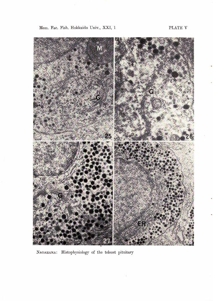
Mem. Fac. Fish. Hokkaido Univ., XXI, 1 PLATE V
NAGAHAMA: Histophysiology of the teleost pituitary

Mem. Fac. Fish. Hokkaido Univ., XXI, 1 PLATE VI
NAGAHAMA: Histophysiology of the teleost pituitary

Mem. Fac. Fish. Hokkaido Univ., XXI, 1 PLATE VII
NAGAHAMA: Histoph;siology of the teleost pituitary

Mem. Fac. Fish. Hokkaido Univ., XXI, 1 PLATE VIII
NAGAHAMA: Histophysiology of the teleost pituitary

Mem. Fac. Fish. Hokkaido Univ. , XXI, 1 PLATE IX
NAGAHAMA ; Histophysiology of the teleost pituitary

Mem. Fac. Fish. Hokkaido Univ., XXI, 1 PLATE X
N AGAHAMA: Histophysiology of the teleost pituitary

Mem. Fac. Fish. Hokkaido Univ., XXI, 1 PLATE XI
NAGAHAMA: Histophysiology of the teleost pituitary

Mem. Fac. Fish. Hokkaido Univ., XXI, 1 PLATE XII
NAGAHAMA : Histophysiology of the teleost pituitary

Mem. Fac. Fish. Hokkaido Univ., XXI, 1 PLATE XIII
NAGAHAMA: Histophysiology of the teleost pituitary

Mem. Fac. Fish. Hokkaido Univ., XXI, 1 PLATE XIV
NAGAHAMA: Histophysiology of the teleost pituitary

Mem. Fac. Fish. Hokkaido Univ., XXI, 1 PLATE XV
NAGAHAMA: Histophysiology of the teleost pituitary

Mem. Fac. Fish. Hokkaido Univ., XXI,~l PLATE XVI
NAGAHAMA: Histophysiology of the teleost pituitary

Mem. Fac. Fish. Hokkaido Univ., XXI, 1 PLATE XVII
NAGAHAMA: Histophysiology of the teleost pituitary

Mem. Fac. Fish. Hokkaido Univ., XXI, 1 PLATE XVIII
NAGAHAMA: Histophysiology of the teleost pituitary

Mem. Fac. Fish. Hokkaido Univ., XXI, 1 PLATE XIX
NAGAHAMA: Histophysiology of the teleost pituitary
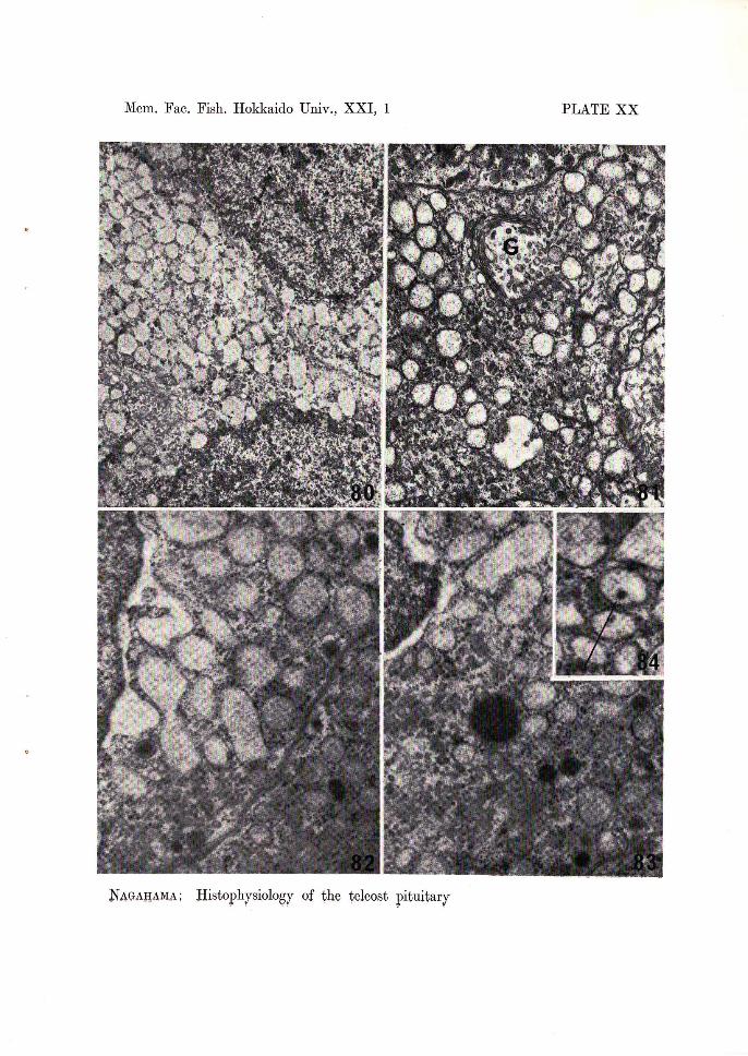
Mem. Fac. Fish. Hokkaido Univ., XXI, 1 PLATE XX
NAGAH;AMA; Histophysiology of the teleost pituitary

Mem. Fac. Fish. Hokkaido Univ., XXI, 1 PLATE XXI
NAGARAMA; Histophysiology of the teleost pituitary

Mem. Fac. Fish. Hokkaido Univ., XXI, 1 PLATE XXII
NAGAHAMA: Histophysiology of the teleost pituitary

Mem. Fac. Fish. Hokkaido Univ., XXI, 1 PLATE XXIII
NAGAHAMA; Histophysiology of the teleost pituitary

Mem. Fac. Fish. Hokkaido Univ., XXI, 1 PLATE XXIV
NAQAHAMA ; Histophysiology of the teleost pituitary

Mem. Fac. Fish. Hokkaido Univ., XXI, 1 PLATE XXV
NAGAIIAMA; Histophysiology of the teleost pituitary

Mem. Fac. Fish. Hokkaido Univ., XXI, 1 PLATE XXVI
NAGAH;AMA: Histophysiology of the teleost pituitary

Mem. Fac. Fish. Hokkaido Univ., XXI, 1 PLATE XXVII
NAQAllAMA; Histophysiology of the teleost pituitary

Mem. Fac. Fish. Hokkaido Univ., XXI, 1 PLATE XXVIII
NAGAHAMA: Histophysiology of the teleost pituitary

Mem. Fac. Fish. Hokkaido_Univ., XXI, 1 PLATE XXIX
NAGAHAMA: Histophysiology of the teleost pituitary

Mem. Fac. Fish. Hokkaido Univ. , XXI, 1 PLATE XXX
NAGAHAMA : Histophysiology of the teleost pituitary

Mem. Fac. Fish. Hokkaido Univ., XXI, 1 PLATE XXXI
NAGAHAMA: Histophysiology of the teleost pituitary

Mem. Fac. Fish. Hokkaido Univ., XXI, 1 PLATE XXXII
NAGAHAMA: Histophysiology of the teleost pituitary

Mem. Fac. Fish. Hokkaido Dniv., XXI, 1 PLATE XXXIII
NAGAHAM,,: Histophysiology of the teleost pituitary

Mem. Fac. Fish. Hokkaido Univ., XXI, 1 PLATE XXXIV
NAGAHAMA: Histophysiology of the teleost pituitary

Mem. Fac. Fish. Hokkaido Univ. , XXI, 1 PLATE XXXV
NAGAHAMA: Histophysiology of the teleost pituitary