Pituitary Dysfunction after Blast Traumatic Brain Injury: The UK BIOSAP Study
Upload
independentCategory
view
0download
0
1
2
3
4567
12
13
14
15
16
17
18
19
20
21
22
23
27
28
29
30
31
32
33
34
35
36
37
38
39
40
41
42
43
44
45
46
47
Psychiatry Research Neuroimaging xxx (2008) xxx-xxx
PSYN-09428 No of Pages 6
Contents lists available at ScienceDirect
Psychiatry Research Neuroimaging
j ourna l homepage wwwe lsev ie rcom locate psychresns
ARTICLE IN PRESS
F
Pituitary gland volume in currently depressed and remitted depressed patients
Valentina Lorenzettiabcd Nicholas B Allenbc Alex Fornitoa Christos PantelisaGiovanni De Platod Anthony Anga Murat YuumlcelabcaMelbourne Neuropsychiatry Centre Department of Psychiatry The University of Melbourne and Melbourne Health VIC AustraliabORYGEN Research Centre Melbourne VIC AustraliacThe University of Melbourne and Melbourne Health VIC AustraliadDepartment of Psychology University of Bologna Viale Berti Pichat 5 40126 Bologna Italy
Corresponding authorMelbourneNeuropsychiatry CSt Carlton South Victoria 3053 Australia Tel +61 3 834
E-mail address muratunimelbeduau (M Yuumlcel)
0925-4927$ ndash see front matter copy 2008 Elsevier Irelanddoi101016jpscychresns200806006
Please cite this article as Lorenzetti V etResearch Neuroimaging (2008) doi10101
O
A B S T R A C T
A R T I C L E I N F O
Article history
Major Depressive Disorder (
Received 13 January 2008Received in revised form 11 April 2008Accepted 12 June 2008Available online xxxx
KeywordsHPA axisStressImagingBrain
D
PROMDD) has been associated with increased pituitary gland volume (PGV) which is
thought to reflect stress-^related dysregulation related to hypothalamusndash
^pituitary-
^axis (HPA) activity However
it is unclear whether PGV alteration reflects a ldquodynamicrdquo change related to current mood instability or if it is astablemarker of illness vulnerability In this studywe investigated PGV in currently depressed patients (cMDD)(n=31) remitted depressed patients (rMDD) (
^n=31) and healthy controls (n=33) using 15 T Magnetic
Resonance Imaging (MRI) The groups were matched for age and gender We found no significant PGV intra-cranial volume (ICV) or whole brain volume (WBV) differences between cMDD patients rMDD patients andhealthy controls Furthermore PGV was not correlated with clinical features of depression (eg age of onsetnumber of episodes scores at the BDI PANAS andMASQ subscales) In conclusion PGV does not appear to be amarker of current or past MDD in adult patients
copy 2008 Elsevier Ireland Ltd All rights reserved
E
48
1 Introduction
49
50
51
52
53
54
55
56
57
58
59
60
61
62
63
64
65
66
67
68
69
UNCO
RRECAbnormal hypothalamicndashpituitaryndashadrenal (HPA) axis activity is
thought to reflect stress-relateddysregulation and is a consistentfindingin patients with mental illnesses particularly those affected by majordepressive disorder (MDD) (Mitchell 1998 Jiang et al 2000 Sheline2000 Pariante and Miller 2001 Davidson et al 2002 Pariante 2003Parker et al 2003 Swaab et al 2005) It has been suggested that chronicHPA activity leads to excessive exposure of certain brain regions toglucocorticoids (Varghese and Brown 2001) which can have adversepotentially neurotoxic effects (Fejograve del Mello et al 2003 Garner 2004Lucassen et al 2006) Specifically several studies to date indirectlysupport the notion that abnormal HPA activity may affect the mor-phology of brain regions involved in the stress response in patients withMDD (Nemeroff et al 1992 Sheline 2000 Davidson et al 2002 FejogravedelMello et al 2003 Pariante 2003 Garner 2004 Herman et al 2005Swaab et al 2005 Lucassen et al 2006)
The pituitary gland is an integral part of the HPA^axis and may be
one of the regions most affected by stress dysregulation occurring inMDD (Krishnan et al 1991 Axelson et al 1992 Schwartz et al 1997MacMaster and Kusumakar 2004 MacMaster et al 2006) Severalstudies have found enlarged pituitary gland volume (PGV) in patients
70
71
72
73
74
entre coAGBuilding161 Barry4 1877 fax +61 3 9348 0469
Ltd All rights reserved
al Pituitary gland volume6jpscychresns200806006
Twith MDD (Krishnan et al 1991 MacMaster and Kusumakar 2004MacMaster et al 2006) which has been hypothesized to representan increased number andor size of corticotropin-releasing hormone(CRH) cells in the area Such conclusions are consistent with evidencethat enlarged pituitary gland volume reflects an increase in the size andnumber of corticotrophs the cells that produce and secrete adreno-corticotrophic hormone (ACTH) (Krishnan et al 1991 Axelson et al1992 Pariante et al 2004b Garner et al 2005) This alteration incorticotrophic cells has been previously hypothesized to be a conse-quence of either HPA axis hyperactivity (HPA hypothesis of depression)or of a specific dysfunction of a subgroup of CRH neurons that pro-vokes HPA axis hyperactivity (CRH-hypothesis of depression) (Parianteand Miller 2001 Swaab et al 2005) A clearer understanding of thevolumetric changes in the pituitary is likely to help shed light on the roleof this structure in the neurobiology of the MDD (Axelson et al 1992)
Despite the robust evidence implicating the HPA axis in MDD(Axelson et al1992 Christensen and Kessing 2001 Swaab et al 2005)only a handful of studies have examined pituitary gland volume (PGV) indepression (Krishnan et al 1991 Axelson et al 1992 Sassi et al 2001MacMaster and Kusumakar 2004 MacMaster et al 2006) Out of fourPGV studies in unipolar depression three reported increased PGV(Krishnan et al 1991 MacMaster and Kusumakar 2004 MacMasteret al 2006)while one foundnosignificantdifference in PGV (Sassi et al2001) in currently depressed patients compared to healthy controls Thestudy of Axelson et al
^(1992) examined the relationship between PGV
and the Dexamethasone Suppression Test (DST) in depressed patientsbut did not have a group of healthy controls and it was not informative
in currently depressed and remitted depressed patients Psychiatry
75
76
77
78
79
80
81
82
83
84
85
86
87
88
89
90
91
92
93
94
95
96
97
98
99
100
101
102
103
104
05
06
07
08
09
10
11
12
13
14
15
16
17
18
19
20
21
22
23
24
25
26
27
28
29
30
31
32
33
34
35
36
37
t11
t12t13
t14
t15
t16
t17
t18
t19
t110
t111
t112
t113
t114
t115
t116
t117
t118
t119
t120
t121
t122
t123
t124
t125
t126
t127
t128
2 V Lorenzetti et al Psychiatry Research Neuroimaging xxx (2008) xxx-xxx
ARTICLE IN PRESS
about PGV differences between MDD patients and healthy subjectsAnother study by Schwartz et al
^(1997) reported no PGV difference
between adult depressed patients and healthy controls but they haveexamined the Seasonal Affective Disorder a depression subtype there-fore their resultsmaynot be comparable toours asweassessedunipolardepression specifically Such knowledge could elucidate whether PGVmay be useful as a biological marker of depression vulnerability (a traitfeature) or a statemarker of depressive illness As a consequence it is notyet clear from the available studies if PGV alteration reflects transientperiods of HPA axis dysregulation such as would occur during a de-pressive episode or if it is a trait feature of MDD suggesting that it maybe stable and chronic in depressed patients
In the current study we investigated PGV in currently depressedpatients (cMDD) and individuals with a history of depression but whoare currently in remission (rMDD) This approachenabledus to examineimportant questions regarding whether differences in PGV reflect stateor trait influences Based on previous findings of increased HPA activityin patients with cMDD (Sheline 2000 Pariante and Miller 2001Davidson et al 2002 Pariante 2003 Parker et al 2003 Swaab et al2005) and the potential for reversibility of structural brain changesfollowing recovery fromdepression(eg adrenal gland in adult humans(Rubin et al 1995) and chronic corticosteroid or stress exposure(eg hippocampal dentate gyrus in mammals (Lucassen et al 2006)we hypothesized that enlarged PGV would be a state marker of de-pressive illness As such we predicted that the cMDD patients wouldhave an enlarged PGV relative to both rMDD and healthy controls
2 Methods
21 Subjects
Ninety-five subjects were recruited into the study of which 31received a current diagnosis of MDD 31 were currently medically
UNCO
RREC
Table 1Demographic neuropsychological and clinical characteristics of the sample
Variable Meanplusmn^SD
cMDD rMDD
Age 3252plusmn828 3507plusmn996Female (male) 22 (7) 18 (9)Melancholicatypical 103 ndash
First episoderecurrent 722 ndash
Age of onset 2107plusmn795 2604plusmn943Number of episodes 367plusmn336 309plusmn263Medication past 6 monthsyesno
216 1213
Current anxiety disorderyesno
1810 423
IQ 10486plusmn874 11142plusmn993WTAR 10748plusmn1139 11174plusmn893BDI 3683plusmn893 1304plusmn1172MASQ GD Mixed 5050plusmn777 4044plusmn1032
GD Dep 4732plusmn919 3504plusmn1167GD Anx 3218plusmn873 2472plusmn766A Anx Arousal 4200plusmn1215 2887plusmn765High PA 4357plusmn1347 6500plusmn1235Loss of Int 3161plusmn635 2348plusmn678
PANAS PA 2161plusmn645 2872plusmn795NA 2121plusmn850 1415plusmn470
AUDIT 537plusmn622 569plusmn477Pituitary volumes (mm3) 72722plusmn13921 74012plusmn12342Whole brain volumes (mm3) 1237176plusmn12411314780 1232334plusmn13295Intra
^-cranial volumes (mm3) 14773534plusmn13810530851 14698426plusmn1502
cMDD = currently depressed patients rMDD = remitted depressed patients HC = healthy contro(WASI) BDI=BeckDepression InventoryMASQ=MoodandAnxietySymptomQuestionnaireGDMarousal High PA = high positive affect Loss of Int = loss of interest PANAS = Positive and NegatiIdentification Test PGVs = pituitary gland volumes WBVs = whole brain volumes ICV = intra cracontrols c = current depressedNpast depressedNcontrols d = current depressedbpast depresseddifference between the degrees of freedom across different measures is due to the fact that we d
Please cite this article as Lorenzetti V et al Pituitary gland volumeResearch Neuroimaging (2008) doi101016jpscychresns200806006
DPR
OOF
1and psychiatrically well individuals with a previous history of a diag-1nosable MDD and 33 were healthy comparison subjects who were1not significantly different in age gender and measures of education1and intelligence Six participants were excluded from further anal-1yses as they were discovered to have brain abnormalitiesincidental1findings (periventricular and frontal hyper-intensities) detected in1the T2 scan Two of those excluded were current depressed (both1males with severe recurrent MDD mean ageplusmn
^SD=3500plusmn989
1mean IQplusmn^SD=10100plusmn989) and four were remitted patients (all
1females mean ageplusmn^SD=4075plusmn629 mean IQplusmn
^SD=11125plusmn464)
1There were no notable differences between the group excluded1because of MRI abnormalities and the main sample on any clinical1or demographical measures The main baseline demographic data1and clinical features of the remaining three groups are presented in1Table 11The patients were recruited through advertisement in the local1media from the general community and via outpatient mental health1clinics (specifically ORYGEN Youth Health and the University of1Melbourne Psychology Clinic Melbourne Australia) The local internal1review board (Mental Health Research amp Ethics Committee Mel-1bourne Health Melbourne Australia) approved the study protocol1and the participants gave written informed consent after a complete1description of the study1Participants inclusion criteria were age
^between 18 and
^50 years
1English as a preferred language current IQN70 and colour vision1and acuity within normal (or corrected-to normal) limits Exclusion1criteria were a history of significant head injury seizures impaired1thyroid function and steroid use neurological diseases electro-1convulsive therapy within the past six months All the depressed1subjects with another current Axis I psychiatric disorder (other than1anxiety disorders) were excluded as well as any healthy controls1who had a personal history of psychiatric illness drug or alcohol1dependence
TE
F(df)^1 P
HC
3403plusmn991 0523 (2 86) 059521 (12) 1159(2) 0560ndash ndash
ndash ndash
ndash 4558 (1 54) 0037 a
ndash 0369 (1 38) 0547ndash 118602 (4) b00005
ndash 84154(2) b00005
11112plusmn1086 4034 (2 85) 0021 b
11164plusmn1229 1407 (2 86) 025355plusmn408 120572 (2 86) b00005 c
2787plusmn828 49209 (2 81) b00005 c
1947plusmn723 66848 (2 82) b00005 c
1641plusmn639 37307 (2 82) b00005 c
2197plusmn444 40472 (2 79) b00005 c
8110plusmn1427 50194 (2 80) b00005 d
1472plusmn504 58682 (2 82) b00005 c
3294plusmn726 18569 (2 82) b00005 b
1119plusmn157 24977 (2 83) b00005 e
461plusmn299 0415 (2 83) 066271028plusmn10328 0452 (2 86) 0638
497718 1250567plusmn14121481640 0154 (2 86) 08585646469 14927358plusmn14314143347 7584 (2 83) 0117
ls IQ = Intelligence Quotient as measured by the Wechsler Abbreviated Scale of Intelligenceixed=generaldistressGDDep=generaldepressionGDAnx=general anxietyAA=anxious
ve Affect Schedule PA = positive affect NA = negative affect AUDIT= Alcohol Use Disordersnial volumes a = current depressedbpast depressed b = current depressedbpast depressedbcontrols e = current depressedNpast depressed controls =χ2 (df)minus = not calculable 1 theid not have information relative to all the patients for the measured variables
in currently depressed and remitted depressed patients Psychiatry
OOF
138
139
140
141
142
143
144
145
146
147
148
149
150
151
152
153
154
155
156
157
158
159
160
161
162
163
164
165
166
167
168
169
170
171
172
173
174
175
176
177
178
179
180
181
182
183
184
185
186
187
188
189
190
191
192
193
194
195
196
197
198
199
200
201
202
203
204
205
206
207
208
209
210
211
212
213
214
215
216
217
218
219
220
221
222
223
224
225
226
227
228
229
230
231
232
233
Fig 1 Pituitary visualized from a coronal slice
3V Lorenzetti et al Psychiatry Research Neuroimaging xxx (2008) xxx-xxx
ARTICLE IN PRESS
UNCO
RREC
22 Clinical measures
All participants underwent a clinical and neuropsychologicalassessment conducted at ORYGEN Youth Health Melbourne Traineepsychologists (DM OS) experienced in recruiting and assessingclinical populations screened participants with the Structured ClinicalInterview for DSM-IV (SCID-IV-TR) (First et al 2001) and a seriesof inventories including the Beck Depression Inventory (BDI) (Beckand Steer 1987) the Mood and Anxiety Symptom Questionnaire(MASQ) (Watson et al 1995) Positive Affect and Negative Affect Scale(PANAS) (Watson et al 1988) and the Alcohol Use Disorders Iden-tification Test (AUDIT) (Babor et al 1992) We also obtained measuresof premorbid and current intelligence using theWechsler Test of AdultReading (WTAR) (Corporation 2001) and the Wechsler AbbreviatedScale of Intelligence (WASI) (Corporation 1999) respectively The ad-ministration of the tests took approximately 25 h and all the par-ticipants received reimbursement of $20 AUD
We also assessed the presenceabsence of medication during the6 months before the screening in the subgroup with a positive historyof lifetime medication (see Table 1) Thirty-three patients were on astable medication regime for at least 6 months preceding the scan Ofthese seventeen patients were on SSRIs (eg fluoxetine fluxovamineparoxetine sertraline citalopram escitolopram) four on SSNRI(eg venlafaxine) three on NaSSAs (eg mirtazapine) two on TCAs(eg amitriptyline doxepin) two on MAOIs (eg tranylcyprominemoclobemide) one on lithium one on NRIs (eg reboxetine) Threepatients were receiving combination therapy (paroxetine and benzo-diazepine escitolopram and mirtazapine lithium and dothiepin re-spectively) while nine were medication-naiumlve
23 MRI data acquisition
All the subjects were scanned with a Siemens MAGNETOMAvanto 15-Tesla scanner at the St Vincents Hospital MelbourneVictoria Each scanning session took approximately 1 h andall the participants received $40 AUD reimbursement A structuralT1-weighted scan was performed Image parameters were as followstime to echo=23 ms time repetition=21 ms flip angle=15deg matrixsize=256times256 voxel dimension=1times1times1 mm Additionally MRIabnormalities were assessed using a high-resolution T2-weightedscan All images were analysed on a LINUX workstation usingANALYZE 75 (Mayo) to delineate the regions of interest and generatevolumetric measures
24 Image analysis
241 Pituitary volumeEach pituitary was traced by the same investigator (VL) who was
blinded to group membership We utilized a modified version(Pariante et al 2004b) of the method previously used by ourselvesand others (Sassi et al 2001 MacMaster and Kusumakar 2004MacMaster et al 2006) Specifically images were traced in the coronalplane where the pituitary is best visualized (Garner 2004 Parianteet al 2004b Garner et al 2005) (see Fig 1) The mean number ofcoronal slices traced per case was 1208 (range 8ndash15) which were1 mm thick We excluded the infindibular stalk from the tracing butincluded the hyper-intense region in the posterior pituitary which isthought to represent high levels of vasopressin concentrations (Sassiet al 2001 Garner 2004 Pariante et al 2004b Garner et al 2005)The borders of the pituitary were clearly defined by the diaphragmasellae superiorly the sphenoid sinus inferiorly and the cavernoussinuses bilaterally (Garner 2004 Pariante et al 2004b Garner et al2005) PGV was calculated by summing the volumes of all the tracedslices Intraclass correlation coefficients (absolute agreement) for intraand inter-rater reliabilities assessed on ten randomly selected imageswere 094 and 098 respectively
Please cite this article as Lorenzetti V et al Pituitary gland volumeResearch Neuroimaging (2008) doi101016jpscychresns200806006
TEDPR242 Whole brain volumes
Whole brain volumes (WBV) were estimated using tools in the FSLsoftware library (website) Briefly all images were stripped ofextracerebral tissue using BET (Brain Extraction Tool) (Smith 2002)Each voxel was classified into grey white or CSF (cerebro-spinal fluid)using FAST (Zhang et al 2001) The number of grey and white mattervoxels was then summed to obtain the WBV estimate
243 Intracranial cavityIntracranial volume (ICV) was delineated from a sagittal reformat
of the original 3D-dataset Themajor anatomical boundarywas the duramater below the inner table and generally visible as awhite lineWherethe duramaterwas not visible the cerebral contourwas outlined Otherlandmarks included the undersurfaces of the frontal lobes the dorsumsellae the clivus and the posterior arch of the craniovertebral junction(Eritaia et al 2000)
25 Statistical analysis
We assessed the distribution of the demographic neuropsycholo-gical and clinical variables across the groups by running a series of chisquare (χ2) and univariate analysis of variance (ANOVA) (see Table 1)
We used ANCOVA with age as a covariate and diagnosis (cMDDrMDD controls) and gender as between-subjects factors to examinegroup differences in PGV Bivariate correlation analyses were used toexamine the association between PGV and other relevant demo-graphic (age) neuropsychological (current intelligence quotient) andclinical variables (age of onset number of episodes scores at the BDIMASQ and PANAS subscales) Finally we used Students t-test toexamine the effects of medication status on PGV by dividing theclinical sample (current and past MDD patients) into those taking ornot taking medication during the 6 months prior to the study For allcomparisons α= 05
3 Results
31 Overall group comparisons
There were no group differences on any of the clinical or demo-graphic measures nor were there any differences in WBV or ICV(see Table 1) ANCOVA assessing group differences in PGV found no
in currently depressed and remitted depressed patients Psychiatry
C
234
235
236
237
238
239
240
241
242
243
244
245
246
247
248
249
250
251
252
253
254
255Q1256
257
258
259
260
261Q2262
263
264
265
266
267
268
269
270
271
272
273
274
275
276
277
278
279
280
281
282
283
284
285
286
287
288
289
290
291
292
293
294
295
296
297
98
99
00
01
02
03
04
05
06
07
08
09
10
11
12
13
14
15
16
17
18
19
20
21
22
23
24
25
26
27
28
29
30
31
32
33
34
35
36
37
38
39
40
41
42
43
44
45
46
47
48
49
50
51
52
53
54
55
56
57
58
59
60
61
62
63
4 V Lorenzetti et al Psychiatry Research Neuroimaging xxx (2008) xxx-xxx
ARTICLE IN PRESS
UNCO
RRE
effect of diagnosis (F^(2 1901)=0343 P=0746) or interaction be-
tween diagnosis and gender (F(2 82) = 0539 P = 0585) Therewas however a significant main effect of gender (F(1 1954) = 23867P = 0041) indicating that males had smaller PGV than females acrossboth groups (males M = 66379
^SD = 10636 females M = 75288
SD = 11802)We found no significant correlation between PGV and age of onset
(r = 0226 P=^077) number of episodes (r=
^minus0049 P=
^0747) or any
of the other clinical measures (eg scores at the BDI MASQ subscalesPANAS subscales AUDIT) Dividing the patient group into those whowere and thosewhowere not takingmedication 6months prior to thescanning revealed no effect of medication status (t=
^0391 P=
^0698
df=^50)
4 Discussion
Our findings suggest that PGV is not altered in patients with acurrent or past Major Depressive Disorder (MDD) The evidence todate suggests that PGV is not affected in adult MDD patients who areeither currently depressed or in remission as the studies that havepreviously examined adult depressed patients did not find evidencefor PGV alterations
Our findings are inconsistent with those of Krishnan et al^(1991)
and MacMaster and colleagues (2004 2006) who have reported in-creased PGV in their samples of currently depressed patients Com-parison of our findings with those of Krishnan et al
^(1991) suggests
their finding may have been driven by an elderly subgroup of patientswhich had an age band (eg over 50 years old) not represented in oursample Indeed their mid-adult depressed subgroup did not showpituitary alterations Interestingly MacMaster and colleagues (20042006) who also found an association between depression and PGVenlargement examined adolescent depressed patients suggestingthat PGV may be a state marker of depression in younger and oldersamples but not through middle adulthood Accordingly Sassi
^et al
(2001) who studied a euthymic sample with age range similar to oursalso failed to find any differences in PGV Together these resultssuggest that adolescent neurodevelopmental and ageing neurodegen-erative changesmay play an important role in the interaction betweenMDD and PGV (Krishnan et al 1991 El-Sheikh et al 2005MacMasteret al 2006) and that a relationship between PGV and MDD is notapparent in younger adult patients Fronto-temporal brain regionsundergo neural changes during adolescence (eg neurodevelopment)and ageing (eg neurodegeneration) (Giedd et al 1999 Sowell et al1999 2001 2004 Gotgay et al 2004) These regions include areaswhich are involved in the HPA axis regulation such as the hippo-campus (Gold et al 1984 Holsboer et al 1987 Sapolsky et al 1991Young et al 1991 Sheline 2000 Pariante and Miller 2001 Davidsonet al 2002 Pariante 2003 Garner 2004) the amygdala (Davidsonet al 2002) the anterior cingulate cortex (ACC) (Chao et al 1989Diorio et al 1993 Cerqueira et al 2005) and the prefrontal cortex(PFC) (Szot et al1994 Sheline 2000 Hermanet al 2003) As such theinteraction between MDD and age
^-related brain changes may further
impair the HPA axis dysregulation in elderly and adolescent depressedpatients leading to an alteration of the PGV only in these clinical sub-groups but not in MDD adults
One possible explanation for the negative finding regarding evi-dence for PGV enlargement in the cMDD group is that 2127 of thesepatients were on stable medication during the 6 months prior to thescan Given that the literature to date consistently demonstrates thatantidepressant medication directly suppress HPA axis activity (Mitch-ell 1998 Pariante 2003 Barden 2004 Pariante et al 2004a Masonand Pariante 2006) it may be that medication may have attenuatedany HPA dysregulation occurring during the depressive episode con-sequently reducing any PGV enlargement Thus while we found nodifferences between patients who were and were not medicated inthe 6 months prior to scanning the medicated group comprised pre-
Please cite this article as Lorenzetti V et al Pituitary gland volumeResearch Neuroimaging (2008) doi101016jpscychresns200806006
TEDPR
OOF
2dominantly cMDD patients while the unmedicated group comprised2predominantly rMDD patients No study has investigated antidepres-3sant effects on PGV in adult depressed patients although reports in3adolescent samples provide evidence for alterations in treatment-3naiumlve patients (MacMaster and Kusumakar 2004 MacMaster et al32006) Also a study by MacMaster et al
^(2007) found a relationship
3between PGV and medication effects (eg volumetric increase3following antipsychotic treatment) suggesting that the PGV may3be a bio-marker of medication effects In our study we used a rela-3tively coarsemeasure ofmedication (presenceabsence during the past36 months) and further studies are required to investigate the rela-3tionship between more detailed parameters of antidepressant treat-3ment (eg length dosage) and stage of illness Moreover we could not3assess the gender by medication interaction due to sample size con-3strains We could only examine medication effects in a separate anal-3ysis collapsed across males and females3A number of studies have consistently found that pituitary size3changes with gender in healthy adults (Lurie et al 1990 Takano et al31999 Kato et al 2002 MacMaster et al 2007) Accordingly our3findings support a strong gender effect on pituitary volume with3males exhibiting smaller pituitary volumes than females in all the3analyses This may be due to the interaction between the HPA axis and3the hypothalamicndashpituitary
^ndashgonadal (HPG) axis which regulates the
3production of sex hormones (Swaab et al 2005) The relationship3between these systems has previously been hypothesized to play a3role in the higher prevalence of mood disorders in women compared3to men and the pituitary gland may be implicated in mechanisms of3gender-related vulnerability to stress (Swaab et al 2005) However3we did not find any gender
^timesdiagnosis interaction effects on the PGV
3and it is still unclear whether the pituitary gland may play a specific3role in the gender-related vulnerability to MDD rather than a more3general role in the gender-related vulnerability to stress3The participants were not matched for intelligence quotient (IQ)3measures as cMDD patients had a significantly lower IQ compared to3the other groups We do not know if it may have affected the PGV but3to our knowledge no study to date has found a relationship between3IQ and PGV measures Moreover a lower IQ is one of the variables3related to the presence of a current depressive episode and this3variable has helped us to characterize the different clinical popula-3tions that we have considered3Our sample was composed of only outpatients as the recruitment3was community-based Therefore we may not have seen the range of3severity and chronicity of MDD to observe structural alterations as3these factors have been hypothesized to be predictive of volumetric3changes in brain structures involved in the stress response and3underlying depression symptomatology Future studies may be useful3in determining whether pituitary volume may be affected in more3severely depressed samples3Whether our PGV findings reflect HPA activity is difficult to deter-3mine as we acquired no direct measures of HPA function in this study3and this may limit the interpretation of our data3Also while a previous study by MacMaster et al
^(2006) has pro-
3vided evidence that a positive family history of mental illness may3affect the PGV of depressed patients we did not assess family history3in this sample3The tracing protocol that we used as well as the ones used by the3previous studies on the PGV (Krishnan et al 1991 Axelson et al 19923Schwartz et al 1997 Sassi et al 2001 MacMaster and Kusumakar32004 Pariante et al 2004b 2005 Garner et al 2005 MacMaster3et al 2006) did not enable us to distinguish the anterior from the3posterior lobe of the pituitary gland The anterior pituitary contains3corticotrophs that produce ACTH (Elster 1993) while the posterior3pituitary secretes oxytocin and vasopressin (Elster 1993) Thus the3anterior portion is most likely to show dynamic changes with stress3and measuring the whole PGV in this study may have obscured these3effects However the anterior pituitary constitutes more than 80 of
in currently depressed and remitted depressed patients Psychiatry
364
365
366
367
368
369
370
371
372
373
374
375
376
377
378
379
380
381
382
383
384
385
386
387
388
389
390
391
392
393
394
395
396
397
398
399
400
401
402
403
404
405
406
407408409410411412413414415416417418419420421422423424425426427428429430431
432 Q3433434435436437438439440441442443444445446447448449450451452453454455456457458459460461462463464465466467468469470471472473474475476477478479480481482483484485486487488489490491492493494495496497498499500501502503504505506507508509510511512513514515516517
5V Lorenzetti et al Psychiatry Research Neuroimaging xxx (2008) xxx-xxx
ARTICLE IN PRESS
UNCO
RREC
the overall pituitary gland (Pariante et al 2004b) and the reportedresults are therefore more likely to reflect changes in the anteriorrather than in the posterior pituitary
Our study has a number of strengths compared to previous studieson the PGV in adults with unipolar depression Firstly we haveprovided a bigger and more homogeneous sample of unipolar adultdepressed patients compared to the ones of Krishnan et al
^(1991)
(eg 19 depressed patients of which 3 were bipolar compared to19 healthy controls) and Sassi et al
^(2001) (eg 13 unipolar patients
of which 7 were euthymic compared to 34 healthy controls) Sec-ondly we considered a more homogeneous age range of adults (eg18ndash50 years) compared to the ones considered by Krishnan et al
^(1991) (eg 23ndash80 years) and Sassi et al^(2001) (eg 24ndash59 years)
which enabled us to rule out the potential confounds of neurodegen-erative and other age-related changes Finally we have selected amore gender-balanced sample than the studies of Krishnan et al
^(1991) (eg 14 females 5 males in each group) and Sassi et al^(2001)
(eg 12 females and one male in the clinical group 14 females and22 males in the control group) As a consequence we believe that ourdata may be more ldquogeneralizablerdquo than the previous studies
Consistent with the data of Sassi et als^(2001) study which effect
sizes (eg negligible effect d=^011) are very close to ours (eg small
effect d=^019) we did not find evidence that the PGV is a state marker
of current depressive illness Also our findings suggest that the PGV isnot a trait marker of vulnerability to MDD Future studies are neededto understand whether and at what extent demographic (eg agegender) and clinical (eg severity and length of illness medication)variables may mediate the relationship between PGV and depressionas this area remains still unexplored
Acknowledgments
This research was supported by grants from the AustralianResearch Council (ID DP0557663) Dr Yuumlcel was supported by aNational Health and Medical Research Council of Australia ClinicalCareer Development Award (509345) a NHMRC Program Grant (ID350241) and the Colonial Foundation Dr Fornito is supported by a JNPeters Fellowship Neuroimaging analysis was facilitated by theNeuropsychiatry Imaging Laboratory managed by Ms Bridget Soulsbyat the Melbourne Neuropsychiatry Centre and supported by Neuros-ciences Victoria Participant recruitment and assessment have beendone by Ms Orli Schwartz and Ms Diana Maud Valentina Lorenzetti issupported by an overseas research project scholarship of theDepartment of Psychology University of Bologna
References
Axelson DA Doraiswamy PM Boyko OB Escalona PR McDonaldWM Ritchie JCPatterson LJ Ellinwood EH Nemeroff CB Krishnan KRR 1992 In vivoassessment of pituitary volume with magnetic resonance imaging and systematicstereology relationship to dexamethasone suppression test results in patientsPsychiatry Research 44 63ndash70
Babor TF De La Fuente JR Saunders J Grant M 1992 The Alcohol Use DisordersIdentification Test Guidelines for use in Primary Health Care World HealthOrganization Geneva Switzerland
Barden N 2004 Implication of the hypothalamicndashpituitaryndashadrenal^axis in the
pathophysiology of depression Journal of Psychiatry amp Neuroscience 29 185ndash193Beck AT Steer RT 1987 Beck Depression Inventory Manual Harcourt Brace
Jovanovich San AntonioCerqueira JJ Catania C Sotiropoulos I Schubert M Kalish R Almeida OFX Auer
DP Sousa N 2005 Corticosteroid status influences the volume of the rat cingulatecortex mdash a magnetic resonance imaging study Journal of Psychiatric Research 39451ndash460
Chao HM Choo PH McEwen BS 1989 Glucocorticoid and mineralocorticoidreceptor mRNA expression in rat brain Neuroendocrinology 50 365ndash371
Christensen MV Kessing LV 2001 The hypothalamo-pituitaryndashadrenal^axis in major
affective disorder a review North Journal of Psychiatry 55 359ndash363Davidson RJ Pizzagalli DA Nitschke JB Putnam K 2002 Depression perspectives
from affective neuroscience Annual Review of Psychology 53 545ndash574Diorio D Viau V Meaney MJ 1993 The role of the medial prefrontal cortex
(cingulate gyrus) in the regulation of hypothalamicndashpituitaryndashadrenal^responses to
stress Journal of Neuroscience 13 3839ndash3847
Please cite this article as Lorenzetti V et al Pituitary gland volumeResearch Neuroimaging (2008) doi101016jpscychresns200806006
TEDPR
OOF
El-Sheikh RM MacMaster FP Upadhyaya AR Nutche J Rosenberg DR KeshavanMS 2005 Specific effect of antipsychotics on pituitary volume a structural MRIstudy Biological Psychiatry
Elster AD 1993 Modern imaging of the pituitary Radiology 187 1ndash14Eritaia J Wood SJ Stuart GW 2000 An optimized method for estimating
intracranial volume from magnetic resonance images Magnetic Resonance inMedicine 44 973ndash977
Fejograve del Mello ADA Fejograve de Mello M Carpenter LL Price LH 2003 Update onstress and depression the role of the hypothalamicndashpituitaryndashadrenal
^(HPA) axis
Revista Brasileira de Psiquiatria 25 231ndash238First MB Spitzer RL Gibbon M Williams JBW 2001 Structured Clinical Interview
for Axis 1 DSM-IV Disorders New York State Psychiatric Institute New YorkGarner BA 2004 PhD An Investigation of the Two-Hit Neurodevelopmental Hypo-
thesis of Schizophrenia Animal and Clinical Studies Department of PsychiatryThe University of Melbourne Melbourne 236
Garner BA Pariante CMWood SJ Velakoulis D Phillips L Soulsby B BrewerWJSmith DJ Dazzan P Berger G Yung AR Van Den Buuse M Murray R McGorryPD Pantelis C 2005 Pituitary volume predicts future transition to psychosis inindividuals at ultra-high risk of developing psychosis Biological Psychiatry 58417ndash423
Giedd JN Blumenthal J Jeffries NO Castellanos FX Liu H Zijdenbos A Paus TEvans AC Rapoport JL 1999 Brain development during childhood andadolescence a longitudinal MRI study Nature Neuroscience 2 861ndash863
Gold P Chrousis G Kellner C Post R Roy A Augerinos P Schulte H Olfield ELoriaux DL 1984 Psychiatric implications of the basic and clinical studies withcorticotropin-releasing factor American Journal of Psychiatry 141 619ndash627
Gotgay N Giedd JN Lusk L Hayashi KM Greenstein D Vaituzis AC Nurgent III TFHerman DH Clasen LS Toga AW Rapoport JL Thompson PM 2004 Dynamicmapping of human cortical development during childhood through early adulthoodProceedings of the National Academy of Sciences of the United States of America 1018174ndash8179
Herman JP Figueiredo H Mueller NK Ulrich-Lai Y Ostrander MM Choi DCCullinanWE 2003 Centralmechanisms of stress integration hierarchical circuitrycontrolling hypothalamo-pituitary
^ndashadrenocortical responsiveness Frontiers in
Neuroendocrinology 24 151ndash180Herman JP Ostrander MM Mueller NK Figueiredo H 2005 Limbic system
mechanisms of stress regulation hypothalamo-pituitary^ndashadrenocortical axis
Progress in Neuro-Psychopharmacology amp Biological Psychiatry 29 1201ndash1213Holsboer F Gerken A Stalla G Muller O 1987 Blunted aldosterone and ACTH release
after human CRH administration in depressed patients American Journal ofPsychiatry 144 229ndash231
Jiang HK Wang JY Lin JC 2000 The central mechanism of hypothalamicndashpituitaryndashadrenocortical
^system hyperfunction in depressed patients Psychiatry and Clinical
Neurosciences 54 227ndash234Kato K Saeki N Yamaura A 2002 Morphological changes on MR imaging of the
normal pituitary gland related to age and sex Main emphasis on pubescentfemales Journal of Clinical Neuroscience 9 53ndash56
Krishnan KRR Doraiswamy PM Lurie SN Figiel GS Husain MM Boyko OBEllinwood EHJ Nemeroff CB 1991 Pituitary size in depression Journal of ClinicalEndocrinology and Metabolism 72 256ndash259
Lucassen PJ Heine VMMuller MB VanDer Beek EMWiegant VM De Kloet EMJoels M Fuchs E Swaab DF Czeh B 2006 Stress depression and hippocampalapoptosis CNS amp Neurological Disorders mdash Drug Targets 5 531ndash546
Lurie SN Doraiswamy PM Husain MM Boyko OB Ellinwood EHJ Figiel GSKrishnan KR 1990 In vivo assessment of pituitary gland volume with mag-netic resonance imaging the effect of age Journal of Clinical Endocrinology andMetabolism 71 505ndash508
MacMaster FP Kusumakar V 2004 MRI study of the pituitary gland in adolescentdepression Journal of Psychiatric Research 38 231ndash236
MacMaster FP Russell A Mirza Y Keshavan MS Taormina SP R B Boyd C LynchM RoseM Ivey JMoore GJ Rosenberg DR 2006 Pituitary volume in treatment-naiumlve pediatric major depressive disorder Biological Psychiatry 60 862ndash866
MacMaster FP Keshavan M Mirza Y Carrey N Upadhyaya AR El-Sheikh RBuhagiar CJ Taormina SP Boyd C Lynch M Rose M Ivey J Moore GJRosenberg DR 2007 Development and sexual dimorphism of the pituitary glandLife Sciences 80 940ndash944
Mason BL and Pariante CM 2006 The Effects of Antidepressants on the HypothalamicndashPituitaryndashAdrenal
^Axis Drug News and Perspectives 19 603
Mitchell AJ 1998 The role of corticotropin releasing factor in depressive illnessa critical review Neuroscience and Biobehavioral Reviews 22 635ndash651
Nemeroff CB Krishnan KR Reed D Leder L Beam C Dunnik NR 1992 Adrenalgland enlargement in major depression A computer tomographic study Archivesof General Psychiatry 49 384ndash387
Pariante CM 2003 Depression stress and the adrenal axis Neuroendocrinology 15811ndash812
Pariante CM Miller AH 2001 Glucocorticoid receptors in major depressionrelevance to pathophysiology and treatment Biological Psychiatry 49 391ndash404
Pariante CM Thomas SA Lovestone S Makoff A Kerwin RW 2004a Do anti-depressants regulate how cortisol affects the brain Psychoneuroendocrinology 29423ndash447
Pariante CM Vassilopoulou K Velakoulis D Phillips L Soulsby B Wood SJBrewer W Smith DJ Dazzan P Yung AR Zervas IM Christidoulou GNMurray R McGorry PD Pantelis C 2004b Pituitary volume in psychosis BritishJournal of Psychiatry 185 5ndash10
Pariante CM Dazzan P Danese A Morgan KD Brudaglio F Morgan C Fearon POrr K Hutchinson G Pantelis C Velakoulis D Jones PB Leff J Murray RM
in currently depressed and remitted depressed patients Psychiatry
518519520521522523524525526527528529530531532533534535536537538539540541542543544
454647484950515253545556575859606162636465666768697071
6 V Lorenzetti et al Psychiatry Research Neuroimaging xxx (2008) xxx-xxx
ARTICLE IN PRESS
2005 Increased pituitary in antipsychotic-free and antipsychotic-treated pa-tients of the AEsop first-onset psychosis study Neuropsychopharmacology 301923ndash1931
Parker KJ Schatzberg AF Lyons DM 2003 Neuroendocrine aspects of hypercorti-solism in major depression Hormones and Behavior 43 60ndash66
Rubin RT Phillips JJ Sadow TF McCracken JT 1995 Adrenal gland volume inmajordepression increase during the depressive episode and decrease with successfultreatment Archives of General Psychiatry 52
Sapolsky RM Zola-Morgan S Squire L1991 Inhibitionof glucocorticoid secretionby thehippocampal formation in the primate The Journal of Neuroscience 11 3695ndash3704
Sassi R Nicoletti M Brambilla P Harenski K Mallinger AG Frank E Kupfer DJKeshavan MS Soares JC 2001 Decreased pituitary volumes in patients withbipolar disorder Biological Psychiatry 50 271ndash280
Schwartz PJ Loe JA Bash CN Bove K Turner EH Frank JA Wehr TA RosenthalNE 1997 Seasonality and pituitary volume Psychiatry Research NeuroimagingSection 74 151ndash157
Sheline YI 2000 3DMRI studies of neuroanatomic changes inunipolarmajordepressionthe role of stress and medical comorbility Biological Psychiatry 48 791ndash800
Smith SM 2002 Fast robust automated brain extraction Human Brain Mapping 17143ndash155
Sowell ER Thompson PM Holmes CJ Jernigan TL Toga AW 1999 In vivoevidence for post-adolescent brain maturation in frontal and striatal regionsNature Neuroscience 2 859ndash861
Sowell ER Thompson PM Tessner KD Toga AW 2001 Mapping continued braingrowth and gray matter density reduction in dorsal frontal cortex inverserelationships during postadolescent brain maturation The Journal of Neuroscience21 8819ndash8829
UNCO
RREC
Please cite this article as Lorenzetti V et al Pituitary gland volumeResearch Neuroimaging (2008) doi101016jpscychresns200806006
OF
5Sowell ER Thompson PM Toga AW 2004 Mapping changes in the human cortex5throughout the span of life The Neuroscientist 10 372ndash3925Swaab DF Bao AM Lucassen PJ 2005 The stress system in the human brain in5depression and neurodegeneration Ageing Research Reviews 4 141ndash1945Szot P Bale TL Dorsa DM 1994 Distribution of messenger RNA for the vasopressin5V1a receptor in the CNS of male and female rats Brain Research 24 1ndash105Takano K Utsonomiya H Ono H Ohfu M Okazaki M 1999 Normal development of5the pituitary gland assessment with three-dimensional MR volumetry American5Journal of Neuroradiology 20 312ndash3155Varghese FP Brown ES 2001 The hypothalamic
^ndashpituitary
^ndashadrenal axis in major
5depressive disorder a brief primer for primary care physicians Primary Care5Companion to the Journal of Clinical Psychiatry 3 151ndash1555Watson D Clark L Tellegen A 1988 Development and validation of brief measures of5positive and negative affect the PANAS scales Journal of Personality and Social5Psychology 54 1063ndash10705Watson D Clark L Weber K Assenheimer J Strauss M McCormick R 1995 Testing5a tripartite model I Evaluating the convergent and discriminant validity of anxiety5and depression symptom scales Journal of Abnormal Psychology 104 3ndash145Wechsler D 1999 Manual for the Wechsler Abbreviated Scale of Intelligence The5Psychological Corporation San Antonio TX5Wechsler D 2001 Manual for the Wechsler Test of Adult Reading (WTAR) The5Psychological Corporation San Antonio TX5Young EA Haskett RF Murphy-Weinberg V Watson SJ Akil H 1991 Loss of gluco-5corticoids fast feedback in depression Archives of General Psychiatry 48 693ndash6985Zhang Y Brady M Smith SM 2001 Segmentation of brain MR images through a5hidden Markov random field model and the expectation maximization algorithm5IEEE Transactions on Medical Imaging 20 45ndash57
TEDPR
O
in currently depressed and remitted depressed patients Psychiatry

OOF
138
139
140
141
142
143
144
145
146
147
148
149
150
151
152
153
154
155
156
157
158
159
160
161
162
163
164
165
166
167
168
169
170
171
172
173
174
175
176
177
178
179
180
181
182
183
184
185
186
187
188
189
190
191
192
193
194
195
196
197
198
199
200
201
202
203
204
205
206
207
208
209
210
211
212
213
214
215
216
217
218
219
220
221
222
223
224
225
226
227
228
229
230
231
232
233
Fig 1 Pituitary visualized from a coronal slice
3V Lorenzetti et al Psychiatry Research Neuroimaging xxx (2008) xxx-xxx
ARTICLE IN PRESS
UNCO
RREC
22 Clinical measures
All participants underwent a clinical and neuropsychologicalassessment conducted at ORYGEN Youth Health Melbourne Traineepsychologists (DM OS) experienced in recruiting and assessingclinical populations screened participants with the Structured ClinicalInterview for DSM-IV (SCID-IV-TR) (First et al 2001) and a seriesof inventories including the Beck Depression Inventory (BDI) (Beckand Steer 1987) the Mood and Anxiety Symptom Questionnaire(MASQ) (Watson et al 1995) Positive Affect and Negative Affect Scale(PANAS) (Watson et al 1988) and the Alcohol Use Disorders Iden-tification Test (AUDIT) (Babor et al 1992) We also obtained measuresof premorbid and current intelligence using theWechsler Test of AdultReading (WTAR) (Corporation 2001) and the Wechsler AbbreviatedScale of Intelligence (WASI) (Corporation 1999) respectively The ad-ministration of the tests took approximately 25 h and all the par-ticipants received reimbursement of $20 AUD
We also assessed the presenceabsence of medication during the6 months before the screening in the subgroup with a positive historyof lifetime medication (see Table 1) Thirty-three patients were on astable medication regime for at least 6 months preceding the scan Ofthese seventeen patients were on SSRIs (eg fluoxetine fluxovamineparoxetine sertraline citalopram escitolopram) four on SSNRI(eg venlafaxine) three on NaSSAs (eg mirtazapine) two on TCAs(eg amitriptyline doxepin) two on MAOIs (eg tranylcyprominemoclobemide) one on lithium one on NRIs (eg reboxetine) Threepatients were receiving combination therapy (paroxetine and benzo-diazepine escitolopram and mirtazapine lithium and dothiepin re-spectively) while nine were medication-naiumlve
23 MRI data acquisition
All the subjects were scanned with a Siemens MAGNETOMAvanto 15-Tesla scanner at the St Vincents Hospital MelbourneVictoria Each scanning session took approximately 1 h andall the participants received $40 AUD reimbursement A structuralT1-weighted scan was performed Image parameters were as followstime to echo=23 ms time repetition=21 ms flip angle=15deg matrixsize=256times256 voxel dimension=1times1times1 mm Additionally MRIabnormalities were assessed using a high-resolution T2-weightedscan All images were analysed on a LINUX workstation usingANALYZE 75 (Mayo) to delineate the regions of interest and generatevolumetric measures
24 Image analysis
241 Pituitary volumeEach pituitary was traced by the same investigator (VL) who was
blinded to group membership We utilized a modified version(Pariante et al 2004b) of the method previously used by ourselvesand others (Sassi et al 2001 MacMaster and Kusumakar 2004MacMaster et al 2006) Specifically images were traced in the coronalplane where the pituitary is best visualized (Garner 2004 Parianteet al 2004b Garner et al 2005) (see Fig 1) The mean number ofcoronal slices traced per case was 1208 (range 8ndash15) which were1 mm thick We excluded the infindibular stalk from the tracing butincluded the hyper-intense region in the posterior pituitary which isthought to represent high levels of vasopressin concentrations (Sassiet al 2001 Garner 2004 Pariante et al 2004b Garner et al 2005)The borders of the pituitary were clearly defined by the diaphragmasellae superiorly the sphenoid sinus inferiorly and the cavernoussinuses bilaterally (Garner 2004 Pariante et al 2004b Garner et al2005) PGV was calculated by summing the volumes of all the tracedslices Intraclass correlation coefficients (absolute agreement) for intraand inter-rater reliabilities assessed on ten randomly selected imageswere 094 and 098 respectively
Please cite this article as Lorenzetti V et al Pituitary gland volumeResearch Neuroimaging (2008) doi101016jpscychresns200806006
TEDPR242 Whole brain volumes
Whole brain volumes (WBV) were estimated using tools in the FSLsoftware library (website) Briefly all images were stripped ofextracerebral tissue using BET (Brain Extraction Tool) (Smith 2002)Each voxel was classified into grey white or CSF (cerebro-spinal fluid)using FAST (Zhang et al 2001) The number of grey and white mattervoxels was then summed to obtain the WBV estimate
243 Intracranial cavityIntracranial volume (ICV) was delineated from a sagittal reformat
of the original 3D-dataset Themajor anatomical boundarywas the duramater below the inner table and generally visible as awhite lineWherethe duramaterwas not visible the cerebral contourwas outlined Otherlandmarks included the undersurfaces of the frontal lobes the dorsumsellae the clivus and the posterior arch of the craniovertebral junction(Eritaia et al 2000)
25 Statistical analysis
We assessed the distribution of the demographic neuropsycholo-gical and clinical variables across the groups by running a series of chisquare (χ2) and univariate analysis of variance (ANOVA) (see Table 1)
We used ANCOVA with age as a covariate and diagnosis (cMDDrMDD controls) and gender as between-subjects factors to examinegroup differences in PGV Bivariate correlation analyses were used toexamine the association between PGV and other relevant demo-graphic (age) neuropsychological (current intelligence quotient) andclinical variables (age of onset number of episodes scores at the BDIMASQ and PANAS subscales) Finally we used Students t-test toexamine the effects of medication status on PGV by dividing theclinical sample (current and past MDD patients) into those taking ornot taking medication during the 6 months prior to the study For allcomparisons α= 05
3 Results
31 Overall group comparisons
There were no group differences on any of the clinical or demo-graphic measures nor were there any differences in WBV or ICV(see Table 1) ANCOVA assessing group differences in PGV found no
in currently depressed and remitted depressed patients Psychiatry
C
234
235
236
237
238
239
240
241
242
243
244
245
246
247
248
249
250
251
252
253
254
255Q1256
257
258
259
260
261Q2262
263
264
265
266
267
268
269
270
271
272
273
274
275
276
277
278
279
280
281
282
283
284
285
286
287
288
289
290
291
292
293
294
295
296
297
98
99
00
01
02
03
04
05
06
07
08
09
10
11
12
13
14
15
16
17
18
19
20
21
22
23
24
25
26
27
28
29
30
31
32
33
34
35
36
37
38
39
40
41
42
43
44
45
46
47
48
49
50
51
52
53
54
55
56
57
58
59
60
61
62
63
4 V Lorenzetti et al Psychiatry Research Neuroimaging xxx (2008) xxx-xxx
ARTICLE IN PRESS
UNCO
RRE
effect of diagnosis (F^(2 1901)=0343 P=0746) or interaction be-
tween diagnosis and gender (F(2 82) = 0539 P = 0585) Therewas however a significant main effect of gender (F(1 1954) = 23867P = 0041) indicating that males had smaller PGV than females acrossboth groups (males M = 66379
^SD = 10636 females M = 75288
SD = 11802)We found no significant correlation between PGV and age of onset
(r = 0226 P=^077) number of episodes (r=
^minus0049 P=
^0747) or any
of the other clinical measures (eg scores at the BDI MASQ subscalesPANAS subscales AUDIT) Dividing the patient group into those whowere and thosewhowere not takingmedication 6months prior to thescanning revealed no effect of medication status (t=
^0391 P=
^0698
df=^50)
4 Discussion
Our findings suggest that PGV is not altered in patients with acurrent or past Major Depressive Disorder (MDD) The evidence todate suggests that PGV is not affected in adult MDD patients who areeither currently depressed or in remission as the studies that havepreviously examined adult depressed patients did not find evidencefor PGV alterations
Our findings are inconsistent with those of Krishnan et al^(1991)
and MacMaster and colleagues (2004 2006) who have reported in-creased PGV in their samples of currently depressed patients Com-parison of our findings with those of Krishnan et al
^(1991) suggests
their finding may have been driven by an elderly subgroup of patientswhich had an age band (eg over 50 years old) not represented in oursample Indeed their mid-adult depressed subgroup did not showpituitary alterations Interestingly MacMaster and colleagues (20042006) who also found an association between depression and PGVenlargement examined adolescent depressed patients suggestingthat PGV may be a state marker of depression in younger and oldersamples but not through middle adulthood Accordingly Sassi
^et al
(2001) who studied a euthymic sample with age range similar to oursalso failed to find any differences in PGV Together these resultssuggest that adolescent neurodevelopmental and ageing neurodegen-erative changesmay play an important role in the interaction betweenMDD and PGV (Krishnan et al 1991 El-Sheikh et al 2005MacMasteret al 2006) and that a relationship between PGV and MDD is notapparent in younger adult patients Fronto-temporal brain regionsundergo neural changes during adolescence (eg neurodevelopment)and ageing (eg neurodegeneration) (Giedd et al 1999 Sowell et al1999 2001 2004 Gotgay et al 2004) These regions include areaswhich are involved in the HPA axis regulation such as the hippo-campus (Gold et al 1984 Holsboer et al 1987 Sapolsky et al 1991Young et al 1991 Sheline 2000 Pariante and Miller 2001 Davidsonet al 2002 Pariante 2003 Garner 2004) the amygdala (Davidsonet al 2002) the anterior cingulate cortex (ACC) (Chao et al 1989Diorio et al 1993 Cerqueira et al 2005) and the prefrontal cortex(PFC) (Szot et al1994 Sheline 2000 Hermanet al 2003) As such theinteraction between MDD and age
^-related brain changes may further
impair the HPA axis dysregulation in elderly and adolescent depressedpatients leading to an alteration of the PGV only in these clinical sub-groups but not in MDD adults
One possible explanation for the negative finding regarding evi-dence for PGV enlargement in the cMDD group is that 2127 of thesepatients were on stable medication during the 6 months prior to thescan Given that the literature to date consistently demonstrates thatantidepressant medication directly suppress HPA axis activity (Mitch-ell 1998 Pariante 2003 Barden 2004 Pariante et al 2004a Masonand Pariante 2006) it may be that medication may have attenuatedany HPA dysregulation occurring during the depressive episode con-sequently reducing any PGV enlargement Thus while we found nodifferences between patients who were and were not medicated inthe 6 months prior to scanning the medicated group comprised pre-
Please cite this article as Lorenzetti V et al Pituitary gland volumeResearch Neuroimaging (2008) doi101016jpscychresns200806006
TEDPR
OOF
2dominantly cMDD patients while the unmedicated group comprised2predominantly rMDD patients No study has investigated antidepres-3sant effects on PGV in adult depressed patients although reports in3adolescent samples provide evidence for alterations in treatment-3naiumlve patients (MacMaster and Kusumakar 2004 MacMaster et al32006) Also a study by MacMaster et al
^(2007) found a relationship
3between PGV and medication effects (eg volumetric increase3following antipsychotic treatment) suggesting that the PGV may3be a bio-marker of medication effects In our study we used a rela-3tively coarsemeasure ofmedication (presenceabsence during the past36 months) and further studies are required to investigate the rela-3tionship between more detailed parameters of antidepressant treat-3ment (eg length dosage) and stage of illness Moreover we could not3assess the gender by medication interaction due to sample size con-3strains We could only examine medication effects in a separate anal-3ysis collapsed across males and females3A number of studies have consistently found that pituitary size3changes with gender in healthy adults (Lurie et al 1990 Takano et al31999 Kato et al 2002 MacMaster et al 2007) Accordingly our3findings support a strong gender effect on pituitary volume with3males exhibiting smaller pituitary volumes than females in all the3analyses This may be due to the interaction between the HPA axis and3the hypothalamicndashpituitary
^ndashgonadal (HPG) axis which regulates the
3production of sex hormones (Swaab et al 2005) The relationship3between these systems has previously been hypothesized to play a3role in the higher prevalence of mood disorders in women compared3to men and the pituitary gland may be implicated in mechanisms of3gender-related vulnerability to stress (Swaab et al 2005) However3we did not find any gender
^timesdiagnosis interaction effects on the PGV
3and it is still unclear whether the pituitary gland may play a specific3role in the gender-related vulnerability to MDD rather than a more3general role in the gender-related vulnerability to stress3The participants were not matched for intelligence quotient (IQ)3measures as cMDD patients had a significantly lower IQ compared to3the other groups We do not know if it may have affected the PGV but3to our knowledge no study to date has found a relationship between3IQ and PGV measures Moreover a lower IQ is one of the variables3related to the presence of a current depressive episode and this3variable has helped us to characterize the different clinical popula-3tions that we have considered3Our sample was composed of only outpatients as the recruitment3was community-based Therefore we may not have seen the range of3severity and chronicity of MDD to observe structural alterations as3these factors have been hypothesized to be predictive of volumetric3changes in brain structures involved in the stress response and3underlying depression symptomatology Future studies may be useful3in determining whether pituitary volume may be affected in more3severely depressed samples3Whether our PGV findings reflect HPA activity is difficult to deter-3mine as we acquired no direct measures of HPA function in this study3and this may limit the interpretation of our data3Also while a previous study by MacMaster et al
^(2006) has pro-
3vided evidence that a positive family history of mental illness may3affect the PGV of depressed patients we did not assess family history3in this sample3The tracing protocol that we used as well as the ones used by the3previous studies on the PGV (Krishnan et al 1991 Axelson et al 19923Schwartz et al 1997 Sassi et al 2001 MacMaster and Kusumakar32004 Pariante et al 2004b 2005 Garner et al 2005 MacMaster3et al 2006) did not enable us to distinguish the anterior from the3posterior lobe of the pituitary gland The anterior pituitary contains3corticotrophs that produce ACTH (Elster 1993) while the posterior3pituitary secretes oxytocin and vasopressin (Elster 1993) Thus the3anterior portion is most likely to show dynamic changes with stress3and measuring the whole PGV in this study may have obscured these3effects However the anterior pituitary constitutes more than 80 of
in currently depressed and remitted depressed patients Psychiatry
364
365
366
367
368
369
370
371
372
373
374
375
376
377
378
379
380
381
382
383
384
385
386
387
388
389
390
391
392
393
394
395
396
397
398
399
400
401
402
403
404
405
406
407408409410411412413414415416417418419420421422423424425426427428429430431
432 Q3433434435436437438439440441442443444445446447448449450451452453454455456457458459460461462463464465466467468469470471472473474475476477478479480481482483484485486487488489490491492493494495496497498499500501502503504505506507508509510511512513514515516517
5V Lorenzetti et al Psychiatry Research Neuroimaging xxx (2008) xxx-xxx
ARTICLE IN PRESS
UNCO
RREC
the overall pituitary gland (Pariante et al 2004b) and the reportedresults are therefore more likely to reflect changes in the anteriorrather than in the posterior pituitary
Our study has a number of strengths compared to previous studieson the PGV in adults with unipolar depression Firstly we haveprovided a bigger and more homogeneous sample of unipolar adultdepressed patients compared to the ones of Krishnan et al
^(1991)
(eg 19 depressed patients of which 3 were bipolar compared to19 healthy controls) and Sassi et al
^(2001) (eg 13 unipolar patients
of which 7 were euthymic compared to 34 healthy controls) Sec-ondly we considered a more homogeneous age range of adults (eg18ndash50 years) compared to the ones considered by Krishnan et al
^(1991) (eg 23ndash80 years) and Sassi et al^(2001) (eg 24ndash59 years)
which enabled us to rule out the potential confounds of neurodegen-erative and other age-related changes Finally we have selected amore gender-balanced sample than the studies of Krishnan et al
^(1991) (eg 14 females 5 males in each group) and Sassi et al^(2001)
(eg 12 females and one male in the clinical group 14 females and22 males in the control group) As a consequence we believe that ourdata may be more ldquogeneralizablerdquo than the previous studies
Consistent with the data of Sassi et als^(2001) study which effect
sizes (eg negligible effect d=^011) are very close to ours (eg small
effect d=^019) we did not find evidence that the PGV is a state marker
of current depressive illness Also our findings suggest that the PGV isnot a trait marker of vulnerability to MDD Future studies are neededto understand whether and at what extent demographic (eg agegender) and clinical (eg severity and length of illness medication)variables may mediate the relationship between PGV and depressionas this area remains still unexplored
Acknowledgments
This research was supported by grants from the AustralianResearch Council (ID DP0557663) Dr Yuumlcel was supported by aNational Health and Medical Research Council of Australia ClinicalCareer Development Award (509345) a NHMRC Program Grant (ID350241) and the Colonial Foundation Dr Fornito is supported by a JNPeters Fellowship Neuroimaging analysis was facilitated by theNeuropsychiatry Imaging Laboratory managed by Ms Bridget Soulsbyat the Melbourne Neuropsychiatry Centre and supported by Neuros-ciences Victoria Participant recruitment and assessment have beendone by Ms Orli Schwartz and Ms Diana Maud Valentina Lorenzetti issupported by an overseas research project scholarship of theDepartment of Psychology University of Bologna
References
Axelson DA Doraiswamy PM Boyko OB Escalona PR McDonaldWM Ritchie JCPatterson LJ Ellinwood EH Nemeroff CB Krishnan KRR 1992 In vivoassessment of pituitary volume with magnetic resonance imaging and systematicstereology relationship to dexamethasone suppression test results in patientsPsychiatry Research 44 63ndash70
Babor TF De La Fuente JR Saunders J Grant M 1992 The Alcohol Use DisordersIdentification Test Guidelines for use in Primary Health Care World HealthOrganization Geneva Switzerland
Barden N 2004 Implication of the hypothalamicndashpituitaryndashadrenal^axis in the
pathophysiology of depression Journal of Psychiatry amp Neuroscience 29 185ndash193Beck AT Steer RT 1987 Beck Depression Inventory Manual Harcourt Brace
Jovanovich San AntonioCerqueira JJ Catania C Sotiropoulos I Schubert M Kalish R Almeida OFX Auer
DP Sousa N 2005 Corticosteroid status influences the volume of the rat cingulatecortex mdash a magnetic resonance imaging study Journal of Psychiatric Research 39451ndash460
Chao HM Choo PH McEwen BS 1989 Glucocorticoid and mineralocorticoidreceptor mRNA expression in rat brain Neuroendocrinology 50 365ndash371
Christensen MV Kessing LV 2001 The hypothalamo-pituitaryndashadrenal^axis in major
affective disorder a review North Journal of Psychiatry 55 359ndash363Davidson RJ Pizzagalli DA Nitschke JB Putnam K 2002 Depression perspectives
from affective neuroscience Annual Review of Psychology 53 545ndash574Diorio D Viau V Meaney MJ 1993 The role of the medial prefrontal cortex
(cingulate gyrus) in the regulation of hypothalamicndashpituitaryndashadrenal^responses to
stress Journal of Neuroscience 13 3839ndash3847
Please cite this article as Lorenzetti V et al Pituitary gland volumeResearch Neuroimaging (2008) doi101016jpscychresns200806006
TEDPR
OOF
El-Sheikh RM MacMaster FP Upadhyaya AR Nutche J Rosenberg DR KeshavanMS 2005 Specific effect of antipsychotics on pituitary volume a structural MRIstudy Biological Psychiatry
Elster AD 1993 Modern imaging of the pituitary Radiology 187 1ndash14Eritaia J Wood SJ Stuart GW 2000 An optimized method for estimating
intracranial volume from magnetic resonance images Magnetic Resonance inMedicine 44 973ndash977
Fejograve del Mello ADA Fejograve de Mello M Carpenter LL Price LH 2003 Update onstress and depression the role of the hypothalamicndashpituitaryndashadrenal
^(HPA) axis
Revista Brasileira de Psiquiatria 25 231ndash238First MB Spitzer RL Gibbon M Williams JBW 2001 Structured Clinical Interview
for Axis 1 DSM-IV Disorders New York State Psychiatric Institute New YorkGarner BA 2004 PhD An Investigation of the Two-Hit Neurodevelopmental Hypo-
thesis of Schizophrenia Animal and Clinical Studies Department of PsychiatryThe University of Melbourne Melbourne 236
Garner BA Pariante CMWood SJ Velakoulis D Phillips L Soulsby B BrewerWJSmith DJ Dazzan P Berger G Yung AR Van Den Buuse M Murray R McGorryPD Pantelis C 2005 Pituitary volume predicts future transition to psychosis inindividuals at ultra-high risk of developing psychosis Biological Psychiatry 58417ndash423
Giedd JN Blumenthal J Jeffries NO Castellanos FX Liu H Zijdenbos A Paus TEvans AC Rapoport JL 1999 Brain development during childhood andadolescence a longitudinal MRI study Nature Neuroscience 2 861ndash863
Gold P Chrousis G Kellner C Post R Roy A Augerinos P Schulte H Olfield ELoriaux DL 1984 Psychiatric implications of the basic and clinical studies withcorticotropin-releasing factor American Journal of Psychiatry 141 619ndash627
Gotgay N Giedd JN Lusk L Hayashi KM Greenstein D Vaituzis AC Nurgent III TFHerman DH Clasen LS Toga AW Rapoport JL Thompson PM 2004 Dynamicmapping of human cortical development during childhood through early adulthoodProceedings of the National Academy of Sciences of the United States of America 1018174ndash8179
Herman JP Figueiredo H Mueller NK Ulrich-Lai Y Ostrander MM Choi DCCullinanWE 2003 Centralmechanisms of stress integration hierarchical circuitrycontrolling hypothalamo-pituitary
^ndashadrenocortical responsiveness Frontiers in
Neuroendocrinology 24 151ndash180Herman JP Ostrander MM Mueller NK Figueiredo H 2005 Limbic system
mechanisms of stress regulation hypothalamo-pituitary^ndashadrenocortical axis
Progress in Neuro-Psychopharmacology amp Biological Psychiatry 29 1201ndash1213Holsboer F Gerken A Stalla G Muller O 1987 Blunted aldosterone and ACTH release
after human CRH administration in depressed patients American Journal ofPsychiatry 144 229ndash231
Jiang HK Wang JY Lin JC 2000 The central mechanism of hypothalamicndashpituitaryndashadrenocortical
^system hyperfunction in depressed patients Psychiatry and Clinical
Neurosciences 54 227ndash234Kato K Saeki N Yamaura A 2002 Morphological changes on MR imaging of the
normal pituitary gland related to age and sex Main emphasis on pubescentfemales Journal of Clinical Neuroscience 9 53ndash56
Krishnan KRR Doraiswamy PM Lurie SN Figiel GS Husain MM Boyko OBEllinwood EHJ Nemeroff CB 1991 Pituitary size in depression Journal of ClinicalEndocrinology and Metabolism 72 256ndash259
Lucassen PJ Heine VMMuller MB VanDer Beek EMWiegant VM De Kloet EMJoels M Fuchs E Swaab DF Czeh B 2006 Stress depression and hippocampalapoptosis CNS amp Neurological Disorders mdash Drug Targets 5 531ndash546
Lurie SN Doraiswamy PM Husain MM Boyko OB Ellinwood EHJ Figiel GSKrishnan KR 1990 In vivo assessment of pituitary gland volume with mag-netic resonance imaging the effect of age Journal of Clinical Endocrinology andMetabolism 71 505ndash508
MacMaster FP Kusumakar V 2004 MRI study of the pituitary gland in adolescentdepression Journal of Psychiatric Research 38 231ndash236
MacMaster FP Russell A Mirza Y Keshavan MS Taormina SP R B Boyd C LynchM RoseM Ivey JMoore GJ Rosenberg DR 2006 Pituitary volume in treatment-naiumlve pediatric major depressive disorder Biological Psychiatry 60 862ndash866
MacMaster FP Keshavan M Mirza Y Carrey N Upadhyaya AR El-Sheikh RBuhagiar CJ Taormina SP Boyd C Lynch M Rose M Ivey J Moore GJRosenberg DR 2007 Development and sexual dimorphism of the pituitary glandLife Sciences 80 940ndash944
Mason BL and Pariante CM 2006 The Effects of Antidepressants on the HypothalamicndashPituitaryndashAdrenal
^Axis Drug News and Perspectives 19 603
Mitchell AJ 1998 The role of corticotropin releasing factor in depressive illnessa critical review Neuroscience and Biobehavioral Reviews 22 635ndash651
Nemeroff CB Krishnan KR Reed D Leder L Beam C Dunnik NR 1992 Adrenalgland enlargement in major depression A computer tomographic study Archivesof General Psychiatry 49 384ndash387
Pariante CM 2003 Depression stress and the adrenal axis Neuroendocrinology 15811ndash812
Pariante CM Miller AH 2001 Glucocorticoid receptors in major depressionrelevance to pathophysiology and treatment Biological Psychiatry 49 391ndash404
Pariante CM Thomas SA Lovestone S Makoff A Kerwin RW 2004a Do anti-depressants regulate how cortisol affects the brain Psychoneuroendocrinology 29423ndash447
Pariante CM Vassilopoulou K Velakoulis D Phillips L Soulsby B Wood SJBrewer W Smith DJ Dazzan P Yung AR Zervas IM Christidoulou GNMurray R McGorry PD Pantelis C 2004b Pituitary volume in psychosis BritishJournal of Psychiatry 185 5ndash10
Pariante CM Dazzan P Danese A Morgan KD Brudaglio F Morgan C Fearon POrr K Hutchinson G Pantelis C Velakoulis D Jones PB Leff J Murray RM
in currently depressed and remitted depressed patients Psychiatry
518519520521522523524525526527528529530531532533534535536537538539540541542543544
454647484950515253545556575859606162636465666768697071
6 V Lorenzetti et al Psychiatry Research Neuroimaging xxx (2008) xxx-xxx
ARTICLE IN PRESS
2005 Increased pituitary in antipsychotic-free and antipsychotic-treated pa-tients of the AEsop first-onset psychosis study Neuropsychopharmacology 301923ndash1931
Parker KJ Schatzberg AF Lyons DM 2003 Neuroendocrine aspects of hypercorti-solism in major depression Hormones and Behavior 43 60ndash66
Rubin RT Phillips JJ Sadow TF McCracken JT 1995 Adrenal gland volume inmajordepression increase during the depressive episode and decrease with successfultreatment Archives of General Psychiatry 52
Sapolsky RM Zola-Morgan S Squire L1991 Inhibitionof glucocorticoid secretionby thehippocampal formation in the primate The Journal of Neuroscience 11 3695ndash3704
Sassi R Nicoletti M Brambilla P Harenski K Mallinger AG Frank E Kupfer DJKeshavan MS Soares JC 2001 Decreased pituitary volumes in patients withbipolar disorder Biological Psychiatry 50 271ndash280
Schwartz PJ Loe JA Bash CN Bove K Turner EH Frank JA Wehr TA RosenthalNE 1997 Seasonality and pituitary volume Psychiatry Research NeuroimagingSection 74 151ndash157
Sheline YI 2000 3DMRI studies of neuroanatomic changes inunipolarmajordepressionthe role of stress and medical comorbility Biological Psychiatry 48 791ndash800
Smith SM 2002 Fast robust automated brain extraction Human Brain Mapping 17143ndash155
Sowell ER Thompson PM Holmes CJ Jernigan TL Toga AW 1999 In vivoevidence for post-adolescent brain maturation in frontal and striatal regionsNature Neuroscience 2 859ndash861
Sowell ER Thompson PM Tessner KD Toga AW 2001 Mapping continued braingrowth and gray matter density reduction in dorsal frontal cortex inverserelationships during postadolescent brain maturation The Journal of Neuroscience21 8819ndash8829
UNCO
RREC
Please cite this article as Lorenzetti V et al Pituitary gland volumeResearch Neuroimaging (2008) doi101016jpscychresns200806006
OF
5Sowell ER Thompson PM Toga AW 2004 Mapping changes in the human cortex5throughout the span of life The Neuroscientist 10 372ndash3925Swaab DF Bao AM Lucassen PJ 2005 The stress system in the human brain in5depression and neurodegeneration Ageing Research Reviews 4 141ndash1945Szot P Bale TL Dorsa DM 1994 Distribution of messenger RNA for the vasopressin5V1a receptor in the CNS of male and female rats Brain Research 24 1ndash105Takano K Utsonomiya H Ono H Ohfu M Okazaki M 1999 Normal development of5the pituitary gland assessment with three-dimensional MR volumetry American5Journal of Neuroradiology 20 312ndash3155Varghese FP Brown ES 2001 The hypothalamic
^ndashpituitary
^ndashadrenal axis in major
5depressive disorder a brief primer for primary care physicians Primary Care5Companion to the Journal of Clinical Psychiatry 3 151ndash1555Watson D Clark L Tellegen A 1988 Development and validation of brief measures of5positive and negative affect the PANAS scales Journal of Personality and Social5Psychology 54 1063ndash10705Watson D Clark L Weber K Assenheimer J Strauss M McCormick R 1995 Testing5a tripartite model I Evaluating the convergent and discriminant validity of anxiety5and depression symptom scales Journal of Abnormal Psychology 104 3ndash145Wechsler D 1999 Manual for the Wechsler Abbreviated Scale of Intelligence The5Psychological Corporation San Antonio TX5Wechsler D 2001 Manual for the Wechsler Test of Adult Reading (WTAR) The5Psychological Corporation San Antonio TX5Young EA Haskett RF Murphy-Weinberg V Watson SJ Akil H 1991 Loss of gluco-5corticoids fast feedback in depression Archives of General Psychiatry 48 693ndash6985Zhang Y Brady M Smith SM 2001 Segmentation of brain MR images through a5hidden Markov random field model and the expectation maximization algorithm5IEEE Transactions on Medical Imaging 20 45ndash57
TEDPR
O
in currently depressed and remitted depressed patients Psychiatry
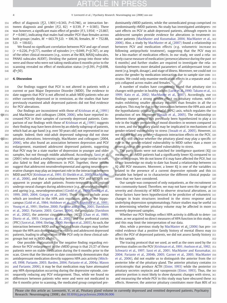
Copyright © 2022 FDOKUMEN