Natural killer cell function is intact after direct exposure to infectious hepatitis C virions

Sensors 2014, 14, 14488-14499; doi:10.3390/s140814488
sensors ISSN 1424-8220
www.mdpi.com/journal/sensors
Article
Genotyping Single Nucleotide Polymorphisms Using Different Molecular Beacon Multiplexed within a Suspended Core Optical Fiber
Linh Viet Nguyen 1,*, Sara Giannetti 2, Stephen Warren-Smith 1, Alan Cooper 3, Stefano Selleri 2,
Annamaria Cucinotta 2 and Tanya Monro 1
1 Institute for Photonics and Advanced Sensing (IPAS) and The ARC Centre for
Nanoscale Biophotonics, The University of Adelaide, Adelaide 5005, Australia;
E-Mails: [email protected] (S.W.-S.); [email protected] (T.M.) 2 Information Engineering Department, University of Parma, Parma 43100, Italy;
E-Mails: [email protected] (S.G.); [email protected] (S.S.);
[email protected] (A.C.) 3 Australian Centre for Ancient DNA (ACAD), The University of Adelaide, Adelaide 5005,
Australia; E-Mail: [email protected]
* Author to whom correspondence should be addressed; E-Mail: [email protected];
Tel.: +61-8-8313-2329; Fax: +61-8-8313-4380.
Received: 25 June 2014; in revised form: 1 August 2014 / Accepted: 5 August 2014 /
Published: 8 August 2014
Abstract: We report a novel approach to genotyping single nucleotide polymorphisms
(SNPs) using molecular beacons in conjunction with a suspended core optical fiber (SCF).
Target DNA sequences corresponding to the wild- or mutant-type have been accurately
recognized by immobilizing two different molecular beacons on the core of a SCF.
The two molecular beacons differ by one base in the loop-probe and utilize different
fluorescent indicators. Single-color fluorescence enhancement was obtained when the
immobilized SCFs were filled with a solution containing either wild-type or mutant-type
sequence (homozygous sample), while filling the immobilized SCF with solution
containing both wild- and mutant-type sequences resulted in dual-color fluorescence
enhancement, indicating a heterozygous sample. The genotyping was realized
amplification-free and with ultra low-volume for the required DNA solution (nano-liter).
This is, to our knowledge, the first genotyping device based on the combination of optical
fiber and molecular beacons.
OPEN ACCESS

Sensors 2014, 14 14489
Keywords: genotyping; suspended core optical fiber; molecular beacon; multiplexing;
DNA detection
1. Introduction
Single-nucleotide substitutions represent the largest source of diversity in the human genome. Although
the vast majority are neutral, these variations have also been directly linked to human disease [1].
Even neutral variations are important because they provide markers for the preparation of detailed
maps of the human genome, serving as essential elements in linkage analyses that identify genes
responsible for complex disorders [2]. Although sequencing is adequate for the initial discovery of
single-nucleotide variations, simpler, faster and more automated genotyping methods are needed to
understand the distribution of genetic variations in populations, as well as for identifying the genes
responsible for genetic disorders [1]. There have been various protocols and methods proposed for
genotyping single-nucleotide polymorphisms (SNPs). For example, methods that utilize gel
electrophoresis to identify single or double-stranded DNA polymorphisms [3,4]). SNPs can also be
determined using solid-phase chemical cleavage [5] in which commercially available chemicals are
used to modify cytosine and thymine, respectively. The modification of the mismatch is then followed
by cleavage with piperidine and the resulting DNA fragments are analyzed by denaturing
polyacrylamide gel-electrophoresis to identify the mismatch sites [1]. SNPs genotyping can as well be
carried out through DNA sequencing, in liquid phase [6] or solid-phase [7,8] such as using
oligonucleotide microarrays [8]. In addition 5'-nuclease reaction [9], or mass spectroscopy [10] have
all been utilized for SNPs genotyping. A comprehensive list of typical SNP genotyping methods and
their details can be found in Reference [1]. Despite their sophistication, typically the SNP genotyping
techniques mentioned above requires either polymerase chain reaction (PCR) or electrophoresis or both.
Molecular beacons (MBs) are single-stranded oligonucleotide hybridization probes that form a
stem-and-loop structure [11]. The loop contains a probe sequence that is complementary to a target
sequence, and the stem is formed by the annealing of complementary arm sequences located on either
side of the probe sequence. A fluorophore is covalently linked to the end of one arm and a quencher is
covalently linked to the end of the other arm. In the absence of target DNA, the probe is dark because the
stem places the fluorophore so close to the quencher that they transiently share electrons and the
fluorescence is efficiently quenched. When the probe encounters a target molecule it forms a probe-target
hybrid, which is longer and more stable than the stem hybrid. Consequently, the molecular beacon
undergoes a spontaneous conformational reorganization that forces the stem hybrid to dissociate and
the fluorophore and the quencher to move away from each other, restoring fluorescence [11]. MBs are
well known to be highly specific and capable of real-time monitoring of DNA amplification during a
polymerase chain reaction [11] and are thus widely used as a probe for DNA detection in various
applications [12], including SNP genotyping [1]. With its capability to recognize the target sequence in
a pool of many different sequences and thus eliminating the process of sorting out DNA segments by
electrophoresis, attaching MBs on an ultra sensitive transduction platform in which a very low
concentration of target sequence can be detected will lead to a much simpler SNP genotyping device.
Sensors 2014, 14 14490
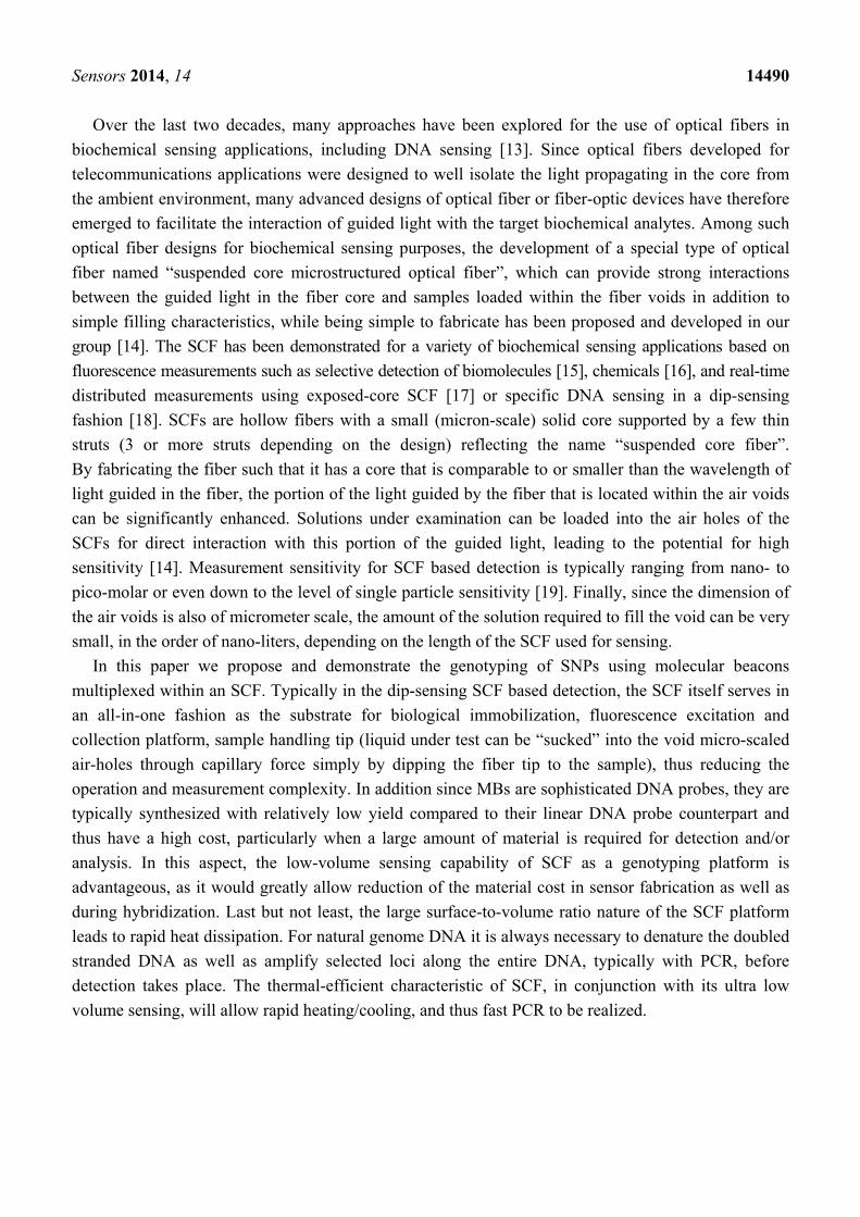
Over the last two decades, many approaches have been explored for the use of optical fibers in
biochemical sensing applications, including DNA sensing [13]. Since optical fibers developed for
telecommunications applications were designed to well isolate the light propagating in the core from
the ambient environment, many advanced designs of optical fiber or fiber-optic devices have therefore
emerged to facilitate the interaction of guided light with the target biochemical analytes. Among such
optical fiber designs for biochemical sensing purposes, the development of a special type of optical
fiber named “suspended core microstructured optical fiber”, which can provide strong interactions
between the guided light in the fiber core and samples loaded within the fiber voids in addition to
simple filling characteristics, while being simple to fabricate has been proposed and developed in our
group [14]. The SCF has been demonstrated for a variety of biochemical sensing applications based on
fluorescence measurements such as selective detection of biomolecules [15], chemicals [16], and real-time
distributed measurements using exposed-core SCF [17] or specific DNA sensing in a dip-sensing
fashion [18]. SCFs are hollow fibers with a small (micron-scale) solid core supported by a few thin
struts (3 or more struts depending on the design) reflecting the name “suspended core fiber”.
By fabricating the fiber such that it has a core that is comparable to or smaller than the wavelength of
light guided in the fiber, the portion of the light guided by the fiber that is located within the air voids
can be significantly enhanced. Solutions under examination can be loaded into the air holes of the
SCFs for direct interaction with this portion of the guided light, leading to the potential for high
sensitivity [14]. Measurement sensitivity for SCF based detection is typically ranging from nano- to
pico-molar or even down to the level of single particle sensitivity [19]. Finally, since the dimension of
the air voids is also of micrometer scale, the amount of the solution required to fill the void can be very
small, in the order of nano-liters, depending on the length of the SCF used for sensing.
In this paper we propose and demonstrate the genotyping of SNPs using molecular beacons
multiplexed within an SCF. Typically in the dip-sensing SCF based detection, the SCF itself serves in
an all-in-one fashion as the substrate for biological immobilization, fluorescence excitation and
collection platform, sample handling tip (liquid under test can be “sucked” into the void micro-scaled
air-holes through capillary force simply by dipping the fiber tip to the sample), thus reducing the
operation and measurement complexity. In addition since MBs are sophisticated DNA probes, they are
typically synthesized with relatively low yield compared to their linear DNA probe counterpart and
thus have a high cost, particularly when a large amount of material is required for detection and/or
analysis. In this aspect, the low-volume sensing capability of SCF as a genotyping platform is
advantageous, as it would greatly allow reduction of the material cost in sensor fabrication as well as
during hybridization. Last but not least, the large surface-to-volume ratio nature of the SCF platform
leads to rapid heat dissipation. For natural genome DNA it is always necessary to denature the doubled
stranded DNA as well as amplify selected loci along the entire DNA, typically with PCR, before
detection takes place. The thermal-efficient characteristic of SCF, in conjunction with its ultra low
volume sensing, will allow rapid heating/cooling, and thus fast PCR to be realized.

Sensors 2014, 14 14491
2. Experimental Section
2.1. Immobilization of Dual-Type MBs on the Surface of the SCF Core
The immobilization of two types of MBs on the surface of the SCF core was carried out following
the procedure detailed in our previous publication [18] using a combination of the fuzzy
nanoassembly technique [20] and the biotin-streptavidin binding mechanism [21]. An in-house made
pump system was used to flow the required solutions through the SCF air-holes for deposition of
different immobilization layers. Typically one end of SCF was immersed in solution vial placed in a
sealed chamber and the other end was left free outside of the chamber. A pressure difference
between the inside and outside of the sealed chamber was created by a pump connecting to the
chamber, forcing the liquid to move from inside to outside through the SCF air-holes. Since the SCF
surface is partly negatively changed by OH− groups on the surface, positively charged
poly(allylamine) hydrochloride (PAH, 2 mg/mL in 1 M NaCl solution, Sigma Aldrich) and
negatively charged poly(sodium 4-styrene sulfonate) (PSS, 2 mg/mL in 1 M NaCl solution, Sigma
Aldrich) were deposited alternately onto the fiber core surface using the layer by layer deposition
technique described in [20], ending with a PAH layer (PAH/PSS/PAH). The PAH provides amino
groups for immobilization of biotinylated MB through a biotin-streptavidin-biotinylated MB
link [21]. The flow time for each polyeletrolyte layer was 20 min, followed by extensive rinsing
using deionized (DI) water. NHS-LC-Biotin (0.5 mg/mL, Thermo Fisher) was prepared freshly and
flowed through for 1 h, followed by extensive rinsing using phosphate buffer solution (PBS, Sigma
Aldrich) to remove unbound biotin on the surface. Non-specific blocking solution (Candor) was
flowed through the fiber for 2 h and rinsed by PBS. Streptavidin (0.5 mg/mL, Thermo Fisher) was
flowed though for 40 min at room temperature then left inside the air-holes overnight and then rinsed
thoroughly with PBS. In this case Streptavidin only binds to Biotin rather than non-specifically
deposition on the SCF surface thanks to the use of non-specific blocker. A biotinylated MB solution
containing a mixture of two different MBs (2.5 µM for each MB, Midland Certified Reagent
Company Inc.) was flowed through for another 1 h, rinsed with PBS and DI water and then dried
with Nitrogen for 15 min. The functionalized fiber was cut into several pieces of 65 mm length each
for measurement. Figure 1 shows the cross section of the SCF used in this work and a sketch of the
final state of the SCF core surface after immobilization. The SCF was a silica glass SCF made
in-house with a core diameter of approximately 13 µm and has four air-holes. We chose to use a
relatively large core SCF principally to ensure high optical coupling stability during the fluorescence
measurement. Despite the large core diameter, we found that the dual-type MB immobilized SCF
presented in this work can detect DNA solution whose concentration of as low as 100 nM. If one
desires to increase further the sensitivity of the sensor, a smaller core size should be used, however,
at the expense of coupling instability. In this case, advanced fiber processing techniques such as
well-optimized fusion splicing are required to ensure good optical coupling stability [22,23].

Sensors 2014, 14 14492
Figure 1. Schematic diagram of the final stage of the suspended core optical fiber (SCF)
core functionalized with dual-type molecular beacons. Picture of the SCF cross section
shown on the right side is a scanning electron microscope (SEM) image of the SCF used in
this work.
2.2. Measurement Setup
A schematic diagram of the measurement setup for fluorescence measurement of the dual-MB
immobilized SCF is shown in Figure 2. Two lasers operating at two excitation wavelengths corresponding
to HEX and Cy5 dyes have been used. Excitation lights from a 532 nm laser (Crystal Laser) and a 638 nm
laser (Toptica) were first combined by a RGB fiber beam combiner (Thorlabs), power-adjusted by a
variable attenuator (a pair of half-wave plate and polarizer), and then directed to the SCF using a beam
splitter and a 40× microscope objective. The same microscope objective and beam splitter also serves
as the collecting objective for the backscattered fluorescence from the fiber. Backscattered light was
directed through a system including mirrors, a filter (either 532 nm or 638 nm filter depending on what
MB signal is being interrogated) to block the residual pump light, and coupled to a large core
multimode fiber using a 20× microscope objective. The multimode fiber guides the backscattered light
to a spectrometer. A shutter that was synchronized with the spectrometer was placed after the output of
the beam combiner. The consistent mode coupling in each measurement was achieved by means of
maximizing the power transmitted through the SCF core. It should be noted that Cy5 dye is known to
have photoswitching characteristic [24], i.e., exposure of Cy5 under continuous wave laser light at
638 nm turns the dye into a dark state, which then can be switch back to a fluorescent state with 532 nm
exposure [25]. Therefore when maximizing light coupling to the SCF core, the green laser (532 nm)
was mainly used, followed by a weak red light (638 nm) beam to check the power coupling to the SCF
core for the red wavelength. The continuous red light exposure of the immobilized SCF was typically 3 s
to minimize the potential photoswitching effect on the Cy5. In the measurement of fluorescence
enhancement upon filling the immobilized SCF with DNA solution, the green laser was always used
first. In this way the green light was used both to excite the fluorescence from HEX dye as well to
reactivate the Cy5 dye to a fluorescent state, in case it might have been photoswitched to a dark state,
even by the weak red beam.

Sensors 2014, 14 14493
Figure 2. Experiment setup for fluorescence measurement of the dual-type molecular
beacons (MBs) immobilized SCF filled with DNA solutions. SMF and MMF are
abbreviations for single mode and multimode mode optical fiber, respectively. The green
laser operating at 532 nm was used to excite HEX dyes (for wild-type sequence) and the red
laser operating at 638 nm was used to excite Cy5 dyes (for mutant-type sequence).
2.3. Hybridization Experiment
The hybridization test between the dual-type MB immobilized SCF and the solution containing
either homozygous single stranded DNA (wild or mutant) or both types (presenting a heterozygous
mixture) were carried out in a manufacturer-recommended buffer solution containing 20 mM Tris-HCl,
5 mM MgCl2, and 50 mM KCl, at room temperature (25 °C). The background of each piece of fiber
under test was recorded first and all the fluorescence spectra were normalized with their own
backgrounds to extract the fluorescence enhancements. Not all of the fiber pieces showed a proper
fluorescence background; some of the pieces cut from the same functionalized SCF exhibited a very
weak background, indicating that the immobilization along a long fiber might not be ideally uniform.
Therefore those poorly functionalized fiber pieces were dismissed and only the pieces with clear and
similar fluorescence background at HEX and Cy5 wavelengths were used for measurement. The filling
time for all the fiber pieces was approximately 30 s. After filling, fluorescence from HEX dye
(associated with wild-type MB) was collected first, followed by changing the filter and switching the
laser beam to excite and collect the fluorescence from Cy5 (associated with mutant-type MB).
The MBs and DNA sequences used in this work are given in Table 1. For the purpose of demonstrating
the proof-of-concept, in this work the wild and mutant MBs and the corresponding DNA sequences
were arbitrarily designed by the manufacturer with a quoted discriminating capability, between the
target and the one-base mismatching sequence by a factor of 10, as measured in solution. However,
virtually any sequences of practical interest could be detected by immobilizing the correspondingly
designed MBs on the SFC core.

Sensors 2014, 14 14494
Table 1. Molecular beacons and DNA sequences used for testing the immobilized SCF.
Samples were synthesized by the Midland Certified Reagent Company Inc. Concentration
of DNA solution used in the hybridization experiment is 100 nM.
Molecular Beacon Wild-type MB: 5'-(HEX)AGCGGATGTTAAAGACCTATGCCGC(BHQ1-dT)(spacer 18)(3'-Biotin)-3' Mutant-type MB: 5'-(HEX)AGCGGATGTTAAAAACCTATGCCGC(BHQ1-dT)(spacer 18)(3'-Biotin)-3' DNA Sequences Wild-type sequence: 5'-CATAGGTCTTTAACAT-3' Mutant-type sequence: 5'-CATAGGTTTTTAACAT-3'
Figure 3. Comparison of fluorescence enhancement between dual-type MB immobilized
SCF and control SCF upon filling with target DNA solution. The control SCF shows
negligible fluorescence enhancement.
(a) (b)
3. Results and Discussion
3.1. Verification of the Immobilization Process with Control SCF
Another SCF that serves as the control fiber was put through the same immobilization process as
described in Section 2, except that the biotin-streptavidin linking step was omitted. The two fibers,
dual-type MB immobilized fiber and the control fiber, were loaded with solution containing either
wild-type or mutant-type sequence with a concentration of 100 nM and the fluorescence were
measured. In this work the functionalized was not optimized for any specific concentration as the
functionalization involved multiple coating layers which makes optimizing the probe surface
density difficult. In principle, detection in a biosensor is based on the binding between
functionalized probe (molecular beacon in our case) and analyte suspended in the solution under
test and therefore there should be an optimized pair of probe density on the sensor surface and

Sensors 2014, 14 14495
analyte concentration. As can be seen in Figure 3, it is clear that the two MBs are successfully
immobilized on the surface of the SCF core through the biotin-streptavidin link as the fluorescence
of the MB immobilized fiber increased significantly upon hybridizing with target DNA while that
of the control fiber remains approximately unchanged. This indicates that without the intended
biotin-streptavidin link, biotinylated MBs cannot form a stable chemical attachment to the fiber
surface and are removed when rinsed. The use of the non-specific blocking layer helps to ensure
that biotinylated MB binds only to the surface through the biotin-streptavidin link and not directly
to the surface due to physical adsorption, which would be too close to the surface and thus might be
associated with high steric hindrance.
3.2. Genotyping SNP with Dual-Type MB Functionalized SCF
The results of the hybridization test, in which the dual-MB immobilized SCFs were loaded with
different DNA solution containing either one type of DNA or both and fluorescence enhancements
were recorded, is shown in Figure 4. When the fiber was filled with a buffer solution containing one
type of DNA, either wild-type or mutant type (Figure 4a,b), essentially only one type of MB was
undergoing conformational change due to target sequence binding to the probe in the MB loop, as
evidenced by the enhancement of fluorescence at only one wavelength (Figure 4a is for fiber filled
with wild-type DNA solution and Figure 4c is for fiber filled with mutant-type DNA solution).
On the other hand, once the fiber is filled with DNA solution containing both the wild and mutant
sequence (Figure 4c), both MBs experienced conformational changes as the fluorescence
enhancement were obtained at both wavelengths. Of course, the level of enhancement should be
lower than the case of single DNA sequence since it is a competing reaction, consistent with the case
of genotyping SNPs using MBs in solution [1]. Therefore, the proposed dual-type MB immobilized
SCF clearly functions as a SNP genotyping platform. Figure 4d shows the integrated intensity for the
HEX (left) and Cy5 (right) fluorescence. Here sample is considered heterozygous if the two
integrated intensities of the two dyes are close to each other while well-separated values of the
fluorescence indicate homozygosity. It should be noted that the performance of the MBs
immobilized on the surface of the SCF was somewhat deteriorated compared with the case of
in-solution measurement. This is partly due to the steric hindrance of the surface on the MB as well
as MBs to each other. In general it is not possible to obtain similar performance for in-solution (same
phase) reaction and immobilized/liquid (different phase) reaction however it might be improved by
optimizing the buffer condition as well as fine-tuning the design of the MBs.
It should be noted that all hybridization and measurement procedures were performed at room
temperature. For the purpose of simplifying this proof-of-concept experiment as well as limit of
equipment temperature control was not included in this work. While it is important to have strict
temperature control of the functionalized SCF for highly specific allele discrimination in the case of
real-world genome DNA samples (in such case the buffer should contain many different DNA
sequences extracted from the genome DNA), it is known that MB can still achieve good discrimination
between sequences of only one base difference [11], even at room temperature. With proper
temperature control and performing the allele discrimination at optimized temperature for a specific
pair of MB and target, as well as optimizing buffer and MB design, the discrimination should be

Sensors 2014, 14 14496
greatly enhanced. In addition, for genome DNA temperature control is often essential, not only for
enhancing the allele discrimination but also for performing other tasks such as denaturing the double
stranded genome DNA as well as amplifying selected loci on the entire sequence before detection.
Finally, with temperature control on the functionalized SCF, the sensor can in principle be regenerated
by denaturing the DNA-probe binding using heat and flowing fresh buffer through the air-holes.
DNA-probe binding in MB is well known to be reversible, as evidence by its usage in real-time PCR [26],
and therefore as long as the DNA is removed MB should resume its original stem-loop and regenerate
the sensor.
Figure 4. Hybridization test of the dual-type MB immobilized SCF filled with solution
containing either one type of DNA sequence, e.g., wild or mutant sequence (homozygous) or
both type (heterozygous). Fluorescence enhancement clearly indicates (a,b) the homozygous
type or (c) heterozygous type; (d) The averaged value over four measurements of spectra as
shown in (a–c), crossing point of HEX and Cy5 fluorescence indicate heterozygousity and
well separated values of HEX and Cy5 fluorescence represent homozygousity.
CATAGGTCTTTAACAT
CATAGGTCTTTAACAT
CATAGGTTTTTAACAT
CATAGGTTTTTAACAT
(a) (b)
CATAGGTCTTTAACAT
CATAGGTTTTTAACAT
(c) (d)

Sensors 2014, 14 14497
4. Conclusions/Outlook
We have presented the first demonstration of the use of SCF in conjunction with MBs for
genotyping SNPs. The proposed device is based on the functionalization of multiple MBs on the core of
an SCF and is capable of genotyping SNPs in a DNA solution whose concentration is as low as 100 nM.
The sensing volume is in the nano-liter range and the genotyping protocol can be as simple as dipping
the free end of the functionalized SCF into the solution under test, leading to potentially highly
accurate and cost-effective genotyping SNPs protocol. Further work is expected to increase the
sensitivity of the device by reducing the SCF core size, optimizing the coupling as well as the MB
designs and buffer conditions. By further driving the sensitivity of the sensor down to the level of a few,
or even a single, DNA template, the proposed genotyping scheme should be able to perform the
genotyping of SNPs in a simpler manner, without the need for PCR or electrophoresis.
Acknowledgments
This work is supported by the Australian Research Council (ARC) Super Science Fellowships for
Linh Viet Nguyen and Stephen Warren-Smith and the ARC Centre of Excellence in
Nanoscale BioPhotonics. Sara Giannetti acknowledges the support of a Spinner 2013—International
Mobility exchange fellowship. Tanya Monro acknowledges the support of an ARC Georgina Sweet
Laureate Fellowship and Alan Cooper acknowledges the support of an ARC Future Fellowship.
Authors thank Roman Koteski, Erik Schartner, Peter Henry and Alastair Dowler for help with the
silica fiber fabrication.
Author Contributions
LVN proposed the work, designed and supervised the experiment. LVN and SG performed the
experiment. All authors wrote and commented on the paper. TM supervised the entire project.
Conflicts of Interest
The authors declare no conflict of interest.
References
1. Kwok, P.-Y., Ed. Single Nucleotide Polymorphisms: Methods and Protocols. In Methods in
Molecular Biology; Volume 212; Humana Press: Totowa, NJ, USA, 2003.
2. Sachidanandam, R.; Weissman, D.; Schmidt, S.C.; Kakol, J.M.; Stein, L.D.; Marth, G.; Sherry, S.;
Mullikin, J.C.; Mortimore, B.J.; Willey, D.L.; et al. A map of human genome sequence variation
containing 1.42 million single nucleotide polymorphisms. Nature 2001, 409, 928–933.
3. Orita, M.; Suzuki, Y.; Sekiya, T.; Hayashi, K. Rapid and sensitive detection of point mutations
and DNA polymorphisms using the polymerase chain reaction. Genomics 1989, 5, 874–879.

Sensors 2014, 14 14498
4. Ganguly, A.; Rock, M.J.; Prockop, D.J. Conformation- sensitive gel electrophoresis for rapid
detection of single-base differences in double-stranded PCR products and DNA fragments:
Evidence for solvent-induced bends in DNA heteroduplexes. Proc. Natl. Acad. Sci. USA 1993, 90,
10325–10329.
5. Ellis, T.P.; Humphrey, K.E.; Smith, M.J.; Cotton, R.G.H. Chemical cleavage of mismatch: A new
look at an established method/recent developments. Hum. Mutat. 1998, 11, 345–353.
6. Taillon-Miller, P.; Kwok, P.-Y. Efficient approach to unique single nucleotide polymorphism
discovery. Genome Res. 1999, 9, 499–505.
7. Hacia, J.G. Resequencing and mutational analysis using oli- gonucleotide microarrays. Nat. Genet.
1999, 21, 42–47.
8. Syvanen, A.-C.; Sajantila, A.; Lukka, M. Identification of individuals by analysis of biallelic
DNA markers, using PCR and solid-phase minisequencing. Am. J. Hum. Genet. 1993, 52, 46–59.
9. Holland, P.M.; Abramson, R.D.; Watson, R.; Gelfand, D.H. Detection of specific polymerase
chain reaction product by utilizing the 5' in place of 3' exonuclease activity of Thermus aquaticus
DNA polymerase. Proc. Natl. Acad. Sci. USA 1991, 88, 7276–7280.
10. Karas, M.; Glueckmann, M.; Schaefer, J. Ionization in matrix-assisted laser desorption/ionization:
Singly charged molecular ions are the lucky survivors. J. Mass Spectrom. 2000, 35, 1–12.
11. Tyagi, S.; Kramer, F.R. Molecular Beacons: Probes that Fluoresce upon Hybridization.
Nat. Biotechnol. 1996, 14, 303–308.
12. Molecular Beacons. Available online: http://www.molecular-beacons.org/ (accessed on 12
January 2014).
13. Wolfbeis, O.S. Fiber-Optic Chemical Sensors and Biosensors. Anal. Chem. 2006, 78, 3859–3874.
14. Monro, T.M.; Warren-Smith, S.C.; Schartner, E.P.; François, A.; Heng, S.; Ebendorff-Heidepriem, H.;
Afshar, V.S. Sensing with suspended-core optical fibers. Opt. Fiber Technol. 2010, 16, 343–356.
15. Ruan, Y.; Foo, T.C.; Warren-Smith, S.C.; Hoffmann, P.; Moore, R.C.; Ebendorff-Heidepriem, H.;
Monro, T.M. Antibody immobilization within glass microstructured fibers: A route to sensitive
and selective biosensors. Opt. Express 2008, 16, 18514–18523.
16. Warren-Smith, S.C.; Heng, S.; Ebendorff-Heidepriem, H.; Abell, A.D.; Monro, T.M.
Fluorescence-based aluminum ion sensing using a surface functionalized microstructured optical
fiber. Langmuir 2011, 27, 5680–5685.
17. Warren-Smith, S.C.; Sinchenko, E.; Stoddart, P.R.; Monro, T.M. Distributed fluorescence
sensing using exposed-core microstructured optical fiber. IEEE Photonics Technol. Lett. 2010, 22,
1385–1387.
18. Nguyen, L.-V.; Warren-Smith, S.C.; Cooper, A.; Monro, T.M. Molecular beacons immobilized within
suspended core optical fiber for specific DNA detection. Opt. Express 2012, 20, 29378–29385.
19. Zhao, J.; Jin, D.; Schartner, E.P.; Lu, Y.; Liu, Y.; Zvyagin, A.V.; Zhang, L.; Dawes, J.M.; Xi, P.;
Piper, J.A.; et al. Single-nanocrystal sensitivity achieved by enhanced upconversion luminescence.
Nat. Nanotechnol. 2013, 8, 729–734.
20. Decher, G. Fuzzy Nanoassemblies: Toward layered polymeric multicomposites. Science 1997,
277, 1232–1237.
21. Liu, X.; Tan, W. A Fiber-Optic Evanescent Wave DNA Biosensor Based on Novel Molecular
Beacons. Anal. Chem. 1999, 71, 5054–5059.

Sensors 2014, 14 14499
22. Zaroszewics, L.R.; Murawski, M.; Nasilowski, T.; Stasiewicz, K.; Marc, P.; Szymanski, M.;
Mergo, P. Methodology of splicing large air filling factor suspended core photonic crystal fibers.
Opto-Electron. Rev. 2011, 19, 256–259.
23. Warren-Smith, S.C.; Monro, T.M. Exposed core microstructured optical fiber Bragg gratings:
Refractive index sensing. Opt. Express 2014, 22, 1480–1489.
24. Dempsey, G.; Bates, M.; Kowtoniuk, W.; Liu, D.; Tsien, R.; Zhuang, X. Photoswitching
mechanism of Cyanine Dyes. J. Am. Chem. Soc. 2009, 131, 18192–18193.
25. Bates, M.; Blosser, T.R.; Zhuang, X. Short-range spectroscopic ruler based on a single-molecule
optical switch. Phys. Rev. Lett. 2005, 94, 108101, doi:10.1103/PhysRevLett.94.108101.
26. Marras, S.A.E.; Tyagi, S.; Kramer, S.R. Real-time assays with molecular beacons and other
fluorescent nuclei acid hybridization probes. Clin. Chim. Acta 2006, 363, 48–60.
© 2014 by the authors; licensee MDPI, Basel, Switzerland. This article is an open access article
distributed under the terms and conditions of the Creative Commons Attribution license
(http://creativecommons.org/licenses/by/3.0/).
Copyright © 2022 FDOKUMEN