Characterization of peptides by capillary zone electrophoresis and electrospray ionization...
11
Original Paper Bindila, Zamfir, Peter-Katalinic ´ 1101 Laura Bindila, Alina Zamfir, Jasna Peter-Katalinic ´ Institute for Medical Physics and Biophysics, University of Mɒnster, Robert Koch Str. 31, D-48149 Mɒnster, Germany Characterization of peptides by capillary zone electrophoresis and electrospray ionization quadrupole time-of-flight tandem mass spectrometry High performance capillary electrophoresis (HPCE) in an off-line combination with electrospray ionization quadrupole time-of-flight (ESI-QTOF) mass spectrometry (MS) has been developed for separation, identification, and compositional analysis of peptides. Two mixtures of peptides from an extracellular matrix have been separated and profiled by CE with UV detection. Introduction of ammonium formate in aqueous/ methanol solution at low pH values provided suitable buffer conditions for a good separation of components and reproducibility of experiments. The analytes were identified by subjecting the CE-collected fractions to (+)nanoESI-QTOF-MS and tan- dem MS with low energy collision induced dissociation (CID). CE/MS analysis of the mixture generated by peptide hydrolysis has been designed to allow both determina- tion of the peptide amino acid composition and ladder sequencing. The strategy pre- sented here could represent a useful preliminary method to serve as a basis for on- line experiments and for further analysis of proteins and their full structural elucida- tion. Key Words: Capillary electrophoresis; Nanoelectrospray QTOF mass spectrometry; Peptides; Amino acids; Received: July 1, 2002; revised: September 18, 2002; accepted: September 18, 2002 1 Introduction In the post genome era a great deal of effort is being invested in the characterization of proteins and their com- positional analysis, which are essential for an understand- ing of their biological function and mechanisms of interac- tion [1, 2]. Global proteome characterization is based on analysis of peptide mixtures released from protein digests and their amino acid sequence. Therefore, the separation and identification of peptides and their amino acid compo- sition represent a crucial step in elucidation of protein structure. An important class of proteins and related peptides is located in the extracellular matrix. Proteins covalently linked to the glycosaminoglycans, forming proteoglycans, are general constituents of the connective tissues. The biological significance of these substances is currently driving enormous efforts directed to the development of specific and sensitive methods for their structural elucida- tion. The most common contemporary approach to identifica- tion and separation of peptides released by protein diges- tion is two-dimensional gel electrophoresis (2-D PAGE) in combination with mass spectrometry [3 – 5]. After enzy- matic digestion of a protein from a 2D-gel spot, the com- ponents in the generated peptide mixture can be identified by matrix assisted laser/desorption ionization (MALDI- MS) or electrospray (ESI-MS) mass spectrometry accord- ing to their molecular ion values, which are submitted to a database search for peptide identification according to their mass. MALDI-MS as a rapid method for identification of peptides is used predominantly for molecular weight determination of components larger than 500 Da [6 – 10]. Another widespread method for separation and identifica- tion of peptides is high performance liquid chromatogra- phy (HPLC) in combination with mass spectrometry. This Correspondence: Jasna Peter-Katalinic ´, Institute for Medi- cal Physics and Biophysics, University of Mɒnster, Robert Koch Str. 31, D-48149 Mɒnster, Germany. E-mail: [email protected] Fax: +49 251 83 55140 Abbreviations: A, alanine; BGE, background electrolyte; CE, capillary electrophoresis; CID collision-induced dissocia- tions; CIEF, capillary isoelectric focusing; ESI, electrospray ionization; FAPP, fibronectin adhesion promoting peptide; FBIP, fibrinogen binding inhibitor peptide; FF, fibronectin fragment; FTIII, fibronectin type III connecting segment frag- ment; LE, leucine enkephaline; LF, laminin fragment; I, iso- leucine; MALDI, matrix assisted laser/desorption ionization; MS, mass spectrometry/spectrometer; MS/MS, tandem mass spectrometry; MW, molecular weight; P, proline; Q, glutamine; QTOF-MS, quadrupole time-of-flight mass spec- trometry/spectrometer; R, arginine; W, tryptophan. J. Sep. Sci. 2002, 25, 1101–1111 i 2002 WILEY-VCH Verlag GmbH & Co. KGaA, Weinheim 1615-9306/2002/1511–1101$17.50+.50/0
-
Upload
independent -
Category
Documents
-
view
1 -
download
0
Transcript of Characterization of peptides by capillary zone electrophoresis and electrospray ionization...
Original
Pap
er
Bindila, Zamfir, Peter-Katalinic 1101
Laura Bindila,Alina Zamfir,Jasna Peter-Katalinic
Institute for Medical Physicsand Biophysics, University ofM�nster, Robert Koch Str. 31,D-48149 M�nster, Germany
Characterization of peptides by capillary zoneelectrophoresis and electrospray ionizationquadrupole time-of-flight tandemmassspectrometry
High performance capillary electrophoresis (HPCE) in an off-line combination withelectrospray ionization quadrupole time-of-flight (ESI-QTOF) mass spectrometry(MS) has been developed for separation, identification, and compositional analysis ofpeptides. Two mixtures of peptides from an extracellular matrix have been separatedand profiled by CE with UV detection. Introduction of ammonium formate in aqueous/methanol solution at low pH values provided suitable buffer conditions for a goodseparation of components and reproducibility of experiments. The analytes wereidentified by subjecting the CE-collected fractions to (+)nanoESI-QTOF-MS and tan-dem MS with low energy collision induced dissociation (CID). CE/MS analysis of themixture generated by peptide hydrolysis has been designed to allow both determina-tion of the peptide amino acid composition and ladder sequencing. The strategy pre-sented here could represent a useful preliminary method to serve as a basis for on-line experiments and for further analysis of proteins and their full structural elucida-tion.
Key Words: Capillary electrophoresis; Nanoelectrospray QTOF mass spectrometry;Peptides; Amino acids;
Received: July 1, 2002; revised: September 18, 2002; accepted: September 18, 2002
1 Introduction
In the post genome era a great deal of effort is beinginvested in the characterization of proteins and their com-positional analysis, which are essential for an understand-ing of their biological function and mechanisms of interac-tion [1, 2]. Global proteome characterization is based onanalysis of peptide mixtures released from protein digestsand their amino acid sequence. Therefore, the separationand identification of peptides and their amino acid compo-
sition represent a crucial step in elucidation of proteinstructure.
An important class of proteins and related peptides islocated in the extracellular matrix. Proteins covalentlylinked to the glycosaminoglycans, forming proteoglycans,are general constituents of the connective tissues. Thebiological significance of these substances is currentlydriving enormous efforts directed to the development ofspecific and sensitive methods for their structural elucida-tion.
The most common contemporary approach to identifica-tion and separation of peptides released by protein diges-tion is two-dimensional gel electrophoresis (2-D PAGE) incombination with mass spectrometry [3–5]. After enzy-matic digestion of a protein from a 2D-gel spot, the com-ponents in the generated peptide mixture can be identifiedby matrix assisted laser/desorption ionization (MALDI-MS) or electrospray (ESI-MS) mass spectrometry accord-ing to their molecular ion values, which are submitted to adatabase search for peptide identification according totheir mass. MALDI-MS as a rapid method for identificationof peptides is used predominantly for molecular weightdetermination of components larger than 500 Da [6–10].Another widespread method for separation and identifica-tion of peptides is high performance liquid chromatogra-phy (HPLC) in combination with mass spectrometry. This
Correspondence: Jasna Peter-Katalinic, Institute for Medi-cal Physics and Biophysics, University of M�nster, RobertKoch Str. 31, D-48149 M�nster, Germany.E-mail: [email protected]: +49 251 83 55140Abbreviations: A, alanine; BGE, background electrolyte;CE, capillary electrophoresis; CID collision-induced dissocia-tions; CIEF, capillary isoelectric focusing; ESI, electrosprayionization; FAPP, fibronectin adhesion promoting peptide;FBIP, fibrinogen binding inhibitor peptide; FF, fibronectinfragment; FTIII, fibronectin type III connecting segment frag-ment; LE, leucine enkephaline; LF, laminin fragment; I, iso-leucine; MALDI, matrix assisted laser/desorption ionization;MS, mass spectrometry/spectrometer; MS/MS, tandemmass spectrometry; MW, molecular weight; P, proline; Q,glutamine; QTOF-MS, quadrupole time-of-flight mass spec-trometry/spectrometer; R, arginine; W, tryptophan.
J. Sep. Sci. 2002, 25, 1101–1111
i 2002WILEY-VCH Verlag GmbH&Co. KGaA,Weinheim 1615-9306/2002/1511–1101$17.50+.50/0
1102 Bindila, Zamfir, Peter-Katalinic J. Sep. Sci. 2002, 25, 1101–1111
method is advantageous for analysis of smaller moleculesrequiring minute sample quantity, buffer volume, andrather simple instrumentation. This popular approach per-mits high throughput analysis; however, the efficiency ofthe separation is frequently the limiting factor. Therefore,there is a continuous demand for alternative strategies forstructural investigation of complex peptide mixtures toenhance detection and identification of a large number ofcomponents in complex mixtures at high sensitivity andresolution [11, 13].
We have recently demonstrated [14] that CE is a powerfulanalytical tool for analysis of biopolymers, characterizedby high sensitivity and high resolving power, efficiency ofseparation, and different type of applications in the field ofstructural analysis of biopolymers.
A novel strategy for high-efficiency capillary isoelectricfocusing (CIEF) separation of peptides, by which a simul-taneous sample concentration and separation can beachieved, has been recently proposed by Shen et al. [15].They demonstrated sensitivity better than 1 ng/nL inCIEF/UV detection. Waterval et al. [16] reported a methodfor identification of therapeutically active peptides usingcapillary electrophoresis with UV detection in combinationwith mass spectrometry, after on-capillary preconcentra-tion based on a cation exchange mechanism. The methoddemonstrated a fair sensitivity and selectivity, but anextensive optimization of the sample pretreatment proce-dures is required.
Due to their excellent performance, hyphenated CE/MSmethods have been used for characterization of pep-tides [17–20]. Since the introduction of CE into proteomeanalysis, different type of buffers for CE/ESI-MS havebeen implemented and optimized. The best electropho-retic separation was so far achieved by using borate andphosphate buffers, which are, however, not compatiblewith the desolvation requirements of ESI desorption forMS analysis. Therefore, efforts were focused on thesearch for volatile electrolytes showing, on the one hand,no significant background in mass spectra, and providingsufficient separation and resolution, on the other. Ammo-nium acetate and acetic acid solutions are currently themost suitable and widely used electrolytes in CE/MS ofpeptides and proteins. However, further efforts wererequired for analytical studies in this field toward optimiza-tion of novel options for CE/ESI-MS-compatible buffersystems and implementation of improved strategies inproteome analysis. On assessing the volatile electrolytesfor capillary electrophoresis-electrospray ionization massspectrometry of proteins, Huber et al. [21] found that byusing 100–125 mM formic acid/ammonia, pH 3.10, fullscan spectra could be acquired with high sensitivity in on-line CE/ESI-MS experiments. However, compared to UVdetection, considerable band broadening was observed
using direct ESI-MS detection, which was mainly attribu-ted to the band spreading in the interface, column over-loading, and scanning data acquisition in the on-line mea-surements of proteins. In this context, as part of the workfocused on elaboration of novel CE/MS methodolo-gies [22, 23], our study presents another practical CE/MSapproach for structural analysis of components in complexpeptide mixtures and determining their amino acidsequence. The current methodology has been applied toextracellular matrix peptides but might represent a generaltool in structural elucidation of proteins.
2 Materials andmethods
2.1 Reagents andmaterials
Formic acid (98–100%), aq. ammonia solution (32%),37%HCl, andmethanol were obtained fromMerck (Darm-stadt, Germany). Distilled and deionized water from aMilli-Q water system Millipore (Bedford, MA, USA) hasbeen used for preparation of peptide solutions and CE buf-fers.
Aqueous solutions of peptides were dried in a Speed VacSPD 111V system from Savant (D�sseldorf, Germany).The pH value of the CE buffer was adjusted by a 766 pHmeter from Calimatic (Knick, Germany). Prior to use, allsolvents were filtered through 0.2 lmmembrane of dispo-sable filter units (Schleicher-Schuell, Dassel, Germany).Externally polyimide coated fused silica CE capillary tub-ing (BGB Analytik Vertrieb, Essen, Germany) was used inall CE experiments. Omega glass capillaries used in thenanoESI experiments (Hillenberg, Germany) were pulledby a vertical pipette puller model 720, David Kopf Instru-ments (Tujunga, CA, USA).
2.2 Samples
Six standard amino acids, W, Q, P, R, A, and I, wereobtained from Sigma (Deisenhofen, Germany) and pre-pared in a concentration of 2.0 mg/mL in CE buffer for CE/UV profiling. In all experiments, the CE buffer had thesame concentration and pH as the background electrolyte(BGE). For fraction collection, all amino acids were dis-solved in the buffer at a concentration of 10 mg/mL for I, A,P, R, and 8 mg/mL for W and Q. Commercially availableextracellular matrix peptides FAPP (MW 1023.00 Da),FBIP (MW 1189.00 Da), LF (MW 967.1 Da), FTIII (MW2732.00 Da), FF (MW 1001.00 Da), and LE (MW555.6 Da) were purchased from Sigma (Deisenhofen,Germany). The peptides were prepared in a concentrationof 2.0 mg/mL buffer for UV profiling and 20 mg/mL bufferfor fraction collection.
J. Sep. Sci. 2002, 25, 1101–1111 Characterization of peptides by capillary zone electrophoresis 1103
2.3 Capillary electrophoresis with UV detection
CE experiments were carried out on a P/ACETM 5000 ser-ies instrument (Beckman-Coulter, Fullerton, CA, USA)equipped with an UV detector (deuterium lamp, 2 nmwavelength accuracy, 190–380 nm wavelength rangewith filter selection) and interfaced with an 486 IBM PS/2Model 56SX computer utilizing System Gold dedicatedsoftware package to control the instrument and collectexperimental data. The electrophoresis capillaries usedmeasured 50 lm ID6375 mm OD and had an overalllength of 100 cm. The detection window was located at7 cm from the outlet of the CE capillary. Before each CEexperiment, the capillary was conditioned by 25 min rin-sing with methanol, 15 min drying with air, and then by40 min flushing with the running buffer, 0.5 M ammoniumformate, pH 2.2, water/methanol (40/60; v/v) solution.Before each injection of the sample, the capillary wasrinsed for at least 15 min with the BGE. In both CE/UV andoff-line CE/MS experiments the sample was injected byapplying a constant nitrogen pressure of 3.45 kPa for 3 s.The temperature of the capillary cartridge was set at 158Cfor all experiments. UV absorption was recorded at200 nm. All experiments were conducted in a forwardpolarity.
2.4 Mass spectrometry
Mass spectrometrywas performed on an orthogonalhybrid quadrupole time-of-flight mass spectrometer,QTOFTM (Micromass, Manchester, UK) equipped with anESI-ion source in the Micromass Z-spray geometry. TheQTOF mass spectrometer is interfaced with a PC runningthe MassLynx N.T. software system to control the instru-ment, and acquire and process the MS data. In nanoESIexperiments, the voltage was applied to the solution via astainless steel wire inserted into the capillary.
Using the advantage of amino acid and peptide cationiza-tionat acidicpH, allmassspectrawereacquired in theposi-tive ion mode ESI-MS. The nanoESI parameters such ascapillary and sampling cone potential, source block tem-perature, desolvation and nebulizer gas pressure wereoptimized in order to obtain a stable spray,minimize the in-source fragmentation of the peptide, and enhance thedecomposition of the analyte-buffer clusters. The low sig-nal-to-noise ratio, caused by reduced concentration of theanalyte in the CE collected fractions, could be readily com-pensated by recording the ESI signal for longer than10 min.
Tandem mass spectrometry has been performed byemploying collision-induced dissociation (CID) at lowenergy with Ar as a collisional gas. The collision energyand the gas pressure were adjusted in order to obtain anoptimal degree of fragmentation of the precursor ion.
2.5 Off-line CE/ESI-QTOF-MSmethod
Off-line experiments were based on the fraction collectionprinciple as described before [22, 23]. All CE-collectedfractions were subjected to (+) nanoESI-QTOF-MS analy-sis.
3 Results and discussion
3.1 CE/UV and CE/ESI-QTOF-MS experiments onFAPP and LEmixture
An equimolar mixture of 2 mg/mL FAPP and LE was sub-mitted to the CE/UV experiment, in 0.5 M ammonium for-mate in aqueous methanol solution (40/60, v/v) at pH 2.2used as a CE buffer and background electrolyte. The lowpH value of 2.2 has been chosen in order to enhance theformation of the positively charged molecules and hencemigration towards the cathode without significant interac-tion with the capillary walls. At this pH value, separation ofthe peptidemixtures can be performed without coating thecapillary or derivatizing the substrate.
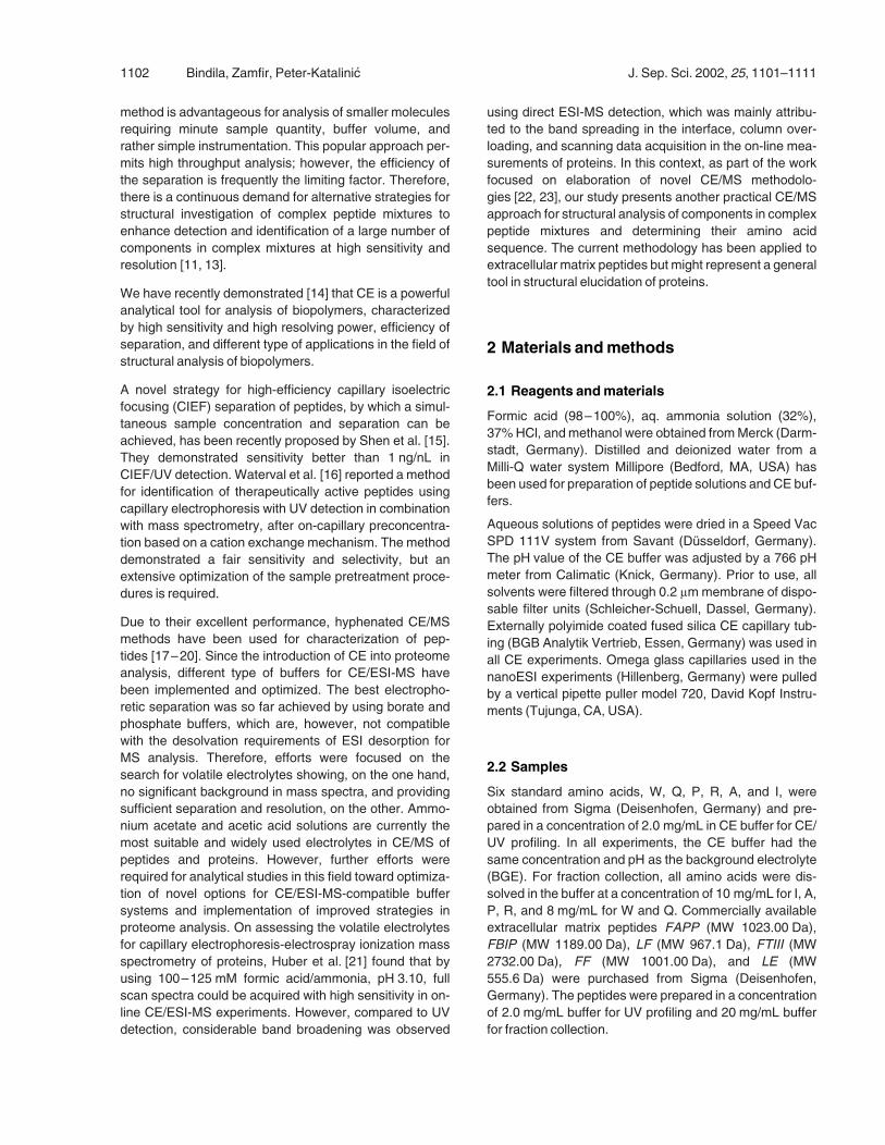
The sample was injected for the CE/UV experiment byapplying the operating pressure for 3.0 s. The CE/UVseparation was performed for 30 min under 30 kV, wherean electric current of about 15 lA was generated. The CE/UV profile of the binary mixture separation is presented inFigure 1; both components are seen to elute in less than10 min after the injection. High separation efficiency andresolution have been achieved using ammonium formateas a buffer as demonstrated by the difference of 3 min inmigrationtimevaluesforcomponentsofthisbinarymixture.
Based on the CE/UV profiles two CE fractions were col-lected, one at 6 and the other at 11 min after injection, in
Figure 1. CE/UV profile of the equimolar mixture of LE andFAPP. Fused silica CE capillary [50 lm ID6375 lm OD,100 cm length]; CE voltage 30 kV; BGE 0.5 M H2O/MeOHammonium formate; pH 2.2; substrate concentration was2 mg/mL buffer (in 10 lL); 3 s injection by pressure; detectionUV (200 nm).
1104 Bindila, Zamfir, Peter-Katalinic J. Sep. Sci. 2002, 25, 1101–1111
vials containing 20 lL of the CE carrier. The CE fractionswere subjected to ESI-MS analysis without any post-separation treatment of the sample. ESI-MS screening ofthe fractions was carried out under mild ESI ion sourcedesorption conditions such as a low ESI capillary potentialwithin the 800–1000 V range and a sampling cone poten-tial of 20–50 V. Such source parameters were shown tobe favorable for ionization of peptide components col-lected from the CE buffer. The desolvation temperatureset to 60–808C, as well as the use of nebulizer gas,enhanced the decomposition of the analyte-buffer clustersand the detection of free peptides as both singly and multi-ply chargedmolecular ions.
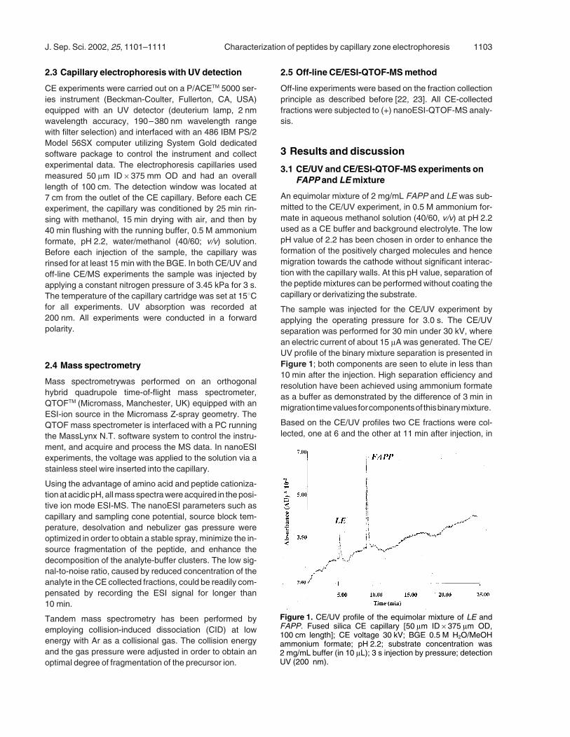
The nanoESI-QTOF mass spectrum of the first collectedfraction is depicted in Figure 2. A relatively abundant[M+H]+ ion at m/z 556.52, representing the molecular ionof LE, as well as its sodium adduct at m/z 578.53 weredetected. The ion at m/z 520.56 was generated by theneutral loss of two water molecules of the pseudomolecu-lar ion of LE. As previously described by us [22], CE col-
lection of fractions presents, de-facto, a drawback relatedto the lack of sensitivity, requiring rather high concentra-tion of the injected material. In this procedure, separationresolution is deliberately sacrificed in order to increase theconcentration of the collected component. The directeffect of this approach is that in the first CE fraction a smalltrace of the second peptide was observed as well, beingdetected in the ESI-MS analysis as a doubly charged ionat m/z 512.2. This could also be attributed to the fractioncollection procedure, in which interruption of both CEseparation voltage and pressure is necessary betweentwo consecutive collections.
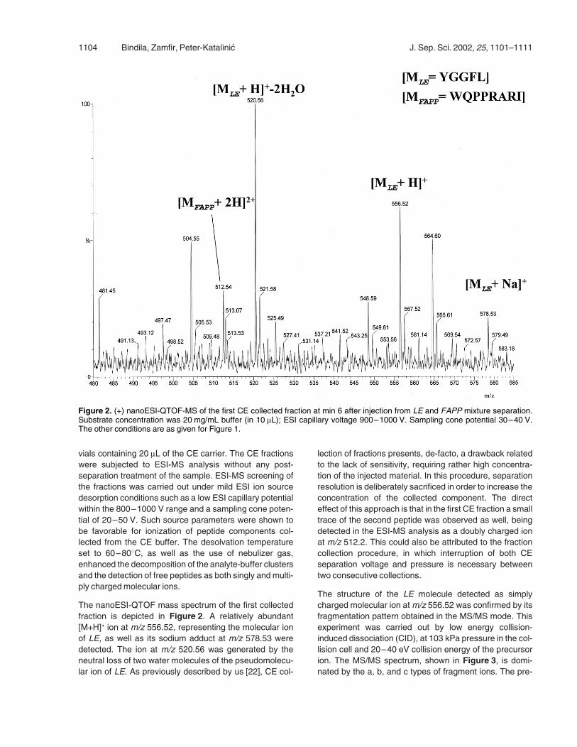
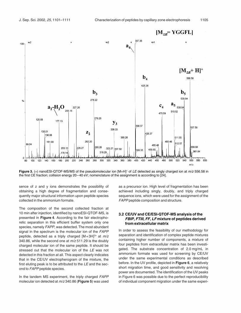
The structure of the LE molecule detected as simplycharged molecular ion atm/z 556.52 was confirmed by itsfragmentation pattern obtained in the MS/MS mode. Thisexperiment was carried out by low energy collision-induced dissociation (CID), at 103 kPa pressure in the col-lision cell and 20–40 eV collision energy of the precursorion. The MS/MS spectrum, shown in Figure 3, is domi-nated by the a, b, and c types of fragment ions. The pre-
Figure 2. (+) nanoESI-QTOF-MS of the first CE collected fraction at min 6 after injection from LE and FAPP mixture separation.Substrate concentration was 20 mg/mL buffer (in 10 lL); ESI capillary voltage 900–1000 V. Sampling cone potential 30–40 V.The other conditions are as given for Figure 1.
J. Sep. Sci. 2002, 25, 1101–1111 Characterization of peptides by capillary zone electrophoresis 1105
sence of z and y ions demonstrates the possibility ofobtaining a high degree of fragmentation and conse-quently major structural information upon peptide speciescollected in the ammonium formate.
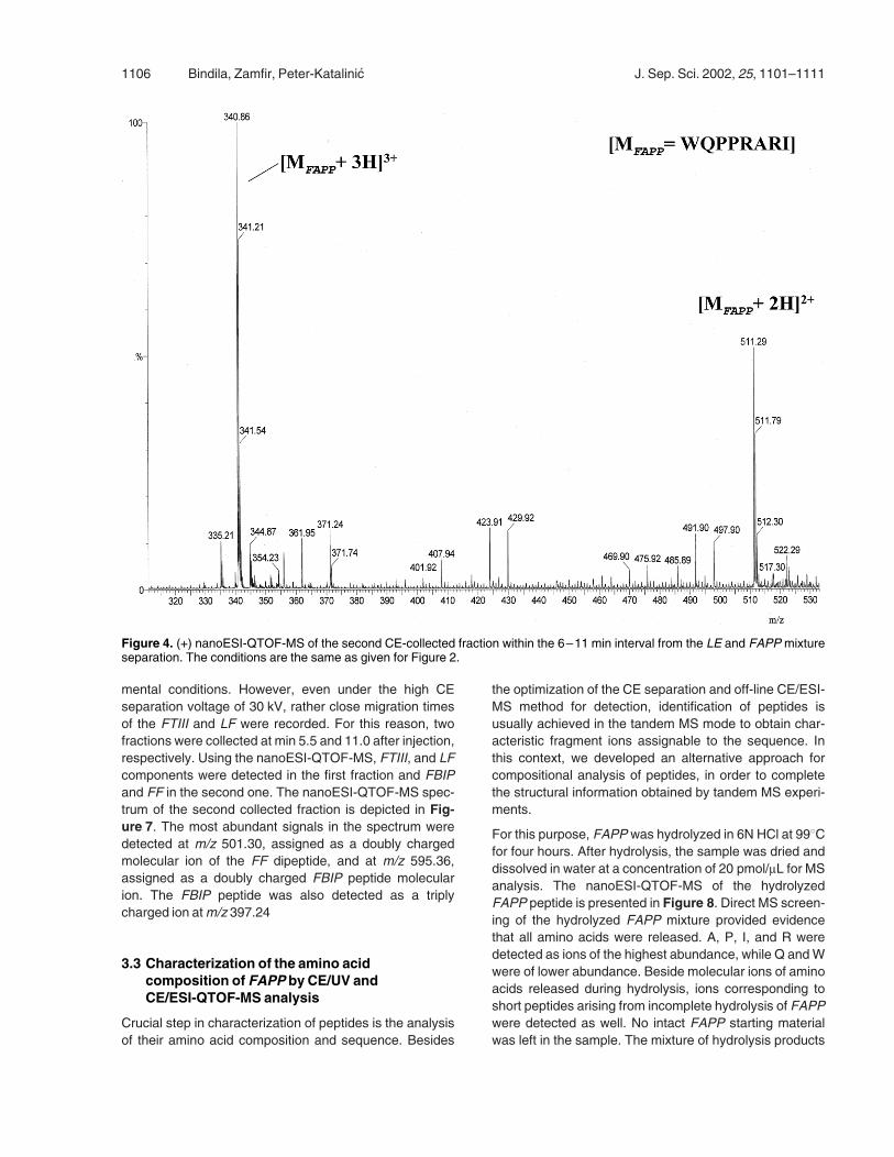
The composition of the second collected fraction at10 min after injection, identified by nanoESI-QTOF-MS, ispresented in Figure 4. According to the fair electropho-retic separation in this efficient buffer system only onespecies, namely FAPP, was detected. Themost abundantsignal in the spectrum is the molecular ion of the FAPPpeptide, detected as a triply charged [M+3H]3+ at m/z340.86, while the second one at m/z 511.29 is the doublycharged molecular ion of the same peptide. It should bestressed out that the molecular ion of the LE was notdetected in this fraction at all. This aspect clearly indicatesthat in the CE/UV electropherogram of the mixture, thefirst eluting peak is to be attributed to the LE and the sec-ond to FAPP peptide species.
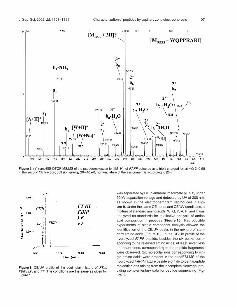
In the tandem MS experiment, the triply charged FAPPmolecular ion detected atm/z 340.86 (Figure 5) was used
as a precursor ion. High level of fragmentation has beenachieved including singly, doubly, and triply chargedsequence ions, which were used for the assignment of theFAPP peptide composition and structure.
3.2 CE/UV and CE/ESI-QTOF-MS analysis of theFBIP, FTIII, FF, LFmixture of peptides derivedfrom extracellular matrix
In order to assess the feasibility of our methodology forseparation and identification of complex peptide mixturescontaining higher number of components, a mixture offour peptides from extracellular matrix has been investi-gated. The substrate concentration of 2.0 mg/mL inammonium formate was used for screening by CE/UVunder the same experimental conditions as describedbefore. In the UV profile, depicted in Figure 6, a relativelyshort migration time, and good sensitivity and resolvingpower are documented. The identification of the UV peaksin Figure 6 was possible due to the perfect reproducibilityof individual component migration under the same experi-
Figure 3. (+) nanoESI-QTOF-MS/MS of the pseudomolecular ion [M+H]+ of LE detected as singly charged ion at m/z 556.58 inthe first CE fraction; collision energy 20–40 eV; nomenclature of the assignment is according to [24].
1106 Bindila, Zamfir, Peter-Katalinic J. Sep. Sci. 2002, 25, 1101–1111
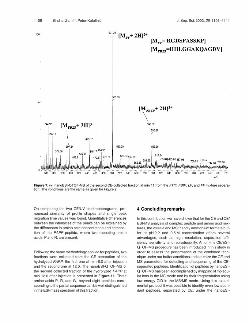
mental conditions. However, even under the high CEseparation voltage of 30 kV, rather close migration timesof the FTIII and LF were recorded. For this reason, twofractions were collected at min 5.5 and 11.0 after injection,respectively. Using the nanoESI-QTOF-MS, FTIII, and LFcomponents were detected in the first fraction and FBIPand FF in the second one. The nanoESI-QTOF-MS spec-trum of the second collected fraction is depicted in Fig-ure 7. The most abundant signals in the spectrum weredetected at m/z 501.30, assigned as a doubly chargedmolecular ion of the FF dipeptide, and at m/z 595.36,assigned as a doubly charged FBIP peptide molecularion. The FBIP peptide was also detected as a triplycharged ion atm/z 397.24
3.3 Characterization of the amino acidcomposition of FAPP by CE/UV andCE/ESI-QTOF-MS analysis
Crucial step in characterization of peptides is the analysisof their amino acid composition and sequence. Besides
the optimization of the CE separation and off-line CE/ESI-MS method for detection, identification of peptides isusually achieved in the tandem MS mode to obtain char-acteristic fragment ions assignable to the sequence. Inthis context, we developed an alternative approach forcompositional analysis of peptides, in order to completethe structural information obtained by tandem MS experi-ments.
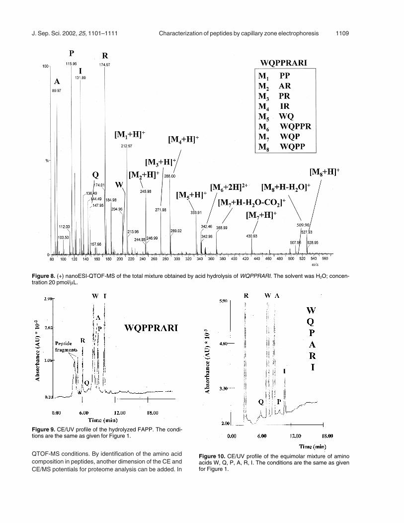
For this purpose, FAPP was hydrolyzed in 6N HCl at 998Cfor four hours. After hydrolysis, the sample was dried anddissolved in water at a concentration of 20 pmol/lL for MSanalysis. The nanoESI-QTOF-MS of the hydrolyzedFAPP peptide is presented in Figure 8. Direct MS screen-ing of the hydrolyzed FAPP mixture provided evidencethat all amino acids were released. A, P, I, and R weredetected as ions of the highest abundance, while Q andWwere of lower abundance. Beside molecular ions of aminoacids released during hydrolysis, ions corresponding toshort peptides arising from incomplete hydrolysis of FAPPwere detected as well. No intact FAPP starting materialwas left in the sample. The mixture of hydrolysis products
Figure 4. (+) nanoESI-QTOF-MS of the second CE-collected fraction within the 6–11 min interval from the LE and FAPP mixtureseparation. The conditions are the same as given for Figure 2.
J. Sep. Sci. 2002, 25, 1101–1111 Characterization of peptides by capillary zone electrophoresis 1107
was separated by CE in ammonium formate pH 2.2, under30 kV separation voltage and detected by UV at 200 nm,as shown in the electropherogram reproduced in Fig-ure 9. Under the same CE buffer and CE/UV conditions, amixture of standard amino acids, W, Q, P, A, R, and I, wasanalyzed as standards for qualitative analysis of aminoacid composition in peptides (Figure 10). Reproducibleexperiments of single component analysis allowed theidentification of the CE/UV peaks in the mixture of stan-dard amino acids (Figure 10). In the CE/UV profile of thehydrolyzed FAPP peptide, besides the six peaks corre-sponding to the released amino acids, at least seven lessabundant ones, corresponding to the peptide fragments,were observed. Six molecular ions corresponding to sin-gle amino acids were present in the nanoESI-MS of thehydrolyzed FAPPmixture beside eight di- to pentapeptidemolecular ions arising from the incomplete cleavage, pro-viding complementary data for peptide sequencing (Fig-ure 8).
Figure 5. (+) nanoESI-QTOF-MS/MS of the pseudomolecular ion [M+H]+ of FAPP detected as a triply charged ion at m/z 340.98in the second CE fraction; collision energy 20–40 eV; nomenclature of the assignment is according to [24].
Figure 6. CE/UV profile of the equimolar mixture of FTIII,FBIP, LF, and FF. The conditions are the same as given forFigure 1.
1108 Bindila, Zamfir, Peter-Katalinic J. Sep. Sci. 2002, 25, 1101–1111
On comparing the two CE/UV electropherograms, pro-nounced similarity of profile shapes and single peakmigration time values was found. Quantitative differencesbetween the intensities of the peaks can be explained bythe differences in amino acid concentration and composi-tion of the FAPP peptide, where two repeating aminoacids, P and R, are present.
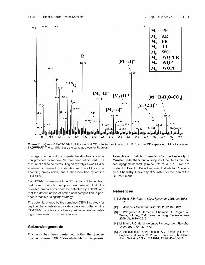
Following the samemethodology applied for peptides, twofractions were collected from the CE separation of thehydrolyzed FAPP, the first one at min 6.0 after injectionand the second one at 12.0. The nanoESI-QTOF-MS ofthe second collected fraction of the hydrolyzed FAPP atmin 12.0 after injection is presented in Figure 11. Threeamino acids P, R, and W, beyond eight peptides corre-sponding to the partial sequence can be well distinguishedin the ESI-mass spectrum of this fraction.
4 Concluding remarks
In this contribution we have shown that for the CE and CE/ESI-MS analysis of complex peptide and amino acid mix-tures, the volatile and MS friendly ammonium formate buf-fer at pH 2.2 and 0.5 M concentration offers severaladvantages, such as high resolution, separation effi-ciency, sensitivity, and reproducibility. An off-line CE/ESI-QTOF-MS procedure has been introduced in this study inorder to assess the performance of the combined tech-nique under our buffer conditions and optimize the CE andMS parameters for detecting and sequencing of the CE-separated peptides. Identification of peptides by nanoESI-QTOF-MS has been accomplished bymapping of molecu-lar ions in the MS mode and by their fragmentation usinglow energy CID in the MS/MS mode. Using this experi-mental protocol it was possible to identify even low abun-dant peptides, separated by CE, under the nanoESI-
Figure 7. (+) nanoESI-QTOF-MS of the second CE-collected fraction at min 11 from the FTIII, FBIP, LF, and FF mixture separa-tion. The conditions are the same as given for Figure 2.
J. Sep. Sci. 2002, 25, 1101–1111 Characterization of peptides by capillary zone electrophoresis 1109
QTOF-MS conditions. By identification of the amino acidcomposition in peptides, another dimension of the CE andCE/MS potentials for proteome analysis can be added. In
Figure 8. (+) nanoESI-QTOF-MS of the total mixture obtained by acid hydrolysis of WQPPRARI. The solvent was H2O; concen-tration 20 pmol/lL.
Figure 9. CE/UV profile of the hydrolyzed FAPP. The condi-tions are the same as given for Figure 1.
Figure 10. CE/UV profile of the equimolar mixture of aminoacids W, Q, P, A, R, I. The conditions are the same as givenfor Figure 1.
1110 Bindila, Zamfir, Peter-Katalinic J. Sep. Sci. 2002, 25, 1101–1111
this regard, a method to complete the structural informa-tion provided by tandem MS has been introduced. Themixture of amino acids resulting on hydrolysis was CE/UVscreened, compared to a standard mixture of the corre-sponding amino acids, and further identified by off-lineCE/ESI-MS.
NanoESI-MS screening of the CE fractions obtained fromhydrolyzed peptide samples emphasized that thereleased amino acids could be detected by ESI/MS andthat the determination of amino acid composition in pep-tides is feasible using this strategy.
The potential offered by the combined CE/MS strategy forpeptide characterization provide a basis for further on-lineCE-ESI/MS studies and allow a positive estimation relat-ing to its extension to protein analysis.
Acknowledgements
This work has been carried out within the Sonder-forschungsbereich 492 “Extracellular Matrix: Biogenesis,
Assembly and Cellular Interactions” at the University ofM�nster under the financial support of the Deutsche For-schungsgemeinschaft (Project Z2 to J.P.-K). We aregrateful to Prof. Dr. Peter Bruckner, Institute for Physiolo-gical Chemistry, University of M�nster, for the loan of theCE instrument.
References
[1] J. Peng, S.P. Gygi, J. Mass Spectrom. 2001, 36, 1083–1091.
[2] T. Manabe, Electrophoresis 1999, 20, 3116–3121.
[3] R. Wildgruber, A. Harder, C. Obermaier, G. Boguth, W.Weiss, S.J. Fey, P.M. Larsen, A. Gorg, Electrophoresis2000, 21, 2610–2616.
[4] M. Mann, R.C. Hendrickson, A. Pandey, Annu. Rev. Bio-chem. 2001, 70, 437–473.
[5] A. Schevchenko, O.N. Jensen, A.V. Podtelejnikov, F.Sagliocco, M. Wilm, O. Vorm, H. Boucherie, M. Mann,Proc. Natl. Acad. Sci. USA 1996, 93, 14440–14445.
Figure 11. (+) nanoESI-QTOF-MS of the second CE collected fraction at min 12 from the CE separation of the hydrolyzedWQPPRARI. The conditions are the same as given for Figure 2.
J. Sep. Sci. 2002, 25, 1101–1111 Characterization of peptides by capillary zone electrophoresis 1111
[6] H. Katayama, Y. Ishihama, Y. Oda, N. Asakawa, RapidCom.Mass Spectrom. 2000, 14, 1167–1178.
[7] W.J. Henzel, T.M. Billeci, J.T. Stults, S.C. Wong, C.Grimley, C. Watanabe, Proc. Natl. Acad. Sci. USA 1993,90, 5011–5015.
[8] S.D. Patterson,Anal. Biochem. 1994, 221, 1–15.
[9] E. Mortz, P.B O’Connor, P. Roepstorff, N.L. Kelleher,T.D. Wood, F.W. McLafferty, M. Mann, Proc. Natl. Acad.Sci. USA 1996, 93, 8264–8267.
[10] M. Wilm, G. Neubauer, M. Mann, Anal. Chem. 1996, 68,527–533.
[11] S.P. Gygi, G.L. Corthals, Y. Zhang, Y. Rochon, R.Aebershold, Proc. Natl. Acad. Sci. USA 2000, 97,9390–9395.
[12] J.R. Yates, TrendsGenet. 2000, 16, 5–8.
[13] D. Figeys, R. Aebersold, Electrophoresis 1998, 19,885–892.
[14] A. Zamfir, D.G. Seidler, H. Kresse, J. Peter-Katalinic,Rapid Commun. Mass Spectrom. 2002, in press.
[15] Y. Shen, S.J. Berger, G.A. Anderson, R.D. Smith, Anal.Chem. 2000, 72, 2154–2159.
[16] J. Waterval, G. Hommels, P. Bestebreurtje, C. Versluis,A.J. Heck, A. Bult, H. Lingeman, W.J. Underberg, Elec-trophoresis 2001, 22, 2709–2716.
[17] A. von Brocke, G. Nicholson, E. Bayer, Electrophoresis2001, 22, 1251–1266.
[18] M.T. Hearn, Biologicals 2001, 29, 159–178.
[19] W. Nashabeh, J.T. Smith, Z. El Rassi, Electrophoresis1993, 14, 407–416.
[20] B. Zhang, F. Foret, B.L. Karger, Anal. Chem. 2000, 72,1015–1022.
[21] C.G. Huber, A. Premstaller, G. Kleindienst, J. Chroma-togr. A 1999, 854, 141–154.
[22] A. Zamfir, S. Konig, J. Althoff, J. Peter-Katalinic, J. Chro-matogr. A 2000, 895, 291–298.
[23] A. Zamfir, Z. Vukelic, J. Peter-Katalinic, Electrophoresis2002, 23, 2894–2903.
[24] P. Roepstorff, J. Fohlman, Biomed. Mass Spectrom.1984, 11, 601–609.
[JSS 1325]