Cholesterol Sulfate Imaging in Human Prostate Cancer Tissue by Desorption Electrospray Ionization...
8
Cholesterol Sulfate Imaging in Human Prostate Cancer Tissue by Desorption Electrospray Ionization Mass Spectrometry Livia S. Eberlin 1 , Allison L. Dill 1 , Anthony B. Costa 1 , Demian R. Ifa 1 , Liang Cheng 2 , Timothy Masterson 3 , Michael Koch 3 , Timothy L. Ratliff 4 , and R. Graham Cooks 1,* 1 Department of Chemistry and Center for Analytical Instrumentation Development, Purdue University, West Lafayette, IN 47907, USA 2 Department of Pathology and Laboratory Medicine, Indiana University School of Medicine, Indianapolis, IN 46202, USA 3 Department of Urology, Indiana University School of Medicine, Indianapolis, IN 46202, USA 4 Purdue University Center for Cancer Research, Purdue University, West Lafayette, IN 47907, USA Abstract Development of methods for rapid distinction between cancerous and non-neoplastic tissues is an important goal in disease diagnosis. To this end, desorption electrospray ionization mass spectrometry (DESI-MS) imaging was applied to analyze the lipid profiles of thin tissue sections of 68 samples of human prostate cancer and normal tissue. The disease state of the tissue sections was determined by independent histopathological examination. Cholesterol sulfate was identified as a differentiating compound, found almost exclusively in cancerous tissues including tissue containing precancerous lesions. The presence of cholesterol sulfate in prostate tissues might serve as a tool for prostate cancer diagnosis although confirmation through larger and more diverse cohorts and correlations with clinical outcome data is needed. Keywords ambient ionization; prostate cancer; mass spectrometry imaging; tumor marker; biomarker; principal component analysis Evidence is provided that cholesterol sulfate might serve as an indicator for prostate cancer, following its detection by desorption electrospray ionization (DESI) imaging mass spectrometry (MS) in prostate tissue samples shown by histopathological examination to be cancerous or precancerous. The desire to distinguish rapidly between diseased and healthy tissues (even in vivo or intra-operatively1) makes it important to identify new molecular targets for disease detection and diagnosis. As part of a program aimed at addressing these issues, DESI-MS imaging was applied to analyze the lipid profiles of thin tissue sections of human prostate cancer under ambient conditions and to compare these images with those for non- cancerous tissue, typed as normal tissue in routine histopathological examination. Cholesterol sulfate (CS) was observed almost exclusively in cancerous tissues and high grade prostatic intraepithelial (PIN) neoplasia as determined by histopathological examination, but it was not *Corresponding Author Professor R. Graham Cooks, Department of Chemistry, Purdue University, West Lafayette, IN, 47907, Tel: (765) 494-5262, Fax: (765) 494-9421, [email protected]. Supporting Information Additional information as noted on text. NIH Public Access Author Manuscript Anal Chem. Author manuscript; available in PMC 2011 May 1. Published in final edited form as: Anal Chem. 2010 May 1; 82(9): 3430–3434. doi:10.1021/ac9029482. NIH-PA Author Manuscript NIH-PA Author Manuscript NIH-PA Author Manuscript
Transcript of Cholesterol Sulfate Imaging in Human Prostate Cancer Tissue by Desorption Electrospray Ionization...

Cholesterol Sulfate Imaging in Human Prostate Cancer Tissue byDesorption Electrospray Ionization Mass Spectrometry
Livia S. Eberlin1, Allison L. Dill1, Anthony B. Costa1, Demian R. Ifa1, Liang Cheng2, TimothyMasterson3, Michael Koch3, Timothy L. Ratliff4, and R. Graham Cooks1,*1Department of Chemistry and Center for Analytical Instrumentation Development, PurdueUniversity, West Lafayette, IN 47907, USA2Department of Pathology and Laboratory Medicine, Indiana University School of Medicine,Indianapolis, IN 46202, USA3Department of Urology, Indiana University School of Medicine, Indianapolis, IN 46202, USA4Purdue University Center for Cancer Research, Purdue University, West Lafayette, IN 47907, USA
AbstractDevelopment of methods for rapid distinction between cancerous and non-neoplastic tissues is animportant goal in disease diagnosis. To this end, desorption electrospray ionization massspectrometry (DESI-MS) imaging was applied to analyze the lipid profiles of thin tissue sections of68 samples of human prostate cancer and normal tissue. The disease state of the tissue sections wasdetermined by independent histopathological examination. Cholesterol sulfate was identified as adifferentiating compound, found almost exclusively in cancerous tissues including tissue containingprecancerous lesions. The presence of cholesterol sulfate in prostate tissues might serve as a tool forprostate cancer diagnosis although confirmation through larger and more diverse cohorts andcorrelations with clinical outcome data is needed.
Keywordsambient ionization; prostate cancer; mass spectrometry imaging; tumor marker; biomarker; principalcomponent analysis
Evidence is provided that cholesterol sulfate might serve as an indicator for prostate cancer,following its detection by desorption electrospray ionization (DESI) imaging massspectrometry (MS) in prostate tissue samples shown by histopathological examination to becancerous or precancerous. The desire to distinguish rapidly between diseased and healthytissues (even in vivo or intra-operatively1) makes it important to identify new molecular targetsfor disease detection and diagnosis. As part of a program aimed at addressing these issues,DESI-MS imaging was applied to analyze the lipid profiles of thin tissue sections of humanprostate cancer under ambient conditions and to compare these images with those for non-cancerous tissue, typed as normal tissue in routine histopathological examination. Cholesterolsulfate (CS) was observed almost exclusively in cancerous tissues and high grade prostaticintraepithelial (PIN) neoplasia as determined by histopathological examination, but it was not
*Corresponding Author Professor R. Graham Cooks, Department of Chemistry, Purdue University, West Lafayette, IN, 47907, Tel: (765)494-5262, Fax: (765) 494-9421, [email protected] InformationAdditional information as noted on text.
NIH Public AccessAuthor ManuscriptAnal Chem. Author manuscript; available in PMC 2011 May 1.
Published in final edited form as:Anal Chem. 2010 May 1; 82(9): 3430–3434. doi:10.1021/ac9029482.
NIH
-PA Author Manuscript
NIH
-PA Author Manuscript
NIH
-PA Author Manuscript
detected in normal tissue by DESI-MS. This is the first report of CS as a potential tumor markerfor human prostate cancer.
Mass spectrometry has long been used to image the distributions of proteins, lipids and otherspecific compounds in tissue. This information is potentially valuable in the development ofnew disease diagnostic methods and in advancing the fundamentals of disease biology.2–4
Amongst the MS imaging methods, DESI-MS allows ambient tissue imaging5, 6 and it appearsfrom preliminary studies to have significance in disease diagnosis in several organs, includingprovision of information on tumor margins from the spatial distributions of phospholipids.7Correlations between the spatial distributions of a broad range of biomolecules in tissuesamples provide information that could be important in the diagnosis and treatment of disease.8 The abundance of ions of any particular m/z value and hence the distribution of thecorresponding molecule in the tissue sample can be represented in the form of a DESI-MSimage. Although the methodology can be used to generate an image of any ion observed in themass spectrum, selected ion images from limited set of molecular species for each sampleanalyzed are most useful and these are displayed here.
Amongst the most abundant signals detected in the negative ion DESI mass spectrum of tissuesamples are those due to fatty acids and polar lipids. It is known that alterations in the lipidcomposition of tissues occur in various forms of cancer.9, 10 In particular, cholesterol sulfate(CS), an important sterol sulfate present in a variety of mammalian tissues and known for itsstabilizing and regulatory role as a cell membrane component, has been reported11 as a potentialtumor marker in human uterine cervical carcinoma tissue. The same study showed that CS isco-expressed with transglutaminase-1 and cytokeratin in well-differentiated types of squamouscell cancers as a tumor marker. Increased accumulation of CS has also been observed intumorigenic esophageal rat cells.12
DESI-MS imaging data on human prostate tissue presented here show that CS is found almostexclusively in cancerous tissues and high grade prostatic intraepithelial neoplasia (PIN), apathology considered to represent precancerous lesions. These correlations are based onstandard histological hematoxylin and eosin (H&E) stained sections and DESI-MS acquiredimages. This behavior of CS is qualitatively different to that of other lipids, including particularglycerophospholipids (GPs) and free fatty acids (FAs) which occur in both diseased and healthytissue samples although with differences in relative amounts.
Mass spectra and ion images from the analysis of 68 prostate tissue samples from 34 differentpatients were obtained by DESI-MS. Identification of the compounds responsible for the peaksobserved in the mass spectra was made based on collision-induced dissociation (CID) tandemMS experiments and by comparison of the product-ion mass spectra to reported literature dataor to spectra generated from the standard compounds.13 The ions observed correspond to threemajor lipid classes; glycerophosphoinositols (PI), glycerophosphoserines (PS) and fatty acids(FA). A peak at mass/charge ratio (m/z) 465.4 was observed at high relative abundance duringthe analysis of cancerous prostatic tissues (Figure 1A) and was not detected in normal tissue(Figure 1B). This peak was tentatively assigned as cholesterol sulfate and when these ions weresubjected to tandem MS experiments for structure elucidation, the main fragment ion wasobserved at m/z 97.0 and assigned as the [HSO4]− ion (Figure 1C). The standard compound,sodium cholesteryl sulfate (Sigma-Aldrich Inc., Milwaukee, WI), was subjected to tandem MSexperiments under the same conditions and gave identical results (Figure 1D). For furtherconfirmation, the isotopic distribution of the molecular ion at m/z 465.4 (insets of Figures 1Cand 1D) was found to agree with the proposed molecular formula.14
The association of cholesterol sulfate, identified by the DESI-MS data, with diseased tissue isprovided by pathological examination of the H&E stained sections. The latter information is
Eberlin et al. Page 2
Anal Chem. Author manuscript; available in PMC 2011 May 1.
NIH
-PA Author Manuscript
NIH
-PA Author Manuscript
NIH
-PA Author Manuscript

used to label the tissue sections in the figures shown below. All human tissue samples werehandled in accordance with approved institutional review board (IRB) protocols at IndianaUniversity School of Medicine. Standard DESI-MS imaging conditions were used to analyzethe tissue samples in the negative ion mode (Materials and methods are available in theSupporting Information). In addition to displaying cholesterol sulfate images from individualtissue sections, principal component analysis (PCA) was employed to generate images of thesesections from the entire set of DESI mass spectra. . The resulting PCA images represent asimple and unsupervised way to visually inspect all the DESI data for identification of tumorand normal regions based on color-coded differences between the tumor and normal samples.Further analysis of the PCA-derived loading plots shows the peak corresponding to cholesterolsulfate to be a primary contribution to separation of tumor and control in the generated images(Figure S1, Supporting Information), reinforcing the conclusions of this work. Since diagnosisin this case, based on the detection of cholesterol sulfate, is so clear-cut no advantage is gainedby extending the data analysis to other compounds.
Representative tumor and normal tissue section images from one patient, case numberMH0107-01, are shown in Figure 2 (tumor and normal tissue appear on the left and right sideof the images, respectively). Similar intensities were observed for m/z 788.4 (PS(18:0/18:1))(Figure 2A) and m/z 885.5 (PI(18:0/20:4)) (Figure 2B). The notation (X:Y) represents thenumber of carbon atoms and double bonds in the fatty acid chains, respectively. Remarkably,cholesterol sulfate (m/z 465.4) was observed exclusively in the cancerous tissue, enabling itsclear identification (Figure 2C). To broaden the analysis, the diagnosis was supported by thePCA generated image which also reveals clear differences between the tumor and normaltissues (Figure S3). Cancerous and normal prostate tissue samples from another patient, casenumber MH0301-17 (Figure S4) show the same relative intensity in disease and normal tissuefor the selected PS, PI and FA ions. The cancerous tissue is most clearly identified by observingthe significantly increased intensity of CS which is undetectable in the normal tissue.
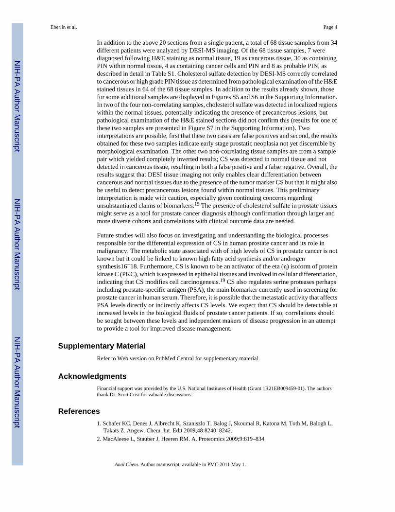
H&E stained and DESI-MS images of CS are shown for another 10 tissue samples in Figure3. For the samples shown in panels A, B and C, CS signals are observed in certain regions ofthe otherwise normal tissue in the DESI ion images, which correspond to precancerous lesionsor high grade prostatic intraepithelial neoplasia (PIN) as determined by pathologicalexamination. The sample shown Figure 3D consists of another case in which one tissue sectionis completely normal and the other has PIN, and where CS is undetectable in the normal tissueand present in regions of the PIN section. Figure 3E shows a case in which both tissue sectionscontain PIN, with corresponding CS by DESI-MS. High grade PIN is characterized by aproliferation of malignant prostatic epithelial cells in prostatic ducts and acini and it is aprecursor to the majority of prostatic adenocarcinomas.
A close examination of the image obtained for CS shown in Figure 2C, reveals that within thenormal tissue, a small localized signal for CS was observed at a spot near the bottom of thetissue. This indicated that a region of PIN might be present in the normal tissue section and inorder to follow up on this indication, twenty additional serial sections for this tissue specimen(case number MH0107-01) comprising the entire tissue were analyzed. Ten serial sectionsspaced throughout the 20 sections used for DESI-MS analysis were subjected to independentH&E staining. Pathological examination of these additional H&E stained tissue sections aswell as the original H&E stained section (Figure 2D) confirmed that the specific region inwhich CS is observed contains precancerous lesions, or high grade prostatic intraepithelialneoplasia (PIN). Ion images of CS for four of these sections are shown in Figure 2 (E-H) andthe additional sixteen sections are shown in Figure S2. In all the tissue sections analyzed, CSsignal was undetectable in the normal tissue region while it was observed as the main peak inmass spectra recorded for the cancerous tissue as well as in the small region of the normaltissue identified as containing PIN.
Eberlin et al. Page 3
Anal Chem. Author manuscript; available in PMC 2011 May 1.
NIH
-PA Author Manuscript
NIH
-PA Author Manuscript
NIH
-PA Author Manuscript
In addition to the above 20 sections from a single patient, a total of 68 tissue samples from 34different patients were analyzed by DESI-MS imaging. Of the 68 tissue samples, 7 werediagnosed following H&E staining as normal tissue, 19 as cancerous tissue, 30 as containingPIN within normal tissue, 4 as containing cancer cells and PIN and 8 as probable PIN, asdescribed in detail in Table S1. Cholesterol sulfate detection by DESI-MS correctly correlatedto cancerous or high grade PIN tissue as determined from pathological examination of the H&Estained tissues in 64 of the 68 tissue samples. In addition to the results already shown, thosefor some additional samples are displayed in Figures S5 and S6 in the Supporting Information.In two of the four non-correlating samples, cholesterol sulfate was detected in localized regionswithin the normal tissues, potentially indicating the presence of precancerous lesions, butpathological examination of the H&E stained sections did not confirm this (results for one ofthese two samples are presented in Figure S7 in the Supporting Information). Twointerpretations are possible, first that these two cases are false positives and second, the resultsobtained for these two samples indicate early stage prostatic neoplasia not yet discernible bymorphological examination. The other two non-correlating tissue samples are from a samplepair which yielded completely inverted results; CS was detected in normal tissue and notdetected in cancerous tissue, resulting in both a false positive and a false negative. Overall, theresults suggest that DESI tissue imaging not only enables clear differentiation betweencancerous and normal tissues due to the presence of the tumor marker CS but that it might alsobe useful to detect precancerous lesions found within normal tissues. This preliminaryinterpretation is made with caution, especially given continuing concerns regardingunsubstantiated claims of biomarkers.15 The presence of cholesterol sulfate in prostate tissuesmight serve as a tool for prostate cancer diagnosis although confirmation through larger andmore diverse cohorts and correlations with clinical outcome data are needed.
Future studies will also focus on investigating and understanding the biological processesresponsible for the differential expression of CS in human prostate cancer and its role inmalignancy. The metabolic state associated with of high levels of CS in prostate cancer is notknown but it could be linked to known high fatty acid synthesis and/or androgensynthesis16–18. Furthermore, CS is known to be an activator of the eta (η) isoform of proteinkinase C (PKC), which is expressed in epithelial tissues and involved in cellular differentiation,indicating that CS modifies cell carcinogenesis.19 CS also regulates serine proteases perhapsincluding prostate-specific antigen (PSA), the main biomarker currently used in screening forprostate cancer in human serum. Therefore, it is possible that the metastatic activity that affectsPSA levels directly or indirectly affects CS levels. We expect that CS should be detectable atincreased levels in the biological fluids of prostate cancer patients. If so, correlations shouldbe sought between these levels and independent makers of disease progression in an attemptto provide a tool for improved disease management.
Supplementary MaterialRefer to Web version on PubMed Central for supplementary material.
AcknowledgmentsFinancial support was provided by the U.S. National Institutes of Health (Grant 1R21EB009459-01). The authorsthank Dr. Scott Crist for valuable discussions.
References1. Schafer KC, Denes J, Albrecht K, Szaniszlo T, Balog J, Skoumal R, Katona M, Toth M, Balogh L,
Takats Z. Angew. Chem. Int. Edit 2009;48:8240–8242.2. MacAleese L, Stauber J, Heeren RM. A. Proteomics 2009;9:819–834.
Eberlin et al. Page 4
Anal Chem. Author manuscript; available in PMC 2011 May 1.
NIH
-PA Author Manuscript
NIH
-PA Author Manuscript
NIH
-PA Author Manuscript

3. Seeley EH, Caprioli RM. Proteom. Clin. Appl 2008;2:1435–1443.4. Walch A, Rauser S, Deininger SO, Hofler H. Histochem. Cell Biol 2008;130:421–434. [PubMed:
18618129]5. Kertesz V, Van Berkel GJ, Vavrek M, Koeplinger KA, Schneider BB, Covey TR. Anal. Chem
2008;80:5168–5177. [PubMed: 18481874]6. Wiseman JM, Ifa DR, Venter A, Cooks RG. Nat. Protoc 2008;3:517–524. [PubMed: 18323820]7. Dill AL, Ifa DR, Manicke NE, Costa AB, Vara JAR, Knapp DW, Cooks RG. Anal. Chem
2009;81:8758–8764. [PubMed: 19810710]8. Hardesty WM, Caprioli RM. Anal. Bioanal. Chem 2008;391:899–903. [PubMed: 18365184]9. Glunde K, Jie C, Bhujwalla ZM. Cancer Res 2004;64:4270–4276. [PubMed: 15205341]10. Iorio E, Mezzanzanica D, Alberti P, Spadaro F, Ramoni C, D'Ascenzo S, Millimaggi D, Pavan A,
Dolo V, Canevari S, Podo F. Cancer Res 2005;65:9369–9376. [PubMed: 16230400]11. Kiguchi K, Iwamori M, Yamanouchi S, Ishiwata L, Saga M, Amemiya A. Clin. Cancer Res
1998;4:2985–2990. [PubMed: 9865910]12. Rearick JI, Stoner GD, George MA, Jetten AM. Cancer Res 1988;48:5289–5295. [PubMed: 3409253]13. Pulfer M, Murphy RC. Mass Spectrom. Rev 2003;22:332–364. [PubMed: 12949918]14. Metzger K, Rehberger PA, Erben G, Lehmann WD. Anal. Chem 1995;67:4178–4183.15. Hughes V. Nat. Med 2009;15:1339–1343. [PubMed: 19966757]16. Migita T, Ruiz S, Fornari A, Fiorentino M, Priolo C, Zadra G, Inazuka F, Grisanzio C, Palescandolo
E, Shin E, Fiore C, Xie W, Kung AL, Febbo PG, Subramanian A, Mucci L, Ma J, Signoretti S,Stampfer M, Hahn WC, Finn S, Loda M. J. Natl. Cancer Inst 2009;101:519–532. [PubMed:19318631]
17. Titus MA, Schell MJ, Lih FB, Tomer KB, Mohler JL. Clin. Cancer Res 2005;11:4653–4657.[PubMed: 16000557]
18. Williams ML, Rutherford SL, Feingold KR. J. Lipid. Res 1987;28:955–967. [PubMed: 2444666]19. Kuroki T, Ikuta T, Kashiwagi M, Kawabe S, Ohba M, Huh N, Mizuno K, Ohno S, Yamada E, Chida
K. Mutat. Res. Rev. Mutat. Res 2000;462:189–195.
Eberlin et al. Page 5
Anal Chem. Author manuscript; available in PMC 2011 May 1.
NIH
-PA Author Manuscript
NIH
-PA Author Manuscript
NIH
-PA Author Manuscript

Figure 1.Typical negative ion mode mass spectra of (A) human prostate cancer and (B) normal tissuein the range of m/z 150 – 1000. Tandem mass spectra (C) of assigned cholesterol sulfate (m/z 465.4) from human prostate cancer tissue and (D) of standard cholesterol sulfate sample. Theinsets show the isotopic patterns of respective cholesterol sulfate ions. The characteristicfragment ion at m/z 97 was observed for both the standard and the molecule detected in prostatictissue.
Eberlin et al. Page 6
Anal Chem. Author manuscript; available in PMC 2011 May 1.
NIH
-PA Author Manuscript
NIH
-PA Author Manuscript
NIH
-PA Author Manuscript

Figure 2.Negative ion mode tissue imaging of prostate tissue showing areas of cancer and normal tissuein sample MH0107-01 (left and right sides, respectively, for each of eight images). (A) Ionimage of m/z 788.4, PS(18:0:18:1), (B) Ion image of m/z 885.5, PI(18:0/20:4), (C) Ion imageof m/z 465.4, cholesterol sulfate and (D) H&E stained tissue sections of the tumor tissue andnormal. Ion images of m/z 465.4, cholesterol sulfate, in four additional sections taken fromamong 135 serial sections obtained by cutting a 2.7 mm thick tissue block into serial sectionsare shown as E-H.
Eberlin et al. Page 7
Anal Chem. Author manuscript; available in PMC 2011 May 1.
NIH
-PA Author Manuscript
NIH
-PA Author Manuscript
NIH
-PA Author Manuscript
Figure 3.DESI-MS ion image of m/z 465.4, cholesterol sulfate, in the negative ion mode, and H&Estained image of prostate samples (A) MH0202-37, cancer and adjacent normal tissue withPIN, (B) MH212-04, cancer and normal tissue with PIN, (C) UH0003-32, cancer and normaltissue with PIN, (D) UH0003-12, PIN and normal tissue and (E) UH0002-29, PIN detectedwithin normal regions of tissue.
Eberlin et al. Page 8
Anal Chem. Author manuscript; available in PMC 2011 May 1.
NIH
-PA Author Manuscript
NIH
-PA Author Manuscript
NIH
-PA Author Manuscript