Nanotubes/nanowires-based, microfluidic-integrated transistors for detecting biomolecules
Upload
independentCategory
view
2download
0
Review Article
Analysis of Biomolecules through Surface-Assisted Laser
Desorption/Ionization Mass Spectrometry Employing Nanomaterials
Wen-Tsen Chen,a Iva Tomalová,b Jan Preislerb* and Huan-Tsung Changa*aDepartment of Chemistry, National Taiwan University, 1, Section 4, Roosevelt Road,
Taipei 106, Taiwan, R.O.C.bCEITEC MU and Department of Chemistry, Faculty of Science, Masaryk University,
Kamenice 5, Brno, Czech Republic
Received May 10, 2011; Accepted June 14, 2011; Published Online June 22, 2011
Nanomaterials, primarily nanoparticles (NPs), can serve as an alternative matrix for the analysis of
various biomolecules through surface-assisted laser desorption/ionization mass spectrometry (SALDI
MS). SALDI MS has been developed to overcome poor reproducibility and high background in the low-
mass region commonly occurring in matrix-assisted laser desorption/ionization mass spectrometry
(MALDI MS). Various nanomaterials, unmodified or functionalized with recognition ligands, can have a
strong affinity toward certain analytes and thus are applicable for their concentration and enrichment from
complex biological matrices. In mass spectrometry imaging (MSI), the use of NPs instead of the conven-
tional matrices can improve the spatial resolution up to the cellular level. In this review, the nature of NPs,
the methods of sample preparation and approaches for quantitation of biomolecules through SALDI MS
are discussed. Practical applications and limitations of SALDI MS employing NPs for separate samples
and MSI are mentioned. With regard to the nature of MSI analysis, the use of nanostructured surfaces for
MSI is also reflected in this review.
Keywords: Nanomaterials; Biomolecules; Surface-assisted laser desorption/ionization mass spec-
trometry; Mass spectrometry imaging.
INTRODUCTION
Matrix-assisted laser desorption/ionization mass
spectrometry (MALDI MS), nowadays an essential mass
spectrometric technique for the analysis and characteriza-
tion of biomolecules, was developed independently by two
groups in the late 80’s. Karas et al. presented soft ioniza-
tion of amino acids and dipeptides, when embedding these
non-absorbing analytes in a UV-absorbing matrix, first
tryptophan followed by nicotinic acid.1-3 Instead of using
an organic matrix, Tanaka dissolved analytes in a suspen-
sion of cobalt nanoparticles (NPs) in glycerol.4 With many
developments, such as the discovery of new more efficient
matrices, the former technique has become widely and rou-
tinely used whereas Tanaka’s method has not attracted
much attention because of the lower sensitivity and limited
mass range compared to the results obtained with organic
matrices. Among a few others who continued to develop
Tanaka’s original idea, for instance Sunner et al. and Dell et
al., used larger particles (2-150 µm) of graphite or silicon
in glycerol.5-7 Another approach for desorption and ioniza-
tion of small molecules using the organic matrices, desorp-
tion/ionization on porous silica (DIOS), was presented by
Wei et al.8 DIOS employs a porous silica surface obtained
by electrochemical etching and provides “clean” mass
spectra with no or little chemical background.
With the recent development of nanotechnology,
Tanaka’s original idea of using the nano-sized metal parti-
cles has been enjoying a renaissance. Without the low va-
por liquid matrix such as glycerol, Au,9,10 Ag,11 SiO2,12
TiO2,13 Fe3O414,15 and other NPs16 have become widely
used as efficient concentrations and desorption probes for
various analytes. Similar to organic matrices for MALDI
Journal of the Chinese Chemical Society, 2011, 58, 769-778 769
Special issue for the nanotechnology-related analytical chemistry
* Corresponding author. Tel: 011-886-2-33661171; 420-549-49-6629; E-mail: [email protected], [email protected]

MS, NPs have high molar absorption coefficients allowing
absorption of the laser energy, leading to soft and efficient
desorption of the analytes (Fig. 1).
When performing the MALDI MS analysis, the ma-
trices are usually mixed in to excess over the analyte to
avoid its fragmentation from laser irradiation and the for-
mation of co-crystals of a size up to hundreds of microme-
ters, resulting in the formation of “sweet spots”. This leads
to poor shot-to-shot and sample-to-sample reproducibility.
In addition, because of strong background signals arising
from matrix fragments in the low-mass range, the MALDI
MS has succeeded neither as a quantitative tool nor a tool
for analysis of low-weight molecules.17,18
Compared to traditional matrices, the use of NPs
overcomes these problems and provides further advantages
including large surface areas available for large analyte
loading capacity (e.g. > 1000 small molecules per single
NP),9 low matrix interference,19 concentration of the ana-
lyte20 or the option of direct combination of the analyte
analysis with its extraction,21 simple preparation of homo-
geneous samples or low-cost and large-scale preparation of
the NPs.
The terminology in this field could appear unclear
and inconsistent. Several new terms, such as NALDI (nano-
structured-assisted LDI),22,23 nano-PALDI (nanoparticles-
assisted LDI)24 or GALDI (colloidal graphite-assisted
LDI)12,25,26 have been used in recent years for numerous
methods employing nanomaterials, but the term SALDI
(surface assisted laser desorption/ionization), which was
first introduced by Sunner et al. for his technique to distin-
guish it from MALDI, remains the most common.
In this review, we describe the properties of NPs,
sample preparation methods for SALDI MS as well as the
effect of the selection of NPs on the sensitivity and repro-
ducibility of analysis. Potential applications and limits of
NP-based SALDI MS techniques are discussed. The appli-
cations and practical considerations for mass spectrometry
imaging (MSI) are mentioned in brief.
NANOPARTICLES, THEIR PROPERTIES AND
ROLE IN SALDI MS
NPs are particles with sizes in the range of 1-100 nm
and, as already comprehensively reviewed, they can be pre-
pared in several ways.27,28 Briefly, two main ways could be
used for their preparation – either the bottom-up approach
by disintegration of bulk materials or top-down by synthe-
sis from atomic or molecular species.29 Metal and metal ox-
ide NPs for SALDI MS are usually synthesized by hydro-
thermal synthesis which provides the most controllable
method for the production of high quality NPs with a de-
fined size. Furthermore, NPs can be used as seeds to pre-
pare nanocomposites (multi-elemental NPs) and core-shell
nanostructures.30,31 In SALDI MS, NPs play important
roles in capturing the analytes, absorbing and transferring
the desorption laser energy leading to the analyte desorp-
tion and ionization. With N2 (337 nm) and Nd:YAG (355
nm) lasers being the most commonly used, NPs with high
absorption in near the UV region are most suitable — for
instance, Au, Ag, and TiO2 NPs.
By decreasing the size of NPs, the surface area per
mass unit increases and thus, the number of adsorbed ana-
lytes increases. More analyte molecules can be desorbed
and ionized when using small-diameter NPs.32,33 The laser
threshold for analyte desorption is inversely related to par-
ticle size: the smaller the NP, the higher the calculated sur-
face temperature generated during irradiation and thus, the
smaller the required laser threshold.33 Relative to larger
NPs, smaller NPs provide greater desorption/ionization ef-
ficiency leading to higher sensitivity. An elevated MS
background over the m/z region from 0 to 3500 was ob-
served when using larger-sized Au NPs (Fig. 2). On the
other hand, smaller NPs having large surface energy can
tend to aggregate and adsorb on a vial surface.34 Based on
our experience, we suggest that 14-nm Au NPs are the opti-
mal substrates.20 Relative to the size, the shape of the NPs
plays a lesser role in determining the sensitivity of SALDI
MS when using UV–VIS lasers.
770 J. Chin. Chem. Soc., Vol. 58, No. 6, 2011 Chen et al.
Fig. 1. Scheme of SALDI MS. A rapid laser-induced
heating of nanoparticles leads to the laser
desorption of analytes from their surface and
ionization.
Selection of NPs
It is well known that various NPs and microspheres,
owing their great surface area, were found to be applicable
as efficient solid phase extraction and concentration probes
for numerous analytes from complicated biological mix-
tures, including trace components. Selective enrichment
and concentration with NPs reduces signal suppression
from multiple analytes, improves sensitivity and can be di-
rectly combined with MALDI MS through simple mixing
the extracted analyte with organic matrices prior to the MS
analysis.35-38
Moreover, when employing suitable NPs, the extract
(target analyte with NPs) can be directly analyzed through
SALDI MS without any further treatment. Nanomaterials
have immense potential for the one-step extraction, con-
centration and detection of diverse analytes, because of the
wide variety of NPs and the possibility of their surface
modification with numerous functional groups. For in-
stance, for thiol-containing molecules Au NPs can be ap-
plied because of the specific Au-S interaction,20 TiO2 NPs
with a high affinity to enediol compounds were used for the
determination of catecholamines13 or for the detection of
alkanes or waxes, the Ag NPs can be useful due to the low
reactivity of silver with alkanes and thus the generation of
silver adducts with no analyte fragmentation.39,40
Two studies comparing a range of nanomaterials for
various analytes have been published in recent years –
Yonezawa et al. investigated the applications of bare metal
NPs (Ag, Au, Cu and Pt) for the SALDI MS of peptides.41
Using a model sample, angiotensin I, the highest perfor-
mance of Pt NPs was demonstrated, whereas the analyte
was not detected with Ag NPs or Cu NPs. Chiang et al.
studied six different nanomaterials (Au NPs, Fe3O4 NPs,
TiO2 NPs, Se NPs, CdTe quantum dots and Pt nano-
sponges) for their applicability in the analyses of small
analytes, peptides and proteins.42 Au NPs were reported as
the most suitable substrate for small analytes; for larger
molecules, the Fe2O3 and Pt nanosponges were demon-
strated as the most efficient substrate allowing the detec-
tion of molecules up to 25 kDa. To increase the specificity
toward an analyte, NP surface modification or conjugation
with recognition biomolecules, such as aptamers, can be
employed.9,43 Modified NPs were reported to provide a
higher ionization efficiency and reduced analyte ion frag-
mentation than the unmodified ones in many cases.19,44-46
Sample preparation
Similar to MALDI MS practice, the samples for
SALDI MS can be prepared in several ways: The analyte
can be mixed with NPs and a suitable buffer prior deposi-
tion (dry droplet method),47 loaded directly onto a plate
coated with single- or multi-layer NPs or having a nano-
structured surface (matrix-first method)48 or deposited first
and subsequently covered by NPs (sample-first method).18
The deposition methods were investigated by Wu et al. for
the analysis of �-cyclodextrins with bare Au NPs.18 The
sample-first method was presented as the most efficient not
only for �-cyclodextrins but also for the determination of
biomolecules as neutral steroids or carbohydrates in high-
salt solutions without need for diluting prior analysis.49
Analysis of Biomolecules through SALDI MS J. Chin. Chem. Soc., Vol. 58, No. 6, 2011 771
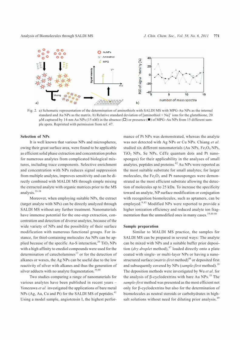
Fig. 2. a) Schematic representation of the determination of aminothiols with SALDI MS with MPG-Au NPs as the internal
standard and Au NPs as the matrix. b) Relative standard deviation of [aminothiol + Na]+ ions for the glutathione, 20
�M captured by 14-nm Au NPs (15 nM) in the absence (�) or presence (�) of MPG–Au NPs from 15 different sam-
ple spots. Reprinted with permission from ref. 47.

Usually, a sample diluting or washing step is required be-
fore analysis to avoid interference and signal suppres-
sion.34 In many cases, the nanoparticles (Au NPs, for exam-
ple) only assist in the desorption process and lack the capa-
bility to ionize analytes. Therefore, ammonium citrate is
usually added as a common proton source.14 The presence
of ammonium citrate reduces the formation of multiple
sodiated adducts, but causes interference in the mass spec-
tra. On the other hand, the functional groups of NPs can act
as the ionization agent as demonstrated by Taira et al.50
Manganese oxide magnetic NPs conjugated with hydroxyl
and amino groups were presented as efficient desorption/
ionization agents without the addition of any external
proton donor.
For MSI, reviews were published about the prepara-
tion of samples with traditional organic matrices.51,52 Sev-
eral methods, such as droplet deposition, electrospray de-
position, spraying or sublimation have been developed in
order to achieve high homogeneity and minimize the ana-
lyte migration. When replacing the organic matrices with
NPs, the colloidal dispersion is usually deposited by spray-
ing onto tissue samples. In order to achieve the highest pos-
sible homogeneity of samples with NPs, the concentration
of NPs, the deposition time, as well as the distance between
the nozzle and sample target require further optimiza-
tion.26,53
Sensitivity and reproducibility
Whereas the organic matrices are mixed with the
analyte to a large excess (e.g., 103–105 matrix/analyte),
NPs allow more molecules to be adsorbed on their surfaces
(e.g., ca. 107–109 analytes per single Au NP);32 thus, more
analytes can desorb and ionize from the NP surfaces per la-
ser shot, theoretically leading to greater sensitivity.
Actually, the sensitivity of SALDI MS is usually at
the micromolar (picomole) level — not better than that of
MALDI MS — primarily because of the relatively poor
desorption and ionization efficiency. One easy approach to
improve the sensitivity of SALDI MS is to use NPs as cap-
ture and concentration probes. For example, SALDI MS
performed without and with preconcentration of the ana-
lyte decreased the limits of detection (LOD) from 1.0 �M
to 25 nM, respectively, for glutathione.20 As mentioned
above, another advantage of SALDI over MALDI is the
high sample homogeneity, which eventually leads to im-
proved reproducibility. Unlike organic matrices, NPs do
not form crystals, but simply allow the preparation of
more-homogeneous samples through adsorption of the
analytes onto their surface. In addition, during laser irradia-
tion, only a few peaks associated with clusters from NPs
appear in the low-mass region, whereas many strong peaks
appear from the fragments originated from organic matri-
ces.20,47,54
The quantitative SALDI MS analyses without any
standard are routinely performed, the intensity of the ana-
lyte signal varies usually within less than 15% from spot-
to-spot.13,55 Using an internal standard, Chiang et al. im-
proved the accuracy of SALDI MS analysis to RSD < 10%
(for inter-day measurements).47 For the determination of
glutathione (GSH) with Au NPs he used the N-2-mercapto-
propionylglycine (MPG) as the standard (Fig. 2). Similarly,
4-mercaptobenzoic acid (MBA)–modified Au NPs were
applied as an internal standard for the determination of the
concentration of captopril (CAP) through SALDI MS mea-
surement in the negative ion mode.54 The spot-to-spot vari-
ations in the absence and presence of the internal standard
(n = 15) were 26 and 9%, respectively. The main advantage
of the quantitation through SALDI MS with NPs consists
of its simple sample preparation, low-cost and fast analyses
without the need for any isotopically labeled standards or
eluents; on the other hand the quantitation restricts the rela-
tively low dynamic range, usually not higher than two or
three orders of magnitude.
APPLICATIONS OF NPS IN SALDI-MS
With its advantages of simple sample preparation,
high sensitivity and high reproducibility, SALDI MS has
become an important analytical tool for the analysis of bio-
logical samples. The main shortfall of SALDI MS is its
limit to the low MS region in most cases; of course it de-
pends on the nature of the NPs and their interactions with
the analytes.
Peptides, proteins and amino acids
The first report of the use of nanoparticles as matrixes
for SALDI MS without the use of low-vapor liquid was
published by McLean et al.32 Au NPs of different sizes (2, 5
and 10 nm) were used for peptide systems, phosphopep-
tides and small proteins, where the smallest NPs showed
the best performance. Castellana et al. furthermore re-
ported higher selectivity and an improvement in the analyte
ion yields and a decrease in ion fragmentation when modi-
772 J. Chin. Chem. Soc., Vol. 58, No. 6, 2011 Chen et al.

fying the Au NPs with 4-aminophenol.45
Because of the strong Au–S bonding (418 kJ/mol),
Nile Red–adsorbed Au NPs were selected as probes by
Huang et al. for the determination of aminothiols through
SALDI MS.20 In the absence and presence of a concentra-
tion process, the LODs of the SALDI MS method for GSH
at a signal-to-noise ratio of 3 were 1.0 �M and 25 nM, re-
spectively. Chiang et al. further improved the limit of de-
tection of GSH to 2 nM when using mixtures of two differ-
ently sized (3.5 and 14 nm) Au NPs and validated this
method through analyses of GSH in cell lysates and cysteine
in plasma.34 The selective capture and determination of
biothiols and sulfur drugs in urine was also demonstrated
through SALDI MS and atmospheric pressure-MALDI ion
trap MS with silver NPs capped with various functional
groups.56 Hua et al. demonstrated SALDI MS of peptides
with Ag NPs.57 Compared to the MALDI MS analysis with
�-cyano-4-hydroxy-cinnamic acid (CHCA), the peptides
were detected in the presence of sodium dodecyl sulfate
with reduced interference of background ions originating
from the detergent. Chen et al. applied SALDI-MS using
silanized Fe3O4 NPs for the analyses of peptides and pro-
teins.14 Coating of each Fe3O4 NP with a shell of SiO2 mini-
mized the leakage of Fe ions, which can cause interference
during analysis. Moreover, these silanized Fe3O4 NPs pos-
sessing negatively charged functionalities were used to se-
lectively capture oppositely charged peptides from sample
solutions. Taira et al. presented manganese oxide magnetic
NPs functionalized with amino and hydroxyl groups as an
efficient probe for desorption/ionization and post-source
decay structural analysis of small proteins, peptides and
drugs.50
Quantum dots (QDs) have unique and fascinating op-
tical properties, including broad absorption spectra, sharp
and symmetrical emission spectra with long Stokes shifts,
high quantum yields and size-dependent emission wave-
lengths, mainly due to quantum confinement effects. Shrivas
et al. demonstrated their applicability for SALDI-MS using
11-mercaptoundecanoic acid (MUA)–modified CdSe QDs
for the detection of proteins.58 Ke et al. investigated three
different surface-modified TiO2 NPs for SALDI MS and
found TiO2-CdS NPs as an efficient matrix for back-
ground-free detection of peptides and proteins with an up-
per detectable limit of 17 kDa.59
A hybrid approach between SALDI and MALDI MS
was presented by Duan et al.46 Au NPs were modified with
CHCA and used for the analysis of protein digests. Com-
pared to the use of the citrate- or cysteamine-capped Au
NPs, CHCA-modified Au NPs provided higher ionization
efficiency, lower analyte fragmentation and cleaner spec-
tra. Carbon-based particles and surfaces have also been
widely used for SALDI MS. Carbon-based substrates offer
diverse types of substrate materials with different proper-
ties, such as thermal hydrophobicity, conductivity, specific
heat capacity or ionization efficiency.60 Micro-sized graph-
ite carbon dispersed in glycerol was first used by Sunner et
al. for the analyses of peptides and small analytes.5 Carbon
nanotubes (CNTs) were applied to sensitive and reproduc-
ible detection of small peptides and proteins by Xu et al.
and Chen et al.61,62 Silicon wafers coated with layer-by-
layer (LBL) films of Au NPs and poly(allylamine hydro-
chloride) (PAHC) have been employed for the SALDI MS
analyses of peptides and environmental pollutants.63 Com-
pared to dispersed Au NPs, the use of LBL films provided
increased sensitivity (sub-femtomole) for angiotensin I and
cytochrome C and a wider analyte mass range (up to 12
kDa).
Because Pt is inert and has a high-melting tempera-
ture, high crystallinity and low thermal conductivity,
highly stable Pt-based nanomaterials absorbing laser light
were reported as useful matrices for SALDI MS analyses.
Kawasaki et al. applied SALDI MS using Pt nanoflowers
for the analyses of various biomolecules, mainly proteins.64
Further, sulfonate group-modified PtFeCu NPs were em-
ployed as selective capture probes for positively charged
proteins as well as efficient laser desorption/ionization
(LDI) agents.65
Chiang et al. employed HgTe nanostructures as a new
matrix for SALDI MS–based analyses of proteins and their
complexes with small analytes.16 The mass limit when us-
ing SALDI MS with HgTe nanostructures as matrices
reached as high as 150 kDa. The applicability of this
method provided a LOD of 200 pM for angiotensin I and
was validated through the detections of recombinant pro-
teins transformed in E. coli, a specific complex between
bovine serum albumin (BSA) and L-tryptophan and a car-
bonic anhydrase–acetazolamide complex (Fig. 3).
Other analytes
Besides the proteins and peptides, SALDI MS with
NPs has become popular for the fast analyses of other mol-
ecules such as carbohydrates, lipids, metabolites, drugs or
Analysis of Biomolecules through SALDI MS J. Chin. Chem. Soc., Vol. 58, No. 6, 2011 773

polymers.
Selective ionization of olefinic compounds, for ex-
ample cholesterol or carotenoids, directly from complex
mixtures by Ag NPs was described by Sherrod et al.66 Su et
al. analyzed and quantified small neutral carbohydrates in
urine through SALDI MS using Au NPs capped with vari-
ous agents.55 Lee et al. found that TiO2 NPs having diame-
ters of less than 20 nm interacted with enediol compounds,
resulting in increases of absorption in the UV–Vis region.13
The SALDI MS approach provided LODs on a micromolar
level for several catechins and was applied to determine the
concentration of EGC and EGCG in tea samples. Chiu et al.
demonstrated and tested on real urine samples for the deter-
mination of three estrogens – estrone (E1), estradiol (E2),
and estriol (E3) – with SALDI MS using Ag NPs (34 ± 3
nm).11 NPs featuring anisotropic shapes are also attractive
matrices for the analysis of small analytes with SALDI MS.
For example, Watanabe et al. applied SALDI MS using
ZnO NPs with cubic or rectangular parallelepiped shapes
for the detection of verapamil hydrochloride, testosterone,
phospholipids, oligosaccharides and synthetic polymers.67
(ZnS)-semiconductor NPs capped with different functional
groups were investigated for the analysis of cyclodextrins
and small proteins.68 The best efficiency was exhibited
when using NPs 3-mercaptopropanoic acid as a matrix.
Cysteine capped ZnS NPs doped with Mn2+ were further
developed by this group for the detection of coccidiostats
and peptide mixtures.69 Amini et al. employed oxidized
graphitized carbon black (GCB) particles for the analysis
of pharmaceutical compounds.70 The advantages of using
GCB materials rather than CNTs are their low cost and
greater content of carboxylic acid groups on their surface,
thereby enhancing the efficiency of the ionization process
and the desorption of hydrophobic compounds.
Coatings of nanostructured diamond-like carbon on
digital versatile disks have been employed as targets for the
SALDI MS analyses of various analytes, including amino
acids, carbohydrates, lipids, peptides, and other metabo-
lites.71 Tseng et al. presented a hybrid of immobilized silica
and 2,5-dihydrobenzoic acid (DHB) on iron oxide mag-
netic NPs as an efficient material providing superior soft
ionization of a variety of new types of synthetic materials
used for solar cells, light emitting devices, dendrimers and
glycolipids, including analytes with either thermally labile
structures or with poor protonation tendencies.19 Wen et al.
employed SALDI MS using silicon powder (5–50 nm) for
the analysis of several small molecules, including drugs,
pesticides, peptides and acids.12 Single-crystalline EuF3
hollow hexagonal microdisks were reported as an efficient
material for SALDI MS, allowing the analysis of polyeth-
ylene glycols with an upper detectable range of 35 kDa.72
“Advanced” applications
Nanoparticles for SALDI MS have found applica-
tions not only as an effective matrix for the detection and
quantitation of analytes but also for the investigation of re-
actions of nanoparticles with analytes or for structural
characterization of analytes.
Watanabe et al. demonstrated that the use of ZnO NPs
enhances the fragmentation of oligosaccharides in a post-
source decay analysis, producing far more fragments than
MALDI MS analysis did when using the DHB as the ma-
trix.67 Lin et al. provided strong evidence for the chemical
reactions of functionalized Au NPs with analyte.73 Via
SALDI MS, Hg2+ ions were demonstrated to induce 3-mer-
captopropionic acid (MPA)�Au NPs aggregation in the
presence of 2,6-pyridinedicarboxylic acid (PDCA) and
H2O2-induced fluorescence quenching of Au nanodots
774 J. Chin. Chem. Soc., Vol. 58, No. 6, 2011 Chen et al.
Fig. 3. SALDI mass spectra of (a, b) solutions contain-
ing HgTe nanostructures, BSA, and (a) L-tryp-
tophan (L-Y) and (b) D-tryptophan and (c, d)
Triton X-100–protected HgTe nanostructures
and human carbonic anhydrase I (hCAI) in the
(c) presence and (d) absence of acetazolamide
(ACZ). When using HgTe nanostructures as
SALDI matrices, weak protein–ligand interac-
tions can be observed, such as the specific com-
plex formed between BSA and L-Y or the hCAI-
ACZ complex. Reprinted with permission from
ref. 16

modified with 11-mercaptoundecanoic acid (MUA). PDCA-
Hg2+-MPA coordination was proved as responsible for Au
NPs aggregation, while the formation of 11-MUA disulfide
compounds that are released into the bulk solution as re-
sponsible for H2O2-induced fluorescence quenching. In ad-
dition to providing information about the chemical struc-
tures, SALDI MS was also reported as being selective and
sensitive for the quantitation of Hg2+ ions and H2O2.
Mass Spectrometry Imaging
MSI has emerged in the last decade as an effective
tool for simultaneous tracing and visualizing the spatial
distribution of proteins, peptides, drugs and their metabo-
lites in tissues.74,75 Rapid development of MSI methodol-
ogy and its countless applications have become possible
mainly due to the recent introduction of commercial high-
throughput mass spectrometers capable of fast data acqui-
sition and equipped with reliable solid-state UV lasers with
high repetition rates, power density and pulse-to-pulse sta-
bility adequate for MALDI. This approach eliminates the
need for labeling because molecular mass is used as an en-
dogenous label, allowing also detection of post-transla-
tional modifications. Besides the above mentioned disad-
vantages, such as the high background noise in the low
mass range, in MALDI MSI other critical limitations of the
spatial resolution are the size of the organic matrix crystals,
analyte migration and laser focus size. With their confined
size and the absence of the crystallization process, MSI em-
ploying nanoparticles were reported to overcome some of
these obstacles and to be able to achieve cellular resolu-
tion.24 So far, the use of several micro- and nano-materials,
including Ag, Au colloidal graphite or magnetic NPs have
been demonstrated for the analysis of small molecules in
MSI.
Cha et al. used an aerosol spray containing cca. 1 �m
colloidal graphite particles for the direct lipid profiling and
imaging of cerebrosides and sulphatides in rat brain tissue
and flavonoids on different plant surfaces and tissue sec-
tions.25,53 Zhang et al. utilized a similar approach for the
imaging of organic acids, flavonoids and oligosaccharides
in various fruit species.26 Cha et al. also presented the use
of colloidal Ag for MSI of epicuticular wax metabolites on
the surfaces of leaves and flowers.39 Taira et al. achieved
cellular (15 µm) resolution when employing 3.7 nm diame-
ter functionalized iron-based magnetic NPs as SALDI ma-
terial in the analysis of lipids and peptides in rat cerebellum
tissues through MSI.24 The same NPs were used by Ageta
et al. to detect different sulfatide species in rat hippocam-
pus;76 gold NPs functionalized with alkylamine were re-
ported as suitable for the detection of minor glycosphingo-
lipids in mouse brain sections77 and silver NPs function-
alized with alkylcarboxylate and alkylamine for fatty acids
in mouse retinal and liver sections.78
Rowel et al. demonstrated direct detection of drugs
and their metabolites in latent fingerprints (LFP) applying
MSI with a hydrophobic silica dusting agent containing
carbon black.79 LFPs were studied also by Tang et al. who
presented Au NPs as suitable for both their visualization
and molecular MSI.80 This approach enables the conserva-
tion of individual identity and also provides information
about drugs or hazardous substances. In 2011, Tang et al.
also demonstrated solvent-free gold-NP-assisted LDI MSI
as a sensitive and minimally destructive method for the di-
rect detection and imaging of ink and visible and/or fluo-
rescent dyes printed on banknotes or written on questioned
documents.81
An innovative method was introduced by Northen et
al.82 Nanostructure-initiator mass spectrometry (NIMS)
was presented as a tool for spatially confined mass analy-
sis. It uses nanostructured surfaces with trapped ‘initiator’
molecules or ‘clathrates’ to release and ionize intact mole-
cules adsorbed on the surface. This surface responds to
both ion and laser irradiation. The lateral resolution (ion-
NIMS about 150 nm), sensitivity, matrix-free and reduced
fragmentation of NIMS allows direct characterization of
peptide microarrays, direct mass analysis of single cells,
tissue imaging, and direct characterization of blood and
urine. The method and its applications have been re-
viewed.83 Vidova et al. also avoided the time consuming
step of MSI - matrix deposition and used the commercially
available nanostructured surfaces as the substrates for the
imprinting of tissue section for lipid analysis (Fig. 4).22
CONCLUSIONS AND PERSPECTIVES
In this review, we have examined several SALDI MS
approaches using NPs for the detection of biomolecules
and MSI. It is obvious that the nature, the size of the NPs as
well as the sample preparation play a crucial role in deter-
mining the sensitivity of the process.
In contrast to MALDI MS, SALDI MS with NPs can
provide great reproducibility, allowing quantitative deter-
mination of the concentrations of small analytes. For the
Analysis of Biomolecules through SALDI MS J. Chin. Chem. Soc., Vol. 58, No. 6, 2011 775

detection of small analytes (< 500 Da), SALDI MS using
Au NPs is superior to MALDI MS and is applicable to
metabolomics and MSI, mainly because of the absence of
background noise in MS spectra originated from organic
matrices. Functionalized NPs with an affinity towards a tar-
get analyte can be applied for both enrichment and MS
analysis of biomolecules making nanomaterials attractive
in terms of sample treatment and analysis of trace analytes.
In addition, the SALDI approach has been reported as al-
lowing ionization of molecules hardly ionizable by MALDI.
On the other hand, single-crystalline EuF3 hollow
hexagonal microdisks and HgTe nanostructures are all ef-
fective substrates for the detection of molecules having
values of m/z of up to 35 and 150 kDa, respectively. The
SALDI MS approach using HgTe nanostructures also al-
lows the detection of weak complexes of proteins with
small analytes revealing its potential for investigating the
complexes of proteins with other proteins and DNA. Their
toxicity is a concern though. The further development of
new nanomaterials allowing the selective capture of larger
proteins and their more efficient desorption/ionization is
essential. A combination of SALDI MS with further analyt-
ical methods is expected over the coming years as well as
more studies of the desorption-ionization process which
has not yet been fully explained and is essential for further
developments.
MSI performed in conjunction with NPs provides
better spatial resolution mainly due to the absence of the
matrix-analyte co-crystallization process and no analyte
migration. New approaches employing nanostructured sur-
faces avoid the matrix deposition steps and therefore make
the MSI analyses significantly easier and faster. We note
that the number of studies dealing with MSI and nanomate-
rials remains very low and more application and method-
ological studies are needed for overall conclusions.
ACKNOWLEDGEMENTS
This study was supported by the National Science
Council of Taiwan under contracts NSC 98-2113-M-002-
011-MY3 and NSC 99-2923-M-002-004-MY3, the Czech
Science Foundation P206/10/J012, the Ministry of Educa-
tion, Youth and Sports of the Czech Republic
MSM0021622415 and CZ.1.05/1.1.00/02.0068. Iva
Tomalová is supported by Brno City Municipality Scholar-
ships for Talented Ph.D. Students.
REFERENCES
1. Karas, M.; Bachmann, D.; Hillenkamp, F. Anal. Chem. 1985,
57, 2935.
2. Karas, M.; Bachmann, D.; Bahr, U.; Hillenkamp, F. Int. J.
Mass Spectrom. 1987, 78, 53.
3. Karas, M.; Hillenkamp, F. Anal. Chem. 1988, 60, 2299.
4. Tanaka, K.; Waki, H.; Ido, Y.; Akita, S.; Yoshida, Y.;
Yoshida, T. Rapid Commun. Mass Spectrom. 1988, 2, 151.
5. Sunner, J.; Dratz, E.; Chen, Y. C. Anal. Chem. 1995, 67,
4335.
6. Dale, M. J.; Knochenmuss, R.; Zenobi, R. Anal. Chem. 1996,
68, 3321.
7. Dale, R.; Knochenmuss, R.; Zenobi, R. Rapid Commun.
Mass Spectrom. 1997, 11, 136.
8. Wei, J.; Buriak, J. M.; Siuzdak, G. Nature 1999, 399, 243.
9. Huang, Y. F.; Chang, H. T. Anal. Chem. 2007, 79, 4852.
10. Huang, Y. F.; Lin, Y. W.; Chang, H. T. Nanotechnology 2006,
17, 4885.
11. Chiu, T. C.; Chang, L. C.; Chiang, C. K.; Chang, H. T. J. Am.
Soc. Mass Spectrom. 2008, 19, 1343.
12. Wen, X. J.; Dagan, S.; Wysocki, V. H. Anal. Chem. 2007, 79,
434.
13. Lee, K. H.; Chiang, C. K.; Lin, Z. H.; Chang, H. T. Rapid
776 J. Chin. Chem. Soc., Vol. 58, No. 6, 2011 Chen et al.
Fig. 4. Comparison of standard MALDI imaging with
the matrix-free NALDI approach. The overlay
images (last row) show the different lipid com-
position of pelvis (orange) and adrenal gland
(yellow) from the rest of the kidney. The exact
m/z of imaged ions are: 760.58, 772.58, 792.59,
and 810.60. Reprinted with permission from
ref. 22.

Commun. Mass Spectrom. 2007, 21, 2023.
14. Chen, W. Y.; Chen, Y. C. Anal. Bioanal. Chem. 2006, 386,
699.
15. Lin, P. C.; Tseng, M. C.; Su, A. K.; Chen, Y. J.; Lin, C. C.
Anal. Chem. 2007, 79, 3401.
16. Chiang, C. K.; Yang, Z. S.; Lin, Y. W.; Chen, W. T.; Lin, H.
J.; Chang, H. T. Anal. Chem. 2010, 82, 4543.
17. Tholey, A.; Heinzle, E. Anal. Bioanal. Chem 2006, 386, 24.
18. Wu, H. P.; Su, C. L.; Chang, H. C.; Tseng, W. L. Anal. Chem.
2007, 79, 6215.
19. Tseng, M. C.; Obena, R.; Lu, Y. W.; Lin, P. C.; Lin, P. Y.; Yen,
Y. S.; Lin, J. T.; Huang, L. D.; Lu, K. L.; Lai, L. L.; Lin, C.
C.; Chen, Y. J. J. Am. Soc. Mass Spectrom. 2010, 21, 1930.
20. Huang, Y. F.; Chang, H. T. Anal. Chem. 2006, 78, 1485.
21. Chen, C. T.; Chen, Y. C. Anal. Chem. 2005, 77, 5912.
22. Vidova, V.; Novak, P.; Strohalm, M.; Pol, J.; Havlicek, V.;
Volny, M. Anal. Chem. 2010, 82, 4994.
23. Wyatt, M. F.; Ding, S. J.; Stein, B. K.; Brenton, A. G.;
Daniels, R. H. J. Am. Soc. Mass Spectrom. 2010, 21, 1256.
24. Taira, S.; Sugiura, Y.; Moritake, S.; Shimma, S.; Ichiyanagi,
Y.; Setou, M. Anal. Chem. 2008, 80, 4761.
25. Cha, S. W.; Yeung, E. S. Anal. Chem. 2007, 79, 2373.
26. Zhang, H.; Cha, S. W.; Yeung, E. S. Anal. Chem. 2007, 79,
6575.
27. Modeshia, D. R.; Walton, R. I. Chem. Soc. Rev. 2010, 39,
4303.
28. Zhu, Y.; Yitai, Q. In Nanorods, Nanotubes, and Nanomateri-
als Research Progress; Prescott, W. V.; Schwartz, A. I., Eds.;
Nova Science Publishers, Inc.: New York, 2008; p 279.
29. Murray, W. A.; Barnes, W. L. Adv. Mater. 2007, 19, 3771.
30. Chuang, H. Y.; Chen, D. H., Nanotechnology 2009, 20, 10.
31. Wu, W.; He, Q. G.; Chen, H.; Tang, J. X.; Nie, L. B. Nano-
technology 2007, 18, 8.
32. McLean, J. A.; Stumpo, K. A.; Russell, D. H. J. Am. Chem.
Soc. 2005, 127, 5304.
33. Schurenberg, M.; Dreisewerd, K.; Hillenkamp, F. Anal.
Chem. 1999, 71, 221.
34. Chiang, N. C.; Chiang, C. K.; Lin, Z. H.; Chiu, T. C.; Chang,
H. T. Rapid Commun. Mass Spectrom. 2009, 23, 3063.
35. Zhang, L. J.; Xu, Y. W.; Yao, H. L.; Xie, L. Q.; Yao, J.; Lu, H.
J.; Yang, P. Y. Chem. Eur. J. 2009, 15, 10158.
36. Li, Y.; Liu, Y.; Tang, J.; Lin, H.; Yao, N.; Shen, X.; Deng, C.;
Yang, P.; Zhang, X. J. Chromatogr., A 2007, 1172, 57.
37. Tang, J.; Liu, Y. C.; Qi, D. W.; Yao, G. P.; Deng, C. H.;
Zhang, X. M. Proteomics 2009, 9, 5046.
38. Chen, H. M.; Deng, C. H.; Zhang, X. M. Angew. Chem. Int.
Ed. 2010, 49, 607.
39. Cha, S. W.; Song, Z. H.; Nikolau, B. J.; Yeung, E. S. Anal.
Chem. 2009, 81, 2991.
40. Chen, R.; Li, L. J. Am. Soc. Mass Spectrom. 2001, 12, 367.
41. Yonezawa, T.; Kawasaki, H.; Tarui, A.; Watanabe, T.;
Arakawa, R.; Shimada, T.; Mafune, F. Anal. Sci. 2009, 25,
339.
42. Chiang, C. K.; Chiang, N. C.; Lin, Z. H.; Lan, G. Y.; Lin, Y.
W.; Chang, H. T. J. Am. Soc. Mass Spectrom. 2010, 21, 1204.
43. Gulbakan, B.; Yasun, E.; Shukoor, M. I.; Zhu, Z.; You, M.
X.; Tan, X. H.; Sanchez, H.; Powell, D. H.; Dai, H. J.; Tan,
W. H. J. Am. Chem. Soc. 2010, 132, 17408.
44. Chiu, Y. C.; Chen, Y. C. Anal. Lett. 2008, 41, 260.
45. Castellana, E. T.; Russell, D. H. Nano Lett. 2007, 7, 3023.
46. Duan, J. C.; Linman, M. J.; Chen, C. Y.; Cheng, Q. J. J. Am.
Soc. Mass Spectrom. 2009, 20, 1530.
47. Chiang, C. K.; Lin, Y. W.; Chen, W. T.; Chang, H. T. Nano-
med.-Nanotechnol. 2010, 6, 530.
48. Gulbakan, B.; Park, D.; Kang, M. C.; Kececi, K.; Martin, C.
R.; Powell, D. H.; Tan, W. H. Anal. Chem. 2010, 82, 7566.
49. Wu, H. P.; Yu, C. J.; Lin, C. Y.; Lin, Y. H.; Tseng, W. L. J. Am.
Soc. Mass Spectrom. 2009, 20, 875.
50. Taira, S.; Kitajima, K.; Katayanagi, H.; Ichiishi, E.;
Ichiyanagi, Y. Sci. Technol. Adv. Mat. 2009, 10.
51. Kaletas, B. K.; van der Wiel, I. M.; Stauber, J.; Dekker, L. J.;
Guzel, C.; Kros, J. M.; Luider, T. M.; Heeren, R. M. A.
Proteomics 2009, 9, 2622.
52. van Hove, E. R. A.; Smith, D. F.; Heeren, R. M. A. J.
Chromatogr., A 2010, 1217, 3946.
53. Cha, S. W.; Zhang, H.; Ilarslan, H. I.; Wurtele, E. S.;
Brachova, L.; Nikolau, B. J.; Yeung, E. S. Plant J. 2008, 55,
348.
54. Chen, W. T.; Chiang, C. K.; Lin, Y. W.; Chang, H. T. J. Am.
Soc. Mass Spectrom. 2010, 21, 864.
55. Su, C. L.; Tseng, W. L. Anal. Chem. 2007, 79, 1626.
56. Shrivas, K.; Wu, H. F. Rapid Commun. Mass Spectrom.
2008, 22, 2863.
57. Hua, L.; Chen, J. R.; Ge, L.; Tan, S. N. J. Nanopart. Res.
2007, 9, 1133.
58. Shrivas, K.; Kailasa, S. K.; Wu, H. F. Proteomics 2009, 9,
2656.
59. Ke, Y. T.; Kailasa, S. K.; Wu, H. F.; Nawaz, M. J. Sep. Sci.
2010, 33, 3400.
60. Tang, H. W.; Ng, K. M.; Lu, W.; Che, C. M. Anal. Chem.
2009, 81, 4720.
61. Chen, W. Y.; Wang, L. S.; Chiu, H. T.; Chen, Y. C.; Lee, C. Y.
J. Am. Soc. Mass Spectrom. 2004, 15, 1629.
62. Xu, S. Y.; Li, Y. F.; Zou, H. F.; Qiu, J. S.; Guo, Z.; Guo, B. C.
Anal. Chem. 2003, 75, 6191.
63. Kawasaki, H.; Sugitani, T.; Watanabe, T.; Yonezawa, T.;
Moriwaki, H.; Arakawa, R. Anal. Chem. 2008, 80, 7524.
64. Kawasaki, H.; Yonezawa, T.; Watanabe, T.; Arakawa, R. J.
Phys. Chem. C 2007, 111, 16278.
65. Kawasaki, H.; Akira, T.; Watanabe, T.; Nozaki, K.;
Yonezawa, T.; Arakawa, R. Anal. Bioanal. Chem. 2009, 395,
1423.
66. Sherrod, S. D.; Diaz, A. J.; Russell, W. K.; Cremer, P. S.;
Russell, D. H. Anal. Chem. 2008, 80, 6796.
Analysis of Biomolecules through SALDI MS J. Chin. Chem. Soc., Vol. 58, No. 6, 2011 777

67. Watanabe, T.; Kawasaki, H.; Yonezawa, T.; Arakawa, R. J.
Mass Spectrom. 2008, 43, 1063.
68. Kailasa, S. K.; Kiran, K.; Wu, H. F. Anal. Chem. 2008, 80,
9681.
69. Kailasa, S. K.; Wu, H. F. Analyst 2010, 135, 1115.
70. Amini, N.; Shariatgorji, M.; Thorsen, G. J. Am. Soc. Mass
Spectrom. 2009, 20, 1207.
71. Najam-ul-Haq, M.; Rainer, M.; Huck, C. W.; Hausberger, P.;
Kraushaar, H.; Bonn, G. K. Anal. Chem. 2008, 80, 7467.
72. Chen, Z. M.; Geng, Z. R.; Shao, D. L.; Mei, Y. H.; Wang, Z.
L. Anal. Chem. 2009, 81, 7625.
73. Lin, Y. W.; Chen, W. T.; Chang, H. T. Rapid Commun. Mass
Spectrom. 2010, 24, 933.
74. Todd, P. J.; Schaaff, T. G.; Chaurand, P.; Caprioli, R. M. J.
Mass Spectrom. 2001, 36, 355.
75. Chaurand, P.; Schwartz, S. A.; Caprioli, R. M. Curr. Opin.
Chem. Biol. 2002, 6, 676.
76. Ageta, H.; Asai, S.; Sugiura, Y.; Goto-Inoue, N.; Zaima, N.;
Setou, M. Med. Mol. Morphol. 2009, 42, 16.
77. Goto-Inoue, N.; Hayasaka, T.; Zaima, N.; Kashiwagi, Y.;
Yamamoto, M.; Nakamoto, M.; Setou, M. J. Am. Soc. Mass
Spectrom. 2010, 21, 1940.
78. Hayasaka, T.; Goto-Inoue, N.; Zaima, N.; Shrivas, K.;
Kashiwagi, Y.; Yamamoto, M.; Nakamoto, M.; Setou, M. J.
Am. Soc. Mass Spectrom. 2010, 21, 1446.
79. Rowell, F.; Hudson, K.; Seviour, J. Analyst 2009, 134, 701.
80. Tang, H. W.; Lu, W.; Che, C. M.; Ng, K. M. Anal. Chem.
2010, 82, 1589.
81. Tang, H. W.; Wong, M. Y. M.; Chan, S. L. F.; Che, C. M.; Ng,
K. M. Anal. Chem. 2011, 83, 453.
82. Northen, T. R.; Yanes, O.; Northen, M. T.; Marrinucci, D.;
Uritboonthai, W.; Apon, J.; Golledge, S. L.; Nordstrom, A.;
Siuzdak, G. Nature 2007, 449, 1033.
83. Greving, M. P.; Patti, G. J.; Siuzdak, G. Anal. Chem. 2011,
83, 2.
778 J. Chin. Chem. Soc., Vol. 58, No. 6, 2011 Chen et al.
Copyright © 2022 FDOKUMEN