Preparation and characterization of nitrilotriacetic-acid-terminated self-assembled monolayers on...
12
ANALYTICAL BIOCHEMISTRY Analytical Biochemistry 345 (2005) 258–269 www.elsevier.com/locate/yabio 0003-2697/$ - see front matter 2005 Elsevier Inc. All rights reserved. doi:10.1016/j.ab.2005.07.009 Preparation and characterization of nitrilotriacetic-acid-terminated self-assembled monolayers on gold surfaces for matrix-assisted laser desorption ionization-time of Xight-mass spectrometry analysis of proteins and peptides Jianwei Shen a,¤ , Tanveer Ahmed a , Andrew Vogt b , Jieyi Wang c , Jean Severin d , Richard Smith d , Sally Dorwin d , Robert Johnson a , John Harlan d , Tom Holzman d a Department R418, Global Pharmaceutical Research and Development, Abbott Laboratories, 200 Abbott Park Road, Abbott Park, IL 60064, USA b Department R45M, Global Pharmaceutical Research and Development, Abbott Laboratories, 100 Abbott Park Road, Abbott Park, IL 60064, USA c Department R47A, Global Pharmaceutical Research and Development, Abbott Laboratories, 100 Abbott Park Road, Abbott Park, IL 60064, USA d Department R46Y, Global Pharmaceutical Research and Development, Abbott Laboratories, 100 Abbott Park Road, Abbott Park, IL 60064, USA Received 10 May 2005 Available online 3 August 2005 Abstract On-target aYnity capture, enrichment and puriWcation of biomolecules improve detection of speciWc analytes from complex bio- logical samples in matrix-assisted laser desorption ionization-time of Xight-mass spectrometry (MALDI-TOF-MS) analysis. In this paper, we report a simple method for preparation of a self-assembled nitrilotriacetic acid (NTA) monolayer on gold surface which can be used as a MALDI-TOF-MS sample target speciWcally for recombinant oligohistidine-tagged proteins/peptides and phosphor- ylated peptides. The NTA functional groups are immobilized to the gold surface via the linkage of 1,8-octanedithiol which forms a self-assembled monolayer on gold. Characterization by X-ray photoelectron spectroscopy and MALDI analysis of the modiWed sur- face are described. The chemically modiWed surface shows strong aYnity toward the analytes of interest, which allows eVective removal of the common interferences, e.g. salts and detergents, and therefore leads to improved signal/noise ratio and detection limit. The use of the modiWed surface simpliWes the sample preparation for MALDI analysis of these targeted analytes. 2005 Elsevier Inc. All rights reserved. Keywords: MALDI; Mass spectrometry; Self-assembled monolayer; Recombinant protein; Posttranslational modiWcation; Phosphorylation IdentiWcation and detailed structural characterization of protein targets is of importance to pharmaceutical research and development. Structure-based drug design demands a thorough understanding of the target protein structure, such as posttranslational modiWcation and protein conformation. Thanks to the development of the soft ionization techniques such as matrix-assisted laser desorption ionization (MALDI) 1 [1,2] and electrospray ionization [3], mass spectrometry has now been applied to analysis of complex biological macromolecules. Rou- tine applications include protein identiWcation by pep- tide mass mapping or sequencing [4–8], large intact protein or protein complex analysis [9,10], and analysis of intact microorganisms [11,12]. MALDI-time of Xight (TOF) is well known for its high sensitivity, high throughput, and relatively simple sample preparation. * Corresponding author. E-mail address: [email protected] (J. Shen). 1 Abbreviations used: MALDI, matrix-assisted laser desorption ionization; TOF, time of Xight; SAM, self-assembled monolayer; NTA, nitrilotri- acetic acid; IMAC, immobilized metal chelate aYnity chromatography; TFA, triXuoroacetic acid; SA, sinnapinic acid.
-
Upload
independent -
Category
Documents
-
view
0 -
download
0
Transcript of Preparation and characterization of nitrilotriacetic-acid-terminated self-assembled monolayers on...
ANALYTICALBIOCHEMISTRY
Analytical Biochemistry 345 (2005) 258–269
www.elsevier.com/locate/yabio
Preparation and characterization of nitrilotriacetic-acid-terminated self-assembled monolayers on gold surfaces for matrix-assisted laser desorption ionization-time of Xight-mass spectrometry analysis of
proteins and peptides
Jianwei Shen a,¤, Tanveer Ahmed a, Andrew Vogt b, Jieyi Wang c, Jean Severin d, Richard Smith d, Sally Dorwin d, Robert Johnson a, John Harlan d, Tom Holzman d
a Department R418, Global Pharmaceutical Research and Development, Abbott Laboratories, 200 Abbott Park Road, Abbott Park, IL 60064, USAb Department R45M, Global Pharmaceutical Research and Development, Abbott Laboratories, 100 Abbott Park Road, Abbott Park, IL 60064, USAc Department R47A, Global Pharmaceutical Research and Development, Abbott Laboratories, 100 Abbott Park Road, Abbott Park, IL 60064, USAd Department R46Y, Global Pharmaceutical Research and Development, Abbott Laboratories, 100 Abbott Park Road, Abbott Park, IL 60064, USA
Received 10 May 2005Available online 3 August 2005
Abstract
On-target aYnity capture, enrichment and puriWcation of biomolecules improve detection of speciWc analytes from complex bio-logical samples in matrix-assisted laser desorption ionization-time of Xight-mass spectrometry (MALDI-TOF-MS) analysis. In thispaper, we report a simple method for preparation of a self-assembled nitrilotriacetic acid (NTA) monolayer on gold surface whichcan be used as a MALDI-TOF-MS sample target speciWcally for recombinant oligohistidine-tagged proteins/peptides and phosphor-ylated peptides. The NTA functional groups are immobilized to the gold surface via the linkage of 1,8-octanedithiol which forms aself-assembled monolayer on gold. Characterization by X-ray photoelectron spectroscopy and MALDI analysis of the modiWed sur-face are described. The chemically modiWed surface shows strong aYnity toward the analytes of interest, which allows eVectiveremoval of the common interferences, e.g. salts and detergents, and therefore leads to improved signal/noise ratio and detection limit.The use of the modiWed surface simpliWes the sample preparation for MALDI analysis of these targeted analytes. 2005 Elsevier Inc. All rights reserved.
Keywords: MALDI; Mass spectrometry; Self-assembled monolayer; Recombinant protein; Posttranslational modiWcation; Phosphorylation
IdentiWcation and detailed structural characterization ionization [3], mass spectrometry has now been applied
of protein targets is of importance to pharmaceuticalresearch and development. Structure-based drug designdemands a thorough understanding of the target proteinstructure, such as posttranslational modiWcation andprotein conformation. Thanks to the development of thesoft ionization techniques such as matrix-assisted laserdesorption ionization (MALDI)1 [1,2] and electrospray0003-2697/$ - see front matter 2005 Elsevier Inc. All rights reserved.doi:10.1016/j.ab.2005.07.009
* Corresponding author.E-mail address: [email protected] (J. Shen).
1 Abbreviations used: MALDI, matrix-assisted laser desorption ionizatioacetic acid; IMAC, immobilized metal chelate aYnity chromatography; TF
to analysis of complex biological macromolecules. Rou-tine applications include protein identiWcation by pep-tide mass mapping or sequencing [4–8], large intactprotein or protein complex analysis [9,10], and analysisof intact microorganisms [11,12]. MALDI-time of Xight(TOF) is well known for its high sensitivity, highthroughput, and relatively simple sample preparation.
n; TOF, time of Xight; SAM, self-assembled monolayer; NTA, nitrilotri-A, triXuoroacetic acid; SA, sinnapinic acid.

Preparation of nitrilotriacetic-acid-terminated self-assembled monolayers / J. Shen et al. / Anal. Biochem. 345 (2005) 258–269 259
Reports show that careful sample preparation prior toMALDI analysis, such as sample puriWcation, uniformcrystallization with matrices, and sample enrichment,can lead to improved sensitivity and better peak shape[13–17]. Many of these sample preparations involveMALDI target surface modiWcation and on-targetcleanup, such as the use of membranes, thin Wlms, andmonolayer surfaces [18–22].
The use of self-assembled monolayer (SAM) surfacesas MALDI sample preparation platforms have recentlybeen a focus of research [23,24]. For MALDI analysis ofpeptides and proteins, Orlando and his co-workers [25]have developed various SAMs as a medium for on-probesolid-phase extraction based on either hydrophobicinteraction or ion pairing mechanisms. It is demon-strated that MALDI probes modiWed with those SAMsare a practical solution for analyzing very small volumesof peptide/protein solutions contaminated with high lev-els of inorganic salts, buVers, detergents, chaotropicagents, and other solubilizing agents [26]. Polymer graft-ing on SAM surfaces has been used to enhance the sur-face binding capacity [27]. MALDI sample probemodiWed with diVerent functionalities including hydro-phobic, cationic, or anionic sites signiWcantly enhancedthe peptide sequence coverage. Selective detection ofphosphorylated peptides can also be achieved usingmetal-ion-immobilized surface or polycationic surfaces[28]. Furthermore, hydrophobic SAM boundaries pro-duced by microcontact printing [29] enable analytes tobe concentrated in small spots. In addition to the appli-cations of using SAM surface for MALDI sample treat-ment, it has been shown that SAM surfaces have thepotential for preparation of protein or peptide arrays.Ouyang et al. [30] recently reported an interestingapproach of using various SAMs or liquid thin Wlm onhydrophilic SAM surfaces as substrates for biomolecu-lar ion soft landing. The bioactivities of the depositedenzymes were examined in situ by MALDI analysis ofthe bioassay products [30]. The combination of MALDI-TOF-MS and biocompatible self-assembled monolayersis well suited to characterizing biological activities atinterfaces. Min et al. [31] recently demonstrated the com-bination of the immobilized peptide SAMs surface anddirect MALDI analysis for kinase assay. The simplicityof sample preparation using SAM surfaces combinedwith the MALDI technique and other surface techniques[32,33] show the potential for many biological researchareas.
In this paper, we report a simple method for prepara-tion of a nitrilotriacetic acid (NTA) self-assembledmonolayer surface on gold. The modiWed surfaceshowed strong selectivity for oligohistidine-tagged pep-tides or oligohistidine-tagged recombinant proteins,which provided an easy solution for sample preparationof these targeted analytes for MALDI analysis. Oligoh-istidine tag is one of the many important aYnity tags
commonly used for isolation and puriWcation of recom-binant proteins. The use of recombinant DNA tech-niques to produce fusion proteins with oligohistidine orother aYnity tags at either N or C terminus greatlyfacilitates the puriWcation and isolation of proteins ofinterest from complex cellular systems [34]. The popu-larity of oligohistidine tag is attributed to the fact thatits corresponding oligonucleotide sequence can be eas-ily fused to the target gene, and the small size of the tagis generally believed to not interfere with the activity ofthe target protein. The oligohistidine-tagged fusion pro-teins can easily be enriched and puriWed using simpleimmobilized metal chelate aYnity chromatography(IMAC). As the fusion protein is overexpressed in livingorganisms, particularly in higher eukaryotic cells, theyundergo various posttranslational modiWcations such asN-terminal acetylation, formylation and myristolation,phosphorylation, or glycosylation. PosttranslationalmodiWcation can occur at the N- or C-terminal oligohis-tidine tag region, e.g., acetylation and glucuronylation,causing undesirable eVects. For example, the heteroge-neity at N-terminal His tag is blamed for the failed crys-tallization of SH3 domains from chicken src tyrosinekinase [35]. A clear understanding of these modiWca-tions is often necessary in the molecular-structure-baseddrug design. The modiWed NTA-SAM surface can pro-vide an easy approach for on-probe enrichment andcleanup for oligohistidine-tagged proteins or peptidesfor MALDI-TOF or MALDI-TOF/TOF analysis. Also,as numerous literature reports using IMAC techniquesfor phosphopeptide analysis have indicated [36–38], themodiWed surface demonstrated the expected selectivityfor phosphopeptides. Case studies here showed that thismodiWed surface can potentially be used to improve thedetection limit for phosphopeptides in MALDIanalysis.
Materials and methods
Materials
Gold foil (0.05 mm thick, 99.99%) was obtained fromAldrich. 1,8-Octanedithiol (HS(CH2)8SH; >97%) waspurchased from Aldrich and used as received withoutfurther puriWcation. N-[5-(3�-maleimidopropylamido)-1-carboxypentyl]-iminodiacetic acid, disodium salt(Maleimido-C3-NTA; 97%) was obtained from DojindoMolecular Technologies (Gaithersburg, MD). Recombi-nant human oligohistidine-tagged ubiquitin (expressedin Escherichia coli), �-casein and �-casein from bovinemilk, horse heart myoglobin, horse heart cytochrome c,bovine serum albumin, and 4-hydroxy-a-cyanocinnamicacid, and sinnapinic acid (SA) were all obtained fromSigma. Phosphopeptides were kindly provided bythe molecular probes group (Abbott Lab, IL). All the
260 Preparation of nitrilotriacetic-acid-terminated self-assembled monolayers / J. Shen et al. / Anal. Biochem. 345 (2005) 258–269
oligohistidine-tagged recombinant proteins were in-house prepared and puriWed.
SAM surface preparation
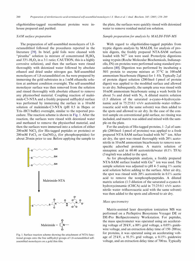
The preparation of self-assembled monolayers of 1,8-octanedithiol followed the procedures reported in theliterature [39]. In brief, gold foils were cleaned with“piranha” solution (a mixture of concentrated H2SO4and 33% H2O2 in a 3:1 ratio; CAUTION, this is a highlycorrosive solution), and then the surfaces were rinsedthoroughly with deionized water followed by absoluteethanol and dried under nitrogen gas. Self-assembledmonolayers of 1,8-octanedithiol on Au were prepared byimmersing the gold substrates in a 1 mM ethanolic solu-tion at ambient condition overnight. The self-assembledmonolayer surface was then removed from the solutionand rinsed thoroughly with absolute ethanol to removeany physisorbed material. Coupling reaction of malei-mide-C3-NTA and a freshly prepared sulfhydryl surfacewas performed by immersing the surface in a 10 mMsolution of maleimide-C3-NTA (pH 8.5 in Hepes orTris–HCl buVer) overnight, similar to the reported pro-cedure. The reaction scheme is shown in Fig. 1. After thereaction, the surfaces were rinsed with deionized waterand methanol to remove the physisorbed material, andthen the surfaces were immersed into a solution of either200 mM NiCl2 (for His-tagged peptides or proteins) or200 mM FeCl3 or Ga(NO3)3 (for phosphopeptides) forabout 20 min prior to use. Before applying the sample to
Fig. 1. Surface reaction scheme showing the attachment of NTA func-tional groups onto the free sulfhydryl groups of 1,8-octanedithiol self-assembled monolayers on a gold thin Wlm.
SSH
SSH
SSH
Maleimide-NTA
S S N
O
O
NH
NO
O-OO-
O-
OO
N
O
O
Maleimide =
the plate, the surfaces were quickly rinsed with deionizedwater to remove residual metal ion solution.
Sample preparation for analysis by MALDI-TOF-MS
With regard to oligohistidine-tagged peptides fromtryptic digests analysis by MALDI, for analysis of pro-tein digests, the freshly prepared NTA-SAM surfacesloaded with Ni2+ ion were used. Proteolytic digestionsusing trypsin (Roche Molecular Biochemicals, Indianap-olis, IN) on proteins were performed using standard pro-cedures [40]. Digestion was performed by incubating a50/1 protein to enzyme mixture at 37 °C in 100 mMammonium bicarbonate (Sigma) for 1–8 h. Typically 2�lof protein digest solution (200 fmol–1 pmol of proteindigest) was applied to the modiWed surface and allowedto air dry. Subsequently, the sample area was rinsed with10 mM ammonium bicarbonate using a wash bottle forabout 5 s and dried with N2. A diluted matrix solution(1:3 dilution of the saturated a-cyano-4-hydroxycin-namic acid in 75:25:0.1 v/v/v acetonitrile–water–triXuo-roacetic acid with the same solvent) was then added tothe spots and allowed to air dry. In the case of the con-trol sample on conventional gold surface, no rinsing wasincluded, and matrix was added and mixed with the sam-ple on the plate.
For the analysis of His-tagged proteins, 1–2 �l of sam-ple (200 fmol–1 pmol of proteins) was applied to a freshprepared NTA-SAM surface loaded with Ni2+ ion. Afterair dry, the spot was rinsed thoroughly with 20% aceto-nitrile in 10 mM ammonium bicarbonate to remove non-speciWc adsorbed proteins. A matrix solution ofsinnapinic acid in 60:40 acetonitrile/water (0.1% TFA)(3 mg/ml) was added to the spot.
As for phosphopeptide analysis, a freshly preparedNTA-SAM surface loaded with Ga3+ ion was used. Thesample solution was adjusted to pH 4–5 using 1% aceticacid solution before adding to the surface. After air dry,the spot was rinsed with 20% acetonitrile in 0.1% aceticacid to remove the nonphosphopeptides. A dilutedmatrix solution (1:3 dilution of the saturated a-cyano-4-hydroxycinnamic (CHCA) acid in 75:25:0.1 v/v/v aceto-nitrile–water–triXuoroacetic acid with the same solvent)was then added to the spots and allowed to air dry.
Mass spectrometry
Matrix-assisted laser desorption ionization MS wasperformed on a PerSeptive Biosystems Voyager DE orDE-Pro BioSpectrometry Workstation. For peptides,the mass spectrometer was operated using an accelerat-ing voltage of 20 kV, a 80% grid voltage, a 0.05% guide-wire voltage, and an extraction delay time of 150–200 ns;for proteins, it was operated using an accelerating volt-age of 25 kV, a 91.5% grid voltage, a 0.15% guidewirevoltage, and an extraction delay time of 700 ns. Typically
Preparation of nitrilotriacetic-acid-terminated self-assembled monolayers / J. Shen et al. / Anal. Biochem. 345 (2005) 258–269 261
average of 100–250 scans was accumulated to get thespectrum. External calibration was performed using thepeptide calibration mixture 1 or 2 or 3 purchased fromPerSeptive Biosystems. Calibration sample was depos-ited on unmodiWed gold foil which was carefullyattached to the standard stainless steel target (PerSeptiveBiosystems) using a double adhesive tape, in the sameway that the modiWed SAM surfaces were analyzed. TheMALDI matrices included CHCA (dissolved in 0.1%TFA acetonitrile/water 2:1 v/v) and SA (3 mg/ml in60:40:0.1 v/v/v acetonitrile/water/TFA). For CHCA, asaturated solution (dissolved in 0.1% TFA acetonitrile/water 2:1 v/v) was further diluted (1:3 or 1:10 ratio)using the same solvent when performing analysis onmodiWed SAMs surfaces.
X-ray photoelectron spectroscopy (XPS)
X-ray photoelectron spectroscopy was done with aPerkin-Elmer PHI 5600 ESCA system using a Al K�(1487 eV) monochromatic X-ray source. A survey spec-trum (binding energy, 1100–0 eV) and Au 4f, Ni 3p, O1 s, N 1 s, and S 2p high-resolution spectra with 20-eVranges were collected. The survey spectrum was collectedusing a pass energy of 187 eV and the high-resolution
spectra were collected using a pass energy of 58 eV. Sam-ples were examined at take-oV angles of 25° and 45°. Thehigh-resolution S 2p spectrum was curve Wtted usingMultipak 7.1A software.
Results and discussion
SAMs surface characterization
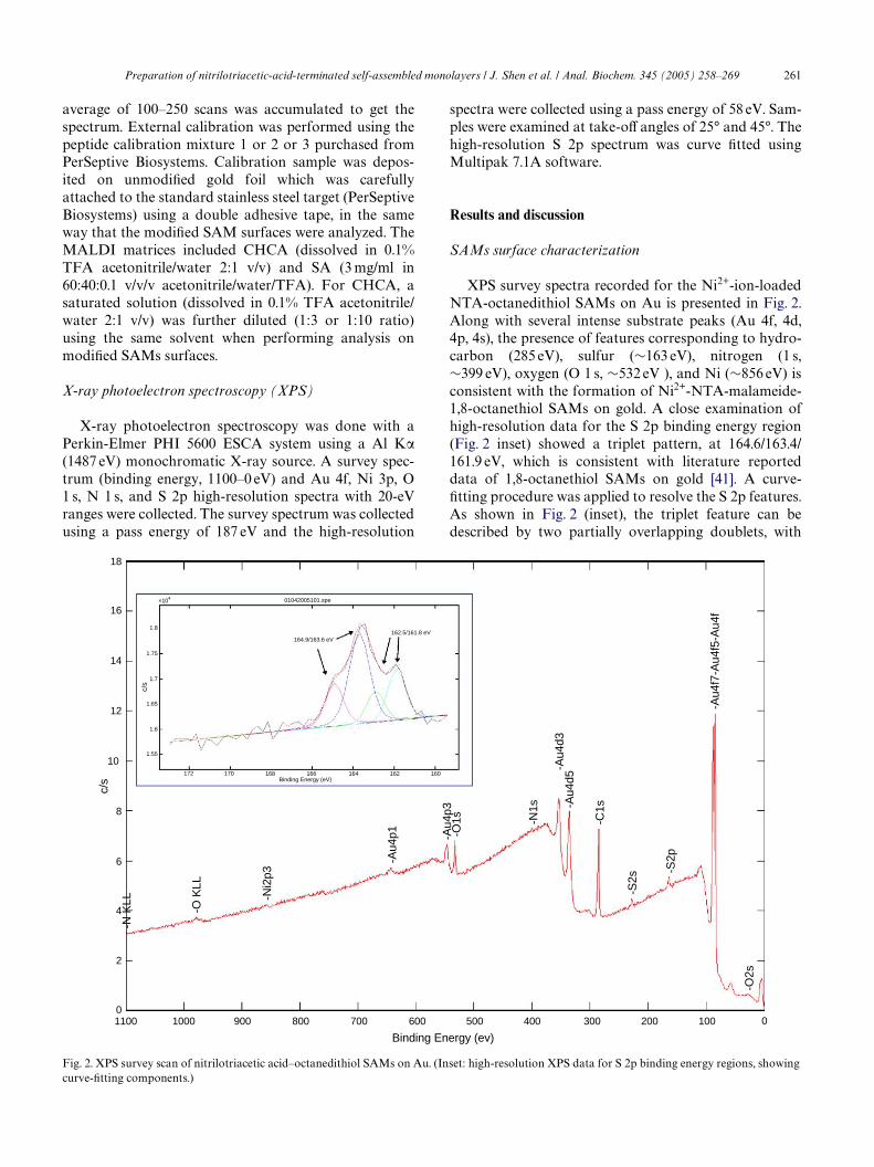
XPS survey spectra recorded for the Ni2+-ion-loadedNTA-octanedithiol SAMs on Au is presented in Fig. 2.Along with several intense substrate peaks (Au 4f, 4d,4p, 4s), the presence of features corresponding to hydro-carbon (285 eV), sulfur (»163 eV), nitrogen (1 s,»399 eV), oxygen (O 1 s, »532 eV ), and Ni (»856 eV) isconsistent with the formation of Ni2+-NTA-malameide-1,8-octanethiol SAMs on gold. A close examination ofhigh-resolution data for the S 2p binding energy region(Fig. 2 inset) showed a triplet pattern, at 164.6/163.4/161.9 eV, which is consistent with literature reporteddata of 1,8-octanethiol SAMs on gold [41]. A curve-Wtting procedure was applied to resolve the S 2p features.As shown in Fig. 2 (inset), the triplet feature can bedescribed by two partially overlapping doublets, with
Fig. 2. XPS survey scan of nitrilotriacetic acid–octanedithiol SAMs on Au. (Inset: high-resolution XPS data for S 2p binding energy regions, showingcurve-Wtting components.)
18
16
14
12
10
8
6
4
2
01100 1000 900 800 700 600 500 400 300 200 100 0
Binding Energy (ev)
c/s
-N K
LL
-O K
LL
-Ni2
p3
-Au4
p1
-Au4
d3
-Au4
f7-A
u4f5
-Au4
f
-Au4
p3
-Au4
d5
-O1s -N
1s
-C1s
-S2s
-S2p
-O2s
1.8
1.75
1.7
1.65
1.6
1.55
172 170 168 166 164 162 160
01042005101.spe
162.5/161.8 eV164.9/163.6 eV
×104
c/s
Binding Energy (eV)
262 Preparation of nitrilotriacetic-acid-terminated self-assembled monolayers / J. Shen et al. / Anal. Biochem. 345 (2005) 258–269
one occurring at 164.9/163.6 eV and another at 162.5/161.8 eV. Again, the data suggest that there are twodiVerent types of sulfur present at the surface, possiblydue to the molecules of 1,8-octanethiol oriented in a fash-ion of “head-down, tail-up” [41]. No features at theregion of 169 eV were observed, suggesting minimal pres-ence of sulfonate species. It should be noted that XPSanalysis of 1,8-octanedithiol on gold before NTA-malei-mide immobilization (data not shown) is consistent withthe literature data [41]. The high-resolution data showedthat the similar triplet pattern for the S 2p appeared at164.8/163.6/161.9 eV; however, in contrast to the NTA-maleimide-immobilized surface, no nitrogen signal at»400 eV was detected. The presence of nitrogen signal inthe NTA-maleimide-treated surface suggests that thefunctional group was incorporated onto the surface.
MALDI-TOF-MS has been reported to characterizeself assembly monolayer surfaces [42,43]. In this study,NTA-malameide-1,8-octanethiol SAMs on Au wereanalyzed in both positive and negative ion mode usingMALDI-TOF. In both cases, the signal was dominatedby the matrix ions. However, some features suggestedthe formation of NTA-malameide-1,8-octanethiol ongold. For example, the observation of ions at m/z
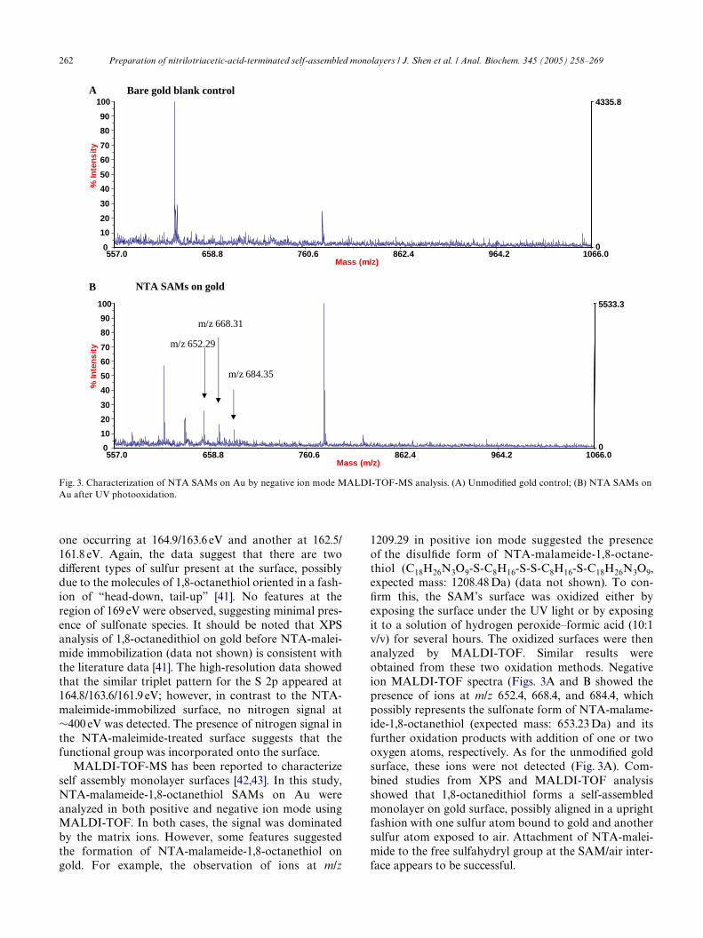
1209.29 in positive ion mode suggested the presenceof the disulWde form of NTA-malameide-1,8-octane-thiol (C18H26N3O9-S-C8H16-S-S-C8H16-S-C18H26N3O9,expected mass: 1208.48 Da) (data not shown). To con-Wrm this, the SAM’s surface was oxidized either byexposing the surface under the UV light or by exposingit to a solution of hydrogen peroxide–formic acid (10:1v/v) for several hours. The oxidized surfaces were thenanalyzed by MALDI-TOF. Similar results wereobtained from these two oxidation methods. Negativeion MALDI-TOF spectra (Figs. 3A and B showed thepresence of ions at m/z 652.4, 668.4, and 684.4, whichpossibly represents the sulfonate form of NTA-malame-ide-1,8-octanethiol (expected mass: 653.23 Da) and itsfurther oxidation products with addition of one or twooxygen atoms, respectively. As for the unmodiWed goldsurface, these ions were not detected (Fig. 3A). Com-bined studies from XPS and MALDI-TOF analysisshowed that 1,8-octanedithiol forms a self-assembledmonolayer on gold surface, possibly aligned in a uprightfashion with one sulfur atom bound to gold and anothersulfur atom exposed to air. Attachment of NTA-malei-mide to the free sulfahydryl group at the SAM/air inter-face appears to be successful.
Fig. 3. Characterization of NTA SAMs on Au by negative ion mode MALDI-TOF-MS analysis. (A) UnmodiWed gold control; (B) NTA SAMs onAu after UV photooxidation.
557.0 658.8 760.6 862.4 964.2 1066.0Mass (m/z)
0
4335.8
0
10
20
30
40
50
60
70
80
90
100%
Inte
ns
ity
557.0 658.8 760.6 862.4 964.2 1066.0Mass (m/z)
0
5533.3
0
10
20
30
40
50
60
70
80
90
100
% In
ten
sit
y m/z 652.29
m/z 668.31
m/z 684.35
Bare gold blank control
NTA SAMs on gold
A
B
Preparation of nitrilotriacetic-acid-terminated self-assembled monolayers / J. Shen et al. / Anal. Biochem. 345 (2005) 258–269 263
Analysis of oligohistidine tag peptides
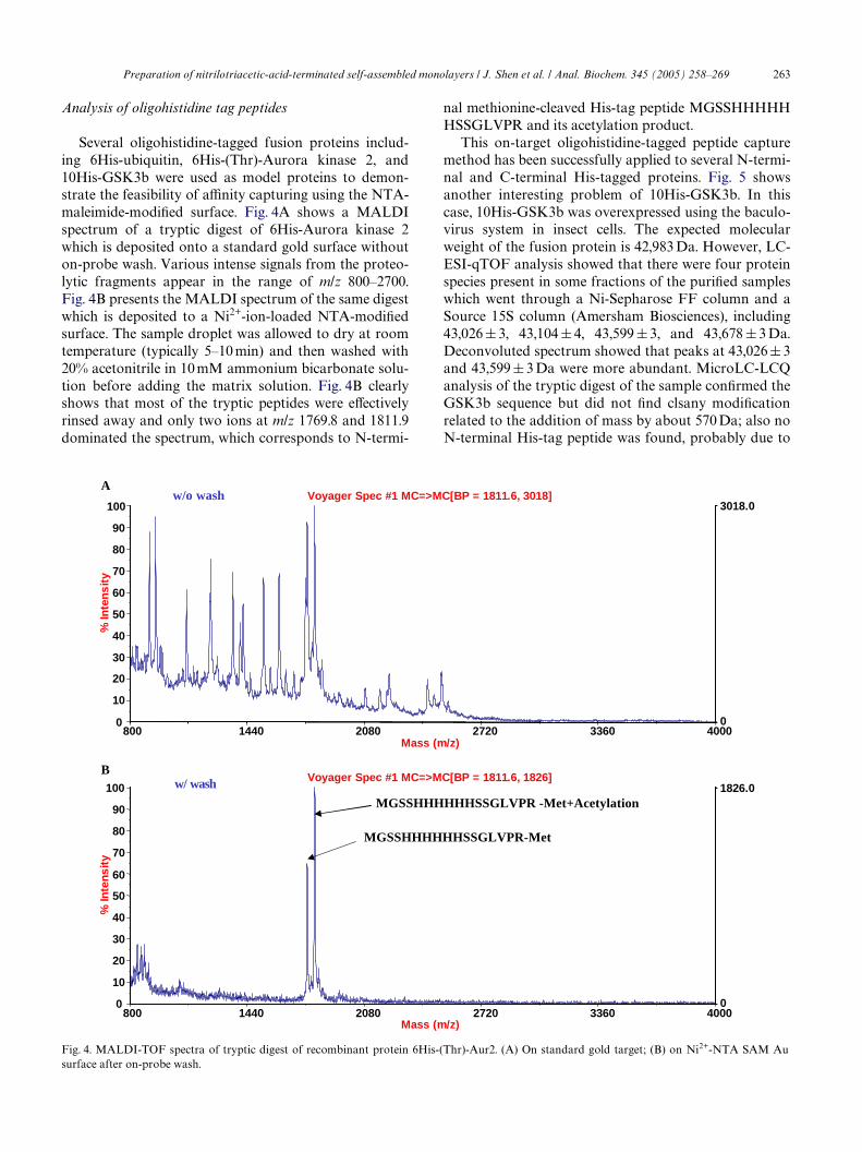
Several oligohistidine-tagged fusion proteins includ-ing 6His-ubiquitin, 6His-(Thr)-Aurora kinase 2, and10His-GSK3b were used as model proteins to demon-strate the feasibility of aYnity capturing using the NTA-maleimide-modiWed surface. Fig. 4A shows a MALDIspectrum of a tryptic digest of 6His-Aurora kinase 2which is deposited onto a standard gold surface withouton-probe wash. Various intense signals from the proteo-lytic fragments appear in the range of m/z 800–2700.Fig. 4B presents the MALDI spectrum of the same digestwhich is deposited to a Ni2+-ion-loaded NTA-modiWedsurface. The sample droplet was allowed to dry at roomtemperature (typically 5–10 min) and then washed with20% acetonitrile in 10 mM ammonium bicarbonate solu-tion before adding the matrix solution. Fig. 4B clearlyshows that most of the tryptic peptides were eVectivelyrinsed away and only two ions at m/z 1769.8 and 1811.9dominated the spectrum, which corresponds to N-termi-
nal methionine-cleaved His-tag peptide MGSSHHHHHHSSGLVPR and its acetylation product.
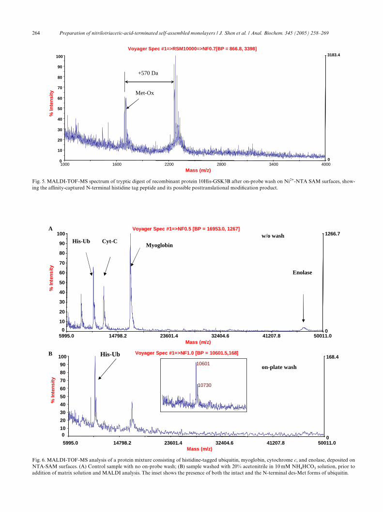
This on-target oligohistidine-tagged peptide capturemethod has been successfully applied to several N-termi-nal and C-terminal His-tagged proteins. Fig. 5 showsanother interesting problem of 10His-GSK3b. In thiscase, 10His-GSK3b was overexpressed using the baculo-virus system in insect cells. The expected molecularweight of the fusion protein is 42,983 Da. However, LC-ESI-qTOF analysis showed that there were four proteinspecies present in some fractions of the puriWed sampleswhich went through a Ni-Sepharose FF column and aSource 15S column (Amersham Biosciences), including43,026 § 3, 43,104 § 4, 43,599 § 3, and 43,678 § 3 Da.Deconvoluted spectrum showed that peaks at 43,026 § 3and 43,599 § 3 Da were more abundant. MicroLC-LCQanalysis of the tryptic digest of the sample conWrmed theGSK3b sequence but did not Wnd clsany modiWcationrelated to the addition of mass by about 570 Da; also noN-terminal His-tag peptide was found, probably due to
Fig. 4. MALDI-TOF spectra of tryptic digest of recombinant protein 6His-(Thr)-Aur2. (A) On standard gold target; (B) on Ni2+-NTA SAM Au
MGSSHHHHHHSSGLVPR -Met+Acetylation
800 1440 2080 2720 3360 4000Mass (m/z)
0
1826.0
0
10
20
30
40
50
60
70
80
90
100
% In
ten
sit
y
Voyager Spec #1 MC=>MC[BP = 1811.6, 1826]
800 1440 2080 2720 3360 4000Mass (m/z)
0
3018.0
0
10
20
30
40
50
60
70
80
90
100
% In
ten
sit
y
Voyager Spec #1 MC=>MC[BP = 1811.6, 3018]
MGSSHHHHHHSSGLVPR-Met
w/o wash
w/ wash
A
B
surface after on-probe wash.
264 Preparation of nitrilotriacetic-acid-terminated self-assembled monolayers / J. Shen et al. / Anal. Biochem. 345 (2005) 258–269
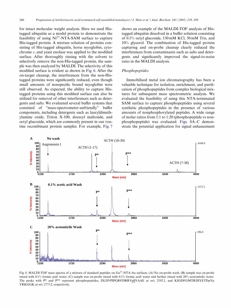
Fig. 6. MALDI-TOF-MS analysis of a protein mixture consisting of histidine-tagged ubiquitin, myoglobin, cytochrome c, and enolase, deposited onNTA-SAM surfaces. (A) Control sample with no on-probe wash; (B) sample washed with 20% acetonitrile in 10 mM NH4HCO3 solution, prior toaddition of matrix solution and MALDI analysis. The inset shows the presence of both the intact and the N-terminal des-Met forms of ubiquitin.
5995.0 14798.2 23601.4 32404.6 41207.8 50011.0Mass (m/z)
0
1266.7
0
10
20
30
40
50
60
70
80
90
100
% In
ten
sit
y
Voyager Spec #1=>NF0.5 [BP = 16953.0, 1267]
Enolase
MyoglobinCyt-CHis-Ub
w/o wash
5995.0 14798.2 23601.4 32404.6 41207.8 50011.0Mass (m/z)
0
168.4
010
20
30
40
50
60
70
80
90
100
% In
ten
sit
y
Voyager Spec #1=>NF1.0 [BP = 10601.5,168]
on-plate wash
His-Ub10601
10730
A
B
Fig. 5. MALDI-TOF-MS spectrum of tryptic digest of recombinant protein 10His-GSK3B after on-probe wash on Ni2+-NTA SAM surfaces, show-ing the aYnity-captured N-terminal histidine tag peptide and its possible posttranslational modiWcation product.
1000 1600 2200 2800 3400 4000
Mass (m/z)
0
3183.4
0
10
20
30
40
50
60
70
80
90
100%
In
ten
sity
Voyager Spec #1=>RSM10000=>NF0.7[BP = 866.8, 3398]
Met-Ox
+570 Da

Preparation of nitrilotriacetic-acid-terminated self-assembled monolayers / J. Shen et al. / Anal. Biochem. 345 (2005) 258–269 265
its highly hydrophilic nature, resulting in its early elutionand suppression by the salt cluster signals. To conWrm theN-terminal sequence, the tryptic digest was applied to theNi2+-NTA-modiWed surface and rinsed with 20% acetoni-trile in 10mM ammonium bicarbonate solution beforeMALDI analysis. MALDI spectrum (Fig. 5) showed twomajor peaks at m/z 1690.83 and m/z 2260.91 (monoiso-topic peak). The expected N-terminal His tag sequence isMHHHHHHHHHHK (expected mass: 1647.74 Da). Thedata suggest that N-terminal acetylation occurred, whichis consistent with q-TOF MW measurement. What isinteresting is the observation of peak at m/z 2260.91,which is 570 Da higher than the ion m/z 1690.83. Theresult suggests that the modiWcation may occur at the Histag peptide. In addition to the His-tag peptide peak at1690.83, peaks at m/z 1706.78 and 1722.76 were alsoobserved, these are possibly the methionine oxidationproducts. Several oxidation peaks were also observed forthe modiWed peptide, including m/z 2276.92, 2292.86,2308.84, and 2324.97. These oxidation products were notobserved in the intact protein MW measurement; theywere probably formed during the processes of trypsin
digestion and sample preparation. Independent studyusing IMAC Ziptip (Millipore) extraction and MALDI-TOF analysis showed similar results. Even though theidentity of the modiWcation (with mass addition of570Da) remains unknown, a followup study using LC-MS/MS of enriched sample by IMAC solid-phase extrac-tion failed to detect the modiWed peptide but detected onlythe unmodiWed peptides, suggesting that the modiWcationmay be labile and could be hydrolyzed or degraded. Fur-ther investigations using other techniques are underwaybut beyond the scope of this paper. The demonstratedexample here showed clearly that the NTA-modiWedSAMs surface is useful to capture and characterize oligoh-istidine tag peptides. We noted in some cases that nonspe-ciWc bindings are diYcult to eliminate; however, theoverall quality of the MALDI signal from these oligohisti-dine peptides improves dramatically.
Oligohistidine-tagged protein analysis
The NTA-modiWed surfaces provide an easy solutionto capture histidine-tagged proteins on MALDI target
Fig. 7. MALDI-TOF-MS analysis of histidine-tagged ubiquitin (A) in the presence of detergent and salt (0.1% octyl glucoside, 150 mM KCl, 50 mM2+
His-Ub
3999.0 7199.4 10399.8 13600.2 16800.6 20001.0Mass (m/z)
0
5848.3
0
10
20
30
40
50
60
70
80
90
100
% In
ten
sit
y
Voyager Spec #1[BP = 10600.3, 5848]
3999.0 7199.4 10399.8 13600.2 16800.6 20001.0Mass (m/z)
0
485.0
0
10
20
30
40
50
60
70
80
90
100
% In
ten
sit
y
Voyager Spec #1[BP = 5298.0, 485]A
B
Tris, and 10% glycerol), and (B) after on-probe wash on Ni -NTA SAMs surface.
266 Preparation of nitrilotriacetic-acid-terminated self-assembled monolayers / J. Shen et al. / Anal. Biochem. 345 (2005) 258–269
for intact molecular weight analysis. Here we used His-tagged ubiquitin as a model protein to demonstrate thefeasibility of using Ni2+-NTA-SAM surface to captureHis-tagged protein. A mixture solution of proteins con-sisting of His-tagged ubiquitin, horse myoglobin, cyto-chrome c, and yeast enolase was applied to the modiWedsurface. After thoroughly rinsing with the solvent toselectively remove the non-His-tagged protein, the sam-ple was then analyzed by MALDI. The selectivity of thismodiWed surface is evident as shown in Fig. 6. After theon-target cleanup, the interferences from the non-His-tagged proteins were signiWcantly reduced, even thoughsmall amounts of nonspeciWc bound myoglobin werestill observed. As expected, the ability to capture His-tagged proteins using this modiWed surface can also beutilized for removal of other interferences such as deter-gents and salts. We evaluated several buVer systems thatconsisted of “mass-spectrometer-unfriendly” buVercomponents, including detergents such as lauryldimeth-ylamine oxide, Triton X-100, docecyl maltoside, andoctyl glucoside, which are commonly present in our rou-tine recombinant protein samples. For example, Fig. 7
shows an example of the MALDI-TOF analysis of His-tagged ubiquitin dissolved in a buVer solution consistingof 0.1% octyl glucoside, 150 mM KCl, 50 mM Tris, and10% glycerol. The combination of His-tagged proteincapturing and on-probe cleanup clearly reduced theinterferences from contaminants such as salts and deter-gents and signiWcantly improved the signal-to-noiseratio in the MALDI analysis.
Phosphopeptides
Immobilized metal ion chromatography has been avaluable technique for isolation, enrichment, and puriW-cation of phosphopeptides from complex biological mix-tures for subsequent mass spectrometric analysis. Weevaluated the feasibility of using this NTA-terminatedSAM surface to capture phosphopeptides using severalsynthetic phosphopeptides in the presence of variousamounts of nonphosphorylated peptides. A wide rangeof molar ratios from 1:1 to 1:20 (phosphopeptide vs non-phosphopeptide) was evaluated. Figs. 8A–C demon-strate the potential application for signal enhancement
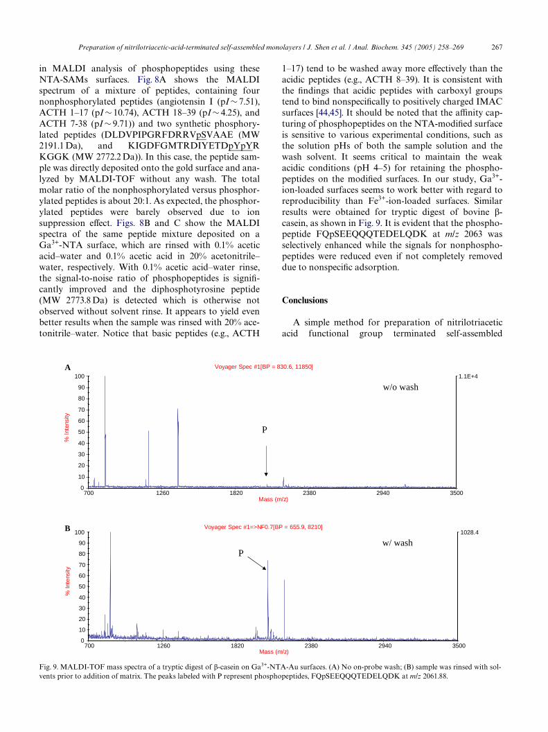
Fig. 8. MALDI-TOF mass spectra of a mixture of standard peptides on Ga3+-NTA-Au surfaces. (A) No on-probe wash; (B) sample was on-proberinsed with 0.1% formic acid–water; (C) sample was on-probe rinsed with 0.1% formic acid–water and further rinsed with 20% acetonitrile–water.The peaks with P* and P** represent phosphopeptides, DLDVPIPGRFDRRVpSVAAE at m/z 2192.1, and KIGDFGMTRDIYETDpYpYRKGGK at m/z 2773.2, respectively.
1150 1720 2290 2860 3430 4000Mass (m/z)
0102030405060708090
100
% I
nten
sit
y
1150 1720 2290 2860 3430 4000Mass (m/z)
0102030405060708090
100
% I
nten
sit
y
1150 1720 2290 2860 3430 4000Mass (m/z)
0102030405060708090
100
% I
nten
sit
y
No wash
0.1% acetic acid Wash
Angiotensin IACTH (1-17)
ACTH (18-39)
ACTH (7-38)
20% acetonitrile Wash
P*
P*
P*
P**
P**
P**
4160.0
1375.5
296.0
A
B
C
Preparation of nitrilotriacetic-acid-terminated self-assembled monolayers / J. Shen et al. / Anal. Biochem. 345 (2005) 258–269 267
in MALDI analysis of phosphopeptides using theseNTA-SAMs surfaces. Fig. 8A shows the MALDIspectrum of a mixture of peptides, containing fournonphosphorylated peptides (angiotensin I (pI » 7.51),ACTH 1–17 (pI » 10.74), ACTH 18–39 (pI » 4.25), andACTH 7-38 (pI » 9.71)) and two synthetic phosphory-lated peptides (DLDVPIPGRFDRRVpSVAAE (MW2191.1 Da), and KIGDFGMTRDIYETDpYpYRKGGK (MW 2772.2 Da)). In this case, the peptide sam-ple was directly deposited onto the gold surface and ana-lyzed by MALDI-TOF without any wash. The totalmolar ratio of the nonphosphorylated versus phosphor-ylated peptides is about 20:1. As expected, the phosphor-ylated peptides were barely observed due to ionsuppression eVect. Figs. 8B and C show the MALDIspectra of the same peptide mixture deposited on aGa3+-NTA surface, which are rinsed with 0.1% aceticacid–water and 0.1% acetic acid in 20% acetonitrile–water, respectively. With 0.1% acetic acid–water rinse,the signal-to-noise ratio of phosphopeptides is signiW-cantly improved and the diphosphotyrosine peptide(MW 2773.8 Da) is detected which is otherwise notobserved without solvent rinse. It appears to yield evenbetter results when the sample was rinsed with 20% ace-tonitrile–water. Notice that basic peptides (e.g., ACTH
1–17) tend to be washed away more eVectively than theacidic peptides (e.g., ACTH 8–39). It is consistent withthe Wndings that acidic peptides with carboxyl groupstend to bind nonspeciWcally to positively charged IMACsurfaces [44,45]. It should be noted that the aYnity cap-turing of phosphopeptides on the NTA-modiWed surfaceis sensitive to various experimental conditions, such asthe solution pHs of both the sample solution and thewash solvent. It seems critical to maintain the weakacidic conditions (pH 4–5) for retaining the phospho-peptides on the modiWed surfaces. In our study, Ga3+-ion-loaded surfaces seems to work better with regard toreproducibility than Fe3+-ion-loaded surfaces. Similarresults were obtained for tryptic digest of bovine �-casein, as shown in Fig. 9. It is evident that the phospho-peptide FQpSEEQQQTEDELQDK at m/z 2063 wasselectively enhanced while the signals for nonphospho-peptides were reduced even if not completely removeddue to nonspeciWc adsorption.
Conclusions
A simple method for preparation of nitrilotriaceticacid functional group terminated self-assembled
Fig. 9. MALDI-TOF mass spectra of a tryptic digest of �-casein on Ga3+-NTA-Au surfaces. (A) No on-probe wash; (B) sample was rinsed with sol-vents prior to addition of matrix. The peaks labeled with P represent phosphopeptides, FQpSEEQQQTEDELQDK at m/z 2061.88.
700 1260 1820 2380 2940 3500Mass (m/z)
1.1E+4
0
10
20
30
40
50
60
70
80
90
100
% I
nten
sity
Voyager Spec #1[BP = 830.6, 11850]
700 1260 1820 2380 2940 3500Mass (m/z)
1028.4
0
10
20
30
40
50
60
70
80
90
100
% I
nten
sity
Voyager Spec #1=>NF0.7[BP = 655.9, 8210]
P
P
w/o wash
w/ wash
A
B
268 Preparation of nitrilotriacetic-acid-terminated self-assembled monolayers / J. Shen et al. / Anal. Biochem. 345 (2005) 258–269
monolayer on gold is described. The modiWed surfacedemonstrated high aYnity toward oligohistidine-taggedpeptides/proteins and phosphopeptides. The strongaYnity capture allows on-target removal of contami-nants such as salts and detergents and interferences fromother proteins and peptides. The on-target sample prepa-ration leads to improved detection for oligohistidine-tagged peptides, proteins, and phosphopeptides. The useof NTA-modiWed surface not only simpliWes the samplepreparation but also potentially minimizes the sampleloss in the oV-line methods such as solid-phase extrac-tion. The aYnity capture of protein or peptides ispotentially valuable for studying protein–ligand or pro-tein–protein interactions by in situ MALDI analysis. Inthe latter case, several issues such as speciWcity and selec-tivity may need to be further improved. One of the issuesis competitive nonspeciWc adsorption at the surface. Tocapture and analyze more complex samples, such ascrude whole cell lysate, high-aYnity selection and toler-ance to various wash conditions are desirable. However,the versatility of using various functional self-assembledmonolayers to immobilize proteins and peptides, in com-bination with sensitive MALDI mass spectrometricanalysis, makes it promising both in characterization ofprotein/peptide analysis and in studying protein–ligandinteraction and bioassays.
References
[1] M. Karas, F. Hillenkamp, Laser desorption ionization of proteinswith molecular masses exceeding 10,000 daltons, Anal. Chem. 60(20) (1988) 2299–2301.
[2] F. Hillenkamp, M. Karas, R.C. Beavis, B.T. Chait, Matrix-assistedlaser desorption/ionization mass spectrometry of biopolymers,Anal. Chem. 63 (1991) 1193A–1203A.
[3] J.B. Fenn, M. Mann, C.K. Meng, S.F. Wong, C.M. Whitehouse,Electrospray ionization for mass spectrometry of large biomole-cules, Science 246 (1989) 64–71.
[4] D.A. Wolters, M.P. Washburn, J.R. Yates III, An automated mul-tidimensional protein identiWcation technology for shotgun pro-teomics, Anal. Chem. 73 (2001) 5683–5690.
[5] R. Aebersold, D. Goodlett, Mass spectrometry in proteomics,Chem. Rev. 101 (2001) 269–295.
[6] M. Mann, R. Hendrickson, A. Pandey, Analysis of proteins andproteomes by mass spectrometry, Annu. Rev. Biochem. 70 (2001)437–473.
[7] W. Zhang, A.N. Krutchinsky, B.T. Chait, “De novo” peptidesequencing by MALDI-quadrupole-ion trap mass spectrometry: apreliminary study, J. Am. Soc. Mass Spectrom. 14 (2003) 1012–1021.
[8] M. Mann, M. Wilm, Error-tolerant identiWcation of peptides insequence databases by peptide sequence tags, Anal. Chem. 66(1994) 4390–4399.
[9] Y. Ge, B.G. Lawhorn, M. ElNaggar, E. Strauss, J.-H. Park, T.P.Begley, F.W. McLaVerty, Top down characterization of largerproteins (45 kDa) by electron capture dissociation mass spectrom-etry, J. Am. Chem. Soc. 124 (2002) 672–678.
[10] M.A. Tito, K. Tars, K. Valegard, J. Hajdu, C.V. Robinson, Elec-trospray time-of-Xight mass spectrometry of the intact MS2 viruscapsid, J. Am. Chem. Soc. 122 (2000) 3550–3551.
[11] C. Fenselau, P.A. Demirev, Characterization of intact microorgan-isms by MALDI mass spectrometry, Mass Spectrom. Rev. 20(2002) 157–171.
[12] S.D. Fuerstenau, W.H. Benner, J.J. Thomas, C. Brugidou, B. Both-ner, G. Siuzdak, Mass spectrometry of an intact virus, Angew.Chem. Int. Ed. 40 (2001) 541–544.
[13] Y. Kim, G.B. Hurst, M.J. Doktycz, M.V. Buchanan, Improvingspot homogeneity by using polymer substrates in matrix-assistedlaser desorption/ionization mass spectrometry of oligonucleotides,Anal. Chem. 73 (2001) 2617–2624.
[14] K. Gevaert, H. Demol, T. Sklyarova, J. Vandekerckhove, T. Hou-thaeve, A peptide concentration and puriWcation method for pro-tein characterization in the subpicomole range using matrix-assisted laser desorption/ionization-postsource decay (MALDI-PSD) sequencing, Electrophoresis 19 (1998) 909–917.
[15] D.I. Papac, J. Hoyes, K.B. Tomer, Direct analysis of aYnity-bound analytes by MALDI/TOF MS, Anal. Chem. 66 (1994)2609–2613.
[16] B.O. Keller, L. Li, Nanoliter solvent extraction combined withmicrospot MALDI TOF mass spectrometry for the analysis ofhydrophobic biomolecules, Anal. Chem. 73 (2001) 2929–2936.
[17] M. Cadene, B.T. Chait, A. Robust, Detergent-friendly method formass spectrometric analysis of integral membrane proteins, Anal.Chem. 72 (2000) 5655–5658.
[18] D. Schleuder, F. Hillenkamp, K. Strupat, IR-MALDI-mass analy-sis of electroblotted proteins directly from the membrane: com-parison of diVerent membranes, application to on-membranedigestion, and protein identiWcation by database searching, Anal.Chem. 71 (1999) 3238–3247.
[19] J.A. Blackledge, A.J. Alexander, Polyethylene membrane as a sam-ple support for direct matrix-assisted laser desorption/ionizationmass spectrometric analysis of high mass proteins, Anal. Chem. 67(1995) 843–848.
[20] M.M. Vestling, C. Fenselau, Poly(vinylidene diXuoride) mem-branes as the interface between laser desorption mass spectrome-try, gel electrophoresis, and in situ proteolysis, Anal. Chem. 66(1994) 471–477.
[21] E.J. Zaluzec, D.A. Gage, J. Allison, J.T. Watson, Quantitativeassessment of cysteine and cystine in peptides and proteins follow-ing organomercurial derivatization and analysis by matrix-assisted laser desorption ionization mass spectrometry, J. Am. Soc.Mass Spectrom. 5 (1994) 230–237.
[22] R.W. Nelson, The use of bioreactive probes in protein character-ization, Mass Spectrom. Rev. 16 (1997) 353–376.
[23] Y. Xu, M.L. Bruening, J.T. Watson, Non-speciWc, on-probecleanup methods for MALDI-MS samples, Mass Spectrom. Rev.22 (2003) 429–440.
[24] N. Tang, P. Tornatore, S.R. Weinberger, Current developments inSELDI aYnity technology, Mass Spectrom. Rev. 23 (2004) 34–44.
[25] M.E. Warren, A.H. Brockman, R. Orlando, On-probe solid-phaseextraction/MALDI-MS using ion-pairing interactions for thecleanup of peptides and proteins, Anal. Chem. 70 (1998) 3757–3761.
[26] A.H. Brockman, N.N. Shah, R. Orlando, Optimization of a hydro-phobic solid-phase extraction interface for matrix-assisted laserdesorption/ionization, J. Mass Spectrom. 33 (1998) 1141.
[27] Y. Xu, J.T. Watson, M.L. Bruening, Patterned monolayer/polymerWlms for analysis of dilute or salt-contaminated protein samplesby MALDI-MS, Anal. Chem. 75 (2003) 185–190.
[28] Y. Xu, M.L. Bruening, J.T. Watson, Use of polymer-modiWedMALDI-MS probes to improve analyses of protein digests andDNA, Anal. Chem. 76 (2004) 3106–3111.
[29] J.L. Wilbur, A. Kumar, H.A. Biebuyck, E. Kim, G.M. Whitesides,Microcontact printing of self-assembled monolayers: applicationsin microfabrication, Nanotechnology 7 (1996) 452–457.
[30] Z. Ouyang, Z. Takáts, T.A. Blake, B. Gologan, A.J. Guymon, J.M.Wiseman, J.C. Oliver, V.J. Davisson, R.G. Cooks, Preparing pro-
Preparation of nitrilotriacetic-acid-terminated self-assembled monolayers / J. Shen et al. / Anal. Biochem. 345 (2005) 258–269 269
tein microarrays by soft-landing of mass-selected ions, Science 301(2003) 1351–1354.
[31] D.-H. Min, J. Su, M. Mrksich, ProWling kinase activities by using apeptide chip and mass spectrometry, Angew. Chem. Int. Ed. 43(2004) 5973–5977.
[32] G.B. Sigal, C. Bamdad, A. Barberis, J. Strominger, G.M. White-sides, A self-assembled monolayer for the binding and study ofhistidine-tagged proteins by surface plasmon resonance, Anal.Chem. 68 (1996) 490–497.
[33] G.J. Wegner, H.J. Lee, G. Marriott, R.M. Corn, Fabrication of his-tidine-tagged fusion protein arrays for surface plasmon resonanceimaging studies of protein–protein and protein–DNA interac-tions, Anal. Chem. 75 (2003) 4740–4746.
[34] M.T.W. Hearn, D. Acosta, Applications of novel aYnity cassettemethods: use of peptide fusion handles for the puriWcation ofrecombinant proteins, J. Mol. Recogn. 14 (2001) 323–369.
[35] K.M. Kim, E.C. Yi, D. Baker, K.Y. Zhang, Post-translationalmodiWcation of the N-terminal His tag interferes with the crystal-lization of the wild-type and mutant SH3 domains from chickensrc tyrosine kinase, Acta Crystallogr. D Biol. Crystallogr. 57(2001) 759–762.
[36] P. Cao, J.T. Stults, Phosphopeptide analysis by on-line immobi-lized metal-ion aYnity chromatography-capillary electrophoresis-electrospray ionization mass spectrometry, J. Chromatogr. A 853(1999) 225–235.
[37] T. Kocher, G. Allmaier, M. Mann, Nanoelectrospray based detec-tion and sequencing of substoichiometric amounts of phospho-peptides in complex mixture, J. Mass Spectrom. 38 (2003) 131–137.
[38] C.S. Raska, C.E. Parker, Z. Dominski, W.F. MarzluV, G.L. Glish,R.M. Pope, C.H. Borchers, Direct MALDI-MS/MS of phospho-peptides aYnity-bound to immobilized metal ion aYnity chroma-tography beads, Anal. Chem. 74 (2002) 3429–3433.
[39] C.E.D. Chidsey, C.R. Bertozzi, T.M. Putvinski, A.M. Mujsce,Coadsorption of ferrocene-terminated and unsubstituted alkan-ethiols on gold: electroactive self-assembled monolayers, J. Am.Chem. Soc. 112 (1990) 4301–4306.
[40] In: J. Walker (Ed.), Protein Protocols, Humana Press, Totowa,1998, Section 18.2.
[41] H. Rieley, G.K. Kendall, F.W. Zemicael, T.L. Smith, S. Yang, X-ray studies of self-assembled monolayers on coinage metals. 1.Alignment and photooxidation in 1,8-octanedithiol and 1-octane-thiol on Au, Langmuir 14 (1998) 5147–5153.
[42] J. Su, M. Mrksich, Using MALDI-TOF mass spectrometry tocharacterize interfacial reactions on self-assembled monolayers,Langmuir 19 (2003) 4867–4870.
[43] L. Hanley, O. Kornienko, E.T. Ada, E. Fuoco, J.L. Trevor, Surfacemass spectrometry of molecular species, J. Mass Spectrom. 34(1999) 705–723.
[44] G. Muszynska, G. Dobrowolska, A. Medin, P. Ekman, J.O.Porath, Model studies on iron(III) ion aYnity chromatography.II. Interaction of immobilized iron(III) ions with phosphorylatedamino acids, peptides and proteins, J. Chromatogr. 604 (1992) 19–28.
[45] S.B. Ficarro, M.L. McCleland, P.T. Stukenberg, D.J. Burke, M.M.Ross, J. Shabanowitz, D.F. Hunt, F.M. White, Phosphoproteomeanalysis by mass spectrometry and its application to Saccharomy-ces cerevisiae, Nat. Biotechnol. 20 (2002) 301–306.