
A novel and efficient excitation- and ionization-scheme for laser resonance ionization of mercury
7
Ž . Spectrochimica Acta Part B 54 1999 1793]1799 A novel and efficient excitation- and ionization-scheme for laser resonance ionization of mercury A.A. Podshivalov U,1 , O.I. Matveev, B.W. Smith, J.D. Winefordner Department of Chemistry, Uni ¤ ersity of Florida, Gaines ¤ ille, FL 32611]7200, USA Received 23 June 1999; accepted 21 August 1999 Abstract A novel, efficient scheme for mercury atomic resonance ionization is proposed and experimentally studied with wavelengths l s254 nm, l s313 nm and l s626 nm. The cross-sections of mercury photoionization from 1 2 3 different excited states were estimated using a new imaging method of resonance ionization signal measurement. Almost 100% efficiency of Hg resonance ionization was achieved using the first harmonic of a dye laser at 626 nm to ionize mercury atoms excited into the 6 3 D state. The photoionization cross-section from this state was found to be 2 y18 2 Ž . 1.5 =10 cm . Suppression of the ionization signal by coherent effects electromagnetically-induced transparency and the efficiency of resonance ionization were also studied. Q 1999 Elsevier Science B.V. All rights reserved. Keywords: Ionization; Laser spectrometry; Mercury; Resonance ionization imaging 1. Introduction In the fields of analytical laser resonance ion- w x ization mercury atom spectroscopy 1 ] 3 and res- w x onance ionization photon detection 4 ] 6 , effi- cient and simple photoionization schemes using the minimal number of lasers and minimal energy U Corresponding author. Fax: q1-352-392-4651. Ž . E-mail address: [email protected]fl.edu A.A. Podshivalov 1 Present address: International Laser Center of Moscow State University, Moscow, Russia, 119899. of laser radiation are important. Two- and three- step laser resonance ionization schemes for the photoionization of mercury have been the subject w x of many studies 1 ] 9 . One of the most often used schemes for mercury resonance ionization is with two lasers, namely l s 253.7 nm and l s l s 1 2 3 w x 435.8 nm 1 ] 6 . Using this scheme with short Ž . 7-ns pulse duration dye lasers, a maximum ion- wx ization efficiency of ; 10% was reached 4 and with a 30-ns dye laser pulse, the ionization effi- ciency was 2%. Our experiments showed that the low ioniza- tion efficiency was possibly caused by coherent 0584-8547r99r$ - see front matter Q 1999 Elsevier Science B.V. All rights reserved. Ž . PII: S 0 5 8 4 - 8 5 4 7 99 00112-3
-
Upload
independent -
Category
Documents
-
view
0 -
download
0
Transcript of A novel and efficient excitation- and ionization-scheme for laser resonance ionization of mercury
Ž .Spectrochimica Acta Part B 54 1999 1793]1799
A novel and efficient excitation- and ionization-schemefor laser resonance ionization of mercury
A.A. PodshivalovU ,1, O.I. Matveev, B.W. Smith, J.D. WinefordnerDepartment of Chemistry, Uni ersity of Florida, Gaines ille, FL 32611]7200, USA
Received 23 June 1999; accepted 21 August 1999
Abstract
A novel, efficient scheme for mercury atomic resonance ionization is proposed and experimentally studied withwavelengths l s254 nm, l s313 nm and l s626 nm. The cross-sections of mercury photoionization from1 2 3different excited states were estimated using a new imaging method of resonance ionization signal measurement.Almost 100% efficiency of Hg resonance ionization was achieved using the first harmonic of a dye laser at 626 nm toionize mercury atoms excited into the 63D state. The photoionization cross-section from this state was found to be2
y18 2 Ž .1.5=10 cm . Suppression of the ionization signal by coherent effects electromagnetically-induced transparencyand the efficiency of resonance ionization were also studied. Q 1999 Elsevier Science B.V. All rights reserved.
Keywords: Ionization; Laser spectrometry; Mercury; Resonance ionization imaging
1. Introduction
In the fields of analytical laser resonance ion-w xization mercury atom spectroscopy 1]3 and res-
w xonance ionization photon detection 4]6 , effi-cient and simple photoionization schemes usingthe minimal number of lasers and minimal energy
U Corresponding author. Fax: q1-352-392-4651.Ž .E-mail address: [email protected] A.A. Podshivalov
1Present address: International Laser Center of MoscowState University, Moscow, Russia, 119899.
of laser radiation are important. Two- and three-step laser resonance ionization schemes for thephotoionization of mercury have been the subject
w xof many studies 1]9 . One of the most often usedschemes for mercury resonance ionization is withtwo lasers, namely l s253.7 nm and l sl s1 2 3
w x435.8 nm 1]6 . Using this scheme with shortŽ .7-ns pulse duration dye lasers, a maximum ion-
w xization efficiency of ;10% was reached 4 andwith a 30-ns dye laser pulse, the ionization effi-ciency was 2%.
Our experiments showed that the low ioniza-tion efficiency was possibly caused by coherent
0584-8547r99r$ - see front matter Q 1999 Elsevier Science B.V. All rights reserved.Ž .PII: S 0 5 8 4 - 8 5 4 7 9 9 0 0 1 1 2 - 3
( )A.A. Podshi alo¨ et al. r Spectrochimica Acta Part B: Atomic Spectroscopy 54 1999 1793]17991794
population trapping, where electromagnetically-Ž .induced transparency EIT is a type of CPT, in
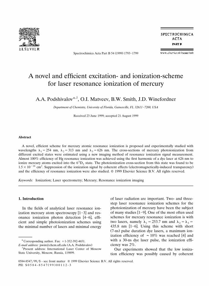
w xatomic mercury vapor 10,11 . To eliminate thesuppressing influence of the CPT effect in thiswork, a novel, efficient two-laser, three-wave-length scheme for mercury photoionization wasstudied. In order to compare the figures of meritof this new scheme, some characteristics of the
Ž .traditionally used scheme l sl s435.8 nm2 3Ž .were also studied see Fig. 1 .
2. Experimental setup
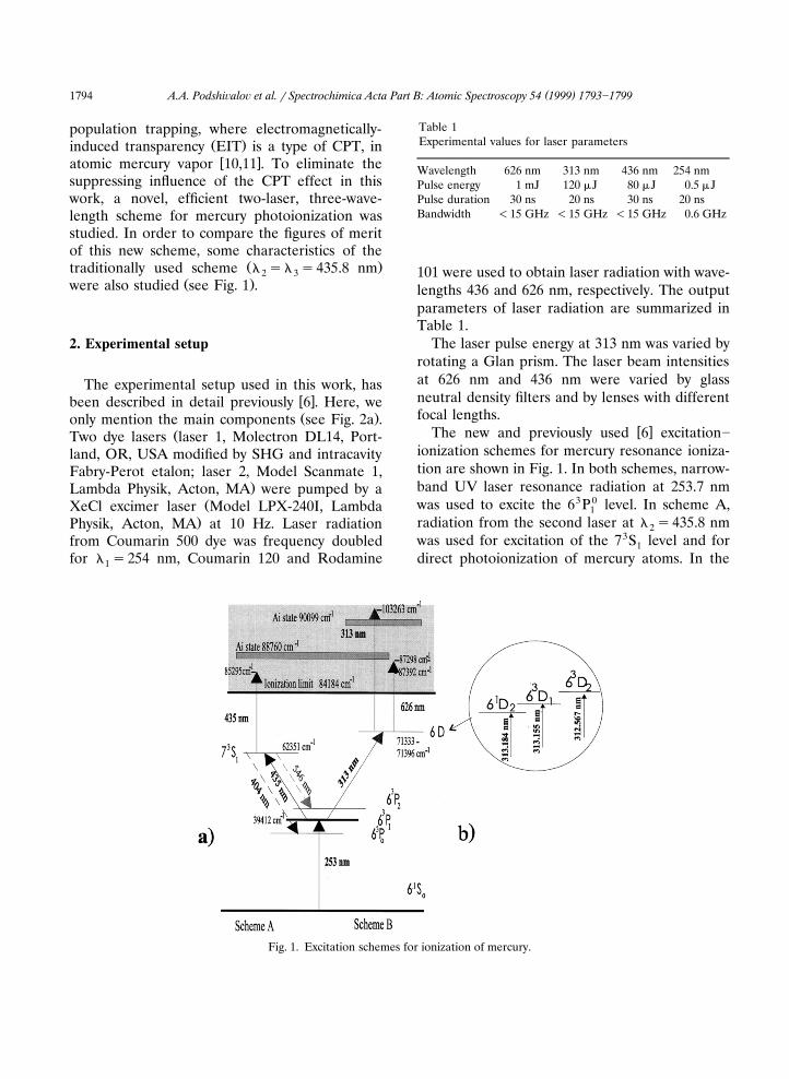
The experimental setup used in this work, hasw xbeen described in detail previously 6 . Here, weŽ .only mention the main components see Fig. 2a .
ŽTwo dye lasers laser 1, Molectron DL14, Port-land, OR, USA modified by SHG and intracavityFabry-Perot etalon; laser 2, Model Scanmate 1,
.Lambda Physik, Acton, MA were pumped by aŽXeCl excimer laser Model LPX-240I, Lambda
.Physik, Acton, MA at 10 Hz. Laser radiationfrom Coumarin 500 dye was frequency doubledfor l s254 nm, Coumarin 120 and Rodamine1
Table 1Experimental values for laser parameters
Wavelength 626 nm 313 nm 436 nm 254 nmPulse energy 1 mJ 120 mJ 80 mJ 0.5 mJPulse duration 30 ns 20 ns 30 ns 20 nsBandwidth -15 GHz -15 GHz -15 GHz 0.6 GHz
101 were used to obtain laser radiation with wave-lengths 436 and 626 nm, respectively. The outputparameters of laser radiation are summarized inTable 1.
The laser pulse energy at 313 nm was varied byrotating a Glan prism. The laser beam intensitiesat 626 nm and 436 nm were varied by glassneutral density filters and by lenses with differentfocal lengths.
w xThe new and previously used 6 excitation]ionization schemes for mercury resonance ioniza-tion are shown in Fig. 1. In both schemes, narrow-band UV laser resonance radiation at 253.7 nmwas used to excite the 63P0 level. In scheme A,1radiation from the second laser at l s435.8 nm2was used for excitation of the 73S level and for1direct photoionization of mercury atoms. In the
Fig. 1. Excitation schemes for ionization of mercury.
( )A.A. Podshi alo¨ et al. r Spectrochimica Acta Part B: Atomic Spectroscopy 54 1999 1793]1799 1795
Fig. 2. Experimental setup for laser resonance ionization ofŽ .mercury: a experimental set-up with two-electrode Hg cell;
Ž .b experimental set-up with RIID.
new scheme B, mercury atoms from the 63P01
state were excited to one of the three 6D levels bylaser radiation at 313 nm. The final ionizationstep was at 626 nm. The essence of the newscheme was that for the second excitation stepŽ .313 nm , a second harmonic of the tunable dyelaser was used, and the remainder of non-con-
verted radiation at the fundamental frequencyŽ .626 nm was used for the third ionization step.The advantages of this scheme are: first, only onelaser is used for both the second excitation stepexcitation and for ionization; second, the sum ofthe energy of the 6D state and the 626 nm quantais close to a Hg autoionization state, which candramatically increase the probability of Hg pho-toionization; and third, the 626 nm laser beam ismuch more intense than the 426 nm beam used inScheme A. It should be indicated that withScheme A, no 313- or 626-nm light is present andduring Scheme B, no 436-nm light is present.
The laser beams were directed into the cells13 y3.containing a high concentration f10 cm of
mercury. One of the cells, shown in Fig. 1b,Žcontained two planar parallel Ni electrodes not
.shown in the figure , separated by 10 mm, tomeasure the resonance ionization signal. The sec-ond cell was designed to produce images of 253.7-nm radiation focused on the surface of the inputwindow. The input quartz window of this cell was
Žcoated on the inside surface by a thin ;1]3 nm.thickness semi-transparent Pt film to eliminate
image distortion caused by surface charge. Theionization signal was visualized on the phosphor
Ž .screen Levy Hill Laboratories, Inc. Tarmac, FLwhich was designed using a 50-mm diameter,6-mm thick lead glass substrate coated with 7.5-mm particle size P22G phosphor at 7.5 mg cmy2
and aluminized with a 5-nm thick film. A DCvoltage of 10]15 kV was applied between thePt-coated input window and the phosphor screen.Free electrons, created as a result of multiphotonionization were accelerated by the electric fieldand visualized on the screen. The imaging signalfrom the screen, proportional to the number ofionized Hg atoms, was observed by a TV camera,digitized and measured using image analysis soft-
Ž .ware Sigma Scan Pro, SPSS Inc. Chicago, IL .
3. Experimental results and discussion
3.1. Experiments with two-electrode cell
Both schemes for mercury ionization were stud-
( )A.A. Podshi alo¨ et al. r Spectrochimica Acta Part B: Atomic Spectroscopy 54 1999 1793]17991796
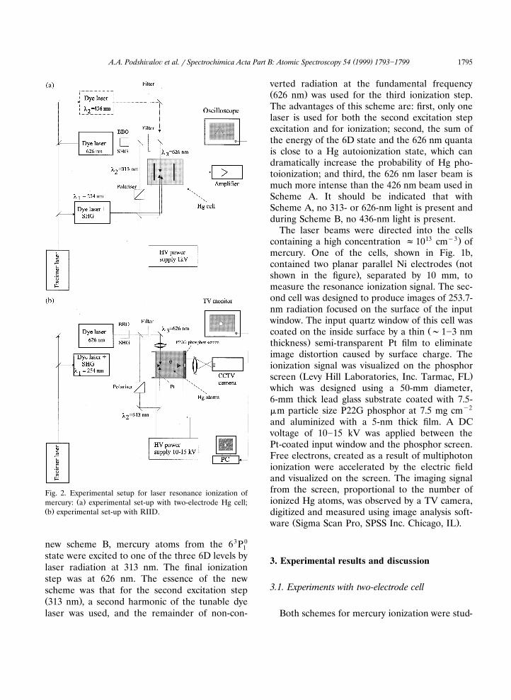
ied with the two electrode cell at 60 mtorr. Forthe experiments with scheme A, laser beams at254 and 436 nm, with diameters 1 and 3 mm,respectively, were directed into the cell from op-posite sides. The 436-nm laser beam was focusedto a diameter of 0.1 mm. A pulsed resonanceionization signal was observed when the 436-nm
Ž 3 3laser was exactly tuned to 435.834 nm 6 P ]7 S1 1.transition . When the 436-nm laser beam was
focused, the ionization signal had a clear depen-dence on the time delay between the 254- and436-nm laser pulses. The maximum signal wasobserved at a 50-ns time delay as shown in Fig. 3.We attribute this effect to coherent populationtrapping, CPT, of which electromagnetically-in-
w xduced transparency is one possibility 11 , whenthe intense second step resonance radiation de-creases the probability for absorption of 254-nmresonance quanta. This coherent effect can takeplace when the Rabi frequency of populationoscillations c1rT , where T is lifetime of 73S2 2 1
w xexcited state, or the laser bandwidth 10 . In ourcase, T s8 ns, the laser bandwidth is 15 GHz,2and the oscillator strength, fs0.11. It is easy toestimate that a CPT coherent effect can takeplace for our experimental conditions at a laserintensity )10 kW cmy2 . It is apparent that inour experiments the laser intensity exceeded thethreshold for CPT coherent effects. A detailedstudy of the observed CPT effect in Hg will be
w xpublished later 11 . At the optimal delay betweenexcitation pulses, when one can neglect coherenteffects, the ionization signal had a linear depen-dence on the 436-nm laser pulse energy with aslope of 2 mV mJ. No saturation of the ionizationsignal vs . irradiance was observed up to the maxi-mum possible value, ;200 mJ cmy2 , when the436-nm beam was focused into the cell by a shortfocusing lens. It should be stressed that the 436-nm laser pulse had a temporal double peak with aFWHM of approximately 34 nm as well as a tail,which caused the need for a 50-ns time delay.
When scheme B was used, two laser beams at254 and 313 nm, with diameters of 1 and 3 mm,respectively, were directed into the Hg cell fromopposite sides. A pulsed resonance ionization sig-nal was observed when the 313-nm laser was
Ž 3 0 1exactly tuned to 313.184 nm 6 P y6 D transi-1 2
Fig. 3. Ionization signal amplitude vs . l s436 nm pulse2energy and time delay.
. Ž 3 0 3 .tion , 313.155 nm 6 P y6 D transition and1 1Ž 3 0 3 .312.567 nm 6 P y6 D transition . The relative1 2
amplitude of these ionization signals in the non-saturated regime had a ratio of 10:3:4, respec-
Žtively. Also a very weak 50 times less than from.313 q 254-nm excitation resonance ionization
signal, due only to the 254-nm laser radiation, wasobserved. The amplitude of the ionization signalwas increased approximately 40 times when the626-nm beam of 3-mm diameter was also directedinto the cell. The dependence of the ionizationsignal vs . the 626-nm laser pulse energy was lin-ear with a slope of 3 mV mJ. However, even witha focused 626-nm laser beam, we did not observea saturated ionization signal up to an irradianceof 600 mJ cmy2 .
The laser fluence necessary to saturate they2 w xionization signal for scheme A is 87 mJ cm 1 .
However, we have not observed any saturationeffects up to irradiances of 200 and 600 mJ cmy2
for scheme A and B, respectively. We assume thisis because the volume within the mercury cellwhere saturation took place was always much lessthan the volume where ionization of mercuryoccurred. Therefore, the ionization signal fromthe volume where saturation occurred was con-siderably less than the signal from the entireilluminated volume. To examine the effect ofsaturation of the ionization signal and to obtaindata about the cross-section for ionization fromthe excited state, we used an imaging procedureto evaluate the signals in subsequent experiments.
( )A.A. Podshi alo¨ et al. r Spectrochimica Acta Part B: Atomic Spectroscopy 54 1999 1793]1799 1797
3.2. Experiments with resonance ionization image( )detector RIID detector
Using both schemes A and B, clear images ofthe volume illuminated by the ionizing lasers wereobserved. The images were seen only when thevoltage between the input window and the phos-phor screen exceeded 5 kV. Under 254-nm illumi-nation, no additional electrons due to a photo-electric effect from the Pt film were observed.When the source of Hg atoms was closed, noionization or photoelectric effect signals wereobserved. It is important to emphasize that therewas also no visible multiphoton ionization whenonly 254-, 436-, 626- or only 313-nm beams werefocused into the RIID cell with the maximumŽ .5-mtorr concentration of Hg atoms. The level ofnoise in the RIID was very low; only random 1]2pixel events in the image were observed, pre-sumably due to cosmic rays. The advantage ofmeasuring the signal via an imaging approach liesin the ability to spatially resolve the ionizationprocess. In the imaging mode, the signal canalways be detected in a small constant volumeusing the maximum laser power. In conventionaldetection from the bulk atomic volume with elec-trodes, it is possible to measure the effect ofsignal saturation only if the laser beams have
Ž .relatively large diameters )2]3 mm .For scheme A, even when the laser beam was
Ž y2 .strongly focused ;200 mJ cm , we did notobserve saturation of the ionization signal. There-fore, it was concluded that the fluence required tosaturate the ionization signal has to be severaltimes larger than the theoretical value of 87 mJcmy2 .
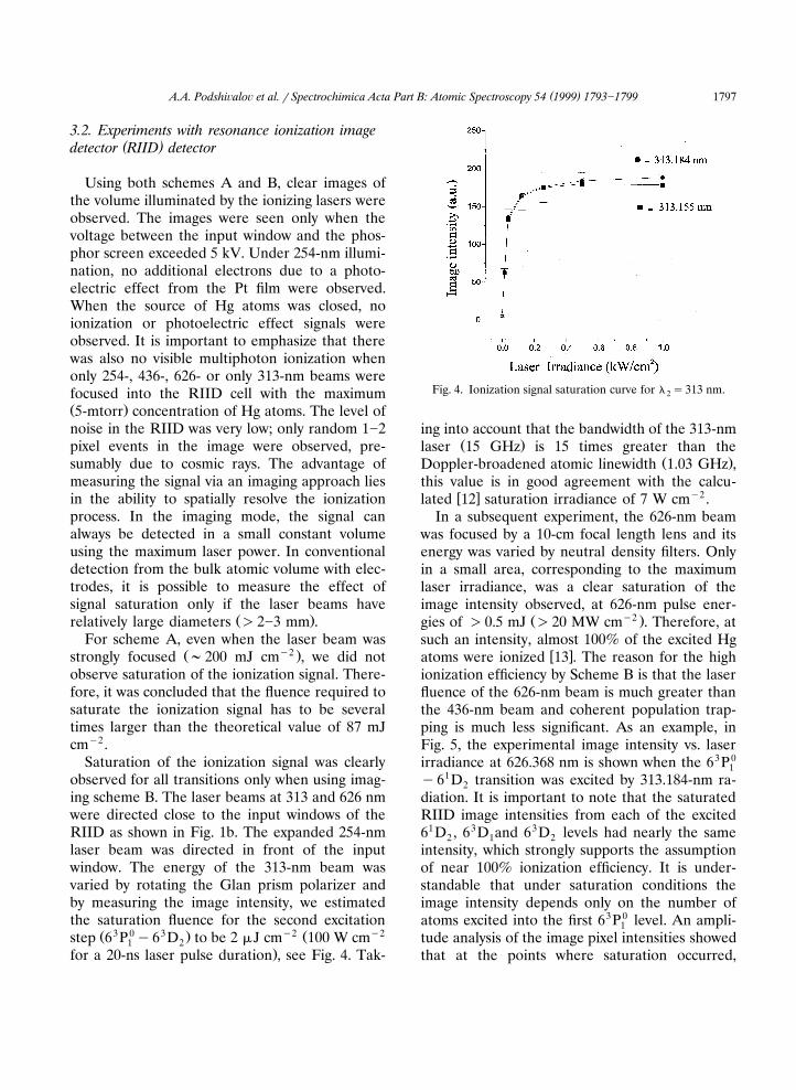
Saturation of the ionization signal was clearlyobserved for all transitions only when using imag-ing scheme B. The laser beams at 313 and 626 nmwere directed close to the input windows of theRIID as shown in Fig. 1b. The expanded 254-nmlaser beam was directed in front of the inputwindow. The energy of the 313-nm beam wasvaried by rotating the Glan prism polarizer andby measuring the image intensity, we estimatedthe saturation fluence for the second excitation
Ž 3 0 3 . y2 Ž y2step 6 P y6 D to be 2 mJ cm 100 W cm1 2.for a 20-ns laser pulse duration , see Fig. 4. Tak-
Fig. 4. Ionization signal saturation curve for l s313 nm.2
ing into account that the bandwidth of the 313-nmŽ .laser 15 GHz is 15 times greater than the
Ž .Doppler-broadened atomic linewidth 1.03 GHz ,this value is in good agreement with the calcu-
w x y2lated 12 saturation irradiance of 7 W cm .In a subsequent experiment, the 626-nm beam
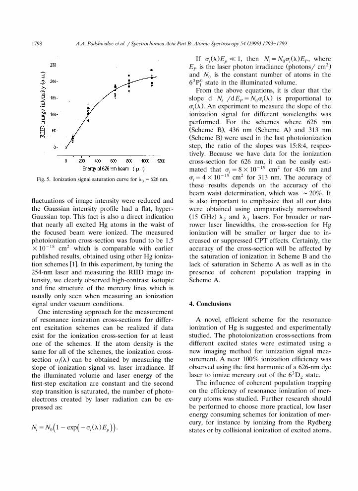
was focused by a 10-cm focal length lens and itsenergy was varied by neutral density filters. Onlyin a small area, corresponding to the maximumlaser irradiance, was a clear saturation of theimage intensity observed, at 626-nm pulse ener-
Ž y2 .gies of )0.5 mJ )20 MW cm . Therefore, atsuch an intensity, almost 100% of the excited Hg
w xatoms were ionized 13 . The reason for the highionization efficiency by Scheme B is that the laserfluence of the 626-nm beam is much greater thanthe 436-nm beam and coherent population trap-ping is much less significant. As an example, inFig. 5, the experimental image intensity vs. laserirradiance at 626.368 nm is shown when the 63P0
1y61D transition was excited by 313.184-nm ra-2diation. It is important to note that the saturatedRIID image intensities from each of the excited61D , 63D and 63D levels had nearly the same2 1 2intensity, which strongly supports the assumptionof near 100% ionization efficiency. It is under-standable that under saturation conditions theimage intensity depends only on the number ofatoms excited into the first 63P0 level. An ampli-1tude analysis of the image pixel intensities showedthat at the points where saturation occurred,
( )A.A. Podshi alo¨ et al. r Spectrochimica Acta Part B: Atomic Spectroscopy 54 1999 1793]17991798
Fig. 5. Ionization signal saturation curve for l s626 nm.3
fluctuations of image intensity were reduced andthe Gaussian intensity profile had a flat, hyper-Gaussian top. This fact is also a direct indicationthat nearly all excited Hg atoms in the waist ofthe focused beam were ionized. The measuredphotoionization cross-section was found to be 1.5=10y18 cm2 which is comparable with earlierpublished results, obtained using other Hg ioniza-
w xtion schemes 1 . In this experiment, by tuning the254-nm laser and measuring the RIID image in-tensity, we clearly observed high-contrast isotopicand fine structure of the mercury lines which isusually only seen when measuring an ionizationsignal under vacuum conditions.
One interesting approach for the measurementof resonance ionization cross-sections for differ-ent excitation schemes can be realized if dataexist for the ionization cross-section for at leastone of the schemes. If the atom density is thesame for all of the schemes, the ionization cross-
Ž .section s l can be obtained by measuring theislope of ionization signal vs . laser irradiance. Ifthe illuminated volume and laser energy of thefirst-step excitation are constant and the secondstep transition is saturated, the number of photo-electrons created by laser radiation can be ex-pressed as:
Ž .N sN 1yexp ys l E .Ž .Ž .i 0 i p
Ž . Ž .If s l E <1, then N sN s l E , wherei p i 0 i PŽ 2 .E is the laser photon irradiance photonsr cmP
and N is the constant number of atoms in the063P0 state in the illuminated volume.1
From the above equations, it is clear that theŽ .slope d N rd E sN s l is proportional toi P 0 i
Ž .s l . An experiment to measure the slope of theiionization signal for different wavelengths wasperformed. For the schemes where 626 nmŽ . Ž .Scheme B , 436 nm Scheme A and 313 nmŽ .Scheme B were used in the last photoionizationstep, the ratio of the slopes was 15:8:4, respec-tively. Because we have data for the ionizationcross-section for 626 nm, it can be easily esti-mated that s s8=10y19 cm2 for 436 nm andis s4=10y19 cm2 for 313 nm. The accuracy ofithese results depends on the accuracy of thebeam waist determination, which was ;20%. Itis also important to emphasize that all our datawere obtained using comparatively narrowbandŽ .15 GHz l and l lasers. For broader or nar-2 3rower laser linewidths, the cross-section for Hgionization will be smaller or larger due to in-creased or suppressed CPT effects. Certainly, theaccuracy of the cross-section will be affected bythe saturation of ionization in Scheme B and thelack of saturation in Scheme A as well as in thepresence of coherent population trapping inScheme A.
4. Conclusions
A novel, efficient scheme for the resonanceionization of Hg is suggested and experimentallystudied. The photoionization cross-sections fromdifferent excited states were estimated using anew imaging method for ionization signal mea-surement. A near 100% ionization efficiency wasobserved using the first harmonic of a 626-nm dyelaser to ionize mercury out of the 63D state.2
The influence of coherent population trappingon the efficiency of resonance ionization of mer-cury atoms was studied. Further research shouldbe performed to choose more practical, low laserenergy consuming schemes for ionization of mer-cury, for instance by ionizing from the Rydbergstates or by collisional ionization of excited atoms.
( )A.A. Podshi alo¨ et al. r Spectrochimica Acta Part B: Atomic Spectroscopy 54 1999 1793]1799 1799
Acknowledgements
This work was supported by a Phase I contractfrom DARPA and the University of Florida.
References
w x Ž .1 E.B. Saloman, Spectrochim. Acta Part B 46 1991 319.w x Ž .2 B.A. Bushaw, Anal. Chem. 57 1985 2397.w x3 W.L. Clevenger, O.I. Matveev, S. Cabredo, N. Omenetto,
Ž .B.W. Smith, J.D. Winefordner, Anal. Chem. 69 19972232.
w x4 O.I. Matveev, B.W. Smith, N. Omenetto, J.D. Wineford-Ž .ner, Spectrochim. Acta Part B 51 1996 563.
w x5 O.I. Matveev, B.W. Smith, J.D. Winefordner, Appl. Opt.Ž .36 1997 8833.
w x6 O.I. Matveev, B.W. Smith, J.D. Winefordner, Opt. Lett.Ž .23 1998 304.
w x Ž .7 P. Dyer, G.C. Baldwin, Appl. Phys. Lett. 42 1983 311.w x8 O.I. Matveev, P. Cavalli, N. Omenetto, AIP Conference
Proceedings of the 7th International Symposium onResonance Ionization Spectroscopy and its applicationsŽ .RIS 94 , American Institute of Physics, New York,1994. p. 269]272.
w x9 P. Bisling, C. Weitkamp, H. Zobel. P. Bisling, C. Wi-etkamp, H. Zobel, AIP Conference Proceedings of the8th International Symposium on Resonance Ionization
Ž .Spectroscopy and its Applications RIS 96 , AmericanInstitute of Physics, New York, 1996, p. 283]286.
w x10 S.E. Harris, Physics Today, 1997 36]42.w x11 A.A. Podshivalov, O.I. Matveev, B.W., Smith, J.D. Wine-
fordner, Opt. Lett. to be published.w x12 E.C. Benck, J.E. Lawler, J.T. Dankin, J. Opt. Soc. Am. B
Ž .6 1989 11.w x13 V.S. Letokhov, Laser Photoionization Spectroscopy,
Academic Press, Inc., 1987.