Organic and Inorganic Mercury in Neonatal Rat Brain after Prenatal Exposure to Methylmercury and...
7
242 VOLUME 118 | NUMBER 2 | February 2010 • Environmental Health Perspectives Research Mercury (Hg) toxicity is the focus of substan‑ tial public concern. Pronouncements about the safety of fish in the diet and about adverse health effects attributable to dental amalgams are the primary bases underlying this concern. Hg occurs in different forms in the environ‑ ment; Hg species are classified as elemental (Hg 0 ), inorganic (Hg 2+ , Hg + ), and organic [e.g., methylmercury (MeHg)]. However, many populations are exposed to more than one form. Almost all of our knowledge is based on research devoted to only one Hg species. How different species of Hg act in combina‑ tion remains unclear, which leads to significant gaps in our understanding of Hg toxicity. MeHg, to which humans are primarily exposed through consumption of contami‑ nated fish, shellfish, and sea mammals [U.S. Environmental Protection Agency (EPA) 1997], is a potent neurotoxicant to both the mature and developing central nervous system. Several studies have shown potential adverse effects on child development from prenatal exposure to MeHg (for review, see Clarkson and Magos 2006). Observations in human populations demonstrate that MeHg readily crosses the placental barrier, as indicated by cord blood levels higher than those seen in maternal blood (Vahter et al 2000). MeHg can penetrate into the fetal brain (Aschner and Aschner 1990; Choi et al. 1978; Davis et al. 1994; Day et al. 2005; Newland et al. 2006; Newland and Reile 1999; Stern et al. 2001), which allows for accumulation in the central nervous system. Because MeHg is slowly con‑ verted to inorganic Hg in brain tissue, and as such resides there for many years (Charleston et al. 1995; Davis et al. 1994), the relative con‑ tribution of the intact organomercurial versus the inorganic metabolite to neuronal damage remains an open question (Magos 1987), with significant implications for risk assessment. Another major source of Hg exposure is inorganic Hg in the form of Hg vapor. Exposure of the general population to Hg vapor occurs primarily through inhala‑ tion of Hg vapor released from dental amal‑ gams [Brownawell et al. 2005; International Programme on Chemical Safety (IPCS) 1991]. Although the levels from such exposures are considered “low” with respect to some known health effects, we know little about how those exposures might interact with MeHg toxicity. Approximately 80% of inhaled Hg vapor is retained and absorbed in blood during pulmo‑ nary circulation (Hursh et al. 1976). Although Hg vapor introduced into the body is trapped and oxidized to divalent inorganic Hg in erythrocytes, part of the Hg vapor remains in the bloodstream long enough for it to be dis‑ tributed to other tissues and reach the blood– brain barrier (Yoshida 2002). Non‑ionized Hg readily penetrates the placental barrier (Clarkson et al. 1972; Khayat and Dencker 1982) and is taken up by fetal tissues, including brain. e ability of inhaled Hg vapor to accu‑ mulate in the fetal brain has also been shown in human and animal studies (Drasch et al. 1994; Morgan et al. 2002, 2006). Inhaled Hg vapor has been known to damage the adult central nervous system (Echeverria et al. 1998; Goldwater 1972). On the other hand, only a limited number of studies have shown behav‑ ioral changes in animals prenatally exposed to Hg vapor (Danielsson et al. 1993; Fredriksson et al. 1996). However, the study designs of these experiments were not relevant to human exposure because of rather high concentrations of Hg vapor and short durations of exposure. Although studies of combined exposures are limited, we do know that both agents a) produce prenatal damage; b) produce behavioral changes in animals at levels that do not produce clinically apparent toxicity; and c) result in deposition in the brain of the same toxic species of Hg, namely, mercuric Hg. Yet there are reasons to suggest that combined exposure to MeHg and inhaled Hg vapor might produce effects different from those seen from exposure to either agent alone: • MeHg is well known to affect the develop‑ ing central nervous system, whereas effects of inhaled Hg vapor on that system are not clear. • Metabolism of MeHg differs from that of Hg vapor (Clarkson and Magos 2006). • Brain pathology and the signs and symp‑ toms of MeHg poisoning differ from those of inorganic Hg poisoning (Clarkson and Magos 2006). • Although both MeHg and Hg vapor result in deposition in the brain as mercuric Hg, divalent inorganic Hg (Hg 2+ ) is believed to be the proximate toxic agent in the case of poisoning from inhaled Hg vapor, but this Address correspondence to B. Weiss, Department of Environmental Medicine, Box EHSC, School of Medicine and Dentistry, University of Rochester, Rochester, NY 14642 USA. Telephone: (585) 275‑1736. Fax: (585) 256‑2591. E‑mail: Bernard_ [email protected] We thank T. Zarcone, M. Balys, A. Lunts, and M. Miller for their contributions and assistance. is study was supported by Research Grant 1‑R01‑ ES013247 to B.W., by Center Grant ES‑01247 from the National Institute of Environmental Health Sciences, and by 5 UL1 RR024160‑03 from the National Center for Research Resources. The authors declare they have no competing financial interests. Received 8 May 2009; accepted 29 September 2009. Organic and Inorganic Mercury in Neonatal Rat Brain after Prenatal Exposure to Methylmercury and Mercury Vapor Hiromi Ishitobi, 1 Sander Stern, 1 Sally W. Thurston, 2 Grazyna Zareba, 1 Margaret Langdon, 1 Robert Gelein, 1 and Bernard Weiss 1 1 Department of Environmental Medicine, and 2 Department of Biostatistics and Computational Biology, School of Medicine and Dentistry, University of Rochester, Rochester, New York, USA BACKGROUND: Many populations are exposed to multiple species of mercury (Hg), predominantly organic Hg as methylmercury (MeHg) from fish, and inorganic Hg as Hg vapor from dental amal- gams. Most of our knowledge of the neurotoxicity of Hg is based on research devoted to studying only one form at a time, mostly MeHg. OBJECTIVES: In this study we investigated the effects of prenatal exposure to MeHg and Hg vapor on Hg concentrations in the brain of neonatal rats. METHODS: Female Long-Evans hooded rats were exposed to MeHg (0, 3, 6, or 9 ppm as drinking solution), Hg vapor (0, 300, or 1,000 µg/m 3 for 2 hr/day), or the combination of both, from 30 days before breeding through gestational day 18. On postnatal day 4, whole brains were taken from one male and one female from each of four litters in each treatment group to assess organic and inorganic Hg in the brain by cold vapor atomic absorption spectrometry. RESULTS: Statistical analysis using linear mixed effects models showed that MeHg dose was the pri- mary determinant of both organic and inorganic brain Hg levels. For both outcomes, we also found significant interactions between MeHg and Hg vapor exposure. ese interactions were driven by the fact that among animals not exposed to MeHg, animals exposed to Hg vapor had significantly greater organic and inorganic brain Hg levels than did unexposed animals. CONCLUSION: is interaction, heretofore not reported, suggests that coexposure to MeHg and Hg vapor at levels relevant to human exposure might elevate neurotoxic risks. KEY WORDS: brain, coexposure, inorganic mercury, mercury vapor, methylmercury, organic mer- cury, prenatal. Environ Health Perspect 118:242–248 (2010). doi:10.1289/ehp.0900956 available via http://dx.doi.org/ [Online 29 September 2009]
-
Upload
independent -
Category
Documents
-
view
1 -
download
0
Transcript of Organic and Inorganic Mercury in Neonatal Rat Brain after Prenatal Exposure to Methylmercury and...

242 volume 118 | number 2 | February 2010 • Environmental Health Perspectives
Research
Mercury (Hg) toxicity is the focus of substan‑tial public concern. Pronouncements about the safety of fish in the diet and about adverse health effects attributable to dental amalgams are the primary bases underlying this concern. Hg occurs in different forms in the environ‑ment; Hg species are classified as elemental (Hg0), inorganic (Hg2+, Hg+), and organic [e.g., methylmercury (MeHg)]. However, many populations are exposed to more than one form. Almost all of our knowledge is based on research devoted to only one Hg species. How different species of Hg act in combina‑tion remains unclear, which leads to significant gaps in our understanding of Hg toxicity.
MeHg, to which humans are primarily exposed through consumption of contami‑nated fish, shellfish, and sea mammals [U.S. Environmental Protection Agency (EPA) 1997], is a potent neuro toxicant to both the mature and developing central nervous system. Several studies have shown potential adverse effects on child development from prenatal exposure to MeHg (for review, see Clarkson and Magos 2006). Observations in human populations demonstrate that MeHg readily crosses the placental barrier, as indicated by cord blood levels higher than those seen in maternal blood (Vahter et al 2000). MeHg can penetrate into the fetal brain (Aschner and Aschner 1990; Choi et al. 1978; Davis et al. 1994; Day et al. 2005; Newland et al. 2006;
Newland and Reile 1999; Stern et al. 2001), which allows for accumulation in the central nervous system. Because MeHg is slowly con‑verted to inorganic Hg in brain tissue, and as such resides there for many years (Charleston et al. 1995; Davis et al. 1994), the relative con‑tribution of the intact organomercurial versus the inorganic metabolite to neuronal damage remains an open question (Magos 1987), with significant implications for risk assessment.
Another major source of Hg exposure is inorganic Hg in the form of Hg vapor. Exposure of the general population to Hg vapor occurs primarily through inhala‑tion of Hg vapor released from dental amal‑gams [Brownawell et al. 2005; International Programme on Chemical Safety (IPCS) 1991]. Although the levels from such exposures are considered “low” with respect to some known health effects, we know little about how those exposures might interact with MeHg toxicity. Approximately 80% of inhaled Hg vapor is retained and absorbed in blood during pulmo‑nary circulation (Hursh et al. 1976). Although Hg vapor introduced into the body is trapped and oxidized to divalent inorganic Hg in erythro cytes, part of the Hg vapor remains in the bloodstream long enough for it to be dis‑tributed to other tissues and reach the blood–brain barrier (Yoshida 2002). Non‑ionized Hg readily penetrates the placental barrier (Clarkson et al. 1972; Khayat and Dencker
1982) and is taken up by fetal tissues, including brain. The ability of inhaled Hg vapor to accu‑mulate in the fetal brain has also been shown in human and animal studies (Drasch et al. 1994; Morgan et al. 2002, 2006). Inhaled Hg vapor has been known to damage the adult central nervous system (Echeverria et al. 1998; Goldwater 1972). On the other hand, only a limited number of studies have shown behav‑ioral changes in animals prenatally exposed to Hg vapor (Danielsson et al. 1993; Fredriksson et al. 1996). However, the study designs of these experiments were not rele vant to human exposure because of rather high concentrations of Hg vapor and short durations of exposure.
Although studies of combined exposures are limited, we do know that both agents a) produce prenatal damage; b) produce behavioral changes in animals at levels that do not produce clinically apparent toxicity; and c) result in deposition in the brain of the same toxic species of Hg, namely, mercuric Hg. Yet there are reasons to suggest that combined exposure to MeHg and inhaled Hg vapor might produce effects different from those seen from exposure to either agent alone: • MeHg is well known to affect the develop‑
ing central nervous system, whereas effects of inhaled Hg vapor on that system are not clear.
• Metabolism of MeHg differs from that of Hg vapor (Clarkson and Magos 2006).
• Brain pathology and the signs and symp‑toms of MeHg poisoning differ from those of inorganic Hg poisoning (Clarkson and Magos 2006).
• Although both MeHg and Hg vapor result in deposition in the brain as mercuric Hg, divalent inorganic Hg (Hg2+) is believed to be the proximate toxic agent in the case of poisoning from inhaled Hg vapor, but this
Address correspondence to B. Weiss, Department of Environmental Medicine, Box EHSC, School of Medicine and Dentistry, University of Rochester, Rochester, NY 14642 USA. Telephone: (585) 275‑1736. Fax: (585) 256‑2591. E‑mail: [email protected]
We thank T. Zarcone, M. Balys, A. Lunts, and M. Miller for their contributions and assistance.
This study was supported by Research Grant 1‑R01‑ES013247 to B.W., by Center Grant ES‑01247 from the National Institute of Environmental Health Sciences, and by 5 UL1 RR024160‑03 from the National Center for Research Resources.
The authors declare they have no competing financial interests.
Received 8 May 2009; accepted 29 September 2009.
Organic and Inorganic Mercury in Neonatal Rat Brain after Prenatal Exposure to Methylmercury and Mercury VaporHiromi Ishitobi,1 Sander Stern,1 Sally W. Thurston,2 Grazyna Zareba,1 Margaret Langdon,1 Robert Gelein,1 and Bernard Weiss1
1Department of Environmental Medicine, and 2Department of Biostatistics and Computational Biology, School of Medicine and Dentistry, University of Rochester, Rochester, New York, USA
Background: Many populations are exposed to multiple species of mercury (Hg), predominantly organic Hg as methylmercury (MeHg) from fish, and inorganic Hg as Hg vapor from dental amal-gams. Most of our knowledge of the neurotoxicity of Hg is based on research devoted to studying only one form at a time, mostly MeHg.
oBjectives: In this study we investigated the effects of prenatal exposure to MeHg and Hg vapor on Hg concentrations in the brain of neonatal rats.
Methods: Female Long-Evans hooded rats were exposed to MeHg (0, 3, 6, or 9 ppm as drinking solution), Hg vapor (0, 300, or 1,000 µg/m3 for 2 hr/day), or the combination of both, from 30 days before breeding through gestational day 18. On postnatal day 4, whole brains were taken from one male and one female from each of four litters in each treatment group to assess organic and inorganic Hg in the brain by cold vapor atomic absorption spectrometry.
results: Statistical analysis using linear mixed effects models showed that MeHg dose was the pri-mary determinant of both organic and inorganic brain Hg levels. For both outcomes, we also found significant interactions between MeHg and Hg vapor exposure. These interactions were driven by the fact that among animals not exposed to MeHg, animals exposed to Hg vapor had significantly greater organic and inorganic brain Hg levels than did unexposed animals.
conclusion: This interaction, heretofore not reported, suggests that coexposure to MeHg and Hg vapor at levels relevant to human exposure might elevate neurotoxic risks.
key words: brain, coexposure, inorganic mercury, mercury vapor, methylmercury, organic mer-cury, prenatal. Environ Health Perspect 118:242–248 (2010). doi:10.1289/ehp.0900956 available via http://dx.doi.org/ [Online 29 September 2009]

Organic and inorganic Hg in neonatal rat brain
Environmental Health Perspectives • volume 118 | number 2 | February 2010 243
does not appear to be the case for MeHg (Clarkson and Magos 2006).
• Delayed manifestation of the effects of MeHg is seen in humans and animals (typical exam‑ples of latent toxicity in humans, including both acute and chronic MeHg exposures have been described) (Weiss et al. 2005a), and latent toxicities in animals have been shown (Newland and Rasmussen 2000; Newland et al. 2004; Rice 1996; Yoshida et al. 2008), but these are not known for inorganic vapor.
Even so, despite the considerable public health implications of coexposure, only Fredriksson et al. (1996) have shown that exposure to MeHg can further worsen adverse behavioral performance compared with inhaled Hg vapor alone. In their experiment, rat dams were exposed to Hg vapor [1,800 µg Hg/m3 for 1.5 hr/day from gestational day (GD) 14 to GD19), MeHg (2 mg/kg/day from GD6 to GD9], or both.
We designed the following experiments to broaden our understanding of the effects of joint exposure to Hg vapor and MeHg, a situa‑tion that more realistically models human expo‑sures, which are both concurrent and chronic and generally begin long before pregnancy.
Materials and MethodsSubjects. We conducted two experiments (experiments 1 and 2); we used the prelimi‑nary analysis of experiment 1 to select the Hg doses for experi ment 2. The basic experimen‑tal schemes were the same between the two experi ments except for the Hg doses. For the experiments, we used 99 (experiment 1) and 96 (experiment 2) female Long‑Evans rats (Harlan, Indianapolis, IN) 8–9 weeks of age when they arrived at the University of Rochester Medical Center Vivarium (a facility certified by the Association for Assessment and Accreditation of Laboratory Animal Care). Animals were housed individually, in rooms maintained at 23 ± 2°C with a 12‑hr light–dark cycle with light onset at 0600 hours, in polycarbonate breeder cages with wire covers and filter tops. The females were allowed free access to Teklad Global 2018 Rodent Diet (Harlan), which does not contain fish meal or animal pro‑tein, and drinking water solutions (described below) except when the rats were in the expo‑sure chambers. We chose that diet to minimize uncontrolled Hg contamination (Weiss et al. 2005b). The rats were randomly assigned to groups as shown in Table 1. Exposure chamber space and exposure durations limited the num‑ber of groups per experiment, so the 6‑ppm MeHg × 300 µg/m3 Hg vapor group was excluded in experiment 1. Long‑Evans male rats (Harlan), 75 retired breeders in experi‑ment 1 and 80 rats 8–9 weeks of age in experi‑ment 2, were received 3 weeks after the females and housed under the same conditions and in the same rooms as the females.
All animals were treated humanely and with regard for alleviation of suffering. All were monitored daily by the research staff and per‑sonnel from the Division of Laboratory Animal Medicine of the University of Rochester Medical Center. All experimental procedures were approved by the University Committee on Animal Research.
Exposure to Hg. Groups of rats were exposed to a) MeHg as MeHg chloride via drinking water, b) Hg vapor, or c) both MeHg and Hg vapor; d) a separate group was given Na2CO3 vehicle (controls). Exposures occurred daily for 30 days before breeding to attain a stable Hg burden in the females so as to simu‑late the human exposure pattern. Exposures continued until GD18.
Hg vapor. Before beginning the exposures, the females were adapted for 1 week to the routine of being transported from their home quarters to the inhalation facility and placed in the exposure chambers. This was done to preclude excessive uptake of Hg during ini‑tial exposures, which would be expected in the absence of adaptation (Stern et al. 1996). The 2‑hr exposure of female rats to Hg vapor was conducted in two adjacent hexagonal Rochester chambers (one control, one expo‑sure), each having an internal capacity of 2 m3 (Chen and Moss 1989). The chambers were supplied with filtered and conditioned outside air drawn into the chamber through an intake duct at the top and discharged into an exhaust manifold with an Hg‑trapping filter at the bottom. Chamber temperatures were main‑tained at 22–24°C. Metallic Hg was heated in a flask located adjacent to the exposure cham‑ber. Vapor concentrations were controlled by adjusting the flow of heated air passing over the Hg, which was then mixed with the main airstream flowing into the chamber. During exposure, the Hg vapor concentration was monitored continuously with an ultraviolet Hg monitor. The calibration of the monitor was confirmed using a Jerome 431‑X mer‑cury vapor analyzer (Arizona Instrument LLC, Chandler, AZ), which was certified by an independent testing laboratory. The out‑put was connected to a desktop computer for both online monitoring and temporary stor‑age of sampling data files. For exposures, the subjects were held individually in open‑mesh metal cages. The 2‑hr session was designated as beginning when the Hg concentration reached 66% of the target concentration (~ 10 min; it reached 90% of the target value in about an additional 1–2 min). Once attained, the concentration was maintained at the tar‑get value (± 5%). At the end of the scheduled exposure, the Hg‑vapor generator was turned off, and the airflow over the Hg was stopped. Chamber concentration declined rapidly, falling to ≤ 30 µg/m3 in just a few minutes. The females were then removed after another
30 min in order to completely exhaust the chambers. Exposure continued from 30 days before breeding through GD18.
MeHg. The females were dosed with an MeHg chloride (CH3HgCl) drinking solu‑tion 30 days before breeding through GD18. For dosing, we prepared 100 ppm MeHg stock solution weekly by dissolving crystal‑line CH3HgCl (Alfa Aesar, Ward Hill, MA) in a buffer solution of 5 mM sodium car‑bonate (Na2CO3; Mallinckrodt & Baker, Inc., Phillipsburg, NJ). The stock solution was diluted to produce requisite quantities of the dosing solutions. The Na2CO3 solution was also used for the 0‑ppm control group. Solutions were stored in glass bottles with neoprene stoppers and stainless steel spouts.
MeHg concentration was confirmed by cold vapor atomic absorption spectrometry and found to be within 5% of the target value.
Breeding and litters. After 30 days of exposure, individual females were randomly placed with the males at 1600–1700 hours. The female’s drinking solution was always kept on the cage in which she was located, so it was on the breeding cage only when she was physi‑cally present with the male. Vaginal smears were obtained at 0600 hours and examined microscopically for the presence of sperm, and the female was then returned to her home cage. The day a sperm‑positive smear was observed was defined as GD0. A male was paired with the same female(s) until a sperm‑positive smear was observed, or after three successive nights, whichever came first. Some males were paired with a second female, which was always a member of a different exposure group. Immediately after the Hg vapor expo‑sure on GD18, blood was drawn from the tail of four females in each group into hepa‑rinized calibrated micropipets (Drummond Scientific Co., Broomall, PA). Blood samples were stored in 0.9% NaCl (Sigma‑Aldrich, St. Louis, MO) at 4°C until assayed.
The day on which a litter was discovered (up to 1300 hours) was designated as post‑natal day (PND) 1. Litter size and sex, body weight, and overall health status of each pup were checked and recorded. Table 1. Group assignment in the two experiments.
MeHg (ppm)Hg vapor (µg/m3) 0 3 6 9Experiment 1
0 15a 12a 12a300 12 121,000 12a 12a 12a
Experiment 20 12a 12a 12a 121,000 12a 12a 12a 12
Values shown are the number of females assigned to each group at the beginning of the experiments. In experiment 1, the 6‑ppm MeHg × 300 µg/m3 Hg vapor group was pre‑cluded because exposure chamber space and exposure durations limited the number of groups per experiment.aGroup included in both experiments.

Ishitobi et al.
244 volume 118 | number 2 | February 2010 • Environmental Health Perspectives
On PND4, litters were culled via random sampling to three pups of each sex per litter in experiment 1 and four in experiment 2. For culling, pups were separated by sex and then separated, held individually in a fixed order, and weighed. A pup was culled immediately if its weight fell below 75% of the mean for that sex in that litter, unless needed for brain sam‑pling. Then, after a list of random numbers equal to the number of remaining pups for that litter/sex, pups were kept or culled.
Brains were sampled on PND4 from one male and one female litter mate from each of four litters in each group. Some brains were collected from pups weighing < 75% of the mean for that sex in that litter. Pups were sacrificed after carbon dioxide anesthesia, and the whole brain was removed and weighed. The brains were stored at –12°C until assayed.
Hg assays. We determined Hg levels in brain and blood by cold vapor atomic absorp‑tion spectrophotometry using a flameless atomic absorption monitor (model 1235; Laboratory Data Control, Riviera Beach, FL). All details of analysis and sample prepara‑tion have been previously described (Magos 1971; Magos and Clarkson 1972). The method is based on the rapid conversions of Hg compounds into atomic Hg. Cadmium
chloride/stannous chloride reagent is used to reduce total Hg (organic plus inorganic), whereas stannous chloride selectively reduces inorganic Hg. Organic Hg is the difference between total and inorganic Hg. We deter‑mined Hg in brain after digestion with 40% sodium hydroxide, and in blood after sample dilution with saline. For standards prepara‑tion, we used Mercury Reference Standard Solution (SM 114‑100; Fisher Scientific, Fairmont, NJ). Detection limits (LODs) and quantification limits were calculated from blank measurements following the recom‑mendations of the International Union of Pure and Applied Chemistry (Currie 1999). The LODs (3 × SD for blanks) were 19.5 ng/g for total Hg, 9.75 ng/g for inorganic Hg for brain, and 11.5 µg/L for both total and inorganic Hg for blood, whereas limits of quantification were 65.0 ng/g, 32.5 ng/g, and 38.3 µg/L, respec‑tively. For samples with values < LOD, we used one‑half the LOD for statistical analysis. The method imprecision, calculated as the coef‑ficient of variation for duplicate preparations measurements, was 4%. We evaluated the ana‑lytical accuracy of Hg determination using ref‑erence material (certified human blood samples from the Centre de Toxicologie du Quebec, International Comparison Program, Quebec,
Canada). Our result was 76.65 ± 1.41 ng/g for lot M‑08‑14, compared with 79 ng/g (range, 58–100 ng/g) the results from the Centre de Toxicologie du Quebec. Participation in exter‑nal quality control programs also rendered highly satis factory data.
Statistical analysis. Breeding outcomes and Hg in dam’s blood. For breeding outcomes, we used the litter as the unit for analysis, and for dam’s blood, we used the dam. We evalu‑ated outcomes (of the averaged parameters if needed for the breeding outcomes) using one‑way analysis of variance (ANOVA) to deter‑mine differences among groups (defined by Hg vapor and MeHg concentrations), and using two‑way ANOVA to determine the effects of MeHg, Hg vapor, and their interaction. Values of p < 0.05 were considered statistically signifi‑cant. We used a logarithmic (base e) transfor‑mation of many outcomes in order to satisfy model assumptions.
Models for pup’s brain Hg. We applied linear mixed‑effects models (McCulloch and Searle 2001) using R software (Faraway 2006; see also R Foundation for Statistical Computing 2009) to examine the relations among MeHg dose, Hg vapor dose, sex, experiment, and two outcomes: organic and inorganic Hg brain levels. The two outcomes were examined in
Table 2. Number of litters, number of pups per litter, and body weight (mean ± SD) on PND1 and PND4 in experiment 1.
Group [MeHg (ppm) × Hg vapor (µg/m3)]0 × 0 3 × 0 6 × 0 0 × 300 3 × 300 0 × 1,000 3 × 1,000 6 × 1,000
No. of littersPND1 14 10 10 12 10 10 9 11PND4 14 10 9 12 10 10 9 11
No. of pups per litterPND1 10.8 11.7 8.4 11.8 11.9 10.7 10.4 11.1PND4 10.8 11.6 9.2 11.8 11.5 9.3 10.1 10.7
Body weight (g)Males
PND1 6.40 ± 0.07 6.34 ± 0.11 6.61 ± 0.14 6.21 ± 0.16 6.10 ± 0.16 6.47 ± 0.14 6.32 ± 0.11 6.48 ± 0.21PND4 9.44 ± 0.80 9.11 ± 0.96 9.85 ± 1.24 9.07 ± 1.27 8.92 ± 0.85 8.94 ± 1.17 9.09 ± 1.19 9.55 ± 1.72
FemalesPND1 6.11 ± 0.10 6.06 ± 0.09 6.10 ± 0.11 5.92 ± 0.13 5.82 ± 0.17 6.11 ± 0.11 6.01 ± 0.08 6.19 ± 0.16PND4 9.02 ± 0.67 8.66 ± 1.03 9.36 ± 1.16 8.65 ± 1.11 8.52 ± 1.04 8.38 ± 1.61 8.45 ± 1.32 9.10 ± 1.41
Group means were calculated using the mean litter weight per group per day. Body weight on PND1 did not differ among groups.
Table 3. Number of litters, number of pups per litter, and body weight (mean ± SD) on PND1 and PND4 in experiment 2.
Group [MeHg (ppm) × Hg vapor (µg/m3)]0 × 0 3 × 0 6 × 0 9 × 0 0 × 1,000 3 × 1,000 6 × 1,000 9 × 1,000
No. of littersPND1 10 11 7 11 10 10 8 11PND4 10 11 7 7 10 9 8 9
No. of pups per litterPND1 9.4 10.7 8.1 10.9 11.4 10.2 11.3 8.1PND4 9.2 10.6 7.4 12.6 10.3 11.1 7.8 6.8
Body weight (g)Males
PND1 6.42 ± 0.56 6.28 ± 0.26 6.53 ± 0.52 5.89 ± 0.64 6.05 ± 0.42 6.22 ± 0.78 5.96 ± 0.49 6.07 ± 0.65PND4 9.03 ± 1.37 8.87 ± 0.54 9.15 ± 0.85 7.33 ± 0.58 8.58 ± 1.47 9.18 ± 0.80 8.20 ± 1.97 8.20 ± 1.58
FemalesPND1 6.05 ± 0.46 5.94 ± 0.25 6.10 ± 0.62 5.59 ± 0.62 5.73 ± 0.46 6.07 ± 0.42 5.69 ± 0.78 5.71 ± 0.55PND4 8.73 ± 1.17 8.40 ± 0.60 8.45 ± 1.20 7.17 ± 0.92 8.12 ± 1.32 8.47 ± 0.93 7.47 ± 2.60 7.06 ± 1.53
Group means were calculated using the mean litter weight per group per day. More pups were lost in the 9‑ppm MeHg groups with or without Hg vapor exposure between PND1 and PND4. Body weight on PND1 did not differ among groups. There was a main effect of MeHg on body weight on PND4 (p = 0.0203 for male and p = 0.0055 for female).

Organic and inorganic Hg in neonatal rat brain
Environmental Health Perspectives • volume 118 | number 2 | February 2010 245
separate models. Data from experiments 1 and 2 were combined for both outcomes. The inclusion of the experiment term in the model allows the two experiments to have different intercepts. The mixed model includes a ran‑dom litter effect, which models the correlation between pups within a litter and allows the pup to be the unit of analysis. This general approach was also used by Gray et al. (2009).
We started by treating both MeHg dose and Hg vapor dose as continuous variables; when the linearity assumption held, we treated them as continuous variables in the final model. For both outcomes we started by con‑sidering a full model, which also included all two‑way and three‑way interactions among Hg vapor dose, MeHg dose, and sex. When the three‑way inter action was not significant, we fit a model without this term. We then consid‑ered models without two‑way interactions and without main and/or covariate effects, when these terms were not significant.
To satisfy model assumptions, it was neces‑sary to use a logarithmic (base e) transforma‑tion of each Hg brain level outcome. For both outcomes there was no difference in response between low and high Hg vapor dose, so this variable could be collapsed into two catego‑ries: no versus any Hg vapor exposure. Neither sex of the pup nor experiment was a signifi‑cant predictor for either outcome, indicating in part that data from both experiments could be combined into a single model. For organic Hg brain levels, we found a linear relationship between the logarithm of organic Hg brain levels and the logarithm of MeHg dose (after adding 0.l to avoid taking the logarithm of 0), and our final model included three terms: log(MeHg dose + 0.1), any Hg vapor exposure, and their interaction. The interaction allows the slope relating MeHg dose to organic brain Hg outcome to differ for Hg vapor–exposed compared with non‑Hg vapor–exposed pups.
For inorganic Hg brain levels, the relation‑ship to MeHg dose was not quite linear. Our final model for this outcome included three terms or groups of terms: three indicator vari‑ables for MeHg dose, any Hg vapor exposure, and their interactions. Sex and experiment were not significant predictors.
ResultsBreeding outcomes. The number of litters and number of pups per litter in each group on PND1 and PND4 are shown in Tables 2 and 3. On PND1, the groups in experiments 1 and 2 did not differ in litter size (the group average varied from 8.7 to 11.9 pups in experiment 1 and from 8.1 to 11.5 in experiment 2) or in sex ratio within a litter (1.07–1.58 and 0.97–2.08 in experiments 1 and 2, respectively). Body weights on PND1 did not differ. The num‑ber of pups in the 6‑ and 9‑ppm MeHg dose groups decreased between PND1 and PND4 in
experiment 2. Two‑way ANOVA showed that MeHg had a main effect on body weight of both male (p = 0.0203) and female (p = 0.0055) pups on PND4 in experiment 2 but not in experiment 1. We found no effects of Hg vapor and interaction of MeHg and Hg vapor.
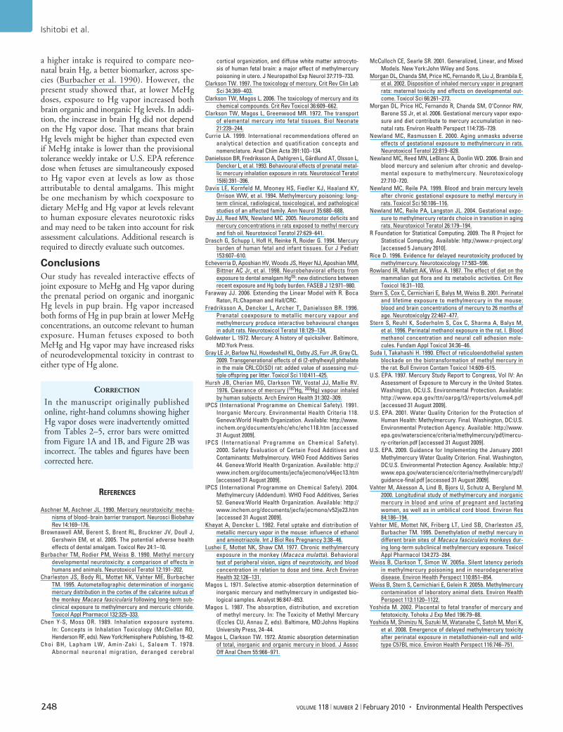
Hg in dam blood. We compared the logarithm of Hg levels (total, inorganic, and organic) in blood on GD18 across groups, sepa‑rately by experiment, as shown in Figure 1. We found significant differences among the groups (p < 0.0001 for total, inorganic, and organic Hg) in both experiments. Two‑way ANOVA with interactions showed that in experiment 1, the interactions between MeHg and Hg vapor were significant for all three outcomes (p = 0.0006 for total Hg, p = 0.002 for inor‑ganic Hg, and p = 0.0002 for organic Hg, each for a 3 df test). In experiment 2, the interactions were significant for inorganic Hg (p < 0.0001) but not for total or organic Hg. MeHg was a very strong predictor of total, inorganic, and organic Hg in both experiments (p < 0.001). With the exception of inorganic Hg in experi‑ment 2, Hg vapor did not affect Hg levels.
Hg in pup brain. Total, inorganic, and organic Hg levels in pup brains for each group are summarized in Tables 4 and 5. Figure 2 shows the observed (points) and fit‑ted values (lines) of organic Hg in pup brain, where both the brain levels and the MeHg dose are on the logarithmic scale. The vapor and no‑vapor points are offset slightly in Figure 2A for clarity. We found an interac‑tion of MeHg and Hg vapor on organic brain Hg (p < 0.0001; Figure 2A). Among animals exposed to Hg vapor, the predicted levels of brain organic Hg were larger among animals not exposed to MeHg, but organic Hg lev‑els rose less steeply with increasing MeHg exposure compared with those in animals not exposed to Hg vapor. Figure 2B shows the relationship between organic brain Hg and
Hg vapor, separately by MeHg concentration. As Figure 2 illustrates, organic brain Hg was very strongly predicted by MeHg dose.
MeHg was also a very strong predic‑tor of inorganic brain levels (p < 0.001 for each MeHg dose; Figure 3A). The observed (points) and fitted values (lines) of inorganic Hg in pup brains are shown in Figure 3. For inorganic Hg brain levels, we coded MeHg dose with three indicator variables to dis‑tinguish among the four MeHg dose levels. This analysis showed interactions between Hg vapor and the MeHg dose indicator variables (p = 0.02 for the 3 df test).
In summary, the dose of MeHg drove levels of both organic and inorganic Hg in pup brains. Exposure to Hg vapor lowered pup brain Hg levels at high MeHg doses and increased them at low MeHg doses com‑pared with animals not exposed to Hg vapor. Separate analysis showed that among the 40 animals with no MeHg exposure, exposure to any Hg vapor (n = 24 animals) was associ‑ated with a higher brain Hg level compared with no exposure to Hg vapor (n = 16 ani‑mals; p = 0.02 for both organic and inorganic Hg; Figure 4).
DiscussionPrenatal exposure to the combination of MeHg and Hg vapor had interactive effects on the levels of organic and inorganic Hg in rat neonatal brain. Surprisingly, Hg levels were increased by Hg vapor at low MeHg doses, a finding relevant to human exposures, which typically occur at low concentrations.
Total Hg concentration in brains of pups exposed to 6 ppm MeHg without Hg vapor is comparable to that on PND1 in previ‑ous studies, in which MeHg dose and dura‑tion of exposure were similar to ours (Day et al. 2005; Newland et al. 2006; Newland and Reile 1999). In these studies, rat dams
Figure 1. Concentrations (mean ± SD) of organic Hg (A and C) and inorganic Hg (B and D) in dam blood on GD18 from experiment 1 (A and B) and experiment 2 (C and D).
6
4
2
0
–2
–4
6
4
2
0
–2
–4
4
2
0
–2
–4
–6
4
2
0
–2
–4
–6
0 300
Log
(org
anic
Hg)
(µg/
mL)
Log
(org
anic
Hg)
(µg/
mL)
Log
(inor
gani
c H
g)(µ
g/m
L)Lo
g (in
orga
nic
Hg)
(µg/
mL)
1,000
0 1,000 0 1,000
0 300 1,000Hg vapor (µg/m3)
Hg vapor (µg/m3) Hg vapor (µg/m3)
Hg vapor (µg/m3)
Organic Hg, experiment 1
0 ppm3 ppm6 ppm9 ppm
Inorganic Hg, experiment 1
Organic Hg, experiment 2 Inorganic Hg, experiment 2

Ishitobi et al.
246 volume 118 | number 2 | February 2010 • Environmental Health Perspectives
were exposed daily to MeHg for 30 days or longer before breeding, simulating the pre‑dominant human exposure pattern of a stable diet. Although we did not determine whether steady‑state levels had been attained in the pregnant females, Newland and Reile (1999) found no differences in brain Hg levels of PND0 offspring of females that had been exposed for 28 or 45 days before breeding. Baseline Hg concentrations achieved by these studies are consistent over time, and thus are preferable to short‑term exposures as a basis for risk assessments.
Pup brain Hg (total, inorganic, and organic) concentrations increased with the dose of MeHg, but this increase was not lin‑ear across the exposure groups. That is, total brain and organic Hg concentrations in pups exposed to 6 or 9 ppm MeHg were greater than two or three times those of pups exposed to the lowest MeHg dose (3 ppm). Newland and Reile (1999) also found that total Hg in pup brain at birth increased nonlinearly with the concentration of MeHg (0.5 or 6.4 ppm in drinking water) when rat dams were exposed to MeHg beginning 28 or 49 days before breeding and through gestation, similar to the present study design. Such non linearities have also been seen in non human primates (Lushei et al. 1977). The present data suggest that extrapolation from high‑concentration expo‑sures may distort estimates of brain Hg levels at lower MeHg doses. The present data also suggest that with a logarithmic transformation of both pup brain Hg and MeHg dose, a lin‑ear dose–response relationship may be reason‑able. The increase in inorganic Hg associated with increased MeHg doses was not similar
to that measured by organic Hg levels. The increase seen in total and organic Hg as MeHg dose increased was higher between 6 and 9 ppm MeHg than between 0 and 3 ppm or between 3 and 6 ppm, whereas the increase of inorganic Hg was lower. This complex pattern indicates that attempts to estimate brain levels of inorganic Hg on the basis of dose requires that experiments rely on the lower MeHg exposure levels that are rele vant to human exposure levels.
In these experiments, inorganic Hg in pup brain increased with increasing MeHg dose. This result is ascribed to the process by which MeHg is converted to the inorganic form (Clarkson 1997; Clarkson and Magos 2006). Moreover, elevated levels of inorganic Hg have been found in the brains of humans and monkeys exposed to MeHg (Davis et al. 1994; Vahter et al. 1995). One site for the process would be phagocytic cells present in many mammalian tissues, including the brain, that are capable of breaking the carbon–Hg bond (Suda and Takahashi 1990). Thus, inor‑ganic Hg in brain tissue may arise from in situ metabolism of MeHg. However, it is still pos‑sible that some of the inorganic Hg does not represent the in situ conversion of MeHg but instead is derived from some distant source via the vapor pathway. Intestinal micro flora are also capable of cleaving the carbon–Hg bond (Rowland et al. 1987). Because the vapor pro‑duced by reduction of inorganic Hg in the intestine or phagocytic cells in the liver readily crosses the blood–brain barrier to be oxidized in brain tissue (Clarkson 1997), some portion of the inorganic Hg in pup brain may arise from this process. However, it is not clear whether
it occurs in the fetus or in the mother. Because MeHg readily crosses the placental barrier (Vahter et al. 2000), MeHg transferred from mother to fetus may be converted to inorganic Hg in the fetus. Also, inorganic Hg converted from MeHg in the mother may be transferred to the fetus via placenta and reach fetal brain as non‑ionized Hg, which also crosses the pla‑cental and blood–brain barrier (Clarkson et al. 1972; Khayat and Dencker 1982; Yoshida 2002). Contributions to pup brain inorganic Hg may differ between fetus and mother.
Exposure to Hg vapor increased brain Hg levels (total, inorganic, and organic) in the pups not exposed to MeHg. The increase in brain Hg was not related to the concentration of Hg vapor, which may suggest that the Hg level in the pup brains reached steady state before the time of brain sampling. However, Morgan et al. (2002, 2006) showed that, in the brains of neo nates perinatally exposed to Hg vapor (1, 2, 4, or 8 mg/m3 for 2 hr/day during GD6–GD15), total Hg concentrations increased with increasing exposure dose. The exposure concentrations they employed were higher, and the duration shorter, than those in the present study, which might contribute to the differing results. Although data on the elimination of inorganic Hg in the fetus and/or neo nate after gestational exposure are sparse, factors of retention and/or elimination should be considered to evaluate the effect of prenatal exposure to Hg vapor on the brain Hg levels because of the time lag (6–7 days) between the last exposure and the brain sampling. There is unlikely to be any loss of inorganic Hg due to this process, because the methylation of inor‑ganic Hg does not appear to take place to any
Table 4. Total, inorganic, and organic Hg concentrations (mean ± SD) in pup brain on PND4 in experiment 1.
Group [MeHg (ppm) × Hg vapor (µg/m3)]0 × 0 3 × 0 6 × 0 0 × 300 3 × 300 0 × 1,000 3 × 1,000 6 × 1,000
Total Hg (ng/g)Males 20 ± 7 2,773 ± 231 8,997 ± 2,949 42 ± 13 2,946 ± 208 50 ± 11 3,244 ± 626 8,064 ± 1,524Females 26 ± 15 2,723 ± 86 9,262 ± 2,810 43 ± 7 2,928 ± 383 51 ± 14 2,685 ± 571 7,900 ± 2,150
Inorganic Hg (ng/g)Males 5 ± 0 140 ± 23 490 ± 209 11 ± 5 150 ± 35 18 ± 5 172 ± 37 350 ± 113Females 7 ± 3 140 ± 15 505 ± 169 13 ± 6 148 ± 30 20 ± 3 151 ± 32 290 ± 149
Organic Hg (ng/g)Males 15 ± 7 2,632 ± 231 8,507 ± 2,749 31 ± 13 2,796 ± 180 33 ± 7 3,072 ± 593 7,715 ± 1,420Females 20 ± 12 2,583 ± 101 8,757 ± 2,658 31 ± 4 2,780 ± 359 32 ± 12 2,534 ± 539 7,510 ± 2002
Table 5. Total, inorganic, and organic Hg concentrations (mean ± SD) in pup brain on PND4 in experiment 2.
Group [MeHg (ppm) × Hg vapor (µg/m3)]0 × 0 3 × 0 6 × 0 9 × 0 0 × 1,000 3 × 1,000 6 × 1,000 9 × 1,000
Total Hg (ng/g)Males 26 ± 13 3,469 ± 254 8,254 ± 1,078 16,122 ± 4,419 32 ± 8 3,286 ± 321 6,796 ± 1,684 11,795 ± 3,156Females 29 ± 16 3,703 ± 72 9,703 ± 2,216 16,545 ± 5,941 33 ± 11 3,379 ± 287 6,468 ± 1,727 13,177 ± 5,007
Inorganic Hg (ng/g)Males 8 ± 6 167 ± 19 452 ± 95 612 ± 52 7 ± 3 160 ± 48 370 ± 75 518 ± 107Females 8 ± 7 165 ± 18 475 ± 83 678 ± 151 5 ± 0 156 ± 22 366 ± 58 668 ± 203
Organic Hg (ng/g)Males 18 ± 11 3,302 ± 240 7,803 ± 1,004 15,511 ± 4,376 25 ± 7 3,127 ± 293 6,426 ± 1,622 11,277 ± 3,052Females 21 ± 15 3,538 ± 62 9,228 ± 2,150 15,867 ± 5,811 28 ± 11 3,223 ± 276 6,102 ± 1,670 12,509 ± 4,812

Organic and inorganic Hg in neonatal rat brain
Environmental Health Perspectives • volume 118 | number 2 | February 2010 247
significant extent in either human or animal tissues (Clarkson and Magos 2006).
So far, only one study has addressed pre‑natal coexposure to MeHg and Hg vapor (Fredriksson et al. 1996). That study showed that brains of rat offspring (on PND3) pre‑natally exposed to both MeHg and Hg vapor contained more total Hg than those exposed to either form alone. Statistical analyses of the joint and single contributions of MeHg and Hg vapor to total Hg seem not to have been performed, although coexposure resulted in slightly higher Hg levels in the brain (12 ng/g) than what would had been expected, consider‑ing the concentrations obtained after expo‑sure to either MeHg (4 ng/g) or Hg vapor (5 ng/g) alone. The present data suggest that coexposure to Hg vapor slightly lowered brain Hg levels at high MeHg doses and increased them at low MeHg doses. In the study by Fredriksson et al. (1996), the MeHg dose was
2 mg/kg/day, which is higher than our highest dose, which would be approximately 700–750 µg/kg/day if we extrapolate from Newland and Reile (1999). Similarly, Fredriksson et al.’s dose of Hg vapor was 1.8 mg/m3 for 1.5 hr/day, which is also higher than our highest dose (1.0 mg/m3 for 2 hr/day). In addition, MeHg exposure occurred only during GD6–GD9, and that of Hg vapor occurred during GD14–GD19, which means that the two exposures did not occur simultaneously. Different doses and duration of exposures might explain the different outcomes.
Several metabolic processes could account for the observation that exposure to Hg vapor lowered brain Hg levels at high MeHg doses. Inorganic Hg, but not MeHg, can induce the metal‑binding protein metallothionein. Binding to this protein is generally regarded as a detoxication process (Clarkson 1997; Clarkson and Magos 2006) and has been
proven to play an important role in the reten‑tion of Hg in tissue. Metallothionein induced by Hg vapor in the mother and/or in the fetus might prevent MeHg and perhaps inorganic Hg from reaching the fetal brain. The conver‑sion of MeHg to inorganic Hg may also need to be considered (Clarkson 1997; Clarkson and Magos 2006). Inorganic Hg in the form of oxidized Hg has a limited capacity to cross the blood–brain and placental barriers (Clarkson 1997; Clarkson and Magos 2006). The pres‑ence of large amounts of Hg vapor might pro‑mote the oxidation of MeHg in the mother and/or in the fetus, resulting in less Hg reach‑ing the brain. However, this hypothesis has not yet been tested.
The main source of human exposure to MeHg is the diet, especially fish and seafood (U.S. EPA 1997). Dietary intake of MeHg is estimated at 0.1–2.0 µg/kg body weight per week for numerous national diets (IPCS 2004). The U.S. EPA’s current reference dose for MeHg is 0.1 µg/kg body weight/day (U.S. EPA 2001, 2009). The 53rd meeting of the Joint Food and Agriculture Organization/World Health Organization Expert Committee on Food Additives established provisional tol‑erance weekly intake of 200 µg MeHg (3.3 µg/kg body weight) for the general population but noted that fetuses and infants might be more sensitive than adults to its toxic effects (IPCS 2000). Because there has been no definitive separation of prenatal and postnatal exposure that would permit dose–response modeling, there are currently no data that would support the derivation of a child (vs. general popula‑tion) reference dose (U.S. EPA 2001). Those values are relatively low compared with doses we used in rats in the present study. Rat blood has approximately 10 times as much hemoglo‑bin as does mouse, monkey, or human blood (Magos 1987) and binds Hg. That means that rat blood has a higher capacity to bind Hg, so
Figure 2. Organic Hg in pup brain on PND4 by log scale of MeHg (A) and absolute value of Hg vapor (B). Data points show individual pups. The fitted values in (A) exhibit a clear linear relationship between the logarithm of organic Hg brain levels and the logarithm of MeHg dose. The interaction allows the slope relating MeHg dose to organic brain Hg outcome to differ for Hg vapor–exposed compared with non‑Hg vapor–exposed pups. Interaction of MeHg and Hg vapor on organic Hg values, p < 0.0001; organic brain Hg is strongly predicted by MeHg dose.
10
8
6
4
2
Log
[org
anic
Hg
(ng/
g)]
Log
[org
anic
Hg
(ng/
g)]
–2 –1 1 20 0 200 400 600 800 1,000
Log [MeHg + 0.1 (ppm)] Hg vapor (µg/m3)
No Hg vapor Hg vapor9 ppm MeHg 0 ppm MeHg
×
10
8
6
4
2
9 ppm MeHg 6 ppm MeHg 3 ppm MeHg0 ppm MeHg
×+
Figure 3. Inorganic Hg in pup brain on PND4 by log scale of MeHg (A) and absolute value of Hg vapor (B). Data points show individual pups. The fitted values are shown in (A). For the interaction between Hg vapor and the MeHg dose indicator variables, p = 0.02 for the 3 df test. MeHg was a very strong predictor of inorganic brain levels (p < 0.001 for each MeHg dose).
7
6
5
4
3
2
7
6
5
4
3
2
Log
[inor
gani
c H
g (n
g/g)
]
Log
[inor
gani
c H
g (n
g/g)
]
–2 –1 1 20 0 200 400 600 800 1,000
Log [MeHg + 0.1 (ppm)] Hg vapor (µg/m3)
9 ppm MeHg 6 ppm MeHg 3 ppm MeHg0 ppm MeHg
×+
No Hg vapor Hg vapor9 ppm MeHg 0 ppm MeHg
×
Figure 4. Box plots of log scale brain concentra‑tions on PND4 for organic Hg (A) and inorganic Hg (B) for pups exposed to no Hg versus pups exposed to any Hg vapor. The solid line within the box shows the median; the top and bottom of the boxes are the 75th and 25th percentiles, respec‑tively; and the whiskers extend to the largest or smallest observation that is within 1.5 times the length of the box.
3.5
3.0
2.5
2.0
1.5
3.0
2.5
2.0
No Hg vapor Hg vapor No Hg vapor Hg vapor
Log
[inor
gani
c H
g (n
g/g)
]
Log
[org
anic
Hg
(ng/
g)]
Ishitobi et al.
248 volume 118 | number 2 | February 2010 • Environmental Health Perspectives
a higher intake is required to compare neo‑natal brain Hg, a better biomarker, across spe‑cies (Burbacher et al. 1990). However, the present study showed that, at lower MeHg doses, exposure to Hg vapor increased both brain organic and inorganic Hg levels. In addi‑tion, the increase in brain Hg did not depend on the Hg vapor dose. That means that brain Hg levels might be higher than expected even if MeHg intake is lower than the provisional tolerance weekly intake or U.S. EPA reference dose when fetuses are simultaneously exposed to Hg vapor even at levels as low as those attributable to dental amalgams. This might be one mechanism by which coexposure to dietary MeHg and Hg vapor at levels relevant to human exposure elevates neurotoxic risks and may need to be taken into account for risk assessment calculations. Additional research is required to directly evaluate such outcomes.
ConclusionsOur study has revealed interactive effects of joint exposure to MeHg and Hg vapor during the prenatal period on organic and inorganic Hg levels in pup brain. Hg vapor increased both forms of Hg in pup brain at lower MeHg concentrations, an outcome rele vant to human exposure. Human fetuses exposed to both MeHg and Hg vapor may have increased risks of neuro develop mental toxicity in contrast to either type of Hg alone.
correction
In the manuscript originally published online, right‑hand columns showing higher Hg vapor doses were inadvertently omitted from Tables 2–5, error bars were omitted from Figure 1A and 1B, and Figure 2B was incorrect. The tables and figures have been corrected here.
RefeRences
Aschner M, Aschner JL. 1990. Mercury neurotoxicity: mecha‑nisms of blood–brain barrier transport. Neurosci Biobehav Rev 14:169–176.
Brownawell AM, Berent S, Brent RL, Bruckner JV, Doull J, Gershwin EM, et al. 2005. The potential adverse health effects of dental amalgam. Toxicol Rev 24:1–10.
Burbacher TM, Rodier PM, Weiss B. 1990. Methyl mercury developmental neurotoxicity: a comparison of effects in humans and animals. Neurotoxicol Teratol 12:191–202.
Charleston JS, Body RL, Mottet NK, Vahter ME, Burbacher TM. 1995. Autometallographic determination of inorganic mercury distribution in the cortex of the calcarine sulcus of the monkey Macaca fascicularis following long‑term sub‑clinical exposure to methylmercury and mercuric chloride. Toxicol Appl Pharmacol 132:325–333.
Chen Y‑S, Moss OR. 1989. Inhalation exposure systems. In: Concepts in Inhalation Toxicology (McClellan RO, Henderson RF, eds). New York:Hemisphere Publishing, 19–62.
Choi BH, Lapham LW, Amin‑Zaki L, Saleem T. 1978. Abnormal neuronal migration, deranged cerebral
cortical organization, and diffuse white matter astrocyto‑sis of human fetal brain: a major effect of methylmercury poisoning in utero. J Neuropathol Exp Neurol 37:719–733.
Clarkson TW. 1997. The toxicology of mercury. Crit Rev Clin Lab Sci 34:369–403.
Clarkson TW, Magos L. 2006. The toxicology of mercury and its chemical compounds. Crit Rev Toxicol 36:609–662.
Clarkson TW, Magos L, Greenwood MR. 1972. The transport of elemental mercury into fetal tissues. Biol Neonate 21:239–244.
Currie LA. 1999. International recommendations offered on analytical detection and quatification concepts and nomenclature. Anal Chim Acta 391:103–134.
Danielsson BR, Fredriksson A, Dahlgren L, Gårdlund AT, Olsson L, Dencker L, et al. 1993. Behavioural effects of prenatal metal‑lic mercury inhalation exposure in rats. Neurotoxicol Teratol 15(6):391–396.
Davis LE, Kornfeld M, Mooney HS, Fiedler KJ, Haaland KY, Orrison WW, et al. 1994. Methylmercury poisoning: long‑term clinical, radiological, toxicological, and pathological studies of an affected family. Ann Neurol 35:680–688.
Day JJ, Reed MN, Newland MC. 2005. Neuromotor deficits and mercury concentrations in rats exposed to methyl mercury and fish oil. Neurotoxicol Teratol 27:629–641.
Drasch G, Schupp I, Hofl H, Reinke R, Roider G. 1994. Mercury burden of human fetal and infant tissues. Eur J Pediatr 153:607–610.
Echeverria D, Aposhian HV, Woods JS, Heyer NJ, Aposhian MM, Bittner AC Jr, et al. 1998. Neurobehavioral effects from exposure to dental amalgam Hg(0): new distinctions between recent exposure and Hg body burden. FASEB J 12:971–980.
Faraway JJ. 2006. Extending the Linear Model with R. Boca Raton, FL:Chapman and Hall/CRC.
Fredriksson A, Dencker L, Archer T, Danielsson BR. 1996. Prenatal coexposure to metallic mercury vapour and methylmercury produce interactive behavioural changes in adult rats. Neurotoxicol Teratol 18:129–134.
Goldwater L. 1972. Mercury: A history of quicksilver. Baltimore, MD:York Press.
Gray LE Jr, Barlow NJ, Howdeshell KL, Ostby JS, Furr JR, Gray CL. 2009. Transgenerational effects of di (2‑ethylhexyl) phthalate in the male CRL:CD(SD) rat: added value of assessing mul‑tiple offspring per litter. Toxicol Sci 110:411–425.
Hursh JB, Cherian MG, Clarkson TW, Vostal JJ, Mallie RV. 1976. Clearance of mercury (197Hg, 203Hg) vapour inhaled by human subjects. Arch Environ Health 31:302–309.
IPCS (International Programme on Chemical Safety). 1991. Inorganic Mercury. Environmental Health Criteria 118. Geneva:World Health Organization. Available: http://www.inchem.org/documents/ehc/ehc/ehc118.htm [accessed 31 August 2009].
IPCS (International Programme on Chemical Safety). 2000. Safety Evaluation of Certain Food Additives and Contaminants: Methylmercury. WHO Food Additives Series 44. Geneva:World Health Organization. Available: http://www.inchem.org/documents/jecfa/jecmono/v44jec13.htm [accessed 31 August 2009].
IPCS (International Programme on Chemical Safety). 2004. Methylmercury (Addendum). WHO Food Additives, Series 52. Geneva:World Health Organization. Available: http://www.inchem.org/documents/jecfa/jecmono/v52je23.htm [accessed 31 August 2009].
Khayat A, Dencker L. 1982. Fetal uptake and distribution of metallic mercury vapor in the mouse: influence of ethanol and aminotriazole. Int J Biol Res Pregnancy 3:38–46.
Lushei E, Mottet NK, Shaw CM. 1977. Chronic methylmercury exposure in the monkey (Macaca mulatta). Behavioral test of peripheral vision, signs of neurotoxicity, and blood concentration in relation to dose and time. Arch Environ Health 32:126–131.
Magos L. 1971. Selective atomic‑absorption determination of inorganic mercury and methylmercury in undigested bio‑logical samples. Analyst 96:847–853.
Magos L. 1987. The absorption, distribution, and excretion of methyl mercury. In: The Toxicity of Methyl Mercury (Eccles CU, Annau Z, eds). Baltimore, MD:Johns Hopkins University Press, 24–44.
Magos L, Clarkson TW. 1972. Atomic absorption determination of total, inorganic and organic mercury in blood. J Assoc Off Anal Chem 55:966–971.
McCulloch CE, Searle SR. 2001. Generalized, Linear, and Mixed Models. New York:John Wiley and Sons.
Morgan DL, Chanda SM, Price HC, Fernando R, Liu J, Brambila E, et al. 2002. Disposition of inhaled mercury vapor in pregnant rats: maternal toxicity and effects on developmental out‑come. Toxicol Sci 66:261–273.
Morgan DL, Price HC, Fernando R, Chanda SM, O’Connor RW, Barone SS Jr, et al. 2006. Gestational mercury vapor expo‑sure and diet contribute to mercury accumulation in neo‑natal rats. Environ Health Perspect 114:735–739.
Newland MC, Rasmussen E. 2000. Aging unmasks adverse effects of gestational exposure to methylmercury in rats. Neurotoxicol Teratol 22:819–828.
Newland MC, Reed MN, LeBlanc A, Donlin WD. 2006. Brain and blood mercury and selenium after chronic and develop‑mental exposure to methylmercury. Neurotoxicology 27:710–720.
Newland MC, Reile PA. 1999. Blood and brain mercury levels after chronic gestational exposure to methyl mercury in rats. Toxicol Sci 50:106–116.
Newland MC, Reile PA, Langston JL. 2004. Gestational expo‑sure to methylmercury retards choice in transition in aging rats. Neurotoxicol Teratol 26:179–194.
R Foundation for Statistical Computing. 2009. The R Project for Statistical Computing. Available: http://www.r‑project.org/ [accessed 5 January 2010].
Rice D. 1996. Evidence for delayed neurotoxicity produced by methylmercury. Neurotoxicology 17:583–596.
Rowland IR, Mallett AK, Wise A. 1987. The effect of diet on the mammalian gut flora and its metabolic activities. Crit Rev Toxicol 16:31–103.
Stern S, Cox C, Cernichiari E, Balys M, Weiss B. 2001. Perinatal and lifetime exposure to methylmercury in the mouse: blood and brain concentrations of mercury to 26 months of age. Neurotoxicolgy 22:467–477.
Stern S, Reuhl K, Soderholm S, Cox C, Sharma A, Balys M, et al. 1996. Perinatal methanol exposure in the rat. I. Blood methanol concentration and neural cell adhesion mole‑cules. Fundam Appl Toxicol 34:36–46.
Suda I, Takahashi H. 1990. Effect of reticuloendothelial system blockade on the biotransformation of methyl mercury in the rat. Bull Environ Contam Toxicol 14:609–615.
U.S. EPA. 1997. Mercury Study Report to Congress, Vol IV: An Assessment of Exposure to Mercury in the United States. Washington, DC:U.S. Environmental Protection. Available: http://www.epa.gov/ttn/oarpg/t3/reports/volume4.pdf [accessed 31 August 2009].
U.S. EPA. 2001. Water Quality Criterion for the Protection of Human Health: Methylmercury. Final. Washington, DC:U.S. Environmental Protection Agency. Available: http://www.epa.gov/waterscience/criteria/methylmercury/pdf/mercu‑ry‑criterion.pdf [accessed 31 August 2009].
U.S. EPA. 2009. Guidance for Implementing the January 2001 Methylmercury Water Quality Criterion. Final. Washington, DC:U.S. Environmental Protection Agency. Available: http://www.epa.gov/waterscience/criteria/methylmercury/pdf/guidance‑final.pdf [accessed 31 August 2009].
Vahter M, Akesson A, Lind B, Bjors U, Schutz A, Berglund M. 2000. Longitudinal study of methylmercury and inorganic mercury in blood and urine of pregnant and lactating women, as well as in umbilical cord blood. Environ Res 84:186–194.
Vahter ME, Mottet NK, Friberg LT, Lind SB, Charleston JS, Burbacher TM. 1995. Demethylation of methyl mercury in different brain sites of Macaca fascicularis monkeys dur‑ing long‑term subclinical methylmercury exposure. Toxicol Appl Pharmacol 134:273–284.
Weiss B, Clarkson T, Simon W. 2005a. Silent latency periods in methylmercury poisoning and in neurodegenerative disease. Environ Health Perspect 110:851–854.
Weiss B, Stern S, Cernichiari E, Gelein R. 2005b. Methylmercury contamination of laboratory animal diets. Environ Health Perspect 113:1120–1122.
Yoshida M. 2002. Placental to fetal transfer of mercury and fetotoxicity. Tohoku J Exp Med 196:79–88.
Yoshida M, Shimizu N, Suzuki M, Watanabe C, Satoh M, Mori K, et al. 2008. Emergence of delayed methylmercury toxicity after perinatal exposure in metallothionein‑null and wild‑type C57BL mice. Environ Health Perspect 116:746–751.