Synthesis, spectroscopic and magnetic resonance studies of mercury(II) and methylmercury(II)...
14
Synthesis, Spectroscopic and Magnetic Resonance Studies of Mercury(U) and Methylmercury(I1) Complexes of Azathioprine, a Biologically Active Mercaptopurine Derivative Helen T. Chifotides, Kim R. Dunbar, Nikos Katsaros, and George Pneumatikakis HTC, NK N.RC2.S. “Demokritos, ” Attikis, Greece.-HTC, KRD. Department of Chemistry, Michigan State University, East Lansing, Michigan, U.S.A.--GP. Deparhnent of Chemistq Inorganic Chemistry Laboratoy, University of Athens, Athens, Greece ABSTRACT Synthetic and spectroscopic studies of the Hg(I1)and MeHg(II) complexes of azathioprine (AZAJ, a biologically active 6-mercaptopurine derivative, were undertaken. The altered coordination behavior of AZ4 with respect to the parent mercaptopurine, with sulfur no longer being the primary donor atom, was confirmed. As concluded by the ‘H NMR, 13C NMR, and IR spectro- scopic data, Hg(I1) binds to the N(9) position of deprotonated AZA, while in the MeHg(II) compound, coordination occurs through the N(3) and N(9) positions of the puke ring. The values of the coupling constants ‘J (lWHg - ‘HI, ‘J(lWHg -13C) for the MeHg(I1) compound further support complexation via nitrogen atoms of the purine. Elemental analyses confirmed the com- pounds to be HgO, (1) and [(MeHg),(AZA)KNO,) (2); conductivity measurement values show that 1 is a nonelectrolyte and 2 is a 1: 1 electrolyte. Furthermore, the FAR-MS of the compounds confnms direct binding of the metal to the ligand, and in the case of the MeHg(I1) compound, the successive loss of one and two MeHg(I1) moieties can be clearly observed. INTRODUCTION Nucleic acids and nucleic acid derivatives are naturally occurring multidentate ligands whose interactions with metal-containing species have stimulated great Address reprint requests and correspondence to: Dr. G. Pneumatikakis, Department of Chemistry, Inorganic Chemistry Laboratory, University of Athens, Panepistimiopolis, Kouponia, 15701 Athens, Greece. Joumnl of InorganicBiocm, 55,203-216 (1994) 203 8 1994 Elsevier Science Inc., 655 Avenue of the Americas, NY, NY 10010 0162-0134/!M/S~.OO
Transcript of Synthesis, spectroscopic and magnetic resonance studies of mercury(II) and methylmercury(II)...

Synthesis, Spectroscopic and Magnetic Resonance Studies of Mercury(U) and Methylmercury(I1) Complexes of Azathioprine, a Biologically Active Mercaptopurine Derivative
Helen T. Chifotides, Kim R. Dunbar, Nikos Katsaros, and George Pneumatikakis
HTC, NK N.RC2.S. “Demokritos, ” Attikis, Greece.-HTC, KRD. Department of Chemistry, Michigan State University, East Lansing, Michigan, U.S.A.--GP. Deparhnent of Chemistq Inorganic Chemistry Laboratoy, University of Athens, Athens, Greece
ABSTRACT
Synthetic and spectroscopic studies of the Hg(I1) and MeHg(II) complexes of azathioprine (AZAJ, a biologically active 6-mercaptopurine derivative, were undertaken. The altered coordination behavior of AZ4 with respect to the parent mercaptopurine, with sulfur no longer being the primary donor atom, was confirmed. As concluded by the ‘H NMR, 13C NMR, and IR spectro- scopic data, Hg(I1) binds to the N(9) position of deprotonated AZA, while in the MeHg(II) compound, coordination occurs through the N(3) and N(9) positions of the puke ring. The values of the coupling constants ‘J (lWHg - ‘HI, ‘J(lWHg -13C) for the MeHg(I1) compound further support complexation via nitrogen atoms of the purine. Elemental analyses confirmed the com- pounds to be HgO, (1) and [(MeHg),(AZA)KNO,) (2); conductivity measurement values show that 1 is a nonelectrolyte and 2 is a 1: 1 electrolyte. Furthermore, the FAR-MS of the compounds confnms direct binding of the metal to the ligand, and in the case of the MeHg(I1) compound, the successive loss of one and two MeHg(I1) moieties can be clearly observed.
INTRODUCTION
Nucleic acids and nucleic acid derivatives are naturally occurring multidentate ligands whose interactions with metal-containing species have stimulated great
Address reprint requests and correspondence to: Dr. G. Pneumatikakis, Department of Chemistry, Inorganic Chemistry Laboratory, University of Athens, Panepistimiopolis, Kouponia, 15701 Athens, Greece.
Joumnl of Inorganic Biocm, 55,203-216 (1994) 203 8 1994 Elsevier Science Inc., 655 Avenue of the Americas, NY, NY 10010 0162-0134/!M/S~.OO


STUDIES OF MERCURY(I1) AND METHYLMERCURY 205
binding sites of the drug {N(l), N(3), N(7), N(9), and S}; In the crystal structures of 6-MI’ with metals, 6-MP acts either as a monodentate bridging or nonbridg ing ligand through S@) or as a S(6)/N(7) chelating ligand [ 151; in the case of the Hg@) complex, 6MP acts as a monodentate ligand through sulfur [161, a behavior in good agreement with the well-known affinity of mercury for sulfur donors. Despite ‘the biological and chemical relevance of AZA to 6-MB, AZA seems to follow binding modes other than those of 6-m; in the x-ray crystal structure of AZA with the bis adduct .Rh,(OA& AZA is coordinated to the metals through the purine nitrogen atom N(3) [17]. Apart from our efforts to study the coordination chemistry of AZA, and possibly elucidate the prevailing factors determining the binding sites in the interactions of AZA with various heavymetals, we probed the binding sites of AZA with H@I(II) and MeHg(I1) by
I using “H and ,13C nuclear magnetic resonance studies as well as infrared spectroscopy and mass spectrometry.
EXPERIMENTAL
Starting Materials and Methods
Azathioprine (AZ& as well as its sodium salt (AZA-Na+) were purchased from Sigma or donated by the Wellcome Co.; in both instances, they were used without further purification. Mercuric chloride (HgCl,) was purchased from Merck Chemicals and was used as received. Methylmercuric nitrate (MeHgNO,) was prepared by a published method [18] from methylmercuric chloride (MeHgCl) and silver nitrate @NO,). MeHgCl was purchased from Ventron GMBH and was used as received. (CD,),SO-d6 (99.96 atom % D) was purchased from Isotech Inc.
‘H NMR spectra were recorded on a Varian-VXR 300 MHz or 500 MHz spectrometer. Chemical shifts were referenced relative to the residual proton impurity of (CD,),SO-d6 (2.49 ppm with respect to TMS). 13C NMR spectra were recorded on a Varian-VXR 500 instrument. operating at 125 MHz with proton noise decoupling. The pulse duration was 6 ps and the relaxation delay 1 set with a minimum of lo3 accumulations. (CD,),SO-d, containing TMS as an internal standard was used as the solvent and provided the internal deuterium lock All spectra were recorded at room temperature (25 f 2°C). DEPT (Distor- tionless Enhancement by Polarization Transfer) [19] spectra were detected with proton decoupling and in order to edit the CH, subspectra, three DEPT experiments for the polarization transfer angles 8, = 45”, 0, = 90”, and 0, = 135” were recorded; methylene and methyl subspectra arise from linear combi- nation of the three experiments: CH: 0,; CH,: 0, - 8,; CH,; 0, + 0, - 0.7070,; the quaternary carbon subspectrum results by subtraction of the other CH, (n = 1,2,3) subspectra from the regular 13C NMR spectrum.
Infrared spectra were registered as 1% (w/w) KBr discs on a Nicolet 740 ET-IR or BOMEM Michelson 100 spectrophotometer. In the case of complexes with NO, - ions in their coordination sphere, an infrared spectrum was recorded on a suspension of the compound in spectroquality Nujol between CsI plates. Fast atom bombardment (FAB) mass spectrometry studies were performed on a JEOL HX 110 double-focusing mass spectrometer housed in the National Institute of Health/Michigan State University Mass Spectrometry Facility; the

206 H. T. Chifotides et al.
sample, was dissolved in a 3-nitrobenzyl alcohol matrix. Conductivity measure- mentswere .performed using an E365B conductoscope (Metrohm Ltd., Herisau, Switzerland). The melting points were determined on a Fisher John’s melting point apparatus and are uncorrected. Elemental analyses were performed at the Centre National de la Recherche Scientifique (C.N.R.S.), France.
Preparation of the Complexes
Hg(AZA), (1). An aqueous (ca. 150 mL) suspension of azathioprine (0.277 g, 1.0 mmol) was refluxed for 40 min until the compound was completely dissolved. Partial cooling,of the previous yellow solution was followed by addition of white solid HgCl, (0.135 g, 0.5 mm&. In a few minutes change of the solution color to pale yellow and .precipitation of yellow solid ensued. To ensure completion of the reaction stirring was continued for ca. 1 hr. The yellow precipitate was collected by filtration, washed with acetone (2 X 3 mL) and ether (2 X 3 mL) and dried in vacua above P,O,, for 24 hr; yield: 0.342 g (91%). MS, m/z (relative intensity): 724 {(202Hg@ZA)2-2 X CH3)+,301, 722 ((200Hg(AZA)2 - 2 X CH,)+, 24). Anal. Calcd. for C,,H,,N,,O,S,Hg: C, 28.70; H, 1.61; N, 26.04; S, 8.51. Found: C, 28.62; H, 1.76; N, 25.96; S, 8.48. M.p. 21s”C with decomposition. IR spectral data (KBr pellet, 4000-200 cm-‘), cm-‘; v (C-H(2),,,,} = 3118 s, v (C-H(8),,,} = 3103 m, v{C-H(ll),,,,) = 3018 m (v (C=C) + v(C=N)} = 1570 vs, 1566 vs, vasym (NO,) = 1527 vs, v_ (NO,) 7 1376 vs, (6 (N(9)-H) + 6 (N(7)-C(8)-N(9))} = 1234 W, 1220 W, 1199 m, Y(C-H)nitroimidazole = 830 S, {v(C-S-C) + rti,,} = 647 m (v = stretching mode, 6 7 bending mode, y = out of plane bending). Au (ohm-’ cm2 mol-‘) of 10m3 M in (CH3)zSO at 25 + O.l”C: 6.21.
[(@$-Ig),(AZA)E(NO,) (2). A suspension of ‘azathioprine (0.138 g, 0.5 mmol) in ethanol (50 mL) was boiled under reflux for 30 min until the compound was completely dissolved. The resulting solution was added to a stirred colorless solution of MeHgNO, (0.278 g, 1.0 mmol) in ethanol ‘(15 mL). The reaction mixture was stirred for a few hours to allow for completion. A yellow precipitate was obtained after slow evaporation of the solvent in the air for several days. The solid was washed several times with acetone (3 x 3 mL) and dried in vacua at room temperature; yield: 0.676 g (87.7%). MS, m/z (relative intensity): 708 (Ml+? 75), 493 (Ml+- Me200Hg = M,, 33), 278 (M2 7 Mem2Hg = AZAH + H, 35). Anal. Calcd. for C,,H,,NsO,SHg,: C, 17.15; H, 1.57; N, 14.55; S, 4.17. Found: C, 17.65; H, 1.65; N, 14.90; S, 4.45. M.p. 193°C with decomposition. IR spectral data (Nujol suspension, 4000-200 cm-‘), cm-‘: v (C-H(2),,,,} = 3114 s, v K~H(83,,,} =3080 m, v {C-H(U),,,,} = 3016 m, vsym (MeHg+) = 2922 m, ;8&I;yyH) + v(C=C) i-,v(C?N)l = 1591 sh, (v(C=C) + v(C=N)J = 1570 vs,
(NO,) = 1539 vs, Vet (NO,-) .= I376 sbr, asy,,, (MeHg+) = 1188 m, (8 ” $6)-H) + S (N(7)-C&-N(9))} = 1231 w, 1221 w*. 1200 m, Vvm (No,-) = 1037 m, (Y(C-H)n‘itroimidazo,~ ) = 832,s, {v(C-S-C)+ ?u$ = 651 m, v(Hg-0 = 568 s, v(Hg-C) = 518 w ‘(v = stretching mode, 8 i burdlng mode, y= out of plane bending). AM (ohm-’ cm2’mo~~‘) of 10~~ ,M~‘im’(.CH3)2S0 at 25 f O.l?C:.23.1.
I >
,/ ,, I ,./.I ., , ,..’ ‘,I/. I ,.., ,‘,~Q.~. .’ I ~uL’Fs’~.D~&~ION ” ,s. 3 ’ !’ I_,‘\ “:;;“‘:,;. ’
5:’ The elemental analyses of, the’ ‘timpleries’listed in the” Experimental section establish the 1: 2 and 2 : 1 ratio of metal: AZA for ‘the”&(B) ‘and MeHg(II)

STUDIES OF MERCURY(R) AND METHYIMERCURY(I1) 207
compounds, respectively. The conductivity measurement values confirm that the MeHg(I1) compound is a 1: 1 electrolyte and the H&Ii) com,pound a non- electrolyte, results consistent with the elemental analyses and the other spectroscopic data (vide i&a).
‘NMR Spectroscopy
Table 1 summarizes the ‘H NMR data for AZA and its sodium salt as well as for its Hg(II) and MeHglII) complexes’in KD,),SO-d,.
At room temperature, in (CD,),SO-d,, the ‘H NMR spectrum of AZA reveals three resonances in the aromatic region .at S + 8.58, + 8.55, and + 8.25 ppm with a 1: 1: 1 ratio, corresponding to the three aromatic protons H(2), H(8), and H(11) of azathioprine, respectively, and one resonance at 6 + 13.88 ‘ppm corresponding to the N(9)-H proton (the previous resonance is not observed in the spectrum of the azathioprine salt because in that case N(9) is deprotonated) (see Table 1). The proton H(D) of the l-methyl-C nitroimidazole ring resonates upfield from the purine protons presumably because, in a purine, the pyrimidine ring is a r-electron deficient system while the imidazole ring is a w-electron rich system [19]. The assignment of the purine protons H(2) and H(8) is based on the ‘H NMR spectrum of 8-deuterioazathioprine (at elevated temperatures in D,O, purines exchange the H(8) proton with deuterium [20, 211; in 50% and 100% deuteriated samples of 8-deuterioazathioprine, the integration area of the resonance due to H(8) diminishes to half of that of H(2) in. the former and disappears entirely in the latter (Table 1).
At room temperature, in (CD&SO-d,, the ‘H NMR spectrum of the Hg(II)/azathioprine complex reveals three resonances in the aromatic region at S + 8.58, + 8.14; and + 8.25 pRm with a 1: 1: 1 ratio, corresponding to the three aromatic protons H(2), H(8), and H(D) of azathioprine, respectively; only the resonance corresponding to H(8) shifts upfieZd by 0.26 ppm and the N(9) proton resonance vanishes while the other proton resonances of the’ ligand remain unchanged (see Table 1). This unusual behavior of the metal complex (since it is
:
TABLE i. ‘H NMR’data fur AZA and Its Hg(II) and MeHg(IT) Complexesa
Coupling
Deuterated Chemical Shifts, 6 @pm) z Constan@, Hz
Compbuud Solvent H(2) H(8) If(ll) 6X9)-H CH3(25) .*J (lH-lwHg)
Azk AZA-d,
&ISO:d, j 858 :s.55 8.25, 13@. ‘%kSO-!, 8.58 - 8.25 -
3.70 ’ ; 3:70
j&&-N;+_ DMSO-d, 8.15 8.14 ‘7.87 L 3.61 &@Z&, .’ DMSO-d, 8,57 8.28 ‘8.23 - ‘3.70 ,, ,H&,+$f
” WHgNO, .Dy?+& ,855 8.26 8.21. - .’ 3.70, DMS;Q-d,,. - ,- ,-- -
[(ti&g&(AzAsFJci,’ DMSO-dQ 8.75 b.65 825 - ‘. _‘I. $ >“, .260.4b
; 222.F
“All chemical shift values are reported in ppm for spectra recorded at room temperature. bCoupling constant for MeHg(I1) complexed with .,D&+Of323 chemical *ii% c ‘value 6 (MeHg+) = +0.83 ppm.
I, C..,_. 1.
cCo~pli~,~nstant $07 nitrpgen;bound, MeHg(II& chemical &I#-y&e 6 @4eHg+),p: +0.93 ppm. ’ *i&i&iry ci$ipleX’~repared fbm the axathibprine +odium salt.

208 H. T. Chifbtides et aL
well known that upon metal coordination to ring nitrogens of purines, the protons adjacent to the binding site become more acidic, thus exhiiiting down- field shifts [14, 20, 221) is consistent with binding of Hg(J1) via N(9) with concomitant proton displacement [23] and can be explained in terms of two opposing effects: the metal complexation at N(9) causing deshielding of the adjacent proton [14, 22, 241 and the localization of the negative charge of the AZA anion at the same position due to deprotonation causing shield- ing of the adjacent proton atom [23, 251. Consequently, complexation of Hg(I1) to AZA causes an overall shielding of H(8). Similar behavior has been observed in the ‘H NMR chemical shifts of other complexes where complexation is followed by deprotonation [6, 7, 26, 271; e.g., the complex of MeHg(I1) with 6-dimethylamino- and 6-diethylaminopurine, where binding takes place through N(9), a N 0.28 ppm upfield shift of H(8) is observed [6, 271. Additional evidence supporting Hg(I1) Complexation through position N(9) of the deprotonated purine ring arises from the fact that the H NMR spectrum of the complex prepared from a reaction with the axathioprine sodium salt (AZA-Na+) is the same as that of the product synthesized from the neutral ligand (see Table 1). Thus, on the basis of ‘H NMR evidence, complexation through the nitroimida- zol ring or the sulfur atom of AZA is precluded in the Hg(I1) complex.
In the case of the MeHg(I1) compounds ‘H NMR spectra not only yield information relating to the ligand but also to.the metal ion via: (i) the chemical shift of the methyl group and (ii) the two bond coupling constant between the “‘Hg isotope (I = 3, isotopic abundance 16.9%) and the methyl protons, 2J (’ 9Hg - H). This two bond coupling constant is a useful indicator of the nature of the donor atom on the ligand as well as of ,the ligand-Hg bond strength 1281 and is correlated with the logarithm of the formation constant K for binding at a given #site with Rabenstein’s [29] approximate linear equation:
1 ( 2J ‘H-199 Hg) = 249 - 5.09 log K) (1)
This relationship presumably holds because a strongly bound ligand to MeHg(I1) weakens the Hg-C bond truns to it and thus decreases the “s” character of the orbitals taking part in the Hg-C bond. Since the Fermi contact term 1301 dominates the two bond proton coupling, a decrease in the “s” character of the orbitals taking part in the .Hg-C bond results in a smaller 2J (‘H - ‘99Hg) value
. [28]. Consequently, a. decrease in the value of the coupling constant is expected 1 in the order of increasing strength of mercury-ligand bond Hg-C > Hg-S > Hg-N [31] and from the value of the coupling constant, it is judged if MeHg(II) is bound to nitrogen, sulfur, or carbon. In the ‘H NMR spectrum of MeHgNO,, the coupling constant value 2J (‘H - 199 Hg) = 260.4 Hz corresponds to MeH$(CI&SOl+ [321, while in the ‘H NMR spectrum of 2 (I?$. 1) the “satellites” due to spin-spin coupling between the ligand protons and be observed and 2J (‘H - lW
9Hg can HgI = 222.8 Hz, a value characteristic of N-bound
MeHgUI) 17, 8, 27, 31, 331. The 2J (‘H - 199Hgl value for N-bound MeHg(I1) is .greater than that for S-bound, MeHg(II) where 2J (‘H - ?!IgJ N 185 I-Ix [28, 31,33] and less than C-bound MeHgCII) where 2J clH - lWHgJ N 157 Hz [7,8]. Furthermore,, the ‘H NMR MeHgW resonance of 2, 6,= + 0193 ‘ppm (Table 11, exhibits a 0.1 ppm downfield shift with respect to MeHgNO, and, its value is

STUDIES OF MERCURY(H) AND METHYLMERCURY 209
% d
222.8 Hz I+tl
I""I""I""l""l""I""I""I""l""l""l"rrp"'l""l""lr 14 13 12 11 10 9 a 7 6 5 4 3 2 1 tvm
FIGURE 1. ‘H NMR 6 (O-14 ppm) spectrum of [(MeH@,WA)XNO,) (2) in DMSO-ds.
good evidence for N-bound MeHg(II) [7, 311. As for the AZA ligand of the MeHg(II) complex, a downfield shift of 0.17 ppm and 0.10 ppm is observed for H(2) and H(8), respectively, compared to the free ligand, while H(11) is not shifted at all (Table 1). These observations preclude the nitroimidazole ring from binding and comply well with binding through the N(3) and N(9) positions of the purine ring, the preferable binding sites of MeHg(I1) with other purines [27, 34-381. Apart from monitoring the coordination sites by ‘H NMR, the 2 : 1 stoichiometry of the MeHg(II)/AZA complex has been confirmed from the 2: 1 peak integration of the methyl group of MeHg(I1) to the N-CH, of
% NMR Spectroscopy
To complement the ‘H NMR data, 13C NMR spectra were obtained for AZA and the complexes under examination. Table 2 summarizes the “C NMR data for AZA as well as for its Hg(II) and MeHg(I1) complexes in (CD$,SO-d,. In the 13C NMR spectrum of AZA (Figs. 2, 3a), the three tertiary carbon atoms 0X?), CX81, CXl 111 were separated from the five remaining quaternary atoms KX4, c(5), c(6), c(13), (X14)] by use of the DEPT (Distortionless Enhancement by Polarization Transfer) pulse sequence by which sorting of CH, mirltiplets is achieved 119,391. The DEPT subspectrum of Figure 3b reveals the resonances of the three tertiary carbon atoms of AZA {c(2), C(8), C(ll)} at S + 151.6, 144.5, and 139.4 ppm, respectively. Unequivocal assignment of. the resonance at S + 144.5 ppm to c(8) was made by running the DEPT experiment for 8-deuterioaxathioprine, (Fig. 3~) for which the “washing out” effect for c(8) is observed 1401. Between C(2) and all), carbon CXll) is more shielded because it belongs to an-imidaxole ring [191, hence it appears upfield compared to c(2).

210 H. T. Chifoides et al.
TABLE 2. r3C NMR data run in DMSO-d, for AZA and Its Hg(I1) and MeHg(II) Complexes”
Chemical Shifts, S (ppm) Coupling
Constants. Hz __
Compound c(6) c(2) c(4) c(I3) c(8) C(11) c(5) Ct14) CH,(15) ‘J (“C-“Hg)
AZA 154.6 151.6150.8 149.7 144.5 139.4 129.9 117.2 32.9 Hgor 153.7 150.8155.6 149.7 151.2 139.3 130.5 117.8 32.9 MeHgNOs __------ -
[(MeHg)2(AZAJ~0,153.8d 151.9 153.1d 149.5 * 139.5 128.6 117.3 33.0 1973b 1755c
“All chemical shift values are reported in ppm for spectra recorded at room temperature. bCoupling constant for MeHg(I1) complexed with DMSO [321; chemical shift value G(MeHg+) = -0.575 ppm. cCoupling constant for nitrogen bound MeHgfII); chemical shift value 15 (MeHg+) = +0.18 ppm. dTentative assignment. *Resonance interfering with others in the spectrum.
60 20 0 PO”
FIGURE 2. r3C NMR 6 (O-230 ppm) spectrum of AZA in DMSO-d,.
Peaks associated with the quaternary carbon atoms are broader and weaker than those of the carbon atoms bound to protons due to the absence of the NOE effect [19,391. Of the remaining quatemary purine carbon atoms, C(5) is theoretically predicted to have the highest +electron density in the purine skeleton [41-431 as it has been observed for C(5) in a variety of purine spectra [41, 441, thus it is assigned the most upfield resonance at 130.0 Ppm. The assignment of the remaining c(4) and c(a) carbon atoms was accomplished by comParing.their relative Peak intensities, as the c(s) signal is expected to have a reduced intensity signal due to the longer Tr relaxation time associated with bridgehead carbons WI. Moreover, C(4) and c(s) are exPected to exhibit a broad line pattern caused by tautomeric exchange of the Iabile proton [N(7)H + N(9)H][44]. Consequentlyi the sequence for the r3C NMR resonances of the purine ring is c(6)<CX2)<d4)< c(s) <c(s) in the direction of increasing

STUDIES OF MERCURY(I1) AND METHYIMERCURY(I1) 211
I”“I”“I’“‘I’“‘I”“I”“I’“‘I”“I”“~‘”’~ 160 153 150 145 140 135 130 125 120 115 ppm
FIGURE 3. (a) 13C NMR S (110-160 ppm) subspectrum of AZ.4 in DMSO-d,; (b) subspectrum of quaternary carbons of AZA obtained from DEFT experiment pulse sequences in DMSO-d,; Cc) subspectrum of quaternary carbons of AZA-ds obtained from DEFT experiment pulse sequences in DMSO-d,; the resonance of the carbon connected to the D has disappeared.
field; this sequence is in agreement with that reported for other substituted mercaptopurines [42, 441 and confirms the assignment given in the literature without explanation for AZA [lo].
The assignment of the 13C NMR spectrum of the Hg(II)/AZA complex (Fig. 4a) is provided in Table 2. The separation of the quatemary carbon reson- ances from the tertiary ones was achieved by use of the DERT pulse sequence (Fig. 4b), as in the case of the free ligand (vide supra). Among the purine carbon resonances, those at S + 155.6 and 153.7 ppm correspond to c(4) and c(6), respectively (C(4) has a reduced intensity peak compared to C(6) because it is a bridgehead carbon [443). The quaternary carbon C(5), CX14), c(ll), and c(13) resonances exhibit minimal shifts compared to the free ligand, while those of C@) and C(4) exhibit a downfield shift of A 6 + 6.7 and + 4.6 ppm, respectively; the pronounced downfield shifts for c(8) and c(4) imply absence of S-metal interaction and coordination of the metal to the neighboring position N(9) formerly occupied by the acidic proton; the shifts are of comparable magnitude to those observed for the MeHg+ complexes of adenine [6], 6-dimethylamino- and 6-diethylaminopurine [27]. Thus, the 13C NMR data support the ‘H NMR data conclusion~of Hg(II) binding through N(9) of J&ZA, although caution must be exercised in assigning binding sites on the basis of “C NMR data [4S] when a combination of deprotonation and complexation takes place, due to opposing effects experienced by the ‘adjacent atoms f46]. ,The opposing tendency of H(8) shifting upfields (Table 1) to C(8) shifting downfield (Table 2), in the Hg(II)

212 H. T. Chifotids et aL
(a)
I”“I”“I”’ 220 200 180 160 140 120 100 80 60 pm
160 150 140 130 120 110 pm
FIGURE 4. (a) 13C NMR S (O-230 ppm) spectrum of HgO, (1) in DMSO = d,; (b) subspectrum of quaternary carbons of HgtAZA), (1) obtained from DEPT experi- ment pulse sequences in DMSO-d,.
complex is attributed to the varying degree of contribution of the chemical effects, depending on the nucleus, to the magnetic shielding constant u [47].
The relevant 13C NMR data for the MeHg(I1) complex (Figs. 5a, 5b, Table 2) are complementary to the ‘H NMR data since 13C NMR chemical shifts are inherently more sensitive to the environment of the metal ion than are the proton chemical shifts [7]. The carbon resonance at 6 + 0.18 ppm (Fig. 5a) associated with the methyl of MeHg(I1) is a feature characteristic of N-bound methylmercurated adducts, since in those cases - 1.0 ppm < 6 < + 2.0 ppm [6-8,, 3,1], while when MeHg(I1) is S-bound + 8.0 < 6 < + 9.0, ppm [28, 311. As was mentioned earlier in the ‘H NMR discussion, the two bond coupling constant *J (199Hg - ‘H) is a useful indicator of the nature of the donor atom of the ligane this also holds true for the one bond coupling constant ‘J (13C - ‘99Hg), since the two quantities are related by the empirical equation 13 11:
{‘J(“C -I,,) = 8.4602J(‘H -r’,,) - 155.6} (2)

STUDIES OF MERCURY(II1 AND METHYLME RCURY@) 213
1' I* ’ I um ‘I II ‘8 ‘If r 8 c 1 c r 160 150 140 130
FIGORE 5. (a) 13C NMR spectrum of [(MeHg),WW~NO,) (2) in the region + 8 < S < - 8 ppm in DMSO-d,; (b) 13C NMR spectrum of [(MeH&CAZA)KNO,) (2) in the region + 170 < S <. 110 ppm in DMSO-d,; (c) subspectmm of quaternary carbons of [(MeHg),(AZA)~NO,) (2) obtained from DEPT experiment pulse sequences in DMSO-d,.
For compound 2, ’ J(13C - 199 Hg) = 1755 Hz (Fig. Sal, a coupling constant value close to the one expected from Eq. (21, is indicative of N-bound MeHg(I1) [311 because in S-bound MeHg(II), ‘J(13C - ‘*Hg) N 1450 Hz [31], since the Hg-S bond is stronger than the Hg-N one. The 13C NMR spectrum of the MeHg(II) complex exhibits only seven of the eight resonances of AZA in the aromatic region (Rig. 5b). The missing resonance is tentatively assigned to C(8), since it is also absent from the DEPT spectrum (Fig. 5c) of the compound; most likely the c(g) peak is shifted downfield where it interferes with other peaks in the 150-155 pprn region; similar behavior has been reported for other purine complexes [451. The resonances were assigned, as in the case of the free ligand,
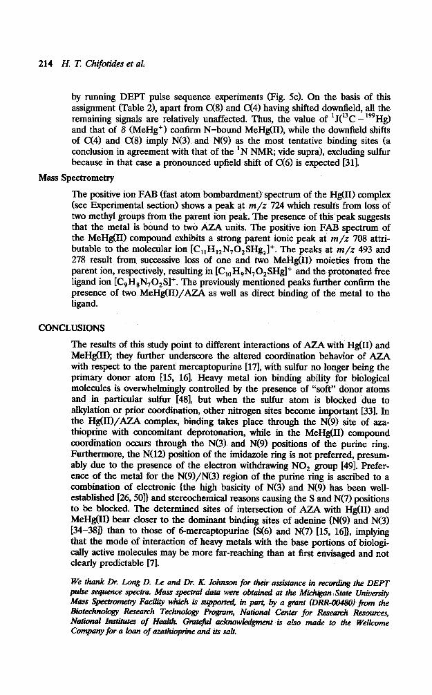
214 H. T. Chif&ides et al.
by running DEPT pulse sequence experiments (Fig. 5~). On the basis of this assignment (Table 21, apart from c(8) and CX4) having shifted downfield, all the remaining signals are relatively unaffected. Thus, the value of ‘J(13C - 199Hg) and that of S (MeHg+) confirm N-bound MeHg(II), while the downfield shifts of C(4) and c(8) imply N(3) and N(9) as the most tentative binding sites (a conclusion in agreement with that of the ‘N NMR; vide supra), excluding sulfur because in that case a pronounced upfield shift of c(6) is expected [311.
Mass Spectrometry
The positive ion FAR (fast atom bombardment) spectrum of the Hg(I1) complex (see Experimental section) shows a peak at m/z 724 which results from loss of two methyl groups from the parent ion peak. The presence of this peak suggests that the metal is bound to two AZA units. The positive ion FAR spectrum of the MeHg(I1) compound exhibits a strong parent ionic peak at m/z 708 attri- butable to the molecular ion [C,,H,,N,O,SHg,]+. The peaks at m/z 493 and 278 result from successive loss of one and two MeHg(II) moieties from the parent ion, respectively, resulting in [Cl,H,N702SHgl+ and the protonated free ligand ion [C,HsN,O,Sl+. The previously mentioned peaks further confirm the presence of two MeHg(II)/AZA as well as direct binding of the metal to the ligand.
CONCLUSIONS
The results of this study point to different interactions of AZA with Hg(I1) and MeHg(I1); they further underscore the altered coordination behavior of AZA with respect to the parent mercaptopurine [17], with sulfur no longer being the primary donor atom 115, 161. Heavy metal ion binding ability for biological molecules is ovenvhehningly controlled by the presence of “soft” donor atoms and in particular sulfur 1481, but when the sulfur atom is blocked due to alkylation or prior coordination, other nitrogen sites become important [33]. In the Hg(II)/AZA complex, binding takes place through the N(9) site of axa- thioprine with concomitant deprotonation, while in the MeHg(I1) compound coordination occurs through the N(3) and N(9) positions of the purine ring. Furthermore, the N(12) position of the imidaxole ring is not preferred, presum- ably due to the presence of the electron withdrawing NO, group [491. Prefer- ence of the metal for the N(9)/N(3) region of the purine ring is ascribed to a combination of electronic {the high basicity of N(3) and N(9) has been well- established [26,5Ol) and stereochemical reasons causing the S and N(7) positions to be blocked. The determined sites of intersection of AZA with Hg(I1) and MeHg01) bear closer to the dominant binding sites of adenine (N(9) and N(3) E34-38D than to those of 6-mercaptopurine (S(6) .and N(7) [15, 16]}, implying that the mode of interaction of heavy metals with the base portions of biologi- cally active molecules may be more far-reaching than at first envisaged and not clearly predictable [7].
We thank Dr. Long D. Le and Dr. K Johnson for their assistance in nxord@ the DEPT puke sequence spectra. Mass spectral dota were obtained at the Mich@n State Ukiuersity Mass Spectmmetty Fadity which is suppotted in part, by a grtlnt (DIM-tW480) )‘km the Biotechnology Research Techrw&y Rogram, National Center for Research Resources, Ndonal Inrliactes of Heat% Gmtefd acknowkdgment is also made to the Welkome Company for a kmn of azathkpine and its salt.

STUDIES OF MERCURY(H) AND METHYLMERCURY 215
REFERENCES
1.
2.
F. Basolo and R. G. Pearson, in Mechdnisms of Inorganic Reactions, Wiley, New York, 1967, p. 351. L. G. Marzilli, T. J. Kistenmacher, and G. L. Eichhom, in Metal Ions in Biology, T. G. Spiro, Ed., Interscience, New York, 1980, Vol. I, pp. 179, 187, and references therein.
3.
4.
J. K. Barton and S. J. Lippard, in Metal Ions in Biology, T. G. Spiro, Ed., Interscience, New York, 1980, Vol. I, pp. 31,66,88. J. G. Wright, M. J. Natan, F. M. MacDonnell, D. M. Ralston, and T. V. G’Halloran, Prog. Irwrg. Chem.: Biotitganic Chemistry, 1990, Vol. 38, p. 323 and references therein.
5.
6.
7. 8.
9. 10.
11.
12. 13.
E. Buncel, C. Boone, H. Joly, R. Kumar, and A. R. Norris, J. Znoq. Biochem. 25,61 (1983) and references therein. J. P. Charland, M. T. P. Viet, M. St-Jacques, and A. L. Beauchamp, J. Am. Chem Sot. 107,8202 (1985). E. Buncel, A. R. Norris, W. J. Racz, and S. E. Taylor, Inorg. Chem. 20,98 (1981). E. Buncel, B. K Hunter, R. Kumar, and A. R. Norris, J. Ino& B&hem. 20, 171 (1984). H. N. YeoweiI and G. B. Elion, J. Heterocycl. Chem. lo,1017 (1973). W. P. Wilson and S. A. Benezra, in Analytical profiles of Drug Substances, K. Florey, Ed., Academic Press, New York, 1981, Vol. 10, p. 29. K. G. Van Scoik, C. A. Johnson, and W. R. Porter, Drug Metabolism Rev. 160, 21, 157 (1985) and references therein. A. K. Mitra and M. M. Narurkar, Int. Jour. Pharmaceutics 35, 165 (1986). G. L. C. Ghan, D. M. Cat&ax, and C. A. Johnson, Pharmacotherapy 7(5), 165 (1987) and references therein.
14. N. Hadjiliadis and T. Theophanides, Inorg. Chim. Acta 15,167 (1975) and references therein.
15. 16. 17.
E. Dubler and E. Gyr, Znorg. Chem. 27,1466 (1988) and references therein. P. Lavertue, J. Hubert, and A. L. Beauchamp, Znorg. Chem. 15,322 (1976). H. T. Chifotides, K R. Dunbar, J. H. Matonic, and N. Katsaros, Znoq. Chm. 31, 4628 (1992) and references therein.
18.
19.
20.
R. D. Bach and H. B. Vardhan, in Inorganic Synthesis, J. M. Shreeve, Ed., Wiley, New York, 1986, Vol. 24, p. 144. E. Breitmaier and W. Voelter, Carbon-Cl3 NMR S’ctmscopy, VCH Publishers, New York, 1990, pp. 80,281. G. Pneumatikakis and N. Hadjiliadis, J. Chem. Sot., Dalton Trans., 596 (1979) and references therein.
21. 22.
N. Hadjiliadis and T. Theophanides, Inorg. Chim. Acta, 16,67 (1976). N. Hadjiliadis and T. Theophanides, Inorg. Chim. Acta, 16, 77 (1976) and references therein.
23. 24. 25.
A. R. Norris, E. Buncel, and S. E. Taylor, J. Inorg. Biochem 16, 279 (1982). R. D. Faust and P. C. Ford, J. Am. Chem. Sot. 94,5686 (1972). M. Louloudi, N. Hadjiliadis, and I. S. Butler, J. Chem. SW, Dalton Tmns. 1401(1992) and references therein.
26. W. M ,Beck, J. C. Celabrese, and N. D. Kottmair, Itwrg. Chem, 18, 136 (1979). 27. L. Grenier, J. P. Charland, and A. Beauchamp, Can. J. Chem. 66,1663 (1988). 28. A. R. Norris, R. Kumar, and E. Buncel, J. Inorg. Biochem. 22,11(1984). 29. D. L. Rebcnstein, Act. Chem. Res 11, 100 (1978). 30. A. J. Car@ and A. Marker, Iraq. Chem 15,425 (1976) and references therein. 31. E. Buncel, R. Kumar, and A. R. Norris, Can. J. Chem 64,442 (1986).

216 H. T. Chifotids et al.
32. A. J. Brown, 0. W. Howarth, and P. Moore, J. Chem. Sot., Dalton Tmns., 1589 (1976).
33. E. Buncel, A. R. Norris, S. E. Taylor, and W. J. Racz, Can. J. Chem. 60,3033 (1982). 34. L. Prizant, M. J. Olivier, R. Rivest, and A. L. Beauchamp, Can. 1. Chem. 59, 1311
(1981) and references therein. 35. J. P. Charland and A. L. Beauchamp, Croat. Chem. Acta 57,679 (1984). 36. J. P. Charland, J. F. B&ten, and A. L. Beauchamp, Inorg< @m, Acta 124,161(1986). 37. J. Hubert and A. L. Beauchamp, Can. J. Chem. S&l439 (1980). 38. J. Hubert and A. L. Beauchamp, Acta Cryst. B36,2613 (1980). 39. A. E. Derome, in manic Chemistry Series, J. E. Bakhvin, Pergamon Press, Oxford,
1990, Vol. 6, p. 143. 40. A. J, Jones, D. M. Grant, M. W. Winkley, and R.‘K. Robins, J. Am. Chem. Sot. 92;
4079 (1970). 41. L. G. Pumell and D. J. Hodgson, Org. Magn. bes. 10, 1 (1977). 42. M. C. Thorpe, W. C. Cobum, and J. A. Montgomery, J. Magn. Res. 1$,98 (1974). 43. R. J. Pugmire, D. M. Grant, R. K. Robins, and G. W. Rhodes, J Am. Chem. Sot. 87,
2226 (1965). 44. M. T. Chenon, R. J. Pugmire, D. M. Grant, R. P. Pair&a, and L. B. Townsend, J.
Am. Chem. Sot. 97,4636 (1975) and references therein. 45. D. W. Abbott and C. Woods, Znorg. Ch 22,597 (1983). 46. D. W. Abbott and C. Woods, Ztwqg. Chem. 22,1918 (1983). 47. G. C. Levy, R. L. Lichter, and G. L. Nelson, Carbon C-13 NMR S’ctmscopy, Wiley,
New York, 1980. 48. K. W. JeMette, S. J. Lippard, and D. A. Ucko, Biochim Bioplys. Acta 402, 403
(1975). 49. J. R. Bales, M. A. Mazid, P. J. Sadler, A. AggarwaI, R. Kuroda, S. Neidle,
D. W. Gihnour, B. J. Peat?, and C. A. Ramsden, J. Chem. Sot., Dalton Tmns., 795 (1985).
50. B. Pullman and A. Pullman, Quamm Biochemistry, Interscience, New York, 1963, pp. 230, 235.
Receiued September 14, 1993; accepted October 7, 1993