Methylmercury in Fish and Hair Samples from the Balbina Reservoir, Brazilian Amazon
Manuscript submitted to Journal of Pediatrics (MS 2300443, accepted for publication)
Cardiac autonomic activity in methylmercury neurotoxicity: 14-year follow-up of a Faroese birth cohort
Philippe Grandjean, MD, PhD, Katsuyuki Murata, MD, Esben Budtz-Jørgensen, PhD, and Pál
Weihe, MD
From the Institute of Public Health, University of Southern Denmark, Odense, Denmark; the
Department of Environmental Health, Harvard University School of Public Health, Boston; the
Division of Environmental Health Sciences, Akita University School of Medicine, Akita, Japan;
the Department of Biostatistics, Institute of Public Health, University of Copenhagen,
Copenhagen, Denmark; and the Department of Occupational Medicine and Public Health,
Faroese Hospital System, Tórshavn, Faroe Islands.
Supported by grants from the US National Institute of Environmental Health Sciences
(ES09797), the Danish Medical Research Council and the Nissan Science Foundation. The
contents of this paper are solely the responsibility of the authors and do not represent the official
views of the NIEHS, NIH or any other funding agency.
Reprint requests: Philippe Grandjean, MD, Department of Environmental Health, Harvard School
of Public Health, Landmark Center East room 3-110, 665, P.O.Box 15967, Boston, MA 02115.
Correspondence to: Philippe Grandjean, Institute of Public Health, University of Southern
Denmark, Winsløwparken 17, 5000 Odense C, Denmark. Phone: +45-65503768. Fax: +45-
65911458. Email: [email protected]
Key words: Electrophysiologic techniques, cardiac Environmental pollution Food
contamination Methylmercury compounds Prenatal exposure delayed effects

2Objective: To determine whether heart function in childhood is affected by exposure to
methylmercury (MeHg) from seafood.
Study design: Prospective study of a Faroese birth cohort (N = 1,022). Examinations at ages 7
and 14 years included blood pressure, heart rate variability (HRV) and its frequency components
of autonomic origin, and brainstem auditory evoked potentials (BAEP). Mercury concentrations
were determined in cord blood, and in the child’s hair.
Results: Both low-frequency (LF) and high-frequency (HF) activities decreased by about 25%
from 7 to 14 years; they correlated well with the blood pressures. A doubling of prenatal MeHg
exposure was associated with a decrease in LF and HF powers of about 6.7 % (p = 0.04) and in
the coefficient of variation of the R-R interval of 2.7% (p = 0.04) at age 14 years. No discernible
effect on blood pressure was apparent. Decreased LF variability was associated with increased
latency of BAEP peak III, but adjustment for MeHg exposure substantially attenuated this
correlation.
Conclusions: MeHg exposure was associated with decreased sympathetic (LF) and
parasympathetic (HF) modulation of the HRV. Parallel MeHg-related delays of BAEP latencies
may be due to underlying MeHg neurotoxicity to brainstem nuclei.
BAEP Brainstem auditory evoked potentials C-CVHF Component coefficient of variance for the high-frequency band C-CVLF Component coefficient of variance for the low-frequency band CVRR Coefficient of variation for the R-R interval HF High frequency HRV Heart rate variability LF Low frequency MeHg Methylmercury
3A National Research Council committee recently suggested that cardiovascular function may be
vulnerable to developmental methylmercury (MeHg) exposure.1 The evidence reviewed included
a prospective birth cohort study from the Faroe Islands, where an increase in blood pressure and a
decrease in the coefficient of variation for the R-R interval (CVRR) at age 7 years was observed
at increased intrauterine exposure levels.2 In the same children, MeHg exposure was also
associated with delayed brainstem auditory evoked potential latencies3 and deficits in
neuropsychological tests that reflect attention and several other functional modalities.4
The CVRR and its frequency components are easy to apply and highly reproducible in
children.5-9 Sympathetic hyperactivity and parasympathetic hypoactivity have been documented
in connection with cardiovascular diseases.6,10 The CVRR increases with age up to 6-10
years,11,12 followed by a decrease related to reduced parasympathetic activity.13 Boys have a
greater CVRR than girls,13 and HRV is increased by physical training.14 However, very little
information on children is available regarding the sensitivity of HRV to toxicants, especially
those that may affect the nervous system.
In adults, HRV is sensitive to dysfunctions of autonomic tone in smokers15-17 and
alcohol drinkers.17-20 Abnormalities have also been documented after exposure to neurotoxic
agents, such as MeHg, mercury vapor, other metals, certain solvents, and nerve gases.21-28 In
most cases, the CVRR was apparently affected mainly through a relative depression of
parasympathetic activity.21 In particular, a decreased HRV in adult patients that had been born
with congenital MeHg poisoning appeared to be mainly due to parasympathetic hypofunction.29
Lesions located in the brainstem may affect the HRV,30 and neuropathological abnormalities
caused by developmental MeHg exposure include this location.31
In a prospective study of a birth cohort with increased intrauterine exposure to MeHg,

4exposure-associated changes in CV-RR and blood pressures were recorded at age 7 years.2 This
population has now been re-examined at age 14 years with electrophysiologic parameters of heart
function, blood pressure and evoked potentials. In addition, CVRR data from both examinations
were analyzed in regard to the frequency components. It was hypothesized that MeHg exposure
would affect HRV and blood pressure, and that effects, if mediated through brainstem nuclei,
would be associated with known MeHg-related delays in BAEP latencies.
METHODS
Study Population and Follow-up
A cohort of 1,022 births was assembled in the Faroe Islands during a 21-month period of 1986-
1987.32 The first follow-up examination was carried out seven years later and included
determination of HRV and evoked potential latencies, neuropsychological testing, pediatric
examination, and exposure assessment.2-4 At age 14 years, a total of 878 of 1,010 live cohort
members (86.9%) were examined by comparable methods (Table I).33 The examinations were
conducted by a team of health professionals who had no access to information on individual
exposure levels. The 438 boys and 440 girls examined had an average age of 13.83 (SD, 0.32)
years. The two examinations involved a total of 963 cohort children (483 boys and 480 girls), of
whom 813 (402 boys and 411 girls) were examined twice.
In a small number of cases, complete heart rate records were not obtained. One child
had a very high systolic blood pressure and was referred to a specialist. When re-examined
several weeks later, the blood pressure had substantially decreased, but matching HRV data were
not obtained, and the child was therefore excluded. One child with a low birth weight2 and 18
other children diagnosed with neurological or other serious diseases that may affect the nervous
5system33 were excluded. None of the children had diabetes.
At each examination, a questionnaire was filled in by a parent and provided information
about past medical history and other relevant factors. Physical activity was rated much, average
and none, with all children participating in such sports as soccer being rated ‘much’. The
pediatrician interviewed the child about smoking and use of alcohol. Birth weight, family history
of hypertension / maternal hypertension (maternal hypertension risk), and smoking during
pregnancy were previously recorded.2 The study protocol was approved by the ethical review
committee for the Faroe Islands and the Institutional Review Board at Boston University, and
parental informed consent was obtained.
The primary indicator of intrauterine exposure to MeHg was the mercury concentration
in cord blood; it was supplemented by the concentration in maternal hair at parturition.32
Exposure levels varied widely, with a 1,000-fold difference between the lowest and the highest
mercury concentrations.32 Children participating in the follow-up had prenatal exposures similar
to those recorded for the full cohort.33 Analysis of hair obtained at the two follow-up
examinations showed that the intrauterine exposure level averaged about 7-fold and 4-fold higher
than the two sets of postnatal levels.33 Results given in �g may be converted to nmol by
multiplying by 5.0.
Cardiovascular Function Assessment
Two pediatricians carried out a thorough physical examination that included assessment of
systolic and diastolic blood pressures thrice, and the average was calculated. With the child
relaxing in a chair, a cuff that covered between 1/2 and 2/3 of the upper arm was applied on the
left arm, and the pressures were read in mm Hg on a sphygmomanometer. Body weight was

6measured in kg on an electronic scale to the nearest single digit after the decimal point. Standing
height was measured with a stadiometer to the nearest millimeter.
At both examinations, the heart rate was measured as the average R-R interval on an
electrocardiographic amplifier (NEC-Sanei 1271SP) connected to a computer.2 After the child
had been lying in a relaxed, supine position and breathing normally for at least 5 min, 300 R-R
intervals were measured in real time (sampling time, 1 ms); 100 consecutive R-R intervals with
the minimal standard deviation were automatically extracted for calculation of the average heart
rate and its relative standard deviation (SD). The CVRR is the ratio of the SD of the R-R
intervals to the average value (RRmean).
The cardiac sinus rhythm shows fluctuations around the mean heart rate due to
continuous changes in the autonomic balance.34-36 The main periodic fluctuations consist of the
respiratory sinus arrhythmia and the baroreflex-related heart rate variation at a lower frequency.
Their frequencies reflect the dependence on inspiratory inhibition of the vagal tone, and the
slower rhythm originating from intrinsic oscillation in the vasomotor part of the baroreflex loop.
They therefore indicate parasympathetic and sympathies activities. Spectral analysis can provide
estimates of the frequencies and powers (e.g., the magnitude of a cyclical component) from time-
series data. We therefore applied autoregressive spectral analysis to partition the HRV into
independent components.17,37 The results of the autoregressive spectral analysis were expressed
in low frequency (LF) and high frequency (HF) components, i.e., 0.01-0.15 Hz and 0.15-0.40 Hz.
As the square root of the total power spectral density is equal to the standard deviation of the R-R
intervals, each component coefficient of variation (i.e., C-CVLF and C-CVHF) was defined as
the ratio of the square root of each component power spectral density (PSD-LF and PSD-HF in
ms2) to the RRmean: C-CVLF (or C-CVHF) = 100 * sqrt (PSD-LF (or PSD-HF))/RRmean. The
7LF/HF power ratio indicates the balance of cardiac autonomic activity, with LF/HF>>1 and
LF/HF<<1 reflecting sympathetic and parasympathetic prominence, respectively.
Of 841 subjects with HRV data at age 14, BAEPs were recorded in all but two, and in all
of the 781 children who were also examined at age 7 years. A four-channel electromyograph
(Medelec Sapphire-4ME) was employed as previously described.33 Peak III latency at 20 Hz and
40 Hz at both examinations showed delays associated with intrauterine MeHg exposure.2,33 Peak
III is thought to reflect the volume-conducted electric activity from the pons (superior olivary
nucleus).38
Data Analysis
Because of skewed distributions, logarithmic transformation of the contaminant concentrations
was used to limit the dependence upon small numbers of children with very high exposure levels.
Likewise, outcome variables other than blood pressure and heart rate required logarithmic
transformation to obtain a better fit of the regression models. For transformed variables,
geometric means were supplemented by interquartile ranges (25th and 75th percentiles).
Pearson’s correlation coefficients were used to assess bivariate relationships, and adjustment for
confounders was included in partial correlation coefficients. Differences between paired
examination data were assessed by matched-pairs t test, while those between sexes were analyzed
by unpaired t-test.
Regression analysis was carried out to determine the association between MeHg
exposure and the outcome variables. Birth weight, maternal hypertension risk and maternal
smoking during pregnancy, and the child’s age, sex, height and weight were included as
confounders. Tanner stage was assessed at age 14 years but was not included because of the
8close association with age and anthropometric parameters, and because this variable was missing
from several subjects. Additional covariates at age 14 years were regular smoking (information
was missing from 31 subjects), and physical activity rated ‘much’. Blood pressure was adjusted
for examiner.
For log transformed outcome variables, the MeHg regression coefficients were modified
to indicate the relative change (in percent) of the average of the dependent variable associated
with a doubling of the MeHg exposure. The heart rate was subsequently added in models where
other outcomes were used as effect parameters. To determine whether changes between the two
examinations were associated with MeHg exposure, the paired result obtained seven years before
was introduced as predictor of the outcome at age 14 years. Potentially nonlinear exposure-effect
relationships2 were again explored in generalized additive models, which do not require linearity
assumptions while providing a smooth non-parametric dose-response curve.39
RESULTS
HRV Changed with Age but Remained a Significant Predictor of Blood Pressure
Paired HRV results from ages 7 and 14 years showed substantial changes between the two
examinations, with decreases of about 25% in both low frequency (LF) and high frequency (HF)
powers and their variabilities (Table II). However, the paired data on heart rate, CVRR, and the
HF power correlated well. Boys and girls generally had similar results, except for a higher heart
rate in girls at 7 years and a higher systolic blood pressure in boys at 14 years.
A lower CVRR, a lower HF power, and a higher LF / HF ratio were significant
predictors of increased systolic blood pressure at both examinations, and a lower LF power was
also significant at age 7 (Table III). Diastolic blood pressure was only weakly related to the HRV
9parameters.
Both LF and HF Decreased at Higher MeHg Exposures
Adjusted regression coefficients showed significant negative impacts of MeHg on
several parameters (Table IV). Both LF and HF decreased at higher prenatal MeHg exposure.
Postnatal exposure at age 7 years seemed mainly related to LF, while mainly to HF at age 14
years. Inclusion of more than one MeHg exposure indicator as independent variables resulted in
attenuated MeHg regression coefficients in the same direction. The correlation between prenatal
and postnatal exposures (correlation coefficients of 0.33 and 0.35)33 therefore did not allow a
clear separation of the impact of exposures at different developmental stages.
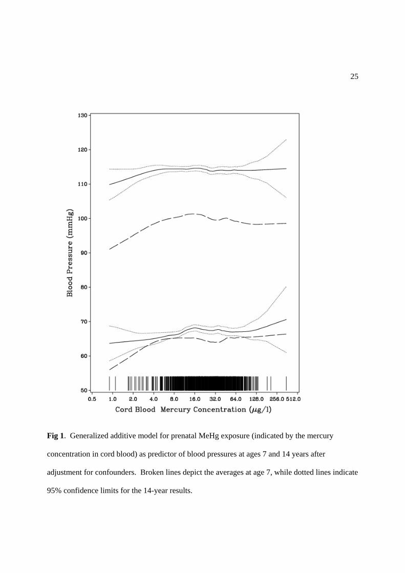
A non-linear association between prenatal MeHg exposure and blood pressure and
CVRR was previously found at age 7 years, where the strongest effect was seen at cord-blood
concentrations below 10 �g/L.2 This association was again explored using generalized additive
models. Fig 1 shows that, while significant at age 7 years,2 the effect on blood pressure is no
longer significant at age 14 years. However, similar modeling of CVRR showed that an effect of
prenatal MeHg exposure is clearly present also at age 14 years (Fig 2). While the change in
CVRR within the low-level exposure range below 10 �g/L was not statistically significant, the
heart rate increased by 2.67 (95% CI, 0.08; 5.27) for each doubling in MeHg exposure within that
interval. Heart rate was associated with almost all outcomes, but adjustment for this variable
resulted in only marginal changes of the MeHg regression coefficients. However, when the
previous outcome at age 7 years was included as an additional independent variable, the MeHg
effects on the same outcome at 14 years was attenuated and became non-significant.
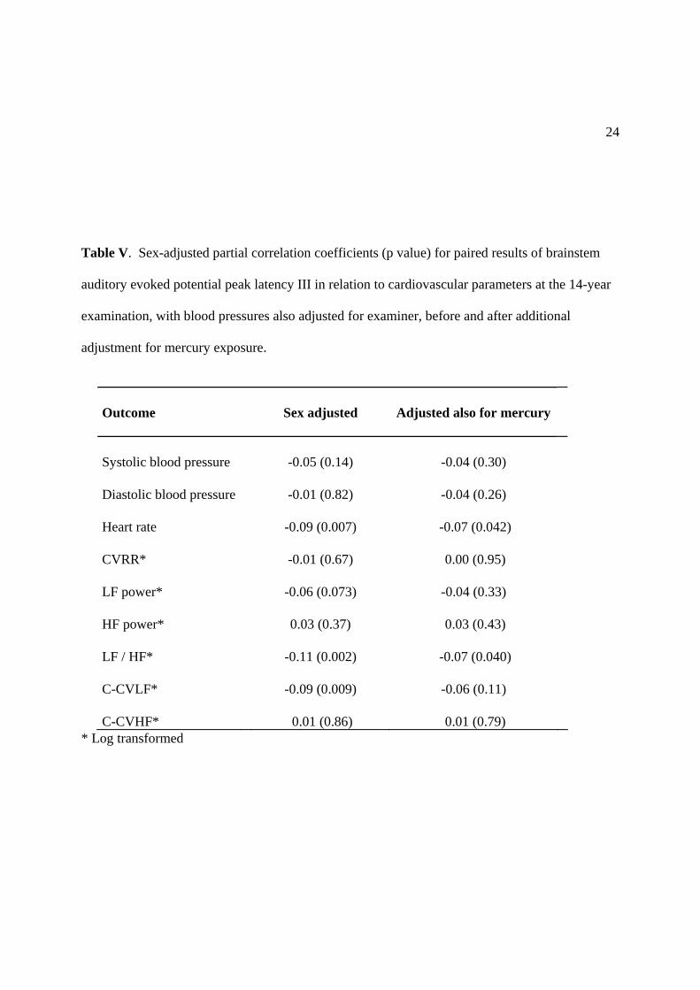
LF Power Was Associated with BAEP Latencies
To ascertain possible associations with brainstem functions, correlations were calculated
10with the latency of BAEP peak III, which appears to be increased by prenatal MeHg exposure.3,33
The LF power and its component CV showed clear negative associations with peak III latencies,
viz., the greater the BAEP latency, the less the LF power and its CV (Table V). To determine
whether these associations were innate, partial correlation coefficients were calculated with
adjustment for prenatal and postnatal MeHg exposure biomarkers. These adjusted correlations
were substantially attenuated and tended to lose statistical significance.
DISCUSSION
This prospective study of a birth cohort provides information on developmental changes in
cardiac autonomic activity and on the impact of MeHg neurotoxicity. A major advantage of the
present study is that birth cohort members were examined prospectively first at school age and
then at early adolescence. Maturation of the cardiac autonomic activity results in an increase in
the CVRR with gestational age and during early postnatal life,34 then followed by a decline of
CVRR as well as in C-CVLF and C-CVHF. Infants particularly have a high sympathetic activity
that then decreases between ages 5 and 10 years, while the sympatho-vagal balance as expressed
by the LF / HF ratio changes less.12 The age-related decreases in heart rate, CVRR, C-CVHF,
and HF power now documented are in accordance with the expectation from smaller, cross-
sectional studies.9,11-13 Despite maturation changes, the CVRR and HF results at the two
examinations correlated well, although the LF results varied more. The blood pressure levels and
the age-associated increase in systolic blood pressure are in accordance with data from previous
cross-sectional studies.40 Only limited sex-related differences were seen at the two ages
examined, while some,9,12 but not all,17,21 previous studies reported an HRV difference between
small numbers of boys and girls. An increased LF / HF ratio is thought to act as a predictor of
11increased blood pressure,10 and the results of the present study support this notion by the
significant associations between increased systolic blood pressure and decreases in CVRR and
HF results. Conversely, physical training has been reported decrease blood pressures and
increase both LF and HF powers.14 Again, the present results are in agreement with previous
research.
Only sparse information is available on adverse effects on HRV in children, e.g.,
following developmental exposure to neurotoxicants. The paucity of information is perhaps not
surprising, given the substantial age-dependency of the HRV parameters that might complicate
interpretation of cross-sectional studies. Studies of adults with occupational exposure to
neurotoxicants21-28 clearly show that HRV may be affected by chemical exposures that damage
the nervous system. However, effects on children would be difficult to predict from studies
conducted in adults.
A central origin of autonomic oscillations of the HRV is indicated by experimental
data41 and by decreases in HRV parameters in relation to central damage of the brainstem nuclei
associated with autonomic nervous function.30 In a study of lead-exposed workers,
electrophysiological parameters showed a significant exposure-associated HRV depression,
although not the anticipated delayed latencies of BAEP peaks.24 This finding may suggest that
changes in HRV could be an earlier effect than increased BAEP latencies. Another study of
occupational lead exposure reported an association between lead-induced changes in HRV and
peripheral nerve conduction.42
The impact of prenatal MeHg poisoning on HRV has recently been documented in
survivors who had reached adult age.29 Although only a small number of patients was examined,
a significant decrease in HF results was reported as a lasting abnormality. The present study

12deals with a much larger number of subjects exposed to much lower levels of MeHg from
contaminated marine food. At both examinations, both HF and LF components appeared to be
affected, but the effect on LF seemed to be less at age 7 years. A difference in sensitivity of
HRV components could be due to normal age-related changes in cardiac autonomic activity.
Despite significant associations between exposure indicators and several HRV outcomes, a
MeHg effect on blood pressure previously documented at age 7 years2 was not discernible at age
14 years. Also, the significant associations of 14-year outcomes with prenatal MeHg exposure
were attenuated after adjustment for the 7-year outcomes. Thus, the developmental MeHg
exposure did not result in substantial further changes beyond those observed at age 7 years.
The plausibility of the findings reported here is supported by the strong evidence of
MeHg neurotoxicity.1,29,31 Children with acute mercury vapor poisoning often have increased
heart rate and increased blood pressure.43 In rats exposed to high doses of MeHg chloride,
changes in normal heart rate variations were induced,44 and an increase in systolic blood pressure
seen in another study persisted for at least 9 months.45
A link between brainstem functions and autonomic tone is supported by the associations
in the present study between BAEP latencies and HRV parameters, especially the LF results.
Although only weak and mostly non-significant after adjustment for MeHg exposure, the
possibility cannot be completely discounted that such correlations are normal and unrelated to
neurotoxicant exposures. However, the fact that MeHg affects both parameters would suggest
that the exposure-related changes in HRV at least in part reflect MeHg neurotoxicity exerted in
the brainstem nuclei. In addition, the existence of neurotoxic effects on the brainstem is
suggested by highly significant MeHg-associated deficits in neuropsychological tests of
attention.4 This hypothesis therefore deserves to be examined in experimental studies.

13 While HRV parameters are highly reproducible under standardized conditions,5-9 a
possible limitation of the present study is that HRV was assessed only during a brief period, and
that respiratory patterns were not controlled. Respiratory activity may have affected the HF
component,34,35 but the children examined were resting in a darkened room, and the 100-heart-
beat sequence with the lowest variation was selected for statistical analysis. Also, the time of the
testing, relation to meals and exercise, temperature of the laboratory, etc., have limited effects on
HRV results.35 Some circadian variation has been described,13 but the cohort children were all
examined between 8 a.m. and 5 p.m., where the variation is expected to be the least. While some
HRV results may be positively associated with heart rate,17 adjustment for this variable affected
the MeHg regression coefficients only slightly. Also, by examining the CVRR rather than the
RR itself, an adjustment for dependence on heart rate was included. As mentioned above, age is
known to affect HRV results, but at the time of each examination, the subjects were of virtually
the same age, and both age and sex were incorporated as mandatory covariates in the statistical
analyses. At age 14 years, Tanner stage was not an important predictor, but rapid changes in
height, weight, and other developmental factors at puberty age may have decreased the sensitivity
of the study in identifying puberty-related effects on, e.g., blood pressure. CVRR and other HRV
parameters may be affected by alcohol and smoking habits,15-20 but few of the subjects examined
had used tobacco and alcohol, and adjustment for smoking was included as a covariate. While
several diseases, such as congenital heart disease6 and asthma,8 are known to affect the CVRR,
this cohort was population-based, and children with relevant diagnoses were excluded.
An important short-coming is that MeHg exposure was assessed only at three points in
time, and that correlations between the three measures prevented distinction between effects of
prenatal and postnatal exposures. Most likely, the three biomarkers are imprecise indicators of

14the MeHg concentrations that may have caused the effects during the 14-year lifespan of the
children. Such non-differential misclassification is likely to bias the finding toward the null
hypothesis. Thus, the present study has the advantage of size, prospective follow-up, and
adjustment for relevant confounders. However, imprecision, especially of the exposure
assessment would tend to cause an underestimation of the true MeHg effects.
Clinical studies of adults have reported that a decreased cardiac vagal tone is associated
with an increased risk of sudden cardiac death or coronary artery disease, and measurements of
HRV and the quantification of its spectral components are therefore considered powerful
predictors of cardiovascular morbidity and mortality.35 Recent findings in adults suggest that
MeHg exposure is associated with increased cardiovascular mortality.46,47 Although several toxic
mechanisms may be involved, these findings in conjunction with the present study suggest that
the impact of neurotoxic MeHg effects on autonomic regulation of heart function deserves
attention.
We are grateful to the cohort families for their loyal support, and to Drs Nicolina Sørensen and
Flemming Juul Hansen and the highly competent clinical staff in Tórshavn.
REFERENCES
1. National Research Council. Toxicological Effects of Methylmercury. Washington, DC:
National Academy Press; 2000.
2. Sørensen N, Murata K, Budtz-Jørgensen E, Weihe P, Grandjean P. Prenatal methylmercury
exposure as a cardiovascular risk factor at seven years of age. Epidemiol 1999; 10: 370-5.
3. Murata K, Weihe P, Araki S, Budtz-Jørgensen E, Grandjean P. Evoked potentials in Faroese
15children prenatally exposed to methylmercury. Neurotoxicol Teratol 1999; 21: 471-2.
4. Grandjean P, Weihe P, White RF, Debes F, Araki S, Murata K, et al. Cognitive deficit in 7-
year-old children with prenatal exposure to methylmercury. Neurotoxicol Teratol 1997; 19:
417-28.
5. Massin M, von Bernuth G. Normal ranges of heart rate variability during infancy and
childhood. Pediatr Cardiol 1997; 18: 297-302.
6. Massin MM, Derkenne B, von Bernuth G. Correlations between indices of heart rate
variability in healthy children and children with congenital heart disease. Cardiology 1999;
91: 109-13.
7. Batten LA, Urbina EM, Berenson GS. Interobserver reproducibility of heart rate variability
in children (the Bogalusa Heart Study). Am J Cardiol 2000; 86:1264-6.
8. Kazuma N, Otsuka K, Miyakawa M, Shirase E, Matsuoka I, Murata M. Seasonal variation in
heart rate variability in asthmatic children. Chronobiol Int 2000; 17: 503-11.
9. Silvetti MS, Drago F, Ragonese P. Heart rate variability in healthy children and adolescents
is partially related to age and gender. Int J Cardiol 2001; 81: 169-74.
10. Stewart JM. Does heart rate variability explain increased blood pressure in adolescents? J
Pediatr 2000; 137: 6-8.
11. Finley JP, Nugent ST. Heart rate variability in infants, children and young adults. J Auton
Nerv Syst 1995; 51: 103-8.
12. Goto M, Nagashima M, Baba R, Nagano Y, Yokota M, Nishibata K, Tsuji A. Analysis of
heart rate variability demonstrates effects of development on vagal modulation of heart rate in
healthy children. J Pediatr 1997; 130: 725-9.
13. Otsuka K, Cornelissen G, Halberg F. Age, gender and fractal scaling in heart rate variability.
16Clin Sci (Lond) 1997; 93: 299-308.
14. Mandigout S, Melin A, Fauchier L, N'Guyen LD, Courteix D, Obert P. Physical training
increases heart rate variability in healthy prepubertal children. Eur J Clin Invest 2002; 32:
479-87.
15. Robertson D, Tseng CJ, Appalsamy M. Smoking and mechanisms of cardiovascular control.
Am Heart J 1988; 115: 258-63.
16. Hayano J, Yamada M, Sakakibara Y, Fujinami T, Yokoyama K, Watanabe Y, Takata K.
Short- and long-term effects of cigarette smoking on heart rate variability. Am J Cardiol
1990; 65: 84-8.
17. Murata K, Landrigan PJ, Araki S. Effects of age, heart rate, gender, tobacco and alcohol
ingestion on R-R interval variability in human ECG. J Auton Nerv Syst 1992; 37: 199-206.
18. Weise F, Krell D, Brinkhoff N. Acute alcohol ingestion reduces heart rate variability. Drug
Alcohol Depend 1986; 17: 89-91.
19. Yokoyama A, Takagi T, Ishii H, Muramatsu T, Akai J, Kato S, et al. Impaired autonomic
nervous system in alcoholics assessed by heart rate variation. Alcohol Clin Exp Res 1991;
15: 761-5.
20. Murata K, Araki S, Yokoyama K, Sata F, Yamashita K, Ono Y. Autonomic neurotoxicity of
alcohol assessed by heart rate variability. J Auton Nerv Syst 1994; 48: 105-11.
21. Araki S, Murata K, Yokoyama K. Application of neurophysiological methods in
occupational medicine in relation to psychological performance. Ann Acad Med Singapore
1994; 23: 710-8.
22. Murata K, Araki S, Yokoyama K, Maeda K. Autonomic and peripheral nervous system
dysfunction in workers exposed to mixed organic solvents. Int Arch Occup Environ Health
171991; 63: 335-40.
23. Murata K, Araki S, Yokoyama K. Assessment of the peripheral, central and autonomic
nervous system function in styrene workers. Am J Ind Med 1991; 20: 775-84.
24. Murata K, Araki S, Yokoyama K, Nomiyama K, Nomiyama H, Tao YX, et al. Autonomic
and central nervous system effects of lead in female glass workers in China. Am J Ind Med
1995; 28: 233-44.
25. Murata K, Araki S, Yokoyama K, Okumura T, Ishimatsu S, Takasu N, et al. Asymptomatic
sequelae to acute sarin poisoning in the central and autonomic nervous system 6 months after
the Tokyo subway attack. J Neurol. 1997; 244: 601-6.
26. Magari SR, Schwartz J, Williams PL, Hauser R, Smith TJ, Christiani DC. The association of
particulate air metal concentrations with heart rate variability. Environ Health Perspect 2002;
110: 875-80.
27. Bockelmann I, Pfister EA, McGauran N, Robra BP. Assessing the suitability of cross-
sectional and longitudinal cardiac rhythm tests with regard to identifying effects of
occupational chronic lead exposure. J Occup Environ Med 2002; 44: 59-65.
28. Jhun HJ, Yim SH, Kim R, Paek D. Heart-rate variability of carbon disulfide-poisoned
subjects in Korea. Int Arch Occup Environ Health 2003; 76: 156-60.
29. Oka T, Matsukura M, Okamoto M, Harada N, Kitano T, Miike T, et al. Autonomic nervous
functions in fetal type Minamata disease patients: assessment of heart rate variability.
Tohoku J Exp Med 2003; 198: 215-21.
30. Shimomura C, Matsuzaka T, Koide E, Kinoshita S, Ono Y, Tsuji Y, et al. Spectral analysis
of heart rate variability in the dysfunction of brainstem. Brain Development 1991; 23: 26-31
(In Japanese).
1831. Takeuchi T, Eto K. The Pathology of Minamata Disease. A Tragic Story of Water Pollution.
Fukuoka: Kyushu University Press, 1999.
32. Grandjean P, Weihe P, Jørgensen PJ, Clarkson T, Cernichiari E, Viderø T. Impact of
maternal seafood diet on fetal exposure to mercury, selenium, and lead. Arch Environ Health
1992; 47: 185-95.
33. Murata K, Weihe P, Budtz-Jørgensen E, Jørgensen PJ, Grandjean P. Delayed brainstem
auditory evoked potential latencies in 14-year-old children exposed to methylmercury. J
Pediatr (submitted).
34. van Ravenswaaij-Arts CMA, Kollee LAA, Hopman JCW, Stoelinga GBA, van Geijn HP.
Heart rate variability. Ann Int Med 1993; 118: 436-47.
35. Task Force of the European Society of Cardiology and the North American Society of Pacing
and Electrophysiology. Heart rate variability: standards of measurement, physiological
interpretation and clinical use. Circulation 1996; 93: 1043-65.
36. Murata K, Araki S. Assessment of autonomic neurotoxicity in occupational and
environmental health as determined by ECG R-R interval variability: a review. Am J Ind
Med 1996; 30: 155-163.
37. Akselrod S. Spectral analysis of fluctuations in heart rate and other cardiovascular
parameters as a tool for the assessment of autonomic control. In: Korczyn AD, editor.
Handbook of Autonomic Nervous System Dysfunction. New York: Marcel Dekker, 1995, p.
469-93.
38. Chiappa KH, Hill RA. Brainstem auditory evoked potentials: interpretation. In: Chiappa
KH, editor. Evoked potentials in clinical medicine, 3rd ed. Philadelphia, PA: Lippincott-
Raven, 1997, p. 199-268.
1939. Hastie TJ, Tibshirani RJ. Generalized Additive Models. Boca Raton: CRC Press, 1990.
40. Brotons C, Singh P, Nishio T, Labarthe DR. Blood pressure by age in childhood and
adolescence: a review of 129 surveys worldwide. Int J Epidemiol 1989; 18: 824-9.
41. Grasso R, Rizzi G, Schena F, Cevese A. Arterial baroreceptors are not essential for low
frequency oscillation of arterial pressure. J Auton Nerv Syst. 1995; 50: 323-31.
42. Murata K, Araki S. Autonomic nervous system dysfunction in workers exposed to lead, zinc,
and copper in relation to peripheral nerve conduction: a study of R-R interval variability. Am
J Ind Med 1991; 20: 663-71.
43. Warkany J, Hubbard DM. Acrodynia and mercury. J Pediatr 1953; 42: 365-86.
44. Arito H, Takahashi M. Effect of methylmercury on sleep patterns in the rat. In: Suzuki T,
Imura N, Clarkson TW, editors. Advances in Mercury Toxicology. New York: Plenum,
1991, p. 381-94.
45. Wakita Y. Hypertension induced by methyl mercury in rats. Toxicol Appl Pharmacol 1987;
89: 144-7.
46. Salonen JT, Seppanen K, Nyyssönen K, Korpola H, Kauhanen J, Kantola M, Tuomilehto J,
Esterbauer H, Tatzber F, Salonen R. Intake of mercury from fish, lipid peroxidation, and the
risk of myocardial infarction and coronary, cardiovascular, and any death in eastern Finnish
men. Circulation 1995; 91: 645-55.
47. Guallar E, Sanz-Gallardo MI, van't Veer P, Bode P, Aro A, Gomez-Aracena J, Kark JD,
Riemersma RA, Martin-Moreno JM, Kok FJ. Mercury, fish oils, and the risk of myocardial
infarction. N Engl J Med 2002; 347: 1747-54.

20Table I. Characteristics of 424 boys and 433 girls from the Faroese birth cohort examined at age
14 years
Variable
Maternal age in years 27.4 (5.4)
Previous births (none / one / at least two in %) 33.4 / 34.5 / 32.1
Smoking during pregnancy (no / yes in %) 59.3 / 40.7
Alcohol consumption during pregnancy (never / ever in %) 76.5 / 23.5
Gestational age in weeks 40.3 (1.3)
Birth weight in g 3680 (528)
Age at 7-year examination in years* 6.84 (0.31)
Body weight in kg* 24.45 (3.85)
Height in cm* 122.4 (5.07)
Age at 14-year examination in years 13.83 (0.32)
Year of examination (% in 2000 / 2001) 55.8 / 44.2
Body weight in kg 54.58 (11.20)
Height in cm 162.9 (7.26)
Smoking (no / yes in %) 94.6 / 5.4
Data for continuous variables are given as mean (SD).
*Data for 401 boys and 411 girls who were also examined at age 7 years

21
Table II. Cardiovascular function results in boys and girls at 7 years and 14 years after exclusion of children with relevant diagnoses. For blood pressures and heart rate, the results are given as mean (SD). For the other outcomes, data are given as geometric mean (interquartile range).
7 years
14 years
Parameter
Boys
Girls
Boys
Girls
Correlation coefficient§
Systolic blood pressure (mm Hg)
Diastolic blood pressure (mm Hg)
Heart rate (min-1)
CVRR (%)
LF power (n.u.)
HF power (n.u.)
LF / HF
C-CVLF
C-CVHF
100.2 (8.02)*
64.7 (8.63)
80.7 (9.52)*†
7.62 (5.17-11.53)*
954 (481-2130)
1779 (807-5270)*
0.539 (0.273-1.007)
4.13 (2.96-5.99)*
5.64 (3.78-8.97)*
100.0 (8.57)*
65.0 (8.38)
85.0 (10.93)*
7.93 (5.55-11.42)*
1076 (475-2457)*
1787 (709-5025)*
0.603 (0.302-1.232)
4.61 (3.18-6.57)*
5.94 (4.11-9.25)*
115.5 (8.27)†
63.1 (8.45)
69.6 (10.86)
5.91 (4.26-8.17)
829 (413-1833)
1252 (578-2826)
0.663 (0.343-1.341)
3.31 (2.39-4.86)
4.06 (2.89-5.88)
113.0 (7.88)
64.1 (7.80)
71.5 (10.96)
6.16 (4.58-8.25)
849 (439-1796)
1439 (644-3312)
0.590 (0.320-1.101)
3.43 (2.51-4.99)
4.47 (3.26-6.09)
0.319
0.194
0.437
0.406
0.249
0.406
0.190
0.210
0.374
*p < 0.01 for difference between outcome at ages 7 and 14 years for fixed sex; †p < 0.01 for difference between outcome in boys and
girls at the same age; §Partial correlation coefficient (after logarithmic transformation of HRV parameters) with adjustment for sex,
blood pressure also adjusted for examiner (p < 0.0001)

22
Table III. Heart rate variability parameters (log transformed) as predictors of blood pressures in
Faroese cohort members at 7-year and 14-year examinations. Results are given as partial
correlation coefficients adjusted for sex and (at age 14 years) examiner (P value).
7 years
14 years
Parameter
Systolic
Diastolic
Systolic
Diastolic
CVRR
LF power
HF power
LF / HF
C-CVLF
C-CVHF
-0.12 (<0.001)
-0.09 (0.006)
-0.13 (<0.001)
0.07 (0.031)
-0.07 (0.040)
-0.12 (<0.001)
-0.09 (0.012)
-0.07 (0.040)
-0.07 (0.051)
0.01 (0.842)
-0.05 (0.117)
-0.06 (0.096)
-0.07 (0.030)
-0.03 (0.427)
-0.13 (<0.001)
0.12 (<0.001)
0.01 (0.800)
-0.11 (0.002)
-0.05 (0.175)
-0.03 (0.420)
-0.06 (0.072)
0.04 (0.242)
0.00 (0.944)
-0.04 (0.292)

23Table IV. Estimated effects of a doubling of MeHg exposure as indicated by three different
exposure biomarkers after adjustment for confounders (p value). For log transformed HRV
outcomes, the effect is expressed as the relative change (%) in the untransformed variable.
Outcome
Cord blood
Hair at 7 yrs
Hair at 14 yrs
7 years
Heart rate (min-1)
0.250 (0.38)
-0.029 (0.90)
-
CVRR*
-1.27 (0.36)
-0.344 (0.76)
-
LF power*
-5.98 (0.047)
-5.13 (0.041)
-
HF power*
-5.16 (0.19)
-0.179 (0.96)
-
LF / HF*
-0.860 (0.79)
-4.95 (0.055)
-
C-CVLF*
-2.81 (0.052)
-2.67 (0.025)
-
C-CVHF*
-2.38 (0.19)
-0.159 (0.92)
-
14 years
Systolic blood pressure (mm Hg)
0.045 (0.84)
0.040 (0.83)
-0.017 (0.91)
Diastolic blood pressure (mm Hg)
0.121 (0.60)
0.018 (0.93)
-0.086 (0.61)
Heart rate (min-1)
0.355 (0.26)
0.393 (0.13)
0.408 (0.073)
CVRR*
-2.73 (0.035)
-1.12 (0.30)
-1.52 (0.11)
LF power*
-6.70 (0.038)
-2.97 (0.27)
-2.06 (0.40)
HF power*
-6.78 (0.038)
-4.70 (0.088)
-4.61 (0.056)
LF / HF*
0.112 (0.97)
1.82 (0.45)
2.69 (0.21)
C-CVLF*
-2.90 (0.070)
-0.978 (0.47)
-0.483 (0.68)
C-CVHF*
-2.95 (0.046)
-1.87 (0.13)
-1.79 (0.099)
* Log transformed
24
Table V. Sex-adjusted partial correlation coefficients (p value) for paired results of brainstem
auditory evoked potential peak latency III in relation to cardiovascular parameters at the 14-year
examination, with blood pressures also adjusted for examiner, before and after additional
adjustment for mercury exposure.
Outcome
Sex adjusted
Adjusted also for mercury
Systolic blood pressure
-0.05 (0.14)
-0.04 (0.30)
Diastolic blood pressure
-0.01 (0.82)
-0.04 (0.26)
Heart rate
-0.09 (0.007)
-0.07 (0.042)
CVRR*
-0.01 (0.67)
0.00 (0.95)
LF power*
-0.06 (0.073)
-0.04 (0.33)
HF power*
0.03 (0.37)
0.03 (0.43)
LF / HF*
-0.11 (0.002)
-0.07 (0.040)
C-CVLF*
-0.09 (0.009)
-0.06 (0.11)
C-CVHF*
0.01 (0.86)
0.01 (0.79)
* Log transformed
25
Fig 1. Generalized additive model for prenatal MeHg exposure (indicated by the mercury
concentration in cord blood) as predictor of blood pressures at ages 7 and 14 years after
adjustment for confounders. Broken lines depict the averages at age 7, while dotted lines indicate
95% confidence limits for the 14-year results.

26
Fig 2. Generalized additive model with 95% confidence limits for prenatal MeHg exposure
(indicated by the mercury concentration in cord blood) as predictor of the CVRR at 14 years of
age after adjustment for confounders.
Copyright © 2022 FDOKUMEN