Tat Hong Equipment Service Co., Ltd. - :: HKEX :: HKEXnews ::

Virology 415 (2011) 56–68
Contents lists available at ScienceDirect
Virology
j ourna l homepage: www.e lsev ie r.com/ locate /yv i ro
Inhibition of Tat-mediated HIV-1 replication and neurotoxicity by novelGSK3-beta inhibitors
Kylene Kehn-Hall a,1, Irene Guendel a,1, Lawrence Carpio a, Leandros Skaltsounis b, Laurent Meijer b,Lena Al-Harthi c, Joseph P. Steiner d, Avindra Nath d, Olaf Kutsch e, Fatah Kashanchi a,⁎a Department of Molecular and Microbiology, National Center for Biodefense & Infectious Diseases, George Mason University, Manassas, VA 20110, USAb Protein Phosphorylation & Human Disease, CNRS, Station Biologique de Roscoff, 29680-Roscoff, Francec Department of Immunology and Microbiology, Rush Medical College, Chicago, IL, USAd Department of Neurology and Neuroscience Johns Hopkins University, Baltimore, MD, USAe Department of Medicine, University of Alabama at Birmingham, AL, USA
⁎ Corresponding author at: Department of MoleculaCenter for Biodefense & Infectious Diseases, George MasoBoulevard, MS 1H9, Manassas, VA 20110, USA. Fax: +1
E-mail address: [email protected] (F. Kashanchi).1 Authors contributed equally to the article.
0042-6822/$ – see front matter © 2011 Elsevier Inc. Aldoi:10.1016/j.virol.2011.03.025
a b s t r a c t
a r t i c l e i n f oArticle history:Received 14 December 2010Returned to author for revision10 January 2011Accepted 27 March 2011Available online 22 April 2011
Keywords:HIV-1Transcriptional inhibitionGSK-3TatHANDNeuroprotectionIndirubinTherapeutic target
The HIV-1 protein Tat is a critical regulator of viral transcription and has also been implicated as a mediator ofHIV-1 induced neurotoxicity. Here using a high throughput screening assay, we identified the GSK-3 inhibitor6BIO, as a Tat-dependent HIV-1 transcriptional inhibitor. Its ability to inhibit HIV-1 transcription wasconfirmed in TZM-bl cells, with an IC50 of 40 nM. Through screening 6BIO derivatives, we identified 6BIOder,which has a lower IC50 of 4 nM in primarymacrophages and 0.5 nM in astrocytes infected with HIV-1. 6BIOderdisplayed an IC50 value of 0.03 nM through in vitro GSK-3β kinase inhibition assays. Finally, we demonstrated6BIO and 6BIOder have neuroprotective effects on Tat induced cell death in rat mixed hippocampal cultures.Therefore 6BIO and its derivatives are unique compounds which, due to their complex mechanisms of action,are able to inhibit HIV-1 transcription as well as to protect against Tat induced neurotoxicity.
r and Microbiology, Nationaln University, 10900 University703 993 4280.
l rights reserved.
© 2011 Elsevier Inc. All rights reserved.
Introduction
Although current antiretroviral therapy has had a major impact onprolonging the life span of HIV-1 infected individuals it also hasimportant limitations. Most of these therapies prevent viral entry intothe cells or formation of proviral DNA. However, once the proviralDNA is integrated into the chromosome, the protease inhibitors arethe only class of drugs available that prevents the formation ofinfectious viral particles. These compounds do not prevent theformation of the early viral proteins such as Tat. The HIV-1 Tatprotein is a critical regulator of HIV replication and can be releasedextracellularly where it may have a multitude of effects. This isparticularly important within the brain, where the virus enters earlyin the course of infection and establishes reservoirs in glial cells.Extracellular Tat is a potent neurotoxin and may be an importantmediator of neuronal dysfunction associated with HIV-1-associated
neurocognitive disease (HAND) (Magnuson et al., 1995; Maragoset al., 2003; Sabatier et al., 1991; Weeks et al., 1995). Recent studiessuggest that despite excellent control of HIV replication in theperiphery, nearly 50–70% of individuals develop neurocognitiveimpairment. Hence approaches to prevent the effects of HIVreplication in the brain are critically necessary.
Elongation of HIV-1 transcripts is dependent on the association ofHIV-1 Tat with the nascent RNA stemloop structure of the transactiva-tion response element (TAR). This process is accompanied by thebinding of several cellular proteins, in particular the P-TEFb complexcomprising of cyclin T1 or CDK9 to the RNAP II complex associatedwiththeHIV-1 LTR (Fujinaga et al., 1998; Herrmann and Rice, 1995; Laspia etal., 1989; Nabel and Baltimore, 1987; Peng et al., 1998; Rice andMathews, 1988; Sobhian et al., 2010; Wei et al., 1998). This uniqueinteraction of HIV-1 Tat with TAR it provides an ideal target forpharmaceutical intervention. In vitro a variety of compounds have beendemonstrated to inhibit HIV-1 replication. Some of these compoundshave been described to specifically interfere with Tat–TAR interaction.The Novartis compound CGP 40336A was described to selectively bindto the AU base pair above the trinucleotide bulge with additionalstacking interactions to the bulge (Hamy et al., 1998). The multicyclicdyes, Hoechst 33258, DAPI and berenil bind to the cavity created by the
57K. Kehn-Hall et al. / Virology 415 (2011) 56–68
trinucleotide bulge (Bailly et al., 1996; Dassonneville et al., 1997;Edwards and Sigurdsson, 2002; Mestre et al., 1999). Neomycin binds totheminor groove of the lower helix,whereas argininamidewas found tobind to the U23–A27–U38 base triple (Aboul-ela et al., 1995; Brodskyet al., 1998; Brodsky and Williamson, 1997; Faber et al., 2000; Nifosi etal., 2000). Other compounds inhibit HIV-1 transcription withoutinterfering with Tat–TAR interaction (K-12, Ro24-7429) (Baba et al.,1998; Baba et al., 1997; Hsu et al., 1993). However, none of thesecompounds have proven clinically useful. Ro24-7429 was evenadvanced into early clinical trials (Hsu et al., 1993), but no inhibitoryeffect on HIV-1 replication in patients was observed during a Phase I/IIclinical trial, despite sufficiently high drug plasma levels and nodevelopment of viral resistance (Haubrich et al., 1995). While somedrug targets, such as the Tat–TAR interaction are clearly defined, themore recent discoveries that WP631, Temacrazine or CDK inhibitors(Agbottah et al., 2005; Galons et al., 2010; Guendel et al., 2010;Kashanchi and Kehn-Hall, 2009; Malumbres et al., 2008; Van Duyne etal., 2008), would also inhibit HIV-1 transcription hint at the possibilitythat there are potential features of HIV-1 transcription that are notappreciated as drug targets yet.
Tat is released by infected lymphoid (Ensoli et al., 1993) and glial cells(Tardieu et al., 1992). Both forms of Tat are released (i.e. Tat formed bythe first exon only, and that formed by both first and second exons)(Malim and Cullen, 1991) and are cytotoxic to neurons (Magnuson et al.,1995; Maragos et al., 2003; Sabatier et al., 1991; Weeks et al., 1995). Tateffects on neurons involve excitotoxic mechanisms, and this is similarlytrue for gp120. αv integrin subunit-containing receptors (Barillari et al.,1993; Etienne-Manneville and Hall, 2001; Noonan and Albini, 2000),vascular endothelial growth factor-1 receptor (VEGF-1 receptor or flt-1)(Krum and Rosenstein, 1998), low-density lipoprotein receptor-relatedprotein (LPR) (Liu et al., 2000), and NMDA receptors (Haughey et al.,2001) (NMDA receptor activationmay be secondary to GPCR activation)(HaugheyandMattson, 2002;Nathet al., 1996)haveall beenproposedastargets for Tat (Noonan and Albini, 2000; Rusnati and Presta, 2002).Interactionswithexcitatoryaminoacid receptors (HaugheyandMattson,2002; Magnuson et al., 1995; Nath et al., 1996), with accompanyingincreases in Ca2+ and reactive oxygen species (Bonavia et al., 2001;Kruman et al., 1998; Nath et al., 1996),may be especially detrimental. Tatinjection into the brain (Jones et al., 1998; Philippon et al., 1994; Sabatieret al., 1991), including the striatum(Bansal et al., 2000) causes gliosis andinfiltration of macrophages, production of cytotoxic cytokines, andchemokines such as MCP-1 (Conant et al., 1998; Weiss et al., 1999).Intrastriatal Tat injections induce neurodegenerative changes (Aksenovet al., 2003; Philippon et al., 1994), which precedes peak increases inmacrophages/microglia at 24 hours (Aksenov et al., 2003). Dyingneurons are no longer seen at 7 days following Tat exposure duringpeak periods of astrogliosis suggesting the neuronal losses are notsecondary to reactive astroglial changes. There is also evidence fromculture studies that Tat is directly neurotoxic because toxicity occurs inhighly enriched cultures of striatal neurons (Bonavia et al., 2001). Verybrief exposures to Tat can cause neuronal death (Magnuson et al., 1995;Nath et al., 1999). The core domain of Tat, amino acids 21–40 can inducecytopathic effects in monocytes and angiogenesis (Boykins et al., 1999).
Here we present data on small chemical molecules that can inhibitboth Tat-dependent transcription and Tat induced neurotoxicity. Thesesmall chemical molecules are inhibitors of GSK-3β, which is known tohave important implications in HAND. We reasoned that signalingmolecules downstreamof Tat thatmay be common to its effects onHIV-1 replication and neurotoxicity may be important therapeutic targets.
Results
HIV-1 transcriptional inhibitor screening system
We have previously developed an HTS-compatible reporter cellline in which a stably integrated chronically active HIV-1 derived from
a patient isolate drives eGFPexpression controlled by aHIV-1 LTR (CUCYcells-Jurkat based) (Kempf et al., 2006). eGFP expression thus serves as adirect and quantitativemarker of HIV-1 expression. By the inclusion of asecond fluorescent protein that can be spectrally separated from GFP(DsRedExpress) and which is controlled by an activation-independentpromoter (MSCV-LTR), we obtained a reporter cell line inwhichwe cansimultaneously measure the influence of compounds on HIV-1transcription (on-target) and general transcription (off-target). By thismeans, false positive hits, in which the compound would non-specifically inhibit global transcription, could be minimized.
However, the requirement for a BSL2+/BSL3 facility to performthe actual drug screen is very restrictive and also significantlyincreases the costs of drug screening. To overcome these limitations,we had earlier attempted to establish a non-infectious drug screeningsystem similar to CUCY cells, in which the integrated LTR-eGFPpromoter construct in the parental JLTRG cells would be activatedthrough stably integrated HIV-1 Tat expression plasmids. However,initial attempts to obtain high eGFP expressing cells through eitherstable transfection or retroviral transduction with Tat expressionvectors failed. For example, retroviral vectors expressing HIV-1 Tatunder the control of the murine leukemia promoter were found toefficiently transduce JLTRG cells, as indicated by the initially highmeasurable levels of eGFP expression, but then gradually lost eGFPexpression, probably due to promoter methylation. We solved thisproblem by using retroviral vectors that express HIV-1 Tat under thecontrol of the murine stem cell virus promoter (Swindle et al., 2004).Through an iterative process of retrovirally transducing JLTRG cellswith the Tat vectors, sorting the transduced cells for high eGFPexpression, supertransducing this eGFP-positive population, etc., wewere able to obtain a cell population that generates a HIV-1 Tat-driveneGFP fluorescence intensity similar to that observed in CUCY cells(Fig. 1A). We were then able to transduce these cells with retroviralvectors expressing high levels of RFP, which similarly as in CUCY cellscan serve as a simultaneously accessible marker for drug toxicities(Fig. 1B). Final single cell cloning for high eGFP/RFP expressing cellsresulted in the establishment of the clonal TiGR cell line. eGFP andDsRed expression in TiGR cells is stable. Over 6 months of continuousculture, we only observed a 10% decrease of eGFP expression at thepopulation basis. However, at this time-point, a 100% double-positivecell population can be easily regenerated by cell sorting.
We initially established the ideal per well cell density by titratingTiGR cells at various cell densities into 384-well plates. The eGFP/RFPsignals increase in a linear manner from a cell density of 5×10³ cells/well, with 2×10⁵ cells (15-fold signal increase over background)being optimal for cell screening. At this cell density Z′ was defined as0.89, indicating that the assay is very robust (Fig. 1C). Theexperimentally determined Z′ factor is a dimensionless statisticalvalue designed to reflect the dynamic range as well as the variation ofthe assay. It calculates as Z′=1−(3σp+3σn)/|μp−μn). With 3σn
representing the standard deviation of the negative control samples,3σp representing the standard deviation of the positive samples, μnthe mean of the negative control samples and μp the mean of thepositive samples. Z′=1 would be an ideal assay and 1NZ′N0.5 areconsidered very good to excellent assays.
We next verified the TiGR based HTS assay in a small manuallyperformed1000 compound screen and included someknown inhibitorsof HIV-1 transcription (Ro24-7429, WP631) as controls, which wereefficiently detected (80–90% eGFP signal reduction on day 7; data notshown). As TiGR cells use fluorescence intensities as quantitativemarkers for HIV-1 expression and drug toxicities, drug effects can beassessed intervention free. As such, the assay determines not only thecumulative inhibition at a defined time point but also the inhibitory/toxic onset kinetics of a respective compound, which gives additionalinsights into the possible drug efficiency. Therefore, this assay systemserves as a standardized platform to screen for Tat LTR transcriptionalinhibitors.

Fig. 1. HIV-1 transcriptional inhibitor screening system. Comparison of eGFP (A) and RFP (B) expression in CUCY cells and the TIGR reporter cells used in this application. (C) Z-testresults for TiGR cells. 2×10⁵ TiGR cells were loaded into 48 wells of a 384-well plate and eGFP fluorescence intensity was measured (black circles) compared to eGFP fluorescence ofthe parental JLTRG cells (gray squares) loaded into 48 wells of the same plate.
Table 1Tat-dependent Transcription Inhibitors.
Inhibitor Description and target % Inhibitionat 1 μM
Dactiniomycin Antineoplastic, intercalating agent 986BIO Glycogen synthase kinase 3α/β inhibitor 62Quinine ethyl carbonate Antimalarial 34Indirubin-3′-oxime CDK inhibitor 25Epirubicin hydrochloride Antineoplastic 17
58 K. Kehn-Hall et al. / Virology 415 (2011) 56–68
Identification of Tat-dependent transcription inhibitors
LOPAC-Sigma-Aldrich (1280 compounds) and Spectrum-Micro-source (2000 compounds) small compound libraries were screenedusing the TiGR cells described above to identify novel HIV-1 transcrip-tion inhibitors. These libraries were chosen as they contain pharmaco-logically active compounds, known FDA-approved drugs, experimentalbioactives, and pure natural products which provided a wide range ofcompounds with potential biological activity. Compounds were dilutedin DMSO and screened at 10 μM for Tat transcriptional inhibition. TiGRcells were analyzed at days 0, 1, 2, 3, 4 and 7 post drug treatments todetermine the GPF and RFP signals and those compounds that exhibitedgreater than 30% inhibition of GFP expression without affecting the RFPexpression were selected for further analysis. We identified fivecandidates based on these criteria. The top five candidates that wereidentified through the TiGR cell system were tested in TZM-bl cells,which contain an integrated LTR-luciferase reporter. As the TZM-blsystem was utilized as our secondary screen, we chose 1 μM as a morestringent cut-off for selection of our lead candidate. TZM-bl cells weretransfected with Tat followed by compound treatment the next day.Results shown in Table 1 indicate the %inhibition observed at 1 μM ascompared to the DMSO control. The compound with the greatest
inhibition was the chemotherapeutic agent Dactinomycin, which is atranscriptional inhibitor and anti-proliferation compound (Ho et al.,2008). Its mechanism of action is non-specific as it binds to multipleDNA structures such as GC-rich duplexes, single-stranded forms, orhairpin forms, as well as interfering with RNA polymerase (Kang andPark, 2009). Although toxicity was not observed in the TIGR cells, theabove characteristics suggest the limited HIV-1 therapeutic potentialof this compound; therefore we chose not to further pursue Dactino-mycin. Out of the remaining compounds, the GSK-3 inhibitor 6BIO (6-bromoindirubin-3′-oxime) (Meijer et al., 2003; Polychronopoulos et al.,2004) was ranked the secondmost potent LTR transcriptional inhibitor
59K. Kehn-Hall et al. / Virology 415 (2011) 56–68
(Table 1). Therefore, these results indicate that 6BIO reproduciblyinhibits Tat-dependent LTR transcription.
6BIO inhibits HIV-1 transcription without inducing cellular toxicity
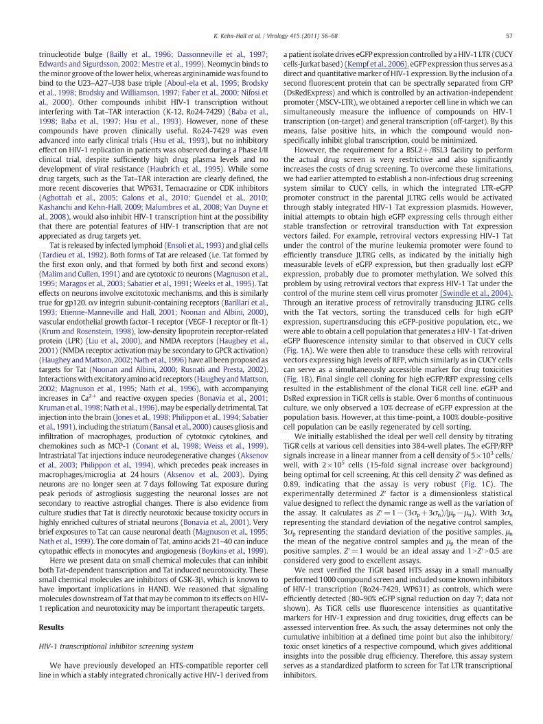
The best candidate identified in our screen, 6BIO, was furthertested using TZM-bl cells to determine its IC50. TZM-bl cells weretransfected with Tat followed by treatment with various concentra-tions of 6BIO the next day. Luciferase assays performed at 2 days post6BIO treatment indicated that 6BIO inhibited HIV-1 LTR Tat-dependent transcription in a dose-dependent manner (Fig. 2A),with an IC50 of 40 nM. MTT assays were performed to determine theinfluence of 6BIO on cell viability. As can be seen in Fig. 2B, at theconcentrations used in our transcriptional assays, 6BIO did not affectcell viability. MTT assays were also performed with 6BIO on multipleother cell types, including uninfected and infected T-cell lines (CEMand ACH2), uninfected and infected monocytic cell lines (U937 andU1) and an astrocytoma cell line (U87MG). No cellular toxicity wasobserved upon treatment with 6BIO (1 μM) as compared to the DMSOcontrol (data not shown). These results indicate that 6BIO can inhibitHIV-1 transcription without inhibiting cellular viability.
To determine whether 6BIO has an effect on HIV-1 replication inprimary cells, we infected activated PBMCs with the dual tropic 89.6virus and subsequently treated cells with vehicle (DMSO) or variousconcentrations of 6BIO (0.1, 0.5, and 1.0 μM). Treatment wasperformed only one time. Cells were maintained for 14 days andsupernatants were collected at days 7 and 14 for RT analysis (Fig. 2C).Results indicate that 1.0 μM of 6BIO inhibited virus replication bymore than 50% after 7 and 14 days. Cells were also collected to
Fig. 2. 6BIO inhibits HIV-1 transcription and replication without inducing cellular toxicity. (A6BIO (0.025, 0.05, 0.1, and 1.0 μM). Cells were processed 48 hours post drug treatment for lustandard deviation. (B) TZM-bl cells were treated with DMSO or 6BIO (0.025, 0.05, 0.1, andperformed in triplicate and samples analyzed at 48 hours. (C) PHA and IL-2 activated PBMCsperformed by following the guidelines of the Centers for Disease Control. Approximately 2.5×of complete media and plated in a 96well plate at 200 μl/well. 6BIO treatment (0, 0.1, 0.5 andon days 7 and 14 and stored at−20 °C for RT assay. Treatments were performed in triplicatedescribed for C. Cells were collected on days 7 and 14, washed 2× with PBS without Ca andanalysis by flow cytometry to determine apoptosis (sub-G1 peak). Triplicate wells were po
determine the influence of 6BIO treatment on cell viability using PIstaining/FACS analysis. Apoptosis was determined through cell cycleanalysis (sub G1 peak). Uninfected PBMCs displayed low levels ofapoptosis at both days 7 and 14 (Fig. 2D). Infected PBMCs alsodemonstrated low levels of apoptosis at day 7. However, at day 14 anincrease in apoptosis was observed in the infected cells probably dueto viral induced cell death as the DMSO control cells were alsobeginning to die and due to the fact that BIO treatment was unable tocompletely suppress viral replication. Collectively these resultsindicate that the IC50 of the 6BIO inhibition is ~0.75 μM in HIV-1infected PBMCs and that 6BIO can inhibit HIV-1 replication withoutinhibiting cellular viability.
Identification of a more potent 6BIO analog
Hit2Lead (Hit2Lead.com) was utilized to identify thirty-eightcommercially available 6BIO analogs. These analogs were tested at1 μM in the TZM-bl cells to test their ability to inhibit Tat-dependentLTR transcription (Fig. 3A). Many of the analogs actually increasedviral transcription as compared to the DMSO control, especiallycompounds 16 and 31 possibly due to removal of inhibitors from thepromoter. In contrast, a few analogs tested displayed LTR transcrip-tional inhibition with different potencies (compounds 4, 6, 18).Derivative #6 showed particularly strong inhibition of LTR transcrip-tion. MTT assays were performed with derivative #6 on multiple celllines, including uninfected and infected T-cells (CEM and ACH2),uninfected and infected monocytes (U937 and U1) and astrocytomacells (U87MG). Results indicated that the transcriptional inhibitionwas not due to cellular toxicity (Fig. 3B) as very little change in
) TZM-bl cells were transfected with 1.0 μg of Tat and treated the next day with DMSO,ciferase assays. Assays were performed in triplicate and an average value is shown plus1.0 μM). Cell proliferation/viability was determined by MTT assays. Treatments were
were kept in culture for 2 days prior to infection. Isolation and treatment of PBMCs were107 PBMCs were infectedwith 89.6 (35,520 RT units). Cells were resuspended in 6.5 ml1.0 μM)was performed (only once) the day following infection. Samples were collectedand the average plus the standard deviation is displayed. (D) PBMCs were processed asMg, resuspended in 70% ethanol, and stained with propidium iodine prior to cell cycleoled for this analysis.

Fig. 3. 6BIO derivatives inhibit HIV-1 transcription without inducing cellular toxicity. (A) TZM-bl cells were transfected with 1.0 μg of Tat and treated the next day with DMSO, 6BIO,derivatives 1–38 at 1 μM. Cells were processed 48 hours post drug treatment for luciferase assays. Assays were performed in triplicate and an average value is shown plus standarddeviation. (B) CEM, ACH2, U937, U1, and U87MG cells were treated with DMSO and derivative #6 (1 μM). Cell proliferation/viability was determined byMTT assays. Treatments wereperformed in triplicate and samples analyzed at 48 hours. Percent viability is expressed as compared to the DMSO control.
60 K. Kehn-Hall et al. / Virology 415 (2011) 56–68
viability was observed upon treatment with derivative #6 ascompared to the DMSO control. Derivative #6 was termed 6BIOderand selected for follow-up analysis.
Effect of 6BIO analogs on inhibition of HIV-1 in monocytes andastrocytoma cells
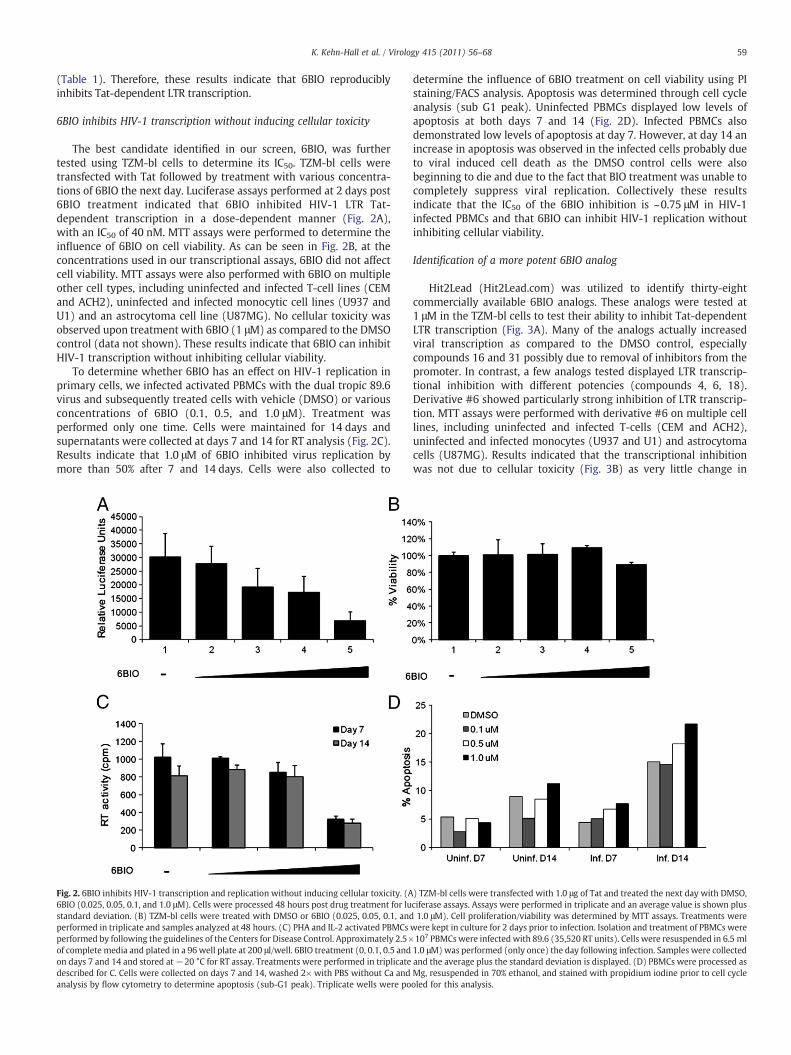
To further test the effect of 6BIOder, we assayed its effect inprimary monocyte/macrophage infection along with the parent 6BIOdrug. Fig. 4A shows the general structure of 6BIO and 6BIOder. Bothcompounds are commercially available, however 6BIOder has notbeen studied in the context of HAND. Data in Fig. 4B utilizedmonocyte/macrophages from a healthy donor infected with HIV-1dual tropic 89.6 for 7 days. Both 6BIO and 6BIOder were added to themedia during the course of infection (once only). Lane 1 showsnormal replication of HIV-1 as evidenced by RT activity in supernatantand lane 2 is with DMSO control. Lane 3 used 6BIO (10 nM) and 4–7utilized 6BIOder (0.1, 1, 10, 100 nM). There was considerableinhibition with 6BIOder at 10 nM. The IC50 for 6BIOder in these cellsis 4 nM. Next, we performed a similar assay in U87MG cells with6BIOder at 0.1, 1 and 10 nM (left hand graph). Again, inhibition wasmostly observed at 1 nM with IC50=0.5 nM. Finally, we performedMTT assays withmonocyte/macrophage from two healthy donors andU87MG at 10, 100, 1000 and 10,000 nM (lanes 2–5) and observed no
apparent toxicity in these cells (Fig. 4C). Collectively, these dataindicate that when searching for 6BIO related compounds to inhibitHIV-1, wewere able to identify 1 out of 38 compounds that had IC50 of0.5–4 nM (depending on the cell type) and were not toxic to cells at10 μM. This is roughly a 3 log difference between HIV-1 inhibitoryactivity and possible cell toxicity.
6BIOder does not inhibit cellular gene expression in the absence of Tatand is specific to GSK-3β
We next asked whether 6BIOder was inhibitory to genes thatrequire CDK9/cyclin T. HIV-1 Tat utilizes CDK9/cyclin T for itsactivation of transcription. We therefore asked if cellular geneexpression would be sensitive to 6BIOder in treated cells. Results inFig. 5A show RT/PCR results of 3 genes that require CDK9/cyclin T fortheir transcription. None of these genes showed a dramatic decreaseafter 6BIOder treatment in U937 or U87MG cells in the absence of Tat.Interestingly, we did observe an increase in expression for MCL-1, IL-8, and cyclin D1 following 6BIOder treatment of U937 cells. Next weasked if 6BIO or 6BIOder was able to inhibit GSK-3β kinase activity.U937 cells were treated with 6BIO or 6BIOder, followed byimmunoprecipitation with anti-GSK-3β. The IPed material wasutilized for in vitro kinase assays with glycogen synthase peptide 2as the substrate. Results in Fig. 5B indicate that BIOder and not 6BIO
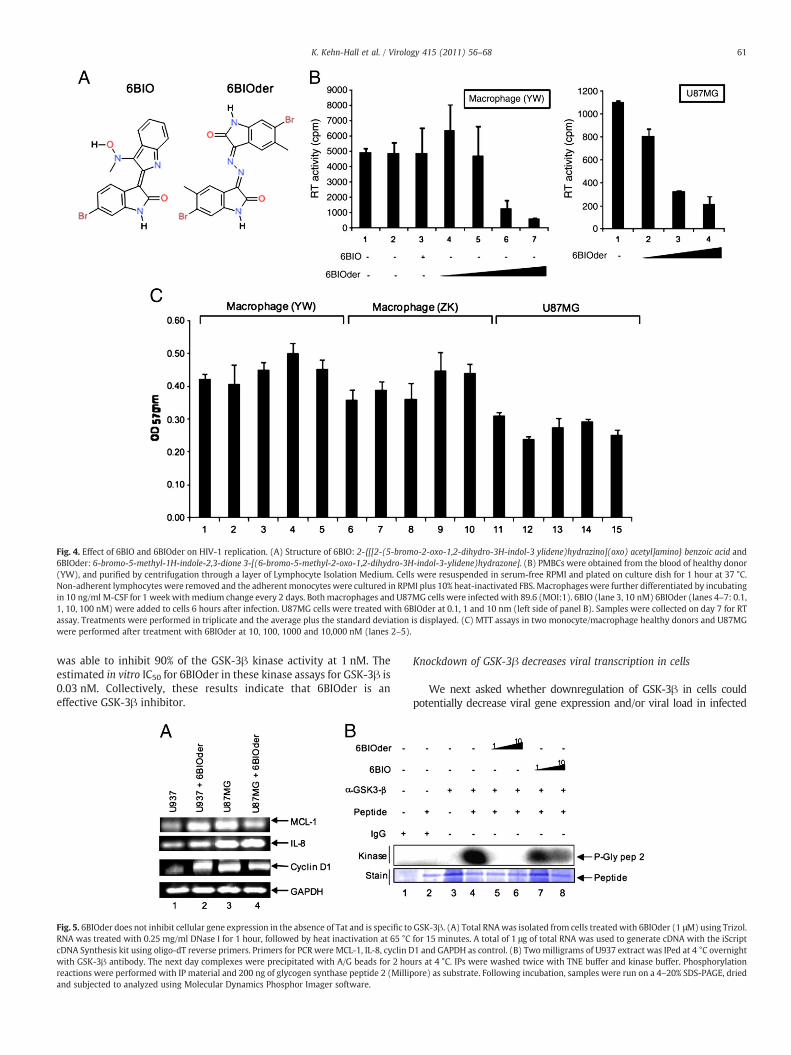
Fig. 4. Effect of 6BIO and 6BIOder on HIV-1 replication. (A) Structure of 6BIO: 2-{[[2-(5-bromo-2-oxo-1,2-dihydro-3H-indol-3 ylidene)hydrazino](oxo) acetyl]amino} benzoic acid and6BIOder: 6-bromo-5-methyl-1H-indole-2,3-dione 3-[(6-bromo-5-methyl-2-oxo-1,2-dihydro-3H-indol-3-ylidene)hydrazone]. (B) PMBCs were obtained from the blood of healthy donor(YW), and purified by centrifugation through a layer of Lymphocyte Isolation Medium. Cells were resuspended in serum-free RPMI and plated on culture dish for 1 hour at 37 °C.Non-adherent lymphocytes were removed and the adherent monocytes were cultured in RPMI plus 10% heat-inactivated FBS. Macrophages were further differentiated by incubatingin 10 ng/ml M-CSF for 1 week withmedium change every 2 days. Bothmacrophages and U87MG cells were infected with 89.6 (MOI:1). 6BIO (lane 3, 10 nM) 6BIOder (lanes 4–7: 0.1,1, 10, 100 nM) were added to cells 6 hours after infection. U87MG cells were treated with 6BIOder at 0.1, 1 and 10 nm (left side of panel B). Samples were collected on day 7 for RTassay. Treatments were performed in triplicate and the average plus the standard deviation is displayed. (C) MTT assays in two monocyte/macrophage healthy donors and U87MGwere performed after treatment with 6BIOder at 10, 100, 1000 and 10,000 nM (lanes 2–5).
61K. Kehn-Hall et al. / Virology 415 (2011) 56–68
was able to inhibit 90% of the GSK-3β kinase activity at 1 nM. Theestimated in vitro IC50 for 6BIOder in these kinase assays for GSK-3β is0.03 nM. Collectively, these results indicate that 6BIOder is aneffective GSK-3β inhibitor.
Fig. 5. 6BIOder does not inhibit cellular gene expression in the absence of Tat and is specific toRNA was treated with 0.25 mg/ml DNase I for 1 hour, followed by heat inactivation at 65 °CcDNA Synthesis kit using oligo-dT reverse primers. Primers for PCR were MCL-1, IL-8, cyclin Dwith GSK-3β antibody. The next day complexes were precipitated with A/G beads for 2 horeactions were performed with IP material and 200 ng of glycogen synthase peptide 2 (Millipand subjected to analyzed using Molecular Dynamics Phosphor Imager software.
Knockdown of GSK-3β decreases viral transcription in cells
We next asked whether downregulation of GSK-3β in cells couldpotentially decrease viral gene expression and/or viral load in infected
GSK-3β. (A) Total RNAwas isolated from cells treated with 6BIOder (1 μM) using Trizol.for 15 minutes. A total of 1 μg of total RNA was used to generate cDNA with the iScript1 and GAPDH as control. (B) Twomilligrams of U937 extract was IPed at 4 °C overnight
urs at 4 °C. IPs were washed twice with TNE buffer and kinase buffer. Phosphorylationore) as substrate. Following incubation, samples were run on a 4–20% SDS-PAGE, dried

62 K. Kehn-Hall et al. / Virology 415 (2011) 56–68
cells. For that we used TZM-bl cells that were transfected with siRNAagainst GSK-3β or luciferase in the presence or absence of Tat and assayedfor luciferase expression 48 hours post-transfection. Results of such anexperiment are shown in Fig. 6Awhere siLuc or siGSK-3β did not controlmuch of basal transcription in the Hela TZM-bl cells (lanes 1 and 2).However, siGSK-3β did significantly reduce Tat activated transcription inthese cells (compare lanes3and4). To confirmtheknockdown,whole cellextract of TZM-bl transfected with siRNAs was run on a 4–20% SDS-PAGEand Western blotted against GSK-3β and β-actin as control. More than90% knockdown was observed with siGSK-3β (lower insert, lane 4).
We next asked whether knockdown of GSK-3β could potentiallydecreasevirus release fromHIV-1 infectedcells. For thatweused J1-1cellswhich are Jurkat derived, contain single copy integrated wild type virus,and release virus into the supernatant without addition of any externalstimuli (i.e., TNF orHDAC inhibitors).Weperformed the experimentwitheither siLuc as control or siGSK-3β using electroporation. Results of suchan experiment are shown in Fig. 6B, where there was a marked decreaseof RT from cells treated with siGSK-3β at days 2 and 4. Collectively thesedata imply that knockdown of GSK-3β in either HeLa or Jurkat (J1-1)based cells down regulated HIV gene expression and viral production.
Effect of 6BIOder on the dox-dependentHIV-rtTA viruses (Tat/TAR specificity)
We next asked if the effect of 6BIOder was specific to Tat functionin HIV-1 expressing cells. For that we obtained two sets of constructs
Fig. 6. Downregulation of GSK-3β decreases viral transcription in cells. (A) TZM-bl cells wabsence of Tat (1 μg) and assayed for luciferase expression 48 hours post-transfection. To cTZM-bl transfected with luciferase siRNA (siLuc), and TZM-bl transfected with GSK-3β (siGS*pb0.01 is related to the comparison between siLuc and siGSK-3β. (B) J1-1 cells were usedelectroporated with either siLuc or siGSK-3β (200 nm) and re-plated in complete media. Streatment was abolished by day 6 (data not shown). *pb0.01 is related to the comparison
from the Berkhout lab which have mutation in Tat/TAR sequence.These viruses can be induced with dox and full particles are recoveredin the supernatant (Verhoef et al., 2001). Briefly, the full-length,infectious HIV-1 molecular clone pLAI was used for construction of anHIV-rtTA virus genome, the transcription of which is controlled bydox. The viral transcriptional elements TAR and Tat were replaced bythe prokaryotic tetO-rtTA elements (Fig. 7A). TAR was inactivated bymutation of multiple nucleotides in the single-stranded bulge andloop domains, the binding sites for Tat and cyclin T, respectively. Also,the inactive TAR motif was inserted in both LTRs to minimize thechance of reversion to the wild-type virus by a recombination event.Inactivation of the Tat protein was accomplished by introduction ofthe Tyr26Ala point mutation. This single amino acid change resultedin a severe loss of Tat transcriptional activity and virus replication.Thus, both LTRs were modified, and this was done in the wild-type(W) and mutant (Y) Tat backgrounds, resulting in four HIV-rtTAconstructs: KWK, KYK, SWS, and SYS. In the current study we used theKWK and KYK sets. The virus variant KWK is most wild type-likebecause it maintained the NF-B sites, SP1 sites, and a wild-type Tatprotein, but it has a mutation in TAR. The KYK clone has similarpromoter elements; however the Tat and TAR are both mutated.These HIV-rtTA replicate in a dox-dependent manner, when trans-fected into either cell lines or primary PBMCs.
We performed our experiments with the KWK and KYK clones inprimary monocytes that had been differentiated into macrophages
ere transfected with siRNA against GSK-3β or luciferase (100 nM) in the presence oronfirm knockdown, 50 μg of whole cell extract of 293T cells (positive control), TZM-bl,K-3β), were run on a 4–20% SDS-PAGE andWestern blotted against GSK-3β and β-actin.for electroporation with siLuc or siGSK-3β. Log phase growing cells (5×10⁵/ml) wereupernatants were processed for presence of RT at days 2 and 4. The effect of siGSK-3βbetween siLuc and siGSK-3β.
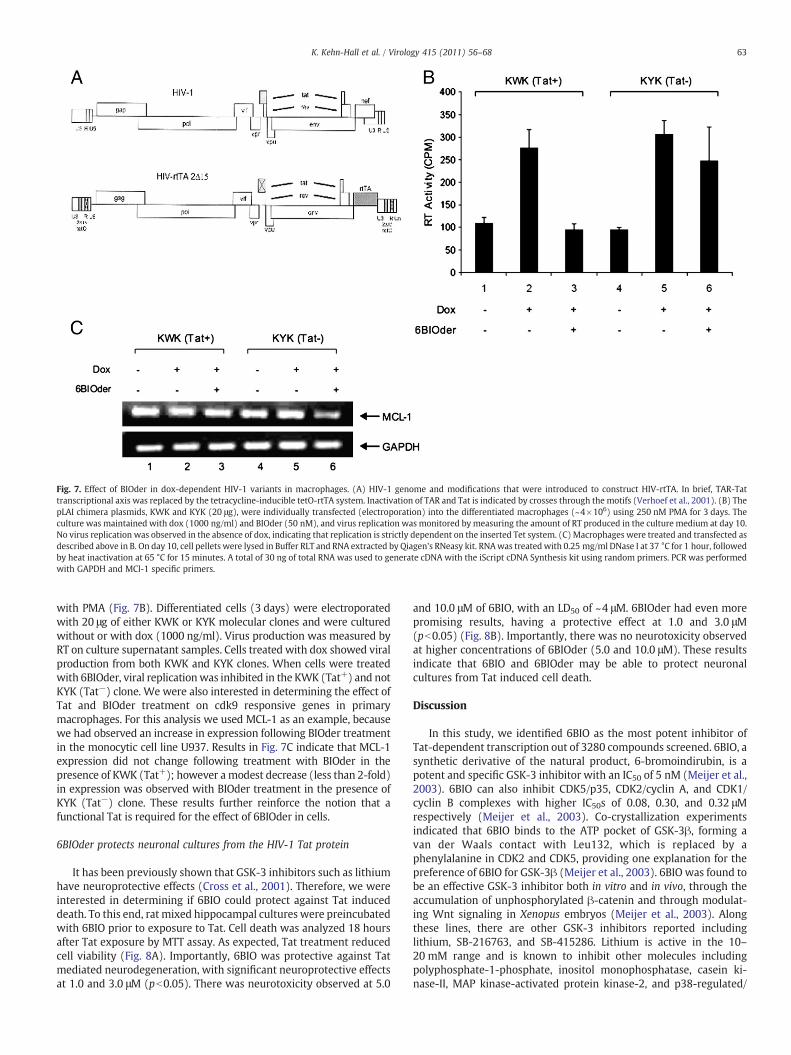
Fig. 7. Effect of BIOder in dox-dependent HIV-1 variants in macrophages. (A) HIV-1 genome and modifications that were introduced to construct HIV-rtTA. In brief, TAR-Tattranscriptional axis was replaced by the tetracycline-inducible tetO-rtTA system. Inactivation of TAR and Tat is indicated by crosses through the motifs (Verhoef et al., 2001). (B) ThepLAI chimera plasmids, KWK and KYK (20 μg), were individually transfected (electroporation) into the differentiated macrophages (~4×10⁶) using 250 nM PMA for 3 days. Theculture was maintained with dox (1000 ng/ml) and BIOder (50 nM), and virus replication was monitored by measuring the amount of RT produced in the culture medium at day 10.No virus replication was observed in the absence of dox, indicating that replication is strictly dependent on the inserted Tet system. (C) Macrophages were treated and transfected asdescribed above in B. On day 10, cell pellets were lysed in Buffer RLT and RNA extracted by Qiagen's RNeasy kit. RNAwas treated with 0.25 mg/ml DNase I at 37 °C for 1 hour, followedby heat inactivation at 65 °C for 15 minutes. A total of 30 ng of total RNA was used to generate cDNA with the iScript cDNA Synthesis kit using random primers. PCR was performedwith GAPDH and MCl-1 specific primers.
63K. Kehn-Hall et al. / Virology 415 (2011) 56–68
with PMA (Fig. 7B). Differentiated cells (3 days) were electroporatedwith 20 μg of either KWK or KYK molecular clones and were culturedwithout or with dox (1000 ng/ml). Virus production was measured byRT on culture supernatant samples. Cells treated with dox showed viralproduction from both KWK and KYK clones. When cells were treatedwith 6BIOder, viral replicationwas inhibited in the KWK (Tat+) and notKYK (Tat−) clone. We were also interested in determining the effect ofTat and BIOder treatment on cdk9 responsive genes in primarymacrophages. For this analysis we used MCL-1 as an example, becausewe had observed an increase in expression following BIOder treatmentin the monocytic cell line U937. Results in Fig. 7C indicate that MCL-1expression did not change following treatment with BIOder in thepresence of KWK (Tat+); however a modest decrease (less than 2-fold)in expression was observed with BIOder treatment in the presence ofKYK (Tat−) clone. These results further reinforce the notion that afunctional Tat is required for the effect of 6BIOder in cells.
6BIOder protects neuronal cultures from the HIV-1 Tat protein
It has been previously shown that GSK-3 inhibitors such as lithiumhave neuroprotective effects (Cross et al., 2001). Therefore, we wereinterested in determining if 6BIO could protect against Tat induceddeath. To this end, rat mixed hippocampal cultures were preincubatedwith 6BIO prior to exposure to Tat. Cell death was analyzed 18 hoursafter Tat exposure by MTT assay. As expected, Tat treatment reducedcell viability (Fig. 8A). Importantly, 6BIO was protective against Tatmediated neurodegeneration, with significant neuroprotective effectsat 1.0 and 3.0 μM (pb0.05). There was neurotoxicity observed at 5.0
and 10.0 μM of 6BIO, with an LD50 of ~4 μM. 6BIOder had even morepromising results, having a protective effect at 1.0 and 3.0 μM(pb0.05) (Fig. 8B). Importantly, there was no neurotoxicity observedat higher concentrations of 6BIOder (5.0 and 10.0 μM). These resultsindicate that 6BIO and 6BIOder may be able to protect neuronalcultures from Tat induced cell death.
Discussion
In this study, we identified 6BIO as the most potent inhibitor ofTat-dependent transcription out of 3280 compounds screened. 6BIO, asynthetic derivative of the natural product, 6-bromoindirubin, is apotent and specific GSK-3 inhibitor with an IC50 of 5 nM (Meijer et al.,2003). 6BIO can also inhibit CDK5/p35, CDK2/cyclin A, and CDK1/cyclin B complexes with higher IC50s of 0.08, 0.30, and 0.32 μMrespectively (Meijer et al., 2003). Co-crystallization experimentsindicated that 6BIO binds to the ATP pocket of GSK-3β, forming avan der Waals contact with Leu132, which is replaced by aphenylalanine in CDK2 and CDK5, providing one explanation for thepreference of 6BIO for GSK-3β (Meijer et al., 2003). 6BIO was found tobe an effective GSK-3 inhibitor both in vitro and in vivo, through theaccumulation of unphosphorylated β-catenin and through modulat-ing Wnt signaling in Xenopus embryos (Meijer et al., 2003). Alongthese lines, there are other GSK-3 inhibitors reported includinglithium, SB-216763, and SB-415286. Lithium is active in the 10–20 mM range and is known to inhibit other molecules includingpolyphosphate-1-phosphate, inositol monophosphatase, casein ki-nase-II, MAP kinase-activated protein kinase-2, and p38-regulated/

Fig. 8. 6BIO protects neuronal cultures from the HIV-1 Tat protein. Rat mixedhippocampal cultures were preincubated with various concentrations of (A) 6BIO(0.05–10 μM) or (B) 6BIOder (0.05–10 μM) for 1 hour at 37 °C prior to an 18-hourexposure to 500 nM Tat1–72. After 18 hours, cell survival was measured by MTT assay.Statistical significance was determined by ANOVA, followed by Newman–Keuls posthoc pair-wise comparisons.
64 K. Kehn-Hall et al. / Virology 415 (2011) 56–68
activated kinase (Berridge et al., 1989; Davies et al., 2000). SB-216763and SB-415286 were originally identified as GSK-3α inhibitorsthrough a high throughput screen of the SmithKline Beechamcompound library against rabbit GSK-3α and subsequently wereshown to inhibit human GSK-3 with IC50s of 34 nM and 78 nMrespectively (Coghlan et al., 2000). 6BIO has the lowest IC50 of allthese GSK-3 inhibitors (5 nM) and therefore has the most therapeuticpotential. In addition, in this study we identified a second generation6BIO analog, 6BIOder, which has an in vitro IC50 of 0.03 nM anddemonstrated neuronal protectionwith less toxicity than 6BIO. At thisstage it cannot be ruled out that 6BIOder acts through anotherunidentified kinase (i.e., CK1, CLK1 or DYRK from infected cells), or byinhibition of a collection of various kinases, which jointly results inselective inhibition of Tat-dependent transcription. Another compli-cating factor at this point is that cell type difference that was alsoobserved when 6BIOder inhibited HIV-1 better in U87MG ascompared to monocyte/macrophages cells.
Glycogen synthase kinase-3 (GSK-3) is a serine/threonine proteinkinase that was originally described as a critical regulator of glycogenmetabolism through the phoshorylation of glycogen synthase (Plyteet al., 1992). The human GSK-3 family comprises two isoforms, GSK-3α and GSK-3β, which share 97% sequence identity in their kinasedomain, but differ in their N- and C-terminus regions. GSK-3α/GSK-3β are implicated in the regulation of glycogen synthesis, the Wnt/β-
catenin signaling pathway, PI3K pathway, cell cycle control, tran-scriptional regulation, and apoptosis (Frame and Cohen, 2001). Theability of GSK-3α/GSK-3β to regulate this vast array of cellularprocesses may be related to its numerous substrates includingglycogen synthase, axin, β-catenin, APC, cyclin D1, c-Jun, c-Myc, C/EBPα/β, NFATc, RelA and CREB to name just a few (Buss et al., 2004;Frame and Cohen, 2001). Interestingly, phosphorylation of somesubstrates such as glycogen synthase, but not of others such as β-catenin, by GSK-3 requires a “priming” phosphorylation of thesubstrate at a serine residue C-terminal to the GSK-3 phosphorylationsite (Ali et al., 2001). GSK-3β is negatively regulated by PKB/AKTphosphorylation of Ser9 (Meijer et al., 2004; vanWeeren et al., 1998).There has been much interest in inhibiting GSK-3β for the treatmentof Alzheimer's disease, and other neurological disorders. This is inlarge part due to its proapoptotic effects in neuronal cells (Hetman etal., 2000). Likewise, GSK-3β inhibitors lithium, SB 216763, and SB415286 have been shown to protect cerebellar granule neurons fromapoptosis (Cross et al., 2001).
Tat induces GSK-3β activity, which can be reversed by the additionof the GSK-3β inhibitor lithium (Maggirwar et al., 1999). Furthermore,the GSK-3β inhibitors lithium and valproic acid (VPA) can protectagainst Tat and gp120mediated neurotoxicity (Dou et al., 2003, 2005;Everall et al., 2002). Rodent and human neurons exposed to culturefluids from HIV-1-infected monocyte-derived macrophages (MDMs)were protected from cell death in the presence GSK-3β inhibitors(lithium, AR-A014418 and 66BIO) (Dou et al., 2005; Nguyen et al.,2009). Importantly, lithium treatment also resulted in neuronalprotection and neurogenesis in SCID HIV-1 encephalitis (HIVE) mice(Dou et al., 2005). Sui et al. investigated the role of GSK-3β in NF-kBregulated neuronal apoptosis (Sui et al., 2006). They found thatneurons exposed to HIVADA-macrophage conditionedmedium (MCM)displayed decreased NF-kB activity in a Tat-dependent manner. GSK-3β inhibition through lithium or indirubin treatment blocked NF-kBinhibition, the suppressive binding of RelA to HDAC3, and neuronalapoptosis (Sui et al., 2006). Lithium treatment also inhibits HIV-1replication of both T- and M-tropic viruses in PBMCs as well as TNFstimulated J1.1 cells (Kumar et al., 2008). Therefore, pharmacologicalinhibition of GSK-3β may have implications for the treatment ofHAND as well as in the inhibition of HIV-1 replication in PBMCs.
The role of GSK-3β in HAND is not entirely clear, but it is wellknown that GSK-3β is important for both inflammation and cellmigration (Jope et al., 2007). An upstream negative regulator of GSK-3β is PI3K, which limits the release of pro-inflammatory cytokinesfrom monocytes and macrophages (Fukao and Koyasu, 2003).Likewise, several studies have demonstrated that the inhibition ofGSK-3β is associated with the suppression of inflammation. Specif-ically, in response to stimulation of Toll-like receptors in bothmonocytes and peripheral blood mononuclear cells, GSK-3β activityis necessary for the production of pro-inflammatory cytokines, such asinterleukin-6 (IL-6), IL-1β, and tumor necrosis factor (TNF), andreduction of the anti-inflammatory cytokine IL-10 (Martin et al.,2005). In terms of cell migration, GSK-3β inhibition preventsextension of lamellipodia in keratinocytes (Koivisto et al., 2004) andreduces axon elongation rates in neurons (Owen and Gordon-Weeks,2003). Inhibition of GSK-3 through 6BIO treatment or RNAi alsoprevented migration of epithelial cells (Farooqui et al., 2006). Thus itwould be expected that GSK-3β inhibition through 6BIO treatmentwould have a profound effect on both inflammatory responses andcellular migration in response to inflammatory signals.
Our studies demonstrate for the first time the ability of 6BIO toinhibit Tat-dependent transcription. GSK-3β regulates a number oftranscription factors and co-factors including β-catenin, c-Jun, c-Myc,C/EBPα/β, NFATc, RelA and CREB (Buss et al., 2004; Frame and Cohen,2001), most of which have also been implicated in Tat mediatedtranscription (Rohr et al., 2003). Studies are underway to identifyfactor occupancy changes at LTR following 6BIO treatment to
65K. Kehn-Hall et al. / Virology 415 (2011) 56–68
elucidate the mechanism of 6BIO inhibition of Tat-dependenttranscription. Of particular interest is the β-catenin/T-cell factor-4(TCF-4) pathway, which has important connections in neuronaldevelopment and multiple neurological disorders (Cerpa et al., 2009).Interestingly, TCF-4 has been shown to inhibit HIV-1 transcription(Carroll-Anzinger et al., 2007; Rossi et al., 2006; Wortman et al.,2002). While initial studies indicated that the TCF-4 mediatedinhibition of HIV-1 transcription was β-catenin independent (Wort-man et al., 2002), later studies utilizing a TCF-4 dominant negativeconstruct, which is mutated in the β-catenin binding site, suggest thatβ-catenin is important for the observed effects (Carroll-Anzinger etal., 2007). β-Catenin binding to TCF-4 results in the release of TCF-4repressors, such as transducin-like enhancer, allowing the TCF-4/β-catenin complex to bind to DNA and regulate transcription. β-Cateninproteasomal degradation is induced by GSK-3β-dependent phosphor-ylation (Amit et al., 2002; Rubinfeld et al., 1996) and thus stabilizationof β-catenin would be expected following 6BIO treatment, as shownin (Polychronopoulos et al., 2004).
In addition to the β-catenin/TCF-4 pathway, the NF-kB pathway ishighly regulated by GSK-3β. Expression of a constitutively active GSK-3β mutant (S9A) and inhibition of the PI3K pathway (thus allowingGSK-3β to remain active) result in astrocyte apoptosis (Rao et al.,2004). This may be due at least in part to the inhibition of the NF-κBpathway (Bournat et al., 2000; Rao et al., 2004; Sanchez et al., 2003).In the presence of constitutively active GSK-3β, inhibition of NF-κBwas observed alongwith stabilization of the NF-κB-inhibitory protein,IκBα and down-regulation of IκB kinase (IKK) activity. GSK-3β candirectly inhibit NF-kB through phosphorylation of RelA at serine 468,resulting in an inactivated form of NF-kB (Buss et al., 2004). However,the reverse has also been demonstrated, where GSK-3βwas shown tobe essential for NF-κB mediated apoptosis in response to TNF-αtreatment (Hoeflich et al., 2000). Thus the influence of GSK-3β on NF-κB activity may be stimulus, cell type and/or promoter specific.
Conclusions
Our studies indicated that 6BIO and 6BIOder can inhibit both Tat-dependent transcription and neuronal cell death. The combined anti-proliferative and anti-inflammatory properties of 6BIO and 6BIOdermake them an attractive treatment towards the control of HAND.While the mechanism of neuroprotection is currently unknown andlikely to be multifaceted; it is clear that 6BIO and 6BIOder have greatpotential to be used as a supplement to current HIV-1 therapies.
Materials and methods
Cell culture and reagents
TZM-bl, U87MG, and 293T Cells were grown and cultured toconfluency in Dulbecco's modified Eagle's medium supplementedwith 10% heat-inactivated FBS, 1% L-glutamine, and 1% streptomycin/penicillin (Gibco/BRL, Gaithersburg, MD, USA). The latently infectedpromonocytic U1 cell line and the uninfected corresponding U937 cellline, as well as infected J1.1, ACH2 and their uninfected counterpartsJurkat and CEM (12D7) cells were cultured up to 1×10⁵ cells per ml(early log phase of growth) in RPMI-1640 medium supplementedwith 10% heat-inactivated FBS, 1% L-glutamine, and 1% streptomycin/penicillin. ACH2, J1-1 contain a single integrated copy of HIV-1genome, whereas U1 cells harbor two copies (one wild type and onemutant) of the viral genome in parental U973 cells. All cells wereincubated at 37 °C and 5% CO2. The reporter T-cell lines JLTRG andTiGR were maintained at an average cell density of 0.5×10⁶ cells/mlin RPMI 1640 (Mediatech, Herndon, VA, USA) supplemented with2 mM L-glutamine, 100 U/ml penicillin, 100 μg/ml streptomycin, and10% heat-inactivated fetal bovine serum (FBS; HyClone, Logan, UT,USA) (Jones et al., 2007). TiGR cells were derived by retroviral
transduction of JLTRG cells with a retroviral MSCV-LTR-driven Tat-expression vector.
PBMCs used to generate infectious viral supernatants wereisolated from the blood of healthy donors by Ficoll-Paque™ densitygradient centrifugation (Amersham 6BIOsciences, Uppsala, Sweden)and were cultured in RPMI 1640 supplemented with 10% heat-inactivated FBS, 2 mM L-glutamine, 100 U/ml penicillin, and 100 μg/ml streptomycin. PBMCs were initially PHA/interleukin-2 stimulatedand infected with HIV-1 89.6 strain 4 days following stimulation. Allantibodies were purchased from BD Pharmingen (San Diego, CA,USA). PHA-L was obtained from Sigma (St. Louis, MO, USA), and IL-2was purchased from 6BIOsource International (Camarillo, CA, USA).
384-Well plate-based fluorometry assays
All plate-based experiments were performed in 384-well opticalbottom black wall plates (Nalgen Nunc International, Rochester, NY,USA) and designed to obtain a final cell density of 1×10⁶ cells/ml in afinal volume of 90 μl phenol red-free RPMI 1640 per well. This optimalnumber was obtained by titrating TiGR cells over a range of cellnumbers per well (1×10³ to 1×10⁶ cell/well). The phenol red-freeRPMI 1640 used in all experiments was supplemented with 2 mM L-glutamine, 100 U/ml penicillin, 100 μg/ml streptomycin, and 2% heat-inactivated FBS. Analysis was performed using a Synergy™ HT Multi-Detection Microplate Reader (6BIO-Tek Instruments, Winooski, VT,USA), equipped with the following filter set: excitation, 488/20 nm;emission, 525/20 nm.
To determine the Z′-factor, JLTRG or TiGR cells were adjusted to acell density of 2×10⁶ cells/ml in phenol red-free RPMI 1640supplemented with 2% FBS, of which 50 μl was loaded per well.
Compounds/inhibitors
The indirubin derivatives used in this study were 1: 2-{[[2-(5-bromo-2-oxo-1,2-dihydro-3H-indol-3-ylidene)hydrazino](oxo)acetyl]amino}benzoic acid, 2: N′~1~,N′~4~-bis(5-methyl-2-oxo-1,2-dihydro-3H-indol-3-ylidene)terephthalohydrazide, 3: 5-bromo-3-({2-[(2-oxo-1,2-dihydro-3H-indol-3-ylidene)amino]phenyl}imino)-1,3-dihydro-2H-indol-2-one, 4: 4-bromo-5-methyl-1H-indole-2,3-dione 3-oxime, 5:4-bromo-5-methyl-1H-indole-2,3-dione 3-(N-phenylsemicarbazone),6: 6-bromo-5-methyl-1H-indole-2,3-dione 3-[(6-bromo-5-methyl-2-oxo-1,2-dihydro-3H-indol-3-ylidene)hydrazone], 7: N′-(5-bromo-7-methyl-2-oxo-1,2-dihydro-3H-indol-3-ylidene)-2-chlorobenzohydra-zide, 8: N′-(5-bromo-2-oxo-1,2-dihydro-3H-indol-3-ylidene)-2-chlor-obenzohydrazide, 9: 5,7-dibromo-1H-indole-2,3-dione 3-(phenylhydrazone), 10: 5,7-dibromo-1H-indole-2,3-dione 3-oxime,11: 2-chloro-N′-(5,7-dibromo-2-oxo-1,2-dihydro-3H-indol-3-ylidene)benzohydrazide, 12: 2-bromo-N′-(5,7-dibromo-2-oxo-1,2-dihydro-3H-indol-3-ylidene)benzohydrazide, 13: N′-(4-bromo-5-methyl-2-oxo-1,2-dihydro-3H-indol-3-ylidene)-3,5-dihydroxybenzohydrazide, 14: N′-(5-bromo-2-oxo-1,2-dihydro-3H-indol-3-ylidene)-2-methyl-3-furohydra-zide, 15: N-(1-{[2-(5-fluoro-2-oxo-1,2-dihydro-3H-indol-3-ylidene)hydrazino]carbonyl}-2-phenylvinyl)benzamide, 16: N′-(5-bromo-2-oxo-1,2-dihydro-3H-indol-3-ylidene)-2,4-dichlorobenzohydrazide, 17:4-bromo-5-methyl-1H-indole-2,3-dione 3-(phenylhydrazone), 18: 6-chloro-7-methyl-1H-indole-2,3-dione 3-oxime, 19: 4-chloro-7-methyl-1H-indole-2,3-dione 3-oxime, 20: 3-[(1H-indazol-5-ylamino)methy-lene]-1,3-dihydro-2H-indol-2-one, 21: 2-(5-bromo-2-methyl-1H-indol-3-yl)-N′-(2-oxo-1,2-dihydro-3H-indol-3-ylidene)acetohydrazide, 22:N′-(2-oxo-1,2-dihydro-3H-indol-3-ylidene)-3-phenyl-1H-pyrazole-5-carbohydrazide, 23: N′-(5-bromo-2-oxo-1,2-dihydro-3H-indol-3-yli-dene)-3-phenyl-1Hpyrazole-5-carbohydrazide, 24: N′-(5,7-dibromo-2-oxo-1,2-dihydro-3H-indol-3-ylidene)-3-phenyl-1Hpyrazole-5-carbo-hydrazide, 25: N-[1-{[2-(5-bromo-2-oxo-1,2-dihydro-3H-indol-3-yli-dene)hydrazino]carbonyl}-2-(3,4-dimethoxyphenyl)vinyl]benzamide,26: N-[1-{[2-(5-bromo-7-methyl-2-oxo-1,2-dihydro-3H-indol-3-

66 K. Kehn-Hall et al. / Virology 415 (2011) 56–68
ylidene)hydrazino]carbonyl}-2-(2,5-dimethoxyphenyl)vinyl]benza-mide, 27: 3-(4-methoxyphenyl)-N′-(2-oxo-1,2-dihydro-3H-indol-3-yli-dene)-1Hpyrazole-5-carbohydrazide, 28: N′-(5-bromo-2-oxo-1,2-dihydro-3H-indol-3-ylidene)-3-(4-methoxyphenyl)-1H-pyrazole-5-carbohydrazide, 29: 3-(4-ethoxyphenyl)-4-methyl-N′-(2-oxo-1,2-dihy-dro-3H-indol-3-ylidene)-1H-pyrazole-5-carbohydrazide, 30: 3-(2-naphthyl)-N′-(2-oxo-1,2-dihydro-3H-indol-3-ylidene)-1H-pyrazole-5-Carbohydrazide, 31: N-(2-{[2-(5-bromo-2-oxo-1,2-dihydro-3H-indol-3-ylidene)hydrazino]carbonyl}phenyl)benzamide, 32: N′-(5-bromo-2-oxo-1,2-dihydro-3H-indol-3-ylidene)-3-methyl-1Hpyrazole-5-carbohy-drazide, 33: 5-(5-bromo-2-oxo-1,2-dihydro-3H-indol-3-ylidene)-2,4-imidazolidinedione, 34: 5-bromo-5′-chloro-3,3′-biindole-2,2′(1H,1′H)-dione, 35: 5-chloro-3,3′-biindole-2,2′(1H,1′H)-dione, 36: 5-fluoro-3,3′-biindole-2,2′(1H,1′H)-dione, 37: 5-bromo-7-methyl-3,3′-biindole-2,2′(1H,1′H)-dione, 38: 6-chloro-7-methyl-3,3′-biindole-2,2′(1H,1′H)-dione. All inhibitors were prepared in 10 mM stock solution dissolvedin DMSO.
Derivative screening and luciferase assay
TZM-bl cells were transfected with pc-Tat (1 μg) or silencing RNA(100 nM) against GSK-3β (Cell Signaling Technology, Danvers, MA,USA) or luciferase (Dharmacon, Lafayette, CO, USA) using theAttracteneLipofectamine reagent (InvitrogenQiagen, Chatsworth, CA,USA) according to themanufacturer's instructions. TZM-bl cells containan integrated copy of the firefly luciferase gene under the control of theHIV-1 promoter (obtained through the NIH AIDS Research andReference Reagent Program). The next day, cells were treated withDMSO or the indicated compound at 1 μM. Forty-eight hours post drugtreatment, luciferase activity of the firefly luciferasewasmeasuredwiththe BrightGlo Luciferase Assay (Promega, Madison, WI, USA) andluminescence was read from a 96-well plate on an EG&G BertholdluminometeR (Berthold Technologies, Oak Ridge, TN, USA).
MTT assay
Fifty thousand cells were plated per well in a 96-well plate and thenext day cells were treated with 1 μM compound or DMSO. Forty-eight hours later, 10 μl MTT reagent (5 mg/ml)was added to eachwelland plates incubated at 37 °C for 2 hours. Next, 100 μl of DMSO wasadded to each well and the plate was shaken for 15 minutes at roomtemperature. The assay was read at 570 nM using a SpectraMax 340plate reader (Molecular Devices, Sunnyvale, CA, USA).
Protein extracts and immunoblotting
Whole cell extracts from were prepared. Cells were collected,washed once with PBS and pelleted. Cells were lysed in a buffercontaining Tris–HCl pH 7.5, 120 mM NaCl, 5 mM EDTA, 0.5% NP-40,50 mM NaF, 0.2 mM Na3VO4, 1 mM DTT and one tablet completeprotease inhibitor cocktail per 50 ml. Lysis was performed under ice-cold conditions, incubated on ice for 30 minutes and spun at 4 °C for5 minutes at 14,000 rpm. The protein concentration for each prepa-ration was determined with a Bio-Rad protein assay kit (Bio-RadLaboratories, Hercules, CA, USA). Cell extracts were resolved by SDSPAGE on a 4–20% tris–glycine gel (Invitrogen, Carlsbad, CA, USA).Proteins were transferred to polyvinylidene difluoride microporousmembranes using the iBlot dry blotting system as described by themanufacturer (Invitrogen). Membranes were blockedwith Dulbecco'sphosphate-buffered saline (PBS) 0.1% Tween-20+3% BSA. Primaryantibody against specified proteins was incubated with the mem-brane in blocking solution overnight at 4 °C. Antibodies against GSK-3β (1V001) and β-actin (C-11) were purchased from Santa CruzBiotechnology (Santa Cruz, CA, USA). Membranes were washed twicewith PBS+0.1% Tween-20 and incubated with HRP-conjugatedsecondary antibody for 1 hour in blocking solution. Presence of
secondary antibody was detected by SuperSignal West Dura ExtendedDuration Substrate (Pierce, Rockford, IL, USA). Luminescence wasvisualized on a Kodak 1D image station (Carestream Health,Rochester, NY, USA).
RT assay
For RT assays, viral supernatants (10 μl) were incubated in a 96-well plate with RT reaction mixture containing 1× RT buffer (50 mMTris–HCl, 1 mM DTT, 5 mM MgCl2, 20 mM KCl), 0.1% Triton, poly(A)(10–2 U), poly(dT) (10–2 U) and [3H]TTP. The mixture was incubatedovernight at 37 °C and 5 μl of the reaction mix was spotted on a DEAEFilter mat paper (PerkinElmer, Shelton, CT, USA) washed four timeswith 5% Na2HPO4 and three times with water, and then driedcompletely. RT activity was measured in a Betaplate counter (Wallac,Gaithersburg, MD).
RT-PCR and primers
For RNA analysis of CDK9/cyclin T-dependent genes (Mcl-1, IL-8,cyclin D1) following drug treatments, total RNA was isolated fromU937 and U87MG cells using Trizol (Invitrogen) according to themanufacturer's protocol. A total of 1 μg of RNA from the RNA fractionwas treated with 0.25 mg/ml DNase I for 60 minutes, followed by heatinactivation at 65 °C for 15 minutes. A total of 1 μg of total RNA wasused to generate cDNA with the iScript cDNA Synthesis kit (BIO-Rad)using oligo-dT reverse primers.
Immunoprecipitation and in vitro kinase assay
For immunoprecipitation (IP) 2 mg of extract from 6BIO or6BIOder-treated (1, 10 μM) U937 cells was immunoprecipitated at4 °C overnight with GSK-3β antibody. The next day complexes wereprecipitated with A/G beads (Calbiochem) for 2 hours at 4 °C. IPs werewashed twice with appropriate TNE buffer and kinase buffer. Reactionmixtures (20 μl) contained final concentrations: 40 mM β-glycero-phosphate pH 7.4, 7.5 mM MgCl2, 7.5 mM EGTA, 5% glycerol, [γ-32P]ATP (0.2 mM, 1 μCi), 50 mMNaF, 1 mM orthovanadate, and 0.1% (v/v)β-mercaptoethanol. Phosphorylation reactions were performed withIP material and 200 ng of glycogen synthase peptide 2 (Millipore) assubstrate in TTK kinase buffer containing 50 mM HEPES (pH 7.9),10 mMMgCl2, 6 mM EGTA, and 2.5 mM dithiothreitol. Reactions wereincubated at 37 °C for 1 hour and stopped by the addition of 1 volumeof Laemmli sample buffer containing 5% β-mercaptoethanol and ranon a 4–20% SDS-PAGE. Gels were subjected to autoradiography andquantitation using a Molecular Dynamics PhosphorImager software(Amersham 6BIOsciences, Piscataway, NJ, USA).
Acknowledgments
We would like to thank Ben Berkhout for the Dox Tat/TAR viruses.This work was supported by NIH grants AI0078859, AI0074410 andNS070740 to Fatah Kashanchi and NIH grant NS060632 to Lena Al-Harthi. The content is solely the responsibility of the authors and doesnot necessarily represent the official views of the National Institutes ofHealth.
References
Aboul-ela, F., Karn, J., Varani, G., 1995. The structure of the human immunodeficiencyvirus type-1 TAR RNA reveals principles of RNA recognition by Tat protein. J. Mol.Biol. 253, 313–332.
Agbottah, E., de La Fuente, C., Nekhai, S., Barnett, A., Gianella-Borradori, A., Pumfery, A.,Kashanchi, F., 2005. Antiviral activity of CYC202 in HIV-1-infected cells. J. Biol.Chem. 280, 3029–3042.
Aksenov, M.Y., Hasselrot, U., Wu, G., Nath, A., Anderson, C., Mactutus, C.F., Booze, R.M.,2003. Temporal relationships between HIV-1 Tat-induced neuronal degeneration,

67K. Kehn-Hall et al. / Virology 415 (2011) 56–68
OX-42 immunoreactivity, reactive astrocytosis, and protein oxidation in the ratstriatum. Brain Res. 987, 1–9.
Ali, A., Hoeflich, K.P., Woodgett, J.R., 2001. Glycogen synthase kinase-3: properties,functions, and regulation. Chem. Rev. 101, 2527–2540.
Amit, S., Hatzubai, A., Birman, Y., Andersen, J.S., Ben-Shushan, E., Mann, M., Ben-Neriah,Y., Alkalay, I., 2002. Axin-mediated CKI phosphorylation of beta-catenin at Ser 45: amolecular switch for the Wnt pathway. Genes Dev. 16, 1066–1076.
Baba, M., Okamoto, M., Makino, M., Kimura, Y., Ikeuchi, T., Sakaguchi, T., Okamoto, T.,1997. Potent and selective inhibition of human immunodeficiency virus type 1transcription by piperazinyloxoquinoline derivatives. Antimicrob. Agents Che-mother. 41, 1250–1255.
Baba, M., Okamoto, M., Kawamura, M., Makino, M., Higashida, T., Takashi, T., Kimura, Y.,Ikeuchi, T., Tetsuka, T., Okamoto, T., 1998. Inhibition of human immunodeficiencyvirus type 1 replication and cytokine production by fluoroquinoline derivatives.Mol. Pharmacol. 53, 1097–1103.
Bailly, C., Colson, P., Houssier, C., Hamy, F., 1996. The bindingmodeof drugs to the TARRNAofHIV-1 studied by electric linear dichroism. Nucleic Acids Res. 24, 1460–1464.
Bansal, A.K., Mactutus, C.F., Nath, A., Maragos, W., Hauser, K.F., Booze, R.M., 2000.Neurotoxicity of HIV-1 proteins gp120 and Tat in the rat striatum. Brain Res. 879, 42–49.
Barillari, G., Gendelman, R., Gallo, R.C., Ensoli, B., 1993. The Tat protein of humanimmunodeficiencyvirus type1, a growth factor forAIDSKaposi sarcomaand cytokine-activated vascular cells, induces adhesion of the same cell types by using integrinreceptors recognizing the RGD amino acid sequence. Proc. Natl Acad. Sci. U. S. A. 90,7941–7945.
Berridge, M.J., Downes, C.P., Hanley, M.R., 1989. Neural and developmental actions oflithium: a unifying hypothesis. Cell 59, 411–419.
Bonavia, R., Bajetto, A., Barbero, S., Albini, A., Noonan, D.M., Schettini, G., 2001. HIV-1 Tatcauses apoptotic death and calcium homeostasis alterations in rat neurons.Biochem. Biophys. Res. Commun. 288, 301–308.
Bournat, J.C., Brown, A.M., Soler, A.P., 2000. Wnt-1 dependent activation of the survivalfactor NF-kappaB in PC12 cells. J. Neurosci. Res. 61, 21–32.
Boykins, R.A., Mahieux, R., Shankavaram, U.T., Gho, Y.S., Lee, S.F., Hewlett, I.K., Wahl, L.M.,Kleinman, H.K., Brady, J.N., Yamada, K.M., Dhawan, S., 1999. Cutting edge: a shortpolypeptide domain of HIV-1-Tat protein mediates pathogenesis. J. Immunol. 163,15–20.
Brodsky, A.S., Williamson, J.R., 1997. Solution structure of the HIV-2 TAR–argininamidecomplex. J. Mol. Biol. 267, 624–639.
Brodsky, A.S., Erlacher, H.A., Williamson, J.R., 1998. NMR evidence for a base triple inthe HIV-2 TAR C-G.C+ mutant–argininamide complex. Nucleic Acids Res. 26,1991–1995.
Buss, H., Dorrie, A., Schmitz, M.L., Frank, R., Livingstone, M., Resch, K., Kracht, M., 2004.Phosphorylation of serine 468 by GSK-3beta negatively regulates basal p65 NF-kappaB activity. J. Biol. Chem. 279, 49571–49574.
Carroll-Anzinger, D., Kumar, A., Adarichev, V., Kashanchi, F., Al-Harthi, L., 2007. Humanimmunodeficiency virus-restricted replication in astrocytes and the ability ofgamma interferon to modulate this restriction are regulated by a downstreameffector of the Wnt signaling pathway. J. Virol. 81, 5864–5871.
Cerpa, W., Toledo, E.M., Varela-Nallar, L., Inestrosa, N.C., 2009. The role of Wnt signalingin neuroprotection. Drug News Perspect. 22, 579–591.
Coghlan, M.P., Culbert, A.A., Cross, D.A., Corcoran, S.L., Yates, J.W., Pearce, N.J., Rausch, O.L.,Murphy, G.J., Carter, P.S., Roxbee Cox, L., Mills, D., Brown, M.J., Haigh, D., Ward, R.W.,Smith, D.G., Murray, K.J., Reith, A.D., Holder, J.C., 2000. Selective small moleculeinhibitors of glycogen synthase kinase-3 modulate glycogen metabolism and genetranscription. Chem. Biol. 7, 793–803.
Conant, K., Garzino-Demo, A., Nath, A., McArthur, J.C., Halliday, W., Power, C., Gallo, R.C.,Major, E.O., 1998. Induction of monocyte chemoattractant protein-1 in HIV-1 Tat-stimulated astrocytes and elevation in AIDS dementia. Proc. Natl Acad. Sci. U. S. A.95, 3117–3121.
Cross, D.A., Culbert, A.A., Chalmers, K.A., Facci, L., Skaper, S.D., Reith, A.D., 2001. Selectivesmall-molecule inhibitors of glycogen synthase kinase-3 activity protect primaryneurones from death. J. Neurochem. 77, 94–102.
Dassonneville, L., Hamy, F., Colson, P., Houssier, C., Bailly, C., 1997. Binding of Hoechst33258 to the TAR RNA of HIV-1. Recognition of a pyrimidine bulge-dependentstructure. Nucleic Acids Res. 25, 4487–4492.
Davies, S.P., Reddy, H., Caivano, M., Cohen, P., 2000. Specificity andmechanism of actionof some commonly used protein kinase inhibitors. Biochem. J. 351, 95–105.
Dou, H., Birusingh, K., Faraci, J., Gorantla, S., Poluektova, L.Y., Maggirwar, S.B., Dewhurst,S., Gelbard, H.A., Gendelman, H.E., 2003. Neuroprotective activities of sodiumvalproate in a murine model of human immunodeficiency virus-1 encephalitis.J. Neurosci. 23, 9162–9170.
Dou, H., Ellison, B., Bradley, J., Kasiyanov, A., Poluektova, L.Y., Xiong, H., Maggirwar, S.,Dewhurst, S., Gelbard, H.A., Gendelman, H.E., 2005. Neuroprotective mechanisms oflithium in murine human immunodeficiency virus-1 encephalitis. J. Neurosci. 25,8375–8385.
Edwards, T.E., Sigurdsson, S.T., 2002. Electron paramagnetic resonance dynamicsignatures of TAR RNA-small molecule complexes provide insight into RNAstructure and recognition. Biochemistry 41, 14843–14847.
Ensoli, B., Buonaguro, L., Barillari, G., Fiorelli, V., Gendelman, R., Morgan, R.A., Wingfield,P., Gallo, R.C., 1993. Release, uptake, and effects of extracellular humanimmunodeficiency virus type 1 Tat protein on cell growth and viral transactivation.J. Virol. 67, 277–287.
Etienne-Manneville, S., Hall, A., 2001. Integrin-mediated activation of Cdc42 controlscell polarity in migrating astrocytes through PKCzeta. Cell 106, 489–498.
Everall, I.P., Bell, C., Mallory, M., Langford, D., Adame, A., Rockestein, E., Masliah, E., 2002.Lithium ameliorates HIV-gp120-mediated neurotoxicity. Mol. Cell. Neurosci. 21,493–501.
Faber, C., Sticht, H., Schweimer, K., Rosch, P., 2000. Structural rearrangements of HIV-1Tat-responsive RNA upon binding of neomycin B. J. Biol. Chem. 275, 20660–20666.
Farooqui, R., Zhu, S., Fenteany, G., 2006. Glycogen synthase kinase-3 acts upstream ofADP-ribosylation factor 6 and Rac1 to regulate epithelial cell migration. Exp. CellRes. 312, 1514–1525.
Frame, S., Cohen, P., 2001. GSK3 takes centre stage more than 20 years after itsdiscovery. Biochem. J. 359, 1–16.
Fujinaga, K., Cujec, T.P., Peng, J., Garriga, J., Price, D.H., Grana, X., Peterlin, B.M., 1998. Theability of positive transcription elongation factor B to transactivate humanimmunodeficiency virus transcription depends on a functional kinase domain,cyclin T1, and Tat. J. Virol. 72, 7154–7159.
Fukao, T., Koyasu, S., 2003. PI3K and negative regulation of TLR signaling. TrendsImmunol. 24, 358–363.
Galons, H., Oumata, N., Meijer, L., 2010. Cyclin-dependent kinase inhibitors: a survey ofrecent patent literature. Expert Opin. Ther. Pat. 20, 377–404.
Guendel, I., Agbottah, E.T., Kehn-Hall, K., Kashanchi, F., 2010. Inhibition of humanimmunodeficiency virus type-1 by cdk inhibitors. AIDS Res. Ther. 7, 7.
Hamy, F., Brondani, V., Florsheimer, A., Stark, W., Blommers, M.J., Klimkait, T., 1998. Anew class of HIV-1 Tat antagonist acting through Tat–TAR inhibition. Biochemistry37, 5086–5095.
Haubrich, R.H., Flexner, C., Lederman, M.M., Hirsch, M., Pettinelli, C.P., Ginsberg, R.,Lietman, P., Hamzeh, F.M., Spector, S.A., Richman, D.D., 1995. A randomized trial ofthe activity and safety of Ro 24–7429 (Tat antagonist) versus nucleoside for humanimmunodeficiency virus infection. The AIDS Clinical Trials Group 213 Team.J. Infect. Dis. 172, 1246–1252.
Haughey, N.J., Mattson, M.P., 2002. Calcium dysregulation and neuronal apoptosis by theHIV-1 proteins Tat and gp120. J. Acquir. Immune Defic. Syndr. 31 (Suppl 2), S55–S61.
Haughey, N.J., Nath, A., Mattson, M.P., Slevin, J.T., Geiger, J.D., 2001. HIV-1 Tat throughphosphorylation of NMDA receptors potentiates glutamate excitotoxicity. J. Neuro-chem. 78, 457–467.
Herrmann, C.H., Rice, A.P., 1995. Lentivirus Tat proteins specifically associate with acellular protein kinase, TAK, that hyperphosphorylates the carboxyl-terminaldomain of the large subunit of RNA polymerase II: candidate for a Tat cofactor.J. Virol. 69, 1612–1620.
Hetman, M., Cavanaugh, J.E., Kimelman, D., Xia, Z., 2000. Role of glycogen synthasekinase-3beta in neuronal apoptosis induced by trophic withdrawal. J. Neurosci. 20,2567–2574.
Ho, C.Y., Wong, C.H., Li, H.Y., 2008. Perturbation of the chromosomal binding of RCC1,Mad2 and survivin causes spindle assembly defects and mitotic catastrophe. J. Cell.Biochem. 105, 835–846.
Hoeflich, K.P., Luo, J., Rubie, E.A., Tsao, M.S., Jin, O., Woodgett, J.R., 2000. Requirement forglycogen synthase kinase-3beta in cell survival and NF-kappaB activation. Nature406, 86–90.
Hsu,M.C., Dhingra, U., Earley, J.V., Holly, M., Keith, D., Nalin, C.M., Richou, A.R., Schutt, A.D.,Tam, S.Y., Potash,M.J., et al., 1993. Inhibition of type 1 human immunodeficiency virusreplication by a tat antagonist to which the virus remains sensitive after prolongedexposure in vitro. Proc. Natl Acad. Sci. U. S. A. 90, 6395–6399.
Jones, M., Olafson, K., Del Bigio, M.R., Peeling, J., Nath, A., 1998. Intraventricular injectionof human immunodeficiency virus type 1 (HIV-1) tat protein causes inflammation,gliosis, apoptosis, and ventricular enlargement. J. Neuropathol. Exp. Neurol. 57,563–570.
Jones, J., Whitford, W., Wagner, F., Kutsch, O., 2007. Optimization of HIV-1 infectivityassays. BioTechniques 43, 589–594.
Jope, R.S., Yuskaitis, C.J., Beurel, E., 2007. Glycogen synthase kinase-3 (GSK3):inflammation, diseases, and therapeutics. Neurochem. Res. 32, 577–595.
Kang, H.J., Park, H.J., 2009. Novel molecular mechanism for actinomycin d activity as anoncogenic promoter g-quadruplex binder. Biochemistry 48, 7392–7398.
Kashanchi, F., Kehn-Hall, K., 2009. Cyclin dependent kinases as attractive targets toprevent transcription from viral genomes. Curr. Pharm. Des. 15, 2520–2532.
Kempf, M.C., Jones, J., Heil, M.L., Kutsch, O., 2006. A high-throughput drug screeningsystem for HIV-1 transcription inhibitors. J. Biomol. Screen. 11, 807–815.
Koivisto, L., Hakkinen, L., Matsumoto, K., McCulloch, C.A., Yamada, K.M., Larjava, H.,2004. Glycogen synthase kinase-3 regulates cytoskeleton and translocation ofRac1 in long cellular extensions of human keratinocytes. Exp. Cell Res. 293,68–80.
Krum, J.M., Rosenstein, J.M., 1998. VEGF mRNA and its receptor flt-1 are expressed inreactive astrocytes following neural grafting and tumor cell implantation in theadult CNS. Exp. Neurol. 154, 57–65.
Kruman, I.I., Nath, A., Mattson, M.P., 1998. HIV-1 protein Tat induces apoptosis ofhippocampal neurons by a mechanism involving caspase activation, calciumoverload, and oxidative stress. Exp. Neurol. 154, 276–288.
Kumar, A., Zloza, A., Moon, R.T., Watts, J., Tenorio, A.R., Al-Harthi, L., 2008. Active beta-catenin signaling is an inhibitory pathway for human immunodeficiency virusreplication in peripheral blood mononuclear cells. J. Virol. 82, 2813–2820.
Laspia, M.F., Rice, A.P., Mathews, M.B., 1989. HIV-1 Tat protein increases transcriptionalinitiation and stabilizes elongation. Cell 59, 283–292.
Liu, Y., Jones, M., Hingtgen, C.M., Bu, G., Laribee, N., Tanzi, R.E., Moir, R.D., Nath, A., He, J.J.,2000. Uptake of HIV-1 tat protein mediated by low-density lipoprotein receptor-related protein disrupts the neuronal metabolic balance of the receptor ligands. Nat.Med. 6, 1380–1387.
Maggirwar, S.B., Tong, N., Ramirez, S., Gelbard, H.A., Dewhurst, S., 1999. HIV-1 Tat-mediated activation of glycogen synthase kinase-3beta contributes to Tat-mediated neurotoxicity. J. Neurochem. 73, 578–586.
Magnuson, D.S., Knudsen, B.E., Geiger, J.D., Brownstone, R.M., Nath, A., 1995. Humanimmunodeficiency virus type 1 tat activates non-N-methyl-D-aspartate excitatoryamino acid receptors and causes neurotoxicity. Ann. Neurol. 37, 373–380.

68 K. Kehn-Hall et al. / Virology 415 (2011) 56–68
Malim, M.H., Cullen, B.R., 1991. HIV-1 structural gene expression requires the binding ofmultiple Rev monomers to the viral RRE: implications for HIV-1 latency. Cell 65,241–248.
Malumbres, M., Pevarello, P., Barbacid, M., Bischoff, J.R., 2008. CDK inhibitors in cancertherapy: what is next? Trends Pharmacol. Sci. 29, 16–21.
Maragos, W.F., Tillman, P., Jones, M., Bruce-Keller, A.J., Roth, S., Bell, J.E., Nath, A., 2003.Neuronal injury in hippocampus with human immunodeficiency virus transacti-vating protein, Tat. Neuroscience 117, 43–53.
Martin, M., Rehani, K., Jope, R.S., Michalek, S.M., 2005. Toll-like receptor-mediatedcytokine production is differentially regulated by glycogen synthase kinase 3. Nat.Immunol. 6, 777–784.
Meijer, L., Skaltsounis, A.L., Magiatis, P., Polychronopoulos, P., Knockaert, M., Leost, M.,Ryan, X.P., Vonica, C.A., Brivanlou, A., Dajani, R., Crovace, C., Tarricone, C.,Musacchio, A., Roe, S.M., Pearl, L., Greengard, P., 2003. GSK-3-selective inhibitorsderived from Tyrian purple indirubins. Chem. Biol. 10, 1255–1266.
Meijer, L., Flajolet, M., Greengard, P., 2004. Pharmacological inhibitors of glycogensynthase kinase 3. Trends Pharmacol. Sci. 25, 471–480.
Mestre, B., Arzumanov, A., Singh,M., Boulme, F., Litvak, S., Gait, M.J., 1999. Oligonucleotideinhibition of the interaction of HIV-1 Tat protein with the trans-activation responsiveregion (TAR) of HIV RNA. Biochim. Biophys. Acta 1445, 86–98.
Nabel, G., Baltimore, D., 1987. An inducible transcription factor activates expression ofhuman immunodeficiency virus in T cells. Nature 326, 711–713.
Nath, A., Psooy, K., Martin, C., Knudsen, B., Magnuson, D.S., Haughey, N., Geiger, J.D.,1996. Identification of a human immunodeficiency virus type 1 Tat epitope that isneuroexcitatory and neurotoxic. J. Virol. 70, 1475–1480.
Nath, A., Conant, K., Chen, P., Scott, C., Major, E.O., 1999. Transient exposure to HIV-1 Tatprotein results in cytokine production inmacrophages and astrocytes. A hit and runphenomenon. J. Biol. Chem. 274, 17098–17102.
Nguyen, T.B., Lucero, G.R., Chana, G., Hult, B.J., Tatro, E.T., Masliah, E., Grant, I., Achim, C.L.,Everall, I.P., Grp, H.N.R., 2009. Glycogen synthase kinase-3 beta (GSK-3 beta)inhibitors AR-A014418 and B6B3O prevent human immunodeficiency virus-mediated neurotoxicity in primary human neurons. J. Neurovirol. 15, 434–438.
Nifosi, R., Reyes, C.M., Kollman, P.A., 2000. Molecular dynamics studies of the HIV-1 TARand its complex with argininamide. Nucleic Acids Res. 28, 4944–4955.
Noonan, D., Albini, A., 2000. From the outside in: extracellular activities of HIV Tat. Adv.Pharmacol. 48, 229–250.
Owen, R., Gordon-Weeks, P.R., 2003. Inhibition of glycogen synthase kinase 3beta insensory neurons in culture alters filopodia dynamics and microtubule distributionin growth cones. Mol. Cell. Neurosci. 23, 626–637.
Peng, J., Zhu, Y., Milton, J.T., Price, D.H., 1998. Identification ofmultiple cyclin subunits ofhuman P-TEFb. Genes Dev. 12, 755–762.
Philippon, V., Vellutini, C., Gambarelli, D., Harkiss, G., Arbuthnott, G., Metzger, D., Roubin,R., Filippi, P., 1994. The basic domain of the lentiviral Tat protein is responsible fordamages in mouse brain: involvement of cytokines. Virology 205, 519–529.
Plyte, S.E., Hughes, K., Nikolakaki, E., Pulverer, B.J., Woodgett, J.R., 1992. Glycogensynthase kinase-3: functions in oncogenesis and development. Biochim. Biophys.Acta 1114, 147–162.
Polychronopoulos, P., Magiatis, P., Skaltsounis, A.L., Myrianthopoulos, V., Mikros, E.,Tarricone, A., Musacchio, A., Roe, S.M., Pearl, L., Leost, M., Greengard, P., Meijer, L.,2004. Structural basis for the synthesis of indirubins as potent and selectiveinhibitors of glycogen synthase kinase-3 and cyclin-dependent kinases. J. Med.Chem. 47, 935–946.
Rao, R., Hao, C.M., Breyer, M.D., 2004. Hypertonic stress activates glycogen synthasekinase 3beta-mediated apoptosis of renal medullary interstitial cells, suppressingan NFkappaB-driven cyclooxygenase-2-dependent survival pathway. J. Biol. Chem.279, 3949–3955.
Rice, A.P., Mathews, M.B., 1988. Transcriptional but not translational regulation of HIV-1 by the tat gene product. Nature 332, 551–553.
Rohr, O., Marban, C., Aunis, D., Schaeffer, E., 2003. Regulation of HIV-1 genetranscription: from lymphocytes to microglial cells. J. Leukoc. Biol. 74, 736–749.
Rossi, A., Mukerjee, R., Ferrante, P., Khalili, K., Amini, S., Sawaya, B.E., 2006. Humanimmunodeficiency virus type 1 Tat prevents dephosphorylation of Sp1 by TCF-4 inastrocytes. J. Gen. Virol. 87, 1613–1623.
Rubinfeld, B., Albert, I., Porfiri, E., Fiol, C., Munemitsu, S., Polakis, P., 1996. Binding ofGSK3beta to the APC–beta–catenin complex and regulation of complex assembly.Science 272, 1023–1026.
Rusnati, M., Presta, M., 2002. HIV-1 Tat protein and endothelium: from protein/cellinteraction to AIDS-associated pathologies. Angiogenesis 5, 141–151.
Sabatier, J.M., Vives, E., Mabrouk, K., Benjouad, A., Rochat, H., Duval, A., Hue, B.,Bahraoui, E., 1991. Evidence for neurotoxic activity of tat from human immuno-deficiency virus type 1. J. Virol. 65, 961–967.
Sanchez, J.F., Sniderhan, L.F.,Williamson, A.L., Fan, S., Chakraborty-Sett, S., Maggirwar, S.B.,2003. Glycogen synthase kinase 3beta-mediated apoptosis of primary corticalastrocytes involves inhibition of nuclear factor kappaB signaling. Mol. Cell. Biol. 23,4649–4662.
Sobhian, B., Laguette, N., Yatim, A., Nakamura, M., Levy, Y., Kiernan, R., Benkirane, M.,2010. HIV-1 Tat assembles a multifunctional transcription elongation complex andstably associates with the 7SK snRNP. Mol. Cell 38, 439–451.
Sui, Z., Sniderhan, L.F., Fan, S., Kazmierczak, K., Reisinger, E., Kovacs, A.D., Potash, M.J.,Dewhurst, S., Gelbard, H.A., Maggirwar, S.B., 2006. Human immunodeficiencyvirus-encoded Tat activates glycogen synthase kinase-3beta to antagonize nuclearfactor-kappaB survival pathway in neurons. Eur. J. Neurosci. 23, 2623–2634.
Swindle, C.S., Kim, H.G., Klug, C.A., 2004. Mutation of CpGs in the murine stem cell virusretroviral vector long terminal repeat represses silencing in embryonic stem cells.J. Biol. Chem. 279, 34–41.
Tardieu, M., Hery, C., Peudenier, S., Boespflug, O., Montagnier, L., 1992. Humanimmunodeficiency virus type 1-infected monocytic cells can destroy human neuralcells after cell-to-cell adhesion. Ann. Neurol. 32, 11–17.
Van Duyne, R., Cardenas, J., Easley, R., Wu, W., Kehn-Hall, K., Klase, Z., Mendez, S., Zeng,C., Chen, H., Saifuddin, M., Kashanchi, F., 2008. Effect of transcription peptideinhibitors on HIV-1 replication. Virology 376, 308–322.
vanWeeren, P.C., de Bruyn, K.M., de Vries-Smits, A.M., van Lint, J., Burgering, B.M., 1998.Essential role for protein kinase B (PKB) in insulin-induced glycogen synthasekinase 3 inactivation. Characterization of dominant-negative mutant of PKB. J. Biol.Chem. 273, 13150–13156.
Verhoef, K., Marzio, G., Hillen, W., Bujard, H., Berkhout, B., 2001. Strict control of humanimmunodeficiency virus type 1 replication by a genetic switch: Tet for Tat. J. Virol.75, 979–987.
Weeks, B.S., Lieberman, D.M., Johnson, B., Roque, E., Green, M., Loewenstein, P., Oldfield,E.H., Kleinman, H.K., 1995. Neurotoxicity of the human immunodeficiency virustype 1 tat transactivator to PC12 cells requires the Tat amino acid 49–58 basicdomain. J. Neurosci. Res. 42, 34–40.
Wei, P., Garber, M.E., Fang, S.M., Fischer, W.H., Jones, K.A., 1998. A novel CDK9-associated C-type cyclin interacts directly with HIV-1 Tat and mediates its high-affinity, loop-specific binding to TAR RNA. Cell 92, 451–462.
Weiss, J.M., Nath, A., Major, E.O., Berman, J.W., 1999. HIV-1 Tat induces monocytechemoattractant protein-1-mediated monocyte transmigration across a model ofthe human blood-brain barrier and up-regulates CCR5 expression on humanmonocytes. J. Immunol. 163, 2953–2959.
Wortman, B., Darbinian, N., Sawaya, B.E., Khalili, K., Amini, S., 2002. Evidence forregulation of long terminal repeat transcription by Wnt transcription factor TCF-4in human astrocytic cells. J. Virol. 76, 11159–11165.
Copyright © 2022 FDOKUMEN