Selective Interaction of Ethidium Derivatives with Quadruplexes: An Equilibrium Dialysis and...
11
Articles Selective Interaction of Ethidium Derivatives with Quadruplexes: An Equilibrium Dialysis and Electrospray Ionization Mass Spectrometry Analysis ² Frederic Rosu, ‡ Edwin De Pauw, § Lionel Guittat, | Patrizia Alberti, | Laurent Lacroix, |,⊥ Patrick Mailliet, # Jean-Franc ¸ ois Riou, ∇ and Jean-Louis Mergny* ,| The Biospectroscopy Laboratory and The Mass Spectrometry Laboratory, UniVersity of Liege, Chemistry Institute, Ba ˆ t. B6c, B-4000 Liege, Belgium, Laboratoire de Biophysique, Muse ´ um National d’Histoire Naturelle USM 0503, INSERM UR 565, CNRS UMR 8646, 43 rue CuVier, 75231 Paris cedex 05, France, Groupe de Biophysique, Ecole Polytechnique, 91128 Palaiseau, France, AVentis Pharma SA, Centre de Recherche de Paris, 94403, Vitry sur Seine, France, and Oncopharmacologie, IFR 53, UFR de Pharmacie, UniVersite ´ de Reims Champagne-Ardenne, 51 rue Cognacq-Jay 51096 Reims Cedex, France ReceiVed April 3, 2003; ReVised Manuscript ReceiVed June 6, 2003 ABSTRACT: The telomeric G-rich single-stranded DNA can adopt in vitro an intramolecular quadruplex structure, which has been shown to directly inhibit telomerase activity. The reactivation of this enzyme in immortalized and most cancer cells suggests that telomerase is a relevant target in oncology, and telomerase inhibitors have been proposed as new potential anticancer agents. In this paper, we have analyzed the selectivity of four ethidium derivatives and ethidium itself toward different G-quadruplex species, with electrospray mass spectrometry and competitive equilibrium dialysis and evaluated their inhibitory properties against telomerase. A selectivity profile may be obtained through electrospray ionization mass spectrometry (ESI-MS), which is in fair agreement with competitive equilibrium dialysis data. It also provides unambiguous data on the number of binding sites per nucleic acid (maximal number of two ethidium derivatives per quadruplex, in agreement with external stacking). Our experiments also demonstrate that one compound (4) is the most active and selective G-quadruplex ligand within this series and the most selective telomerase inhibitor in a modified TRAP-G4 assay. Telomeres protect chromosomal ends from fusion events and provide a mean for complete replication of the chromo- some. The study of telomeres and the telomerase enzyme that maintains their length has acquired importance through the discovery of telomerase activity in most types of cancer cells. The enzyme is normally inactive in somatic cells, and thus, presents a new and potentially very specific therapeutic target for drug development. Chromosomal DNA of ciliates, yeasts, and vertebrates ends in a 3′ single-stranded overhang that is relatively long in vertebrates. These overhangs may be involved in different DNA conformations such as T-loops (1), triplexes (2), or G-quadruplexes (G4) (3, 4). The presence of telomeric quadruplexes has been recently demonstrated in the macronucleus of a ciliate, Stylonychia lemnae (5), and there is a renewed interest for G4 structures because of their putative biological regulatory function during transcription and replication (6, 7). Folding of the telomeric G-rich single strand (GGGTTA) n into G4 DNA has been found to inhibit telomerase activity (8). It was deduced from this observation that a molecule that favors G4 formation locks the telomeric substrate into an inactive conformation that is no longer recognized nor extended by the enzyme (9). Stabilization of G-quadruplexes can then be considered an original strategy to achieve antitumor activity (10). G-quadruplexes are a family of secondary DNA structures formed in the presence of monovalent cations that consist of four-stranded structures stabilized by G-quartets (Figure 1) (11-15). G4 ligands require a structural selectivity (i.e., preferential binding to G4 over duplexes and single strands). The quadruplex itself, which is very different from classical double-stranded B-DNA, provides a good structural basis for selective recognition, and several classes of small molecules that selectively bind to G4 DNA and inhibit telomerase activity have been described such as porphyrins (16-18), perylenes ² This work was supported by ARC Grants (4321 to J.-L.M. and 4691 to J.F.R.); an Action Concerte ´e Incitative “Me ´dicament et Cibles the ´rapeutiques” from the Ministe `re de la Recherche et de la Technologie (to J.-L.M., P.M., and J.F.R.); and an Aventis research grant (to J.- L.M.). The Mass Spectrometry Laboratory acknowledges the F. N. R. S. and the “Fonds Spe ´ciaux de L’Universite ´ de Lie `ge” for financing the Q-TOF instrument. * Corresponding author. Fax: (33-1) 40 79 37 05. E-mail: mergny@ vnumail.com. ‡ The Biospectroscopy Laboratory. § The Mass Spectrometry Laboratory, University of Liege. | Muse ´um National d’Histoire Naturelle USM 0503. ⊥ Ecole Polytechnique. # Aventis Pharma SA. 3 Universite ´ de Reims Champagne-Ardenne. 10361 Biochemistry 2003, 42, 10361-10371 10.1021/bi034531m CCC: $25.00 © 2003 American Chemical Society Published on Web 08/13/2003
Transcript of Selective Interaction of Ethidium Derivatives with Quadruplexes: An Equilibrium Dialysis and...

Articles
Selective Interaction of Ethidium Derivatives with Quadruplexes: An EquilibriumDialysis and Electrospray Ionization Mass Spectrometry Analysis†
Frederic Rosu,‡ Edwin De Pauw,§ Lionel Guittat,| Patrizia Alberti,| Laurent Lacroix,|,⊥ Patrick Mailliet,#
Jean-Franc¸ois Riou,∇ and Jean-Louis Mergny*,|
The Biospectroscopy Laboratory and The Mass Spectrometry Laboratory, UniVersity of Liege, Chemistry Institute,Bat. B6c, B-4000 Liege, Belgium, Laboratoire de Biophysique, Muse´um National d’Histoire Naturelle USM 0503,
INSERM UR 565, CNRS UMR 8646, 43 rue CuVier, 75231 Paris cedex 05, France, Groupe de Biophysique,Ecole Polytechnique, 91128 Palaiseau, France, AVentis Pharma SA, Centre de Recherche de Paris, 94403,
Vitry sur Seine, France, and Oncopharmacologie, IFR 53, UFR de Pharmacie, UniVersitede Reims Champagne-Ardenne,51 rue Cognacq-Jay 51096 Reims Cedex, France
ReceiVed April 3, 2003; ReVised Manuscript ReceiVed June 6, 2003
ABSTRACT: The telomeric G-rich single-stranded DNA can adopt in vitro an intramolecular quadruplexstructure, which has been shown to directly inhibit telomerase activity. The reactivation of this enzymein immortalized and most cancer cells suggests that telomerase is a relevant target in oncology, andtelomerase inhibitors have been proposed as new potential anticancer agents. In this paper, we have analyzedthe selectivity of four ethidium derivatives and ethidium itself toward different G-quadruplex species,with electrospray mass spectrometry and competitive equilibrium dialysis and evaluated their inhibitoryproperties against telomerase. A selectivity profile may be obtained through electrospray ionization massspectrometry (ESI-MS), which is in fair agreement with competitive equilibrium dialysis data. It alsoprovides unambiguous data on the number of binding sites per nucleic acid (maximal number of twoethidium derivatives per quadruplex, in agreement with external stacking). Our experiments also demonstratethat one compound (4) is the most active and selective G-quadruplex ligand within this series and themost selective telomerase inhibitor in a modified TRAP-G4 assay.
Telomeres protect chromosomal ends from fusion eventsand provide a mean for complete replication of the chromo-some. The study of telomeres and the telomerase enzymethat maintains their length has acquired importance throughthe discovery of telomerase activity in most types of cancercells. The enzyme is normally inactive in somatic cells, andthus, presents a new and potentially very specific therapeutictarget for drug development. Chromosomal DNA of ciliates,yeasts, and vertebrates ends in a 3′ single-stranded overhangthat is relatively long in vertebrates. These overhangs maybe involved in different DNA conformations such as T-loops
(1), triplexes (2), or G-quadruplexes (G4) (3, 4). The presenceof telomeric quadruplexes has been recently demonstratedin the macronucleus of a ciliate,Stylonychia lemnae(5), andthere is a renewed interest for G4 structures because of theirputative biological regulatory function during transcriptionand replication (6, 7).
Folding of the telomeric G-rich single strand (GGGTTA)n
into G4 DNA has been found to inhibit telomerase activity(8). It was deduced from this observation that a moleculethat favors G4 formation locks the telomeric substrate intoan inactive conformation that is no longer recognized norextended by the enzyme (9). Stabilization of G-quadruplexescan then be considered an original strategy to achieveantitumor activity (10). G-quadruplexes are a family ofsecondary DNA structures formed in the presence ofmonovalent cations that consist of four-stranded structuresstabilized by G-quartets (Figure 1) (11-15). G4 ligandsrequire a structural selectivity (i.e., preferential binding toG4 over duplexes and single strands). The quadruplex itself,which is very different from classical double-strandedB-DNA, provides a good structural basis for selectiverecognition, and several classes of small molecules thatselectively bind to G4 DNA and inhibit telomerase activityhave been described such as porphyrins (16-18), perylenes
† This work was supported by ARC Grants (4321 to J.-L.M. and4691 to J.F.R.); an Action Concerte´e Incitative “Medicament et Ciblestherapeutiques” from the Ministe`re de la Recherche et de la Technologie(to J.-L.M., P.M., and J.F.R.); and an Aventis research grant (to J.-L.M.). The Mass Spectrometry Laboratory acknowledges the F. N. R.S. and the “Fonds Spe´ciaux de L’Universite´ de Liege” for financingthe Q-TOF instrument.
* Corresponding author. Fax: (33-1) 40 79 37 05. E-mail: [email protected].
‡ The Biospectroscopy Laboratory.§ The Mass Spectrometry Laboratory, University of Liege.| Museum National d’Histoire Naturelle USM 0503.⊥ Ecole Polytechnique.# Aventis Pharma SA.3 Universitede Reims Champagne-Ardenne.
10361Biochemistry2003,42, 10361-10371
10.1021/bi034531m CCC: $25.00 © 2003 American Chemical SocietyPublished on Web 08/13/2003
(19), amidoanthracene-9,10-diones (20), 2,7-disubstitutedamidofluorenones (21), acridines (22), disubstituted-triazines(23), telomestatin (24), and indoloquinolines (25) (for areview, see refs26 and27). General features of moleculesthat bind to G-quadruplexes include a large flat aromaticsurface and cationic charges or a planar and circular structurethat could partially mimic a G-quartet. The former categoryof compounds is generally similar to intercalators or corre-sponds to derivatives of well-known intercalators. Such amolecular motif seems suitable for stacking with the terminalG-quartets and formation of specific electrostatic contacts.Unfortunately, many compounds studied so far that adoptthe terminal stacking mode suffer from insufficient preferencefor G4 over duplexes. The discovery of new G4-specificcompounds is thus of utmost importance for a morecomprehensive understanding of the biological implicationsof these structures and for designing new drugs withenhanced activity and minimized undesired toxicity.
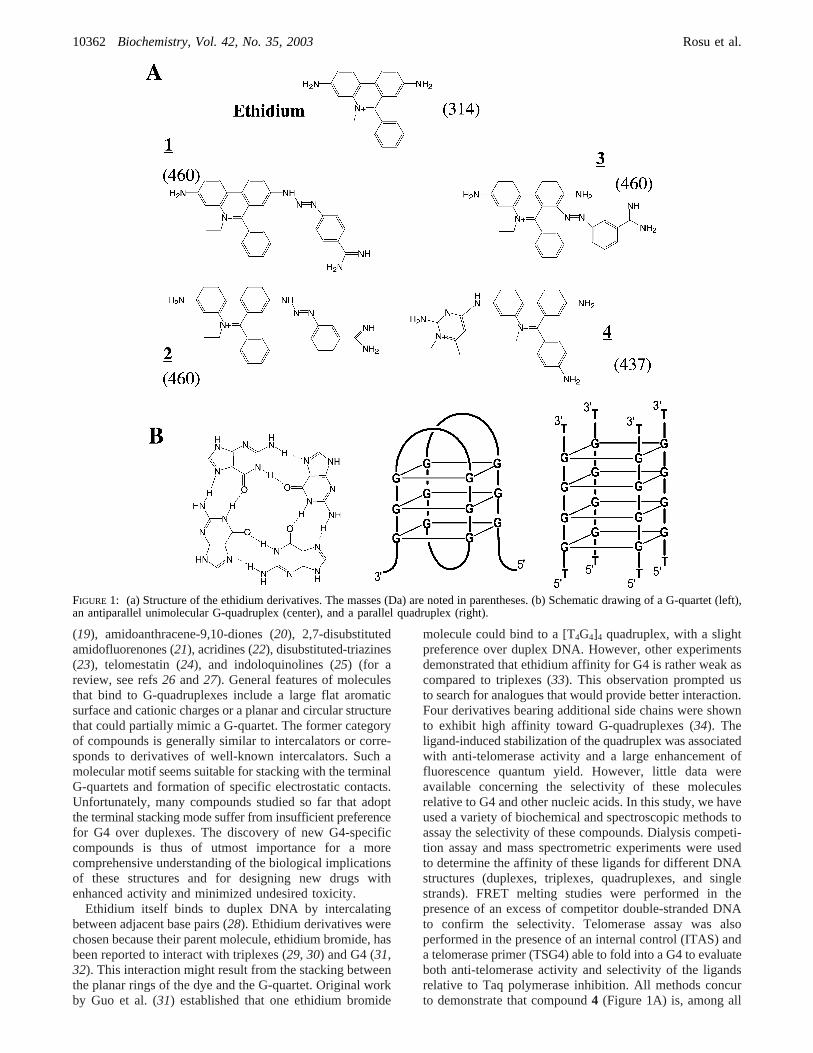
Ethidium itself binds to duplex DNA by intercalatingbetween adjacent base pairs (28). Ethidium derivatives werechosen because their parent molecule, ethidium bromide, hasbeen reported to interact with triplexes (29, 30) and G4 (31,32). This interaction might result from the stacking betweenthe planar rings of the dye and the G-quartet. Original workby Guo et al. (31) established that one ethidium bromide
molecule could bind to a [T4G4]4 quadruplex, with a slightpreference over duplex DNA. However, other experimentsdemonstrated that ethidium affinity for G4 is rather weak ascompared to triplexes (33). This observation prompted usto search for analogues that would provide better interaction.Four derivatives bearing additional side chains were shownto exhibit high affinity toward G-quadruplexes (34). Theligand-induced stabilization of the quadruplex was associatedwith anti-telomerase activity and a large enhancement offluorescence quantum yield. However, little data wereavailable concerning the selectivity of these moleculesrelative to G4 and other nucleic acids. In this study, we haveused a variety of biochemical and spectroscopic methods toassay the selectivity of these compounds. Dialysis competi-tion assay and mass spectrometric experiments were usedto determine the affinity of these ligands for different DNAstructures (duplexes, triplexes, quadruplexes, and singlestrands). FRET melting studies were performed in thepresence of an excess of competitor double-stranded DNAto confirm the selectivity. Telomerase assay was alsoperformed in the presence of an internal control (ITAS) anda telomerase primer (TSG4) able to fold into a G4 to evaluateboth anti-telomerase activity and selectivity of the ligandsrelative to Taq polymerase inhibition. All methods concurto demonstrate that compound4 (Figure 1A) is, among all
FIGURE 1: (a) Structure of the ethidium derivatives. The masses (Da) are noted in parentheses. (b) Schematic drawing of a G-quartet (left),an antiparallel unimolecular G-quadruplex (center), and a parallel quadruplex (right).
10362 Biochemistry, Vol. 42, No. 35, 2003 Rosu et al.
ethidium derivatives tested so far, the most active andselective G4 ligand inhibitor of telomerase.
EXPERIMENTAL PROCEDURES
Materials. All oligonucleotides were synthesized andpurified by Eurogentec, Belgium.
Ethidium derivatives1-4 have been previously described(see Figure 1A) (34, 35) (patent WO 0212194).1 wassimilarly prepared from 2,7-diamino-10-ethyl-9-phenyl-phenanthridinium iodide, using 4-benzamido-diazonium saltin place of 3-benzamido-diazonium salt for the synthesis of2. Compound 2, also known as isomethamidium, waspreviously prepared and identified as a trypanocidal drug(36, 37). Compound3 was identified as the main byproductin the synthesis of2. It occurred through azo-coupling of3-benzamido-diaazonium salt at position 8 of 2,7-diamino-10-ethyl-9-phenyl-phenanthridinium iodide, ortho to the7-amino group. Compound4 was identified as the main by-product in the synthesis of prothidium iodide according tothe May and Baker patent (GB 816236). Solutions of allderivatives were kept at-20 °C in the dark betweenexperiments.
Equilibrium Dialysis. The initial dialysis protocol wasdefined by Ren and Chaires (33). We have adapted this testto accommodate a different set of 19 nucleic acid structures(see Tables 1 and 2) (38-40). The TC, GA, and GT triplexesresult from the association of two strands of different lengths(13 and 30 nucleotides) (41). The 24GA duplex results fromthe self-association of a (GA)12 oligonucleotide. The parallel-stranded duplex (psD) results from the association of twoAT strands. The 24CTG mimics eight repeats of thetrinucleotide unit CTG. ds26 is a duplex formed with a self-complementary oligonucleotide. 22CT is an oligonucleotide
that mimics the cytosine-rich strand of human telomeres (42),whereas 22AG is an oligonucleotide that mimics the guanine-rich strand of human telomeres and adopts an intramolecularquadruplex structure (3). 24G20 may form an intermolecularG-quadruplex, whereas poly(dC) may form an intermoleculari-motif.
All structures used in these experiments were stable atroom temperature under the chosen experimental conditions(15 mM sodium cacodylate pH 6.5, 10 mM MgCl2, and 185mM NaCl, 20 °C) except 22CT (see Table 1) (39). Thepresence of 185 mM sodium chloride promotes quadruplexformation (39). A total of 400 mL of the dialysate solutioncontaining 1µM ligand was used for each competitiondialysis assay. A volume of 200µL at 75 µM monomericunit (nucleotide, base pair, base triplet, or quartet) of eachof the nucleic acids samples was pipeted into a separateDialyzer unit (Pierce). All 19 dialysis units were then placedin the beaker containing the dialysate solution. The beakerwas covered with Parafilm and wrapped in aluminum foil,and its contents were allowed to equilibrate with continuousstirring at room temperature (20-22°C) overnight. This longincubation time is likely to allow proper thermodynamicequilibrium, as most drug-DNA interactions occur on afaster time scale in theµM concentration range. At the endof the equilibration period, DNA samples were carefullyremoved to microfuge tubes and treated with 1% SDS. Theligand concentration in each sample was determined byfluorescence or absorbance spectroscopy. The amount of dyein each dialysis unit is directly proportional to the absorbancesignal of the sample as the nucleic acid-compound complexis dissociated by SDS. This quantification procedure has beenvalidated in a number of independent studies by severalgroups including ours (33, 38, 43-47).
Mass Spectrometry.Oligodeoxynucleotides d-CGTAAATT-TACG (Dk33, M ) 3644.45 Da), d-CGCGAATTCGCG(Dk66, M ) 3646.44 Da), d-CGCGGGCCCGCG(Dk100, M ) 3678.40 Da), d-TGGGGT (M) 1863.26Da), d-GGGGTTTTGGGG (M ) 3788.50 Da), andd-(GGGTTA)3GGG (M) 6653.35 Da) were purchased fromEurogentec (Angleur, Belgium) and used without furtherpurification. Duplex and quadruplex solutions were prepared,respectively, in 100 and 150 mM NH4OAc, pH 7.0. Oligo-nucleotide solutions were heated to 85°C for 5 min andcooled overnight to 20°C to form the duplex or quadruplexstructures. Ammonium acetate was chosen as the electrolytefor its compatibility with electrospray mass spectrometry.The solution conformation of the oligonucleotide in am-monium acetate was characterized by circular dichroism andUV melting experiments (48). Experiments were performedon an LCQ mass spectrometer (Finnigan, San Jose, CA) asdescribed previously (49). The experimental conditions wereoptimized to avoid denaturation of the duplex or quadruplexspecies: the heated capillary temperature of the electrospraysource was set to 185°C, and a good compromise betweensufficient focusing and minimal collisional activation of theions in the 1 Torr region of the source is achieved with atube lens offset of 35 V and a voltage on the heated capillaryof -11 V (the skimmer is at ground). Full scan MS spectrawere recorded in them/z range [1000-2000], and 50 scanswere summed for each spectrum. Spectra of equimolar mix-tures (5µM) of all possible structure (duplex or quadruplex)+ drug combinations were recorded. Methanol (15%) was
Table 1: List of the Nucleic Acids Used in Equilibrium DialysisExperimentsa
number name typeb structureTm
(°C)e
1 (T,C) triplex oligos triplex 382 (G,A) triplex oligos triplex 533 (G,T) triplex oligos triplex 534 poly dA.2polydT poly triplex 715 24GA duplex oligo “duplex”c 376 psD duplex oligos “duplex”c 397 24CTG oligo “duplex”c 648 poly d(A-T) poly duplex 669 poly d(G-C) poly duplex >90
10 calf thymus DNA poly duplex 8611 ds 26 oligo duplex 7512 poly dC poly i-DNA 51f
13 22CT oligo ss/i-DNAd 13f
14 22AG oligo G4 6215 24G20 oligo G4 >9016 poly dT poly single-str -g
17 poly dA poly single-str -g
18 poly rU poly single-str -g
19 poly rA poly single-str -g
a 19 different nucleic acids structures are used (samples labeled 1-19,left column).b poly ) polynucleotide; oligo) oligonucleotide; oligos) structure formed by the association of two different oligonucleotides.Polynucleotides are>100 bases long.c These three duplexes are unusualas they involve the formation of nonclassical base pairs.d 22CT mayform an i-DNA structure but is mainly single stranded at roomtemperature.e Obtained in an buffer identical to the equilibrium dialysisprotocol. f Hysteresis.Tm obtained while heating.g No transition.
Ethidium Derivatives Bind to G4 DNA Biochemistry, Vol. 42, No. 35, 200310363
added to the samples just before injection to obtain a stableelectrospray signal. The rate of sample infusion into the massspectrometers was 4µL/min. As previously discussed (49),the relative intensities of the free and bound DNA in themass spectra are assumed to be proportional to the relativeabundances of these species in solution. As the startingconcentrations are known, the concentrations of all individualspecies at equilibrium (free DNA, 1:1 complex, 2:1 complex,and by difference, the free drug) can be determined fromthe relative intensities of the free DNA and the complexes.The concentration of bound ligand per DNA molecule orper binding unit give us the affinity of the drug for a givenstructure. The concentration of bound ligand per DNAmolecule is obtained with the following equation (1):
whereC0 is the starting DNA concentration (expressed instructure, duplex, or quadruplex),IDNA is the relative intensityof the free DNA, andI(n:1) are relative intensities of thecomplexes (n drug molecules bound to one DNA target).The relative intensities were obtained from a sum of 50spectra. The amount of bound ligand expressed in a molec-ular binding unit (base pair, base triplet, or quartet in the
DNA target) is determined by dividing the total amount ofbound ligand by the number of monomeric units in the DNAtargets.
Fluorescence Melting Experiments.All FRET measure-ments with the doubly fluorescent (F) fluorescein, T)tetramethylrhodamine) F21T oligodeoxynucleotide (5′ fluo-rescein-(GGGTTA)3GGG-3′ tetramethylrhodamine, 0.2µM)were performed on a Spex Fluoromax3 instrument, using abandwidth of 5 nm and 0.2× 1 cm quartz cuvettes,containing 600µL of solution in a pH 7.2, 0.1 M lithiumchloride, 10 mM sodium cacodylate buffer. All ligands weretested at 1µM. In competition experiments, various con-centrations of an autocomplementary unmodified 26 baselong oligodeoxynucleotide (ds26: d-CAATCGGATCGAAT-TCGATCCGATTG) or a single-stranded dT26 oligodeoxy-nucleotide were added. All measurements were made aspreviously described (50, 51).
Circular Dichroism. CD spectra were obtained at 20°Cwith a Jobin Yvon CD6 (Lonjumeau, France) circulardichrograph. A quartz cell (Hellma, Inc.) with a 10 mm pathlength was used to obtain spectra at 0.5 nm intervals from220 to 640 nm. Spectra result from the averaging of threescans, followed by the subtraction of the CD spectrum of a
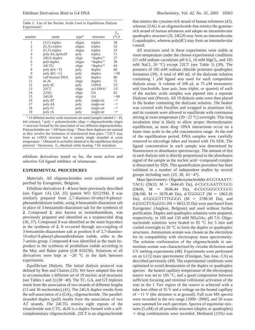
Table 2: Sequence of the Oligonucleotides Used in Equilibrium Dialysisa
a The name of the oligonucleotides is followed by the structure number (see Table 1) and the sequence. Note that the triplexes correspond to theassociation of two strands of different lengths. ds 26 is an autocomplementary oligonucleotide.
[bound ligand]) C0(I(1:1)+ 2I(2:1))/(IDNA + I(1:1)+ I(2:1))
10364 Biochemistry, Vol. 42, No. 35, 2003 Rosu et al.
solution of 0.15 M aqueous NH4OAc. The quadruplexconcentration was 3µM, and the drug was added up to amolar ratio r ) [drug]/[quadruplex] ) 4.2. Results areexpressed in molar circular dichroism.
Telomerase Assay (TRAP-G4).Telomerase extract wasprepared from A549 cells according to Fu et al. (52). TRAP-G4 was performed as described previously (53). PCR wasperformed in a final 50µL reaction volume containingprimers TSG4 (3.5 pmol), TS (18 pmol), CXext (22.5 pmol),NT (7.5 pmol), and TSNT (0.01 amol); 20 mM Tris-HCl(pH 8.0), 50µM dNTPs, 1.5 mM MgCl2, 63 mM KCl, 1mM EGTA, 0.005% Tween 20, 20µg/mL bovine serumalbumin; 2.5 U Taq DNA polymerase (DyNAzyme II DNApolymerase, Ozyme); 100 ng of telomerase extract; andcompounds or distilled water under a volume of 5µL.Samples were incubated for 15 min at 30°C and denaturedfor 1 min at 90°C and further submitted to 30 PCR cycles:30 s, 92°C; 30 s, 52°C; and 30 s, 72°C. After amplification,10 µL of loading buffer containing 20% sucrose, 5X TBE,0.2% bromophenol blue, and 0.2% xylene cyanol were addedto the reaction. After electrophoresis onto a 12% nondena-turing polyacrylamide gel (19:1) in 1X TBE and stainingwith SYBR Green I (Roche), DNA fragments were digi-talized by a CCD camera (Bioprint) and quantified by theBioCapt software. The disappearance of the TSG4 bandcorresponded to a G-quadruplex stabilization of the TSG4oligonucleotide and coincided with inhibition of the telomericrepeats extension from either TSG4 or TS oligonucleotides.The disappearance of the ITAS band corresponded tononspecific inhibition of Taq polymerase activity. For eachcompound, results were expressed by the IC50 of the TSG4and ITAS band formation and indicated as IC50 TRAP G4and IC50 Taq, respectively. The ratio IC50 Taq/ IC50 TRAPG4 represented the selectivity factor of the compound in thisassay.
Cell Culture Conditions and SurViVal Assay.A549 humancell lung carcinoma cell line was from the American TypeCulture Collection. These cells were grown in DMEMmedium with Glutamax (Invitrogen) and supplemented with10% fetal calf serum and antibiotics.
Cells were seeded in 96-well plates at 3× 10³ cells/wellin a final volume of 200µL. Ligand at final concentrationsof 20, 2, and 0.2µM were added 6 h later under a volumeof 20 µL, each concentration in quadruplicate. The MTTsurvival assay was performed after 4 days of incubation, asrecommended by the manufacturer (Sigma). Results wereexpressed as the percent cell survival relative to untreatedcontrol cells. The concentration of the ligand that inhibits50% of cell growth (IC50) was determined from semi-logarithmic plots.
RESULTS
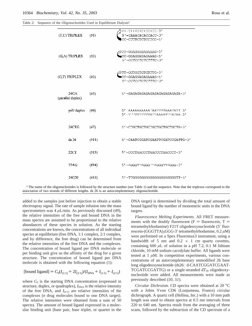
Equilibrium Dialysis. To evaluate the selectivity ofethidium and its derivatives for different DNA structures,we performed competitive dialysis experiments using 19nucleic acid structures (described in Experimental Proce-dures) against a common ligand solution. More productaccumulates in the dialysis tube containing the structural formwith the highest ligand binding affinity. Results for ethidium,2, 3, and4 were shown in Figure 2. The spectral propertiesof compound1 did not allow a precise quantification of the
binding data (not shown). It is possible to correlate theamount of the bound dye to a given structure with the affinityof the dye for that nucleic acid sample. As shown in Figure2 (top left), ethidium interacts preferentially with duplexes(in black and gray), whereas it shows a weak affinity fori-motif (green), single strands (blue), triplexes (orange), andG4 (red). Interestingly, the preferred structure correspondsto an unusual DNA duplex (24CTG, in gray) resulting fromthe intramolecular folding of a (CTG)8 repeat oligodeoxy-nucleotide. Overall, binding to the four regular duplexes (inblack) was stronger than to the triplexes or quadruplexes, inagreement with previous reports (29, 34). For 2 and 4,binding to G4 (in red) is stronger than binding to Watson-Crick duplexes (in black). Binding to single strands (in blue)is virtually nil, demonstrating that these derivatives havesome selectivity for quadruplexes over duplexes and singlestrands. Nevertheless, it is clear from these studies that2(upper right) and3 (lower left) have a lower quadruplex-duplex selectivity than4 (lower right). For all derivatives,binding to the intramolecular telomeric quadruplex is similarto the binding to the parallel quadruplex, showing that thesemolecules are not able to discrimimate between thesequadruplexes. In principle, competitive dialysis data may beused to calculate apparent binding constants (Ka), using thesimple relationKa ) [bound]/([free][NAtotal - bound]),where [bound] is the amount bound to each structure, [free]is the free ligand concentration (kept at 1µM), and NAtotalis the total nucleic acid concentration (75µM). It istheoretically possible to calculate the equilibrium constantfor each structure (i.e., 5× 10⁴ and 7 × 10³ M-1 forcompound4 binding to the parallel quadruplex and the polyd(A-T) duplex, respectively and 8× 10⁴ and 3× 10⁴ M-1
for compound3 binding to the telomeric quadruplex and thepoly d(A-T) duplex, respectively). These values representthe dissociation constant for a monomeric unit (base pair orquartet). However, these values are poorly reliable as: (i)they are based on a single dye/DNA monomeric unitconcentration, (ii) they rely on an accurate determination ofthe absolute value of bound dye: in our hands, the dialysisprofile is reproducible, but the numerical values are not, and(iii) higher affinities should be expected at lower ionicstrength. The binding profile of these compounds relativeto higher-order DNA structures prompted us to confirm thisinteraction using different techniques.
Mass Spectrometry.Electrospray ionization mass spec-trometry (ESI-MS) is a highly sensitive method to studydrug-DNA interactions, as reviewed recently (54, 55).Stoichiometries and relative binding affinities of DNAcomplexes with intercalators and minor groove binders havebeen studied (56, 57). Quantitative determination of bindingconstants for minor groove binders with different DNAduplexes has been demonstrated (49), and the interaction ofdrugs with DNA triplex and quadruplex structures can alsobe investigated (40, 48, 58).
In the present work, electrospray mass spectrometry (ESI-MS) was used to determine the binding affinity and specific-ity of the ethidium derivatives for DNA duplex andquadruplex forms. Our binding assay uses three 12-base pairduplexes with different GC content and three quadruplexes([G4T4G4]2, [TG4T]4, and the human intramolecular telomericsequence (G3T2A)3G3). The later oligonucleotide is identicalto the one used in FRET melting studies and similar to 22AG
Ethidium Derivatives Bind to G4 DNA Biochemistry, Vol. 42, No. 35, 200310365

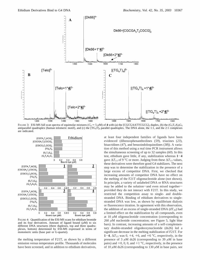
A(GGGTTA)3GGG used for dialysis. Figure 3 illustratestypical ESI-MS mass spectra obtained with an equimolarmixture of the drug4 with the duplex [CGCGAATTCGCG]2
(Dk66; Figure 3A), the parallel quadruplex [TG4T]4 (Figure3C), and the human telomeric sequence (Figure 3B). Drug/DNA (1:1) and (2:1) complexes were observed with all thesestructures. It has been previously reported that, for thequadruplex [TG4T]4, specifically three ammonium ions aredetected within this particular G4 structure (48) and arelocated between the tetrads. For all compounds bound to thisparticular quadruplex, the mass of the complexes indicatesthat the three ammonium ions are conserved in the complex(i.e., a mass shift of 51 Da corresponding to the mass ofthree ammonium ions). As the ammonium ions between thetetrads are not expelled, this suggests that the drugs are notlocated between the tetrads.
The largest amount of the complexes (1:1) and (2:1) isobserved with the intramolecular telomeric sequence (Figure3B) followed by the parallel quadruplex (Figure 3C), andthe Dickerson duplex (Dk66) (Figure 3A). Nevertheless, tomake semiquantitative comparisons with equilibrium dialysisexperiments, the amount of drug bound was expressed interms of the monomeric unit: the amount of bound drugcalculated with eq 1 is divided by the number of base pairs
or tetrads in the oligonucleotide studied. These results aresummarized in Figure 4. Among the duplexes, all drugsinteract preferentially with those of high GC content. Thisis consistent with the GC preference of common intercalatorssuch as ethidium. Concerning the structural selectivity(duplex vs G4), ESI-MS results are in agreement withequilibrium dialysis data: all ethidium derivatives bindpreferentially to the G4 structures. The derivative4 showsthe best selectivity for the quadruplex structure. Bindingconstants obtained with the mass spectrometry experimentwere K1 ) 3.04 × 10⁵ and K2 ) 2.0 × 10⁵ M-1 for thevertebrate intramolecular telomeric sequence (two bindingsites per quadruplex). The same order of magnitude obtainedfor the binding constants indicates that the binding sites areequivalent.
FRET Melting Studies.FRET can be used to probe thesecondary structure of oligodeoxynucleotides mimickingrepeats of the guanine-rich strand of vertebrate telomeres,provided a fluorescein molecule (donor) and a tetramethyl-rhodamine derivative (acceptor) are attached to the 5′ and3′ ends of the oligonucleotide, respectively. The melting ofthe G-quadruplex is usually monitored in the presence of 1µM dye by measuring the fluorescence of the donor. Ligandsknown to specifically interact with a quadruplex increased
FIGURE 2: Equilibrium dialysis. All measurements were performed in a 185 mM NaCl, 10 mM MgCl2, 15 mM pH 6.5 sodium-cacodylatebuffer. The nucleic acid names are given on the left, and structures are described in Tables 1 and 2. The values are normalized for eachcompound. Maximum amount bound corresponds to 2.1µM (ethidium); 4.0µM (compound2); 8.6µM (compound3); and 3.5µM (compound4).
10366 Biochemistry, Vol. 42, No. 35, 2003 Rosu et al.
the melting temperature of F21T, as shown by a differentemission versus temperature profile. Thousands of moleculeshave been screened, and in addition to ethidium derivatives,
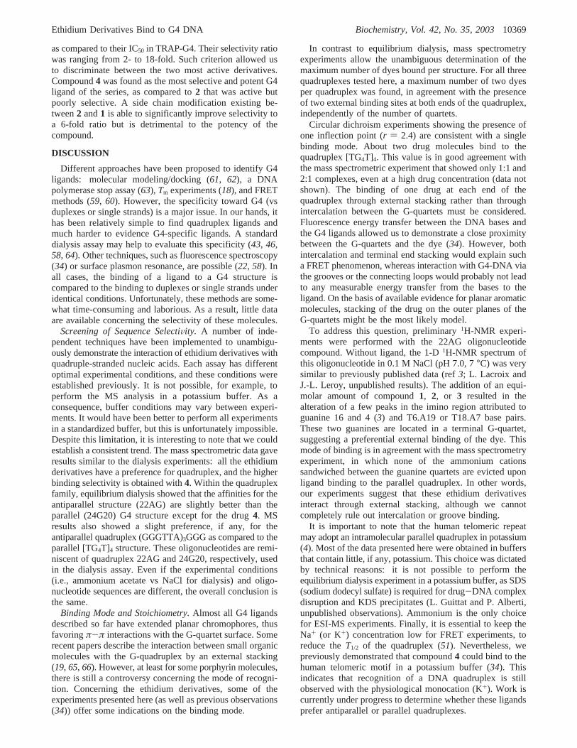
at least four independent families of ligands have beenevidenced (dibenzophenanthrolines (59), triazines (23),bisacridines (47), and benzoindoloquinolines (38)). A varia-tion of this method using a real time PCR instrument allowsthe simultaneous screening of up to 32 samples (60). In thistest, ethidium gave little, if any, stabilization whereas1-4gave∆T1/2 of 9 °C or more. Judging from these∆T1/2 values,these derivatives were therefore good G4 stabilizers. The nextstep was to determine the stabilization in the presence of alarge excess of competitor DNA. First, we checked thatincreasing amounts of competitor DNA have no effect onthe melting of the F21T oligonucleotide alone (not shown).In principle, a variety of unlabeled DNA or RNA structuresmay be added to the solutionsand even mixed togethersprovided they do not interact with F21T. In this study, werestricted the competition assay to single- and double-stranded DNA. Binding of ethidium derivatives to single-stranded DNA was low, as shown by equilibrium dialysisor fluorescence titration. In agreement with this observation,the addition of an excess of single-stranded DNA (dT26) hada limited effect on the stabilization by all compounds, evenat 10 µM oligonucleotide concentration (corresponding to260 µM nucleotide concentration; see Figure 5, light bluebars). In contrast, increasing amounts of a self-complemen-tary double-stranded oligodeoxynucleotide (ds26) led asignificant decrease in the melting stabilization of F21T. For1-4, ∆T1/2 was 0,+4, +6, and+6 °C, respectively, in thepresence of 3µM ds26 (corresponding to 39µM in basepairs) and+0, 0, 0, and+1 °C, respectively, in the presenceof 10 µM ds26 (corresponding to 130µM in base pairs, see
FIGURE 3: ESI-MS full scan spectra of equimolar mixtures (C0 ) 5 µM) of 4 with (a) the [CGCGAATTCGCG]2 duplex, (b) the (G3T2A)3G3antiparallel quadruplex (human telomeric motif), and (c) the [TG4T]4 parallel quadruplex. The DNA alone, the 1:1, and the 2:1 complexesare indicated.
FIGURE 4: Quantification of the ESI-MS scans for ethidium bromideand its four derivatives. Amount of ligand bound (µM) to sixdifferent DNA structures (three duplexes, top and three quadru-plexes, bottom) determined by ESI-MS expressed in terms ofmonomeric units (base pair or G-quartet).
Ethidium Derivatives Bind to G4 DNA Biochemistry, Vol. 42, No. 35, 200310367
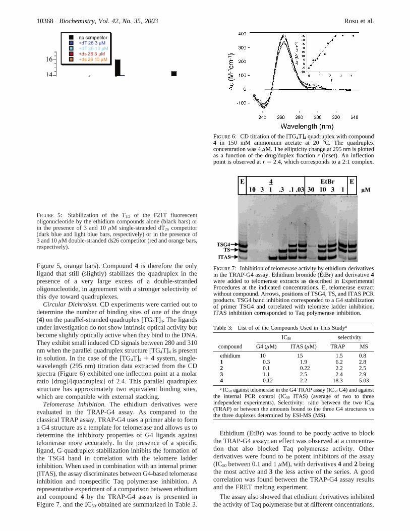
Figure 5, orange bars). Compound4 is therefore the onlyligand that still (slightly) stabilizes the quadruplex in thepresence of a very large excess of a double-strandedoligonucleotide, in agreement with a stronger selectivity ofthis dye toward quadruplexes.
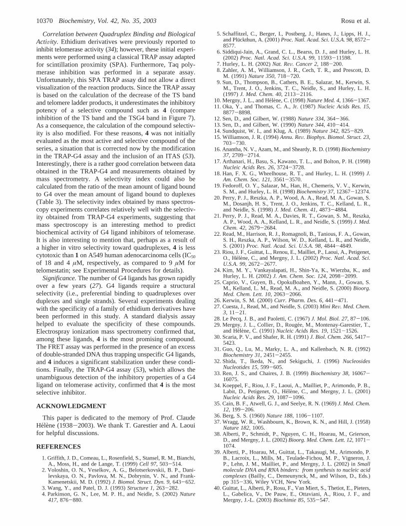
Circular Dichroism.CD experiments were carried out todetermine the number of binding sites of one of the drugs(4) on the parallel-stranded quadruplex [TG4T]4. The ligandsunder investigation do not show intrinsic optical activity butbecome slightly optically active when they bind to the DNA.They exhibit small induced CD signals between 280 and 310nm when the parallel quadruplex structure [TG4T]4 is presentin solution. In the case of the [TG4T]4 + 4 system, single-wavelength (295 nm) titration data extracted from the CDspectra (Figure 6) exhibited one inflection point at a molarratio [drug]/[quadruplex] of 2.4. This parallel quadruplexstructure has approximately two equivalent binding sites,which are compatible with external stacking.
Telomerase Inhibition.The ethidium derivatives wereevaluated in the TRAP-G4 assay. As compared to theclassical TRAP assay, TRAP-G4 uses a primer able to forma G4 structure as a template for telomerase and allows us todetermine the inhibitory properties of G4 ligands againsttelomerase more accurately. In the presence of a specificligand, G-quadruplex stabilization inhibits the formation ofthe TSG4 band in correlation with the telomere ladderinhibition. When used in combination with an internal primer(ITAS), the assay discriminates between G4-based telomeraseinhibition and nonspecific Taq polymerase inhibition. Arepresentative experiment of a comparison between ethidiumand compound4 by the TRAP-G4 assay is presented inFigure 7, and the IC50 obtained are summarized in Table 3.
Ethidium (EtBr) was found to be poorly active to blockthe TRAP-G4 assay; an effect was observed at a concentra-tion that also blocked Taq polymerase activity. Otherderivatives were found to be potent inhibitors of the assay(IC50 between 0.1 and 1µM), with derivatives4 and2 beingthe most active and3 the less active of the series. A goodcorrelation was found between the TRAP-G4 assay resultsand the FRET melting experiment.
The assay also showed that ethidium derivatives inhibitedthe activity of Taq polymerase but at different concentrations,
FIGURE 5: Stabilization of theT1/2 of the F21T fluorescentoligonucleotide by the ethidium compounds alone (black bars) orin the presence of 3 and 10µM single-stranded dT26 competitor(dark blue and light blue bars, respectively) or in the presence of3 and 10µM double-stranded ds26 competitor (red and orange bars,respectively).
FIGURE 6: CD titration of the [TG4T]4 quadruplex with compound4 in 150 mM ammonium acetate at 20°C. The quadruplexconcentration was 4µM. The ellipticity change at 295 nm is plottedas a function of the drug/duplex fractionr (inset). An inflectionpoint is observed atr ) 2.4, which corresponds to a 2:1 complex.
FIGURE 7: Inhibition of telomerase activity by ethidium derivativesin the TRAP-G4 assay. Ethidium bromide (EtBr) and derivative4were added to telomerase extracts as described in ExperimentalProcedures at the indicated concentrations. E, telomerase extractwithout compound. Arrows, positions of TSG4, TS, and ITAS PCRproducts. TSG4 band inhibition corresponded to a G4 stabilizationof primer TSG4 and correlated with telomere ladder inhibition.ITAS inhibition corresponded to Taq polymerase inhibition.
Table 3: List of of the Compounds Used in This Studya
IC50 selectivity
compound G4 (µM) ITAS (µM) TRAP MS
ethidium 10 15 1.5 0.81 0.3 1.9 6.2 2.82 0.1 0.22 2.2 2.53 1.1 2.5 2.4 2.94 0.12 2.2 18.3 5.03a IC50 against telomerase in the G4 TRAP assay (IC50 G4) and against
the internal PCR control (IC50 ITAS) (average of two to threeindependent experiments). Selectivity: ratio between the two IC50
(TRAP) or between the amounts bound to the three G4 structures vsthe three duplexes determined by ESI-MS (MS).
10368 Biochemistry, Vol. 42, No. 35, 2003 Rosu et al.
as compared to their IC50 in TRAP-G4. Their selectivity ratiowas ranging from 2- to 18-fold. Such criterion allowed usto discriminate between the two most active derivatives.Compound4 was found as the most selective and potent G4ligand of the series, as compared to2 that was active butpoorly selective. A side chain modification existing be-tween2 and1 is able to significantly improve selectivity toa 6-fold ratio but is detrimental to the potency of thecompound.
DISCUSSION
Different approaches have been proposed to identify G4ligands: molecular modeling/docking (61, 62), a DNApolymerase stop assay (63), Tm experiments (18), and FRETmethods (59, 60). However, the specificity toward G4 (vsduplexes or single strands) is a major issue. In our hands, ithas been relatively simple to find quadruplex ligands andmuch harder to evidence G4-specific ligands. A standarddialysis assay may help to evaluate this specificity (43, 46,58, 64). Other techniques, such as fluorescence spectroscopy(34) or surface plasmon resonance, are possible (22, 58). Inall cases, the binding of a ligand to a G4 structure iscompared to the binding to duplexes or single strands underidentical conditions. Unfortunately, these methods are some-what time-consuming and laborious. As a result, little dataare available concerning the selectivity of these molecules.
Screening of Sequence SelectiVity. A number of inde-pendent techniques have been implemented to unambigu-ously demonstrate the interaction of ethidium derivatives withquadruple-stranded nucleic acids. Each assay has differentoptimal experimental conditions, and these conditions wereestablished previously. It is not possible, for example, toperform the MS analysis in a potassium buffer. As aconsequence, buffer conditions may vary between experi-ments. It would have been better to perform all experimentsin a standardized buffer, but this is unfortunately impossible.Despite this limitation, it is interesting to note that we couldestablish a consistent trend. The mass spectrometric data gaveresults similar to the dialysis experiments: all the ethidiumderivatives have a preference for quadruplex, and the higherbinding selectivity is obtained with4. Within the quadruplexfamily, equilibrium dialysis showed that the affinities for theantiparallel structure (22AG) are slightly better than theparallel (24G20) G4 structure except for the drug4. MSresults also showed a slight preference, if any, for theantiparallel quadruplex (GGGTTA)3GGG as compared to theparallel [TG4T]4 structure. These oligonucleotides are remi-niscent of quadruplex 22AG and 24G20, respectively, usedin the dialysis assay. Even if the experimental conditions(i.e., ammonium acetate vs NaCl for dialysis) and oligo-nucleotide sequences are different, the overall conclusion isthe same.
Binding Mode and Stoichiometry.Almost all G4 ligandsdescribed so far have extended planar chromophores, thusfavoringπ-π interactions with the G-quartet surface. Somerecent papers describe the interaction between small organicmolecules with the G-quadruplex by an external stacking(19, 65, 66). However, at least for some porphyrin molecules,there is still a controversy concerning the mode of recogni-tion. Concerning the ethidium derivatives, some of theexperiments presented here (as well as previous observations(34)) offer some indications on the binding mode.
In contrast to equilibrium dialysis, mass spectrometryexperiments allow the unambiguous determination of themaximum number of dyes bound per structure. For all threequadruplexes tested here, a maximum number of two dyesper quadruplex was found, in agreement with the presenceof two external binding sites at both ends of the quadruplex,independently of the number of quartets.
Circular dichroism experiments showing the presence ofone inflection point (r ) 2.4) are consistent with a singlebinding mode. About two drug molecules bind to thequadruplex [TG4T]4. This value is in good agreement withthe mass spectrometric experiment that showed only 1:1 and2:1 complexes, even at a high drug concentration (data notshown). The binding of one drug at each end of thequadruplex through external stacking rather than throughintercalation between the G-quartets must be considered.Fluorescence energy transfer between the DNA bases andthe G4 ligands allowed us to demonstrate a close proximitybetween the G-quartets and the dye (34). However, bothintercalation and terminal end stacking would explain sucha FRET phenomenon, whereas interaction with G4-DNA viathe grooves or the connecting loops would probably not leadto any measurable energy transfer from the bases to theligand. On the basis of available evidence for planar aromaticmolecules, stacking of the drug on the outer planes of theG-quartets might be the most likely model.
To address this question, preliminary1H-NMR experi-ments were performed with the 22AG oligonucleotidecompound. Without ligand, the 1-D1H-NMR spectrum ofthis oligonucleotide in 0.1 M NaCl (pH 7.0, 7°C) was verysimilar to previously published data (ref3; L. Lacroix andJ.-L. Leroy, unpublished results). The addition of an equi-molar amount of compound1, 2, or 3 resulted in thealteration of a few peaks in the imino region attributed toguanine 16 and 4 (3) and T6.A19 or T18.A7 base pairs.These two guanines are located in a terminal G-quartet,suggesting a preferential external binding of the dye. Thismode of binding is in agreement with the mass spectrometryexperiment, in which none of the ammonium cationssandwiched between the guanine quartets are evicted uponligand binding to the parallel quadruplex. In other words,our experiments suggest that these ethidium derivativesinteract through external stacking, although we cannotcompletely rule out intercalation or groove binding.
It is important to note that the human telomeric repeatmay adopt an intramolecular parallel quadruplex in potassium(4). Most of the data presented here were obtained in buffersthat contain little, if any, potassium. This choice was dictatedby technical reasons: it is not possible to perform theequilibrium dialysis experiment in a potassium buffer, as SDS(sodium dodecyl sulfate) is required for drug-DNA complexdisruption and KDS precipitates (L. Guittat and P. Alberti,unpublished observations). Ammonium is the only choicefor ESI-MS experiments. Finally, it is essential to keep theNa+ (or K+) concentration low for FRET experiments, toreduce theT1/2 of the quadruplex (51). Nevertheless, wepreviously demonstrated that compound4 could bind to thehuman telomeric motif in a potassium buffer (34). Thisindicates that recognition of a DNA quadruplex is stillobserved with the physiological monocation (K+). Work iscurrently under progress to determine whether these ligandsprefer antiparallel or parallel quadruplexes.
Ethidium Derivatives Bind to G4 DNA Biochemistry, Vol. 42, No. 35, 200310369
Correlation between Quadruplex Binding and BiologicalActiVity. Ethidium derivatives were previously reported toinhibit telomerase activity (34); however, these initial experi-ments were performed using a classical TRAP assay adaptedfor scintillation proximity (SPA). Furthermore, Taq poly-merase inhibition was performed in a separate assay.Unfortunately, this SPA TRAP assay did not allow a directvisualization of the reaction products. Since the TRAP assayis based on the calculation of the decrease of the TS bandand telomere ladder products, it underestimates the inhibitorypotency of a selective compound such as4 (compareinhibition of the TS band and the TSG4 band in Figure 7).As a consequence, the calculation of the compound selectiv-ity is also modified. For these reasons,4 was not initiallyevaluated as the most active and selective compound of theseries, a situation that is corrected now by the modificationin the TRAP-G4 assay and the inclusion of an ITAS (53).Interestingly, there is a rather good correlation between dataobtained in the TRAP-G4 and measurements obtained bymass spectrometry. A selectivity index could also becalculated from the ratio of the mean amount of ligand boundto G4 over the mean amount of ligand bound to duplexes(Table 3). The selectivity index obtained by mass spectros-copy experiments correlates relatively well with the selectiv-ity obtained from TRAP-G4 experiments, suggesting thatmass spectroscopy is an interesting method to predictbiochemical activity of G4 ligand inhibitors of telomerase.It is also interesting to mention that, perhaps as a result ofa higher in vitro selectivity toward quadruplexes,4 is lesscytotoxic than1 on A549 human adenocarcinoma cells (IC50
of 18 and 4µM, respectively, as compared to 9µM fortelomestatin; see Experimental Procedures for details).
Significance.The number of G4 ligands has grown rapidlyover a few years (27). G4 ligands require a structuralselectivity (i.e., preferential binding to quadruplexes overduplexes and single strands). Several experiments dealingwith the specificity of a family of ethidium derivatives havebeen performed in this study. A standard dialysis assayhelped to evaluate the specificity of these compounds.Electrospray ionization mass spectrometry confirmed that,among these ligands,4 is the most promising compound.The FRET assay was performed in the presence of an excessof double-stranded DNA thus trapping unspecific G4 ligands,and4 induces a significant stabilization under these condi-tions. Finally, the TRAP-G4 assay (53), which allows theunambiguous detection of the inhibitory properties of a G4ligand on telomerase activity, confirmed that4 is the mostselective inhibitor.
ACKNOWLEDGMENT
This paper is dedicated to the memory of Prof. ClaudeHelene (1938-2003). We thank T. Garestier and A. Laouifor helpful discussions.
REFERENCES
1. Griffith, J. D., Comeau, L., Rosenfield, S., Stansel, R. M., Bianchi,A., Moss, H., and de Lange, T. (1999)Cell 97, 503-514.
2. Voloshin, O. N., Veselkov, A. G., Belotserkovskii, B. P., Dani-levskaya, O. N., Pavlova, M. N., Dobrynin, V. N., and Frank-Kamenetskii, M. D. (1992)J. Biomol. Struct. Dyn. 9, 643-652.
3. Wang, Y., and Patel, D. J. (1993)Structure 1, 263-282.4. Parkinson, G. N., Lee, M. P. H., and Neidle, S. (2002)Nature
417, 876-880.
5. Schaffitzel, C., Berger, I., Postberg, J., Hanes, J., Lipps, H. J.,and Pluckthun, A. (2001)Proc. Natl. Acad. Sci. U.S.A. 98, 8572-8577.
6. Siddiqui-Jain, A., Grand, C. L., Bearss, D. J., and Hurley, L. H.(2002)Proc. Natl. Acad. Sci. U.S.A. 99, 11593-11598.
7. Hurley, L. H. (2002)Nat. ReV. Cancer 2, 188-200.8. Zahler, A. M., Williamson, J. R., Cech, T. R., and Prescott, D.
M. (1991)Nature 350, 718-720.9. Sun, D., Thompson, B., Cathers, B. E., Salazar, M., Kerwin, S.
M., Trent, J. O., Jenkins, T. C., Neidle, S., and Hurley, L. H.(1997)J. Med. Chem. 40, 2113-2116.
10. Mergny, J. L., and He´lene, C. (1998)Nature Med. 4, 1366-1367.11. Oka, Y., and Thomas, C. A., Jr. (1987)Nucleic Acids Res. 15,
8877-8898.12. Sen, D., and Gilbert, W. (1988)Nature 334, 364-366.13. Sen, D., and Gilbert, W. (1990)Nature 344, 410-414.14. Sundquist, W. I., and Klug, A. (1989)Nature 342, 825-829.15. Williamson, J. R. (1994)Annu. ReV. Biophys. Biomol. Struct. 23,
703-730.16. Anantha, N. V., Azam, M., and Sheardy, R. D. (1998)Biochemistry
37, 2709-2714.17. Arthanari, H., Basu, S., Kawano, T. L., and Bolton, P. H. (1998)
Nucleic Acids Res. 26, 3724-3728.18. Han, F. X. G., Wheelhouse, R. T., and Hurley, L. H. (1999)J.
Am. Chem. Soc. 121, 3561-3570.19. Fedoroff, O. Y., Salazar, M., Han, H., Chemeris, V. V., Kerwin,
S. M., and Hurley, L. H. (1998)Biochemistry 37, 12367-12374.20. Perry, P. J., Reszka, A. P., Wood, A. A., Read, M. A., Gowan, S.
M., Dosanjh, H. S., Trent, J. O., Jenkins, T. C., Kelland, L. R.,and Neidle, S. (1998)J. Med. Chem. 41, 4873-4884.
21. Perry, P. J., Read, M. A., Davies, R. T., Gowan, S. M., Reszka,A. P., Wood, A. A., Kelland, L. R., and Neidle, S. (1999)J. Med.Chem. 42, 2679-2684.
22. Read, M., Harrison, R. J., Romagnoli, B., Tanious, F. A., Gowan,S. H., Reszka, A. P., Wilson, W. D., Kelland, L. R., and Neidle,S. (2001)Proc. Natl. Acad. Sci. U.S.A. 98, 4844-4849.
23. Riou, J. F., Guittat, L., Renou, E., Mailliet, P., Laoui, A., Petigenet,O., Helene, C., and Mergny, J. L. (2002)Proc. Natl. Acad. Sci.U.S.A. 99, 2672-2677.
24. Kim, M. Y., Vankayalapati, H., Shin-Ya, K., Wierzba, K., andHurley, L. H. (2002)J. Am. Chem. Soc. 124, 2098-2099.
25. Caprio, V., Guyen, B., OpokuBoahen, Y., Mann, J., Gowan, S.M., Kelland, L. M., Read, M. A., and Neidle, S. (2000)Bioorg.Med. Chem. Lett. 10, 2063-2066.
26. Kerwin, S. M. (2000)Curr. Pharm. Des. 6, 441-471.27. Cuesta, J., Read, M., and Neidle, S. (2003)Mini ReV. Med. Chem.
3, 11-21.28. Le Pecq, J. B., and Paoletti, C. (1967)J. Mol. Biol. 27, 87-106.29. Mergny, J. L., Collier, D., Rouge´e, M., Montenay-Garestier, T.,
and Helene, C. (1991)Nucleic Acids Res. 19, 1521-1526.30. Scaria, P. V., and Shafer, R. H. (1991)J. Biol. Chem. 266, 5417-
5423.31. Guo, Q., Lu, M., Marky, L. A., and Kallenbach, N. R. (1992)
Biochemistry 31, 2451-2455.32. Shida, T., Ikeda, N., and Sekiguchi, J. (1996)Nucleosides
Nucleotides 15, 599-605.33. Ren, J. S., and Chaires, J. B. (1999)Biochemistry 38, 16067-
16075.34. Koeppel, F., Riou, J. F., Laoui, A., Mailliet, P., Arimondo, P. B.,
Labit, D., Petigenet, O., He´lene, C., and Mergny, J. L. (2001)Nucleic Acids Res. 29, 1087-1096.
35. Cain, B. F., Atwell, G. J., and Seelye, R. N. (1969)J. Med. Chem.12, 199-206.
36. Berg, S. S. (1960)Nature 188, 1106-1107.37. Wragg, W. R., Washbourn, K., Brown, K. N., and Hill, J. (1958)
Nature 182, 1005.38. Alberti, P., Schmidt, P., Nguyen, C. H., Hoarau, M., Grierson,
D., and Mergny, J. L. (2002)Bioorg. Med. Chem. Lett. 12, 1071-1074.
39. Alberti, P., Hoarau, M., Guittat, L., Takasugi, M., Arimondo, P.B., Lacroix, L., Mills, M., Teulade-Fichou, M. P., Vigneron, J.P., Lehn, J. M., Mailliet, P., and Mergny, J. L. (2002) inSmallmolecule DNA and RNA binders: from synthesis to nucleic acidcomplexes(Bailly, C., Demeunynck, M., and Wilson, D., Eds.)pp 315-336, Wiley VCH, New York.
40. Guittat, L., Alberti, P., Rosu, F., Van Miert, S., Thetiot, E., Pieters,L., Gabelica, V., De Pauw, E., Ottaviani, A., Riou, J. F., andMergny, J.-L. (2003)Biochimie 85, 535-547.
10370 Biochemistry, Vol. 42, No. 35, 2003 Rosu et al.
41. Mills, M., Arimondo, P., Lacroix, L., Garestier, T., He´lene, C.,Klump, H. H., and Mergny, J. L. (1999)J. Mol. Biol. 291, 1035-1054.
42. Phan, A. T., and Leroy, J. L. (2002)J. Biomol. Struct. Dyn. Spec.Issue S2, 245-252.
43. Ren, J. S., and Chaires, J. B. (2000)J. Am. Chem. Soc. 122, 424-425.
44. Ren, J. S., Bailly, C., and Chaires, J. B. (2000)FEBS Lett. 470,355-359.
45. Ren, J., Qu, X., Dattagupta, N., and Chaires, J. B. (2001)J. Am.Chem. Soc. 123, 6742-6743.
46. Ren, J. S., and Chaires, J. B. (2001) inDrug Nucleic AcidInteraction(Chaires, J. B., and Waring, M. J., Eds.) pp 99-108,Academic Press Inc., San Diego, CA.
47. Alberti, P., Ren, J., Teulade-Fichou, M. P., Guittat, L., Riou, J.F., Chaires, J. B., He´lene, C., Vigneron, J. P., Lehn, J. M., andMergny, J. L. (2001)J. Biomol. Struct. Dyn. 19, 505-513.
48. Rosu, F., Gabelica, V., Houssier, C., Colson, P., and De Pauw, E.(2002)Rapid Commun. Mass Spectrom. 16, 1729-1736.
49. Rosu, F., Gabelica, V., Houssier, C., and DePauw, E. (2002)Nucleic Acids Res. 30, e82.
50. Mergny, J. L. (1999)Biochemistry 38, 1573-1581.51. Mergny, J. L., and Maurizot, J. C. (2001)ChemBioChem 2, 124-
132.52. Fu, W. M., Begley, J. G., Killen, M. W., and Mattson, M. P. (1999)
J. Biol. Chem. 274, 7264-7271.53. Gomez, D., Mergny, J. L., and Riou, J. F. (2002)Cancer Res. 62,
3365-3368.54. Hofstadler, S. A., and Griffey, R. H. (2001)Chem. ReV. 101, 377-
390.
55. Beck, J., Colgrave, M. L., Ralph, S. F., and Sheil, M. M. (2001)Mass Spectrom. ReV. 20, 61-87.
56. Gabelica, V., DePauw, E., and Rosu, F. (1999)J. Mass Spec-trometry 34, 1328-1337.
57. Wan, K. X., Shibue, T., and Gross, M. L. (2000)J. Am. Chem.Soc. 122, 300-307.
58. Carrasco, C., Rosu, F., Gabelica, V., Houssier, C., De Pauw, E.,Garbay-Jaureguiberry, C., Roques, B., Wilson, W. D., Chaires, J.B., Waring, M. J., and Bailly, C. (2002)ChemBioChem 3, 100-106.
59. Mergny, J. L., Lacroix, L., Teulade-Fichou, M. P., Hounsou, C.,Guittat, L., Hoarau, M., Arimondo, P. B., Vigneron, J. P., Lehn,J. M., Riou, J. F., Garestier, T., and He´lene, C. (2001)Proc. Natl.Acad. Sci. U.S.A. 98, 3062-3067.
60. Darby, R., Sollogoub, M., McKeen, C., Brown, L., Risitano, A.,Brown, N., Barton, C., Brown, T., and Fox, K. (2002)NucleicAcids Res. 30, e39.
61. Chen, Q., Kuntz, I. D., and Shafer, R. H. (1996)Proc. Natl. Acad.Sci. U.S.A. 93, 2635-2639.
62. Shafer, R. H. (1998) inProgress in Nucleic Acid Research(Moldave, K., Ed.) pp 55-94, Academic Press Inc., New York.
63. Han, H. Y., Hurley, L. H., and Salazar, M. (1999)Nucleic AcidsRes. 27, 537-542.
64. Perry, P. J., and Jenkins, T. C. (2001)Mini ReV. Med. Chem. 1,31-41.
65. Read, M., and Neidle, S. (2000)Biochemistry 39, 13422-13432.66. Clark, G. R., Pytel, P. D., Squire, C. J., and Neidle, S. (2003)J.
Am. Chem. Soc. 125, 4066-4067.
BI034531M
Ethidium Derivatives Bind to G4 DNA Biochemistry, Vol. 42, No. 35, 200310371