Analysis of flavonoids from Cyclanthera pedata fruits by liquid chromatography/electrospray mass...
10
Journal of Pharmaceutical and Biomedical Analysis 34 (2004) 295–304 Analysis of flavonoids from Cyclanthera pedata fruits by liquid chromatography/electrospray mass spectrometry Virginia Carbone a , Paola Montoro b , Nunziatina de Tommasi b , Cosimo Pizza b,∗ a Centro di Spettrometria di Massa Proteomica e Biomolecolare, Istituto di Scienze dell’alimentazione—C.N.R., Via Roma 52a-c, 83100 Avellino, Italy b Dipartimento di Scienze Farmaceutiche, Facoltà di Farmacia, Università di Salerno, Via Ponte don Melillo, 84084 Fisciano (SA), Italy Received 10 December 2002; received in revised form 25 July 2003; accepted 7 August 2003 Abstract A liquid chromatography/mass spectrometry (LC/MS) based method was developed for the characterization of fruits of Cyclanthera pedata Scrabs (Caigua), a Peruvian food and medicinal plant. This method is based on the separation of flavonoid glycosides present in the methanolic extracts from C. pedata fruits using high performance liquid chromatography (HPLC) followed by detection with electrospray ionization mass spectrometry (ESI/MS). Chromatographic separation of the analytes of interest was achieved on a Symmetry C-18 column with detection in positive ion mode. Calibration graphs were obtained by determining the area ratio between external standard of each major compound and the internal standard naringine. Due to the sensitivity and the repeatability of the assay, this method is suitable for industrial quality control of raw materials and final products. © 2003 Elsevier B.V. All rights reserved. Keywords: Cyclanthera pedata; Flavonoids; LC/MS; ESI/MS 1. Introduction The fruits of Cyclanthera pedata Scrabs (Caigua), a Peruvian edible plant belonging to the Cucurbitaceae family, are largely used in South America for their anti-inflammatory, hypoglycaemic and hypocholes- terolemic properties. Because of this latter activity, supported by a clinical study [1], C. pedata have a commercial interest in the phytopharmaceutical ∗ Corresponding author. Tel.: +39-089-962813; fax: +39-089-962828. E-mail address: [email protected] (C. Pizza). market of this geographical area. More recently, this medicinal herb has become more popular and has attracted European market. At the present, no official methods are used for the quality control of the plant and its products; and the authentication of commer- cial samples of C. pedata is generally carried out using classical procedures performed by thin layer chromatography (TLC). In our previous work, the chemical composition of fruits and seeds of this plant have been investigated; particularly, we reported the isolation and the struc- ture determination of cucurbitacin glycosides from the seeds, triterpenoid saponins and flavone glycosides from the fruits [2–4]. 0731-7085/$ – see front matter © 2003 Elsevier B.V. All rights reserved. doi:10.1016/S0731-7085(03)00580-6
Transcript of Analysis of flavonoids from Cyclanthera pedata fruits by liquid chromatography/electrospray mass...

Journal of Pharmaceutical and Biomedical Analysis34 (2004) 295–304
Analysis of flavonoids fromCyclanthera pedatafruits byliquid chromatography/electrospray mass spectrometry
Virginia Carbonea, Paola Montorob, Nunziatina de Tommasib, Cosimo Pizzab,∗a Centro di Spettrometria di Massa Proteomica e Biomolecolare, Istituto di Scienze dell’alimentazione—C.N.R.,
Via Roma 52a-c, 83100 Avellino, Italyb Dipartimento di Scienze Farmaceutiche, Facoltà di Farmacia, Università di Salerno, Via Ponte don Melillo,
84084 Fisciano (SA), Italy
Received 10 December 2002; received in revised form 25 July 2003; accepted 7 August 2003
Abstract
A liquid chromatography/mass spectrometry (LC/MS) based method was developed for the characterization of fruits ofCyclanthera pedataScrabs (Caigua), a Peruvian food and medicinal plant. This method is based on the separation of flavonoidglycosides present in the methanolic extracts fromC. pedatafruits using high performance liquid chromatography (HPLC)followed by detection with electrospray ionization mass spectrometry (ESI/MS). Chromatographic separation of the analytes ofinterest was achieved on a Symmetry C-18 column with detection in positive ion mode. Calibration graphs were obtained bydetermining the area ratio between external standard of each major compound and the internal standard naringine. Due to thesensitivity and the repeatability of the assay, this method is suitable for industrial quality control of raw materials and final products.© 2003 Elsevier B.V. All rights reserved.
Keywords: Cyclanthera pedata; Flavonoids; LC/MS; ESI/MS
1. Introduction
The fruits ofCyclanthera pedataScrabs (Caigua), aPeruvian edible plant belonging to the Cucurbitaceaefamily, are largely used in South America for theiranti-inflammatory, hypoglycaemic and hypocholes-terolemic properties. Because of this latter activity,supported by a clinical study[1], C. pedatahavea commercial interest in the phytopharmaceutical
∗ Corresponding author. Tel.:+39-089-962813;fax: +39-089-962828.
E-mail address:[email protected] (C. Pizza).
market of this geographical area. More recently, thismedicinal herb has become more popular and hasattracted European market. At the present, no officialmethods are used for the quality control of the plantand its products; and the authentication of commer-cial samples ofC. pedata is generally carried outusing classical procedures performed by thin layerchromatography (TLC).
In our previous work, the chemical composition offruits and seeds of this plant have been investigated;particularly, we reported the isolation and the struc-ture determination of cucurbitacin glycosides from theseeds, triterpenoid saponins and flavone glycosidesfrom the fruits[2–4].
0731-7085/$ – see front matter © 2003 Elsevier B.V. All rights reserved.doi:10.1016/S0731-7085(03)00580-6
296 V. Carbone et al. / Journal of Pharmaceutical and Biomedical Analysis 34 (2004) 295–304
These last metabolites are the major constituentsof the fruits and have been isolated for the first timein this plant. For this reason, the flavone glycosideshave been selected as “marker compounds” for thechemical evaluation or standardization ofC. pedataand its products.
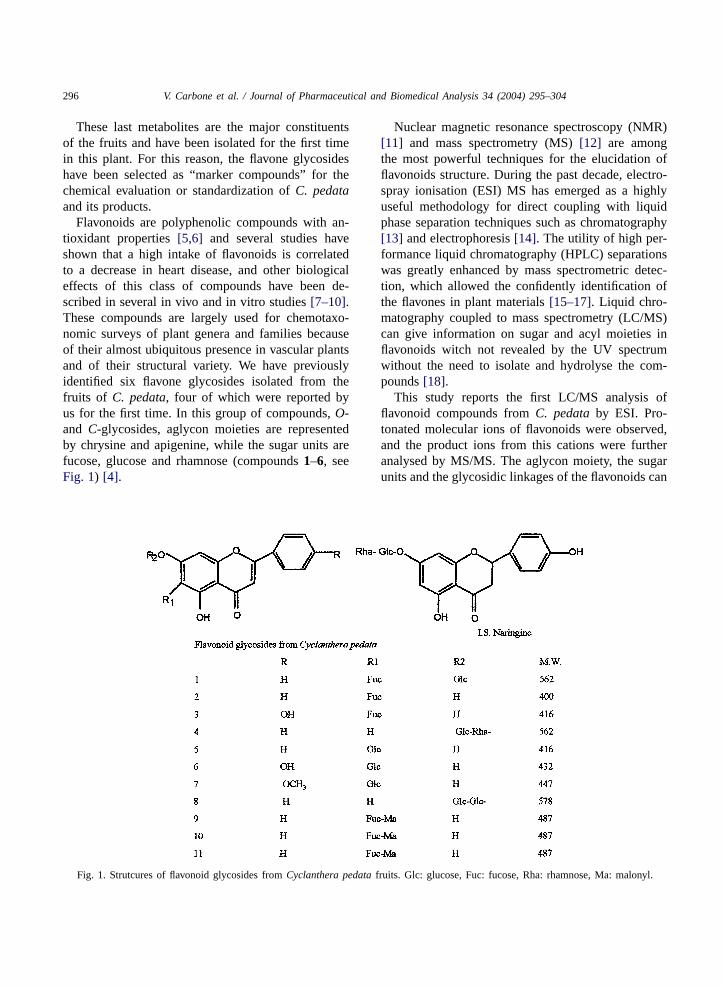
Flavonoids are polyphenolic compounds with an-tioxidant properties[5,6] and several studies haveshown that a high intake of flavonoids is correlatedto a decrease in heart disease, and other biologicaleffects of this class of compounds have been de-scribed in several in vivo and in vitro studies[7–10].These compounds are largely used for chemotaxo-nomic surveys of plant genera and families becauseof their almost ubiquitous presence in vascular plantsand of their structural variety. We have previouslyidentified six flavone glycosides isolated from thefruits of C. pedata, four of which were reported byus for the first time. In this group of compounds,O-and C-glycosides, aglycon moieties are representedby chrysine and apigenine, while the sugar units arefucose, glucose and rhamnose (compounds1–6, seeFig. 1) [4].
Fig. 1. Strutcures of flavonoid glycosides fromCyclanthera pedatafruits. Glc: glucose, Fuc: fucose, Rha: rhamnose, Ma: malonyl.
Nuclear magnetic resonance spectroscopy (NMR)[11] and mass spectrometry (MS)[12] are amongthe most powerful techniques for the elucidation offlavonoids structure. During the past decade, electro-spray ionisation (ESI) MS has emerged as a highlyuseful methodology for direct coupling with liquidphase separation techniques such as chromatography[13] and electrophoresis[14]. The utility of high per-formance liquid chromatography (HPLC) separationswas greatly enhanced by mass spectrometric detec-tion, which allowed the confidently identification ofthe flavones in plant materials[15–17]. Liquid chro-matography coupled to mass spectrometry (LC/MS)can give information on sugar and acyl moieties inflavonoids witch not revealed by the UV spectrumwithout the need to isolate and hydrolyse the com-pounds[18].
This study reports the first LC/MS analysis offlavonoid compounds fromC. pedataby ESI. Pro-tonated molecular ions of flavonoids were observed,and the product ions from this cations were furtheranalysed by MS/MS. The aglycon moiety, the sugarunits and the glycosidic linkages of the flavonoids can

V. Carbone et al. / Journal of Pharmaceutical and Biomedical Analysis 34 (2004) 295–304 297
be determinate from collision induced dissociation(CID) of pseudomolecular ions.
2. Materials and methods
2.1. Materials
Naringine, used as internal standard, was purchasedfrom Sigma (St. Louis, MO, USA).
Standards of pure compounds1–6 were isolated inour previous experimental study[4], and their struc-tures were elucidated by NMR.
HPLC grade methanol (MeOH), acetonitrile (ACN)and trifluoroacetic acid (TFA) were purchased fromJ.T. Baker (Baker Mallinckrodt, Phillipsburg, NJ,USA). HPLC grade water (18 m�) was prepared us-ing a Millipore Milli-Q purification system (MilliporeCorp., Bedford, MA, USA).
2.2. Preparation of flavonoid standards
Stock solutions of the flavonoid standards (1 mg/ml),were prepared by dissolving each compound inMeOH. Four different solutions, containing respec-tively 5, 25, 50 and 125�g ml−1 of each flavonoid (ex-ternal standards) and 40�g ml−1 of naringine, wereprepared in MeOH and used for method development.
2.3. Preparation of plant samples
The fruits ofC. pedataused in this study were sup-plied by IPIFA, Istituto Peruano Investigaciones Fi-toterapica Andina, and were collected in Perù, 1997.They were air dried and stored at room temperature.
A voucher sample is deposited at the Department ofPharmaceutical Science, University of Salerno, Italy(n. 5).
Capsules preparations ofC. pedatafruits were pur-chased from Natural World, Lima, Perù. Powdereddried extracts fromC. pedatafruits were supplied fromLaboratori Biokyma, Anghiari Ar, Italy).
A sample of 5 g of powder from air dried fruits,without the seeds, containing internal standard (10 mg)was extracted with 25 ml of MeOH, using sonicationfor 60 min at room temperature, and then was kept atroom temperature in the dark for a night. The extractwas filtered and diluted 1:10 with MeOH, and 20�l
of the sample were injected in to the analytical sys-tem. In order to establish if qualitative differences inflavonoid profile could be observed when the extrac-tion was carried out for a longer period,C. pedatafruits were also extracted in MeOH for 7 days at roomtemperature.
Capsules contents and dried extracts were extractedaccording the procedure described above.
2.4. Electrospray mass spectrometry (ESI/MS)
ESI/MS in the positive ion mode was performed byusing a Finnigan LCQ Deca ion trap instrument fromThermo Finnigan (San José, CA, USA) equipped withXcalibur software. Samples were dissolved in MeOHand infused in the ESI source by using a syringe pump;the flow rate was 3�l ml−1. The capillary voltage wasat 5 V, the spray voltage was at 5 kV and the tubelens offset was at 35 V. The capillary temperature was220◦C. Data were acquired in MS1 and MS/MS scan-ning mode.
2.5. Liquid chromatography/electrospray massspectrometry (LC/ESI/MS)
The extract was analysed by LC/ESI/MS “on-line”using the same instrument described above equippedwith a Spectra System HPLC (Thermo Finnigan, SanJosé, CA, USA). Individual flavonoids were separatedon a Symmetry C18 column (150 mm×2.1 mm, 5�m)(Waters Corporation, Milford, MA, USA) at a flow rateof 0.3 ml min−1; solvent A was 0.05% trifluoroaceticacid and solvent B was 0.05% (v/v) trifluoroacetic acidin acetonitrile. After a 5 min hold at 10% solvent B,elution was performed by a linear gradient from 10to 40% solvent B in 40 min and from 40 to 65% sol-vent B in 15 min. The eluate was directly injected intothe electrospray ion source and the MS1 and MS/MSspectra were acquired and interpreted using the soft-ware provided by the manufacturer.
Neutral loss scan mass spectrometric analyseswere performed with a triple quadrupole instrumentAPI2000 (Applied Biosystems, Foster City, CA,USA) equipped with a Series 200 HPLC system(Perkin-Elmer, Norwalk, CT, USA). Chromatographicconditions were the same as described above for theLC/ESI/MS experiment. The instrument was used inthe tandem MS mode (neutral loss scan).

298 V. Carbone et al. / Journal of Pharmaceutical and Biomedical Analysis 34 (2004) 295–304
2.6. Calibration, quantification and statisticalanalysis
Standard curves for each of the five flavonoid stan-dards were prepared over a concentration range of5–125�g ml−1 with four different concentration lev-els (5, 25, 50, and 125�g ml−1) and triplicate injec-tions at each level. Peak area ratios between the areaof each flavonoid standard and those of naringine,used as internal standard, were calculated and plot-ted against the corresponding standard concentrationusing weighed linear regression to generate standardcurves.
3. Results and discussion
3.1. ESI/MS and ESI/MS/MS analysis of flavonoidstandards
With the aim of obtaining mass spectrometric-basedinformation focused to evaluate the presence of dif-ferent classes of flavonoids,O- or C- and monoor di-glycosides, direct flow injection experimentsof flavonoid standards were initially performed.[M + H]+ and [M + Na]+ ions were observed whenthe data were acquired in MS1 scanning mode (m/zrange: 150–700) (seeTable 1). In Table 1, the dataobtained from MS/MS spectra of compounds1–11are also shown.
The fragmentation pattern observed for flavonoidC-glycosides was in agreement with the resultsobtained from Li et al.[19] with FAB MS andfrom Waridel et al. [20] using atmospheric pres-sure chemical ionisation–ion trap–mass spectrometry(APCI–IT–MS).
The daughter ion spectrum of the pseudomolecularion atm/z 563 (Compound1), 7-O-�-d-glucopyrano-syl-6-C-fucopyranosylchrysine (C27H30O13), showedan intense ion atm/z 545 corresponding to the lossof water and a minor fragment atm/z 523 due tothe loss of two water molecules, both of them con-nected with the fragmentation of the C-linked pentosesugar unit; another characteristic fragment was ob-served atm/z 489 corresponding to the ion [Glc–A–CH2CHOHCH=OH]+, where A corresponds to theprotonated aglycone moiety. The loss of the O-linkedhexose sugar unit instead produced an intense ion at
m/z 401 and another ion atm/z 383 due to a previousloss of water.
In order to obtain more information about the frag-mentation of the C-linked pentose sugar unit the ionat m/z 401 was subjected to a further MS3 fragmen-tation experiment. The resulting spectra showed twoions atm/z 383 and 365 due to the loss of one andtwo water molecules, respectively and a characteris-tic ion atm/z 321 derived from the Retro-Diels–Alderreaction (loss of acetaldehyde) following the loss oftwo molecules of water. Another ion occurred atm/z297 derived from the rearrangement reaction that pro-duce the ion [A–CH=CH–OH2]+. The relative inten-sity of each fragment ion confirmed the C-6 linkage ofthe sugar moiety excluding the C-8 linkage hypothesis[19].
The MS/MS analyses of compounds2, 6-C-fucopyr-anosylchrysine (C21H20O8), and3, 6-C-fucopyranosylapigenin (C21H20O9), showed the same fragmentationpattern of the C-linked pentose sugar unit previouslyseen. Differences in the intensity of the ion fragmentsare summarized inTable 1.
The tandem mass spectrum of compound4, 7-O-�-d-glucopyranosyl-(1→ 4)-�-l-rhamnopyranosyl-chrysine (C27H30O13), presents the characteristicfragmentation of flavonoidO-diglycosides[21].
The most intense ion observed in the full MS spec-trum was the ion atm/z255, corresponding to the pro-tonated aglycone moiety. Other two peaks, observedat m/z 401 and 417, were due to the loss of the ex-ternal glucose unit according to the Y and Z series offragmentation in polysaccharide chains[22].
Compounds5, 6-C-glucopyranosylchrysine (C21H20O9), and6, isovitexin (C21H20O10), showed frag-mentation patterns characteristic for the C-6-linkedhexose[19].
Compound7 was not observed in the overnight ex-tract of Cyclanthera pedatafruits and was isolatedonly from the 7 days extract. It was identified as amethoxyl derivative of compound6, glucopyranosilapigenin (C22H22O10). It was not an artefact becauseof it is present also in an Ethanolic extract, obtainedin 7 days. The MS/MS spectrum obtained from thefragmentation of the pseudomolecular ion atm/z 447showed the presence of a most abundant ion atm/z415 due to the loss of one molecule of methanol. Theother ions derived from the typical fragmentation ofC-6-glucosyl-flavonoids.

V. Carbone et al. / Journal of Pharmaceutical and Biomedical Analysis 34 (2004) 295–304 299
Table 1Molecular ions and fragment ions of the flavonoids, acquired by FIA (compounds1–7), using ES–MS and by SIM (compounds8–11),using LC/ESMS.
Compound Full MS (intensity)a MS/MS (intensity)a MS/MS/MS
1 585 [M + Na]+ (100) 563 [M + H]+ parent ion (10) 401 [M + H − 162]+ parent ion (20)563 [M + H]+ (80) 545 [M + H − w]+ (100) 383 [M + H − 162 − w]+ (70)
523 [M + H − 2w]+ (16.3) 365 [M + H − 162 − 2w]+ (100)489 [M + H − 74]+ (31.8) 321 [M + H − 162 − 44 − 2w]+ (50)401 [M + H − 162]+ (51.3) 297 [M + H − 162 − 104]+ (20)383 [M + H − 162 − w]+ (23.5)
2 423 [M + Na]+ (100) 401 [M + H]+ parent ion (10)401 [M + H]+ (60) 383 [M + H − w]+ (100)
365 [M + H − 2w]+ (60)321 [M + H − 44 − 2w]+ (51)297 [M + H − 104]+ (18.5)
3 439 [M + Na]+ (100) 417 [M + H]+ parent ion (15)417 [M + H]+ (56) 399 [M + H − w]+ (100)
381 [M + H − 2w]+ (32)337 [M + H − 44 − 2w]+ (21)313 [M + H − 104]+ (16)
4 585 [M + Na]+ (70) 563 [M + H]+ parent ion (30)563 [M + H]+ (100) 417 [M + H − 146]+ (60)255 [M + H − 162 − 146]+ (20) 401 [M + H − 162]+ (20)
255 [M + H − 162 − 146]+ (100)
5 439 [M + Na]+ (50) 417 [M + H]+ parent ion (0)417 [M + H]+ (100) 399 [M + H − w]+ (100)297 [M + H – 120]+ (50) 351 [M + H − 2w − 30]+ (58)
321 [M + H − 2w − 60]+ (25)297 [M + H − 120]+ (41)
6 455 [M + Na]+ (50) 433 [M + H]+ parent ion (0)433 [M + H]+ (100) 415 [M + H − w]+ (100)313 [M + H − 120]+ (50) 397 [M + H − 2w]+ (15)
367 [M + H − 2w − 30]+ (58)337 [M + H − 2w − 60]+ (25)313 [M + H − 120]+ (41)
7 447 [M + H]+ (100) 447 [M + H]+ parent ion (30)415 [M + H − 32]+ (100)397 [M + H − w − 32]+ (20)379 [M + H −2w − 32]+ (22)321 [M + H − 2w − 60]+ (15)295 [M + H − 32 − 120]+ (51)
8 579 [M + H]+ (100) 579 [M + H]+ parent ion (5)417 [M + H − 162]+ (100)255 [M + H − 162 − 162]+ (50)
9–11 487 [M + H]+ (100) 487 [M + H]+ parent ion469 [M + H − w]+451 [M + H − 2w]+401 [M + H − 86]+383 [M + H − 104]+365 [M + H − w − 104]+347 [M + H − 2w − 104]+
a The intensity is determined relative to the maximum mass peak.

300 V. Carbone et al. / Journal of Pharmaceutical and Biomedical Analysis 34 (2004) 295–304
Fig. 2. LC/ESMS analysis ofCyclanthera pedatafruits extract for the separation and identification of seven flavonoid glycosides.
3.2. Liquid chromatography/electrospray massspectrometry analysis of flavonoids
Positive ion electrospray LC/MS analysis, totalion current (TIC) profile, and reconstructed ion chro-matograms ofC. pedatafruits extract are shown inFig. 2. Flavonoids were identified comparing reten-tion times andm/z values in the total ion currentchromatogram to those of the selected standards.Reconstructed ion chromatograms were obtained foreach value ofm/zobserved for standard compounds inorder to improve the separation and the identification
of the single flavonoids; this step is very important toreach a higher selectivity for the quantitative analysis.Using LC/MS experiments, we have identified a largegroup of flavonoid compounds in the extracts withouttime-consuming pre-purification steps or optimisationof chromatographic procedures.
LC/MS analysis of the extract from the plant re-vealed the presence of several other compounds notpreviously detected in this species, and not reportedin Fig. 1. In particular peaks atm/z 649, 579 and487 were observed and the reconstructed ion chro-matograms showed the presence of one peak atm/z

V. Carbone et al. / Journal of Pharmaceutical and Biomedical Analysis 34 (2004) 295–304 301
649, two compounds at different retention times forthe ion with m/z 579 and three compounds in thelast part of the chromatogram with am/z 487. Theseevidences suggest us to use more sophisticated massspectrometric analyses to achieve a partial character-ization of these compounds, identified in the fruits ofthis plant for the first time.
3.3. LC/ESI/MS/MS product ion scan: identificationof new compounds
In order to characterize these unexpected com-pounds and to reveal the presence of other correlatedmolecules in the extract, simultaneous experiments ofLC/ESI/MS in selected ion monitoring (SIM) modeand LC/ESI/MS/MS, selecting the same ions as pre-cursors, were performed.
Them/zvalues selected were the following:m/z563,chosen in order to have a differentiation between thetwo O- andC-glycosides flavonoids showing the samemolecular weight andm/z 579, 487 and 649 chosenbecause of their presence in the total ion current of theLC/MS experiment.
The chromatographic profile obtained from theLC/ESI/MS SIM experiment for the ion atm/z 563showed the same profile of the reconstructed LC/MSchromatogram obtained for this ion in the previousexperiment. In particular, it shows two peaks at reten-tion time of 22.96 and 27.42 min that were attributedrespectively to compounds1 and 4, based on theLC/MS/MS data and on the comparison with theMS/MS spectra originated from the standards.
Analysis of the chromatogram obtained byLC/ESI/MS/MS for the ion atm/z 579 provides struc-tural information on the two flavonoid compoundsnot previously detected. The tandem mass spec-trum produced by the compound at retention time of19.53 min showed a major ion fragment atm/z 401,probably corresponding to the loss of a sugar moi-ety and a subsequent fragmentation, similarly to aC-glycoside flavonoid. Nevertheless, this result wasinsufficient for a complete structural characterizationof this compound. On the other hand, the compoundat retention time 24.03 min produced an unambiguoustandem mass spectrum, with two major ions, due tothe sequential loss of two molecules of hexose sugarO-linked to a flavonoid aglycone. For this reason, wehave hypothised that this compound has the structure
of an O-diglucopyranosilchrysine. The structure andthe spectral data are reported inFig. 1and inTable 1,respectively.
The chromatographic profile obtained from theLC/ESI/MS SIM experiment for the ion atm/z 487revealed the presence of three peaks at different re-tention times (seeFig. 3). Analysis of the tandemmass spectra of the three different species, in thesimultaneous LC/MS/MS experiment, showed thesame fragment ions at different intensities. In par-ticular, the compound at retention time 37.56 minshows an initial loss of one, two or three moleculesof water and a subsequent loss of malonic acid corre-sponding to the loss of a 86 Da fragment. From thisevidence we thought that the three compounds arethe malonyl derivatives of the fucosyl chriysine, eachof them substituted with the malonyl unit in one ofthe three possible free hydroxyl position of the sugarmoiety.
The chromatogram obtained by the LC/ESI/MSSIM experiment for the ion atm/z 649 gave a sin-gle peak at retention time of 26.63 min; the corre-sponding MS/MS spectrum showed a fragment ionat m/z 487, corresponding to the loss of an hexoseunit, followed by the same fragmentation pattern ofmalonyl-derivatives previously described. In this casethe hypothesized structure differs from the one de-scribed above by the presence of an additional glucoseunit, like compound1 differs from compound2.
In order to confirm these structures we carried out astudy focused on fragmentation reactions of the mal-onate moiety. Using a triple quadrupole instrument,these fragmentations allow the specific detection of amalonate containing class of molecules by neutral lossscanning; as a matter of fact when a group is lost as aneutral fragment, neutral loss scanning provides spe-cific detection. In fact, in a neutral loss scan experi-ment, both mass analysers are scanned simultaneouslyin a synchronized fashion but with an offset to lowerm/z values applied to Q3, which is equivalent to theneutral loss to be detected.
Since CID causes the loss of two molecules of wa-ter and a neutral acid malonic unit from [M+H]+ ionsof malonate containing compounds, we performed anLC/ESI/MS/MS experiment scanning for neutral lossof 140 Da. The three peaks revealed were located atthe end of the chromatogram at the same retentiontime already observed in the previous experiment; this

302 V. Carbone et al. / Journal of Pharmaceutical and Biomedical Analysis 34 (2004) 295–304
Fig. 3. LC/ES MS/MS SIM profile of malonyl derivatives of fucosyl chrysine.
allowed us to rule out the presence of other similarmalonyl derivatives. The absence of the peak corre-sponding to the ion atm/z 649 is easily explained, be-cause this compound gives an initial loss of a chargedfragment of 162 Da not detectable with neutral lossscanning.
3.4. Quantitative analisys of flavonoids incyclanthera pedata fruits
The calibration graphs, obtained by plotting area ra-tio between external and internal standard versus theknown concentration of each compound, were linearin the range of 5–125�g ml−1 for all six flavonoids.Five aliquots of crude extract ofC. pedatafruits wereanalysed in order to quantify the content of flavonoids.The method proved to be specific for the flavonoidsand the internal standard, since no interfering com-pounds could be seen in the TIC profile at the elutionpositions of the flavonoid compounds. The flavanone,naringin, was selected as a suitable internal standardfor the present calibration, since no interfering peakswere seen in any extract sample, thus confirming theadvantage in the use of this compound. It is worth
noting that the internal standard was introduced intoboth samples and calibration standards before the ex-traction, improving the precision and accuracy of thequantitative analysis.
Quantitative results for flavonoid glycosides areshown inTable 2, the concentrations are expressed asmilligram of each compound present in 1 g of driedfruit. Quantitative data are not reported for compound4, occurring as a minor constituent. Quantitative datashowed that compounds1 and 2 are the most abun-dant; this evidence is particularly important becausethe same compounds are also structurally characteris-tic for this species.
Five samples from commercial products, capsulescontaining powered dried fruits and powdered driedfruits, were analysed and quantified for flavonoid con-tent using the same procedure.
Quantitative analyses results showed that the rel-ative quantities of each flavonoid compound are inagreement with results obtained from plant samples,and, in particular, compounds1 and 2 appear to bethe most abundant (Table 3). This finding demon-strated a good quality for the commercial productsanalysed.

V. Carbone et al. / Journal of Pharmaceutical and Biomedical Analysis 34 (2004) 295–304 303
Table 2Quantitation of flavonoid glycosides by LC/MS for Peruvian samples ofCyclanthera pedatafruits
Compound1(mg/g of fruit)
Compound2(mg/g of fruit)
Compound3(mg/g of fruit)
Compound4(mg/g of fruit)
Compound5(mg/g of fruit)
Compound6(mg/g of fruit)
Mean (n = 5) 1.84 2.62 0.36 Minor 0.66 1.02S.D. 0.18 0.18 0.23 0.09 0.11r2 0.9996 0.9984 0.9984 0.99 0.9982 0.9952
Table 3Quantitation of flavonoid glycosides by LC/MS for commercialsamples ofCyclanthera pedatafruits (n = 5)
Flavonoids Capsules(mg/g of fruit)
Powdered dried fruits(mg/g of fruit)
1 2.92± 0.16 1.42± 0.062 1.76± 0.04 2.41± 0.063 0.70± 0.08 0.44± 0.064 Minor Minor5 0.59± 0.05 0.61± 0.066 1.41± 0.11 1.25± 0.28 Minor Minor9 Minor Minor
10 Minor Minor11 Minor Minor
Total 7.38± 0.11 6.14± 0.26
Note: For compound7 are not reported data, not founding thiscompound with the standard extraction procedure (seeSection 2).
4. Conclusions
In conclusion, we quantified the flavonoidic con-tent of C. pedataextracts by LC/MS using exter-nal and internal standard. Five other flavonoids,three malonyl derivatives of 6-C-fucosil-chrysine,a 6-C-gentobiosil-chrysine and a glucopyranosilacacetin, were also detected in these plant extracts.Four of them are new compounds, but occurred inC. pedataas minor constituents. With this analyticalapproach we have been able to reveal the presence offlavonoid glycoside malonates inCyclantheraspecies.
LC/MS techniques showed good performances bothin terms of sensitivity and specificity and provide twoindependent parameters, i.e. retention time and massinformation for the identification of the flavonoid con-stituents in a complex mixture such as a plant crudeextract.
To perform MS/MS analyses during the chro-matographic run allows the discrimination of the
O-glycosylation andC-glycosylation of flavonoids, asalready reported in the analysis of one crude extractcontaining daidzein and genistein glycosides[23].
The developed LC/MS/MS analytical method seemsto provide an accurate picture of flavonoid-relatedcompounds present in fruit extracts and, at the sametime, it results in a target-compounds approach.
The neutral loss scanning experiment reported inthis paper did not give additional information aboutsome other malonyl derivatives of flavonoids gly-cosides; but could be a useful tool for a rapid andsensitive screening of natural products conjugate,such as malonyl or acetyl derivatives, in a crude plantextract.
The developed method is straightforward and con-venient, requiring no expensive and time-consumingsample preparation procedures. As such it will be agood tool for industrial quality control in order toquantify marker compounds in both raw materials andin final products.
Acknowledgements
The authors acknowledge the kind support providedby Dr. Marco Biglietto for the analyses performed onthe API 2000 triple quadrupole instrument.
References
[1] A. Brank Egg, in: Dicionario Enciclopedico de Plantas utilesdel Perú, CBC, Centro de Estudios Regional Andinos “Bartolomè de las Casas”, 1999, pp. 171–172.
[2] N. de Tommasi, F. de Simone, C. Pizza, J. Agric. Food Chem.44 (1996) 2020–2025.
[3] N. de Tommasi, F. de Simone, G. Speranza, C. Pizza, J.Agric. Food Chem. 47 (1999) 4512–4519.
[4] P. Montoro, V. Carbone, F. de Simone, C. Pizza, N. deTommasi, J. Agric. Food Chem. 49 (11) (2002) 5156–5161.
304 V. Carbone et al. / Journal of Pharmaceutical and Biomedical Analysis 34 (2004) 295–304
[5] C.A. Rice-Evans, N.J. Miller, G. Panaga, Trends Plant Sci. 2(1997) 152–159.
[6] G. Cao, E. Sofic, R.L. Prior, Free Rad. Biol. Med. 22 (1997)749–760.
[7] C.A. Rice-Evans, N.J. Miller, G. Panaga, Free Rad. Biol.Med. 20 (1996) 933–956.
[8] E.J. Lien, S. Ren, H. Bui, R. Wang, Free Rad. Biol. Med.26 (1999) 285–294.
[9] J. Lunec, Ann. Clin. Biochem. 27 (1990) 173–182.[10] M.J. Sanz, M.L. Ferrandiz, M. Cejudo, M.C. Terencio, B.
Gil, G. Bustos, A. Ubeda, R. Gunasegaran, M.J. Alcaraz,Xenobiotica 24 (1994) 689–699.
[11] P.K. Agrawal, Phytochemistry 31 (1992) 3307–3330.[12] S.J. Gaskell, Anal. Chem. 70 (1998) 647–716.[13] W.M.A. Niessen, A.P. Tinke, J. Chromatogr. A 703 (1995)
37–57.[14] F.A. Tomás-Barberán, Phytochem. Anal. 6 (1995) 177–192.
[15] L.W. Sumner, N.L. Paiva, R.A. Dixon, P.W. Geno, J. MassSpectrom. 31 (1996) 472–485.
[16] M. Careri, L. Eviri, A. Mangia, Rapid Commun. MassSpectrom. 13 (23) (1999) 2399–2405.
[17] U. Justesen, P. Knuthsen, T. Leth, J. Chromatogr. A 799(1998) 101–110.
[18] M. Stobiecki, Phytochemistry 54 (2000) 237–256.[19] Q.M. Li, H. van den Heuvel, L. Dillen, M Claeys, Biol. Mass
Spectrom. 21 (4) (1992) 213–221.[20] J.L. Waridel, K. Wolfender, K.R. Ndjoko, H.J. Hobby, K.
Major, W. Hostettmann, J. Chromatogr. A 926 (2001) 29–32.
[21] Q.M. Li, M. Claeys, Biol. Mass Spectrom. 23 (7) (1994)406–416.
[22] B. Domon, C. Costello, Glycoconjugate J. 5 (1988) 397–409.[23] H. Rong, J.F. Stevens, M.L. Deinzer, L. de Cooman, D. de
Keukeleire, Planta Med. 64 (1998) 620–625.