University of South Florida Crystal engineering of flavonoids Scholar Commons Citation
101
University of South Florida Scholar Commons Graduate eses and Dissertations Graduate School 2008 Crystal engineering of flavonoids Padmini Kavuru University of South Florida Follow this and additional works at: hp://scholarcommons.usf.edu/etd Part of the American Studies Commons is esis is brought to you for free and open access by the Graduate School at Scholar Commons. It has been accepted for inclusion in Graduate eses and Dissertations by an authorized administrator of Scholar Commons. For more information, please contact [email protected]. Scholar Commons Citation Kavuru, Padmini, "Crystal engineering of flavonoids" (2008). Graduate eses and Dissertations. hp://scholarcommons.usf.edu/etd/325
-
Upload
independent -
Category
Documents
-
view
0 -
download
0
Transcript of University of South Florida Crystal engineering of flavonoids Scholar Commons Citation

University of South FloridaScholar Commons
Graduate Theses and Dissertations Graduate School
2008
Crystal engineering of flavonoidsPadmini KavuruUniversity of South Florida
Follow this and additional works at: http://scholarcommons.usf.edu/etdPart of the American Studies Commons
This Thesis is brought to you for free and open access by the Graduate School at Scholar Commons. It has been accepted for inclusion in GraduateTheses and Dissertations by an authorized administrator of Scholar Commons. For more information, please contact [email protected].
Scholar Commons CitationKavuru, Padmini, "Crystal engineering of flavonoids" (2008). Graduate Theses and Dissertations.http://scholarcommons.usf.edu/etd/325

Crystal Engineering of Flavonoids
by
Padmini Kavuru
A thesis submitted in partial fulfillment Of the requirements for the degree of
Master of Science Department of Chemistry
College of Arts and Sciences University of South Florida
Major Professor: Michael J. Zaworotko, Ph.D. Julie P. Harmon, Ph.D.,
Abdul Malik, Ph.D.
Date of Approval: April 11, 2008
Keywords: Polyphenols, Pharmaceutical Cocrystals, Hydrogen bond, French Paradox, Bioavailability
© Copyright 2008, Padmini Kavuru

Dedication
To my husband, parents and in-laws

Acknowledgements
I would like to thank my advisor, Professor Michael J. Zaworotko, for
the opportunity to conduct research under his supervision, and for his advice and
guidance throughout the Graduate Program.
I would also like to thank Dr. Julie P. Harmon and Dr. Abdul Malik, my
committee members, for their helpful comments and encouragements.
In addition, I would like to acknowledge all members of my research
group, as well as Faculty and Staff of the Chemistry Department of University of South
Florida, for their friendly accommodation.
At last, I would like to express my deepest thanks to my husband,
parents, in-laws, other family members and friends who constantly supported me
throughout the period of studies.

i
TABLE OF CONTENTS
LIST OF TABLES iv
LIST OF FIGURES iv
ABSTRACT ix
1. INTRODUCTION 1
1.1 Nutraceuticals 1
1.2 Flavonoids 1
1.3 Structure and classification 3
1.4 Bioactivity and bioavailability 6
1.5 Crystal Engineering 9
1.5.1 Supramolecular Chemistry 10
1.5.2 Supramolecular synthons 11
1.5.3 Cocrystals 12
1.5.4 Pharmaceutical cocrystals 14
2. CSD ANALYSIS 18
2.1 Cambridge Structural Database 18
2.2 CSD statistics for Phenols and Polyphenols 19
2.3 Interactions of phenolic-OH with other functional groups 21
2.4 Discussions 25 3. QUERCETIN 26

ii
3.1 Description 26
3.2 Strategy for Crystal Engineering of Quercetin 27
3.3 Solvates of Quercetin 29
3.3.1 Form I and Form II (Pyridine solvates of Quercetin) 29
3.3.2 Form III (THF solvate of Quercetin) 32
3.3.3 Form III (Quercetin-Acetone solvate) 33
3.3.4 Discussions 35
3.4 Cocrystals of Quercetin 36
3.4.1 Quercetin- Caffeine- Methanol cocrystal solvate 36
3.4.2 Quercetin-Isonicotinamide 39
3.4.4 Discussions 42
4. HESPERETIN 43
4.1 Description 43
4.2 Strategy for Crystal Engineering of Hesperetin 44
4.2.1 Hesperetin-Isonicotinamide cocrystal (Cocrystal 3) 45
4.2.2 Hesperetin-Nicotinic acid zwitterion cocrystals 49
4.2.4 Discussions 54
5. DISSOLUTION STUDIES OF THE COCRYSTALS 55
5.1 Dissolution experiments of quercetin and hesperetin cocrystals 57
5.2 Dissolution study of Quercetin:Caffeine cocrystal (Cocrystal 1) 60
5.2 Dissolution study of Quercetin-Isonicotinamide (Cocrystal 2) 61
5.3 Dissolution study of Hesperetin-Isonicotinamide (Cocrystal 3) 62
5.4 Dissolution study of Hesperetin-NA (Cocrystal 4) 63

iii
5.5 Discussions 64
6. CONCLUSIONS AND FUTURE DIRECTIONS 66
REFERENCES 67
APPENDICIES 76
Appendix A: 77
Appendix B 85

iv
LIST OF TABLES
Table 1.1 Structures of different of Flavonoids 5
Table 1.2 Classification Flavonoids and their common food sources 6
Table 2.1 CSD statistics for Phenols and Polyphenols 20
Table 3.1 Solvates of Quercetin 27

v
LIST OF FIGURES
Figure 1.1 Skeleton of Flavonoid 3 Figure1.2 Structure of Flavonoid with the chromane ring 3 Figure1.3 Aurones (2-benzyl-coumarone) 4 Figure1.4 Example of Glycoside, Rutin (quercetin-3-rutinoside) 4 Figure1.5 Hypothesis of the links between the working mechanisms of flavonoids and their effects on disease. NO, nitrous oxide 7 Figure 1.6 Supramolecular synthons: (a) Carboxylic acid homosynthon, 12 (b) Carboxylic acid•••pyridine heterosynthon
Figure 1.7 Crystal structure of the triclinic form of quinhydrone 13 Figure 1.8 The Hoogsteen base-pairing in the structure of 9-methyladenine 13 Figure 1.9 Occurrence of the term “cocrystal” in papers published between 1991 and 2007 (SciFinder search 2/2008). 14 Figure 1.10 Different forms of drugs in which they are administered. 15 Figure 1.11 Crystal packing of 1:1 co-crystals of 5, 5-diethylbarbituric acid 16 Figure 1.12 (a) Itraconazole•succinic acid cocrystal, (b) Carbamazepine- saccharin cocrystal 16 Figure 2.1 (a) Growth of the CSD since 1972, (b) Predicted growth of the CSD during the decade 2001 - 2010 19 Figure 2.2 Structure of polyphenol for the CSD search 19 Figure 2.3 Supramolecular phenolic OH… OH homosynthon (Synthon I) 22 Figure 2.4 Histogram for synthon I 23 Figure 2.5 Supramolecular phenolic OH…OH heterosynthon (Syn II) 21

vi
Figure 2.6 Histogram for synthon II 21 Figure 2.7 Supramolecular phenolic OH…Narom heterosynthon (Syn III) 22
Figure 2.8 Histogram for synthon II 24 Figure 2.9 Supramolecular phenolic OH…O heterosynthon (Syn IV) 25 Figure 2.10 Histogram for synthon IV 24 Figure 2.11 (a) Supramolecular phenolic OH…CO heterosynthon (Syn V) (b) Supramolecular phenolic OH…NH heterosynthon (Syn VI) 24 Figure 2.12 Histograms for synthon V and synthon VI 25 Figure 3.1 Structure of Quercetin 26 Figure 3.2 Representation of different rings in Quercetin 27 Figure 3.3 Hydrogen bonding in Quercetin dihydrate 28 Figure 3.4 Illustration of dihedral angle between the planes. (Red plane contains rings A and C and the blue plane contains ring B) 29 Figure 3.5 Single crystal of quercetin-pyridine solvate 30 Figure 3.6 (a) Hydrogen bonding in Form I (b) Hydrogen bonding in Form II 31 Figure 3.7 The dihedral angle in From I (a) and Form II (b) 31 Figure 3.8 Single crystal of quercetin-THF solvate (Form II) 32 Figure 3.9 Comparison of hydrogen bonding in Form III (left) with that of Form II (right) 33 Figure 3.10 Crystals of Quercetin-Acetone solvate (Form III) 34 Figure 3.11 Hydrogen bonding in Form III (Acetone molecules colored red) 34 Figure 3.12 Representation of crisscross grid network in Quercetin-Acetone 35 solvate
Figure 3.13 The asymmetric unit of 1:1:1 cocrystal solvate of quercetin, caffeine and methanol 36

vii
Figure 3.14 Single crystal of quercetin:caffeine:methanol cocrystal solvate (Cocrystal I) 37 Figure 3.15 Intermolecular interactions in the 1:1:1 cocrystal solvate of quercetin, caffeine and methanol (Green). 38 Figure 3.16 Illustration of sheets generated in the cocrystal 1. 39 Figure 3.17 The asymmetric unit of the1:1 quercetin – isonicotinamide cocrystal 39 Figure 3.18 Single crystal of quercetin-isonicotinamide cocrystal 39 Figure 3.19 Intermolecular interactions in the 1:1 Quercetin-Isonicotinamide cocrystal. 40 Figure 3.20 Illustration of R4
4 (18) graph set in quercetin and isonicotinamide cocrystal. 41 Figure 4.1 Structure of hesperetin (hydrogen are not included) 43 Figure 4.2 Illustration of rings in Hesperetin 44 Figure 4.3 Hydrogen bonding in (a) Hesperetin (b) Hesperetin monohydrate 44 Figure 4.4 Illustration of the dihedral angles 45 Figure 4.5 The asymmetric unit of hesperetin:isonicotinamide 1:1 cocrystal 45 Figure 4.6 Single crystal of hesperetin-isonicotinamide cocrystal 46 Figure 4.7 Overall hydrogen bonding in the Hesperetin-Isonicotinamide cocrystal 47 Figure 4.8 Illustration supramolecular synthons in the cocrystal 3. 47 Figure 4.9 The cavity formed in hesperetin:isonicotinamide cocrystal. 48 Figure 4.10 Illustration of 8-fold interpenetrated network in Cocrystal 3 48 Figure 4.11 Illustration of dihedral angle in hesperetin molecules (red plane contains rings A and C; blue contains ring C) 49 Figure 4.12 Hesperetin – Nicotinic acid 1:1 zwitterion cocrystal (Form I) 49 Figure 4.13 Single crystals of hesperetin-NA cocrystals. 50

viii
Figure 4.14 The hydrogen bonding in Form I cocrystal.(Blue: R-Hesperetin ) 51 Figure 4.15 Illustration of hydrogen bonds in NA: a) NA in pure form b) NA in the cocrystal, hesperetin molecules are deleted for clarity. 51 Figure 4.16 Representation of dihedral angle between the rings. (red plane contains rings A and C, blue plane contains ring B) 52 Figure 4.17 The hydrogen bonding in Form II cocrystal (yellow and maroon: NA molecules, (+) hesperetin: blue, (-) hesperetin) 53 Figure 4.18 The dihedral angle formed between the planes of rings A, C and ring B in Form II 53 Figure 5.1 Powder dissolution profiles for (1), (2), (3), and (4) in water at 20 °C. 56 Figure 5.2 Dissolution profiles into 0.1 N HCl at 25 °C for Sporanox beads and the cocrystals 56 Figure 5.3 Average plasma time curves of carbamazepine concentrations (±SEM)from a cross-over experiment in fasted beagle dogs (n = 4)given oral doses of 200 mg of the active drug as Tegretol tablets and co-crystal 57 Figure 5.4 UV spectrums of Quercetin and Hesperetin in 1:1 Ethanol/Water 59 Figure 5.5 Dissolution profiles of quercetin dihydrate, solvated and the desolvated cocrystal in 1:1 Ethanol/water 61 Figure 5.6 Dissolution profiles of quercetin dihydrate and quercetin- isonicotinamide cocrystal in 1:1 EtOH/Water (V/V%) 61 Figure 5.7 Dissolution profiles of hesperetin and hesperetin-isonicotinamide (1:1) cocrystal in 1:1 EtOH/Water (V/V%) 62 Figure 5.8 Dissolution profiles of hesperetin and its cocrystals 63

ix
CRYSTAL ENGINEERING OF FLAVONOIDS
PADMINI KAVURU
ABSTRACT
Crystal engineering is attracting attention in the pharmaceutical industry because the
design of new crystal form of drugs can improve their stability, bioavailability and other
relevant physical characteristic properties. Therefore, crystal engineering of
nutraceuticals such as flavonoids by exploring their hydrogen bonding interactions can
generate novel compounds such as pharmaceutical cocrystals. Flavonoids are
polyphenolic secondary plant metabolites that are present in varying levels in fruits,
vegetables and beverages. The “French paradox”, low cardiovascular mortality rate in
spite of high intake of saturated fat among the Mediterranean populations made
flavonoids an appropriate target for therapeutic researchers.
The work herein deals with the crystal engineering of two flavonoids,
quercetin and hesperetin, which are already known to exhibit antioxidant properties and
reduce cardiovascular effects in humans. However, they have limited bioavailability and
poor water solubility. Several new forms of quercetin and hesperetin in the form of
solvates and cocrystals were synthesized. These new crystal forms were characterized by
various techniques: FT-IR, DSC (Differential Scanning Calorimetry), single X-ray
diffraction, powder X-ray diffraction, TGA (Thermal Gravimetric Analysis) and

x
melting point. The new compounds were also studied via dissolution studies performed in
1:1 ethanol/water (V/V%). Thus, crystal engineering proves to be effective way to
enhance the solubility and bioavailability of the target flavonoid molecules.

1
1. INTRODUCTION
1.1 Nutraceuticals
The term ‘Nutraceutical’ is an intermediate of nutrition and pharmaceutical.1 It is used to
describe a medicinal or nutritional component of food, plant or naturally occurring
material in purified or concentrated form claimed to have a medicinal effect on human
health. Such foods are also called functional foods. It can also refer to individual
chemical present in common foods. The position of nutraceuticals on the legislative
grounds is marginal between pharmaceutics and food.
The term nutraceutical was coined by Dr. Stephen De Felice (founder and
chairman of Foundation of Innovation in Medicine) in 1976.1 He defined it as:
“Nutraceuticals are ‘food, or parts of food, that provide medical or health benefits,
including the prevention and treatment of disease”. Nutraceuticals include a wide range
of products, such as polyphenols (Flavonoids), vitamins, oils from fish and flax seed,
glucosamine, chondroitin, resveratrol, calcium-fortified juices etc.1
1.2 Flavonoids Flavonoids are naturally occurring polyphenolic compounds that are found in fruit,
vegetables, grains, bark, roots, stems, flowers, tea, and wine.2 These natural products
were known for their beneficial effects on health long before flavonoids were isolated as

2
the effective compounds. There are more than 4000 varieties of flavonoids which have
been identified till now and are considered responsible for the attractive colors of flowers,
fruit, and leaves.3, 4 These are responsible for many of the plant colors that dazzle us with
their brilliant shades of yellow, red and orange. Many of the flavonoids are dietary
antioxidants and constituents of medicinal herbs. These compounds were discovered in
by a Hungarian scientist, Dr. Albert Szent-Gyorgyi in 1938, who discovered Vitamin C
(Nobel laureate) by mistake. Rusznyák and Szent-Györgi (1936) suggested that
flavonoids be known as vitamin P or vitamin C2.5-8 However, by the 1950’s the vitamin
claim had been abandoned due to a lack of substantive evidence.
Flavonoids are also known as plant secondary metabolites which does not
effect the normal growth, development or reproduction of organisms like the primary
metabolites neither their absence result in immediate death.9 But, these compounds are
used as defenses against predators, parasites and diseases, for interspecies competition,
and to facilitate the reproductive processes (coloring agents, attractive smells, etc) and
hence they are called as the “First Line of Defense”.10 The research on flavonoids took a
rapid momentum with the discovery of the French paradox, i.e., low cardiovascular
mortality rate observed in Mediterranean populations with red wine consumption and a
high saturated fat intake.22, 23 The flavonoids in red wine are responsible for this effect. In
addition, epidemiologic studies suggest a protective role of dietary flavonoids against
coronary heart disease. After a number of subsequent studies on animals it was found that
flavonoid intake is inversely correlated with mortality due to coronary heart disease.11, 12
These potential benefits are being used to promote the consumption of flavonoid-rich
foods, beverages and dietary supplements.
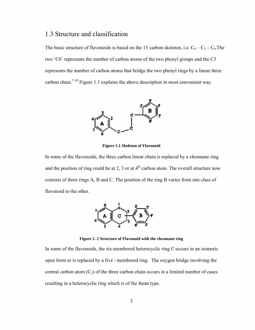
1.3 Structure and classification
The basic structure of flavonoids is based on the 15 carbon skeleton, i.e. C6 – C3 – C6.The
two ‘C6’ represents the number of carbon atoms of the two phenyl groups and the C3
represents the number of carbon atoms that bridge the two phenyl rings by a linear three
carbon chain.7-10 Figure 1.1 explains the above description in most convenient way.
Figure 1.1 Skeleton of Flavonoid
In some of the flavonoids, the three carbon linear chain is replaced by a chromane ring
and the position of ring could be at 2, 3 or at 4th carbon atom. The overall structure now
consists of three rings A, B and C. The position of the ring B varies from one class of
flavonoid to the other.
Figure 1. 2 Structure of Flavonoid with the chromane ring
In some of the flavonoids, the six-membered heterocyclic ring C occurs in an isomeric
open form or is replaced by a five - membered ring. The oxygen bridge involving the
central carbon atom (C2) of the three carbon chain occurs in a limited number of cases
resulting in a heterocyclic ring which is of the furan type.
3

Figure 1.3 Aurones (2-benzyl-coumarone)
Most of the flavonoids occur as glucosides in which the C6-C3-C6 aglycone part of the
molecule is linked with different number of sugars. If the linkage of the sugar to the
flavonoid aglycone is through an OH group then these are called as O-glycosylflavonoids
and if the linkage is through C-C bond then these are called as C-glycosylflavonoids.17
The type of sugar and the position of sugars vary for different glycosides. These have a
more complex structure than the phenolic skeleton materials.
Glycoside = Aglycone + Glycone (sugar)
Figure 1.4 Example of Glycoside, Rutin (quercetin-3-rutinoside)
The most common citrus flavanone-glycosides are rutin, natrirutin, naringin, hesperedin
and neohesperidin. Naringin (in grapefruits) and hesperidin (in oranges) are the two
major flavonoid-glycosides present in the citrus fruits, and are primarily concentrated in
the peel and the tissue of the fruit.18 The table below represents the structures of various
flavonoids.
4

Table 1.1 Structures of different of Flavonoids
Anthocyanidins
Flavanols
Flavonols
Flavonones
Flavones
Isoflavones
Flavonoids are divided into various classes on the basis of their molecular structure.19 The six
main groups of flavonoids are listed in Table 1.2, which includes the known members of
5

6
each group and the food sources in which they are present.
Table 1.2 Classification Flavonoids and their common food source
Flavonoid Subclass
Dietary Flavonoids Some Common Food Sources
Anthocyanidins
Cyanidin, Delphinidin, Malvidin, Pelargonidin,
Peonidin, Petunidin
Red, blue and purple berries, red and purple grapes, red wine
Flavanols Monomers (Catechins): Catechin, Epicatechin,
Epigallocatechin Epicatechin gallate,
Epigallocatechin gallate Dimers and Polymers:
Theaflavins, Thearubigins, Proanthocyanidins
Catechins: Teas (particularly green and white), chocolate, grapes, berries, apples Theaflavins, Thearubigins: Teas (particularly black and oolong) Proanthocyanidins: Chocolate, apples, berries, red grapes, red wine
Flavonones Hesperetin, Naringenin, Eriodictyol
Citrus fruits and juices, e.g., oranges, grapefruits, lemons
Flavonols Quercetin, Kaempferol, Myricetin, Isorhamnetin
Widely distributed: yellow onions, scallions, kale, broccoli, apples, berries, teas.
Flavones Apigenin, Luteolin Parsley, thyme, celery, hot peppers,
Isoflavones Daidzein, Genistein, Glycitein Soybeans, soy foods, legumes
1.4 Bioactivity and bioavailability Flavonoids are widely known for their antioxidant activity. Important effect of flavonoids is
the scavenging of oxygen-derived free radicals. In vitro experimental systems showed that
flavonoids possess anti-inflammatory, anti-allergic, antiviral, and anti-carcinogenic
properties.20 An overview of the hypothetical links between the working mechanisms and
clinical effects of flavonoids is given in Figure 1.5.21

Figure 1.5 Hypothesis of the links between the working mechanisms of flavonoids and their effects on disease. NO, nitrous oxide
One clue to the health benefits of flavonoids comes from studies of the "French paradox”.22
French eat almost four times more butter and three times more lard-and have higher
cholesterol levels and blood pressures-than do Americans and yet the French are 2.5 times
less likely than Americans to die of coronary heart disease. Many people have suggested that
the liberal French consumption of red wine protects against coronary heart disease,
apparently by lowering cholesterol levels or preventing abnormal blood clots. In fact, at least
eight medical studies have found that a glass or two of wine daily protects against heart
disease. But some studies have reported that red wine is better than white wine, suggesting
that some of the benefits might be unrelated to the alcohol. The antioxidant property of
flavonoids can be explained in the following way: Flavonoids are oxidized by radicals,
resulting in a more stable, less-reactive radical. In other words, flavonoids stabilize the
reactive oxygen species by reacting with the reactive compound of the radical. Because of
7

8
the high reactivity of the hydroxyl group of the flavonoids, radicals are made inactive,
according to the following equation: 22
Flavonoid (OH) + R• Flavonoid (O•) + RH (1) where, R•
is a free radical and O• is an oxygen free radical. Selected flavonoids can directly scavenge
super oxides; whereas other flavonoids can scavenge the highly reactive oxygen derived
radical called peroxynitrite. Epicatechin and rutin are also powerful radical scavengers. The
scavenging ability of rutin may be due to its inhibitory activity on the enzyme xanthine
oxidase. By scavenging radicals, flavonoids can inhibit LDL oxidation in vitro.24 This action
protects the LDL particles and, theoretically, flavonoids may have preventive action against
atherosclerosis.
With the exception of flavanols (catechins and proanthocyanidins), flavonoids
occur in plants and most foods as glycosides. Even after cooking, most flavonoid glycosides
reach the small intestine intact. Only flavonoid aglycones and flavonoid glucosides (bound to
glucose) are absorbed in the small intestine, where they are rapidly metabolized to form
methylated, glucuronidated or sulfated metabolites.25 Hollman and Katan suggested that the
glycosylated forms of quercetin are absorbed more readily than are the aglycone forms;
however, this has been questioned by other researchers. The role of flavonoid glycosylation
in facilitating absorption is questioned by the fact that catechin, which is not glycosylated in
nature, is absorbed relatively efficiently. As most of the flavonoids are poorly absorbed by
the body and the major percentage is excreted out which, limits its bioavailability. Even
though numerous strategies exist for enhancing the bioavailability of drugs with low aqueous
solubility, the success of these approaches is not yet able to be guaranteed and is greatly
dependent on the physical and chemical nature of the molecules being developed. Crystal
9
engineering offers a means to improved solubility and dissolution rate and stability, which
can be adopted through an in-depth knowledge of crystallization processes and the molecular
properties of active pharmaceutical ingredients.26
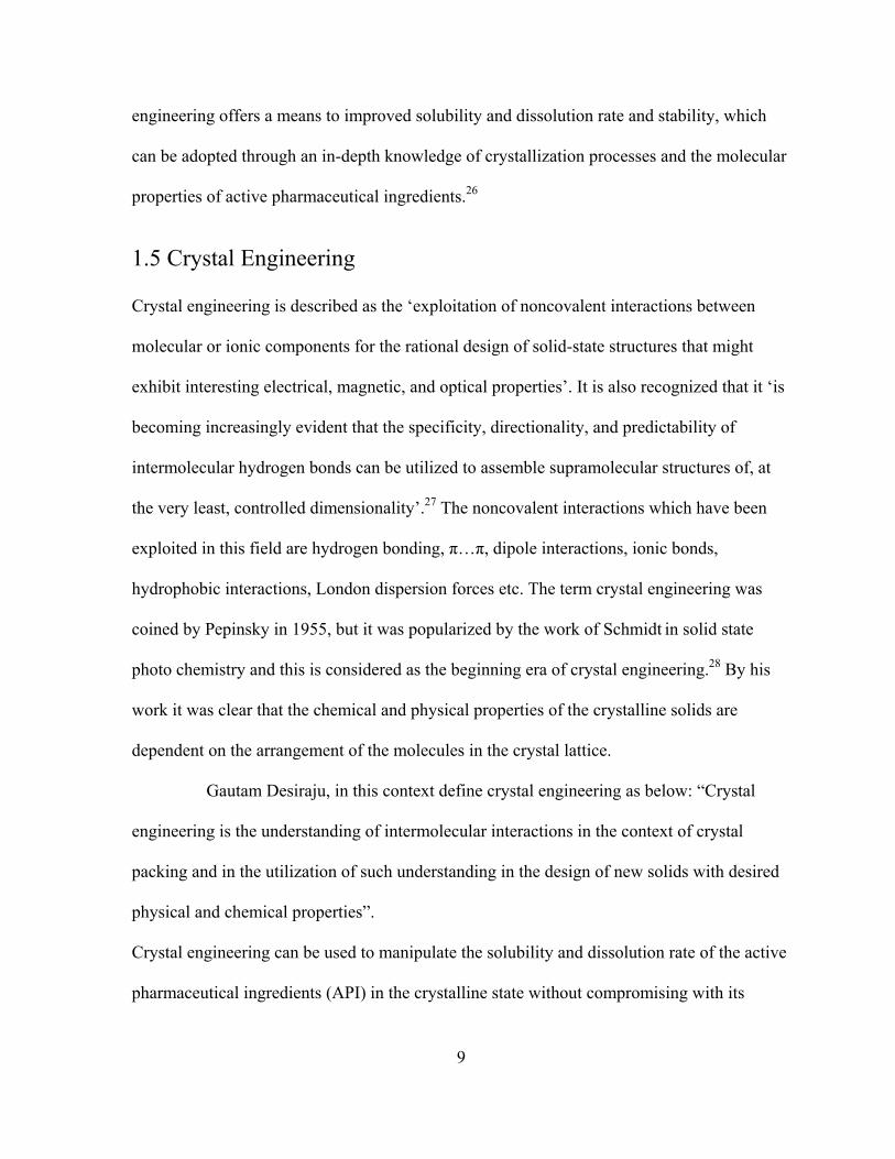
1.5 Crystal Engineering Crystal engineering is described as the ‘exploitation of noncovalent interactions between
molecular or ionic components for the rational design of solid-state structures that might
exhibit interesting electrical, magnetic, and optical properties’. It is also recognized that it ‘is
becoming increasingly evident that the specificity, directionality, and predictability of
intermolecular hydrogen bonds can be utilized to assemble supramolecular structures of, at
the very least, controlled dimensionality’.27 The noncovalent interactions which have been
exploited in this field are hydrogen bonding, π…π, dipole interactions, ionic bonds,
hydrophobic interactions, London dispersion forces etc. The term crystal engineering was
coined by Pepinsky in 1955, but it was popularized by the work of Schmidt in solid state
photo chemistry and this is considered as the beginning era of crystal engineering.28 By his
work it was clear that the chemical and physical properties of the crystalline solids are
dependent on the arrangement of the molecules in the crystal lattice.
Gautam Desiraju, in this context define crystal engineering as below: “Crystal
engineering is the understanding of intermolecular interactions in the context of crystal
packing and in the utilization of such understanding in the design of new solids with desired
physical and chemical properties”.
Crystal engineering can be used to manipulate the solubility and dissolution rate of the active
pharmaceutical ingredients (API) in the crystalline state without compromising with its

10
bioactivity. There are many possible ways that may be achieved from recent developments in
the study of molecular solids and reviews topical issues such as habit modification,
polymorphism, solvation, co-crystal formation and surface modification. Therefore,
particular attention is paid to the area of co-crystallization, which is an emerging area of
strategic importance to the pharmaceutical sector.27
1.5.1 Supramolecular Chemistry Supramolecular chemistry is a relatively new field of chemistry which focuses literally on
going "beyond" molecular chemistry.29 The importance of supramolecular chemistry was
recognized by Donald J. Cram, Jean-Marie Lehn, and Charles J. Pedersen and for their work
in this area they were awarded Nobel Prize for Chemistry in 1987. The important
breakthrough that allowed the elucidation of the double helical structure of DNA occurred
when it was realized that there were two separate strands of nucleotides connected through
hydrogen bonds. The utilization of noncovalent bonds is essential to replicate because they
allow the strands to be separated and used to template new double stranded DNA. The
development of selective "host-guest" complexes, in which a host molecule recognizes and
selectively binds a certain guest, was cited as an important contribution. Unlike, the organic
synthesis that involves the making and breaking of covalent bonds to synthesize a desired
molecule, supramolecular chemistry utilizes far weaker and reversible noncovalent
interactions, such as hydrogen bonding, metal coordination, hydrophobic forces, van der
Waals forces, pi-pi interactions, sometimes electrostatic effects to assemble molecules into
multimolecular complexes. Various fields that are classified under supramolecular chemistry

11
include molecular self-assembly, molecular recognition, host-guest chemistry, mechanically-
interlocked molecular architectures, and dynamic covalent chemistry.30 It is important for the
development of new pharmaceutical therapies by understanding the interactions at a drug
binding site. In addition, supramolecular systems have been designed to disrupt protein-
protein interactions that are important to cellular function. It also has application in green
chemistry where reactions have been developed where the reactions take place in the solid
state directed by non-covalent bonding. Such procedures are highly desirable since they
reduce the need for solvents during the production of chemicals.
1.5.2 Supramolecular synthons Supramolecular synthons are the smallest structural units within which is encoded all the
information inherent in the mutual recognition of molecules to yield solid state
supermolecules, that is, crystals. A key aspect of crystal engineering is therefore the
dissection of a target network into supramolecular synthons not the molecular synthons
which connect the supramolecular synthons.31 Such a dissection simplifies the analysis of a
target network and is important in crystal engineering because it recognizes the
interchangeability of supramolecular synthons in a family of structures.32, 33 Supramolecular
synthons are classified into two categories: supramolecular homosynthons and
supramolecular heterosynthons.34 The supramolecular homosynthons are formed between
the same, self-complementary functional groups and supramolecular heterosynthons are
formed between different but complementary functional groups. Examples of supramolecular
homosynthons include the dimers formed between carboxylic acids 35, 36 and amides 37
whereas supramolecular heterosynthons include carboxylic acid···amide, 38-42

hydroxyl···pyridine, 43-45 and carboxylic acid···pyridine.46-49
(a) (b)
Figure 1.6 Supramolecular synthons: (a) Carboxylic acid homosynthon, (b) Carboxylic acid•••pyridine heterosynthon
The supramolecular homosynthons usually exists in structures of single-component
compounds, for example the carboxylic acids exists as dimers. Where as, if multiple
functional groups are present, a supramolecular heterosynthon is more likely to be formed as
the formation of heterosynthons wins over the homosynthons when ever these two are
competing with each other. When a supramolecular heterosynthon is formed between two
functional groups that are located on different molecules, a multicomponent is formed.
1.5.3 Cocrystals Cocrystals belong to the class of compounds which are long known but little studied. Earlier,
cocrystals were known with different names like; molecular compounds,50 organic molecular
compounds,51 addition compounds,52 molecular complexes,53 solid-state complexes,54 or
heteromolecular crystals.55 In1844, the first cocrystal of benzoquinone and hydroquinone
was synthesized by Wohler, and then followed by studies of halogen derivatives of
quinhydrone.50, 56 Until 1960 the structural information for the cocrystal was absent. The
elucidation of DNA structure 57, 58 through X-ray analysis inspired many people to come out
with numerous nucleobase complexes in 1950’s and 60’s 59-62 and then the term “cocrystal”
12

was first coined in this context 63 later it was subsequently popularized by Etter.64 During this
time, Hoogstein came up with the new base pair called “Hoogstein base pair”, a cocrystal
formed between 9-methyladenine and 1-methylthymine.61
Figure 1.7 Crystal structure of the triclinic form of quinhydrone
Figure 1.8 The Hoogsteen base-pair The term cocrystal sounds very simple but the definition is often a topic of debate.65, 66 The
Zaworotko research group defines cocrystal as: “A cocrystal is a multiple component crystal
in which all components (molecule or ion and a molecular cocrystal former) are solid under
ambient conditions when in their pure form”. All components are solid under ambient
conditions has important practical considerations, because synthesis of cocrystals can be
achieved via the solid-state.67 Aakero¨y and co-workers,68 they defined cocrystals as (i)
compounds constructed from neutral molecules; (ii) made from reactants that are solids at
ambient conditions; and (iii) structurally homogeneous crystalline materials that contains at
13

least two neutral building blocks with a well-defined stoichiometry. A broad definition of a
cocrystal given by Dunitz: “a crystal containing two or more components together”69 would
include molecular adducts, salts, solvates/hydrates, inclusion compounds, etc. What ever way
people define the term cocrystal, the field of cocrystal is attaining the heights of excellence,
and Figure 1.9 reveals the growing interest in the subject. Therefore, it is evident that
cocrystals play a vital role in pharmaceutical science and elsewhere.
14
1991 1992 1993 1994 1995 1996 1997 1998 1999 2000 2001 2002 2003 2004 2005 2006 20070
50
100
150
200
250
300
Num
ber o
f hits
con
tain
ing
the
term
"Coc
ryst
al"
Year
Figure 1.9 Occurrence of the term “cocrystal” in papers published between 1991 and 2007
(SciFinder search 2/2008).
1.5.4 Pharmaceutical cocrystals Most of the active pharmaceutical ingredients (APIs) are administered in the solid state as
part of an approved dosage type like tablets, capsules, etc as it is the convenient and compact
format to store a drug.

Figure 1.10 Different forms of drugs in which they are administered. APIs can exist in a variety of distinct solid forms, where each form may display unique
physicochemical properties such as hygroscopicity, morphology, and (most importantly)
solubility. The solid form of the drug dictates the properties like stability, hygroscopicity,
dissolution rate, solubility and bioavailability. Figure 1.10 represents the different ways in
which drugs are administered.70 This makes the pharmaceutics to look for a crystal form with
the best properties for therapeutic use and manufacturability. Therefore, whatever form of
drug is selected, it must be amenable to handling and processing and as a drug, it must be
effective and safe. Sometimes, the pharmaceutic developers come across a problem, that the
most common polymorphic form of the drug is least stable. Therefore the current approaches
to change the properties of APIs include the utilization of ionic salts, solvates, hydrates, and
polymorphs.71 In this context pharmaceutical cocrystallization is potentially attractive for
improving the material properties while leaving an API unaltered. The intellectual property
implications of creating co-crystals are also highly relevant. A pharmaceutical co-crystal can
described as “a multiple component crystal in which at 20 least one component is molecular
and a solid at room temperature (the co-crystal former) and forms a supramolecular synthon
with a molecular or ionic API”.72, 73 The first application of crystal engineering to generate of
15

pharmaceutical co-crystals was a series of studies by Whitesides et al. with the use of
substituted barbituric acid, including barbital and melamine derivatives and generated a
supramolecular ‘linear tape,’ ‘crinkled tape,’ and ‘rosette’ motifs.74-80
(a) (b)
Figure 1.11 Crystal packing of 1:1 co-crystals of 5, 5-diethylbarbituric acid (barbital)
To date, many pharmaceutical cocrystals have been reported in the scientific and patent
literature and most of the cocrystals have been found to exhibit improved material properties
like solubility, stability, bioavailability etc over the pure API. A few examples
pharmaceutical cocrystals are: carbamazepine (CBZ), aspirin, profens, piracetam,
caffeine, loracarbef, cephalexin, cefaclor, conazoles, topiramate, modafinil, phenytoin,
olanzapine, nabumetone, fluoxetine, theophylline, sulfadimidine, trimethroprim, and
paracetamol.81-97
(a) (b)
Figure 1.12 (a) Itraconazole•succinic acid cocrystal, (b) Carbamazepine•saccharin cocrystal
16

17
Therefore, the applications of concepts of supramolecular synthesis and crystal engineering
to the development of pharmaceutical cocrystals offer many opportunities for the drug
development and delivery. So it would not be exaggeration in saying that sooner or later
pharmaceutical cocrystals will gain a broader foothold in drug formulation.

18
2. CSD ANALYSIS
2.1 Cambridge Structural Database The Cambridge Structural Database (CSD) is an important product of the Cambridge
Crystallographic Data Centre (CCDC), which was established by Olga Kennard at
Cambridge University in 1965.98 It comprises software for database access, structure
visualization and data analysis, and structural knowledge. It has a collection of bibliographic,
chemical and crystallographic information for organic molecules and metal-organic
compounds whose 3D structures have been determined using X-ray diffraction or neutron
diffraction and the information is stored in form of a cif file (.cif). The collection also
includes the results of single crystal studies powder diffraction studies which yield 3D atomic
coordinate data for at least all non-H atoms. The crystal structure data arising from
publications in the open literature and private communications via direct data deposition are
also assimilated in the CSD.
The basic softwares that CSD relies on are: ConQuest (search and information
retrieval), Mercury (structure visualization), Vista (numerical analysis) and PreQuest
(database creation). The CSD helps to compare existing data with that obtained from crystals
grown in their laboratories. It is evident from Figure 2.1 that there is a rapid growth of the
CSD since 1970, and it is predicted growth would increase exponentially during the decade
2001-2010. Thus CSD is proves to be an inexhaustible source for crystallographers and
interpretation of existing crystal structures with complete knowledge on interplay between

supramolecular synthons would help in designing new multicomponent crystals.
(a) (b) (b)
Figure 2.1 (a) Growth of the CSD since 1972, (b) Predicted growth of the CSD during the decade 2001 - 2010
2.2 CSD statistics for Phenols and Polyphenols Flavonoids are known as polyphenolic compounds and to design new crystal forms (solvates,
cocrystals etc) of these compounds it is necessary to know about the statistics of existing
synthons of hydroxyl groups with other functional groups present in the CSD. Although
phenols and alcohols have the hydroxyl group common in their structures as they differ in
acidity. Phenols have relatively higher acidities due to the aromatic ring tightly coupling with
the oxygen and a relatively loose bond between the oxygen and hydrogen. The acidity of the
hydroxyl group in phenols is intermediate between that of aliphatic alcohols and carboxylic
acids (their pKa is between 10 and 12). The polyphenols for the CSD search were recognized
by the presence of more than one hydroxyl group per molecule as represented in the Figure
2.2.
Figure 2.2 Structure of polyphenol for the CSD search
19

20
All the searches were done by considering the constraints: no ions, only organics, R factor: ≤
7.5% and structures with 3D coordinates. The statistics for the occurrence of various
interactions of phenols and polyphenols with other functionalities in the CSD are tabulated
below.
Table 2.1 CSD statistics for Phenols and Polyphenols (January 2008 update)
Moieties present in a structure
No. Struc (Phenols)
Structures with synthon
No. Struc (Polyphenols)
Structures with synthon
*OH
(Phenols only)
6932
O-H…OH (synthon I) 1320/ 6932 (19.0%)
1196 / 6932
(19.2%)
O-H…OH
492 / 1196 (41.1%)
*OH & CO (ket & ald)
960
O-H…CO (synthon II) 569 / 960 (67.0%)
& O-H…OH (phenolic)
202 homosynthons (20.0%)
446
O-H…CO 186 / 446 (41.7%)
& O-H…OH (phenolic)
163 homosynthons (36.6%)
*OH & Narom
566
O-H…Narom (synthon III)315 / 566 (59.3%)
& O-H…OH (phenolic)
88 homosynthons (15.5%)
151
O-H…Narom128 / 151 (84.8%)
& O-H…OH (phenolic)
46 homosynthons (30.5%)
*OH & O
(ether)
2391
O-H…O (synthon IV) 278 / 2391 (11.6%)
& O-H…OH (phenolic)
383 homosynthons (16.0%)
330
O-H…O 66 / 330 (23.5%)
& O-H…OH (phenolic)
131 homosynthons (39.7%)
*OH & CONH2
(1o amides)
85
O-H…O=C (synthon V)16 / 85 (%)
OH…NH2 (synthon VI)
14 / 85, (16.5%)
& O-H…OH (phenolic) 6 homosynthons
(7.1%)
10
O-H…O=C 3 / 10 (30%)
OH…NH2
8 / 10 (80%)
& O-H…OH (phenolic)2 homosynthons
(20%) Constrains: no ions, only organics, R factor: ≤ 7.5% and structures with 3D coordinates * represents phenolic –OH group

2.3 Interactions of phenolic -OH group with other functional groups The CSD search reveals that there are 6232 hits for phenols and 1196 polyphenols
(constraints: no ions, only organics, R factor: ≤ 7.5% and structures with 3D coordinates).
HO OH
Figure 2.3 Supramolecular phenolic OH OH homosynthon (Synthon I) Out of 6232 phenols the OH…OH phenolic supramolecular homosynthon was found in
19.0% structures and out of 1196 polyphenols it was 41.1%. The bond range for synthon I
was determined by the histogram generated by Vista software and it was found to be 2.5-3.07
Å (cut off range). This implies that the remaining structures form supramolecular
heterosynthons in the presence of other competing groups and the Figure 2.4 represents the
cut off range for synthon I.
Figure 2.4 Histogram for synthon I
21

The search for the entries with phenolic –OH group and the carbonyl group (aldehydes and
ketones only) revealed that out of 960 hits 67.0% were found to have phenolic OH…CO
supramolecular heterosynthon (synthon II) and only 20.0% have OH…OH phenolic
supramolecular homosynthon (synthon I)
O
H
O C
Figure 2.5 Supramolecular phenolic OH CO heterosynthon (Synthon II)
In case of polyphenols the presence of synthon II was only 41.7% and 36.6% have synthon I
out of 446 entries with polyphenols and carbonyl compounds. The cut off range for synthon
II was determined as 2.5-3.10 Å as shown in Figure 2.6.
Figure 2.6 Histogram for synthon II Another search for the occurrence of phenolic OH…Narom supramolecular heterosynthon
(synthon III) among the structures containing aromatic nitrogen with phenols and
polyphenols was made.
Figure 2.7 Supramolecular phenolic OH Narom heterosynthon (Synthon III)
22

The search results in 566 hits for compounds containing aromatic nitrogen and phenols. Out
of the 566 hits 59.3% were found to have synthon III and 15.5% synthon I. For polyphenols
the percentage for the occurrence of synthon III is much higher than that of phenols. Out of
151 total structures containing polyphenols and aromatic nitrogen, 84.8% were found to have
synthon III and 30.5% have synthon I. The distance range for synthon III was found to be
2.5-3.125 Å as shown in Figure 2.8.
Figure 2.8 Histogram for synthon III
The search for the phenolic OH…O supramolecular heterosynthon (synthon IV) for ether with
phenols and polyphenols shows that ethers are not good hydrogen bond acceptors as other
functional groups. Even though the occurrence ethers with phenols and polyphenols is high
when compared to other functional groups, out of 2391 hits only 11.6% of the total structures
are capable of forming synthon IV and 16.0% form synthon I.
O
H
O
Figure 2.9 Supramolecular phenolic OH O heterosynthon (Synthon IV)
23

For polyphenols out of 330 hits 23.5% were found to have the synthon IV and 39.7% form
synthon I. The distance range for the occurrence of synthon IV was found to be 2.55-3.07 Å.
Figure 2.10 Histogram for synthon IV The similar kind of search was made for the presence of amides with phenols and
polyphenols was made for the occurrence of phenolic OH…CO (synthon V) and phenolic
OH…NH (synthon VI) supramolecular heterosynthons. Out of 85 hits containing phenols and
amides only 18.8% hits form the synthon V and 16.5% form synthon VI and only 7.1% of the
total hits form synthon I.
O H O
NH2
OH
O
NHH
(a) (b)
Figure 2.11 (a) Supramolecular phenolic OH CO; (b) Supramolecular phenolic OH NH2 heterosynthons synthon
There were only 10 hits for amides and polyphenols and out of which only 3 hits were found
to form synthon V and 8 hits form synthon VI and only 2 hits were found to form synthon I.
24
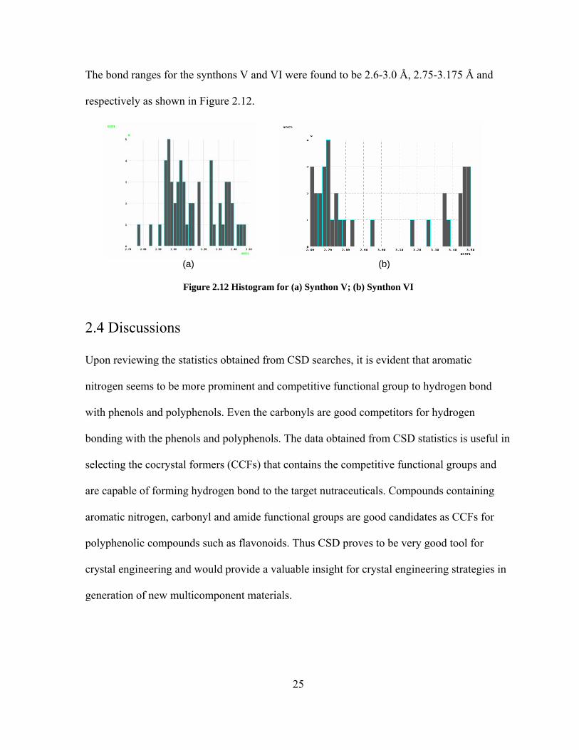
The bond ranges for the synthons V and VI were found to be 2.6-3.0 Å, 2.75-3.175 Å and
respectively as shown in Figure 2.12.
(a) (b)
Figure 2.12 Histogram for (a) Synthon V; (b) Synthon VI
2.4 Discussions
Upon reviewing the statistics obtained from CSD searches, it is evident that aromatic
nitrogen seems to be more prominent and competitive functional group to hydrogen bond
with phenols and polyphenols. Even the carbonyls are good competitors for hydrogen
bonding with the phenols and polyphenols. The data obtained from CSD statistics is useful in
selecting the cocrystal formers (CCFs) that contains the competitive functional groups and
are capable of forming hydrogen bond to the target nutraceuticals. Compounds containing
aromatic nitrogen, carbonyl and amide functional groups are good candidates as CCFs for
polyphenolic compounds such as flavonoids. Thus CSD proves to be very good tool for
crystal engineering and would provide a valuable insight for crystal engineering strategies in
generation of new multicomponent materials.
25

3. QUERCETIN
Quercetin (3, 3’4, 4’, 5-7-pentahydroxyflavone) is a bio flavonoid which is widely
distributed in the plant kingdom.99-102 It is the most abundant of all the flavonoid
molecules found in many often consumed foods including apples, citrus fruits, onions,
tea, berries and vegetables, as well as many seeds, nuts, flowers, barks and leaves.103 It is
also present in medicinal botanicals like Ginkgo biloba, Hypericum perforatum,
Sambuscus Canadensis and many others.104
Figure 3.1 Structure of Quercetin 3.1 Description Quercetin (Mol Wt = 302.2) is a bioflavonoid (pigment), found in almost all herbs,
fruits, and vegetables. Bioflavonoids provide the body with anti-inflammatory and
antioxidant protection, and quercetin is one of the most powerful and effective herbal
anti-inflammatory, and antioxidant supplements in the market today.105, 106 Quercetin has
been shown to prevent the development of a variety of conditions related to inflammation
by direct inhibition of several initial processes of inflammation and free-radical damage,
including arthritis, allergies, macular degeneration, heart disease and various forms of
26
cancer.107 Quercetin also shows anti-tumor properties.108 The estimated average daily
dietary consumption of quercetin is about 30 mg/day. However, the manufacturer’s
recommended daily dose of over-the-counter quercetin supplements ranges from 400–
1200 mg/day.109 There is no recommended standard dose for quercetin but there have
been some occasional reports of nausea when taken as high doses in supplements.106
Quercetin exhibits poor solubility in water, it is practically insoluble in water but it is
soluble in 100% sodium hydroxide and methanol, but in its glycoside form (with a sugar
attached), which is the common form of its occurrence in fruits and vegetables, is more
soluble in the body.110 Some of the glycoside forms of quercetin are rutin and quercitrin.
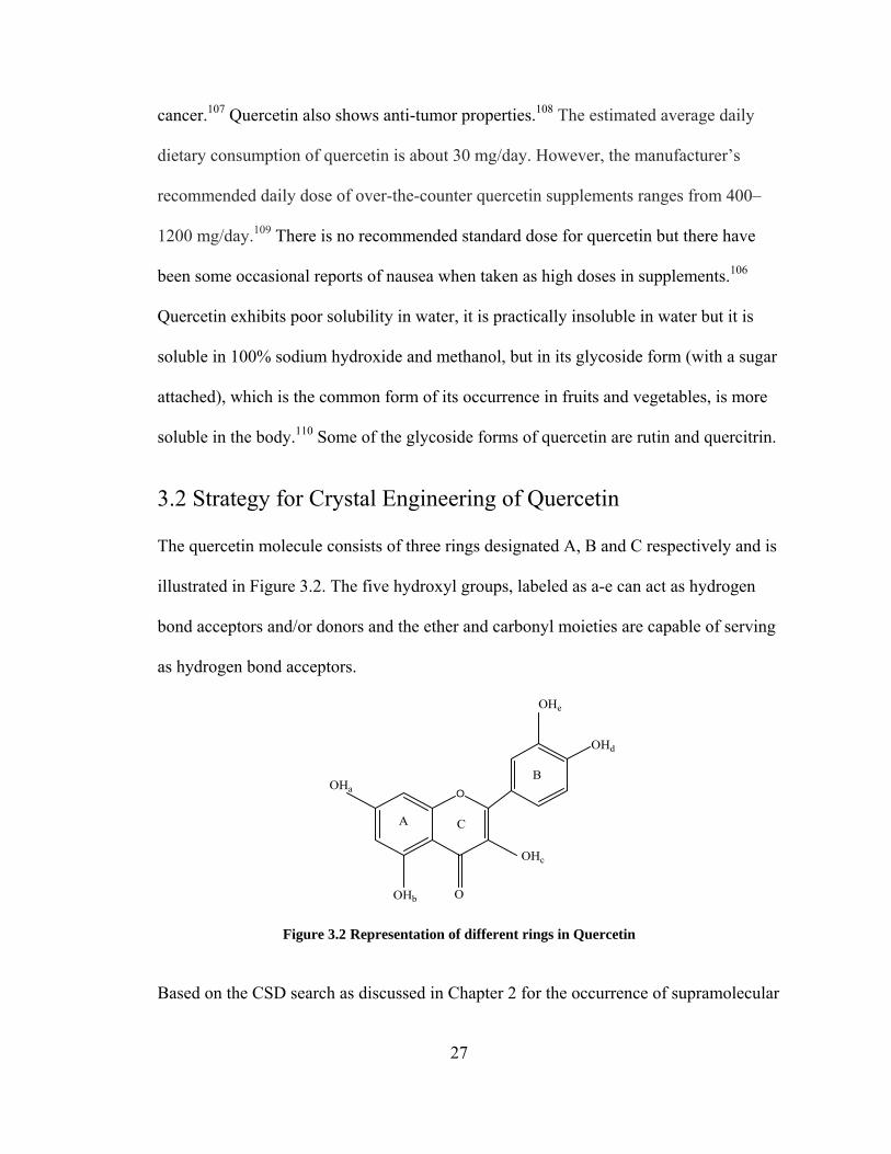
3.2 Strategy for Crystal Engineering of Quercetin
The quercetin molecule consists of three rings designated A, B and C respectively and is
illustrated in Figure 3.2. The five hydroxyl groups, labeled as a-e can act as hydrogen
bond acceptors and/or donors and the ether and carbonyl moieties are capable of serving
as hydrogen bond acceptors.
O
OHd
OHe
OHb
OHa
O
B
CA
OHc
Figure 3.2 Representation of different rings in Quercetin
Based on the CSD search as discussed in Chapter 2 for the occurrence of supramolecular
27

synthons between polyphenols and other functionalities, aromatic nitrogen, carbonyls and
amides look more promising competitors for hydrogen bonding. To date, the crystal
structure of pure quercetin is not known but there are two entries for quercetin dihydrate
with ref codes FEFBEX111 and FEFBEX01112 and a formamide solvate (EVIJUO) 113 and
other hydrates (LORKEI) 114 in the CSD. Figure 3.3 represents the packing in the
quercetin dihydrate. The hydrogen bonding for the dihydrates of quercetin with the ref
codes FEFBEX and FEFBEX01 are same but the R factor for FEFBEX01 (Rfac 4.8) is
better than FEFBEX (Rfac 9.5).
Figure 3.3 Hydrogen bonding in Quercetin dihydrate
In the dihydrate form quercetin crystallizes with two water molecules that participate in
extended hydrogen bonding network through the crystal lattice. In the crystal structure
quercetin exists as a dimer which is formed by the OHb and the adjacent carbonyl moiety
and these are also involved in the intramolecular hydrogen bonding in the dimer (circled
in Fig 3.3). All the hydroxyl and carbonyl groups and the water molecules participate in
hydrogen bonding. All the three rings of quercetin molecule in the crystal lattice are not
28

planar. The rings A and C lie in the same plane whereas the ring B is not. The dihedral
angle between the plane containing rings A and C and the plane containing ring B is
8.10o as shown in Figure 3.4. The overall hydrogen bonding leads to the stacking of
infinite 2 D sheets.
Figure 3.4 Illustration of dihedral angle between the planes. (Red plane contains rings A, C and the blue plane contains ring B)
3.3 Solvates of Quercetin
Three solvates of quercetin with pyridine, tetrahydrofuran (THF) and acetone namely I,
II, III, were obtained by different methods. All the chemicals used during the preparation
were purchased from Sigma Aldrich and used as received.
Form I and II Quercetin-Pyridine solvates
Form III Quercetin-THF solvate
Form IV Quercetin-Acetone solvate
Table 3.1 Solvates of Quercetin
3.3.1 Form I and Form II (Pyridine solvates of Quercetin)
Synthesis: 34 mg of quercetin dihydrate (purchased from Sigma Aldrich and used as
received) was dissolved in 5ml of Pyridine; this solution was layered with 4 mL of water
29

and refrigerated. After 4 hours golden yellow crystals were produced (yield- 25mg). The
crystallization experiment was conducted in an unmodified atmosphere and the solvents
were dried by standard methods prior to use. mp-314 °C.
Figure 3.5 Single crystal of quercetin-pyridine solvate
The attempts to get pure crystals of quercetin using pyridine solvate resulted in two
different forms of quercetin-pyridine solvate namely Form I and Form II. The Form I in
due course of time transforms to Form II. The crystal structure of Form I reveals that two
molecules of pyridine hydrogen bond to each molecule of quercetin. The quercetin
molecules don’t exists as a dimer as found in its dihydrate form (FEFBEX01). But the
quercetin molecules forms a cyclic pattern, described by Etter’s115 graph set R22 (8). The
cyclic pattern includes heterosynthons such as OH…CO (D = 2.724 Å) and OH…OH (D =
2.914 Å). Where as the crystal structure Form II reveals that quercetin molecules exists as
dimer as found in quercetin dihydrate (FEFBEX01). But the only difference being that in
FEFBEX01 the dimer includes OHb and the carbonyl moiety whereas in Form II the
dimer includes OHc and the carbonyl moiety (D = 2.749 Å) and the OHb is just involved
in the intramolecular hydrogen bonding (D = 2.608(3) Ao). Figure 3.6 represents the
hydrogen bonding in Form I and Form II. The R22 (8) graph set in Form I and the
quercetin dimer in Form II are circled in the figure.
30

(a) (b)
Figure 3.6 (a) Hydrogen bonding in Form I (b) Hydrogen bonding in Form II
The dihedral angle between the planes containing the rings A, C and ring B for form I
and Form II are 21.11o and 23.71o respectively. These angles are higher than that is found
in FEFBEX01 (8.10o). This indicates that quercetin molecules in Form I and II are not
that planar as in FEFBEX01 (quercetin dihydrate).
(a) (b)
Figure 3.7 The dihedral angles in From I (a) and Form II (b) The presence of quercetin-pyridine solvate in two different forms indicates that quercetin
exhibits polymorphism.65 The CSD search has no entries for the crystal structure of
polymorphic quercetin but the literature searches reveal that quercetin exhibits
polymorphism.116-119 The Form II could be most stable than Form I because according to
Ostwald’s step rule134, 135 which states: “In general it is not the most stable but the least
stable polymorph that crystallizes first”. In both the forms the pyridine molecules
hydrogen bond to OHa and OHe of quercetin molecules. The hydrogen bond distances for
the OHa…Narom and OHe
…Narom in From I are 2.705 Å and 2.677 Å respectively whereas
31

for Form II it is 2.765 Å and 2.64 Å. The distances are in accordance with the distance
ranges found in CSD as discussed in Chapter 2. Overall supramolecular hydrogen
bonding in Form I results in 2D tape and in Form II it results in a 2D sheet. Desolvation
occurs when the crystals are heated at 150oC for 10 min and they become amorphous.
The amorphous powder could be pure quercetin as it decomposes at 316oC which
coincides with the literature value.120
3.3.2 Form III (THF solvate of Quercetin) Synthesis: 34 mg of quercetin dihydrate (purchased from Sigma Aldrich and used as
received) was dissolved in 5ml of THF; this solution was layered by 4 mL of hexane and
refrigerated. After 4 days golden yellow crystals in the form of plates were produced
(yield-20.5mg). The crystallization experiment was conducted in an unmodified
atmosphere and the solvents were dried by standard methods prior to use. mp 316 °C.
Figure 3.8 Single crystal of quercetin-THF solvate (Form III)
The crystal structure reveals that there are three THF molecules in the asymmetric unit.
Two of the THF molecules are involved in the formation of supramolecular network with
the quercetin molecules and the third distorted THF molecule just act as a guest. All the
hydroxyl and the carbonyl groups of quercetin and the THF molecules are involved in
hydrogen bonding. In Form III also quercetin exists as a dimer (circle in Fig 3.9) with a
32

bond distance of 2.749(3) Ao similar to that found in Form II (quercetin-pyridine solvate)
with hydrogen bond distance of 2.749 Å (OH…CO). Figure 3.9 compares the hydrogen
bonding in Form II and III. The dimer in Form III can also be described as R22 (10) graph
set.115
Figure 3.9 Comparison of hydrogen bonding in Form III (left) with that of Form II (right)
Desolvation of Form III crystals occurs after 15-20 minutes when they are taken out from
the mother liquor and leaves the system amorphous and the decomposition temperature of
the powder coincides with that of quercetin.120
3.3.3 Form III (Quercetin-Acetone solvate)
Synthesis: 34 mg of quercetin dihydrate and 13.3 mg of caprolactam were dissolved in
5ml of acetone and the refrigerated for 3 days. Golden yellow crystals of solvate III were
obtained wherein quercetin and acetone were in 1:1 stoichiometric ratio (yield-20mg). All
the chemicals used during the preparation were purchased from Sigma Aldrich and used
as received. The crystallization experiment was conducted in an unmodified atmosphere
and the solvents were dried by standard methods prior to use. mp 316oC.
33

Figure 3.10 Crystals of Quercetin-Acetone solvate (Form III)
The crystals of solvate III were obtained during an attempt to prepare the cocrystal of
quercetin and caprolactam by using acetone as a solvent. In Form III also quercetin exists
as a dimer, but this dimer is formed by OHd and OHe of quercetin molecule form strong
with hydrogen bond distance of 2.754 Å which falls well with in the range for alcohol-
alcohol supramolecular homosynthon as discussed in Chapter 2. Figure 3.11 represents
the dimer that is formed in Form III. All hydroxyls and the carbonyl groups of quercetin
participate in the hydrogen bonding. Further, OHe of both quercetin molecule hydrogen
bonds with OHd of adjacent quercetin molecule with OH…OH interaction (D = 2.765 Å)
and the OHd hydrogen bonds to the carbonyl group of another adjacent quercetin
molecule and forms a strong OH…O hydrogen bond (D = 2.682 Å).
Figure 3.11 Hydrogen bonding in Form III (Acetone molecules colored red)
34

The OHa of each quercetin molecule is bifurcated in hydrogen bonding with acetone
molecule forming OH…O hydrogen bond (D = 2.596 Å) and OH…OH supramolecular
homosynthon with adjacent OHa quercetin molecules (D = 2.750 Å). All the hydrogen
bond distances mentioned above fall well with in the range of distances found in CSD as
discussed in Chapter 2. The overall hydrogen bonding results in a crisscross grid network
as represented in Figure 3.12.
Figure 3.12 Representation of crisscross grid network in Quercetin-Acetone solvate (Space fill model)
3.3.4 Discussions
The attempts to obtain the pure crystals of quercetin from quercetin dihydrate resulted in
the formation of new forms namely, Form I, Form II, Form III and Form IV. The Form I
and Form II are the polymorphs of quercetin whereas, the Form III and IV are the
solvates of THF and acetone respectively. The quercetin molecule exists as dimer in
Form II and III similar to that of quercetin dihydrate (FEFBEX01)90 where as such kind
of dimer is absent in Form I. Hydrogen bonding in Form I and II results in the formation
of linear tapes where as the Form III results in cross grid network. Desolvation occurs for
Form II after 5-10 minutes when the crystals are taken out of the mother liquor and an
amorphous powder is obtained where as, the Form I and III are pretty stable in absence of
mother liquor. In order to get rid of the solvent molecules in Form I and III, the crystals
were heated at 150 and 55 oC respectively. The amorphous powders from all the three
35

forms were analyzed by DSC and melt temp separately and it was found that in all the
three cases the powder decomposes at 328 oC, that exactly coincides with the
decomposition temperature of quercetin. The amorphous form obtained could be pure
quercetin, but this could not be confirmed as the crystals of pure quercetin were not
produced by other means.
3.4 Cocrystals of Quercetin
Crystal engineering of quercetin lead to the generation of two novel cocrystals namely
Cocrystal I and II with caffeine and isonicotinamide were obtained by slow evaporation.
The functional groups that are more likely to undergo hydrogen boning in quercetin are
the hydroxyls and the carbonyl. These functional groups were utilized to target the
various functional groups in caffeine and isonicotinamide such as the nitrogen in the
imidazole ring and the imide groups in caffeine and the aromatic nitrogen and the amide
groups in isonicotinamide. The CSD searches (Chapter 2) reveal that the polyphenols are
more likely to form supramolecular synthons with functional groups like carbonyl and
aromatic nitrogen. The percentages obtained for OH…Narom and OH…CO supramolecular
heterosynthons in the CSD searches are 84.8% and 74.4% respectively.
3.4.1 Quercetin- Caffeine- Methanol cocrystal solvate
36
Figure 3.13 The asymmetric unit of 1:1:1 cocrystal solvate of quercetin, caffeine and methanol

37
Synthesis: 68 mg of quercetin dihydrate and 38 mg of caffeine were dissolved in
approximately 5 mL of methanol and heated until a clear solution was obtained. Slow
evaporation of this solution in refrigerator resulted in 1:1 crystals after 3 days (yield- 30
mg). All the chemicals used during the preparation were purchased from Sigma Aldrich
and used as received. All crystallization experiments were conducted in an unmodified
atmosphere and the solvents were dried by standard methods prior to use. mp: 246°C.
Figure 3.14 Single crystal of quercetin-caffeine-methanol cocrystal solvate (Cocrystal I)
he crystal structure reveals that the imide group and the aromatic nitrogen of caffeine
are the functional group that interacts with the hydroxyl groups of quercetin. Caffeine
molecules interact with quercetin molecules via the formation of OHc Narom and
OHa CO supramolecular heterosynthons. The former supramolecular heterosynthon is
formed by the interaction of OHc quercetin and the aromatic nitrogen of the imidazole
ring in caffeine (D = 2.821(3) A ) and the latter results due to the hydrogen bonding
between OHa of quercetin and the CO moiety of the imide group of caffeine (D= 2.716(3)
A ). The carbonyl in the caffeine molecule hydrogen bonds to the methanol molecule
(D=2.712(3) A ). All the above distances are in accordance with the OH O synthon and
OHc Narom distance ranges found in CSD as discussed in Chapter 2.
T
…
…
o
o
o …
…

Figure 3.15 Intermolecular interactions in the 1:1:1 cocrystal solvate of quercetin, caffeine and methanol (colored green)
Adjacent quercetin molecules interact by utilizing some of the remaining hydrogen
bonding sites as follows: trifurcation of the OHb moiety of quercetin molecules through
supramolecular homosynthon and OH…CO supramolecular heterosynthon. The OH…OH
supramolecular homosynthon is formed between the OHb of quercetin molecules and
OHd and OHe of adjacent quercetin molecules (D = 2.774(3) Ao, 2.847(3) Ao
respectively). OHb is also engaged in OH…CO intramolecular heterosynthon (, D =
2.602(3) Ao). The bond distances fall in the range found in the CSD searches for the
respective synthons as discussed in Chapter 2. These hydrogen bond interactions afford a
supramolecular sheet that stacks up. These sheets are further connected via the
interaction of methanol molecules with OHe moieties of quercetin molecules through
OH…OH supramolecular homosynthons (D = 2.633(3) Ao). The overall effect of the
hydrogen bonding results in the formation of a network where the methanol could be
described as a guest although as shown in Figure 3.16.
38

Figure 3.16 Illustration of sheets generated in the cocrystal 1.
3.4.2 Quercetin-Isonicotinamide
Figure 3.17 The asymmetric unit of the1:1 quercetin – isonicotinamide cocrystal
Synthesis: 67.6 mg of Quercetin dihydrate and 24.6 mg of isonicotinamide were
dissolved in approximately 5 mL of methanol and heated until a clear solution was
obtained. Slow evaporation of this solution in refrigerator resulted in 1:1 crystals after 2
days (yield-31.5mg). All the chemicals used during the preparation were purchased from
Sigma Aldrich and used as received. The crystallization experiment was conducted in an
unmodified atmosphere and the solvents were dried by standard methods prior to use.
mp: 256-260°C.
Figure 3.18 Single crystal of quercetin-isonicotinamide cocrystal (Cocrystal 2)
39

The aromatic nitrogen and the carbonyl moiety of the amide group and the carbonyl
group in quercetin acts as hydrogen bond acceptors where as all the hydroxyl groups of
quercetin can act as hydrogen bond acceptors/donors. Isonicotinamide molecules interact
with quercetin molecules via, CO…OH and NH…OH supramolecular heterosynthons and
OH…OH supramolecular homosynthons. One of the two hydrogen atoms in the amino
moiety of the isonicotinamide hydrogen bonds to the carbonyl group of adjacent
quercetin molecules and the other hydrogen atom interacts with OHd of a different
quercetin molecule giving rise to NH…CO (D = 3.018(4) Ao) and NH…OH (D = 3.028(4)
Ao) supramolecular heterosynthons, respectively. These distances are in accordance with
the distance ranges found in the CSD search for amides in Chapter 2. The carbonyl of the
amide moiety hydrogen bonds to OHe quercetin molecules whereas the Narom atom of the
isonicotinamide molecule interacts with OHa of quercetin molecules and thereby
generates CO…OH (D = 2.607(3) Ao) and Narom…OH (D = 2.684(3) Ao) supramolecular
heterosynthons respectively. The distances fall within the range found in CSD as
discussed in Chapter 2.
Figure 3.19 Intermolecular interactions in the 1:1 Quercetin-Isonicotinamide cocrystal.
40
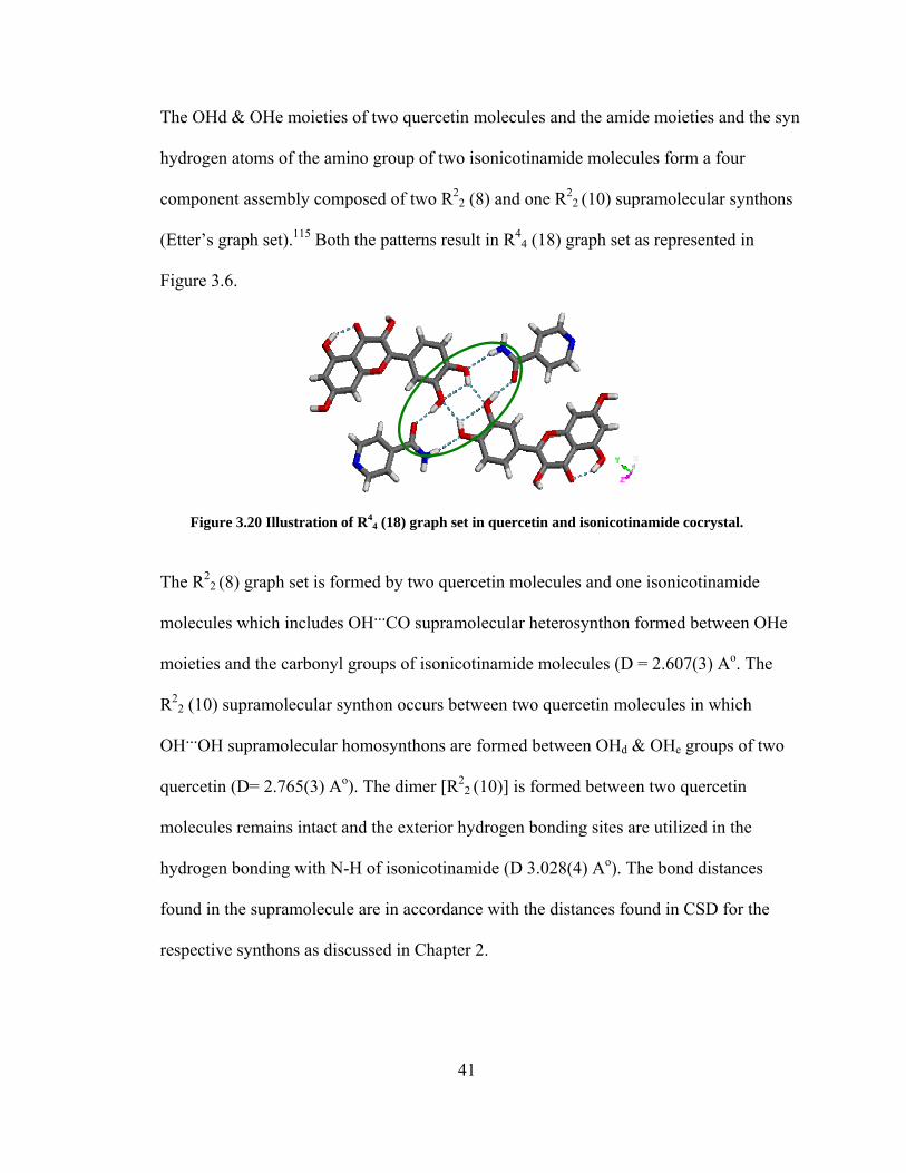
The OHd & OHe moieties of two quercetin molecules and the amide moieties and the syn
hydrogen atoms of the amino group of two isonicotinamide molecules form a four
component assembly composed of two R22 (8) and one R2
2 (10) supramolecular synthons
(Etter’s graph set).115 Both the patterns result in R44 (18) graph set as represented in
Figure 3.6.
Figure 3.20 Illustration of R44 (18) graph set in quercetin and isonicotinamide cocrystal.
The R22 (8) graph set is formed by two quercetin molecules and one isonicotinamide
molecules which includes OH…CO supramolecular heterosynthon formed between OHe
moieties and the carbonyl groups of isonicotinamide molecules (D = 2.607(3) Ao. The
R22 (10) supramolecular synthon occurs between two quercetin molecules in which
OH…OH supramolecular homosynthons are formed between OHd & OHe groups of two
quercetin (D= 2.765(3) Ao). The dimer [R22 (10)] is formed between two quercetin
molecules remains intact and the exterior hydrogen bonding sites are utilized in the
hydrogen bonding with N-H of isonicotinamide (D 3.028(4) Ao). The bond distances
found in the supramolecule are in accordance with the distances found in CSD for the
respective synthons as discussed in Chapter 2.
41

42
3.4.4 Discussions
Crystal engineering has lead to the generation novel cocrystals of quercetin and thus
confirms that flavonoids are capable of generating novel cocrystals with pharmaceutically
acceptable molecules such as caffeine and isonicotinamide. As caffeine and
isonicotinamide have no solubility problems whereas caffeine has problem with physical
stability against hydration. The CSD reveals that there are 17 entries for the cocrystals of
caffeine. There are also evidences which indicate that cocrystallization of caffeine with
suitable cocrystal formers (CCF) overcome the hydration problem.121 Even
isonicotinamide 122 seems to be a good CCF as there are cocrystals of it in the literature.
The CSD statistics as discussed in Chapter 2 reveals that aromatic nitrogen and carbonyl
functional groups are good acceptors to hydrogen bond with the hydroxyls of
polyphenolic compounds. Therefore caffeine and isonicotinamide (isomer of
nicotinamide, known as niacin) 123 stand as suitable CCFs for cocrystallization and are
pharmaceutically acceptable molecules. Thus crystal engineering allows one to choose
complementary components for polyphenolic compounds for designing supramolecules.

4. HESPERETIN Hesperetin (RS-2,3-dihydro-5,7-dihydroxy-2-(3-hydroxy-4-methoxyphenyl)-4H-1-
benzopyran-4-one) is a naturally occurring flavanone which is unique among the others as it
is the only one which is faintly sweet while most of the flavonoids are tasteless or bitter.124
Hesperetin is the aglycone form of hesperedin which is abundantly present in citrus fruits.
Animal studies have shown that hesperetin has very good anti-inflammatory properties.125, 111
Figure 4.1 Structure of hesperetin (hydrogen are not included) 4.1 Description Hesperetin (Mol.Wt 302.27 g/mol) is a bioflavonoid that is present in the plants in its
glycoside form (hesperedin). We have discussed earlier in Chapter 1, how glycoside forms of
flavonoids are more soluble that the aglycone form. Hesperedin is more soluble than
hesperetin because of the sugar ring attached to it.126 It is a phenolic antioxidant127,
antiallergic128, and antimutagenic129. It may scavenge the reactive oxygen species as
superoxide anions and may protect against peroxidation. Studies on rats have shown that
hesperetin helps in reducing the cholesterol by the inhibition of 3-hydroxy-3-ethylglutaryl
coenzyme A.130 In vitro studies have also shown that hesperetin has some anti cancer activity
too.131
43

4.2 Strategy for Crystal Engineering of Hesperetin Hesperetin has similar structure to that of quercetin, the difference being rings B and C. In
ring B there is a methoxy group instead of a hydroxyl and saturated ring C. Figure 4.2
represents the structure of hesperetin with its 3 rings A, B, C. The hydroxyl groups are
labeled a, b and c. The hesperetin molecule has two hydroxyl groups on ring A and one on
ring B act as hydrogen bond acceptors and/or donors. Hesperetin has also has an ether and
carbonyl (ketone) moieties on ring C that can act as hydrogen bond acceptors.
O
OCH3
OHc
OHb
OHa
O
B
CA
Figure 4.2 Illustration of rings in Hesperetin The data available in the CSD reveals that there are two crystal structures of hesperetin
discovered so far, one is the pure hesperetin (YEHROS) 132 and the other is the monohydrate
form (FOYTOC).133
(a) (b)
Figure 4.3 Hydrogen bonding in (a) Hesperetin (b) Hesperetin monohydrate Figure 4.3 represents the hydrogen bonding in both the available forms of hesperetin as a
44

racemate and monohydrate. Hesperetin molecule in YEHROS is not a planar molecule as
represented in Figure 4.4 (a).
(a) YEHROS
(b) FOYTOC
Figure 4.4 Illustration of the dihedral angles (blue plane contains rings A and C; red plane contains the ring C).
The two rings A and C lie in the same plane and the ring B lies in the other plane with a
dihedral angle of 53.59o. Whereas, in FOYTOC hesperetin adopts almost a planar
conformation while the conformation in YEHROS seems to be more favorable as there is less
repulsive steric hindrance between the rings with a dihedral angle of 2.91o as represented in
Figure 4.4 (b).133
4.2.1 Hesperetin-Isonicotinamide cocrystal (Cocrystal 3)
Figure 4.5 The asymmetric unit of hesperetin:isonicotinamide 1:1 cocrystal
45

Synthesis: 60 mg of Hesperetin and 24.6 mg were dissolved in approximately 5 mL of
ethanol and heated until a clear solution was obtained. Slow evaporation of this solution in
refrigerator resulted in 1:1 crystals after 5 days. All crystallization experiments were
conducted in an unmodified atmosphere and the solvents were dried by standard methods
prior to use. All chemicals were purchased from Aldrich and used as obtained. m.p 172-
176oC.
Figure 4.6 Single crystal of hesperetin-isonicotinamide cocrystal Crystallization of hesperetin with isonicotinamide resulted in a 1:1 cocrystal. In the
isonicotinamide molecule, the aromatic nitrogen and the carbonyl moiety of the amide group
act as hydrogen bond acceptors. The two hydrogen atoms of the amino group of the amide
moiety act as the hydrogen bond donors. The X-ray crystal structure reveals the existence of
a supramolecular homosynthon (amide dimer) formed by the interaction of two
isonicotinamide molecules. This results in a graph set notation115 R22 (8) with O---Narom
interaction as represented in Figure 4.8 with bond distance of 2.868(3) Ao that fall well with
the range found in CSD in Chapter 2. The supramolecular synthon formed in the cocrystal
includes OH---N hydrogen bond between the nitrogen atom of isonicotinamide and the OHa
of the adjacent hesperetin molecule with an O---N bond distance of 2.623(2) Ao. OHa is
further bifurcated as it forms a hydrogen bond with the anti N-H of the isonicotinamide
46

dimer with an N---O bond distance of 3.031(3) Ao. The amide dimer remains intact and as
observed in the crystal structure of pure isonicotinamide.
Figure 4.7 Overall hydrogen bonding in the Hesperetin-Isonicotinamide 1:1 cocrystal
The hesperetin molecules lie opposite to each other around the crystallographic inversion
center as represented in Figure 4.8. The adjacent hesperetin molecules interact via OH---
CO utilizing the hydroxyl moieties on ring B with a bond distance of 2.720(2) Ao. There is
also intramolecular hydrogen bonding between the carbonyl (ketone) group on ring C and
the OHc with a bond distance of 2.584(2) Ao. All the hydrogen bond distances found in
hesperetin and isonicotinamide falls well with in the expected as discussed in Chapter 2.
Figure 4.8 Illustration supramolecular synthons in the cocrystal 3. Figure 4.7 represents the how the hesperetin and isonicotinamide molecules arrange
47

themselves in the crystal lattice. The hydrogen bonding in the cocrystal 3, the hesperetin
molecules form a one dimensional tape along Z-axis and these tapes are connected through
centrosymmetric amide dimers formed by isonicotinamide molecules forming a two
dimensional sheets with cavities (31 x 10 Å2).
Figure 4.9 The cavity formed in hesperetin:isonicotinamide cocrystal.
The cavities are filled by similar sheets and its eight fold interpenetrated. The overall
hydrogen-bonding pattern is that of a zigzag 2-D sheet with interpenetration and it means that
all hydrogen bond donors and acceptors in both molecules are satisfied as shown in Figure
4.10.
Figure 4.10 Illustration of 8-fold interpenetrated network formed in Cocrystal 3
48

The hesperetin molecules maintain the angular conformation similar to what is observed in
pure hesperetin crystal structure (YEHROS). The dihedral angle observed for hesperetin
molecules in the cocrystal between the planes containing rings A, C and ring B is greater by
24.2o than found YEHROS. The hesperetin molecules are more stable in angular
conformation as the steric hindrance is reduced. Figure 4.11 represents the dihedral angle
between the planes of the rings in hesperetin molecule.
Figure 4.11 Illustration of dihedral angle in hesperetin molecules (red plane contains rings A and C; blue contains ring C)
4.2.2 Hesperetin-Nicotinic acid cocrystals
Figure 4.12 Hesperetin – Nicotinic acid 1:1 zwitterion cocrystal (Form I) Synthesis: 60 mg of Hesperetin and 24.6 mg of nicotinic acid were dissolved in
approximately 5 mL of methanol and heated until a clear solution was obtained. Slow
49

evaporation of this solution in refrigerator resulted in 1:1 crystals after 5 days. All
crystallization experiments were conducted in an unmodified atmosphere and the solvents
were dried by standard methods prior to use. All chemicals were purchased from Aldrich and
used as obtained. m.p 198-204 oC.
Figure 4.13 Single crystals of hesperetin-NA cocrystals. Crystallization of hesperetin with nicotinic acid results in two1:1 cocrystals in which the
nicotinic acid exists as a zwitterionic state, Form I contains only one of the enantiomer of
R-hesperetin (Cahn-Ingold-Prelog priority rules) and converts to Form II which contains both
forms of (+) hesperetin (Cahn-Ingold-Prelog priority rules) crystallizes with nicotinic
zwitterion. As Form I contains only one of the enantiomer of hesperetin in the crystal
structure it is called as the racemic conglomerate and Form II is the racemate of hesperetin.
During the crystallization process both the cocrystals were generated in the same vial and the
reproduction of the Form I cocrystal was not achieved. This indicates that Form I is
kinetically more favored and Form II is thermodynamically. Figure 4.13 represents the
overall hydrogen bonding in cocrystal Form I. The crystal structure of Form I reveals the
existence of nicotinic acid (NA) as a zwitterionic species in the cocrystal, which is not an
amino acid. The zwitterion of NA can be confirmed by the following structural information:
a) The C-O bond distances in the carboxylate ion were found to be 1.241 Å 7& 1.250 Å and
50

the corresponding C-N-C (protonated pyridine) bond angle equal to 121.54o. Where as, the
C-O & C=O bond distances and the C-N-C bond angle in NA are 1.217Å &1.289 Å and
117.65o respectively.
Figure 4.14 Overall hydrogen bonding in the hesperetin Form I cocrystal. (Blue color- R-Hesperetin)
Pure NA forms a linear tape where they are hydrogen bonded to each other in a head to tail
fashion with a bond distance of 2.666 Å. Similar kind of tape is also present in Form I
cocrystal with hydrogen bond distance of 2.604 Å. Figure 4. 15 represents the tapes that are
formed in NA in pure NA (a) and in Form I cocrystal (b) respectively.
(a)
(b)
Figure 4.15 Illustration of hydrogen bonds in NA: a) NA in pure form b) NA in the cocrystal, hesperetin molecules are deleted for clarity.
51
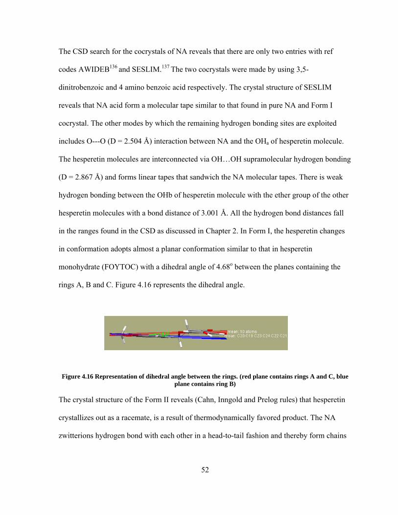
The CSD search for the cocrystals of NA reveals that there are only two entries with ref
codes AWIDEB136 and SESLIM.137 The two cocrystals were made by using 3,5-
dinitrobenzoic and 4 amino benzoic acid respectively. The crystal structure of SESLIM
reveals that NA acid form a molecular tape similar to that found in pure NA and Form I
cocrystal. The other modes by which the remaining hydrogen bonding sites are exploited
includes O---O (D = 2.504 Å) interaction between NA and the OHa of hesperetin molecule.
The hesperetin molecules are interconnected via OH…OH supramolecular hydrogen bonding
(D = 2.867 Å) and forms linear tapes that sandwich the NA molecular tapes. There is weak
hydrogen bonding between the OHb of hesperetin molecule with the ether group of the other
hesperetin molecules with a bond distance of 3.001 Å. All the hydrogen bond distances fall
in the ranges found in the CSD as discussed in Chapter 2. In Form I, the hesperetin changes
in conformation adopts almost a planar conformation similar to that in hesperetin
monohydrate (FOYTOC) with a dihedral angle of 4.68o between the planes containing the
rings A, B and C. Figure 4.16 represents the dihedral angle.
Figure 4.16 Representation of dihedral angle between the rings. (red plane contains rings A and C, blue plane contains ring B)
The crystal structure of the Form II reveals (Cahn, Inngold and Prelog rules) that hesperetin
crystallizes out as a racemate, is a result of thermodynamically favored product. The NA
zwitterions hydrogen bond with each other in a head-to-tail fashion and thereby form chains
52

that are sandwiched by chains of hesperetin molecules. The crystal is still polar as the head-
to-tail chains of NA zwitterions are parallel throughout the crystal structure. The three chains
are linked by lateral hydrogen bonds and therefore form a network that could be described as
a supramolecular tape as a represented in Figure 4.17. The molecular structure of hesperetin
as it exists in Form II reveals that one of the three hydroxyl groups engages in an
intramolecular hydrogen bond with the carbonyl moiety. All the hydrogen bonds formed in
both the forms have their distances that fall well with in the bond ranges obtained in the CSD
searches as discussed in Chapter 2.
Figure 4.17 The hydrogen bonding in the hesperetin Form II cocrystal (yellow and maroon: NA molecules, (+) hesperetin: blue, (-) hesperetin) green)
In Form II, the hesperetin maintains the almost planar conformation as found in Form I but it
is slightly greater. In Form I it is 4.69o and in Form II it is around 11.38o.
Figure 4.18 The dihedral angle formed between the planes of rings A, C and ring B in Form II
53

54
4.2.4 Discussions Crystal engineering has lead to the generation novel cocrystals of hesperetin with
pharmaceutically acceptable molecules such as isonicotinamide and nicotinic acid (NA).
For the first time crystal engineering has been applied to generate cocrystals of
Flavonoids. As we discussed earlier in Chapter 2 that compounds containing aromatic
nitrogen are more likely to form hydrogen bond with phenolic –OH groups.
Both isonicotinamide and NA have aromatic nitrogen and make them good cocrystal
formers for the cocrystallization of hesperetin. In addition to aromatic nitrogen
isonicotinamide and NA have amide and acid groups respectively which are capable of
forming hydrogen bonds with hesperetin. The CSD search reveals that there are almost
26 cocrystals of isonicotinamide. Among the 26 cocrystals three are with polyphenols
such as phloroglucinol,138 hydroquinone,138 and resorcinol.138 NA has two cocrystals
dinitrobenzoic136 acid and aminobenzoic acid.137 Thus isonicotinamide and NA seem to
be good cocrystal former (CCF) for cocrystallization.

55
5. DISSOLUTION STUDIES OF COCRYSTALS
Most of the active pharmaceutical ingredients (APIs) are administered in the solid state as
part of an approved dosage type like tablets, capsules, etc as these are the convenient and
compact format to store a drug.70 The absorption of all the forms of a drug depends on its
physical properties.139 There are examples of drugs that are commercially available that
suffer problems related to solubility, dissolution rate and absorption in the neutral form.
These entire problems thus restrict the bioavailability of drugs. In this context the
application of crystal engineering to the field of pharmaceutics provides a solution. It has
intensified the research in the pharmaceutical industries as it offers a means to alter the
solubilities of drugs by the discovery of new crystal forms there by improving the
bioavailability.140, 141 The new forms include polymorphs, solvates, salts, hydrates and
cocrystals. There are evidences where in it is proved that drugs that are commercially
available, such as fluoxitine HCl (Prozac*)86, Itraconazole (Sporanox*)75 with low
solubility show a drastic improvement in the solubilities via cocrystallization. But not all
cocrystals show improved properties, and they are not a panacea for all problems.
For example the cocrystals67 forms of Fluoxetine hydrochloride, an anti
depressant drug, with a series of acids, has shown variations in its solubility when
compared to the pure API. For fluoxetine HCl: succinic acid cocrystal an approximate 3-
fold increase in the dissolution of the API relative to fluoxetine HCl, the cocrystal of
fluoxetine HCl: benzoic acid dissolves at a rate that is approximately half of the rate for
the API alone, while the dissolution rate for fluoxetine HCl: fumaric acid cocrystal is

roughly similar to that of fluoxetine HCl. These results reveal that, by cocrystallizing an
API with different guest molecules, it is possible to increase or decrease the dissolution
rate of the API or to leave the effective dissolution rate essentially unchanged. The Figure
5.1 represents the results of dissolution studies carried out for various cocrystals of
fluoxetine HCl.
Figure 5.1 Powder dissolution profiles for (1), (2), (3), and (4) in water at 20 °C.
Another example of API that forms cocrystals is Itraconazole (Sporanox), a low
solubility drug that is available commercially in the amorphous form. The dissolution
studies of the cocrystals of Itraconazole drug with carboxylic acids show a wide range of
solubilities ranging from 4-20 fold than that of the crystalline form of API.
Figure 5.2 Dissolution profiles into 0.1 N HCl at 25 °C for Sporanox beads and the cocrystals
56

The carbamazepine (CBZ): saccharin cocrystal is also an appropriate example in this
context. CBZ, commercially available as Tegretol, is another example of low soluble
drug.142 The CZB:saccharin cocrystal has better solubility when compared to CBZ
dihydrate and pure CBZ. The cocrystal even offers more stability (cocrystal is more
stable than pure CBZ for more than 24hrs) to CBZ in the cocrystal. Figure 5.3 compares
the plasma concentration of CBZ and the cocrystal in fated beagle dogs.
Figure 5.3 Average plasma time curves of carbamazepine concentrations (±SEM) from a cross-over
experiment in fasted beagle dogs (n = 4) given oral doses of 200 mg of the active drug as Tegretol tablets and co-crystal
5.1 Dissolution experiments of quercetin and hesperetin cocrystals
Dissolution is a characterization test commonly used by the pharmaceutical industry to
guide formulation design and control product quality.143 It is also the only test that
measures the rate of in vitro drug release as a function of time, which can reflect either
reproducibility of the product manufacturing process or, in limited cases, in vivo drug
release. The common analytical methods used for quantifying drug release in dissolution
tests are: spectrophotometric, chromatographic, mass spectrometric, and
57
58
potentiometric.144 The UV/VIS spectrophotometric method has been the traditional
analytical method for dissolution testing. A compound will exhibit absorption in the UV
region if it contains one or more chromophores, such as aromatic nitro, azoxy, nitroso,
carbonyl, or azo groups. For quantitation of any given drug, the desired absorption (A)
should be 0.3–1.0. In the UV absorption spectra of flavonoid, there are generally two
main absorption bands, band I (300–400nm) and band II (240–300 nm) which are
associated with the absorption of the cinnamoyl system and the absorption of the benzoyl
moiety in the molecules respectively.145, 146 The two absorption bands are more sensitive
to the environment of pH due to the weak acidity of the phenolic hydroxyl groups of
flavonoid. In the UV absorption spectra of hesperetin in ethanol, the main absorption
peak is assigned as band II, centered in about 290 nm, and there is only a weak broad
shoulder peak in about 330 nm.145, 147 The reason for this is because the B ring of
hesperetin is not conjugated with the carbonyl group in ring C. Whereas quercetin has
two bands at 255 nm and 374 nm respectively. The band II for quercetin is broader than
band I and this is due to the conjugation of ring B with ring C.
As quercetin and hesperetin did not show any UV absorbance spectrum in water
raised a situation to look for a suitable solvent in which these compounds are soluble and
suitable for the dissolution studies. Most of the flavonoids are soluble in alcohols;
therefore a mixture of solvent system consisting of 1:1 ethanol/water (V/V%) was
adopted in order to carry out the dissolution experiments. Figure 5.4 represents UV
spectrum of quercetin and hesperetin in 1:1 ethanol/water (V/V%) solvent system.

Figure 5.4 UV spectrums of Quercetin and Hesperetin in 1:1 Ethanol/Water (V/V%)
59
tandard
For cocrystals 2, 3, 4 the dissolution were carried out in 50 % EtOH/Water (v/v) mixture. In
order to avoid the interference from methanol in the quercetin:caffeine:methanol cocrystal
solvate, methanol/water solvent system was employed. A 3:2 methanol/water (V/V%)
solvent system was used for the dissolution of cocrystal 1. The dissolution study for
cocrystal 1 was carried at 400nm as caffeine did not absorb at this wavelength and did not
interfere with the determination of concentration of quercetin. In the similar way the
wavelengths for the dissolution of cocrystals 2, 3, 4 (cocrystal 2 = 360nm, cocrystal 3 & 4 =
300nm) were selected in such way that the interference of the cocrystal formers was not
observed while determining the concentration of respective flavonoids. The standard
calibration curves all the cocrystals were plotted by preparing the standard solutions of the
cocrystals in their respective solvent systems. For the bulk powder dissolution of cocrystals
1-4 the powder was sieved using ASTM s sieve in order to get the particle sizes

60
was
ranging between 53 - 75 µm. All the experiments were carried at room temperature in 3 sets
for each cocrystal in order to have reliability. Approximately 900mg of the sample was added
to the beakers containing 80mL of solvent mixture and the resulting slurry was stirred at 200
rpm with the help of spinning vane on a stir plate. At regular intervals of time an aliquot
drawn out from the beaker and it was filtered using a 0.25 µm nylon filter. Appropriate
amount of the resulting solution was diluted with the solvent mixture and then the UV
absorption was recorded.
5.2 Dissolution study of Quercetin:Caffeine cocrystal (Cocrystal 1) The dissolution studies of cocrystal 1 (solvated and desolvated forms) in water did not show
any improvements in the solubility of quercetin. Therefore, the dissolutions of methanol
solvate of quercetin-caffeine cocrystal (KP05) and desolvated cocrystal were carried out in
3:2 methanol/water (V/ V %) solvent system. The Figure 5.4 represents the comparison of
the dissolution of quercetin dihydrate with the methanol solvate and desolvated cocrystals. It
is evident from the Fig 5.5 that there is an approximate 6-fold improvement in the solubility
of quercetin in the methanol solvate cocrystal and 21-fold improvement in the desolvated
cocrystal in 3:2 methanol/water (V/ V %) solvent system when compared to quercetin
dihydrate. The solubility of quercetin in the desolvated cocrystal (amorphous form) has much
better solubility than the solvated cocrystal in the first hour. But, later (after 1 hour) the
desolvated cocrystal converts to the solvated cocrystal as the solubility goes down and
coincides with the solvated cocrystal. The dissolution experiment demonstrates that the pure
cocrystal has better solubility than the solvated cocrystal. Thus, cocrystallization can tailor
the solubility of a compound and could afford to improve the bioavailability.

0
1
2
3
4
5
6
7
8
0 50 100 150 200 250
Time (Minutes)
Con
cent
ratio
n (m
g/m
L)Quercetin dihydrate
KP05
Desolvated cocrysta
Figure 5.5 Comparison dissolution profiles of quercetin, methanol solvated cocrystal and the desolvated cocrystal in 3:2 MeOH/Water (V/V%) solvent system
5.2 Dissolution study of Quercetin-Isonicotinamide (Cocrystal 2) The dissolution quercetin-isonicotinamide cocrystal (KP10) was carried out in 1:1
ethanol/water (V/V%) solvent system as there was no improvement in the solubility of
quercetin. It is clear from the Figure 5.5 that the solubility of quercetin in the cocrystal
reaches the maximum value within the first five minutes and then the solubility goes down
and gets almost close to that of quercetin dihydrate. The solubility of pure quercetin through
out the experiment is almost constant this indicates that the solubility of pure quercetin
reaches the saturation limit within five minutes. The dissolution of quercetin cocrystal with
isonicotinamide reveals that the solubility of quercetin has been improved 6-folds in 1:1
ethanol/water (V/V%) solvent system.
61

0
0.2
0.4
0.6
0.8
1
1.2
1.4
0 50 100 150 200 250
Time (minutes)
Con
cent
ratio
n (m
g/m
L)
Quercetin dihydrateKP10
Figure 5.6 Dissolution profiles of quercetin and quercetin-isonicotinamide (1:1) cocrystal in 1:1
EtOH/Water (V/V%) solvent mixture
5.3 Dissolution study of Hesperetin-Isonicotinamide (Cocrystal 3) The dissolution of cocrystal 3 (KP15) was also carried out in 1:1 ethanol/water (V/V%)
solvent system as it did not showed any improvement the solubility in water. The solubility
of pure hesperetin remains constant through out the time. But the solubility of hesperetin in
the cocrystal increases slowly reaches a maximum value after 40 min and later it deteriorates
down, if extrapolated probably could reach to the saturated limit similar to that pure
hesperetin. Figure 5.6 represents the comparison of the concentrations of pure hesperetin and
hesperetin in the cocrystal plotted against time. This indicates that the solubility of hesperetin
has been improved 10-12 fold via cocrystallization in 1:1 ethanol/water (V/V%) solvent
system.
62

0
1
2
3
4
5
6
7
8
9
0 50 100 150 200 250
Time (Minutes)
Conc
entra
tion
(mg/
mL)
Hesperetin
KP15
Figure 5.7 Dissolution profiles of hesperetin and hesperetin-isonicotinamide (1:1) cocrystal in50 % EtOH/Water (v/v) solvent system
5.4 Dissolution study of Hesperetin-NA (Cocrystal 4) The dissolution experiment of cocrystal 4 (KP22) reveals that the solubility of hesperetin in
cocrystal increases tremendously from zero to 3.5 mg/mL of 1:1 ethanol/water (V/V%)
solvent system within five minutes of the experiment. In the due course of time the solubility
of hesperetin (cocrystal) decreases reaches around 2 mg/mL after 3 hrs was carried out in 1:1
ethanol/water (V/V%) solvent system. The Figure 5.7 represents the comparison of the
dissolution of hesperetin with its cocrystals with isonicotinamide and nicotinic acid. The NA
cocrystal shows an approximate 5-fold increase in the solubility of hesperetin.
63

0
1
2
3
4
5
6
7
8
9
0 50 100 150 200 250
Time (Minutes)
Con
cent
ratio
n (m
g/m
L)
KP22
Hesperetin
KP15
Figure 5.8 Comparison of dissolution profiles of hesperetin with its cocrystals 5.5 Discussions The application of crystal engineering to the field of nutraceuticals has been demonstrated by
synthesizing the cocrystals with pharmaceutically acceptable molecules. Incorporating
pharmaceutically acceptable molecules with the desired target nutraceuticals provides an
opportunity to tailor the solubilities and thereby generating more chances to improve the
bioavailability. Crystal engineering plays a vital role in setting up the strategies for selecting
appropriate nutraceutical:guest combinations that could form complementary hydrogen
bonds and producing the novel cocrystals.
In order to check whether the cocrystallization has altered the solubility of
hesperetin and quercetin it was important do the dissolution experiments for the pure
flavonoids as well as the cocrystals. But owing to the poor solubility of hesperetin and
quercetin in water has forced to adopt a different solvent for the dissolution of the cocrystals.
64

65
As most of the flavonoids show good solubility in alcohols, 1:1 ethanol-water solvent system
was adopted. It is evident from the dissolution experiments that all the cocrystals of
hesperetin and quercetin showed an improved solubility. Thus confirming the fact that
cocrystallization offers a potential means to tailor the physical properties of the target
molecules in the solid state. As the solubility and bioavailability are interdependent,
cocrystallization could be a better alternative to tailor the solubility, in return the
bioavailability.

66
6. CONCLUSIONS AND FUTURE DIRECTIONS The study of crystal engineering remained as an understudied subject until Schmidt’s work
on topochemical reactions15 popularized it. Later it expanded to larger areas like
Supramolecular Chemistry.16 In the field of pharmaceutics crystal engineering has excelled
greatly as it offers a wide range of application to the drug discovery. It has lead to the
discovery of new forms of drugs as solvates, hydrates, polymorphs58 and cocrystals54 which
allows handling problems pertaining to the physiochemical properties of drugs such as
hygroscopicity, solubility and bioavailability.
In case most of the APIs and nutraceuticals these properties can be tailored
by cocrystallizing pharmaceutically acceptable molecules. In this regard, crystal engineering
proves to be an efficient tool. It directs the one to select appropriate cocrystal formers to
generate cocrystals by exploring the non-covalent interactions such as hydrogen bonding.
The flavonoids have many benefits to the human health; the problem lies with
their solubility and bioavailability that limits their usage.2 Therefore, flavonoids have been
selected for the research work. The results herein demonstrate the wide range application of
crystal engineering to generate new forms of quercetin and hesperetin. The utilization of
crystal engineering to synthesize cocrystals has been demonstrated using the cocrystals of
quercetin and hesperetin with pharmaceutically acceptable molecules like caffeine, nicotinic
acid and isonicotinamide. The work also developed new solvent systems for dissolutions
where, the dissolutions studies in water are not possible. The application of such solvent

67
systems may not be allowed pharmaceutically but could act as templates for other cocrystals
as the results indicate increase in the solubility of the quercetin and hesperetin via
cocrystallization. These results could be the subject of future contributions.

68
REFERNCES: 1. Brian Lockwood, Pharmaceutical Press: London, UK, 2007. 2. Middleton, E.; Kandaswami, C. Biochem Pharmacol. 1992; 43(6):1167-1179 1992 3. Groot, H. Rauen, U. Fundamentals of Clinical Pharmacology, 1998; 12:249–55. 4. Jitka, Psotova1.; Iarka, ChlopCikova1.; Petra, Miketova1.; Jan, Hrbac.; Vilim, Íimanek. Phytother. Res. 2004; 18, 516–521. 5. Divi, R. L.; Chang, H. C.; Doerge, Dr. Biochem Pharmacol. 1997; 54(10):1087- 1096 1997. 6. Gil, M. I.; Ferreres, F.; Tomas, Barberan. F. A. J Agric Food Chem. 1999; 47(6): 2213-7 7. Groff, J. L.; Gropper, S. S.; Hunt, S. M. Advanced Nutrition and Human Metabolism. West Publishing Company, New York, 1995. 8. Ioku, K.; Aoyama, Y.; Tokuno, A. et al. J Nutr Sci Vitaminol (Tokyo) 2001; 41(7): 78-83 2001. 9. Ververidis, Filippos.; Trantas, Emmanouil.; Douglas, Carl.; Vollmer, Guenter.; Kretzschmar, Georg.; Panopoulos, Nickolas. Jou. Biotech2007. 2:10, 1235.
10. Adebamowo, C. A.; Cho, E.; Sampson, L. et al, Int J Cancer. 2005; 114:628-633.
11. Formica, J. V.; Regelson, W.; Food Chem Toxicol, 1995; 33:1061–80.
12. Huxley1, C. C.; Neil, Haw.. Eur. J. Cli Nutr. 2003; 57, 904–908 13. Knekt, P.; Jarvinen, R.; Reunanen, A.; Maatela, J. BMJ. 1996; 312:478–81.
14. Corbett, J. R, Academic Press, 1974.
15. Matsumura, F. Plenum Press, New York, 1985.
16. O'Brien, R. D. Academic Press, 1967.
17. Shimkim, M. B.; Anderson, N. N. Acute toxicities of rotenone and mixed pyrethrins in mammals. Proc. Soc. Exp. Biol. Med. 1936; 34: 135-138..
69
18. Harbone, J. B.; Boner, J.; Verner, E. V. Flavonoid Pigments in Plant Biochemistry, 1965; 618. 19. Jane, Higdon. Linus Pauling Institute, Oregon State University. 2005. 20. Rice-Evans, C. A.; Miller, N. J.; Paganga, G. Biol Med. 1996; 20:933–56. 21. Korkina, L.; G.; Afanasev, I. B. Adv Pharmacol. 1997; 38:151–63. 22. Robert, J. Nijveldt.; Els van, Nood.; Danny, E. C.; van, Hoorn.; Petra, G. Boelens.; Klaske van, Norren.; Paul, A. M.; van, Leeuwen. J Clin Nutr. 2001; 74: 418–25. 23. Garavello, W.; Rossi1, M.; McLaughlin, J. K.;Bosetti1, C.; Negri1, E.; Lagiou, P.; Talamini, R.; Franceschi, S.; Parpinel, M.; Dal Maso, L.; La Vecchia1, C. Annals of Oncology. 2007; 18: 1104–1109. 24. Jack, Challem. The Nutrition Reporter, 1996. 25. Jane, Higdon.; Roderick, H. Dashwood. Linus Pauling Institute, 2005-2007. 26. Beecher, G. R. Journal of Nutr. 2003; 133 (10): 3248-3254. 27. Blagden, N.; de Matas, M.; Gavan, P. T.; York, P. Advanced drug delivery Reviws,
2007; 59, 617-630.
28. Subramanian, S.; Zaworotko, M. J. Can. J. Chem. 1995; 73, 414–424. 29. Schmidt, G. M. Pure Appl. Chem. 1971; 27, 647. 30. Lehn, J. M. Science, 1993; 260 (5115): 1762-3. 31. Gennady, V Oshovsky.; David, N.; Reinhoudt, I., Willem, Verboom. Angew Chem.
2007; 46 (14): 2366-2393. 32. Desiraju, G R. Angew. Chem, 1995; 34, 2311. 33. Desiraju, G. R. Crystal Engineering: The Design of Organic Solids; Science Publ. Co. 1989. 34. Walsh, R. D. B.; Bradner, M. W.; Fleischman, S.; Morales, L. A.; Moulton, B.;
Rodríguez-Hornedo, N.; Zaworotko, M. J. Chem. Comm. 2003; 186. 35. Leiserowitz, L. Acta Cryst. 1976; B32, 775.

70
36. Etter, M. C. J. Am. Chem. Soc. 1982; 104, 1095.
37. Leiserowitz, L.; Schmidt, G. M. J. J. Chem. Soc. 1969; 2372.
38. Leiserowitz, L.; Nader, F. Acta Cryst. 1977; 33, 2719.
39. Videnova-Adrabinska, V.; Etter, M. C. J. Chem. Cryst. 1995; 25, 823.
40. Fleischman, S. G.; Kuduva, S. S.; McMahon, J. A.; Moulton, B.; Walsh, R. D.B.; Rodríguez H N.; Zaworotko, M J. Cryst. Growth Des. 2003; 3, 909. 41. McMahon, J. A.; Bis, J. A.; Vishweshwar, P.; Shattock, T. R.; McLaughlin, O. L.; Zaworotko, M. J. Z. Kristallogr. 2005; 220, 340. 42. Vishweshwar, P.; Nangia, A.; Lynch, V. M. Cryst Eng Comm. 2003; 5, 164.
43. Papaefstathiou, G. S.; MacGillivray, L. R. Org. Lett. 2001; 3, 3835 .
44. MacGillivray, L. R.; Atwood, J. L. J. Solid State Chem. 2000; 152, 199.
45. Aakeröy, C. B.; Beatty, A. M.; Helfrich, B. A. J. Am. Chem. Soc. 2002; 24,14425.
46. Vishweshwar, P.; Nangia, A.; Lynch, V. M. J. Org. Chem. 2002; 67, 556.
47. Bhogala, B. R.; Vishweshwar, P.; Nangia, A. Cryst. Growth Des. 2002; 2, 325.
48. Bond, A. D. Chem. Comm. 2003; 250.
49. Etter, M. C, Adsmond, D. A. J. Chem. Soc. 1990; 589.
50. Ling, A. R.; Baker, J. L. J. Chem. Soc. 1893; 63, 1314. 51. Anderson, J. S. Nature. 1937; 140, 583.
52. Buck, J. S.; Ide, W. S. J. Am. Chem. Soc. 1931; 53, 2784.
53. Vanniekerk, J. N.; Saunder, D. H. Acta Crystallogr. 1948; 1, 44.
54. Hall, B.; Devlin, J. P. J. Phys. Chem. 1967; 71, 465.
55. Pekker, S.; Kováts, É.; Oszlányi, G.; Bényei, G.; Klupp, G.; Bortel, G.; Jalsovszky, I., Jakab, E.; Borondics, F.; Kamarás, K.; Bokor, M.; Kriza, G.; Tompa K.; Faigel, G. Nature Mat. 2005; 4, 764.
56. Wohler, F. Annalen. 1844; 51, 153.

71
57. Watson, J. D.; Crick, F. H. C. Nature. 1953; 171, 737.
58. Pauling, L.; Corey, R. B. Arch. Biochem. Biophys. 1956; 65, 164.
59. Hoogsteen, K. Acta Cryst. 1963; 16, 907
60. Kim, S. H.; Rich, A. J. Mol. Biol. 1969; 42, 87
61. Hoogsteen, K. Acta Crys. 1959; 12, 822.
62. O'Brien, E. J. Acta Cryst. 1967; 23, 92.
63. Schmidt, J.; Snipes, W. Int. J. Radiat. Biol. 1967; 13, 101-109.
64. Etter, M. C. J. Phys. Chem. 1991; 95, 4601-4610. 65. Desiraju, G. R. Cryst. Eng. Comm. 2003; 5, 466-467.
66. Dunitz, J. D. Cryst. Eng. Comm. 2003; 5, 506.
67. Shan, N.; Toda, F.; Jones, W. Chem. Comm. 2002; 2372.
68. Aakeroy, C. B.; Salmon, D. J, Cryst Eng Comm. 2005; 7, 439.
69. Dunitz, J. D. Cryst Eng Comm. 2003; 5, 506. 70. Ann, M. Thayer, C & EN, 2007; 17 71. Almarsson, Ö.; Zaworotko, M. J. Chem. Comm. 2004; 189. 72. Almarsson, Ö.; Bourghol, H. M.; Peterson, M.; Zaworotko M.J.; Moulton B.; Rodríguez, H. N. Int. Appl. WO2004078161, 2004. 73. Zerkowski J. A.; MacDonald, J. C.; Whitesides, G. M. 1997; Chem Mat. 74. Zerkowski, J. A.; Seto, C. T.; Wierda, D.A.; Whitesides, G. M. J Am Chem Soc,
1990; 12:9025–9026. 75. Zerkowski, J,A.; Seto, C.T.; Whitesides, G. M. J Am Chem Soc. 1992;114:5473–
5475. 76. Zerkowski, J. A.; Whitesides, G. M. J Am Chem Soc. 1994; 116:4298–4304.
77. Zerkowski, J.A.; MacDonald, J. C.; Seto, C. T.; Wierda, D. A.; Whitesides, G.M. J
Am Chem Soc. 1994; 116:2382–2391.

72
78. Zerkowski,J.A.; Mathias, J. P.; Whitesides, G. J Am Chem Soc. 1994; 116:4305– 4315. 79. Vishweshwar, P.; McMahon, J. A.; Oliveira, M.; Peterson, M. L.; Zaworotko, M. J. J. Am. Chem. Soc. 2005; 127, 16802. 80. Walsh, R. D. B.; Bradner, M. W.; Fleischman, S.; Morales, L. A.; Moulton, B.; Rodríguez-Hornedo, N.; Zaworotko, M. J. Chem. Comm. 2003; 186. 81. Fleischman, S. G.; Kuduva, S. S.; McMahon, J. A.; Moulton, B.; Walsh, R. D. B.; Rodríguez-Hornedo, N.; Zaworotko, M. J. Cryst. Growth Des. 2003; 3, 909. 82. Remenar, J. F.; Morissette, S. L.; Peterson, M. L.; Moulton, B.; MacPhee, J. M.; Guzman, H. R.; Almarsson, Ö. J. Am. Chem. Soc. 2003; 125, 8456. 83. Trask, A. V.; Motherwell, W. D. S.; Jones, W. Crys. Growth .Des. 2005; 5, 1013. 84. Trask, A. V.; Motherwell, W. D. S.; Jones, W. Chem. Commun. 2004; 890 . 85. Oswald, I. D. H.; Allan, D. R.; McGregor, P. A.; Motherwell, W. D. S.; Parsons, S.; Pulham, C. R. Acta Crys. 2002, 58, 1057. 86. Ghosh, M.; Basak, A. K.; Mazumdar, S. K.; Sheldrick, B. Acta Cryst. 1991; C47, 577. 87. Madarasz, J.; Bombicz, P.; Jarmi, K.; Ban, M.; Pokol, G.; Gal, S. J. Therm. Anal. Cal. 2002; 69, 281.
88. Caira, M. R. J. Cryst. Spectrosc. Res. 1991; 21, 641.
89. (a) Caira, M. R. J. Crys. Spectrosc. Res. 1992; 22, 193. 200. (b) Bettinetti, G.; Caira,
M. R.; Callegari, A.; Merli, M.; Sorrenti, M.; Tadini, C. J. Pharm. Sci. 2000; 89, 478. 90. Almarsson Ö.; Remenar J.; Peterson M. L. US Pat. Appl. US6559293, 2003. 91. Peterson M. L.; Bourghol Hickey, M. O. M.; Almarsson Ö.; Remenar J. US Pat. Appl. US2005267209, 2005. 92. Bourghol Hickey, M.; Remenar J. PCT Int. Appl. WO 2004089313, 2004. 93. Childs, S. L.; Chyall, L. J.; Dunlap, J. T.; Smolenskaya, V. N.; Stahly B. C.;. Stahly, G. P. J. Am. Chem. Soc. 2004; 126, 13335 94. Childs, S. PCT. Int. Appl. WO2004064762, 2004.

73
95. Remenar, J.; MacPhee, M.; Peterson, M. L.; Morissette, S.L.; Almarsson, Ö. PCT Int. Appl. WO03101392, 2003; 152 96. Amos, J. G.; Indelicato, J. M.; Pasini, C. E.; Reutzel, S. M. Eur. Pat. Appl. EP0637587 A1, 1995. 97. Vishweshwar, P.; McMahon, J. A.; Peterson, M. L.; Hickey, M. B.; Shattock, T. R.; Zaworotko, M. J. Chem. Comm. 2005; 4601. 98. www.ccdc.cam.ac.uk/products/csd/
99. Bosetti, C.; Spertini, L.; Parpinel, M. et al. Cancer Epidemiol Biomarkers Prev. 2005;14:805-808.
100. www.vitaminstuff.com/quercetin.html 101. Borghetti, G. S.; Costa, I. M.;. Petrovick, P. R.; Pereira, V.P.; Bassani, V. L. 2006; 61, 9, 802-804. 102. BBC: Onions 'cut heart disease risk. 2007. 103. Miriam, R. L.; Ricklesb, F.; Wendy, A. Halpin. Bioorg Chem., 14, 1985; 55. 104. Jin, G.Z.; Yamagata, Y.; Tomita, K. Acta Cryst. 1990; C46, 310-313. 105. Young, J. F.; Nielsen, S. E.; Haraldsdottir, J. et al. Am J Clin Nutr. 1999; 69(1): 87-94. 106. White, L.; Mavor, S. Herbs, Health. Loveland, Colo: Interweave Press; 1998: 22, 84 107. Akagi K, Hirose M, Hoshiya T, Mizoguchi Y, Ito N, Shirai T. Cancer Lett. 1995, 20; 94(1):113-21. 108. Arai, Y.; Watanabe, S.; Kimira, M.; Shimoi, K.; Mochizuki, R.; Kinae, N. J Nutr. 2000;130(9): 2243-50 109. Josh, W.; Rhona, J.; Kathryn, D.; Raj, P. K. Ped. Dev. Path. 2003; 6, 547–551. 110. Budavari, S. Merck & Co., Whitehouse Station (NJ), 1996. 111. Rossi, M.; Rickles, L.F.; Halpin. W. A. Bioorg.Chem. 1986; 14, 55. 112. Guang-Zhu Jin, Yuriko Yamagata, Ken-Ichi Tomita, Acta Cryst. 1990; 46, 310. 113. Mazur, L.; Tokarska, J. Koziol, A.E.; Kopacz, M. Acta Cryst. 2004; 60, 779.

74
114. Selivanova, I. A.; Tyukavkina, N. A.; Kolesnik, Y. A.; Nesterov, V. N.; Kuleshova, L.N.; Khutoryan. V. A. Russ.Chem.Pharm.J. 1999; 33. 51-4. 115. Margaret, C. Etter, Jonh, C. Macdonald, Acta Cryst.. 1990; B46, 256-262 116. McCrone, W. C. Polymorphism. In Physics and Chemistry of the Organic Solid State, Interscience: New York, 1965. 117. Bernstein, J. Polymorphism in Molecular Crystals.; Claredon Press: Oxford, United Kingdom, 2002. 118. Threlfall, T. L. Analyst. 1995; 120, 2435. 119. Davey, R. J. Chem. Commun. 2003, 1463. 120. Shakhova. et al.; Obshch, Khim. 1962; 32, 390 121. Dejan-Kresimir Bucar, R.; Henry, F.; Xiaochun, Lou.; Richard, W.; Duerst, Thomas. B.; Borchardt, Leonard. R.; MacGillivray.; Geoff, G.; Zhang, Z. Mol Pharm. 2007; 339 vol. 4, 339-346. 122. Christer, B. Aakeröy.; Alicia, M. Beatty.; Brian, A. Helfrich. Angew. Chem. Int. Ed. 2001; 40, No. 17 123. Henderson, L. M. Annu. Rev. Nutr. 1983, 3: 289–307. 124. Yamato, M.; Hashigaki, K.; Mito, K.; Koyama, T. Chem Pharma. Bull. 1978; 26, 2331. 125. Jin, Y. R.; Cho, M.R.; Lim, Y.; Lee, .J. J.; Pyo, H. B.; Yun, Y. P. Journal of
Thromb. Haemos. 2005; 3. 126. Ray, Sahelian. Phytothe.r Res. 2001;15(8):655-69. 127. Miura, S.; Watanabe, J.; Sano, M.; Tomita, T.; Osawa, T.;Hara, Y.; Tomita, I.
Biol. Pharm. Bull. 1995; 18, 1. 128. Matsuo, N.; Yamada, K.; Yamashita, K.; Shoji, K.; Mori, M.; Sugano, M. In Vitro
Cellular & Developmental Biology –Animal. 1996; 32, 340. 129. Yamada, J.; Tomita, Y. Biosci. Biotech. Biochem. 1994; 58, 2197. 130.(a) Lee, S. H.; Park, Y. B.; Kwon, Y. K.; Choi, M. S.; Bok, S. H.; Jeong, T. S. Nutr. Res. 1999; 19, 1245. (b) Kim, H. K.; Jeong, T. S.; Lee, M. K.; Park, Y. B.;
Choi, M. S. Clin. Chimi. Acta, 2003; 327, 129.

75
131. Han, C. Cancer Lett. 1997; 114,153. 132. Fujii, S.; Yamagata, Y.; Guang-Zhu Jin, Tomita, K. Chem.Pharm.Bull. 1994; 42, 1143. 133. Wnanchul, StuN.; Sangsoo, Kmi.; Kwon, Soo Chun. Acta Cryst. 1987; C43, 1946-
1949 134. Ostwald, W. Zeits. Phys Chem. 1897; 22: 289-330. 135. Threlfall, T. Organic Process Research and Development, 2003; 1017-1027. ISSN 1083-6160. 10-30. 136. Zhu, Jun.; Zheng, Ji Min. J. Struc Chem, 2004; 23, 417 137. Samuel, Robinson Jebas.; Balasubramanian, Thailampillai. Acta Cryst, 2006;
E62, 5621–5622 138. Vishweshwar, P.; Nangiam, A.; Lynch, V. M. Crys Eng Comm, 2003, 5, 164 139. Horter, D.; Dresssman, J. B. 1997; 25, 3-14 140. Walsh, B. R. D.; Bradner, M. W.; Fleischman, S.; Morales, L. A.; Moulton,
B.; Rodrı´guez-Hornedo, N.; Zaworotko, M. J. Chem. Commun. 2003; 186-187. 141. Oswald, I. D. H.; Allan, D. R.; McGregor, P. A.; Motherwell, W. D. S.;
Parson, S.; Pulham, C. R. Acta Crystallog, 2002; B58, 1057-1066, 118.
142. Magali B H, Matthew L Peterson, Michael J Zaworotko, Orn Almarsson, Eur. J. Parhm and Biopharm. 2006; 67, 112-119 143. Merck Research Laboratories, Pharmaceutical Research & Development, West Point, PA. 19486. 144. Qingxi, Wang.; Decheng, Ma1.; John, P. Higgins. Disssolution Techonologie. 2006. 145. Zsila, F.; Bikadi Z.; Simonyi, M. Biochem. Pharmacol. 2003; 65, 447– 456. 146. Huang, L.; Yu, D. q. Scientific Press, Beijing, 2000. 147. Garg, a.; Garg, S.; Zaneveld, J. D.; Singla, A. K. 2001; 15 655–669.

76
APPENDICIES

77
Appendix A:
1. Crystallographic data and Experimental data for the new crystal forms of Quercetin
Form I
Form II
Form III
Form IV
Cocrystal 1
Cocrystal 2
Formula
C25H20N2O7
C25H20N2O7
C27H26O10
C16 5H10O8 75
C24H24N4O10
C21H16N2O8
m.p. (°C)
316
(decomp)
316
(decomp)
318
(decomp)
316
(decomp)
246
256-260
Mol. Wt.
460.43
460.43
510.48
351.25
528.47
424.36
Crystal System
Monoclinic
Triclinic
Triclinic
Monoclinic
Monoclinic
Triclinic
Space Group
P2(1)/c
P-1
P-1
C2/c
P2(1)c
P-1
A (Å)
16.727(7)
10.188(2)
9.0830(3)
38.192
10.315(2)
4.9780(10)
B (Å)
17.564(8)
10.224(2)
10.858(3)
3.6344
14.853(4)
12.636(3)
C (Å)
6.919(3)
12.082(3)
13.063(3)
21.227
15.229(4)
15.571(3)
Α (°)
90
104.167(4)
102.344(5)
90
90
110.53(3)
Β (°)
91.456(12)
109.081(3)
93.516(4)
104.57(3)
100.667
97.63(3)
Γ (°)
90
107.775
111.149(4)
90
90
99.39(3)
Volume (Å3)
2032.2(14)
1046.3(4)
1255.5(5)
2851.7(10)
2292.9(4)
885.7(3)
calc density
(mg/cm3)
1.505
1.461
1.350
1.636
1.531
1.591
Solvent
Pyridine
Pyridine
THF
Acetone
Methanol
Methanol

1.1 Experimental data for Form I and Form II
DSC thermogram, FT-IR spectrum and X-ray powder diffraction patterns of bulk sample (black) calculated from the single crystal structure of Form I (green) and From II (red) and TGA.
78
6960
324
821.
5486
4.36
941.
6610
14.3
4
1261
.14
1317
4113
81.2
615
21.2
115
61.0
516
09.0
31664
.71
3277
.13
3411
.55
qdh
70
75
80
85
90
95
100
%T
1000 2000 3000 4000 Wavenumbers (cm-1)
5 10 15 20 25 30 35 40
0
2000
4000
6000
8000
10000
12000
14000
Expt
Form II
Form I
Rel
ativ
e in
tens
ity
2 θ degree

1.2 Experimental data for Form III
DSC thermogram, FT-IR spectrum and X-ray powder diffraction patterns of bulk sample (red) and calculated from the single crystal structure (black) and TGA.
79

60
796.
4787
6.8992
9.88
1016
.48
1088
911159
.2114
39.6
715
94.9
116
20.2
71654
.29
3261
.41
qdh-thf
50
60
70
80
90
100
%T
1000 2000 3000 4000 Wavenumbers (cm-1)
5 10 15 20 25 30 35 40
2000
4000
6000
8000
10000
12000
Calculated (100K)
Experimental (293K)Rel
ativ
e in
tens
ity
2 θ degree
80

1.3 Experimental data for Form IV
DSC thermogram, FT-IR spectrum and X-ray powder diffraction patterns of bulk sample (red) and calculated from the single crystal structure (black) and TGA.
704.
4475
4.32
788.
0582
1.60
879.
1993
1.68
998.
6410
09.7
21041
.10
1092
.75
1140
.01
1163
.9912
08.1
412
44.4
612
87.2
013
16.1
2
1353
.90
1451
.46
1510
.73
1554
.60
1608
.45
1657
.2532
26.2
7
76
78
80
82
84
86
88
90
92
94
96
98
1000 2000 3000 Wavenumbers (cm-1)
81
5 10 15 20 25 30 35 40
0
2000
4000
6000
8000
10000
kp08
Calculated
Rel
ativ
e in
tens
ity
2 θ degree

1.4 Experimental data for Cocrystal 1
DSC thermogram, FT-IR spectrum and X-ray powder diffraction patterns of bulk sample (red) and calculated from the single crystal structure (black) and TGA.
82

1157
.83
1278
.00
1358
.69
1434
.37
1460
.43
1509
.7015
54.2
1
1633
.26
1687
.16
3307
.02
72
74
76
78
80
82
84
86
88
90
92
94
96
98
100
1000 2000 3000 4000 Wavenumbers (cm-1)
5 10 15 20 25 30 35 40
0
1000
2000
3000
4000
5000
6000
Calculated
kpo5
Rel
ativ
e in
tens
ity
2 θ degree
83

1.5 Experimental data for Cocrystal 2
DSC thermogram, FT-IR spectrum and X-ray powder diffraction patterns of bulk sample (red) and calculated from the single crystal structure (black).
84
5 10 15 20 25 30 35 40
0
2000
4000
6000
8000
10000
12000
14000
16000
18000
Experimental
Calculated
Rel
ativ
e in
tens
ity
2 θ degree
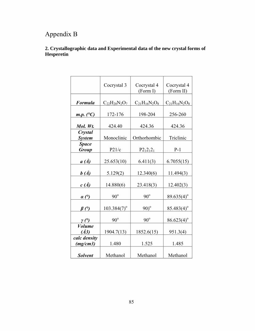
85
Appendix B 2. Crystallographic data and Experimental data of the new crystal forms of Hesperetin
Cocrystal 3
Cocrystal 4
(Form I)
Cocrystal 4 (Form II)
Formula
C22H20N2O7
C21H16N2O8
C21H16N2O8
m.p. (°C)
172-176
198-204
256-260
Mol. Wt.
424.40
424.36
424.36
Crystal System
Monoclinic
Orthorhombic
Triclinic
Space Group
P21/c
P212121
P-1
a (Å)
25.653(10)
6.411(3)
6.7055(15)
b (Å)
5.129(2)
12.340(6)
11.494(3)
c (Å)
14.880(6)
23.418(3)
12.402(3)
α (°)
90o
90o
89.635(4)o
β (°)
103.384(7)o
90)o
85.483(4)o
γ (°)
90o
90o
86.623(4)o
Volume (Å3)
1904.7(13)
1852.6(15)
951.3(4)
calc density (mg/cm3)
1.480
1.525
1.485
Solvent
Methanol
Methanol
Methanol

2.1 Experimental data of Cocrystal 3
DSC thermogram, FT-IR spectrum and X-ray powder diffraction patterns of bulk sample (red) and calculated from the single crystal structure (black).
638.
573
0.61
759.
0680
1.96
816.
0985
8.85
959.
5810
12.6
710
58.3
7
1082
.52
1127
.511
67.7
512
18.9
712
62.113
060
1335
.5213
99.0
11
37.6
51
72.1
215
95.9
1629
.16
96.9
9
2962
.21
3108
.22
3156
.07
322
.5
76
78
80
82
8
86
88
90
92
9
96
98
100
1000 2000 3000 000 Wavenumbers (cm-1)
5 10 15 20 25 30 35 40
0
2000
4000
6000
8000
10000
kp15
Calculated
Rel
ativ
e in
tens
ity
2 θ degree
86

.2 Experimental data of Cocrystal 4 (Form I and Form II)
DSC thermogram, FT-IR spectrum and X-ray powder diffraction patterns of bulk sample
2
(red), calculated from the single crystal of Form I (black) and calculated from the single crystal of Form II (Green).
87
719.
8074
1.61
756.
98
799.
6083
7.11
872.
0294
6.70
971.
7410
12.9
810
46.9
410
72.7
210
85.6
511
52.8
5
1178
.04
1192
.33
1234
.85
1274
.53
1320
.93
1368
.45
1397
.16
1438
.54
1452
.92
1508
.66
1580
.62
1628
.35
3394
.36
72
74
76
78
80
82
84
86
88
90
92
94
96
98
100
3000 2000 1000 Wavenumbers (cm-1)
5 10 15 20 25 30 35 40
0
2000
4000
6000
8000
10000
12000
Form I (P 1 )
Form II (P 212121)
E xp t
Rel
ativ
e in
tens
ity
2 θ deg ree