Silicon: the most underappreciated element in horticultural crops
Upload
khangminh22Category
view
0download
0
Received: 8 July 2021; Accepted: 20 November 2021; Published: 20 January 2022; Corrected and Typeset: 23 March 2022© The Author(s) 2022. Published by Oxford University Press on behalf of Nanjing Agricultural University. This is an Open Access article distributed under the termsof the Creative Commons Attribution License (https://creativecommons.org/licenses/by/4.0/), which permits unrestricted reuse, distribution, and reproductionin any medium, provided the original work is properly cited.
Horticulture Research, 2022, 9: uhab068
https://doi.org/10.1093/hr/uhab068
Review Article
Hydroxylation decoration patterns of flavonoids inhorticultural crops: chemistry, bioactivity, andbiosynthesisYilong Liu1,2, Jiafei Qian1, Jiajia Li1, Mengyun Xing1, Donald Grierson1,3, Chongde Sun1,2, Changjie Xu1, Xian Li1,2,* and Kunsong Chen1,2
1Zhejiang Provincial Key Laboratory of Horticultural Plant Integrative Biology, Zhejiang University, Hangzhou 310058, China2Shandong (Linyi) Institute of Modern Agriculture, Zhejiang University, Linyi 276000, China3Plant and Crop Sciences Division, School of Biosciences, Sutton Bonington Campus, University of Nottingham, Loughborough LE12 5RD, UK
*Corresponding author. E-mail: [email protected]
Abstract
Flavonoids are the most widespread polyphenolic compounds and are important dietary constituents present in horticulturalcrops such as fruits, vegetables, and tea. Natural flavonoids are responsible for important quality traits, such as food colors andbeneficial dietary antioxidants, and numerous investigations have shown that intake of flavonoids can reduce the incidence ofvarious non-communicable diseases. Analysis of the thousands of flavonoids reported so far has shown that different hydroxylationmodifications affect their chemical properties and nutritional values. These diverse flavonoids can be classified based on differenthydroxylation patterns in the B, C, and A rings and multiple structure–activity analyses have shown that hydroxylation decorationat specific positions markedly enhances their bioactivities. This review focuses on current knowledge concerning hydroxylationof flavonoids catalyzed by several different types of hydroxylase enzymes. Flavonoid 3′-hydroxylase (F3′H) and flavonoid 3′5′-hydroxylase (F3′5′H) are important enzymes for the hydroxylation of the B ring of flavonoids. Flavanone 3-hydroxylase (F3H) iskey for the hydroxylation of the C ring, while flavone 6-hydroxylase (F6H) and flavone 8-hydroxylase (F8H) are key enzymes forhydroxylation of the A ring. These key hydroxylases in the flavonoid biosynthesis pathway are promising targets for the futurebioengineering of plants and mass production of flavonoids with designated hydroxylation patterns of high nutritional importance.In addition, hydroxylation in key places on the ring may help render flavonoids ready for degradation, and the catabolic turnover offlavonoids may open the door for new lines of inquiry.
IntroductionFlavonoids are the most widespread polyphenolic com-pounds, characterized by possession of at least one aro-matic ring with one or more hydroxyl groups attached.Current dietary guidance from the World Health Orga-nization recommends people should consume at least400 g, i.e. five portions, of fruits and vegetables per day fortheir optimum health. Besides vitamins, minerals, anddietary fiber, such horticultural crops have the advantageof accumulating high amounts of polyphenols, especiallyflavonoids [1]. These substances in planta are generatedas the products of plant antioxidative defense systemsactivated in rapid response to both biotic and abioticstresses and are responsible for important quality traits,such as color, in horticultural crops [2]. Moreover, overrecent years, numerous epidemiological and clinicalstudies have shown that such flavonoids also exhibitstrong antioxidant properties in vitro and in vivo, aswell as antidiabetic, antiobesity, anti-inflammatory,
anticancer, and antibacterial activities [1, 3, 4] (Fig. 1).Hence, flavonoid-abundant foods might be nature’sbountiful gifts to humankind for their excellent health-promoting benefits.
There are >8000 different flavonoids identified fromplants [5]. Different modifications, such as hydroxylation,glycosylation, methylation, and acylation, play impor-tant roles in generating such diversity of flavonoids, andhydroxylation is the most frequently occurring modi-fication in natural flavonoids (Fig. 2). Hydroxylation offlavonoids improves the chemical solubility and stabilityof an array of flavonoids and is also correlated with theirbioactive properties. For instance, a hydroxylated B ring iscrucial for the antioxidant properties of flavonoids [6]. Anincreased degree of hydroxylation of flavonoids is associ-ated with stronger inhibitory effects on α-glucosidase orα-amylase activities [7, 8]. Inhibition of aldose reductaseactivity was also found to be remarkably enhanced by thehydroxylation of specific positions, such as the C3′ and
Dow
nloaded from https://academ
ic.oup.com/hr/article/doi/10.1093/hr/uhab068/6511828 by guest on 21 April 2022
2 | Horticulture Research, 2022, 9: uhab068
Figure 1. Health-promoting bioactivities of flavonoids in horticulturalcrops.
C4′ of the B ring of flavonoids [9]. Taken together, the datashow that different hydroxylation decoration patternsof flavonoids may affect their chemical properties aswell as their bioactivities. So far, there have been severalreview papers describing the structures and biosynthe-sis of flavonoids and the introduction of various deco-rations, such as glycosylation [10–12], methylation [13,14], and acylation [15]. However, to date, there is nosystematic review on the hydroxylation modification offlavonoids, particularly in horticultural crops.
This review focuses on current knowledge concerninghydroxylation decoration of flavonoids and its effectson their chemical and nutritional value. Diverse dietaryflavonoids are further classified based on differenthydroxylation patterns in the B, C, and A rings offlavonoids and a comparison is made of bioactivitiesof flavonoids that differ only by hydroxylation patterns.Particular attention is paid to key hydroxylases in theflavonoid biosynthesis pathway, with the objective offuture bioengineering-targeted bioactivities in plantresources and mass production of flavonoids withdesirable hydroxylation patterns. We also discuss thedegradation of natural flavonoids and the relationshipbetween hydroxylation modification and flavonoidcatabolism in planta, which requires further investigationin the future.
Divergent hydroxylation patterns anddistribution of flavonoidsFlavonoids consist of two aromatic rings (A and B rings)connected by a three-carbon bridge (C ring) containing anembedded oxygen atom, abbreviated as C6-C3-C6 (Fig. 2).According to the oxidative status and hydroxylationdegree of the C ring and the connection position of theB ring, flavonoids can be grouped into different sub-classes, including flavonols, anthocyanidins, flavones,flavanones, flavanols, and isoflavones (Table 1). Eachsubclass of flavonoids can be analyzed based on itshydroxylation patterns.
Hydroxylation and distribution of flavonolsFlavonols are the most widespread subclass of flavonoidsand possess the 3-OH and 4-oxo groups on the C6-C3-C6 backbone. Their diversity originates from differentsubstitution of the phenolic -OH groups in both the A andthe B ring (Table 1).
The flavonol fisetin is hydroxylated at C7, C3, C3′,and C4′ and is found mainly in strawberry, apple, per-simmon, onion, and grape [16]. Galangin, kaempferol,morin, quercetin, and myricetin are common flavonolshaving the same structures on the A and C rings withhydroxyl groups attached at C5, C7, and C3, but withdifferent hydroxylation modes on their B ring (Table 1).Specifically, galangin, which is a major active componentin the root of galangal [17] and is also found in propolisproduced from plants by bees [18], has no -OH on the Bring. Kaempferol, with an -OH group substituted at C4′, isabundant in green leafy vegetables such as spinach andkale, and berries, including mulberry and strawberry [19,20]. Morin also has two -OH groups on the B ring at the C2′
and C4′ positions and is found mainly in mulberry, guava,grape, and fig [21]. Quercetin, which is hydroxylated atboth C3′ and C4′, is present in a high concentration inonion, chili pepper, apple, Chinese bayberry, mulberry,and apricot [19, 20, 22]. Notably, myricetin has threehydroxyl groups at C3′, C4′, and C5′. It was originallyisolated from the bark of Chinese bayberry [23] and is alsopresent at a relatively high level in its fruit [22]. Myricetinis also widely distributed in many other berries, such asstrawberry, blackberry, and blueberry, as well as vegeta-bles like spinach and cauliflower [20, 24]. Quercetagetinwhich has three -OH groups on the A ring (C5, C6, and C7)is found mainly in Tagetes [25].
Hydroxylation and distribution ofanthocyanidinsAnthocyanins are the glycoside conjugates of antho-cyanidins (Table 1) and constitute a group of naturalpigments that confer a wide spectrum of colors, varyingfrom orange, salmon pink, red, magenta, and violet todark blue in many fruits, colored leafy vegetables, andtubers. The majority of anthocyanins reported are basedon three common anthocyanidins, i.e. pelargonidin,cyanidin, and delphinidin [26]. These three compoundsshare the same chemical structures at the A and C ringswith a characteristic flavylium cation and three -OHgroups attached at C5, C7, and C3. They have differentdegrees of hydroxylation on the B ring (Table 1) and,interestingly, their colors seem to gradually deepen withincreasing hydroxylations, i.e. orange/red (pelargonidin),red/magenta (cyanidin), and violet/blue (delphinidin),respectively [27].
Pelargonidin, hydroxylated at C4′, is abundant instrawberry [28], and distributed in red radish, potato, andbanana [29]. Cyanidin, with two hydroxyl groups at C3′
and C4′ of the B ring, is the most common anthocyanidin.It is especially rich in berries such as Chinese bayberry[22], elderberry, chokeberry, blackberry, black mulberry,
Dow
nloaded from https://academ
ic.oup.com/hr/article/doi/10.1093/hr/uhab068/6511828 by guest on 21 April 2022
Liu et al. | 3
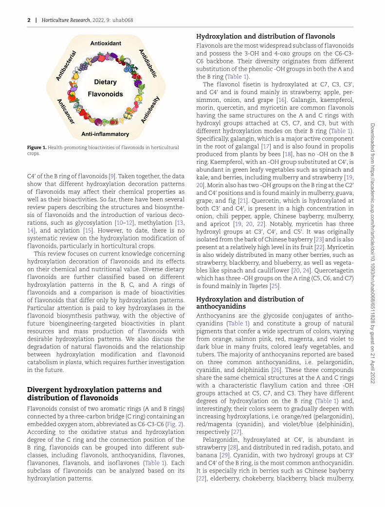
Figure 2. Key hydroxylases that participate in hydroxylation decoration during flavonoid biosynthesis. F3H, flavanone 3-hydroxylase; F3′H, flavonoid3′-hydroxylase; F3′5′H, flavonoid 3′,5′-hydroxylase; F6H, flavone 6-hydroxylase; F8H, flavone 8-hydroxylase; DFR, dihydroflavonol 4-reductase; ANS,anthocyanidin synthase; ANR, anthocyanidin reductase; LAR, leucoanthocyanidin reductase.
and cherry [28, 29]. Cyanidin is also ubiquitously foundin other fruits, like apple, peach, pear, and fig, as wellas colored vegetables like red onion and red cabbage[29]. Delphinidin has three hydroxyl groups at C3′, C4′,and C5′ of the B ring, the major sources of which weredark-colored foods such as bilberry, highbush blueberry,blackcurrant, eggplant, etc. [28, 29]. It is also found inpassion fruit, green bean, and pomegranate [29].
Hydroxylation and distribution of flavonesFlavones lack the hydroxyl group at C3 compared withthe skeleton of flavonols, but have a wide range ofhydroxylation patterns on the A and B rings. Typically,chrysin, apigenin, luteolin, and tricetin are commonflavones that all have hydroxylation at C5 and C7 butdiffer from each other by the substitution of -OH groupson their B rings (Table 1). Chrysin has no hydroxyl onthe B ring and is found in honey, propolis, and passionfruit [30]. Apigenin (4′-OH) is particularly abundant inparsley and dried flowers of chamomile, and exists insome other vegetables, such as celery, broccoli, spices likethyme, and fruits such as cherry, olive, and legumes [31].Major food sources of luteolin (3′,4′-OH) include bird chili[32], celery, broccoli, parsley, chrysanthemum flowers,onion leaves, sweet bell pepper, and carrot [33]. Tricetin(3′,4′,5′-OH) is found mainly in Myrtaceae pollen and
Eucalyptus honey [34]. In addition, other flavones, such asbaicalein or norwogonin, show additional hydroxylationson carbons 6 or 8, based on the structure of chrysin; andscutellarein has an additional -OH group substituted atthe C4′ of baicalein or C6 of apigenin (Table 1). Thesethree flavones specifically exist in Scutellaria baicalensisand Scutellaria barbata [35].
Hydroxylation and distribution of flavanonesFlavanones that have a chiral carbon at C2 are thehydrogenated derivatives of flavones. The flavanoneliquiritigenin has a C7-hydroxyl on the A ring and C4′-hydroxyl on its B ring and is particularly prevalent inlicorice [36] (Table 1). Flavanones including pinocembrin,naringenin, eriodictyol, and 5,7,3′,4′,5′-pentahydroxyflavanone all have dihydroxyl groups at C5 and C7,with the structural difference involving sequentialincreases in B-ring hydroxylation (Table 1). Pinocembrinhas no -OH substitution on the B ring and in the (2S)-form is widely distributed in propolis and Glycyrrhizaglabra [37]. Naringenin (4′-OH) occurs extensively inCitrus fruits like grapefruit, orange, and lemon [38].Additionally, eriodictyol (3′,4′-OH) is also widely dis-tributed in Citrus fruits such as bitter orange, mandarin,tangerine, and lemon, as well as peanuts and loquats
Dow
nloaded from https://academ
ic.oup.com/hr/article/doi/10.1093/hr/uhab068/6511828 by guest on 21 April 2022

4 | Horticulture Research, 2022, 9: uhab068
Table 1. Divergent hydroxylation patterns of flavonoids and their distribution in horticultural crops.
Structure A C B
Flavonoids Distribution References 5 6 7 8 3 2' 3' 4' 5'
H H OH H OH H OH OH H Fisetin Strawberry, apple, persimmon, onion,
grape
16
OH H OH H OH H H H H Galangin Galangal, propolis 17,18
OH H OH H OH H H OH H Kaempferol Spinach, kale, mulberry, strawberry 19,20
OH H OH H OH OH H OH H Morin Mulberry, guava, grape, fig 21
OH H OH H OH H OH OH H Quercetin Onion, chili pepper, apple, Chinese
bayberry, mulberry, apricot 19,20,22
OH H OH H OH H OH OH OH Myricetin
Chinese bayberry, strawberry,
blackberry, blueberry, spinach,
cauliflower
20.22.24
OH OH OH H OH H OH OH H Quercetagetin Tagetes 25
OH H OH H OH H H OH H Pelargonidin Strawberry, red radish, potato, banana
22,28,29OH H OH H OH H OH OH H Cyanidin
Chinese bayberry, elderberry,
chokeberry, blackberry, black mulberry,
cherry, apple, peach, pear, fig, red onion,
red cabbage
OH H OH H OH H OH OH OH Delphinidin
Bilberry, highbush blueberry,
blackcurrant, eggplant, passion fruit,
green bean, pomegranate
OH H OH H H H H H H Chrysin Honey, propolis, passion fruit 30
OH H OH H H H H OH H Apigenin
Parsley, dried flowers of chamomile,
celery, broccoli, thyme, cherry, olive,
legume
31
OH H OH H H H OH OH H Luteolin
Bird chili, celery, broccoli, parsley,
chrysanthemum flowers, onion
leaves, sweet bell pepper, carrot
32,33
OH H OH H H H OH OH OH Tricetin Myrtaceae pollen, Eucalyptus honey 34
OH OH OH H H H H H H Baicalein
Scutellaria baicalensis, Scutellaria barbata
35OH OH OH H H H H OH H Scutellarein
OH H OH OH H H H H H Norwogonin
H H OH H H H H OH H Liquiritigenin Licorice 36
OH H OH H H H H H H Pinocembrin Propolis, Glycyrrhiza glabra 37
OH H OH H H H H OH H Naringenin Grapefruit, orange, lemon 38
OH H OH H H H OH OH H Eriodictyol Bitter orange, mandarin, tangerine,
lemon, peanut, loquat
39
OH H OH H H H OH OH OH 5,7,3',4',5'-Pentahydroxy
flavanone Helichrysum bracteatum 40
OH H OH H OH H H OH H (Epi)Afzelechin Sour jujube, cowpea, peanut 43-45
OH H OH H OH H OH OH H (Epi)Catechin
Tea 41,42OH H OH H OH H OH OH OH (Epi)Gallocatechin
H H OH H Ph H H OH H Daidzein
Soybean, soy-based foods 46
OH H OH H Ph H H OH H Genistein
Different flavonoids are listed within each subclass according to the position of -OH in the order of A ring, followed by B ring with increasing number ofhydroxyl groups. Compounds are listed based on the relative amount of each compound, from high to relatively low, in different horticultural crops.
[39]. Pentahydroxy flavanones also exist in some hor-ticultural crops. For instance, 5,7,3′,4′,5′-pentahydroxy
flavanone has been detected in Helichrysum bracteatum[40].
Dow
nloaded from https://academ
ic.oup.com/hr/article/doi/10.1093/hr/uhab068/6511828 by guest on 21 April 2022
Liu et al. | 5
Hydroxylation and distribution of flavanolsCompared with other subclasses of flavonoids, flavanolsare characterized by the absence of a double bondbetween C2 and C3 and have no C4 carbonyl in theC ring (Table 1). Therefore, two chiral carbons (C2 andC3) exist in flavanols and the fixed hydroxylation at C3means each of them has four possible diastereoisomers.For instance, catechin, the most typical monomericflavanol, exists in four forms, i.e. (−)-catechin (2S,3R),(+)-catechin (2R,3S), (−)-epicatechin (2S,3S), and (+)-epicatechin (2R,3R). Of these, (+)-catechin and (−)-epicatechin are most commonly found in horticulturalcrops, especially in tea leaves [41, 42]. Catechin has fourhydroxyl groups in the basic skeleton of flavan-3-ol at C5,C7, C3′, and C4′, respectively. Afzelechin, which lacks the3′-OH of catechin, is present in sour jujube [43], cowpea[44], and peanut seed skin [45]. Gallocatechin has onemore -OH substitution at C5′ of catechin, and is presentat significant levels in tea [41, 42]. The 3-OH on the C ringof catechins is usually esterified with gallic acid, therebyforming the gallated catechins such as epigallocatechingallate, epicatechin gallate, and catechin gallate.
Hydroxylation and distribution of isoflavonesUnlike other flavonoids, the isoflavone B ring is attachedat C3 rather than C2 (Table 1). Isoflavones such as 4’,7-dihydroxyisoflavone (daidzein) and 4′,5,7-trihydroxyisoflavone (genistein), are specifically accu-mulated in legumes, especially soybeans [46]. Thus,soybean-based foods such as tofu, soy milk, and soyyoghurt are excellent dietary sources of isoflavones.
Bioactivities of flavonoids influenced bydivergent hydroxylation patternsFor the B ringWith the increasing attention paid to the healthcarebenefits of natural products, there are more and morestudies focusing on the structure–activity relation-ships between the different chemical modificationsof flavonoids and their bioactivities [47, 48]. Plentyof studies have shown that hydroxylation decorationat specific positions of flavonoids markedly enhancesefficacy of their bioactivity (Table 2).
The flavonols kaempferol (4′-OH), quercetin (3′,4′-OH), and myricetin (3′,4′,5′-OH) are a typical group offlavonoids with the same structures for the A and Crings but have different hydroxylation modes on the Bring. Results have shown that quercetin has stronger •OHscavenging properties than kaempferol, and myricetin isthe most powerful hydroxyl radical scavenger, indicatingthat radical scavenging activity increases with thegrowing number of -OH groups on the aromatic Bring [49] (Table 2). Furthermore, their acrolein-trappingefficiency decreases in the order of myricetin > quercetin> kaempferol [50]. Exposure to blue light for 20 hoursinduced the death of ∼75% of the photoreceptors inbovine retinal cell cultures, while myricetin conferred
∼100% protection against light-mediated damage tophotoreceptors, whereas quercetin showed relativelypoor protective activity, and kaempferol was inactive[51]. Among these three flavonols, myricetin showedthe strongest inhibitory effects on either α-glucosidase[52, 53] or epidermal growth factor-induced cell trans-formation in JB6 P+ cells [54], followed by quercetinand kaempferol. Such flavonols can also be individuallymixed with a chitosan-based matrix to develop activepackaging films, and myricetin showed the strongestintermolecular interactions with the film matrix, dueto the greater number of hydroxyl groups on the Bring [55]. Hence, films containing myricetin have themost satisfactory mechanical properties and the highestability to provide a barrier to water vapor and oxygen[55]. In addition, studies have reported that myricetin ismore effective than quercetin at inhibiting the inductionof apoptosis in prostate cancer cell line PC-3 [56]. Also,quercetin has a stronger tyrosinase inhibitory effect thankaempferol [57].
Morin (2′,4′-OH), with an additional hydroxyl at the C2′
position of kaempferol, shows enhanced hydroxyl radicalscavenging activity [49]. It has a greater ability to inhibitβ-amyloid fibril formation from amyloid β-peptide anddestabilizes preformed β-amyloid fibrils, demonstratingits greater potential for the prevention and control ofAlzheimer’s disease [58]. Galangin, another flavonol withno hydroxylation on the B ring, as well as morin andkaempferol, all show significant scavenging ability foracrolein, and their trapping efficiency increases in theorder galangin < morin < kaempferol [50].
The activities of three anthocyanidins in O2•− and
ONOO− scavenging and inhibition of lipid peroxi-dation have been ranked in the order pelargonidin(4′-OH) < cyanidin (3′,4′-OH) < delphinidin (3′,4′,5′-OH),indicating that the increasing number of -OH groupspresent on the B ring enhances the antioxidant activitiesof anthocyanidins [59–61] (Table 2). As mentionedpreviously, the degree of hydroxylation is related to thegradually deepening colors of these three anthocyanidinsand, therefore, dark-colored fruits and vegetables mayshow greater health benefits [62, 63]. Similarly, comparedwith cyanidin 3-O-glucoside, delphinidin 3-O-glucosideshowed enhanced ability to stimulate insulin secretion[64], and greater ability to inhibit the viability of cancercells such as HCT 116 cells [65] (Table 2).
Chrysin (no hydroxyl at the B ring), apigenin (4′-OH),and luteolin (3′,4′-OH) belong to group of flavones withincreasing numbers of hydroxyl substituents on the Bring. Their apoptosis-inducing potencies in tumor cells[66], proteasome inhibitory activities [66, 67], matrix met-alloproteinase (MMP) inhibition effects, and scavengingreactive oxygen capacities [68] are in the order of lute-olin > apigenin > chrysin (Table 2). Also, compared withchrysin, apigenin showed a stronger ability to inhibit theproduction of pro-inflammatory cytokines stimulated bylipopolysaccharide in human peripheral blood mononu-clear cells [69].
Dow
nloaded from https://academ
ic.oup.com/hr/article/doi/10.1093/hr/uhab068/6511828 by guest on 21 April 2022
6 | Horticulture Research, 2022, 9: uhab068
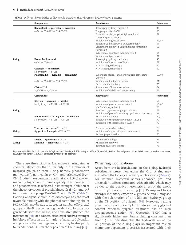
Table 2. Different bioactivities of flavonoids based on their divergent hydroxylation patterns.
Compounds Bioactivities References
Kaempferol → quercetin → myricetin Scavenging hydroxyl radicals ↑ 494′-OH → 3′,4′-OH → 3′,4′,5′-OH Trapping ability of ACR ↑ 50
Protection activity against light-mediatedphotoreceptor damage ↑
51
Inhibition of α-glucosidase ↑ 52, 53Inhibits EGF-induced cell transformation ↑ 54Constituent of active packaging films containingflavonols ↑
55
Induction of apoptosis in tumor cells ↑ 56Inhibition of tyrosinase ↑ 57
B ring Kaempferol → morin Scavenging hydroxyl radicals ↑ 494′-OH → 2′,4′-OH Inhibition of formation of fAβ ↑ 58
ACR trapping efficiency ↑ 50Galangin → kaempferolNo hydroxyl → 4′-OH
ACR trapping efficiency ↑ 50
Pelargonidin → cyanidin → delphinidin Superoxide radical- and peroxynitrite-scavengingactivity ↑
59, 60
4′-OH → 3′,4′-OH → 3′,4′,5′-OH Inhibition of lipid peroxidation ↑ 61Antioxidant activities ↑ 60
C3G → D3G Stimulation of insulin secretion ↑ 643′,4′-OH → 3′,4′,5′-OH Inhibition of viability of cancer cells ↑ 65
Compounds Bioactivities References
Chrysin → apigenin → luteolinNo hydroxyl → 4′-OH → 3′,4′-OH
Induction of apoptosis in tumor cells ↑ 66Inhibition of proteasome activity ↑ 66, 67MMP inhibitory effect ↑ 68
B ring Reactive oxygen scavenging activities ↑ 68Inhibition of pro-inflammatory cytokine production ↑ 69
Pinocembrin → naringenin → eriodictyol Antioxidant activity ↑ 70, 71No hydroxyl → 4′-OH → 3′,4′-OH Inhibition of the phosphorylation of PKCδ ↑ 70
Inhibition of the formation of AGEs ↑ 72
Tricetin → myricetin 3H → OH Pro- and antioxidant activity ↑ 73C ring Apigenin→ kaempferol 3H → OH Inhibition of α-glucosidase or α-amylase ↑ 74
Anti-adipogenic action ↑ 75
Fisetin → quercetin 5H → OH Membrane binding ↑ 76A ring Daidzein → genistein 5H → OH Antioxidant activity ↑ 77
Improves glucose tolerance↑ 78
fAβ, β-amyloid fibrils; C3G, cyanidin 3-O-glucoside; D3G, delphinidin 3-O-glucoside; ACR, acrolein; EGF, epidermal growth factor; MMP, matrix metalloproteinease;PKCδ, protein kinase Cδ; AGEs, advanced glycation end products.
There are three kinds of flavanones sharing similarchemical structures that differ only in the number ofhydroxyl groups on their B ring, namely, pinocembrin(no hydroxyl), naringenin (4′-OH), and eriodictyol (3′,4′-OH). Studies have demonstrated that eriodictyol showedmarkedly higher antioxidant capacity than naringeninand pinocembrin, as reflected in its stronger inhibition ofthe phosphorylation of protein kinase Cδ (PKCδ) and p47in murine macrophage RAW264.7 cells [70, 71] (Table 2).Docking analysis showed that eriodictyol has the mostfavorable binding with the phorbol ester binding site ofPKCδ, which may be due to its greater number of hydroxylgroups on the B ring conferring the ability to form hydro-gen bonds with the kinase, and thus strengthened theinteraction [70]. In addition, eriodictyol showed strongerinhibitory effects on the formation of advanced glycationend products than naringenin, which may be due partlyto its additional -OH in the 3′ position of the B ring [72].
Other ring modificationsApart from the hydroxylations on the B ring, hydroxylsubstituents present on either the C or A ring mayalso affect the biological activity of flavonoids (Table 2).For instance, myricetin shows enhanced pro- andantioxidant effects compared with tricetin, which maybe due to the positive mesomeric effect of the enolic3-hydroxy group on the C-ring [73]. Kaempferol has astronger inhibitory effect on α-glucoside and α-amylasewith the substitution of an additional hydroxyl groupat the C3 position of apigenin [74]. Moreover, treatingpreadipocytes with kaempferol reduces triacylglycerolcontent, whereas apigenin treatment has no suchanti-adipogenic action [75]. Quercetin (5-OH) has asignificantly higher membrane binding constant thanfisetin (5-H), indicating that the hydroxylation at theC5 position of the A ring plays an important role inmembrane-dependent processes associated with their
Dow
nloaded from https://academ
ic.oup.com/hr/article/doi/10.1093/hr/uhab068/6511828 by guest on 21 April 2022
Liu et al. | 7
biological activities [76]. The isoflavone genistein (5-OH)shows stronger antioxidant activity than daidzein (5-H) [77] and supplementing with genistein significantlyimproved glucose tolerance in diabetic db/db mice, whichwas not observed in the daidzein-supplemented group[78].
Key hydroxylases in the biosynthesispathways of flavonoidsFlavonoids are synthesized through the phenylpropanoidmetabolic pathway, initiated by transformation ofphenylalanine into 4-coumaroyl-CoA. This is combinedwith malonyl CoA, and naringenin is then synthesized bythe decarboxylation and cyclization reactions catalyzedby chalcone synthase (CHS) and chalcone isomerase. Fur-ther, naringenin is converted by flavanone 3-hydroxylase(F3H) and other hydroxylases, including flavonoid3′-hydroxylase (F3′H) and flavonoid 3′,5′-hydroxylase(F3′5′H), to produce different dihydroflavonols, which canform leucoanthocyanidins catalyzed by dihydroflavonol4-reductase (Fig. 2). Subsequently, leucoanthocyanidinsare converted into anthocyanidins or flavanols by theaction of anthocyanidin synthase or leucoanthocyanidinreductase, respectively. Anthocyanidins can also betransformed to flavanols by the catalysis of anthocyani-din reductase.
The basic hydroxylation at C5, C7, and C4’ of com-mon flavonoids is shown for naringenin, while additionalhydroxyl groups can also occur at C3, C3’, C5’, C6, andC8 positions (in Table 1). Hydroxylases play a vital rolein the biosynthesis of hydroxylated flavonoids and F3′Hand F3′5′H are important enzymes for the hydroxylationof the B ring of flavonoids (Fig. 2). F3H is key for thehydroxylation of the C ring and flavone 6-hydroxylase(F6H) and flavone 8-hydroxylase (F8H) are key enzymesfor the hydroxylation of the A ring (Fig. 2).
Flavonoid 3′-hydroxylaseF3′H (EC 1.14.13.21) is a cytochrome P450 (CYP450)-dependent monooxygenase requiring NADPH as acofactor. It catalyzes the hydroxylation of flavanones,flavones, dihydroflavonols, and flavonols at the C3′
position of the B ring to their 3′,4′-hyroxylated states(Fig. 2). F3′H was first cloned from petunia (Petuniahybrida) [79], and then isolated from various resourcessuch as soybean (Glycine max) [80], grapevine (Vitisvinifera) [81], sorghum (Sorghum bicolor) [82], apple (Malus× domestica) [83], strawberry (Fragaria × ananassa) [84],rice (Oryza sativa) [85], and tea (Camellia sinensis) [86](Table 3).
Heterologous expression in yeast and analysis ofF3′Hs from divergent plant species showed significantdifferences in substrate specificity and catalytic prop-erties (Fig. 2). For instance, F3′H isolated from eithertea [86, 87] or montbretia (Crocosmia × crocosmiif lora)[88] catalyzed the introduction of a hydroxyl group atthe C3′ position of naringenin, dihydrokaempferol, and
kaempferol to produce eriodictyol, dihydroquercetin,and quercetin, respectively (Fig. 3). Similarly, F3′H fromstrawberry (Fragaria sp.) showed high specificity fornaringenin, dihydrokaempferol, and kaempferol, whileapigenin was just a minor substrate [84]. However, F3′Hencoded by CYP75B4 from rice grain preferred apigeninto other substrates, leading to the formation of luteolin[85] (Fig. 3). Interestingly, CYP75B3, another rice F3′Hgene, encodes an enzyme with a higher preference forkaempferol, and CYP75B3 from black rice showed around2-fold increased catalytic efficiency with naringenin anddihydrokaempferol compared with the enzyme fromeither white or red rice [85]. It is also possible thatdifferent F3′H family members isolated from the samespecies might also have different catalytic functions.
Transgenic analysis has also been used to verifythe functionality of F3′H from different plant sources(Table 3). Heterologous overexpression of grapevineVvF3′H in the petunia ht1 mutant showed a significantaccumulation of 3′,4′-hydroxylated flavonoids, includingpeonidin, an O-methylated anthocyanidin derived fromcyanidin, and quercetin in the transgenic flowers [81].In contrast, transgenic grapevines with the F3′H genesilenced had obviously decreased contents of peonidinas well as seed tannins [89]. Transgenic arabidopsis(Arabidopsis thaliana) tt7 seedlings overexpressing F3′Hgenes isolated from either apple or sorghum showed arestored ability to produce 3′,4′-hydroxylated flavonoidssuch as cyanidin and quercetin [82, 83]. The increasesof these two flavonoids were also shown in transgenictobacco (Nicotiana tabacum) overexpressing apple F3′H[83]. Overexpression of the ginkgo (Ginkgo biloba) geneGbF3′H1 enhanced flavonoid production in transgenicpoplar (Populus davidiana), as shown by the significantlyincreased red pigmentation and higher concentrations ofepigallocatechin, gallocatechin, and catechin comparedwith that in the wild-type plants [90].
Flavonoid 3′,5′-hydroxylaseF3′5′H (EC 1.14.13.88) is another key hydroxylase respon-sible for the B-ring hydroxylation of flavonoids and playsa crucial role in the formation of 3′,4′,5′-trihydroxylatedderivatives (Fig. 2). Initially, F3′5′H was discovered indelphinidin-rich plants, responsible for the production ofblue flower colors, and was therefore known as the bluegene [91]. Later, such genes were also identified widelyin other plants that accumulate proanthocyanidins orflavonols possessing trihydroxyls on the B-ring. However,not all plant species have the ability to accumulatesuch B-ring trihydroxyflavonoids, indicating that thepresence or expression of an appropriate F3′5′H geneis not ubiquitous in plants [92]. For example, thereis no F3′5′H gene in the genome of the model plantarabidopsis.
To date, a number of F3′5′Hs have been isolated fromvarious horticultural crops, such as potato (Solanumtuberosum) [93], grapevine [81], tomato (Solanum lycop-ersicum) [94], pea (Pisum sativum) [95], and tea [96, 97].
Dow
nloaded from https://academ
ic.oup.com/hr/article/doi/10.1093/hr/uhab068/6511828 by guest on 21 April 2022
8 | Horticulture Research, 2022, 9: uhab068
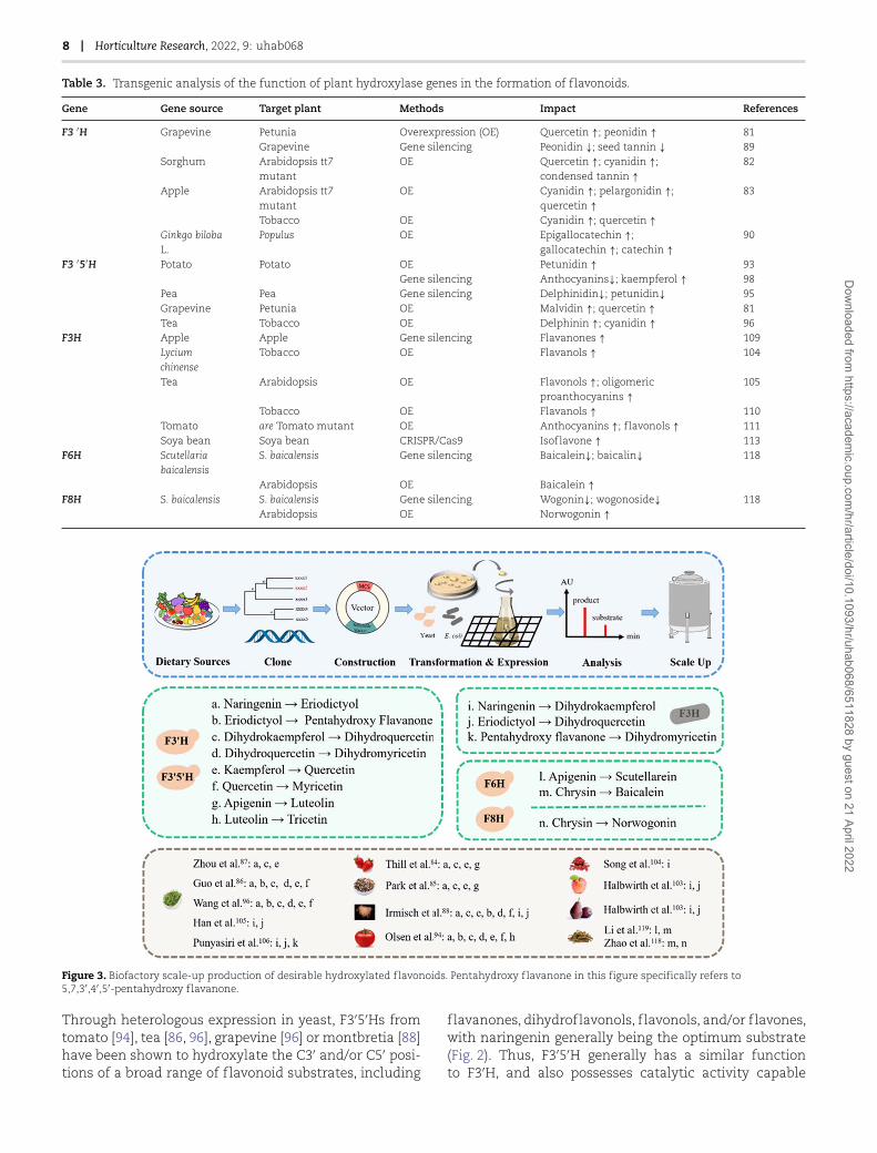
Table 3. Transgenic analysis of the function of plant hydroxylase genes in the formation of flavonoids.
Gene Gene source Target plant Methods Impact References
F3 ′H Grapevine Petunia Overexpression (OE) Quercetin ↑; peonidin ↑ 81Grapevine Gene silencing Peonidin ↓; seed tannin ↓ 89
Sorghum Arabidopsis tt7mutant
OE Quercetin ↑; cyanidin ↑;condensed tannin ↑
82
Apple Arabidopsis tt7mutant
OE Cyanidin ↑; pelargonidin ↑;quercetin ↑
83
Tobacco OE Cyanidin ↑; quercetin ↑Ginkgo bilobaL.
Populus OE Epigallocatechin ↑;gallocatechin ↑; catechin ↑
90
F3 ′5′H Potato Potato OE Petunidin ↑ 93Gene silencing Anthocyanins↓; kaempferol ↑ 98
Pea Pea Gene silencing Delphinidin↓; petunidin↓ 95Grapevine Petunia OE Malvidin ↑; quercetin ↑ 81Tea Tobacco OE Delphinin ↑; cyanidin ↑ 96
F3H Apple Apple Gene silencing Flavanones ↑ 109Lyciumchinense
Tobacco OE Flavanols ↑ 104
Tea Arabidopsis OE Flavonols ↑; oligomericproanthocyanins ↑
105
Tobacco OE Flavanols ↑ 110Tomato are Tomato mutant OE Anthocyanins ↑; f lavonols ↑ 111Soya bean Soya bean CRISPR/Cas9 Isoflavone ↑ 113
F6H Scutellariabaicalensis
S. baicalensis Gene silencing Baicalein↓; baicalin↓ 118
Arabidopsis OE Baicalein ↑F8H S. baicalensis S. baicalensis Gene silencing Wogonin↓; wogonoside↓ 118
Arabidopsis OE Norwogonin ↑
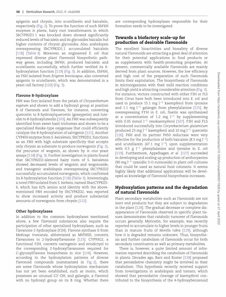
Figure 3. Biofactory scale-up production of desirable hydroxylated flavonoids. Pentahydroxy flavanone in this figure specifically refers to5,7,3′,4′,5′-pentahydroxy flavanone.
Through heterologous expression in yeast, F3′5′Hs fromtomato [94], tea [86, 96], grapevine [96] or montbretia [88]have been shown to hydroxylate the C3′ and/or C5′ posi-tions of a broad range of flavonoid substrates, including
flavanones, dihydroflavonols, flavonols, and/or flavones,with naringenin generally being the optimum substrate(Fig. 2). Thus, F3′5′H generally has a similar functionto F3′H, and also possesses catalytic activity capable
Dow
nloaded from https://academ
ic.oup.com/hr/article/doi/10.1093/hr/uhab068/6511828 by guest on 21 April 2022

Liu et al. | 9
of converting naringenin or eriodictyol to 5,7,3′,4′,5′-pentahydroxy flavanone, dihydrokaempferol or dihy-droquercetin to dihydromyricetin, and kaempferol orquercetin to myricetin, and also transforming luteolininto tricetin (Fig. 3).
In addition, expression levels of genes encodingF3′5′Hs greatly affect the accumulation of flavonoidswith diverse B-ring hydroxylation patterns (Table 3).The red-skinned potato cultivar ‘Désirée’ transformedwith a potato F3′5′H became purple-skinned, and themajor anthocyanins were changed from pelargonidinderivatives to petunidin derivatives [93]. Silencing theF3′5′H in transgenic potato tuber impaired anthocyaninbiosynthesis and caused a 100-fold increased levelof kaempferol [98]. Similarly, F3′5′H mutants of peahave lost the ability to synthesize delphinidin andpetunidin, which are the main pigments in wild-typepea flowers [95]. Additionally, transgenic petunia linescarrying VvF3′5′H1 accumulate the 3′,4′,5′-hydroxylated-based anthocyanin malvidin, and show a shift fromkaempferol to quercetin, compared with the non-transgenic control [81]. Overexpression of tea F3′5′H intobacco plants caused the majority of transgenic flowersto become magenta, compared with the pale pink of theuntransformed host, due to the accumulation of a novel3′,4′,5′-hydroxylated-based anthocyanin, delphinin, andthe cyanidin content was also increased [96]. So far,transgenic studies related to F3′5′H genes have focusedpreferentially on the biosynthesis of anthocyanins thatconfer the blue or purple colors of plant tissues.
Flavanone 3-hydroxylaseF3H (EC 1.14.11.9) belongs to the FeII/2-ketoglutarate-dependent dioxygenase family. It catalyzes the 3β-hydroxylation of 2S-flavanones at the C3 positionto 2R,3R-dihydroflavonols in the presence of O2, 2-oxoglutarate, Fe2+, and ascorbate as cofactors [99, 100](Fig. 2). The first F3H was cloned from Antirrhinum majus[101], and subsequently additional F3Hs were isolatedfrom other plant-based foods, such as soybean (G. max),apple (Malus domestica), pear (Pyrus communis), Lyciumchinense, and tea [102–105]. Heterologous expressionof Malus or Pyrus F3H in yeast, and montbretia or teaF3H in Escherichia coli, showed these proteins could allcatalyze the conversion of naringenin and eriodictyolinto dihydrokaempferol and dihydroquercetin, respec-tively [88, 103, 105] (Fig. 3). Similarly, when LcF3H wasexpressed in E. coli, the recombinant enzyme showedthe ability to convert naringenin to dihydrokaempferol[104]. Further, recombinant tea F3H was able to accept5,7,3′,4′,5′-pentahydroxy flavanone as substrate to formdihydromyricetin [106]. Interestingly, in a heterologousassembly of the fisetin biosynthetic pathway in E. coli,the production of fisetin was achieved by the conversionof liquiritigenin into garbanzol as an intermediate underthe catalytic action of F3H from arabidopsis [107] (Fig. 2).
F3H shares the flavanones as substrates with otherenzymes involved in the synthesis of 3-deoxyflavonoids,
and the dihydroflavonols it produces are the keybiosynthetic precursors of flavonols, anthocyanins,and proanthocyanins [108]. Hence, altered expressionlevels of F3H might redirect the flavonoid accumulationpatterns in planta (Table 3) and silencing of F3H in appleled to an accumulation of flavanones [109]. Transgenictobacco overexpressing F3H from either tea or L. chinenseshowed significantly increased contents of flavanolssuch as catechin, epicatechin, and epigallocatechin [104,110]. The anthocyanin reduced (are) tomato mutant has amutation in the gene encoding F3H and accumulateshigher concentrations of naringenin and lower levelsof flavonols and anthocyanins, compared with those inthe wild type. This phenotype was reversed after thecomplementation of are with the p35S:F3H transgene[111]. The seeds of transgenic arabidopsis carrying teaF3H showed markedly increased contents of kaempferolglycosides and oligomeric proanthocyanidins [105].Additionally, as expected, transgenic expression of F3Hin sorghum resulted in an enrichment in its flavonoidprofile, and CRISPER/Cas9-mediated gene editing ofF3H as well as flavone synthase (FNS) in soya beansignificantly altered the accumulation of isoflavones, asshown by the doubling in leaf isoflavone content in theT3 generation of homozygous triple mutants comparedwith that of the wild type [112, 113].
Flavone 6-hydroxylaseDuring the synthesis of the basic flavonoid skeleton cat-alyzed by CHS, the hydroxyl groups in the C5 and/or C7positions are added to the A ring. It has also been foundthat additional hydroxylations can occur at other carbonsites on the A ring, such as C6 and C8, catalyzed by F6Hand F8H, respectively (Fig. 2).
F6H was first identified from soybean and was shownto be a CYP450-dependent hydroxylase [114]. It efficientlycatalyzes the hydroxylation at the C6 position of variousflavanones but is hardly active with isoflavones. How-ever, in soybean, there are several kinds of isoflavonoidconstituents that possess a 6-hydroxyl group. Furtherinvestigation indicated that such 6-hydroxylation mightoccur before the 1,2-aryl migration of the flavonoidB ring in the process of isoflavanone formation [114].Subsequently, a flavonol 6-hydroxylase was isolated andcharacterized from Chrysosplenium americanum, whichexhibited 2-oxoglutarate-dependent dioxygenase (ODD)activity [115]. Meanwhile, Halbwirth et al. [116] identifiedanother novel flavonol 6-hydroxylase, a microsomalCYP450-dependent monooxygenase in the petals ofTagetes patula and Tagetes erecta that could introduce ahydroxyl at the C6 of quercetin leading to the formationof quercetagetin. An F6H was also found in sweetbasil (Ocimum basilicum L.) that could catalyze the 6-hydroxylation of flavones possessing 5-hydroxyl and7-methoxyl residues [117]. Furthermore, another F6Hwas isolated from S. baicalensis, which was encoded bya CYP450 enzyme, namely SbCYP82D1.1 [118]. This canconvert flavones without 7-O-methyl groups, such as
Dow
nloaded from https://academ
ic.oup.com/hr/article/doi/10.1093/hr/uhab068/6511828 by guest on 21 April 2022
10 | Horticulture Research, 2022, 9: uhab068
apigenin and chrysin, into scutellarein and baicalein,respectively (Fig. 2). To prove the function of such SbF6Henzymes in planta, hairy root transformants in whichSbCYP82D1.1 was knocked down showed significantlyreduced levels of baicalein and its glycosides baicalin buthigher contents of chrysin glycosides. Also, arabidopsisoverexpressing SbCYP82D1.1 accumulated baicalein[118] (Table 3). Moreover, an engineered E. coli thatexpressed diverse plant flavonoid biosynthetic path-way genes, including SbF6H, produced baicalein andscutellarein successfully, which further verified its 6-hydroxylation function [119] (Fig. 3). In addition, EbF6H,an F6H isolated from Erigeron breviscapus, also convertedapigenin to scutellarein, which was demonstrated in ayeast cell factory [120] (Fig. 3).
Flavone 8-hydroxylaseF8H was first isolated from the petals of Chrysanthemumsegetum and shown to add a hydroxyl group at position8 of flavonols and flavones, for instance, convertingquercetin to 8-hydroxylquercetin (gossypetin) and lute-olin to 8-hydroxyluteolin [103]. An F8H was subsequentlyidentified from sweet basil trichomes and shown to be aspecialized Rieske-type oxygenase that could efficientlycatalyze the 8-hydroxylation of salvigenin [121]. AnotherCYP450 enzyme from S. baicalensis, SbCYP82D2, functionsas an F8H with high substrate specificity that acceptsonly chrysin as substrate to produce norwogonin (Fig. 2),the precursor of wogonin, as shown by in vivo yeastassays [118] (Fig. 3). Further investigation in plants foundthat SbCYP82D2-silenced hairy roots of S. baicalensisshowed decreased levels of wogonin and wogonoside,and transgenic arabidopsis overexpressing SbCYP82D2successfully accumulated norwogonin, which confirmedits 8-hydroxylation function [118] (Table 3). Interestingly,a novel F8H isolated from S. barbata, named SbarCYP82D-6, which has 62% amino acid identity with the above-mentioned F8H encoded by SbCYP82D2, was reportedto show increased activity and produce substantialamounts of norwogonin from chrysin [122].
Other hydroxylasesIn addition to the common hydroxylases mentionedabove, a few flavonoid substances also require theparticipation of other specialized hydroxylases, such asflavanone 2-hydroxylase (F2H). Flavone synthase II fromMedicago truncatula, abbreviated as MtFNSII, convertsflavanones to 2-hydroxyflavanone [123]. CYP93G2, afunctional F2H, converts naringenin and eriodictyol tothe corresponding 2-hydroxyflavanones required forC-glycosylflavone biosynthesis in rice [124]. However,according to the hydroxylation patterns of diverseflavonoid compounds (summarized in Fig. 2), thereare some flavonoids whose hydroxylation mechanismhas not yet been established, such as morin, whichpossesses an unusual C2′-OH, and galangin, a flavonolwith no hydroxyl group on its B ring. Whether there
are corresponding hydroxylases responsible for theirformation needs to be investigated.
Towards a biofactory scale-up forproduction of desirable flavonoidsThe excellent bioactivities and biosafety of diversenatural flavonoids are attracting a great deal of attentionfor their potential applications in food products oras supplements with health-promoting properties. Atpresent, commercially available flavonoids are mainlyderived from plant sources. However, the low efficiencyand high cost of the preparation of such flavonoidslimits their exploitation. The biosynthesis of flavonoidsin microorganisms with their mild reaction conditionsand high yield is attracting considerable attention (Fig. 3).For instance, vectors constructed with either F3H or FLSfrom Citrus have both been introduced into E. coli andused to produce 15.1 mg l−1 kaempferol from tyrosineand 1.1 mg l−1 galangin from phenylalanine [125]. Byoverexpressing F3′H in E. coli, fisetin was synthesizedat a concentration of 1.2 mg l−1 by supplementingwith 0.05 mmol l−1 resokaempferol [107]. F3H and FLSintroduced successfully into Corynebacterium glutamicumproduced 23 mg l−1 kaempferol and 10 mg l−1 quercetin[126]. F6H and its partner P450 reductase were veryeffective for the production of both baicalein (8.5 mg l−1)and scutellarein (47.1 mg l−1) upon supplementationwith 0.5 g l−1 phenylalanine and tyrosine in E. coli[119]. Furthermore, Appelhagen et al. [127] succeededin developing and scaling up production of anthocyanins(90 mg l−1 cyanidin 3-O-rutinoside) in plant cell culturesthat could be used as natural food colorants. It seemshighly likely that additional applications will be devel-oped as knowledge of flavonoid biosynthesis increases.
Hydroxylation patterns and the degradationof natural flavonoidsPlant secondary metabolites such as flavonoids are notinert end products but they are subject to degradationin the plant [128]. The gradual decrease or complete dis-appearance of flavonoids observed in specific plant tis-sues demonstrates that catabolic turnover of flavonoidsoccurs generally. Myricetin, for example, was recentlyreported to accumulate to higher levels in younger fruitsthan in mature fruits of Morella rubra [129], althoughhow it is degraded remains unknown. Thus, biosynthe-sis and further catabolism of flavonoids occur for bothsecondary constituents as well as primary metabolites.
There is, however, a quite limited amount of infor-mation reported describing the catabolism of flavonoidsin plants. Decades ago, Barz and Koster [128] proposedthat peroxidative chemistry might be involved in theircatabolism. This hypothesis recently received supportfrom investigations in arabidopsis and tomato, whichshowed that peroxidative cleavage of kaempferol con-tributed to the biosynthesis of the 4-hydroxybenzenoid
Dow
nloaded from https://academ
ic.oup.com/hr/article/doi/10.1093/hr/uhab068/6511828 by guest on 21 April 2022

Liu et al. | 11
moiety of the vital respiratory cofactor ubiquinone [130].In contrast, there is a lot known about the degradation offlavonoids by microbes, which involves reactions such asdehydroxylation, deglycosylation, demethylation, decar-boxylation, or isomerization carried out by rhizospheremicroorganisms [131, 132] or gut microbiota [133].
Notably, the free hydroxyl group on C3 of kaempferolwas proposed to be crucial for peroxidative cleavageof kaempferol [130]. Similarly, for degradation of plantflavonols by pirin proteins from E. coli or human, the3-hydroxyl group of the ‘flavonol backbone’ was foundto be important for the specific enzyme–substrate inter-action [134]. For plant–pathogen interaction, Sclerotiniasclerotiorum was found to be able to catabolize flavonolsthrough enzymes such as quercetin 2,3-dioxygenase[135]. Therefore, hydroxylation in key places on thering may help render flavonoids ready to participate indegradation through the action of dioxygenases.
Conclusions and perspectiveFlavonoids with diverse hydroxylation patterns areimportant secondary metabolites and food constituentsproduced by plants during their development or as adefense against various environmental stresses. With thecontinued development of metabolomics and functionalgenomics, additional flavonoids are likely to be discov-ered in food plants with more diverse hydroxylationpatterns generated by additional hydroxylases. It is antic-ipated that further structure–activity investigations,combined with a better understanding of regulatorymechanisms, will identify new bioactive compoundsof great nutritional value. The rapid development ofsynthetic biology in the food and biomedicine industries,combined with new scale-up strategies, opens theprospect for mass production of desirable bioactiveflavonoids. This is expected to play an important rolein the amelioration and prevention of global high riskof non-communicable diseases. In addition, the recentdiscovery of ubiquinone biosynthesis from catabolicturnover of flavonoids in planta may open the door fornew lines of inquiry.
AcknowledgementsThis work was supported by the Key Research and Devel-opment Program of Zhejiang Province (2021C02001),the National Natural Science Foundation of China(31872067), the Key Project for New Variety Breeding inAgriculture of Zhejiang Province (2021C02066-3), the 111project (B17039), and the Fundamental Research Fundsfor the Central Universities.
Author contributionsY.L. and X.L. designed the review. Y.L., J.Q., J.L., andM.X. conducted the literature review and wrote themanuscript. D.G. and X.L. carefully compiled and revisedthe paper. C.S., C.X., and K.C. provided discussion and
comments on the paper. All authors approved the finalsubmission.
Conflict of interestThe authors declare no conflict of interests.
References
1. Crozier A, Jaganath IB, Clifford MN. Dietary phenolics: chem-istry, bioavailability and effects on health. Nat Prod Rep. 2009;26:1001–43.
2. Saijo Y, Loo EP. Plant immunity in signal integration betweenbiotic and abiotic stress responses. New Phytol. 2020;225:87–104.
3. Sun CD, Liua Y, Zhan L et al. Anti-diabetic effects of naturalantioxidants from fruits. Trends Food Sci Technol. 2021;117:3–14.
4. Del Río-Celestino M, Font R. The health benefits of fruits andvegetables. Foods. 2020;9:369.
5. Alseekh S, Perez de Souza L, Benina M et al. The style andsubstance of plant flavonoid decoration; towards defining bothstructure and function. Phytochemistry. 2020;174:112347.
6. Sekher Pannala A, Chan TS, O’Brien PJ et al. Flavonoid B-ring chemistry and antioxidant activity: fast reaction kinetics.Biochem Biophys Res Commun. 2001;282:1161–8.
7. Xiao J, Kai G, Yamamoto K et al. Advance in dietary polyphenolsas α-glucosidases inhibitors: a review on structure-activityrelationship aspect. Crit Rev Food Sci. 2013;53:818–36.
8. Xiao J, Ni X, Kai G et al. A review on structure-activity relation-ship of dietary polyphenols inhibiting α-amylase. Crit Rev FoodSci. 2013;53:497–506.
9. Xiao J, Ni X, Kai G et al. Advance in dietary polyphenols as aldosereductases inhibitors: structure-activity relationship aspect.Crit Rev Food Sci. 2015;55:16–31.
10. Khodzhaieva RS, Gladkov ES, Kyrychenko A et al. Progressand achievements in glycosylation of flavonoids. Front Chem.2021;9:637994.
11. Hofer B. Recent developments in the enzymatic O-glycosylation of flavonoids. Appl Microbiol Biotechnol. 2016;100:4269–81.
12. Xiao J, Muzashvili TS, Georgiev MI. Advances in the biotech-nological glycosylation of valuable flavonoids. Biotechnol Adv.2014;32:1145–56.
13. Wen LR, Jiang Y, Yang J et al. Structure, bioactivity, and synthesisof methylated flavonoids. Ann NY Acad Sci. 2017;1398:120–9.
14. Koirala N, Thuan NH, Ghimire GP et al. Methylation offlavonoids: chemical structures, bioactivities, progress and per-spectives for biotechnological production. Enzyme Microb Tech-nol. 2016;86:103–16.
15. Chebil L, Humeau C, Falcimaigne A et al. Enzymatic acylationof flavonoids. Process Biochem. 2006;41:2237–51.
16. Arai Y, Watanabe S, Kimira M et al. Dietary intakes of flavonols,flavones and isoflavones by Japanese women and the inversecorrelation between quercetin intake and plasma LDL choles-terol concentration. J Nutr. 2000;130:2243–50.
17. Zou WW, Xu SP. Galangin inhibits the cell progression andinduces cell apoptosis through activating PTEN and caspase-3 pathways in retinoblastoma. Biomed Pharmacother. 2018;97:851–63.
18. Noureddine H, Hage-Sleiman R, Wehbi B et al. Chemical charac-terization and cytotoxic activity evaluation of Lebanese propo-lis. Biomed Pharmacother. 2017;95:298–307.
Dow
nloaded from https://academ
ic.oup.com/hr/article/doi/10.1093/hr/uhab068/6511828 by guest on 21 April 2022
12 | Horticulture Research, 2022, 9: uhab068
19. Dabeek WM, Marra MV. Dietary quercetin and kaempferol:bioavailability and potential cardiovascular-related bioactivityin humans. Nutrients. 2019;11:2288.
20. Sultana B, Anwar F. Flavonols (kaempeferol, quercetin,myricetin) contents of selected fruits, vegetables and medicinalplants. Food Chem. 2008;108:879–84.
21. Akshaya KB, Varghese A, Sudhakar YN et al. Electrocatalyticoxidation of morin on electrodeposited ir-PEDOT nanograins.Food Chem. 2019;270:78–85.
22. Zhang X, Huang H, Zhang Q et al. Phytochemical characteri-zation of Chinese bayberry (Myrica rubra Sieb. et Zucc.) of 17cultivars and their antioxidant properties. Int J Mol Sci. 2015;16:12467–81.
23. Perkin AG, Hummel JJ. LXXVI.—the colouring principle con-tained in the bark of Myrica nagi. Part I. J Chem Soc. 1896;69:1287–94.
24. Skrovankova S, Sumczynski D, Mlcek J et al. Bioactive com-pounds and antioxidant activity in different types of berries.Int J Mol Sci. 2015;16:24673–706.
25. Sun C, Dai L, Gao Y. Binary complex based on zein and propy-lene glycol alginate for delivery of quercetagetin. Biomacro-molecules. 2016;17:3973–85.
26. Andersen OM, Markham KR, eds. Flavonoids: Chemistry, Biochem-istry and Applications. CRC Press/Taylor & Francis, Boca Raton;2006.
27. Tanaka Y, Ohmiya A. Seeing is believing: engineering antho-cyanin and carotenoid biosynthetic pathways. Curr Opin Biotech-nol. 2008;19:190–7.
28. Veberic R, Slatnar A, Bizjak J et al. Anthocyanin compositionof different wild and cultivated berry species. LWT – Food SciTechnol. 2015;60:509–17.
29. Bueno JM, Sáez-Plaza P, Ramos-Escudero F et al. Analysis andantioxidant capacity of anthocyanin pigments. Part II: chemi-cal structure, color, and intake of anthocyanins. Crit Rev AnalChem. 2012;42:126–51.
30. Manzolli ES, Serpeloni JM, Grotto D et al. Protective effects of theflavonoid chrysin against methylmercury-induced genotoxic-ity and alterations of antioxidant status, in vivo. Oxid Med CellLongev. 2015;2015:602360.
31. Lefort ÉC, Blay J. Apigenin and its impact on gastrointestinalcancers. Mol Nutr Food Res. 2013;57:126–44.
32. Miean KH, Mohamed S. Flavonoid (myricetin, quercetin,kaempferol, luteolin, and apigenin) content of edible tropicalplants. J Agric Food Chem. 2001;49:3106–12.
33. Imran M, Rauf A, Abu-Izneid T et al. Luteolin, a flavonoid,as an anticancer agent: a review. Biomed Pharmacother.2019;112:108612.
34. Sun FF, Hu P-F, Xiong Y et al. Tricetin protects rat chondrocytesagainst IL-1β-induced inflammation and apoptosis. Oxid MedCell Longev. 2019;2019:4695381.
35. Xu Z, Gao R, Pu X et al. Comparative genome analysis of Scutel-laria baicalensis and Scutellaria barbata reveals the evolution ofactive flavonoid biosynthesis. Genomics Proteomics Bioinformat-ics. 2020;18:230–40.
36. Li X, Sun R, Liu R. Natural products in licorice for the therapyof liver diseases: progress and future opportunities. PharmacolRes. 2019;144:210–26.
37. Guo L, Chen X, Li L-N et al. Transcriptome-enabled discoveryand functional characterization of enzymes related to (2S)-pinocembrin biosynthesis from Ornithogalum caudatum andtheir application for metabolic engineering. Microb Cell Fact.2016;15:27.
38. Den Hartogh DJ, Tsiani E. Antidiabetic properties of naringenin:a citrus fruit polyphenol. Biomolecules. 2019;9:99.
39. He P, Yan S, Zheng J et al. Eriodictyol attenuates LPS-induced neuroinflammation, amyloidogenesis, and cognitiveimpairments via the inhibition of NF-κB in male C57BL/6J miceand BV2 microglial cells. J Agric Food Chem. 2018;66:10205–14.
40. Soliman FM, Shehata AH, Khaleel AE et al. Caffeoyl derivativesand flavonoids from three Compositae species. PharmacognMag. 2008;4:1–11.
41. Del Rio D, Stewart AJ, Mullen W et al. HPLC-MSn analysis ofphenolic compounds and purine alkaloids in green and blacktea. J Agric Food Chem. 2004;52:2807–15.
42. Ye JH, Augustin MA. Nano- and micro-particles for delivery ofcatechins: physical and biological performance. Crit Rev FoodSci. 2019;59:1563–79.
43. Song W, Liu LL, Ren YJ et al. Inhibitory effects and molec-ular mechanism on mushroom tyrosinase by condensedtannins isolation from the fruit of Ziziphus jujuba Mill. var.spinosa (Bunge) Hu ex H. F. Chow. Int J Biol Macromol. 2020;165:1813–21.
44. Ojwang LO, Yang L, Dykes L et al. Proanthocyanidin profile ofcowpea (Vigna unguiculata) reveals catechin-O-glucoside as thedominant compound. Food Chem. 2013;139:35–43.
45. Tsujita T, Shintani T, Sato H. Preparation and characterisationof peanut seed skin polyphenols. Food Chem 2014;151:15–20.
46. Zaheer K, Humayoun Akhtar M. An updated review of dietaryisoflavones: nutrition, processing, bioavailability and impactson human health. Crit Rev Food Sci. 2017;57:1280–93.
47. Chen L, Teng H, Xie Z et al. Modifications of dietary flavonoidstowards improved bioactivity: an update on structure-activityrelationship. Crit Rev Food Sci Nutr. 2018;58:513–27.
48. Wang S, Alseekh S, Fernie AR et al. The structure and functionof major plant metabolite modifications. Mol Plant. 2019;12:899–919.
49. Husain SR, Cillard J, Cillard P. Hydroxyl radical scavengingactivity of flavonoids. Phytochemistry. 1987;26:2489–91.
50. Zhang D, Jiang X, Xiao L et al. Mechanistic studies of inhibitionon acrolein by myricetin. Food Chem. 2020;323:126788.
51. Laabich A, Manmoto CC, Kuksa V et al. Protective effectsof myricetin and related flavonols against A2E and lightmediated-cell death in bovine retinal primary cell culture. ExpEye Res. 2007;85:154–65.
52. Jia Y, Ma Y, Cheng G et al. Comparative study ofdietary flavonoids with different structures as α-glucosidaseinhibitors and insulin sensitizers. J Agric Food Chem. 2019;67:10521–33.
53. Liu Y, Zhan L, Xu C et al. α-Glucosidase inhibitors from Chinesebayberry (Morella rubra Sieb. et Zucc.) fruit: molecular dockingand interaction mechanism of flavonols with different B-ringhydroxylations. RSC Adv. 2020;10:29347–61.
54. Kumamoto T, Fujii M, Hou DX. Myricetin directly targets JAK1to inhibit cell transformation. Cancer Lett. 2009;275:17–26.
55. Zhang N, Bi F, Xu F et al. Structure and functional propertiesof active packaging films prepared by incorporating differ-ent flavonols into chitosan based matrix. Int J Biol Macromol.2020;165:625–34.
56. Xu R, Zhang Y, Ye X et al. Inhibition effects and induction ofapoptosis of flavonoids on the prostate cancer cell line PC-3 invitro. Food Chem. 2013;138:48–53.
57. Söhretoglu D, Sari S, Barut B et al. Tyrosinase inhibition by someflavonoids: inhibitory activity, mechanism by in vitro and insilico studies. Bioorg Chem. 2018;81:168–74.
Dow
nloaded from https://academ
ic.oup.com/hr/article/doi/10.1093/hr/uhab068/6511828 by guest on 21 April 2022
Liu et al. | 13
58. Ono K, Yoshiike Y, Takashima A et al. Potent anti-amyloidogenicand fibril-destabilizing effects of polyphenols in vitro: impli-cations for the prevention and therapeutics of Alzheimer’sdisease. J Neurochem. 2003;87:172–81.
59. Noda Y, Kaneyuki T, Mori A et al. Antioxidant activitiesof pomegranate fruit extract and its anthocyanidins: delphini-din, cyanidin, and pelargonidin. J Agric Food Chem. 2002;50:166–71.
60. Seeram NP, Nair MG. Inhibition of lipid peroxidation andstructure-activity-related studies of the dietary constituentsanthocyanins, anthocyanidins, and catechins. J Agric Food Chem.2002;50:5308–12.
61. Rahman MM, Ichiyanagi T, Komiyama T et al. Superoxideradical- and peroxynitrite-scavenging activity of anthocyanins;structure-activity relationship and their synergism. Free RadicRes. 2006;40:993–1002.
62. Wallace TC, Bailey RL, Blumberg JB et al. Fruits, vegetables,and health: a comprehensive narrative, umbrella review of thescience and recommendations for enhanced public policy toimprove intake. Crit Rev Food Sci. 2020;60:2174–211.
63. Cömert ED, Mogol BA, Gökmen V. Relationship between colorand antioxidant capacity of fruits and vegetables. Curr Res FoodSci. 2019;2:1–10.
64. Jayaprakasam B, Vareed SK, Olson LK et al. Insulin secretion bybioactive anthocyanins and anthocyanidins present in fruits. JAgric Food Chem. 2005;53:28–31.
65. Mazewski C, Kim MS, Gonzalez de Mejia E. Anthocyanins,delphinidin-3-O-glucoside and cyanidin-3-O-glucoside, inhibitimmune checkpoints in human colorectal cancer cells in vitroand in silico. Sci Rep. 2019;9:11560.
66. Chen D, Chen MS, Cui QC et al. Structure-proteasome-inhibitory activity relationships of dietary flavonoids in humancancer cells. Front Biosci. 2007;12:1935–45.
67. Wu YX, Fang X. Apigenin, chrysin, and luteolinselectively inhibit chymotrypsin-like and trypsin-likeproteasome catalytic activities in tumor cells. Planta Med.2010;76:128–32.
68. Sim GS, Lee B-C, Cho HS et al. Structure activity relationshipof antioxidative property of flavonoids and inhibitory effecton matrix metalloproteinase activity in UVA-irradiated humandermal fibroblast. Arch Pharm Res. 2007;30:290–8.
69. Hougee S, Sanders A, Faber J et al. Decreased pro-inflammatorycytokine production by LPS-stimulated PBMC upon in vitroincubation with the flavonoids apigenin, luteolin or chrysin,due to selective elimination of monocytes/macrophages.Biochem Pharmacol. 2005;69:241–8.
70. Kongpichitchoke T, Hsu JL, Huang TC. Number of hydroxylgroups on the B-ring of flavonoids affects their antioxidantactivity and interaction with phorbol ester binding site of PKCδ
C1B domain: in vitro and in silico studies. J Agric Food Chem.2015;63:4580–6.
71. Habtemariam S. The Nrf2/HO-1 axis as targets for flavanones:neuroprotection by pinocembrin, naringenin, and eriodictyol.Oxid Med Cell Longev. 2019;2019:4724920.
72. Liu J, Yang Z, Cheng Y et al. Eriodictyol and naringenin inhibitthe formation of AGEs: an in vitro and molecular interactionstudy. J Mol Recognit. 2020;33:e2814.
73. Chobot V, Hadacek F, Bachmann G et al. In vitro evaluation ofpro- and antioxidant effects of flavonoid tricetin in comparisonto myricetin. Molecules. 2020;25:5850.
74. Wang H, Du YJ, Song HC. α-Glucosidase and α-amylaseinhibitory activities of guava leaves. Food Chem. 2010;123:6–13.
75. Gómez-Zorita S, Lasa A, Abendaño N et al. Phenolic compoundsapigenin, hesperidin and kaempferol reduce in vitro lipid accu-mulation in human adipocytes. J Transl Med. 2017;15:237.
76. Sinha R, Srivastava S, Joshi A et al. In-vitro anti-proliferativeand anti-oxidant activity of galangin, fisetin and quercetin:role of localization and intermolecular interaction in modelmembrane. Eur J Med Chem. 2014;79:102–9.
77. Zhang J, Du F, Peng B et al. Structure, electronic properties,and radical scavenging mechanisms of daidzein, genistein,formononetin, and biochanin A: a density functional study.J Mol Struc-Theochem. 2010;955:1–6.
78. Ae Park S, Choi M-S, Cho S-Y et al. Genistein and daidzein mod-ulate hepatic glucose and lipid regulating enzyme activities inC57BL/KsJ-db/db mice. Life Sci. 2006;79:1207–13.
79. Brugliera F, Barri-Rewell G, Holton TA et al. Isolation and char-acterization of a flavonoid 3′-hydroxylase cDNA clone corre-sponding to the Ht1 locus of Petunia hybrida. Plant J. 1999;19:441–51.
80. Toda K, Akasaka M, Dubouzet EG et al. Structure of flavonoid3′-hydroxylase gene for pubescence color in soybean. Crop Sci.2005;45:2212–7.
81. Bogs J, Ebadi A, McDavid D et al. Identification of the flavonoidhydroxylases from grapevine and their regulation during fruitdevelopment. Plant Physiol. 2006;140:279–91.
82. Shih CH, Chu IK, Yip WK et al. Differential expression of twoflavonoid 3′-hydroxylase cDNAs involved in biosynthesis ofanthocyanin pigments and 3-deoxyanthocyanidin phytoalex-ins in sorghum. Plant Cell Physiol. 2006;47:1412–9.
83. Han Y, Vimolmangkang S, Soria-Guerra RE et al. Ectopic expres-sion of apple F3′H genes contributes to anthocyanin accumula-tion in the Arabidopsis tt7 mutant grown under nitrogen stress.Plant Physiol. 2010;153:806–20.
84. Thill J, Miosic S, Gotame TP et al. Differential expression offlavonoid 3′-hydroxylase during fruit development establishesthe different B-ring hydroxylation patterns of flavonoids inFragaria × ananassa and Fragaria vesca. Plant Physiol Biochem.2013;72:72–8.
85. Park S, Choi MJ, Lee JY et al. Molecular and biochemical analysisof two rice flavonoid 3′-hydroxylase to evaluate their roles inflavonoid biosynthesis in rice grain. Int J Mol Sci. 2016;17:1549.
86. Guo L, Gao L, Ma X et al. Functional analysis of flavonoid 3′-hydroxylase and flavonoid 3′,5′-hydroxylases from tea plant(Camellia sinensis), involved in the B-ring hydroxylation offlavonoids. Gene. 2019;717:144046.
87. Zhou TS, Zhou R, Yu Y-B et al. Cloning and characterization of aflavonoid 3′-hydroxylase gene from tea plant (Camellia sinensis).Int J Mol Sci. 2016;17:261.
88. Irmisch S, Ruebsam H, Jancsik S et al. Flavonol biosynthe-sis genes and their use in engineering the plant antidiabeticmetabolite montbretin A. Plant Physiol. 2019;180:1277–90.
89. Robinson SP, Pezhmanmehr M, Speirs J et al. Grape and wineflavonoid composition in transgenic grapevines with alteredexpression of flavonoid hydroxylase genes. Aust J Grape WineRes. 2019;25:293–306.
90. Wu Y, Wang T, Xin Y et al. Overexpression of the GbF3′H1gene enhanced the epigallocatechin, gallocatechin, and cate-chin contents in transgenic Populus. J Agr Food Chem. 2020;68:998–1006.
91. Holton TA, Brugliera F, Lester DR et al. Cloning and expressionof cytochrome P450 genes controlling flower colour. Nature.1993;366:276–9.
92. Seitz C, Eder C, Deiml B et al. Cloning, functional identifica-tion and sequence analysis of flavonoid 3′-hydroxylase and
Dow
nloaded from https://academ
ic.oup.com/hr/article/doi/10.1093/hr/uhab068/6511828 by guest on 21 April 2022
14 | Horticulture Research, 2022, 9: uhab068
flavonoid 3′,5′-hydroxylase cDNAs reveals independent evolu-tion of flavonoid 3′,5′-hydroxylase in the Asteraceae family.Plant Mol Biol. 2006;61:365–81.
93. Jung CS, Griffiths HM, De Jong DM et al. The potato P locus codesfor flavonoid 3′,5′-hydroxylase. Theor Appl Genet. 2005;110:269–75.
94. Olsen KM, Hehn A, Jugdé H et al. Identification and characteri-sation of CYP75A31, a new flavonoid 3′5′-hydroxylase, isolatedfrom Solanum lycopersicum. BMC Plant Biol. 2010;10:1–12.
95. Moreau C, Ambrose MJ, Turner L et al. The B gene of pea encodesa defective flavonoid 3′,5′-hydroxylase, and confers pink flowercolor. Plant Physiol. 2012;159:759–68.
96. Wang YS, Xu Y-J, Gao L-P et al. Functional analysis of flavonoid3′,5′-hydroxylase from tea plant (Camellia sinensis): critical rolein the accumulation of catechins. BMC Plant Biol. 2014;14:347.
97. Jin JQ, Ma JQ, Yao MZ et al. Functional natural allelic variantsof flavonoid 3′,5′-hydroxylase gene governing catechin traits intea plant and its relatives. Planta. 2017;245:523–38.
98. Rommens CM, Richael CM, Yan H et al. Engineered nativepathways for high kaempferol and caffeoylquinate productionin potato. Plant Biotechnol J. 2008;6:870–86.
99. Britsch L, Grisebach H. Purification and characterizationof (2S)-flavanone 3-hydroxylase from Petunia hybrida. Eur JBiochem. 1986;156:569–77.
100. Lukacin R, Gröning I, Pieper U et al. Site-directed mutagenesisof the active site serine290 in flavanone 3β-hydroxylase fromPetunia hybrida. Eur J Biochem. 2000;267:853–60.
101. Martin C, Prescott A, Mackay S et al. Control of anthocyaninbiosynthesis in flowers of Antirrhinum majus. Plant J. 1991;1:37–49.
102. Zabala G, Vodkin LO. The wp mutation of Glycine max carriesa gene-fragment-rich transposon of the CACTA superfamily.Plant Cell. 2005;17:2619–32.
103. Halbwirth H, Stich K. An NADPH and FAD dependent enzymecatalyzes hydroxylation of flavonoids in position 8. Phytochem-istry. 2006;67:1080–7.
104. Song X, Diao J, Ji J et al. Molecular cloning and identificationof a flavanone 3-hydroxylase gene from Lycium chinense, andits overexpression enhances drought stress in tobacco. PlantPhysiol Biochem. 2016;98:89–100.
105. Han Y, Huang K, Liu Y et al. Functional analysis of twoflavanone-3-hydroxylase genes from Camellia sinensis: a criticalrole in flavonoid accumulation. Genes. 2017;8:300.
106. Punyasiri PA, ISB A, Kumar V et al. Flavonoid biosynthesis inthe tea plant Camellia sinensis: properties of enzymes of theprominent epicatechin and catechin pathways. Arch BiochemBiophys. 2004;431:22–30.
107. Stahlhut SG, Siedler S, Malla S et al. Assembly of a novelbiosynthetic pathway for production of the plant flavonoidfisetin in Escherichia coli. Metab Eng. 2015;31:84–93.
108. Martens S, Mithöfer A. Flavones and flavone synthases. Phyto-chemistry. 2005;66:2399–407.
109. Flachowsky H, Halbwirth H, Treutter D et al. Silencing offlavanone-3-hydroxylase in apple (Malus × domestica Borkh.)leads to accumulation of flavanones, but not to reduced fireblight susceptibility. Plant Physiol Biochem. 2012;51:18–25.
110. Mahajan M, Yadav SK. Overexpression of a tea flavanone 3-hydroxylase gene confers tolerance to salt stress and Alternariasolani in transgenic tobacco. Plant Mol Biol. 2014;85:551–73.
111. Maloney GS, DiNapoli KT, Muday GK. The anthocyanin reducedtomato mutant demonstrates the role of flavonols in tomatolateral root and root hair development. Plant Physiol. 2014;166:614–31.
112. Wang L, Lui ACW, Lam PY et al. Transgenic expression offlavanone 3-hydroxylase redirects flavonoid biosynthesis andalleviates anthracnose susceptibility in sorghum. Plant Biotech-nol J. 2020;18:2170–2.
113. Zhang P, Du H, Wang J et al. Multiplex CRISPR/Cas9-mediatedmetabolic engineering increases soya bean isoflavone contentand resistance to soya bean mosaic virus. Plant Biotechnol J.2020;18:1384–95.
114. Latunde-Dada AO, Cabello-Hurtado F, Czittrich N et al.Flavonoid 6-hydroxylase from soybean (Glycine max L.), a novelplant P-450 monooxygenase. J Biol Chem. 2001;276:1688–95.
115. Anzellotti D, Ibrahim RK. Molecular characterization and func-tional expression of flavonol 6-hydroxylase. BMC Plant Biol.2004;4:20.
116. Halbwirth H, Forkmann G, Stich K. The A-ring specific hydrox-ylation of flavonols in position 6 in Tagetes sp. is catalyzedby a cytochrome P450 dependent monooxygenase. Plant Sci.2004;167:129–35.
117. Berim A, Gang DR. The roles of a flavone-6-hydroxylase and 7-O-demethylation in the flavone biosynthetic network of sweetbasil. J Biol Chem. 2013;288:1795–805.
118. Zhao Q, Cui M-Y, Levsh O et al. Two CYP82D enzymes functionas flavone hydroxylases in the biosynthesis of root-specific4′-deoxyflavones in Scutellaria baicalensis. Mol Plant. 2018;11:135–48.
119. Li J, Tian C, Xia Y et al. Production of plant-specific flavonesbaicalein and scutellarein in an engineered E. coli from avail-able phenylalanine and tyrosine. Metab Eng. 2019;52:124–33.
120. Liu X, Cheng J, Zhang G et al. Engineering yeast for the produc-tion of breviscapine by genomic analysis and synthetic biologyapproaches. Nat Commun. 2018;9:448.
121. Berim A, Park JJ, Gang DR. Unexpected roles for ancient pro-teins: flavone 8-hydroxylase in sweet basil trichomes is aRieske-type. PAO-family oxygenase. Plant J. 2014;80:385–95.
122. Liu X, Cheng J, Zhu X et al. De novo biosynthesis of multiplepinocembrin derivatives in Saccharomyces cerevisiae. ACS SynthBiol. 2020;9:3042–51.
123. Zhang J, Subramanian S, Zhang Y et al. Flavone synthasesfrom Medicago truncatula are flavanone-2-hydroxylases and areimportant for nodulation. Plant Physiol. 2007;144:741–51.
124. Du Y, Chu H, Chu IK et al. CYP93G2 is a flavanone 2-hydroxylaserequired for C-glycosylflavone biosynthesis in rice. Plant Physiol.2010;154:324–33.
125. Miyahisa I, Funa N, Ohnishi Y et al. Combinatorial biosynthe-sis of flavones and flavonols in Escherichia coli. Appl MicrobiolBiotechnol 2006;71:53–8.
126. Kallscheuer N, Vogt M, Bott M et al. Functional expression ofplant-derived O-methyltransferase, flavanone 3-hydroxylase,and flavonol synthase in Corynebacterium glutamicum for pro-duction of pterostilbene, kaempferol, and quercetin. J Biotechnol.2017;258:190–6.
127. Appelhagen I, Wulff-Vester AK, Wendell M et al. Colour bio-factories: towards scale-up production of anthocyanins inplant cell cultures. Metab Eng. 2018;48:218–32.
128. Barz W, Koster J. Chapter 3: Turnover and Degradation ofSecondary (Natural) Products. In: Conn EE, ed. The Biochemistryof Plants, Vol. 7. New York: Academic Press, 1981, 35–84.
129. Xing MY, Cao Y, Ren C et al. Elucidation of myricetin biosynthe-sis in Morella rubra of the Myricaceae. Plant J. 2021;108:411–25.
130. Soubeyrand E, Johnson TS, Latimer S et al. The peroxidativecleavage of kaempferol contributes to the biosynthesis of thebenzenoid moiety of ubiquinone in plants. Plant Cell. 2018;30:2910–21.
Dow
nloaded from https://academ
ic.oup.com/hr/article/doi/10.1093/hr/uhab068/6511828 by guest on 21 April 2022
Liu et al. | 15
131. Hassan S, Mathesius U. The role of flavonoids in root-rhizosphere signalling: opportunities and challenges forimproving plant-microbe interactions. J Exp Bot. 2012;6:3429–44.
132. Shaw LJ, Morris P, Hooker JE. Perception and modification ofplant flavonoid signals by rhizosphere microorganisms. EnvironMicrobiol. 2006;8:1867–80.
133. Braune A, Blaut M. Bacterial species involved in the conversionof dietary flavonoids in the human gut. Gut Microbes. 2016;7:216–34.
134. Guo B, Zhang Y, Hicks G et al. Structure-dependent mod-ulation of substrate binding and biodegradation activity ofPirin proteins toward plant flavonols. ACS Chem Biol. 2019;14:2629–40.
135. Chen J, Ullah C, Reichelt M et al. Sclerotinia sclerotiorum circum-vents flavonoid defenses by catabolizing flavonol glycosidesand aglycones. Plant Physiol. 2019;180:1975–87.
Dow
nloaded from https://academ
ic.oup.com/hr/article/doi/10.1093/hr/uhab068/6511828 by guest on 21 April 2022
Copyright © 2022 FDOKUMEN