Leishmanicidal Activity of Nine Novel Flavonoids from Delphinium staphisagria
10
The Scientific World Journal Volume 2012, Article ID 203646, 10 pages doi:10.1100/2012/203646 The cientificWorldJOURNAL Research Article Leishmanicidal Activity of Nine Novel Flavonoids from Delphinium staphisagria Inmaculada Ram´ ırez-Mac´ ıas, 1 Clotilde Mar´ ın, 1 Jes ´ us G. D´ ıaz, 2 Mar´ ıa Jos´ e Rosales, 1 Ram ´ on Guti´ errez-S´ anchez, 3 and Manuel S ´ anchez-Moreno 1 1 Department of Parasitology, University of Granada, Severo Ochoa s/n, 18071 Granada, Spain 2 Departmento de Qu´ ımica Org´ anica y Instituto Universitario de Bio-org´ anica “Antonio Gonz´ alez”, Universidad de La Laguna, 38206 La Laguna, Spain 3 Department of Statistics, University of Granada, Severo Ochoa s/n, 18071 Granada, Spain Correspondence should be addressed to Manuel S´ anchez-Moreno, [email protected] Received 17 January 2012; Accepted 19 February 2012 Academic Editors: J. P. Akue and A. Y. Debrah Copyright © 2012 Inmaculada Ram´ ırez-Mac´ ıas et al. This is an open access article distributed under the Creative Commons Attribution License, which permits unrestricted use, distribution, and reproduction in any medium, provided the original work is properly cited. Objectives. To evaluate the in vitro leishmanicidal activity of nine flavonoid derivatives from Delphinium staphisagria against L. infantum and L. braziliensis. Design and Methods. The in vitro activity of compounds 1–9 was assayed on extracellular promastigote and axenic amastigote forms and on intracellular amastigote forms of the parasites. Infectivity and cytotoxicity tests were carried on J774.2 macrophage cells using Glucantime as the reference drug. The mechanisms of action were analysed performing metabolite excretion and transmission electronic microscope ultrastructural alteration studies. Results. Nine flavonoids showed leishmanicidal activity against promastigote as well as amastigote forms of Leishmania infantum and L. braziliensis. These compounds were nontoxic to mammalian cells and were effective at similar concentrations up to or lower than that of the reference drug (Glucantime). The results showed that 2 -acetylpetiolaroside (compound 8) was clearly the most active. Conclusion. This study has demonstrated that flavonoid derivatives are active against L. infantum and L. braziliensis. 1. Introduction Leishmaniasis is an infection caused by different species of the protozoan genus Leishmania, which is transmitted by dipterans of the genera Phlebotomus in the Old World and Lutzomyia in the New World. Leishmaniasis represents one of the most significant of a collection of neglected tropical diseases. According to the latest report of the World Health Organization [1], 350 million people in 88 countries are considered at risk of con- tracting leishmaniasis, and some 2 million new cases occur annually. Drug treatment for leishmaniasis has been available since the beginning of the 20th Century [1], but only a few drugs have been developed for use and there are numerous drawbacks to each of the treatments. Two pentavalent antimonials are available (meglumine antimoniate or Glu- cantime and sodium stibogluconate or pentostan), but they have common side effects such as anorexia, vomiting, nausea, abdominal pain, malaise, and myalgia. They are available in several formulations, which are administered by intravenous infusion, including liposomal amphotericin B, amphotericin B lipid complex, and amphotericin B colloidal dispersion. However, side effects such as fever, chills, rigor and back pain, and transient nephrotoxicity or thrombocytopenia occur in some patients. Other antileishmanial medicines are paromomycin (aminosidine) and pentamidine isethion- ate, which are usually administered intramuscularly. The alkyl phospholipid (hexadecylphosphocholine), commonly known as miltefosine, induces gastrointestinal side effects such as anorexia, nausea, vomiting (38%) and diarrhea (20%), and also skin allergies, elevated hepatic transaminase concentrations, and, occasionally, renal failure. It is now used in combination with different classes of azole oral antifungal agents including ketoconazole, fluconazole, and itraconazole. In addition to the adverse effects of the drugs, resistance to these treatments is appearing in the parasites. For all these
-
Upload
independent -
Category
Documents
-
view
3 -
download
0
Transcript of Leishmanicidal Activity of Nine Novel Flavonoids from Delphinium staphisagria

The Scientific World JournalVolume 2012, Article ID 203646, 10 pagesdoi:10.1100/2012/203646
The cientificWorldJOURNAL
Research Article
Leishmanicidal Activity of Nine Novel Flavonoids fromDelphinium staphisagria
Inmaculada Ramırez-Macıas,1 Clotilde Marın,1 Jesus G. Dıaz,2 Marıa Jose Rosales,1
Ramon Gutierrez-Sanchez,3 and Manuel Sanchez-Moreno1
1 Department of Parasitology, University of Granada, Severo Ochoa s/n, 18071 Granada, Spain2 Departmento de Quımica Organica y Instituto Universitario de Bio-organica “Antonio Gonzalez”, Universidad de La Laguna,38206 La Laguna, Spain
3 Department of Statistics, University of Granada, Severo Ochoa s/n, 18071 Granada, Spain
Correspondence should be addressed to Manuel Sanchez-Moreno, [email protected]
Received 17 January 2012; Accepted 19 February 2012
Academic Editors: J. P. Akue and A. Y. Debrah
Copyright © 2012 Inmaculada Ramırez-Macıas et al. This is an open access article distributed under the Creative CommonsAttribution License, which permits unrestricted use, distribution, and reproduction in any medium, provided the original work isproperly cited.
Objectives. To evaluate the in vitro leishmanicidal activity of nine flavonoid derivatives from Delphinium staphisagria againstL. infantum and L. braziliensis. Design and Methods. The in vitro activity of compounds 1–9 was assayed on extracellularpromastigote and axenic amastigote forms and on intracellular amastigote forms of the parasites. Infectivity and cytotoxicitytests were carried on J774.2 macrophage cells using Glucantime as the reference drug. The mechanisms of action were analysedperforming metabolite excretion and transmission electronic microscope ultrastructural alteration studies. Results. Nine flavonoidsshowed leishmanicidal activity against promastigote as well as amastigote forms of Leishmania infantum and L. braziliensis. Thesecompounds were nontoxic to mammalian cells and were effective at similar concentrations up to or lower than that of the referencedrug (Glucantime). The results showed that 2
′′-acetylpetiolaroside (compound 8) was clearly the most active. Conclusion. This
study has demonstrated that flavonoid derivatives are active against L. infantum and L. braziliensis.
1. Introduction
Leishmaniasis is an infection caused by different species ofthe protozoan genus Leishmania, which is transmitted bydipterans of the genera Phlebotomus in the Old World andLutzomyia in the New World.
Leishmaniasis represents one of the most significant ofa collection of neglected tropical diseases. According to thelatest report of the World Health Organization [1], 350million people in 88 countries are considered at risk of con-tracting leishmaniasis, and some 2 million new cases occurannually.
Drug treatment for leishmaniasis has been available sincethe beginning of the 20th Century [1], but only a fewdrugs have been developed for use and there are numerousdrawbacks to each of the treatments. Two pentavalentantimonials are available (meglumine antimoniate or Glu-cantime and sodium stibogluconate or pentostan), but theyhave common side effects such as anorexia, vomiting, nausea,
abdominal pain, malaise, and myalgia. They are available inseveral formulations, which are administered by intravenousinfusion, including liposomal amphotericin B, amphotericinB lipid complex, and amphotericin B colloidal dispersion.However, side effects such as fever, chills, rigor and backpain, and transient nephrotoxicity or thrombocytopeniaoccur in some patients. Other antileishmanial medicinesare paromomycin (aminosidine) and pentamidine isethion-ate, which are usually administered intramuscularly. Thealkyl phospholipid (hexadecylphosphocholine), commonlyknown as miltefosine, induces gastrointestinal side effectssuch as anorexia, nausea, vomiting (38%) and diarrhea(20%), and also skin allergies, elevated hepatic transaminaseconcentrations, and, occasionally, renal failure. It is now usedin combination with different classes of azole oral antifungalagents including ketoconazole, fluconazole, and itraconazole.
In addition to the adverse effects of the drugs, resistanceto these treatments is appearing in the parasites. For all these

2 The Scientific World Journal
reasons, further novel drug development is necessary to treatthese infections.
Considerable attention is currently being paid to phy-totherapy in the search for new drugs. One method usedto discover new drugs is to investigate natural productsfrom plants used medicinally [2]. The broad range of plantfamilies and species available offers many potentially activeleishmanicidal substances [3, 4]. Leishmanicidal capacity ofalkaloid compounds isolated from medicinal plants has beenstudied [5], and the antileishmania capacity of Aloe Veraexudate against L. donovani has been established [6].
Leishmanicidal properties may reside in phytochemicalssuch as flavonoids, which are hence strong candidates for usein combination therapy against these infections. Flavonoidsare abundant in fruits, vegetables, and saps of plants andare demonstrated to have anticarcinogenic, antimicrobial,and antiparasitic activity [7, 8]. Flavonoid analogues derivedfrom Consolida oliveriana exerted a significant effect on thein vitro growth of species of Leishmania spp. [9].
In this work, the inhibitory effects of flavonoids fromaerial parts of Delphinium staphisagria L. (Ranunculaceae)on the extracellular and intracellular stages of L. infantumand L. braziliensis were investigated. In addition, the cyto-toxic effects of these compounds against a host cell linewere assessed. We also used 1H-NMR spectroscopic analysisto determine the nature and percentage of the excretionmetabolites and to elucidate any inhibitory effect that thecompounds have on the glycolytic pathway. Finally, theeffects of the compounds on the ultrastructure were studied.
2. Material and Methods
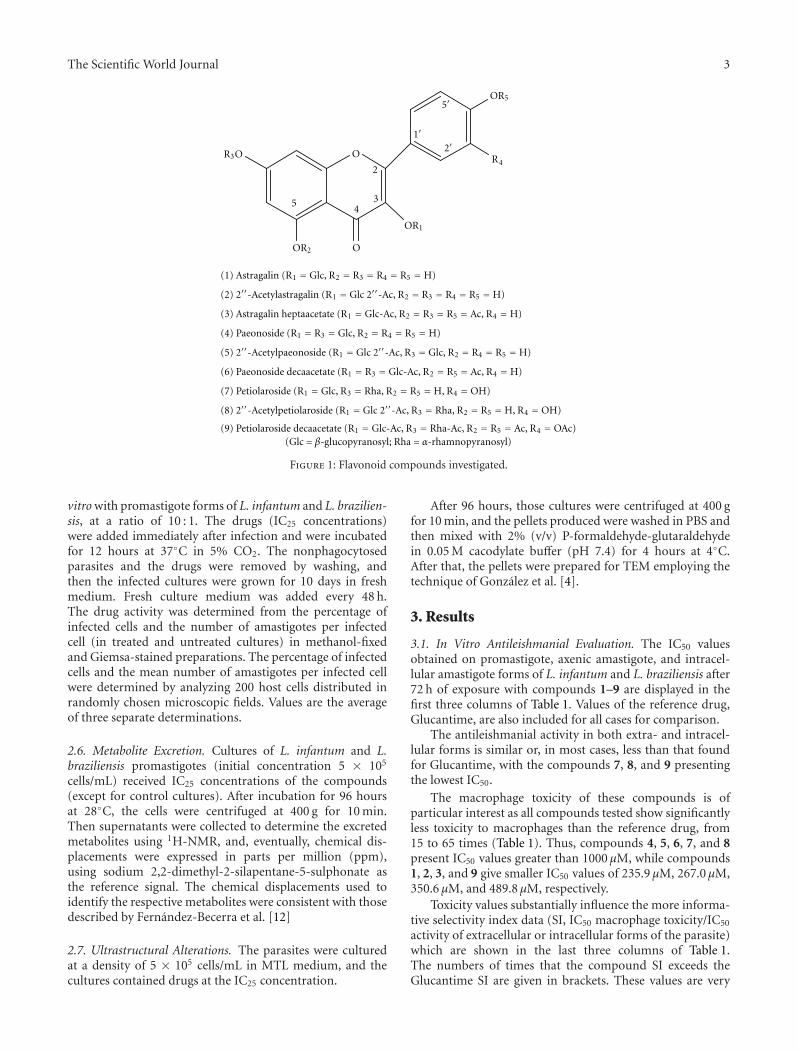
2.1. Plant Material. Aerial parts of the Delphinium staphis-agria were collected and processed as described previouslyDıaz et al. [10]. Nine flavonoids (1–9) were isolated,derivatized, and identified (Figure 1) [10].
2.2. Parasite Strain and Culture. L. infantum (MCAN/ES/2001/UCM-10) and L. braziliensis (MHOM/BR/1975/M2904) were cultivated in vitro in trypanosomes liquidmedium (MTL) with 10% inactive fetal bovine serum andwere kept in an air atmosphere at 28◦C, in Roux flasks(Corning, USA) with a surface area of 75 cm2, according tothe methodology described by Gonzalez et al. [4].
2.3. Cell Culture and Cytotoxicity Tests. J774.2 macrophages(ECACC number 91051511) were originally obtained from atumour in a female BALB/c rat in 1968. The cytotoxicity testfor macrophages was performed according to the method-ology of Gonzalez et al. [4]. After 72 hours of treatment,cell viability was determined by flow cytometry. Thus,100 μL/well of propidium iodide solution (100 mg/mL) wasadded and incubated for 10 min at 28◦C in darkness. After-wards, 100 μL/well of fluorescein diacetate (100 ng/mL) wasadded and incubated under the same conditions. Finally, thecells were recovered by centrifugation at 400 g for 10 min andthe precipitate washed with phosphate buffered saline (PBS).Flow cytometric analysis was performed with a FACSVantage
flow cytometer (Becton Dickinson). The percentage viabilitywas calculated in comparison with the control culture. TheIC50 was calculated using linear regression analysis from theKc values of the concentrations employed.
2.4. In Vitro Activity Assay
2.4.1. Promastigote Forms, Assay. The compounds obtainedwere dissolved in the culture medium, at dosages of 100,50, 25, 10, and 1 μM. The effects of each compound againstpromastigote forms were tested at 72 hours using a Neubauerhaemocytometric chamber. The antileishmanial effect isexpressed as the IC50, that is, the concentration required togive 50% inhibition, calculated by linear regression analysisfrom the Kc values of the concentrations employed.
2.4.2. Amastigote Forms, Assay. J774.2 macrophages weregrown in minimum essential medium (MEM) plus glu-tamine (2 mM), supplemented with 20% inactive fetalbovine serum, and were kept in a humidified atmosphere of95% air and 5% CO2 at 37◦C.
Cells were seeded at a density of 1 × 10⁴ cells/well in24-well microplates (Nunc) with rounded coverslips on thebottom and cultured for 2 days. Afterwards the cells wereinfected in vitro with promastigote forms of L. infantum andL. braziliensis, at a ratio of 10 : 1 during 24 hours. The non-phagocytosed parasites were removed by washing, and thenthe drugs (at 1, 10, 25, 50, 100 μM) were added. Macrophageswith the drugs were incubated for 72 hours at 37◦C in 5%CO2.
Drug activity was determined on the basis of number ofamastigotes in treated and untreated cultures in methanol-fixed and Giemsa-stained preparations. The number ofamastigotes was determined by analyzing 200 host cellsdistributed in randomly chosen microscopic fields. Theantileishmanial effect is expressed as the IC50 values are themeans of three separate determinations.
2.4.3. Axenic Amastigote Forms, Assay. Axenic amastigotesforms of L. infantum and L. braziliensis were culturedfollowing the methodology described previously by Morenoet al. [11]. Thus, promastigote transformation to amastigoteswas obtained after three days of culture in M199 medium(Invitrogen, Leiden, The Netherlands) supplemented with10% heat-inactivated FCS, 1 g/L β-alanine, 100 mg/L L-asparagine, 200 mg/L sacarose, 50 mg/L sodium pyruvate,320 mg/L malic acid, 40 mg/L fumaric acid, 70 mg/L succinicacid, 200 mg/L α-ketoglutaric acid, 300 mg/L citric acid,1.1 g/L sodium bicarbonate, 5 g/L MES, 0.4 mg/L hemin,and 10 mg/L gentamicin pH 5.4 at 37◦C. The effect of eachcompound against axenic amastigotes forms was tested at48 hours using a Neubauer haemocytometric chamber. Theantileishmanial effect is expressed as the IC50.
2.5. Infection Assay. J774.2 macrophage cells were grownunder the same conditions expressed in amastigote formsassay during two days. Afterwards, the cells were infected in
The Scientific World Journal 3
R3O
5
OR2 O
2
34
O
OR1
OR5
R4
1
2
5
(1) Astragalin (R1 = Glc, R2 = R3 = R4 = R5 = H)
(2) 2-Acetylastragalin (R1 = Glc 2-Ac, R2 = R3 = R4 = R5 = H)
(3) Astragalin heptaacetate (R1 = Glc-Ac, R2 = R3 = R5 = Ac, R4 = H)
(4) Paeonoside (R1 = R3 = Glc, R2 = R4 = R5 = H)
(5) 2-Acetylpaeonoside (R1 = Glc 2-Ac, R3 = Glc, R2 = R4 = R5 = H)
(6) Paeonoside decaacetate (R1 = R3 = Glc-Ac, R2 = R5 = Ac, R4 = H)
(7) Petiolaroside (R1 = Glc, R3 = Rha, R2 = R5 = H, R4 = OH)
(8) 2-Acetylpetiolaroside (R1 = Glc 2-Ac, R3 = Rha, R2 = R5 = H, R4 = OH)
(9) Petiolaroside decaacetate (R1 = Glc-Ac, R3 = Rha-Ac, R2 = R5 = Ac, R4 = OAc)(Glc = β-glucopyranosyl; Rha = α-rhamnopyranosyl)
Figure 1: Flavonoid compounds investigated.
vitro with promastigote forms of L. infantum and L. brazilien-sis, at a ratio of 10 : 1. The drugs (IC25 concentrations)were added immediately after infection and were incubatedfor 12 hours at 37◦C in 5% CO2. The nonphagocytosedparasites and the drugs were removed by washing, andthen the infected cultures were grown for 10 days in freshmedium. Fresh culture medium was added every 48 h.The drug activity was determined from the percentage ofinfected cells and the number of amastigotes per infectedcell (in treated and untreated cultures) in methanol-fixedand Giemsa-stained preparations. The percentage of infectedcells and the mean number of amastigotes per infected cellwere determined by analyzing 200 host cells distributed inrandomly chosen microscopic fields. Values are the averageof three separate determinations.
2.6. Metabolite Excretion. Cultures of L. infantum and L.braziliensis promastigotes (initial concentration 5 × 10⁵
cells/mL) received IC25 concentrations of the compounds(except for control cultures). After incubation for 96 hoursat 28◦C, the cells were centrifuged at 400 g for 10 min.Then supernatants were collected to determine the excretedmetabolites using 1H-NMR, and, eventually, chemical dis-placements were expressed in parts per million (ppm),using sodium 2,2-dimethyl-2-silapentane-5-sulphonate asthe reference signal. The chemical displacements used toidentify the respective metabolites were consistent with thosedescribed by Fernandez-Becerra et al. [12]
2.7. Ultrastructural Alterations. The parasites were culturedat a density of 5 × 10⁵ cells/mL in MTL medium, and thecultures contained drugs at the IC25 concentration.
After 96 hours, those cultures were centrifuged at 400 gfor 10 min, and the pellets produced were washed in PBS andthen mixed with 2% (v/v) P-formaldehyde-glutaraldehydein 0.05 M cacodylate buffer (pH 7.4) for 4 hours at 4◦C.After that, the pellets were prepared for TEM employing thetechnique of Gonzalez et al. [4].
3. Results
3.1. In Vitro Antileishmanial Evaluation. The IC50 valuesobtained on promastigote, axenic amastigote, and intracel-lular amastigote forms of L. infantum and L. braziliensis after72 h of exposure with compounds 1–9 are displayed in thefirst three columns of Table 1. Values of the reference drug,Glucantime, are also included for all cases for comparison.
The antileishmanial activity in both extra- and intracel-lular forms is similar or, in most cases, less than that foundfor Glucantime, with the compounds 7, 8, and 9 presentingthe lowest IC50.
The macrophage toxicity of these compounds is ofparticular interest as all compounds tested show significantlyless toxicity to macrophages than the reference drug, from15 to 65 times (Table 1). Thus, compounds 4, 5, 6, 7, and 8present IC50 values greater than 1000 μM, while compounds1, 2, 3, and 9 give smaller IC50 values of 235.9 μM, 267.0 μM,350.6 μM, and 489.8 μM, respectively.
Toxicity values substantially influence the more informa-tive selectivity index data (SI, IC50 macrophage toxicity/IC50
activity of extracellular or intracellular forms of the parasite)which are shown in the last three columns of Table 1.The numbers of times that the compound SI exceeds theGlucantime SI are given in brackets. These values are very

4 The Scientific World Journal
Ta
ble
1:In
vitr
oac
tivi
ty,
toxi
city
,an
dse
lect
ivit
yin
dex
fou
nd
for
the
astr
agal
in-b
ased
(1–3
),pa
eon
osid
e-ba
sed
(4–6
),an
dp
etio
laro
side
-bas
ed(7
–9)
deri
vati
ves
onex
trac
ellu
lar
and
intr
acel
lula
rfo
rms
ofLe
ishm
ania
spp.
(a)
Leis
hm
ania
infa
ntu
m
Com
pou
nds
Act
ivit
yIC
50(μ
M)a
bM
acro
phag
eto
xici
tyIC
50SI
c
Pro
mas
tigo
tefo
rms
Axe
nic
amas
tigo
tefo
rms
Intr
acel
lula
ram
asti
gote
form
sP
rom
asti
gote
form
sA
xen
icam
asti
gote
form
sIn
trac
ellu
lar
amas
tigo
tefo
rms
Glu
can
tim
e18
.0±
3.1
24.2±
2.6
30.0±
2.7
15.2±
1.3
0.8
0.6
0.5
135
.1±
3.3
29.2±
2.8
45.9±
2.7
235.
9±
17.9
6.7
(8)
8.1
(14)
5.1
(10)
234
.1±
1.6
32.7±
5.0
42.3±
3.9
267.
0±
11.0
7.8
(10)
8.2
(14)
6.3
(13)
340
.4±
3.4
29.2±
3.1
38.1±
1.6
350.
6±
26.8
8.7
(11)
12.0
(20)
9.2
(18)
425
.1±
1.9
26.9±
1.4
31.4±
2.0
>10
00.0±
60.0
39.8
(50)
37.2
(62)
31.8
(64)
524
.6±
3.7
29.8±
2.2
37.2±
3.5
>10
000±
71.4
40.7
(51)
33.6
(56)
26.9
(54)
623
.1±
4.6
30.5±
2.3
40.3±
2,4
>10
00.0±
82.0
43.3
(54)
32.8
(55)
24.8
(50)
724
.5±
1.1
19.4±
1.4
27.8±
1.4
>10
00.0±
55.6
40.8
(51)
51.5
(86)
36.0
(72)
819
.1±
0,9
18.4±
1.3
32.1±
2.0
>10
00.0±
38.9
52.4
(66)
54.3
(91)
31.2
(62)
922
.8±
1.7
21.5±
0.9
31.0±
1.7
489.
8±
22.4
21.5
(27)
22.8
(38)
15.8
(32)
(b)
Leis
hm
ania
braz
ilien
sis
Com
pou
nds
Act
ivit
yIC
50(μ
M)a
bM
acro
phag
eto
xici
tyIC
50SI
c
Pro
mas
tigo
tefo
rms
Axe
nic
amas
tigo
tefo
rms
Intr
acel
lula
ram
asti
gote
form
sP
rom
asti
gote
form
sA
xen
icam
asti
gote
form
sIn
trac
ellu
lar
amas
tigo
tefo
rms
Glu
can
tim
e25
.6±
1.6
30.4±
6.1
31.1±
3.0
15.2±
1.3
0.6
0.5
0,5
128
.1±
1.0
22.6±
1.7
32.2±
1.6
235.
9±
17.9
8.4
(14)
10.4
(21)
7.3
(15)
225
.1±
2.5
27.9±
2.7
30.2±
3.8
267.
0±
11.0
10.6
(18)
9.6
(19)
8.8
(18)
328
.4±
3.0
32.9±
3.0
41.9±
4.0
350.
6±
26.8
12.3
(21)
10.7
(21)
8.4
(17)
428
.1±
5.2
23.7±
2.1
32.7±
2.1
>10
00.0±
60.0
35.6
(59)
42.2
(84)
30.6
(61)
534
.1±
6.6
19.4±
0.9
50.9±
6.2
>10
00.0±
71.4
29.3
(49)
51.5
(103
)20
.0(4
0)6
31.3±
3.0
28.0±
1.6
30.4±
2.7
>10
00.0±
82.0
31.9
(53)
35.7
(71)
32.9
(66)
721
.1±
1.7
19.5±
0.8
33.9±
3.5
>10
00.0±
55.6
47.4
(79)
51.3
(103
)29
.5(5
9)8
29.0±
1.5
25.5±
0.7
20.1±
1.7
>10
00.0±
38.9
34.5
(58)
39.2
(78)
49.8
(100
)9
32.3±
3.3
24.6±
1.3
21.0±
1.3
489.
8±
22.4
15.2
(25)
19.9
(40)
23.3
(47)
Res
ult
sar
eav
erag
esof
thre
ese
para
tede
term
inat
ion
s.a IC
50=
the
con
cen
trat
ion
requ
ired
togi
ve50
%in
hib
itio
n,c
alcu
late
dby
linea
rre
gres
sion
anal
ysis
from
the
Kc
valu
esat
con
cen
trat
ion
sem
ploy
ed(1
,10,
25,5
0,an
d10
0μ
M).
bA
gain
stJ7
74.2
mac
roph
ages
afte
r72
hof
cult
ure
.c Se
lect
ivit
yin
dex=
IC50
mac
roph
ages
/IC
50ex
trac
ellu
lar
and
intr
acel
lula
rfo
rmof
the
para
site
.In
brac
kets
:nu
mbe
rof
tim
esth
atco
mpo
un
dSI
exce
eds
the
refe
ren
cedr
ug
SI.

The Scientific World Journal 5
Infe
ctio
n (
%)
80
64
48
32
16
00 1 2 3 4 5 6 7 8 9 10
Time (days)
(a)
Infe
ctio
n (
%)
80
64
48
32
16
00 1 2 3 4 5 6 7 8 9 10
Time (days)
(b)
Nu
mbe
r of
am
asti
gote
/mac
roph
age
50
40
30
20
10
00 1 2 3 4 5 6 7 8 9 10
Time (days)
(c)
Nu
mbe
r of
am
asti
gote
/mac
roph
age
70
56
42
28
14
00 1 2 3 4 5 6 7 8 9 10
Time (days)
(d)
Figure 2: Effects of flavonoids 4, 5, 6, 7, and 8 on the infection and growth rates of Leishmania spp. (a) rate of infection of L. infantum; (c)mean number of amastigotes per infected J774 A.2 macrophage cells of L. infantum; (b) rate of infection of L. braziliensis; (d) mean numberof amastigotes per infected J774 A.2 macrophage cells of L. braziliensis. (-�-, control; -�-, comp. 4; -�-, comp. 5; -�-, comp. 6; -♦-, comp.7; -∇-, comp. 8; -©-, Glucantime). At IC25 conc., values are means of three separate experiments.
illustrative of the in vitro potential of the compounds testedwith respect to the reference drug. Compounds 7, 8, 4, 5,and 6 showed the best SI for L. infantum (Table 1(a)), withindexes exceeding that of the reference drug SI by more than50 times. Compounds 1, 2, 3, and 9 were more effective thanGlucantime; however, SI values of 50 times or greater werenot reached, which is the criterion for a compound to beincluded in subsequent studies [13].
Similar results can be extracted from the L. braziliensisdata shown in Table 1(b). The compounds 7, 8, 4, and 6again gave the best SI results in the three assays performed,with values exceeding those of the reference drugs by 79,103, and 59 times in the case of compound 7, by 58, 78,and 100 times for compound 8, by 59, 84, and 61 times forcompound 4, and by 53, 71, and 66 times for compound 6.Although compound 5 did not give SI values greater thanor equal to 50 times those of the reference drugs, it wasincluded in subsequent studies because promastigote andintracellular amastigote SI values were close to 50 times that
of the reference drug, while the axenic amastigote SI was twotimes (Table 1(b)).
The effectiveness of the compounds on the infection rateand the intracellular replication of the amastigote forms wasdetermined by the infection assay (Figure 2). When selectedflavonoid compounds 4–8 and Glucantime were added attheir respective IC25 concentrations to macrophages infectedwith Leishmania spp. promastigote forms, the infectionrate decreased significantly after 12 h with respect to thecontrol measurement. The infection rate decreases followedthe trend 8 > 5 > 7 = 4 > 6 for L. infantum (Figure 2(a))and 8> 6> 5> 7> 4 for L. braziliensis (Figure 2(b)) withpercentages of inhibition capacity of 95%, 79%, 77%, and71%, respectively, in the case of L. infantum and 88%,82%, 79%, 69%, and 68%, respectively, in the case of L.braziliensis. These values are remarkably higher than thosefor inhibition by Glucantime (63% and 58% for L. infantumand L. braziliensis, resp.).

6 The Scientific World Journal
Metabolites excreted
0
5
10
15
20
25
30
35
40
45
Mal
ate
Succ
inat
e
Ace
tate
Ala
nin
e
Lac
tate
Eth
anol
Compound 4Compound 5Compound 6
Compound 7Compound 8
Var
iati
on in
th
e h
eigh
t of
th
e pe
aks
wit
hre
spec
t to
th
e co
ntr
ol te
st (
mm
)
−5
(a)
Compound 4Compound 5Compound 6
Compound 7Compound 8
Metabolites excreted
−10
−20
−30Var
iati
on in
th
e h
eigh
t of
th
e pe
aks
wit
hre
spec
t to
th
e co
ntr
ol te
st (
mm
)
Mal
ate
Succ
inat
e
Ace
tate
Ala
nin
e
Lac
tate
Eth
anol
0
10
20
30
40
50
(b)
Figure 3: Variation in the height of the peaks corresponding to catabolites excreted by L. infantum (a) and L. braziliensis (b) promastigoteforms in the presence of flavonoid derivatives with respect to the control test.
All five compounds were more effective than Glucantime(only 12% decrease for L. infantum and 33% decreasefor L. braziliensis) at decreasing the average number ofamastigotes per infected macrophage cells (Figures 2(c) and2(d)). Compound 8 was the most effective in L. infantum andL. braziliensis. The amastigote number decreases measuredwere as follows: 8 (78%) > 7 (49%) > 5 (45%) > 4 (37%) > 6(24%) for L. infantum and 8 (82%) > 5 (72%) > 6 (69%) > 4(61%) > 7 (55%) for L. braziliensis.
3.2. Metabolites Excretion Effect. After treatment of theparasites with compounds 4–8 at IC25, the excretion ofthe catabolites was clearly altered. Figure 3 displays themodifications observed in the height of the spectra peakscorresponding to the most representative final excretionproducts. From a careful examination of the data, it wouldappear that there are marked differences in the catabolicpathway. In the case of L. infantum, compounds 4, 5, 6, and8 gave similar spectra, with an increase in the excretion ofsuccinate, and acetate, followed by malate, alanine, lactateand ethanol. On the other hand, compound 7 causes anincrease in the production of malate, succinate, ethanol, andlactate, but a decrease of excreted acetate and alanine levels(Figure 3(a)).
In the case of L. braziliensis, the increase of excretedsuccinate was the most significant change when the parasiteswere treated with the compounds. Compounds 6, 7, and 8caused decreases in the excretion of acetate (Figure 3(b)).
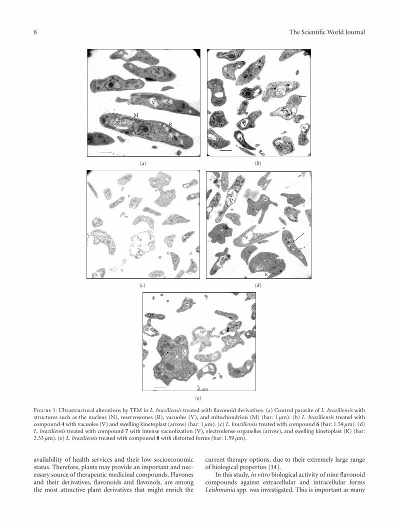
3.3. Ultrastructural Alterations. The transmission electronmicroscope evaluation of flavonoid compounds 4–8 againstLeishmania spp. promastigotes showed notable ultrastruc-tural alterations, as reflected in Figures 4 and 5 (panels b, c,d, e) with respect to the control (Figures 4(a) and 5(a)). All
compounds produced significant alterations to L. infantum,but the compounds most effective were 4 and 8, as illustratedin Figures 4(b) and 4(e). All of the compounds induced theformation of strongly electrodense inclusions that appearedinside and outside vacuoles with different sizes, or directly inthe cell cytoplasm. The major effect of compound 8 was thatmany of the parasites adopted distorted shapes with distortedcytoplasm, while no cytoplasmic organelles were visibledue to the altered appearance of promastigotes. However,large vacuoles containing cellular debris were visible. Withcompound 4 (Figure 4(b)), many of the parasites appeareddead, while another showed pronounced vacuolization,and many of these vacuoles contained strongly electro-dense inclusions. Compound 5 produced similar effects(Figure 4(c)). In the same way, compound 6 induced emptyvacuoles, lipid vacuoles, and a large number of electrodensevesicles (Figure 4(d)). By contrast, compound 7 showed noinduction of substantial alterations (graph not shown).
Studies of ultrastructural alterations made on L.braziliensis showed that the compounds produced changessimilar to those they produced in L. infantum. Another effectof the compounds was the expansion of the kinetoplast,which appeared swollen when treated with compound 4(Figure 5(b)). Compound 8 (Figure 5(e)) induced changes inthe shape of parasites, with them showing completely alteredforms. All these changes occurred after treatment withcompound 7 (Figure 5(d)), while compound 5 producedno significant changes compared to the control (data notshown).
4. Discussion
Many people living in rural areas have no easy access toconventional allopathic treatments, largely due to the limited

The Scientific World Journal 7
N
R
M
V
K
(a) (b)
V
(c)
V
LV
(d)
(e)
Figure 4: Ultrastructural alterations by TEM in L. infantum treated with the flavonoid derivatives. (a) Control parasite of L. infantumshowing organelles with their characteristic features such as the nucleus (N), kinetoplast (K), reservosomes (R), mitochondrion (M), andvacuoles (V) (bar: 1 μm). (b) L. infantum treated with compound 4. Electrodense organelles (arrow) (Bar: 1 μm). (c) L. infantum treated withcompound 5 with intense vacuolization (V) (Bar: 1.59 μm). (d) L. infantum treated with compound 6, vacuoles (V), lipid vacuoles (LV),and electrodense vesicles (arrow) (bar: 1.59 μm). (e) L. infantum treated with compound 8 with distorted parasites (arrow) (Bar: 1 μm).
8 The Scientific World Journal
N
M
R
V
(a)
V
(b)
(c)
V
K
V
(d)
(e)
Figure 5: Ultrastructural alterations by TEM in L. braziliensis treated with flavonoid derivatives. (a) Control parasite of L. braziliensis withstructures such as the nucleus (N), reservosomes (R), vacuoles (V), and mitochondrion (M) (bar: 1 μm). (b) L. braziliensis treated withcompound 4 with vacuoles (V) and swelling kinetoplast (arrow) (bar: 1 μm). (c) L. braziliensis treated with compound 6 (bar: 1.59 μm). (d)L. braziliensis treated with compound 7 with intense vacuolization (V), electrodense organelles (arrow), and swelling kinetoplast (K) (bar:2.33 μm). (e) L. braziliensis treated with compound 8 with distorted forms (bar: 1.59 μm).
availability of health services and their low socioeconomicstatus. Therefore, plants may provide an important and nec-essary source of therapeutic medicinal compounds. Flavonesand their derivatives, flavonoids and flavonols, are amongthe most attractive plant derivatives that might enrich the
current therapy options, due to their extremely large rangeof biological properties [14].
In this study, in vitro biological activity of nine flavonoidcompounds against extracellular and intracellular formsLeishmania spp. was investigated. This is important as many

The Scientific World Journal 9
studies of the activity of compounds against Leishmaniaspp. are performed on promastigote forms, which are mucheasier to work in vitro. However, since extracellular forms arenot the developed forms of the parasite in vertebrate hosts,evaluations made with these forms are merely indicativeof the potential leishmanicidal activity of the compoundstested. Consequently, a preliminary test using extracellularpromastigote forms should always be complemented by asubsequent evaluation using intracellular forms (amastigotesin vertebrate host cells), so that a better understanding of theactivity may be obtained [4].
The petiolaroside-based derivatives (7–9) presented thelowest IC50values against extra- and intracellular forms.
Previously, the nine flavonoids investigated here weretested against epimastigote forms of Trypanosoma cruzi, andthe most effective compounds were astragalin (1) and twoacetylated derivatives (2 and 3). These had significantly lowerIC50 values compared with benznidazole [15]. By contrast, inthis study, we saw that the astragalin and its derivates werethe least effective agents against Leishmania spp.
Petiolaroside, 2′′-acetylpetiolaroside (7 and 8, resp.), andpaeonoside and its derivates (4, 5, and 6) showed the bestselectivity indexes. These indexes exceeded the reference drugSI by more than 50 times. The astragalin and its derivates(1, 2, and 3) and petiolaroside decacetate (9) were all moreeffective than Glucantime.
If we compare the results obtained for the infection rateand the number of amastigotes per infected macrophagecells for both Leishmania species, it can be concluded that2′′-acetylpetiolaroside (8) is clearly the most active. Theacetylated derivatives 2′′-acetylpetiolaroside (8) and 2′′-acetylpaeonoside (5) are more active than petiolaroside (7)and paeonoside (4), respectively.
As far as is known to date, none of the trypanosomatidsstudied are capable of completely degrading glucose to CO2
under aerobic conditions. When this occurs, it results in theexcretion of a major part of the carbon skeleton into themedium as fermented metabolites, which can differ accord-ing to the employed species [16]. 1H-NMR spectra enabledus to determine the fermented metabolites excreted by theparasites during their in vitro culture. The final products ofglucose catabolism in Leishmania are usually CO2, succinate,acetate, L-lactate, pyruvate, L-alanine, and ethanol [17]. Aswith T. cruzi [18], one of the major metabolites excreted byLeishmania spp. is succinate, with the main role likely tobe to maintain the glycosomal redox balance by providingtwo glycosomal oxidoreductase enzymes. These enzymesallow reoxidation of NADH produced by glyceraldehyde-3-phosphate dehydrogenase in the glycolytic pathway. Succinicfermentation offers one significant advantage as it requiresonly half the produced phosphoenolpyruvate (PEP) tomaintain the NAD+/NADH balance. The remaining PEPis converted to acetate, depending on the species underconsideration.
The majority of metabolites excreted by L. infantum aresuccinate and acetate. In the case of L. infantum, variationsin the final catabolism products seem to be dependent onthe structural aspects of the compounds assayed. These dataagree well with those of other authors [18].
Analyses of in vitro and in vivo results of Trypanosomacruzi showed that the best potential flavonoids to treatChagas disease were the acetylated compounds 2, 5, and 6[15]. The present work confirmed this conclusion in the caseof visceral leishmaniasis (L. infantum) and mucocutaneousleishmaniasis (L. braziliensis). Moreover, these data areconsistent with others previously published, which showedthat several acetylated flavonoids derived from Consolidaoliveriana were very active in vitro against both extracellularand intracellular forms of L. (V.) peruviana and L. (V.)braziliensis [9].
In conclusion, this study has demonstrated that flavonoidderivatives are active against L. infantum and L. braziliensis.These results support further investigation of flavonoidcompounds as potential agents against Leishmaniasis.
Acknowledgments
The authors would like to express their thanks to M.J. Martınez-Guerrero, J. Lazuen-Alcon and E. Onorato-Gutierrez for their expert technical help in transmissionelectron microscopy, flow cytometry and nuclear magneticresonance spectroscopy, respectively. They also appreciatethe help of E. Guerrero Lopez and C. Anaya Romero inculture media preparation and in vitro culture. This workwas financially supported by the Consolider Ingenio ProjectCSD2010-00065 (Spain).
References
[1] “Control of the leishmaniases,” WHO Technical Report 949,World Health Organization, Geneva, Switzerland, 2010.
[2] O. Kayser, A. F. Kiderlen, and S. L. Croft, “Natural productsas antiparasitic drugs,” Parasitology Research, vol. 90, no. 2, pp.S55–S62, 2003.
[3] M. J. Chan-Bacab and L. M. Pena-Rodrıguez, “Plant nat-ural products with leishmanicidal activity,” Natural ProductReports, vol. 18, no. 6, pp. 674–688, 2001.
[4] P. Gonzalez, C. Marın, I. Rodrıguez-Gonzalez et al., “In vitroactivity of C20-diterpenoid alkaloid derivatives in promastig-otes and intracellular amastigotes of Leishmania infantum,”International Journal of Antimicrobial Agents, vol. 25, no. 2, pp.136–141, 2005.
[5] C. Di Giorgio, F. Delmas, E. Ollivier, R. Elias, G. Balansard,and P. Timon-David, “In vitro activity of the β-carboline alka-loids harmane, harmine, and harmaline toward parasites ofthe species Leishmania infantum,” Experimental Parasitology,vol. 106, no. 3-4, pp. 67–74, 2004.
[6] A. Dutta, S. Bandyopadhyay, C. Mandal, and M. Chatterjee,“Aloe vera leaf exudate induces a caspase-independent celldeath in Leishmania donovani promastigotes,” Journal ofMedical Microbiology, vol. 56, no. 5, pp. 629–636, 2007.
[7] D. J. Newman and G. M. Cragg, “Natural products as sourcesof new drugs over the last 25 years,” Journal of NaturalProducts, vol. 70, no. 3, pp. 461–477, 2007.
[8] F. Torres, J. Quintana, J. G. Dıaz, A. J. Carmona, and F. Estevez,“Trifolin acetate-induced cell death in human leukemia cellsis dependent on caspase-6 and activates the MAPK pathway,”Apoptosis, vol. 13, no. 5, pp. 716–728, 2008.
[9] C. Marın, S. Boutaleb-Charki, J. G. Dıaz et al., “Antileishma-niasis activity of flavonoids from Consolida oliveriana,” Journalof Natural Products, vol. 72, no. 6, pp. 1069–1074, 2009.
10 The Scientific World Journal
[10] J. G. Dıaz, A. J. Carmona, P. P. de Paz, and W. Herz,“Acylated flavonol glycosides from Delphinium staphisagria,”Phytochemistry Letters, vol. 1, no. 2, pp. 125–129, 2008.
[11] D. Moreno, D. Plano, Y. Baquedano, A. Jimenez-Ruiz, J.Antonio Palop, and C. Sanmartın, “Antileishmanial activity ofimidothiocarbamates and imidoselenocarbamates,” Parasitol-ogy Research, vol. 108, no. 1, pp. 233–239, 2011.
[12] C. Fernandez-Becerra, M. Sanchez-Moreno, A. Osuna, and F.R. Opperdoes, “Comparative aspects of energy metabolismin plant trypanosomatids,” Journal of Eukaryotic Microbiology,vol. 44, no. 5, pp. 523–529, 1997.
[13] S. Nwaka and A. Hudson, “Innovative lead discovery strategiesfor tropical diseases,” Nature Reviews Drug Discovery, vol. 5,no. 11, pp. 941–955, 2006.
[14] L. H. Cazarolli, L. Zanatta, E. H. Alberton et al., “Flavonoids:prospective drug candidates,” Mini Reviews in MedicinalChemistry, vol. 8, no. 13, pp. 1429–1440, 2008.
[15] C. Marın, I. Ramırez-Macıas, A. Lopez-Cespedes et al., “Invitro and in vivo trypanocidal activity of flavonoids fromDelphinium staphisagria against chagas disease,” Journal ofNatural Products, vol. 74, no. 4, pp. 744–750, 2011.
[16] F. Bringaud, L. Riviere, and V. Coustou, “Energy metabolismof trypanosomatids: adaptation to available carbon sources,”Molecular and Biochemical Parasitology, vol. 149, no. 1, pp. 1–9, 2006.
[17] J. J. Cazzulo, “Aerobic fermentation of glucose by trypanoso-matids,” FASEB Journal, vol. 6, no. 13, pp. 3153–3161, 1992.
[18] M. L. Ginger, “Trypanosomatid biology and euglenozoanevolution: new insights and shifting paradigms revealedthrough genome sequencing,” Protist, vol. 156, no. 4, pp. 377–392, 2005.