Genome-wide association analysis identifies multiple loci related to resting heart rate
10
Genome-wide association analysis identifies multiple loci related to resting heart rate Mark Eijgelsheim 1, { , Christopher Newton-Cheh 5,7,8, { , Nona Sotoodehnia 9,10, { , Paul I.W. de Bakker 7,15, { , Martina Mu ¨ ller 16,17,18, { , Alanna C. Morrison 19, { , Albert V. Smith 20, { , Aaron Isaacs 1, { , Serena Sanna 21, { , Marcus Do ¨ rr 22, { , Pau Navarro 25, { , Christian Fuchsberger 26, { , Ilja M. Nolte 27, { , Eco J.C. de Geus 28, { , Karol Estrada 2 , Shih-Jen Hwang 8 , Joshua C. Bis 10,11 , Ina-Maria Ru ¨ ckert 17 , Alvaro Alonso 29 , Lenore J. Launer 30 , Jouke Jan Hottenga 28 , Fernando Rivadeneira 1,2 , Peter A. Noseworthy 5 , Kenneth M. Rice 12 , Siegfried Perz 31 , Dan E. Arking 32,33 , Tim D. Spector 34 , Jan A. Kors 3 , Yurii S. Aulchenko 1 , Kirill V. Tarasov 35 , Georg Homuth 23 , Sarah H. Wild 36 , Fabio Marroni 26,37 , Christian Gieger 17 , Carmilla M. Licht 38 , Ronald J. Prineas 39 , Albert Hofman 1,40 , Jerome I. Rotter 41 , Andrew A. Hicks 26 , Florian Ernst 23 , Samer S. Najjar 35 , Alan F. Wright 25 , Annette Peters 17 , Ervin R. Fox 42 , Ben A. Oostra 4 , Heyo K. Kroemer 43 , David Couper 44 , Henry Vo ¨ lzke 24 , Harry Campbell 36 , Thomas Meitinger 45,46 , Manuela Uda 21 , Jacqueline C.M. Witteman 1,40 , Bruce M. Psaty 10,11,13,14,47 , H-Erich Wichmann 16,17,48 , Tamara B. Harris 30 , Stefan Ka ¨a ¨b 16 , David S. Siscovick 10,11,13 , Yalda Jamshidi 34,49 , Andre ´ G. Uitterlinden 1,2,40 , Aaron R. Folsom 29 , Martin G. Larson 8,50 , James F. Wilson 36 , Brenda W. Penninx 38 , Harold Snieder 27,34 , Peter P. Pramstaller 26,51,52 , Cornelia M. van Duijn 1,40 , Edward G. Lakatta 35 , Stephan B. Felix 22 , Vilmundur Gudnason 20,53 , Arne Pfeufer 45,46 , Susan R. Heckbert 10,13,47 , Bruno H.Ch. Stricker 1,2,3,40,54, ∗ , Eric Boerwinkle 19 and Christopher J. O’Donnell 6,8,55,∗ 1 Department of Epidemiology, 2 Department of Internal Medicine, 3 Department of Medical Informatics and 4 Department of Clinical Genetics, Erasmus Medical Center, Rotterdam, The Netherlands, 5 Center for Human Genetic Research, Cardiovascular Research Center and 6 Cardiology Division, Massachusetts General Hospital, Boston, MA, USA, 7 Program in Medical Population Genetics, Broad Institute of Harvard and MIT, Cambridge, MA, USA, 8 National Heart, Lung and Blood Institute’s Framingham Heart Study, Framingham, MA, USA, 9 Division of Cardiology, Department of Medicine, School of Medicine, 10 Cardiovascular Health Research Unit, Metropolitan Park East Tower, 11 Department of Medicine, 12 Department of Biostatistics, 13 Department of Epidemiology and 14 Department of Health Services, University of Washington, Seattle, WA, USA, 15 Division of Genetics, Department of Medicine, Brigham’s and Women’s Hospital, Boston, MA, USA, 16 Institute of Epidemiology, Helmholtz Center Munich, German Research Center for Environmental Health, Neuherberg, Germany, 17 Institute of Medical Informatics, Biometry and Epidemiology, Chair of Epidemiology and 18 Department of Medicine I, University Hospital Grosshadern, Ludwig- Maximilians-Universita ¨t, Munich, Germany, 19 Human Genetics Center, University of Texas Health Science Center, Houston, TX, USA, 20 Icelandic Heart Association Research Institute, Kopavogur, Iceland, 21 Istituto di Neurogenetica e Neurofarmacologia, Consiglio Nazionale delle Ricerche, Cittadella Universitaria di Monserrato, Monserrato, Cagliari, Italy, 22 Department of Internal Medicine B, 23 Interfaculty Institute for Genetics and Functional Genomics and # The Author 2010. Published by Oxford University Press. All rights reserved. For Permissions, please email: [email protected] † These authors contributed equally to this work. ‡ These authors contributed equally to this work. ∗ To whom correspondence should be addressed at: Department of Epidemiology, Erasmus Medical Center, PO Box 2040, 3000 CA, Rotterdam, The Netherlands. Tel: +31 107043489; Fax: +31 107044657; Email: [email protected] (B.H.Ch.S.); Framingham Heart Study, 73 Mt Wayte Avenue, Suite No. 2, Framingham, MA 01702-5827, USA. Tel: +1 5089353435; Fax: +1 5086261262; Email: [email protected] (C.J.O.). Human Molecular Genetics, 2010, Vol. 19, No. 19 3885–3894 doi:10.1093/hmg/ddq303 Advance Access published on July 16, 2010 at Vrije Universiteit, Library on September 24, 2010 hmg.oxfordjournals.org Downloaded from
-
Upload
hms-harvard -
Category
Documents
-
view
0 -
download
0
Transcript of Genome-wide association analysis identifies multiple loci related to resting heart rate

Genome-wide association analysis identifiesmultiple loci related to resting heart rate
Mark Eijgelsheim1,{, Christopher Newton-Cheh5,7,8,{, Nona Sotoodehnia9,10,{,
Paul I.W. de Bakker7,15,{, Martina Muller16,17,18,{, Alanna C. Morrison19,{, Albert V. Smith20,{,
Aaron Isaacs1,{, Serena Sanna21,{, Marcus Dorr22,{, Pau Navarro25,{, Christian Fuchsberger26,{,
Ilja M. Nolte27,{, Eco J.C. de Geus28,{, Karol Estrada2, Shih-Jen Hwang8, Joshua C. Bis10,11,
Ina-Maria Ruckert17, Alvaro Alonso29, Lenore J. Launer30, Jouke Jan Hottenga28,
Fernando Rivadeneira1,2, Peter A. Noseworthy5, Kenneth M. Rice12, Siegfried Perz31,
Dan E. Arking32,33, Tim D. Spector34, Jan A. Kors3, Yurii S. Aulchenko1, Kirill V. Tarasov35,
Georg Homuth23, Sarah H. Wild36, Fabio Marroni26,37, Christian Gieger17, Carmilla M. Licht38,
Ronald J. Prineas39, Albert Hofman1,40, Jerome I. Rotter41, Andrew A. Hicks26, Florian Ernst23,
Samer S. Najjar35, Alan F. Wright25, Annette Peters17, Ervin R. Fox42, Ben A. Oostra4,
Heyo K. Kroemer43, David Couper44, Henry Volzke24, Harry Campbell36, Thomas Meitinger45,46,
Manuela Uda21, Jacqueline C.M. Witteman1,40, Bruce M. Psaty10,11,13,14,47,
H-Erich Wichmann16,17,48, Tamara B. Harris30, Stefan Kaab16, David S. Siscovick10,11,13,
Yalda Jamshidi34,49, Andre G. Uitterlinden1,2,40, Aaron R. Folsom29, Martin G. Larson8,50,
James F. Wilson36, Brenda W. Penninx38, Harold Snieder27,34, Peter P. Pramstaller26,51,52,
Cornelia M. van Duijn1,40, Edward G. Lakatta35, Stephan B. Felix22, Vilmundur Gudnason20,53,
Arne Pfeufer45,46, Susan R. Heckbert10,13,47, Bruno H.Ch. Stricker1,2,3,40,54,∗, Eric Boerwinkle19
and Christopher J. O’Donnell6,8,55,∗
1Department of Epidemiology, 2Department of Internal Medicine, 3Department of Medical Informatics and4Department of Clinical Genetics, Erasmus Medical Center, Rotterdam, The Netherlands, 5Center for Human Genetic
Research, Cardiovascular Research Center and 6Cardiology Division, Massachusetts General Hospital, Boston, MA,
USA, 7Program in Medical Population Genetics, Broad Institute of Harvard and MIT, Cambridge, MA, USA, 8National
Heart, Lung and Blood Institute’s Framingham Heart Study, Framingham, MA, USA, 9Division of Cardiology,
Department of Medicine, School of Medicine, 10Cardiovascular Health Research Unit, Metropolitan Park East Tower,11Department of Medicine, 12Department of Biostatistics, 13Department of Epidemiology and 14Department of Health
Services, University of Washington, Seattle, WA, USA, 15Division of Genetics, Department of Medicine, Brigham’s and
Women’s Hospital, Boston, MA, USA, 16Institute of Epidemiology, Helmholtz Center Munich, German Research
Center for Environmental Health, Neuherberg, Germany, 17Institute of Medical Informatics, Biometry and
Epidemiology, Chair of Epidemiology and 18Department of Medicine I, University Hospital Grosshadern, Ludwig-
Maximilians-Universitat, Munich, Germany, 19Human Genetics Center, University of Texas Health Science Center,
Houston, TX, USA, 20Icelandic Heart Association Research Institute, Kopavogur, Iceland, 21Istituto di Neurogenetica e
Neurofarmacologia, Consiglio Nazionale delle Ricerche, Cittadella Universitaria di Monserrato, Monserrato, Cagliari,
Italy, 22Department of Internal Medicine B, 23Interfaculty Institute for Genetics and Functional Genomics and
# The Author 2010. Published by Oxford University Press. All rights reserved.For Permissions, please email: [email protected]
†These authors contributed equally to this work.‡These authors contributed equally to this work.
∗To whom correspondence should be addressed at: Department of Epidemiology, Erasmus Medical Center, PO Box 2040, 3000 CA, Rotterdam,The Netherlands. Tel: +31 107043489; Fax: +31 107044657; Email: [email protected] (B.H.Ch.S.); Framingham Heart Study, 73 Mt WayteAvenue, Suite No. 2, Framingham, MA 01702-5827, USA. Tel: +1 5089353435; Fax: +1 5086261262; Email: [email protected] (C.J.O.).
Human Molecular Genetics, 2010, Vol. 19, No. 19 3885–3894doi:10.1093/hmg/ddq303Advance Access published on July 16, 2010
at Vrije U
niversiteit, Library on Septem
ber 24, 2010hm
g.oxfordjournals.orgD
ownloaded from
24Institute for Community Medicine, Ernst-Moritz-Arndt-University, Greifswald, Germany, 25MRC Human Genetics
Unit, Institute of Genetics and Molecular Medicine, Western General Hospital, Edinburgh, UK, 26Institute of Genetic
Medicine, European Academy Bozen/Bolzano (EURAC), Bolzano, Italy (Affiliated Institute of the University of Lubeck,
Germany), 27Unit of Genetic Epidemiology and Bioinformatics, Department of Epidemiology, University Medical
Center Groningen, Groningen, The Netherlands, 28Department of Biological Psychology, VU University, Amsterdam,
The Netherlands, 29Division of Epidemiology and Community Health, School of Public Health, University of Minnesota,
Minneapolis, MN, USA, 30National Institute of Aging’s Laboratory for Epidemiology, Demography, and Biometry,
Bethesda, MD, USA, 31Institute of Biological and Medical Imaging, Helmholtz Zentrum Munchen, Munich, Germany,32McKusick-Nathans Institute of Genetic Medicine and 33Division of Cardiology, Department of Medicine, Johns
Hopkins University School of Medicine, Baltimore, MD, USA, 34Department of Twin Research and Genetic
Epidemiology Unit, St Thomas’ Campus, King’s College London, St Thomas’ Hospital, London, UK, 35Laboratory of
Cardiovascular Science, Intramural Research Program, National Institute on Aging, National Institutes of Health,
Baltimore, USA, 36Centre for Population Health Sciences, University of Edinburgh, Edinburgh, UK, 37Institute of
Applied Genomics, Udine, Italy, 38Department of Psychiatry, VU Medical Centre, Amsterdam, The Netherlands,39EPICARE Center, Winston, Salem, NC, USA, 40Netherlands Genomics Initiative-sponsored Netherlands
Consortium for Healthy Ageing, Rotterdam, The Netherlands, 41Medical Genetics Institute, Cedars-Sinai Medical
Center, Los Angeles, CA, USA, 42Cardiology Division, University of Mississippi Medical Center, Jackson, MS, USA,43Department of Pharmacology, Center for Pharmacology and Experimental Therapeutics, Greifswald, Germany,44Department of Biostatistics, University of North Carolina, Chapel Hill, NC, USA, 45Institute of Human Genetics,
Helmholtz Center Munich, Munich, Germany, 46Institute of Human Genetics, Klinikum Rechts der Isar der
Technischen Universitat Munchen, Munich, Germany, 47Group Health Research Institute, Seattle, WA, USA,48Klinikum Grosshadern, Munich, Germany, 49St George’s University of London, Cranmer Terrace, London, UK,50Department of Mathematics and Statistics, Boston University, Boston, MA, USA, 51Department of Neurology,
General Central Hospital, Bolzano, Italy, 52Department of Neurology, University of Lubeck, Lubeck, Germany,53University of Iceland, Reykjavik, Iceland, 54Inspectorate of Health Care, The Hague, The Netherlands and 55National
Heart, Lung and Blood Institute, Bethesda, MD, USA
Received April 9, 2010; Revised and Accepted July 13, 2010
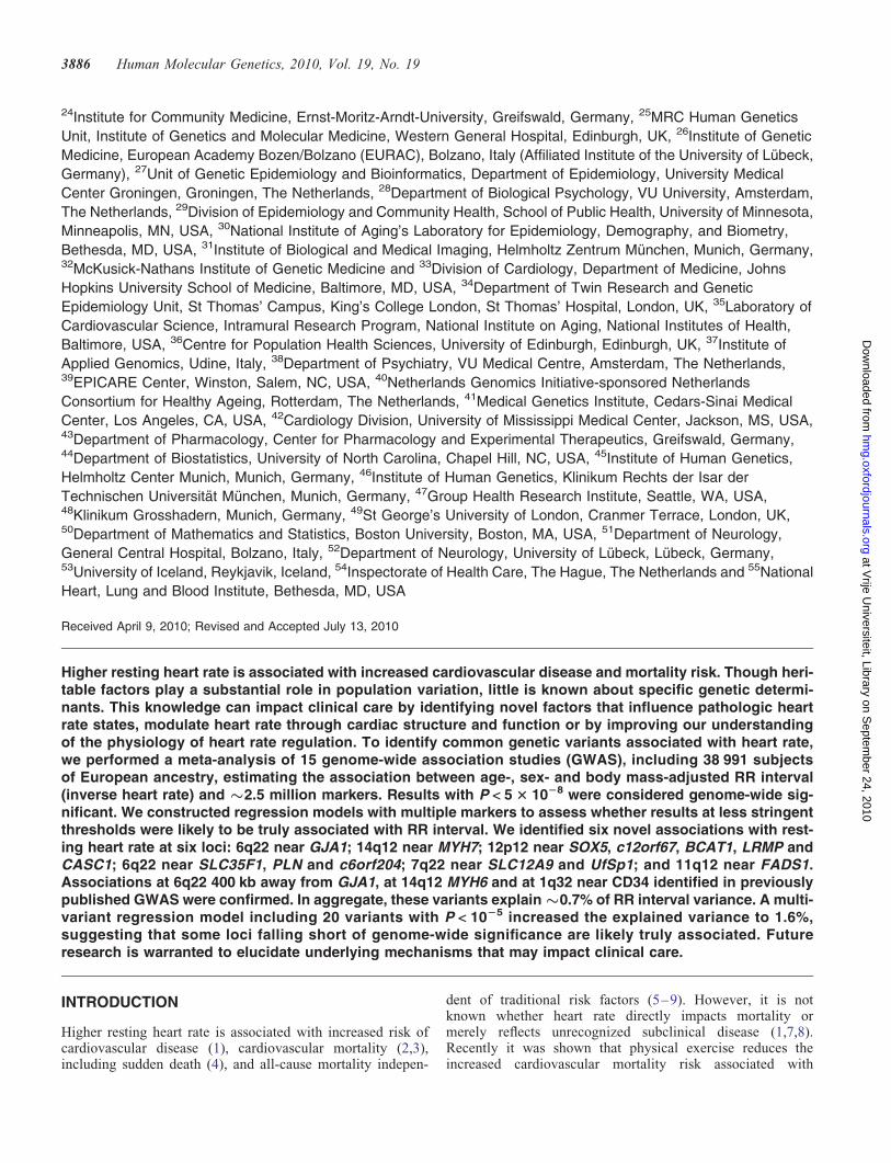
Higher resting heart rate is associated with increased cardiovascular disease and mortality risk. Though heri-table factors play a substantial role in population variation, little is known about specific genetic determi-nants. This knowledge can impact clinical care by identifying novel factors that influence pathologic heartrate states, modulate heart rate through cardiac structure and function or by improving our understandingof the physiology of heart rate regulation. To identify common genetic variants associated with heart rate,we performed a meta-analysis of 15 genome-wide association studies (GWAS), including 38 991 subjectsof European ancestry, estimating the association between age-, sex- and body mass-adjusted RR interval(inverse heart rate) and ∼2.5 million markers. Results with P < 5 3 1028 were considered genome-wide sig-nificant. We constructed regression models with multiple markers to assess whether results at less stringentthresholds were likely to be truly associated with RR interval. We identified six novel associations with rest-ing heart rate at six loci: 6q22 near GJA1; 14q12 near MYH7; 12p12 near SOX5, c12orf67, BCAT1, LRMP andCASC1; 6q22 near SLC35F1, PLN and c6orf204; 7q22 near SLC12A9 and UfSp1; and 11q12 near FADS1.Associations at 6q22 400 kb away from GJA1, at 14q12 MYH6 and at 1q32 near CD34 identified in previouslypublished GWAS were confirmed. In aggregate, these variants explain ∼0.7% of RR interval variance. A multi-variant regression model including 20 variants with P < 1025 increased the explained variance to 1.6%,suggesting that some loci falling short of genome-wide significance are likely truly associated. Futureresearch is warranted to elucidate underlying mechanisms that may impact clinical care.
INTRODUCTION
Higher resting heart rate is associated with increased risk ofcardiovascular disease (1), cardiovascular mortality (2,3),including sudden death (4), and all-cause mortality indepen-
dent of traditional risk factors (5–9). However, it is notknown whether heart rate directly impacts mortality ormerely reflects unrecognized subclinical disease (1,7,8).Recently it was shown that physical exercise reduces theincreased cardiovascular mortality risk associated with
3886 Human Molecular Genetics, 2010, Vol. 19, No. 19
at Vrije U
niversiteit, Library on Septem
ber 24, 2010hm
g.oxfordjournals.orgD
ownloaded from
higher heart rate (3). This suggests that heart rate is a clinicallyrelevant and potentially modifiable risk factor.
Heart rate is a complex trait, determined by multipleenvironmental, genetic and other endogenous factors. Heredityplays a substantial role in the inter-individual variation ofresting heart rate, accounting for 26–32% of heart rate vari-ation in prior studies (10–13). Twin studies report evenhigher heritability estimates up to 55–63% (14,15). Candidategene approaches have identified multiple loci associated withheart rate (12,13,16–20), but the results have been inconsist-ent and difficult to replicate. Genome-wide genotypingarrays of single-nucleotide polymorphisms (SNPs) assaycommon variation in the human genome and can identifygenetic variants with modest influences on a complex traitsuch as heart rate, as shown by two recent genome-wideassociation studies (GWAS) that identified common variationat or near MYH6, GJA1 and CD34 associated with heart rate(21,22). These chromosomal loci identified in an unbiasedgenome-wide study may represent novel risk factors for cardi-ovascular disease outcomes. This knowledge may also have animpact on clinical care (i) by identifying novel factors thatcause pathologic heart rate states (such as sick sinus syndromeor other arrhythmias), (ii) by identifying factors that influencecardiac structure or function (e.g. stroke volume) and therebymodulate heart rate (since cardiac output ¼ heart rate × strokevolume) or (iii) by improving our understanding of the physio-logic basis of heart rate regulation. Altogether this will gener-ate insights into the underlying mechanisms of heart rate as awell-established, but poorly understood, risk indicator for car-diovascular disease and mortality.
To identify additional genetic determinants of heart rate, weperformed a meta-analysis of GWAS of resting heart rate,measured as the RR interval on the electrocardiogram(ECG) in 38 991 individuals of European ancestry derivedfrom 15 studies in the RRGEN consortium.
RESULTS
There were 38 991 individuals available for genotype–pheno-type association analysis after exclusions. Subject character-istics are shown in Table 1. While cohort-specific quantile–quantile plots of P-value distributions approximated expec-tations under the null, the meta-analysis of all resultsshowed a clear excess of low P-values (SupplementaryMaterial, Fig. S1). Study-specific genomic inflation lambdavalues ranged from 0.98 to 1.05, suggesting that populationstratification or other technical artifacts were minimal (Sup-plementary Material, Table S1).
Meta-analysis of results from all studies resulted in 156SNPs reaching the pre-specified genome-wide statistical sig-nificance level (P , 5 × 1028; Supplementary Material,Table S2) before applying post-meta-analysis genomiccontrol. In total, nine independent genome-wide significantsignals were observed across seven chromosomal loci(Table 2, Fig. 1), with all but one locus harboring multipleSNPs reaching the genome-wide significance threshold (Sup-plementary Material, Table S2; Fig. 2A–E).
Genome-wide significant associations were observed at 6q22nearest to GJA1; 14q12 near MYH6 and MYH7; 12p12 near
SOX5, c12orf67, BCAT1, LRMP and CASC1; 6q22 nearSLC35F1, 6orf204 and PLN (.3 Mb away from 6q22 GJA1);7q22 near SLC12A9; 11q12 near FADS1; and 1q32 nearCD34. The genomic inflation lambda value of the meta-analysiswas 1.05. After applying post-meta-analysis genomic control,the signal at 1q34 near CD34 and a second independent signalat 6q22 GJA1 lost genome-wide significance.
The strongest association was observed for an intergenicSNP 370 kb upstream of GJA1 at 6q22 rs9398652 [minorallele frequency (MAF) ¼ 0.10] with a 12.6 ms shorter RRinterval per minor A allele, which is equivalent to a0.95 bpm higher heart rate based on the baseline mean heartrate of 66.8 bpm, across all studies (P ¼ 8.0 × 10216,Pmeta-gc ¼ 3.8 × 10215; Table 2, Fig. 2A). Cho et al. (21)observed genome-wide significant association betweenrs12110693 and pulse rate in an Asian population-basedGWAS. This SNP is in perfect linkage disequilibrium (r2 ¼1) with rs939862 in both Caucasian and Asian HapMap refer-ence populations and thus reflects the same signal 370 kb fromGJA1. A second SNP only 8 kb away from GJA1 also reachedgenome-wide significance pre-genomic control, rs11154022(MAF ¼ 0.33) with a 5.8 ms longer RR interval (0.43 bpmlower heart rate) per A allele (P ¼ 3.5 × 1028, Pmeta-gc ¼7.2 × 1028; Fig. 2B, Table 2). This SNP had low correlationwith rs9398652 (r2 ¼ 0.006 in HapMap CEU), suggesting anovel-independent association signal. Since HapMap islimited to 90 subjects, we assessed the linkage disequilibriumin our data. All observed r2 values ranged between 0.0001 and0.004, which is lower than seen in the HapMap CEU referencepopulation. Conditional analysis confirmed that these twosignals are independent with Pconditional ¼ 2.4 × 10211 andPconditional ¼ 3.3 × 1028, respectively, for rs9398652 andrs1154022 in the subset (n ≤ 33 846) used for this analysis.
The second locus with two signals in low correlation reach-ing genome-wide significance is located on chromosome14q12. The strongest association at this locus was observedfor rs452036 located in intron 19 of MYH6 [MAF ¼ 0.36,with a 7.8 ms shorter RR interval (0.58 bpm higher heartrate) per C allele, P ¼ 8.1 × 10215, Pmeta-gc ¼ 3.8 × 10214,Fig. 2C]. This replicates the finding by Holm et al. (22) whopreviously described an association between rs452036 andheart rate. The non-synonymous coding variant rs365990,which results in an amino acid change at position 1101(Alanine . Valine) of the MYH6 gene product, is a possiblefunctional variant since it showed strong correlation with(r2 ¼ 0.96 in HapMap CEU) and association results are indis-tinguishable from rs452036 (Table 2). A second SNP locatednear MYH7 rs223116 (MAF ¼ 0.24), associated with a7.4 ms shorter RR interval per A allele (0.55 bpm higherheart rate, P ¼ 1.1 × 1028, Pmeta-gc ¼ 2.5 × 1028, Fig. 2D)was in low correlation with rs452036 (r2 ¼ 0.08 in HapMapCEU) and reflects a novel association. We observed r2 valuessimilar to HapMap CEU with values ranging from 0.03 to0.08. Conditional analysis confirmed the presence of two inde-pendent signals with Pconditional ¼ 7.9 × 10214 and Pconditional¼3.7 × 1024 for rs452036 and rs223116, respectively.
For the other five loci, only a single signal of association metour genome-wide significance threshold, meaning that all othergenome-wide significant SNPs have an r2 . 0.1 in HapMapCEU to the most significant SNP at that locus. For these loci,
Human Molecular Genetics, 2010, Vol. 19, No. 19 3887
at Vrije U
niversiteit, Library on Septem
ber 24, 2010hm
g.oxfordjournals.orgD
ownloaded from

the MAF of the index SNPs ranged from 15 to 50%, effect sizesranged between 5.4 and 8.6 ms and Pmeta-gc ranged between2.1 × 1029 and 1.6 × 10210 (Table 2, Fig. 2C–H). The CD34locus lost genome-wide significance upon meta-analyticgenomic control but replicates the association reported byCho et al. (21). Of these five loci, only the index SNP at 7q22SLC12A9 shows strong correlation with a non-synonymouscoding SNP. This coding SNP (rs12666989, r2 ¼ 0.88 tors314370 in HapMap CEU) results in a Leucine . Valine sub-stitution at amino acid position 41 in the UfSp1 gene product.
Additionally, an association result that just missed ourgenome-wide significance threshold (P ¼ 5.2 × 1028,Pmeta-gc ¼ 1.1 × 1027) was observed for a non-synonymouscoding SNP in CCDC141 at 2q31. This SNP, rs17362588(MAF ¼ 0.12, with a 8.3 ms shorter RR interval per Aallele), results in an amino acid substitution at position 360(Tryptophan . Arginine) of the encoded protein.
Evidence for additional causal loci not reachinggenome-wide significance
We used polygenic modeling methods to quantify the geneticvariance explained and to indicate if loci falling short of genome-wide significance are likely to harbor additional variants thatinfluence heart rate. The explained variance of resting heartrate in RS-II (first extended Rotterdam Study cohort; n ¼1589) was �0.7% when the score was calculated based ongenome-wide significant signals. Inclusion of 20 independentvariants with P , 1 × 1025 resulted in the maximal proportionof explained variance of 1.6% (Supplementary Material,Fig. S2). These 20 variants included signals from the 7 genome-wide significant loci and signals from an additional 12 loci,including several loci with genes of potential cardiac relevance(see Supplementary Material, Table S3, for full list).
DISCUSSION
The application of genome-wide association methods in alarge sample of subjects of European ancestry identified
common variants at multiple loci associated with inter-individual variation in resting heart rate. Genetic determinantsof heart rate could alter the function of the sinus node (thedominant pacemaker in the normal heart) either directlythrough altered pacemaking activity (23) or indirectlythrough sympathetic or parasympathetic inputs to the heart.We were therefore encouraged to find that genes at someof the loci identified here are cardiac ion channels or theirregulatory proteins. Besides a direct effect on sinus nodefunction, effects on cardiac structure—either developmentalor through remodeling—and function could underlie theobserved associations.
The most strongly related locus on chromosome region6q22 included two independent association signals 8 and370 kb away from GJA1. The latter finding is in line with arecent GWAS that described an association betweenrs12110693 and pulse rate in an Asian sample (21).rs9398652 described in the present study is in perfectlinkage disequilibrium (r2 ¼ 1) with rs12110693 in both theCaucasian and Asian HapMap reference populations andthus reflects the same signal 370 kb from GJA1. GJA1encodes Cx43, a connexin family protein and a major com-ponent of the cardiac gap junction, crucial in electrical coup-ling of myocytes (24). Mutations in GJA1 cause a Mendelianinherited hypoplastic left heart syndrome (25). To our knowl-edge, a role for Cx43 in pacemaker function in the adult sinusnode has not been reported.
The 14q12 MYH6 locus was also associated with restingheart rate in the RRGEN study. Its gene product, the myosinheavy chain-6 protein, is a component of the hexamericmyosin protein. Mutations in MYH6 have been related to Men-delian forms of hypertrophic cardiomyopathy (26), atrialseptal defect (27) and dilated cardiomyopathy (28). Theindex SNP was strongly correlated with an amino acid-alteringcommon variant in MYH6. The association of the coding SNPwith heart rate raises the possibility that it is the causal variantand MYH6 the causal gene. This finding replicates the resultof Holm et al. (22) who performed a GWAS on heart ratein an Icelandic population-based sample. We do report a
Table 1. Baseline characteristics of samples included by cohort
Samplesize
Male [n (%)] Age [mean(SD), years]
Body mass index[mean (SD), kg/m2]
Heart rate(SD, bpm)
RR interval(SD, ms)
SD ofRR-residual
AGES 1651 622 (37.7) 75.9 (5.5) 26.8 (4.4) 68.3 (10.2) 897.1 (131.0) 128.5ARIC 6308 2855 (45.3) 53.9 (5.6) 26.7 (4.7) 67.3 (9.0) 907.6 (118.2) 116.1CHS 2544 951 (37.4) 72.2 (5.3) 26.2 (4.4) 65.7 (9.2) 930.1 (123.6) 121.3ERF 1275 508 (39.8) 47.1 (14.0) 26.5 (4.5) 64.5 (9.2) 948.5 (128.8) 126.9FHS 7243 3305 (45.6) 40.2 (10.5) 26.1 (5.0) 69.3 (11.1) 888.2 (139.2) 121.8KORA F3 995 480 (48.2) 60.0 (10.1) 27.3 (4.4) 65.6 (9.7) 933.8 (130.8) 128.1KORA S4 1398 654 (46.8) 52.8 (8.7) 27.3 (4.4) 66.4 (9.4) 921.1 (125.6) 122.5MICROS 919 399 (44.4) 44.8 (16.0) 25.6 (4.8) 68.8 (11.7) 897.0 (151.4) 92.0NESDA 1456 437 (30.0) 39.8 (12.2) 25.1 (4.7) 68.1 (9.6) 898.7 (125.4) 125.1ORCADES 546 240 (44.0) 52.6 (14.9) 27.6 (4.9) 62.5 (8.1) 975.2 (119.6) 118.5RS-I 3781 1441 (38.1) 68.5 (8.6) 26.1 (3.6) 71.2 (10.2) 860.6 (126.4) 124.1RS-II 1589 695 (43.7) 64.8 (7.4) 27.0 (4.0) 70.3 (10.1) 871.1 (123.9) 122.5SardiNIA 3977 1678 (42.1) 42.9 (17.3) 25.3 (4.7) 64.5 (10.1) 907.4 (130.0) 127.4SHIP 2582 1260 (48.8) 46.8 (15.7) 26.8 (4.7) 72.1 (11.4) 852.7 (134.4) 133.5TwinsUK 2727 117 (4.3) 51.7 (12.5) 25.7 (4.4) 67.1 (9.6) 911.5 (126.3) 125.5
SD, standard deviation; RR-residual, residuals are from linear regression models adjusting for age, sex and body mass index.
3888 Human Molecular Genetics, 2010, Vol. 19, No. 19
at Vrije U
niversiteit, Library on Septem
ber 24, 2010hm
g.oxfordjournals.orgD
ownloaded from

novel-independent signal located near MYH7. Future researchis warranted to define the allelic architecture of this locus inrelation to heart rate. Of note, cardiac-specific microRNAsencoded within intronic regions of MYH6 (miR-208a) andMYH7 (miR-208b) have regulatory effects on cardiacconduction (29,30).
The locus on chromosome 12p12 includes several genes,but without a clear candidate for association with heart ratein the associated interval. The closest genes are BCAT1,which encodes a cytosolic form of the branched-chain aminoacid transaminase enzyme that catalyzes transamination ofbranched-chain amino acids to their respective alpha-ketoacids essential for cell growth and protein synthesis andSOX5, a member of the Sox family (31) and of incompletelyunderstood function. Sox genes play a major role in cell fatemodulation through transcriptional activity but without aclear cardiac role for SOX5 (32). The variant at 12p12 wedescribe here, rs17287293, is a perfect proxy for rs11047543which was associated with the PR interval (reflecting atrialdepolarization duration and atrioventricular nodal conductiontime) in a GWAS (33), independent of heart rate, suggestingpleiotropic electrophysiological effects.
The second locus on 6q22 is located near SLC35F1 andPLN, which encodes phospholamban. This locus is located.3 Mb away from the loci near GJA1 on 6q22. We have pre-viously described the common variation at this locus to beassociated with heart rate corrected QT interval (34–36).The index SNP reported here was in moderate linkage disequi-librium with two SNPs associated with heart rate-adjusted QTinterval duration [rs11756438 (35), r2 ¼ 0.43 in HapMapCEU, RR interval P ¼ 6.5 × 1026 and rs11970286 (34),r2 ¼ 0.58 in HapMap CEU, RR interval P ¼ 2.7 × 1028],resulting in overlapping signals in independent phenotypes.
In addition to higher heart rate and longer QT interval, thislocus has also been associated with decreased end-diastolicleft ventricular diameter in the EchoGen Study (37). Inhumans, the sinoatrial node shows comparable expression ofphospholamban compared with atrial myocytes (23).However, basal cAMP-mediated, protein kinase A (PKA)-dependent phosphorylation of phospholamban is elevated insinoatrial nodal pacemaker cells compared with othercardiac cell types (38). Basal PKA-dependent phosphorylationis obligatory for spontaneous basal pacemaking activity, andgraded changes in the phosphorylation of phospholambancause graded changes of the pacemaker cell basal heart rate(38,39). The location of the top SNP at this locus in anintron of SLC35F1, which is mainly expressed in the brain(40), raises the possibility that causal variation influencingthis gene in fact underlies the association at the locus.However, the observation that heart rate, QT interval andleft ventricular structure are all associated with genetic vari-ation at this locus makes phospholamban the best candidatein light of its role in excitation–contraction coupling andintracellular calcium signaling, critical to action potentialdevelopment in both the sinoatrial node and ventricles.
A locus without a clear candidate to explain its strongassociation with resting heart rate is located on chromosome7q22 with the index SNP in SLC12A9 encoding a cation-Cl2
co-transporter-interacting protein. However, this SNP wasstrongly correlated with genome-significant SNPs in TRIP6encoding a thyroid receptor-interacting protein, AChE, whichencodes acetylcholinesterase and a non-synonymous-codingSNP in UfSp1. This gene encodes an ubiquitin-fold modifierprotease (41,42).
The exact same signal that we report from the FADS1 locuson 11q12 was previously associated with cholesterol levels
Table 2. Association analysis results for independent index SNPs from loci with P , 5 × 1028 in the meta-analysis
Chr Basepairposition(kb)
SNP Correlationwith indexSNPa
Function/gene Coded/non-codedallele
Allelefrequency
Effectivesample
Effectestimate
SE Two-sidedP
Two-sidedPmeta-gc
6q22 122 187 rs9398652 – Intergenic, 400 kbfrom GJA1
A/C 0.10 37 050 212.6 1.56 7.7 3 10216 3.8 3 10215
6q22 121 790 rs11154022 0.006 Intergenic, 8 kbfrom GJA1
A/G 0.33 31 676 5.8 1.05 3.5 3 1028 7.2 × 1028
14q12 22 935 rs452036 – Intronic MYH6 A/G 0.36 34 640 27.8 1.00 8.1 3 10215 3.8 3 10214
14q12 22 931 rs365990 0.96 Non-synonymouscoding MYH6(Ala-1101-Val)
G/A 0.37 32 627 27.7 1.02 5.4 3 10214 2.1 3 10213
14q12 23 046 rs223116 0.08 Intergenic, nearestto MYH7, NDNG
A/G 0.24 26 899 27.4 1.30 1.1 3 1028 2.5 3 1028
12p12 24 662 rs17287293 – Intergenic G/A 0.15 37 988 8.6 1.31 5.7 3 10211 1.6 3 10210
6q22 118 680 rs281868 – Intronic SLC35F1 G/A 0.50 32 109 26.3 0.99 1.5 3 10210 4.3 3 10210
7q22 100 291 rs314370 – Intronic SLC12A9 C/T 0.19 35 170 27.6 1.21 2.3 3 10210 6.1 3 10210
7q22 100 324 rs12666989 0.88 Non-synonymouscoding UfSp1(Leu-41-Val)
C/T 0.18 35 750 27.0 1.21 9.4 3 1029 2.1 3 1028
11q12 61 327 rs174547 – Intronic FADS1 C/T 0.33 34 907 26.2 1.01 8.2 3 10210 2.1 3 1029
1q32 206 195 rs2745967 – Intergenic nearCD34
G/A 0.37 34 913 5.4 0.98 3.2 3 1028 6.6 × 1028
Chromosomal positions and coded alleles are given relative to the forward strand of NCBI build 36. Effect sizes (on the millisecond scale) are shown as betaestimates from linear regression models for each additional copy of the coded allele. The effective sample size reflects the imputation quality-adjusted sample size.Final column shows the P-value from inverse variance weighted meta-analyses. Chr, chromosome; SE, standard error. Bold values indicate P , 5 × 1028
aCEU HapMap population linkage disequilibrium r2 values to the index SNP.
Human Molecular Genetics, 2010, Vol. 19, No. 19 3889
at Vrije U
niversiteit, Library on Septem
ber 24, 2010hm
g.oxfordjournals.orgD
ownloaded from

(43–45) and fatty acid metabolism (46,47). The direct productof the reaction catalyzed by FADS1 is arachidonyl-CoA,which has been shown to release Ca2+ from the sarcoplasmicreticulum (48).
The 1q32 CD34/C1orf132 locus has no clear potential mech-anism through which it is related to heart rate and it losesgenome-wide significance after applying post-meta-analysisgenomic control. However, it is likely to be truly associatedwith pulse rate since it was previously identified in a non-Caucasian sample (21), and our study thus represents a replica-tion of this finding in a different ethnic group.
The 2q31 CCDC141 locus just missed our genome-widesignificance threshold, but the most significant SNP is a non-synonymous coding variant within CCDC141, a coiled-coildomain containing protein of unknown function. Interestingly,the nearby TTN gene encodes Titin, which is expressed incardiac and skeletal myocytes (49) and plays a key role inmuscle assembly, force transmission and maintenance ofresting tension (50).
In additional analysis, we have shown that based on theresults of this GWAS, the explained variance could beincreased to 1.6% with the inclusion of additional signalswith P , 1025. The goal of this analysis was to describewhether additional variants associated with heart rate arelikely to be present. Although true positives are among theseloci, we cannot discriminate them from the false-positives.We did observe that the loci contributing to the maximalexplained variance include loci of specific interest. Threeloci that stand out are 3q26 near GNB4, 12p13 nearCACNA1C and 14q11 near PRKD1. GNB4 encodes Gb4known to influence G-protein-activated inwardly rectifyingK+ channels (GIRK) that play an important role in heartbeatregulation (51–54) and are activated on binding of acetyl-choline to the muscarinic M2-receptor present in the sinoatrialnode (23,54). CACNA1C encodes the alpha-1 subunit of thevoltage-dependent calcium channel and is related to Timothy’ssyndrome including, among other traits, prolonged QT inter-val, high arrhythmia risk and with a single observation ofin utero bradycardia (55). Lastly, the PRKD1 gene product
is relevant for calcium-/calmodulin-dependent kinase affectingcardiac remodeling and contraction (56). Validation studies inindependent samples have to indicate whether these loci aretruly associated with heart rate.
Strengths and limitations
The large sample derived from several population-basedcohort studies allowed us to identify common variants withmodest effects. In addition, these cohort studies have extensivedata on covariates and disease status that allowed us to harmo-nize exclusion criteria and phenotype modeling prior to ana-lyzing the data.
Additional signals may have been missed due to randomsampling variation, restriction to autosomal SNPs, poor cover-age of certain genomic regions or rare alleles by the genotypingplatforms used or a lack of power to detect even smaller effects.Lastly, heart rate is a very dynamic trait. Strong environmentalinfluences such as chronic physical activity or training as well asvariability in the time at rest or posture at the moment ofmeasurements and other factors, such as anemia or anxiety,which we have not accounted for, would add noise to the phe-notype, which would be expected to bias our study toward thenull but not toward the false inference of association.
Conclusion
RRGEN identified nine signals at seven loci at which commongenetic variation is associated with resting heart rate. Six ofthese signals at six loci are novel, while the other threesignals replicate previous findings from GWAS. Several ofthese loci include genes encoding ion channel regulator pro-teins with known involvement in heart rate regulation, or pro-teins with cardiovascular relevant functions and knownassociated Mendelian disorders. These variants may impactclinical care by identifying novel factors that influence patho-logic heart rate states, are relevant for cardiac structure andthereby modulate heart rate, or by improving our understand-ing of the physiologic basis of heart rate regulation.
Figure 1. RR interval association results for �2.5 million imputed autosomal SNPs in 38 991 individuals from 15 cohorts. Results are shown on the 2log10(P)scale (y-axis). The x-axis depicts chromosomal position. The gray horizontal line corresponds to the genome-wide significance threshold of P ¼ 5 × 1028.
3890 Human Molecular Genetics, 2010, Vol. 19, No. 19
at Vrije U
niversiteit, Library on Septem
ber 24, 2010hm
g.oxfordjournals.orgD
ownloaded from
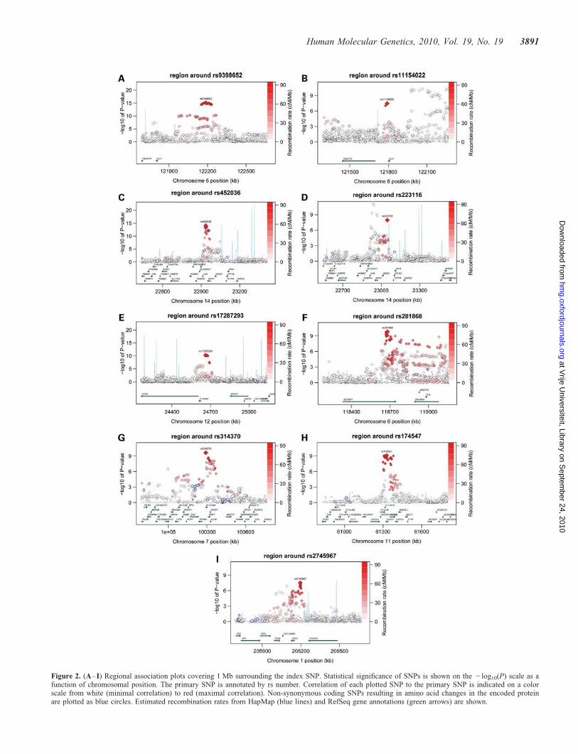
Figure 2. (A–I) Regional association plots covering 1 Mb surrounding the index SNP. Statistical significance of SNPs is shown on the 2log10(P) scale as afunction of chromosomal position. The primary SNP is annotated by rs number. Correlation of each plotted SNP to the primary SNP is indicated on a colorscale from white (minimal correlation) to red (maximal correlation). Non-synonymous coding SNPs resulting in amino acid changes in the encoded proteinare plotted as blue circles. Estimated recombination rates from HapMap (blue lines) and RefSeq gene annotations (green arrows) are shown.
Human Molecular Genetics, 2010, Vol. 19, No. 19 3891
at Vrije U
niversiteit, Library on Septem
ber 24, 2010hm
g.oxfordjournals.orgD
ownloaded from

MATERIALS AND METHODS
Study participants
The RRGEN sample consisted of subjects from the five parti-cipating studies in the Cohorts for Heart and Aging Researchin Genomic Epidemiology Consortium (57)—comprised ofthe Age, Gene, Environment Susceptibility (AGES) Study,the Atherosclerosis Risk in Communities (ARIC) Study, theCardiovascular Health Study (CHS), the Framingham HeartStudy (FHS) and the Rotterdam Study (RS)—as well as theCooperative Health Research in the Region Augsburg(KORA) study, the SardiNIA study, the Study of Health inPomerania (SHIP), TwinsUK, Netherlands Study ofDepression and Anxiety (NESDA) and three populationisolate studies in the European Special Populations Network(EUROSPAN), the Erasmus Rucphen Family (ERF), theSouth Tyrolean Micro-Isolate (MICROS) and the OrkneyComplex Disease Study (ORCADES). For RRGEN, both thebaseline Rotterdam Study (RS-I) and first extended cohort(RS-II) were used (58,59). KORA subjects in RRGEN weredrawn from the F3 and S4 cohorts.
Individuals were excluded if they were non-Caucasian, hadatrial fibrillation, a second- or third-degree atrioventricularblock, a pacemaker, a diagnosis of prevalent myocardialinfarction or prevalent heart failure, used beta-adrenergicblocking agents, non-dihydropyridine calcium antagonists ordigoxin, or had a heart rate ,50 (RR interval . 1200 ms)or .100 bpm (RR interval , 600 ms).
To adhere to STrenghtening the REporting of GeneticAssociations studies (STREGA) statement guidelines (60), weinclude an online supplement with additional information(Supplementary Material). All studies have approval from theirinstitutional review committee, and the subjects of all cohortsprovided written informed consent. A more detailed descriptionof cohorts is given in Supplementary Material, Appendix.
RR interval measurement methods
All cohorts recorded 12-lead ECGs from which the RR inter-val (which equals the inverse heart rate) was measured. Forcohort-specific details on RR interval measurement methods,please refer to the Supplementary Material, Appendix.
Genotyping and imputation
Affymetrix and Illumina arrays were used for genotyping.Using genotype information generated on these platforms, allcohorts imputed genotypes for a common set of �2.5 millionautosomal SNPs based on linkage disequilibrium patternsobserved in HapMap CEU reference samples (Utah residentsof Northern and Western European descent). The genetic traitanalyzed was the imputed allele dosage, a fractional valuebetween 0 and 2, reflecting the estimated number of allelecopies of an SNP for each subject. A more detailed descriptionis given in the Supplementary Material, Appendix and Table S1.
Statistical methods
The resting RR interval was adjusted for age, sex and bodymass index. For each SNP, we tested the genotype for associ-
ation with the covariate-adjusted RR interval under an additivegenetic model using linear regression models (SupplementaryMaterial, Appendix). We then conducted a fixed-effects,inverse variance weighted meta-analysis using beta estimatesand standard errors from each of the cohorts with applyinggenomic control on a per study basis and additionallypost-meta-analysis (Pmeta-gc). Genomic control refers to thecorrection made to the test statistics to account for anyinflation of the test statistic distribution, which can resultfrom unaccounted population substructure or other technicalbiases (61,62). We mapped all SNPs to dbSNP build 129,resulting in a unique set of 2 650 552 autosomal SNPs, afterconfirming the consistency of the coded allele across allstudies. Scripts used for this meta-analysis are availableonline (http://www.broadinstitute.org/~debakker/meta.html).
Genome-wide significance was defined as P , 5 × 1028,based on the estimated multiple testing burden for all commonvariants in populations of European ancestry (63). To identifyindependent signals reaching the genome-wide significancethreshold within a locus, genome-wide association meta-analysisresults were aggregated into bins by index SNP at a linkage dis-equilibrium r2 threshold of 0.1, such that all results within agiven bin were correlated to the index SNP at r2 ≥ 0.1 but toany index SNP in other bins at r2 , 0.1. Four index SNPs attwo loci were subsequently analyzed in conditional regressionmodels (n ≤ 33 846) to assess statistical independence.
Finally, we adopted the polygenic regression modelingapproach as recently described by Purcell et al. (64) andimplemented in PLINK (http://pngu.mgh.harvard.edu/~purcell/plink) to estimate the genetic variance explained by associatedloci at progressively less stringent P-value thresholds withinthe RS-II sample. The outcome of this analysis is a P-valuethreshold at which the explained variance is maximized. Thelist of loci included in the score yielding the maximum explainedvariance will include non-genome-wide loci that in aggregatecontribute to the model’s performance and indicate thatadditional true-positive signals are likely to be present withinthat list. For this analysis, we removed the RS-I and RS-II datafrom the discovery meta-analysis to remove the risk of corre-lation between the discovery and validation sample and overes-timation of the explained variance. Subsequently, we obtained alist of independent signals to be included in the model based ontheir statistical significance from the meta-analysis without RS-Ior RS-II (PLINK-clump option, r2 ≥0.05, 1 Mb window) (65).We summarized variation across associated loci (using signifi-cance thresholds from P , 5 × 1028 to P , 0.05) into quantitat-ive scores per individual. The score was then used as a predictorin a linear regression analysis in the RS-II sample (n ¼ 1589) andthe resulting r2 is reported as the measure for explained variancefor each P-value threshold.
SUPPLEMENTARY MATERIAL
Supplementary Material is available at HMG online.
ACKNOWLEDGEMENTS
We thank all participants and the study staff of the Age, Gene/Environment Susceptibility Reykjavik Study (AGES); the
3892 Human Molecular Genetics, 2010, Vol. 19, No. 19
at Vrije U
niversiteit, Library on Septem
ber 24, 2010hm
g.oxfordjournals.orgD
ownloaded from

Atherosclerosis Risk in Communities (ARIC) study; theCardiovascular Health Study (CHS); the Erasmus RucphenFamily (ERF) study; the Framingham Heart Study (FHS);the Kooperative Gesundheitsforschung in der RegionAugsburg (KORA); the Micros Study; the Netherlands Studyof Depression and Anxiety (NESDA); the Orkney ComplexDisease Study (Orcades); the Rotterdam Study (RS); theSardiNIA study; the Study of Health in Pomerania (SHIP);the TwinsUK study as well as the Cohorts for Heartand Aging Research in Genome Epidemiology (CHARGE)Consortium, for their invaluable contributions.
Conflict of Interest statement. None declared.
FUNDING
This work has been made possible by the generous financialsupport of a variety of funding sources to participatingstudies and individual scientists. A detailed list of acknowl-edgments and funding sources, as well as an author contri-bution list, is included in Supplementary Material.
REFERENCES
1. Fox, K., Borer, J.S., Camm, A.J., Danchin, N., Ferrari, R., Lopez Sendon, J.L.,Steg, P.G., Tardif, J.C., Tavazzi, L. and Tendera, M. (2007) Resting heart ratein cardiovascular disease. J. Am. Coll. Cardiol., 50, 823–830.
2. Greenland, P., Daviglus, M.L., Dyer, A.R., Liu, K., Huang, C.F.,Goldberger, J.J. and Stamler, J. (1999) Resting heart rate is a risk factorfor cardiovascular and noncardiovascular mortality: the Chicago HeartAssociation Detection Project in Industry. Am. J. Epidemiol., 149, 853–862.
3. Nauman, J., Nilsen, T.I., Wisloff, U. and Vatten, L.J. (2010) Combinedeffect of resting heart rate and physical activity on ischaemic heartdisease: mortality follow-up in a population study (the HUNT study,Norway). J. Epidemiol. Community Health, 64, 175–181.
4. Jouven, X., Zureik, M., Desnos, M., Guerot, C. and Ducimetiere, P. (2001)Resting heart rate as a predictive risk factor for sudden death inmiddle-aged men. Cardiovasc. Res., 50, 373–378.
5. Kannel, W.B., Kannel, C., Paffenbarger, R.S. Jr and Cupples, L.A. (1987)Heart rate and cardiovascular mortality: the Framingham Study. Am.Heart J., 113, 1489–1494.
6. Kristal-Boneh, E., Silber, H., Harari, G. and Froom, P. (2000) Theassociation of resting heart rate with cardiovascular, cancer and all-causemortality. Eight year follow-up of 3527 male Israeli employees (theCORDIS Study). Eur. Heart J., 21, 116–124.
7. Chang, M., Havlik, R.J., Corti, M.C., Chaves, P.H., Fried, L.P. andGuralnik, J.M. (2003) Relation of heart rate at rest and mortality in theWomen’s Health and Aging Study. Am. J. Cardiol., 92, 1294–1299.
8. Jouven, X., Empana, J.P., Escolano, S., Buyck, J.F., Tafflet, M., Desnos, M.and Ducimetiere, P. (2009) Relation of heart rate at rest and long-term (.20years) death rate in initially healthy middle-aged men. Am. J. Cardiol., 103,279–283.
9. Hsia, J., Larson, J.C., Ockene, J.K., Sarto, G.E., Allison, M.A., Hendrix, S.L.,Robinson, J.G., LaCroix, A.Z. and Manson, J.E.; Women’s Health InitiativeResearch Group. (2009) Resting heart rate as a low tech predictor of coronaryevents in women: prospective cohort study. Br. Med. J., 338, b219.
10. Russell, M.W., Law, I., Sholinsky, P. and Fabsitz, R.R. (1998) Heritabilityof ECG measurements in adult male twins. J. Electrocardiol., 30 (suppl.),64–68.
11. Singh, J.P., Larson, M.G., O’Donnell, C.J., Tsuji, H., Evans, J.C. andLevy, D. (1999) Heritability of heart rate variability: the FraminghamHeart Study. Circulation, 99, 2251–2254.
12. Martin, L.J., Comuzzie, A.G., Sonnenberg, G.E., Myklebust, J., James, R.,Marks, J., Blangero, J. and Kissebah, A.H. (2004) Major quantitative traitlocus for resting heart rate maps to a region on chromosome 4.Hypertension, 43, 1146–1151.
13. Laramie, J.M., Wilk, J.B., Hunt, S.C., Ellison, R.C., Chakravarti, A.,Boerwinkle, E. and Myers, R.H. (2006) Evidence for a gene influencingheart rate on chromosome 5p13-14 in a meta-analysis of genome-widescans from the NHLBI Family Blood Pressure Program. BMC Med.
Genet., 7, 17.
14. Dalageorgou, C., Ge, D., Jamshidi, Y., Nolte, I.M., Riese, H., Savelieva, I.,Carter, N.D., Spector, T.D. and Snieder, H. (2008) Heritability of QT interval:how much is explained by genes for resting heart rate? J. Cardiovasc.
Electrophysiol., 19, 386–391.
15. De Geus, E.J., Kupper, N., Boomsma, D.I. and Snieder, H. (2007)Bivariate genetic modeling of cardiovascular stress reactivity: does stressuncover genetic variance? Psychosom. Med., 69, 356–364.
16. Wilk, J.B., Myers, R.H., Zhang, Y., Lewis, C.E., Atwood, L., Hopkins, P.N.and Ellison, R.C. (2002) Evidence for a gene influencing heart rate onchromosome 4 among hypertensives. Hum. Genet., 111, 207–213.
17. Ranade, K., Jorgenson, E., Sheu, W.H., Pei, D., Hsiung, C.A., Chiang, F.T.,Chen, Y.D., Pratt, R., Olshen, R.A., Curb, D. et al. (2002) A polymorphismin the beta1 adrenergic receptor is associated with resting heart rate. Am. J.
Hum. Genet., 70, 935–942.
18. An, P., Rice, T., Rankinen, T., Leon, A.S., Skinner, J.S., Wilmore, J.H.,Bouchard, C. and Rao, D.C. (2006) Genome-wide scan to identifyquantitative trait loci for baseline resting heart rate and its response toendurance exercise training: the HERITAGE Family Study. Int. J. Sports
Med., 27, 31–36.
19. Wilk, J.B., Myers, R.H., Pankow, J.S., Hunt, S.C., Leppert, M.F.,Freedman, B.I., Province, M.A. and Ellison, R.C. (2006) Adrenergicreceptor polymorphisms associated with resting heart rate: the HyperGENStudy. Ann. Hum. Genet., 70, 566–573.
20. Wilton, S.B., Anderson, T.J., Parboosingh, J., Bridge, P.J., Exner, D.V.,Forrest, D. and Duff, H.J. (2008) Polymorphisms in multiple genes areassociated with resting heart rate in a stepwise allele-dependent manner.Heart Rhythm, 5, 694–700.
21. Cho, Y.S., Go, M.J., Kim, Y.J., Heo, J.Y., Oh, J.H., Ban, H.J., Yoon, D.,Lee, M.H., Kim, D.J., Park, M. et al. (2009) A large-scale genome-wideassociation study of Asian populations uncovers genetic factorsinfluencing eight quantitative traits. Nat. Genet., 41, 527–534.
22. Holm, H., Gudbjartsson, D.F., Arnar, D.O., Thorleifsson, G.,Thorgeirsson, G., Stefansdottir, H., Gudjonsson, S.A., Jonasdottir, A.,Mathiesen, E.B., Njolstad, I. et al. (2010) Several common variantsmodulate heart rate, PR interval and QRS duration. Nat. Genet., 42,117–122.
23. Chandler, N.J., Greener, I.D., Tellez, J.O., Inada, S., Musa, H., Molenaar, P.,Difrancesco, D., Baruscotti, M., Longhi, R., Anderson, R.H. et al. (2009)Molecular architecture of the human sinus node: insights into the function ofthe cardiac pacemaker. Circulation, 119, 1562–1575.
24. Rohr, S. (2004) Role of gap junctions in the propagation of the cardiacaction potential. Cardiovasc. Res., 62, 309–322.
25. Dasgupta, C., Martinez, A.M., Zuppan, C.W., Shah, M.M., Bailey, L.L.and Fletcher, W.H. (2001) Identification of connexin43 (alpha1) gapjunction gene mutations in patients with hypoplastic left heart syndromeby denaturing gradient gel electrophoresis (DGGE). Mutat. Res., 479,173–186.
26. Niimura, H., Patton, K.K., McKenna, W.J., Soults, J., Maron, B.J.,Seidman, J.G. and Seidman, C.E. (2002) Sarcomere protein genemutations in hypertrophic cardiomyopathy of the elderly. Circulation,105, 446–451.
27. Ching, Y.H., Ghosh, T.K., Cross, S.J., Packham, E.A., Honeyman, L.,Loughna, S., Robinson, T.E., Dearlove, A.M., Ribas, G., Bonser, A.J.et al. (2005) Mutation in myosin heavy chain 6 causes atrial septal defect.Nat. Genet., 37, 423–428.
28. Carniel, E., Taylor, M.R., Sinagra, G., Di Lenarda, A., Ku, L., Fain, P.R.,Boucek, M.M., Cavanaugh, J., Miocic, S., Slavov, D. et al. (2005)Alpha-myosin heavy chain: a sarcomeric gene associated with dilated andhypertrophic phenotypes of cardiomyopathy. Circulation, 112, 54–59.
29. Callis, T.E., Pandya, K., Seok, H.Y., Tang, R.H., Tatsuguchi, M., Huang, Z.P.,Chen, J.F., Deng, Z., Gunn, B., Shumate, J. et al. (2009) MicroRNA-208a is aregulator of cardiac hypertrophy and conduction in mice. J. Clin. Invest., 119,2772–2786.
30. van Rooij, E., Sutherland, L.B., Qi, X., Richardson, J.A., Hill, J. andOlson, E.N. (2007) Control of stress-dependent cardiac growth and geneexpression by a microRNA. Science, 316, 575–579.
Human Molecular Genetics, 2010, Vol. 19, No. 19 3893
at Vrije U
niversiteit, Library on Septem
ber 24, 2010hm
g.oxfordjournals.orgD
ownloaded from

31. Wunderle, V.M., Critcher, R., Ashworth, A. and Goodfellow, P.N. (1996)Cloning and characterization of SOX5, a new member of the human SOXgene family. Genomics, 36, 354–358.
32. Lefebvre, V. (2009) The SoxD transcription factors—Sox5, Sox6 andSox13—are key cell fate modulators. Int. J. Biochem. Cell Biol., 42, 429–432.
33. Pfeufer, A., van Noord, C., Marciante, K.D., Arking, D.E., Larson, M.G.,Smith, A.V., Tarasov, K.V., Muller, M., Sotoodehnia, N., Sinner, M.F.et al. Genome-wide association study of PR interval. Nat. Genet., 42,153–159.
34. Pfeufer, A., Sanna, S., Arking, D.E., Muller, M., Gateva, V., Fuchsberger, C.,Ehret, G.B., Orru, M., Pattaro, C., Kottgen, A. et al. (2009) Common variantsat ten loci modulate the QT interval duration in the QTSCD Study. Nat.Genet., 41, 407–414.
35. Newton-Cheh, C., Eijgelsheim, M., Rice, K.M., de Bakker, P.I., Yin, X.,Estrada, K., Bis, J.C., Marciante, K., Rivadeneira, F., Noseworthy, P.A.et al. (2009) Common variants at ten loci influence QT interval duration inthe QTGEN Study. Nat. Genet., 41, 399–406.
36. Nolte, I.M., Wallace, C., Newhouse, S.J., Waggott, D., Fu, J., Soranzo, N.,Gwilliam, R., Deloukas, P., Savelieva, I., Zheng, D. et al. (2009) Commongenetic variation near the phospholamban gene is associated with cardiacrepolarisation: meta-analysis of three genome-wide association studies.PLoS One, 4, e6138.
37. Vasan, R.S., Glazer, N.L., Felix, J.F., Lieb, W., Wild, P.S., Felix, S.B.,Watzinger, N., Larson, M.G., Smith, N.L., Dehghan, A. et al. (2009)Genetic variants associated with cardiac structure and function: ameta-analysis and replication of genome-wide association data. JAMA,302, 168–178.
38. Vinogradova, T.M., Sirenko, S., Lyashkov, A.E., Younes, A., Li, Y., Zhu, W.,Yang, D., Ruknudin, A.M., Spurgeon, H. and Lakatta, E.G. (2008)Constitutive phosphodiesterase activity restricts spontaneous beating rate ofcardiac pacemaker cells by suppressing local Ca2+ releases. Circ. Res., 102,761–769.
39. Vinogradova, T.M., Lyashkov, A.E., Zhu, W., Ruknudin, A.M., Sirenko, S.,Yang, D., Deo, S., Barlow, M., Johnson, S., Caffrey, J.L. et al. (2006) Highbasal protein kinase A-dependent phosphorylation drives rhythmic internalCa2+ store oscillations and spontaneous beating of cardiac pacemakercells. Circ. Res., 98, 505–514.
40. Nishimura, M., Suzuki, S., Satoh, T. and Naito, S. (2009) Tissue-specificmRNA expression profiles of human solute carrier 35 transporters. DrugMetab. Pharmacokinet., 24, 91–99.
41. Ha, B.H., Ahn, H.C., Kang, S.H., Tanaka, K., Chung, C.H. and Kim, E.E.(2008) Structural basis for Ufm1 processing by UfSP1. J. Biol. Chem.,283, 14893–14900.
42. Kang, S.H., Kim, G.R., Seong, M., Baek, S.H., Seol, J.H., Bang, O.S.,Ovaa, H., Tatsumi, K., Komatsu, M., Tanaka, K. et al. (2007) Two novelubiquitin-fold modifier 1 (Ufm1)-specific proteases, UfSP1 and UfSP2.J. Biol. Chem., 282, 5256–5262.
43. Aulchenko, Y.S., Ripatti, S., Lindqvist, I., Boomsma, D., Heid, I.M.,Pramstaller, P.P., Penninx, B.W., Janssens, A.C., Wilson, J.F., Spector, T.et al. (2009) Loci influencing lipid levels and coronary heart disease riskin 16 European population cohorts. Nat. Genet., 41, 47–55.
44. Kathiresan, S., Willer, C.J., Peloso, G.M., Demissie, S., Musunuru, K.,Schadt, E.E., Kaplan, L., Bennett, D., Li, Y., Tanaka, T. et al. (2009)Common variants at 30 loci contribute to polygenic dyslipidemia. Nat.Genet., 41, 56–65.
45. Sabatti, C., Service, S.K., Hartikainen, A.L., Pouta, A., Ripatti, S.,Brodsky, J., Jones, C.G., Zaitlen, N.A., Varilo, T., Kaakinen, M. et al.(2009) Genome-wide association analysis of metabolic traits in a birthcohort from a founder population. Nat. Genet., 41, 35–46.
46. Gieger, C., Geistlinger, L., Altmaier, E., Hrabe de Angelis, M.,Kronenberg, F., Meitinger, T., Mewes, H.W., Wichmann, H.E.,Weinberger, K.M., Adamski, J. et al. (2008) Genetics meetsmetabolomics: a genome-wide association study of metabolite profiles inhuman serum. PLoS Genet., 4, e1000282.
47. Schaeffer, L., Gohlke, H., Muller, M., Heid, I.M., Palmer, L.J., Kompauer, I.,Demmelmair, H., Illig, T., Koletzko, B. and Heinrich, J. (2006) Commongenetic variants of the FADS1 FADS2 gene cluster and their reconstructedhaplotypes are associated with the fatty acid composition in phospholipids.Hum. Mol. Genet., 15, 1745–1756.
48. Dettbarn, C. and Palade, P. (1993) Arachidonic acid-induced Ca2+release from isolated sarcoplasmic reticulum. Biochem. Pharmacol., 45,1301–1309.
49. Bang, M.L., Centner, T., Fornoff, F., Geach, A.J., Gotthardt, M., McNabb, M.,Witt, C.C., Labeit, D., Gregorio, C.C., Granzier, H. et al. (2001) The completegene sequence of titin, expression of an unusual approximately 700-kDa titinisoform, and its interaction with obscurin identify a novel Z-line to I-bandlinking system. Circ. Res., 89, 1065–1072.
50. Itoh-Satoh, M., Hayashi, T., Nishi, H., Koga, Y., Arimura, T., Koyanagi, T.,Takahashi, M., Hohda, S., Ueda, K., Nouchi, T. et al. (2002) Titin mutationsas the molecular basis for dilated cardiomyopathy. Biochem. Biophys. Res.
Commun., 291, 385–393.
51. Kubo, Y., Baldwin, T.J., Jan, Y.N. and Jan, L.Y. (1993) Primary structureand functional expression of a mouse inward rectifier potassium channel.Nature, 362, 127–133.
52. Fleischmann, B.K., Duan, Y., Fan, Y., Schoneberg, T., Ehlich, A., Lenka, N.,Viatchenko-Karpinski, S., Pott, L., Hescheler, J. and Fakler, B. (2004)Differential subunit composition of the G protein-activated inward-rectifierpotassium channel during cardiac development. J. Clin. Invest., 114, 994–1001.
53. Rosskopf, D., Nikula, C., Manthey, I., Joisten, M., Frey, U., Kohnen, S.and Siffert, W. (2003) The human G protein beta4 subunit: gene structure,expression, Ggamma and effector interaction. FEBS Lett., 544, 27–32.
54. Ruiz-Velasco, V., Ikeda, S.R. and Puhl, H.L. (2002) Cloning, tissuedistribution, and functional expression of the human G protein beta4-subunit. Physiol. Genomics, 8, 41–50.
55. Splawski, I., Timothy, K.W., Decher, N., Kumar, P., Sachse, F.B., Beggs,A.H., Sanguinetti, M.C. and Keating, M.T. (2005) Severe arrhythmia disordercaused by cardiac L-type calcium channel mutations. Proc. Natl Acad. Sci.
USA, 102, 8089–8096; discussion 8086–8088.
56. Avkiran, M., Rowland, A.J., Cuello, F. and Haworth, R.S. (2008) Proteinkinase d in the cardiovascular system: emerging roles in health anddisease. Circ. Res., 102, 157–163.
57. Psaty, B.M., O’Donnell, C.J., Gudnason, V., Lunetta, K.L., Folsom, A.R.,Rotter, J.I., Uitterlinden, A.G., Harris, T.B., Witteman, J.C.M.,Boerwinkle, E. et al. (2009) Cohorts for Heart and Aging Research inGenomic Epidemiology (CHARGE) Consortium: design of prospectivemeta-analyses of genome-wide association studies from 5 cohorts. Circ.
Cardiovasc. Genet., 2, 73–80.
58. Hofman, A., Breteler, M.M., van Duijn, C.M., Krestin, G.P., Pols, H.A.,Stricker, B.H., Tiemeier, H., Uitterlinden, A.G., Vingerling, J.R. andWitteman, J.C. (2007) The Rotterdam Study: objectives and designupdate. Eur. J. Epidemiol., 22, 819–829.
59. Hofman, A., Breteler, M.M., van Duijn, C.M., Janssen, H.L., Krestin, G.P.,Kuipers, E.J., Stricker, B.H., Tiemeier, H., Uitterlinden, A.G., Vingerling,J.R. et al. (2009) The Rotterdam Study: 2010 objectives and design update.Eur. J. Epidemiol., 24, 553–572.
60. Little, J., Higgins, J.P., Ioannidis, J.P., Moher, D., Gagnon, F., von Elm, E.,Khoury, M.J., Cohen, B., Davey-Smith, G., Grimshaw, J. et al. (2009)Strengthening the reporting of genetic association studies (STREGA): anextension of the STROBE statement. Eur. J. Epidemiol., 24, 37–55.
61. de Bakker, P.I., Ferreira, M.A., Jia, X., Neale, B.M., Raychaudhuri, S. andVoight, B.F. (2008) Practical aspects of imputation-driven meta-analysisof genome-wide association studies. Hum. Mol. Genet., 17, R122–R128.
62. Devlin, B., Roeder, K. and Wasserman, L. (2001) Genomic control, a newapproach to genetic-based association studies. Theor. Popul. Biol., 60,155–166.
63. Pe’er, I., Yelensky, R., Altshuler, D. and Daly, M.J. (2008) Estimation ofthe multiple testing burden for genomewide association studies of nearlyall common variants. Genet. Epidemiol., 32, 381–385.
64. Purcell, S.M., Wray, N.R., Stone, J.L., Visscher, P.M., O’Donovan, M.C.,Sullivan, P.F. and Sklar, P. (2009) Common polygenic variationcontributes to risk of schizophrenia and bipolar disorder. Nature, 460,748–752.
65. Purcell, S., Neale, B., Todd-Brown, K., Thomas, L., Ferreira, M.A.,Bender, D., Maller, J., Sklar, P., de Bakker, P.I., Daly, M.J. et al. (2007)PLINK: a tool set for whole-genome association and population-basedlinkage analyses. Am. J. Hum. Genet., 81, 559–575.
3894 Human Molecular Genetics, 2010, Vol. 19, No. 19
at Vrije U
niversiteit, Library on Septem
ber 24, 2010hm
g.oxfordjournals.orgD
ownloaded from