Genome-wide association analysis identifies three new breast cancer susceptibility loci
7
1000 VOLUME 42 | NUMBER 11 | NOVEMBER 2010 NATURE GENETICS LETTERS We carried out a meta-analysis of two recent psoriasis genome-wide association studies 1,2 with a combined discovery sample of 1,831 affected individuals (cases) and 2,546 controls. One hundred and two loci selected based on P value rankings were followed up in a three-stage replication study including 4,064 cases and 4,685 controls from Michigan,Toronto, Newfoundland and Germany. In the combined meta-analysis, we identified three new susceptibility loci, including one at NOS2 (rs4795067, combined P = 4 × 10 −11 ), one at FBXL19 (rs10782001, combined P = 9 × 10 −10 ) and one near PSMA6- NFKBIA (rs12586317, combined P = 2 × 10 −8 ). All three loci were also associated with psoriatic arthritis (rs4795067, combined P = 1 × 10 −5 ; rs10782001, combined P = 4 × 10 −8 ; and rs12586317, combined P = 6 × 10 −5 ) and purely cutaneous psoriasis (rs4795067, combined P = 1 × 10 −8 ; rs10782001, combined P = 2 × 10 −6 ; and rs12586317, combined P =1× 10 −6 ). We also replicated a recently identified 3 association signal near RNF114 (rs495337, combined P = 2 × 10 −7 ). Psoriasis vulgaris (PsV) is one of several immunologically mediated disorders whose genetic provenance is now coming into focus. Although the most evident cellular features of psoriasis are epi- dermal hyperplasia and altered keratinocyte differentiation, mount- ing evidence implicates both innate and acquired immunity in disease pathogenesis 4 . About a third of the individuals with PsV develop psoriatic arthritis (PsA), an inflammatory arthritis that is usually sero- negative for rheumatoid factor 5 . Herein, the abbreviation PsC refers to individuals with purely cutaneous psoriasis (that is, PsV without PsA), and the general term ‘psoriasis’ refers to PsV unless otherwise specified. Individuals with psoriasis are at significantly greater risk of cardiovascular morbidity and mortality than the general population, especially younger individuals with more severe skin disease 6 . Psoriasis is also associated with inflammatory bowel disease and other autoimmune disorders 7 . Psoriasis is strongly influenced by genetic factors. The earliest reported genetic susceptibility factors for psoriasis map to the major histocompatibility complex (MHC) 8 and include haplotypes bearing HLA-Cw6 (ref. 9). More recently, the MHC component of susceptibility has been fine mapped to or very near HLA-Cw6 itself 10 , and genome-wide association studies (GWAS) have identified nine additional regions outside the MHC with genome-wide levels of statistical significance: IL12B, IL23R, IL23A, TNFAIP3, TNIP1, IL4-IL13, LCE3B-LCE3C, DEFB4 and SPATA2-RNF114 (refs. 3,11,12). In this study, we report the discovery of three new genetic suscepti- bility loci for psoriasis, none of which have been definitively impli- cated in any complex genetic disorder to date. We also replicate a previous report 3 of association to the SPATA2-RNF114 locus (com- bined P = 2 × 10 −7 ) and extend the associated region at this locus to include two additional candidate genes. We augmented published GWAS results from the Collaborative Association Study of Psoriasis (CASP) 1 by using genotype imputa- tion 13 to combine the results of that study with those of an independent GWAS of psoriasis from Kiel, Germany 2 using either phase 2 HapMap or 1000 Genomes Project haplotypes as a reference (Supplementary Table 1). Only SNPs imputed with relatively high confidence (esti- mated r 2 between imputed and true genotypes >0.3) in both GWAS were considered for meta-analysis. Our discovery sample consisted of up to 1,831 cases and 2,546 controls with imputed genotypes for 2,502,313 (HapMap) or 7,456,344 (1000 Genomes Project) autosomal SNPs. Genomic control inflation factors for the P values from the meta-analysis of the CASP and Kiel GWAS were low (λ = 1.061 for HapMap and λ = 1.054 for 1000 Genomes Project imputations), and Genome-wide association analysis identifies three psoriasis susceptibility loci Philip E Stuart 1,15 , Rajan P Nair 1,15 , Eva Ellinghaus 2 , Jun Ding 3 , Trilokraj Tejasvi 1 , Johann E Gudjonsson 1 , Yun Li 3 , Stephan Weidinger 4 , Bernadette Eberlein 4 , Christian Gieger 5 , H Erich Wichmann 5 , Manfred Kunz 6 , Robert Ike 7 , Gerald G Krueger 8 , Anne M Bowcock 9 , Ulrich Mrowietz 10 , Henry W Lim 11 , John J Voorhees 1 , Gonçalo R Abecasis 3 , Michael Weichenthal 10 , Andre Franke 2 , Proton Rahman 12 , Dafna D Gladman 13 & James T Elder 1,14 1 Department of Dermatology, University of Michigan Medical School, Ann Arbor, Michigan, USA. 2 Institute for Clinical Molecular Biology, University of Kiel, Kiel, Germany. 3 Center for Statistical Genetics, Department of Biostatistics, School of Public Health, University of Michigan, Ann Arbor, Michigan, USA. 4 Department of Dermatology and Allergy, Technical University Munich, Munich, Germany. 5 Institute of Medical Informatics, Biometry and Epidemiology, Ludwig-Maximilians- University, Munich, Germany. 6 Comprehensive Center for Inflammation Medicine, University of Lübeck, Lübeck, Germany. 7 Department of Medicine, University of Michigan Medical School, Ann Arbor, Michigan, USA. 8 Department of Dermatology, University of Utah, Salt Lake City, Utah, USA. 9 Division of Human Genetics, Department of Genetics, Washington University at St. Louis, St. Louis, Missouri, USA. 10 Department of Dermatology, University of Kiel, Kiel, Germany. 11 Department of Dermatology, Henry Ford Hospital, Detroit, Michigan, USA. 12 Department of Medicine, Memorial University, St. John’s, Newfoundland, Canada. 13 Department of Rheumatology, University of Toronto, Toronto, Ontario, Canada. 14 Ann Arbor Veterans Affairs Medical Center, Ann Arbor, Michigan, USA. 15 These authors contributed equally to this work. Correspondence should be addressed to J.T.E. ([email protected]). Received 9 March; accepted 9 July; published online 17 October 2010; doi:10.1038/ng.693 © 2010 Nature America, Inc. All rights reserved.
-
Upload
independent -
Category
Documents
-
view
1 -
download
0
Transcript of Genome-wide association analysis identifies three new breast cancer susceptibility loci

1000 VOLUME 42 | NUMBER 11 | NOVEMBER 2010 Nature GeNetics
l e t t e r s
Wecarriedoutameta-analysisoftworecentpsoriasisgenome-wideassociationstudies1,2withacombineddiscoverysampleof1,831affectedindividuals(cases)and2,546controls.OnehundredandtwolociselectedbasedonPvaluerankingswerefollowedupinathree-stagereplicationstudyincluding4,064casesand4,685controlsfromMichigan,Toronto,NewfoundlandandGermany.Inthecombinedmeta-analysis,weidentifiedthreenewsusceptibilityloci,includingoneatNOS2(rs4795067,combinedP=4×10⁻¹¹),oneatFBXL19(rs10782001,combinedP=9×10⁻¹⁰)andonenearPSMA6-NFKBIA(rs12586317,combinedP=2×10−8).Allthreelociwerealsoassociatedwithpsoriaticarthritis(rs4795067,combinedP=1×10−5;rs10782001,combinedP=4×10−8;andrs12586317,combinedP=6×10−5)andpurelycutaneouspsoriasis(rs4795067,combinedP=1×10−8;rs10782001,combinedP=2×10−6;andrs12586317,combinedP=1×10−6).Wealsoreplicatedarecentlyidentified3associationsignalnearRNF114(rs495337,combinedP=2×10−7).
Psoriasis vulgaris (PsV) is one of several immunologically mediated disorders whose genetic provenance is now coming into focus. Although the most evident cellular features of psoriasis are epidermal hyperplasia and altered keratinocyte differentiation, mounting evidence implicates both innate and acquired immunity in disease pathogenesis4. About a third of the individuals with PsV develop psoriatic arthritis (PsA), an inflammatory arthritis that is usually seronegative for rheumatoid factor5. Herein, the abbreviation PsC refers to individuals with purely cutaneous psoriasis (that is, PsV without PsA), and the general term ‘psoriasis’ refers to PsV unless otherwise specified. Individuals with psoriasis are at significantly greater risk of cardiovascular morbidity and mortality than the general population,
especially younger individuals with more severe skin disease6. Psoriasis is also associated with inflammatory bowel disease and other autoimmune disorders7.
Psoriasis is strongly influenced by genetic factors. The earliest reported genetic susceptibility factors for psoriasis map to the major histocompatibility complex (MHC)8 and include haplotypes bearing HLA-Cw6 (ref. 9). More recently, the MHC component of susceptibility has been fine mapped to or very near HLA-Cw6 itself10, and genomewide association studies (GWAS) have identified nine additional regions outside the MHC with genomewide levels of statistical significance: IL12B, IL23R, IL23A, TNFAIP3, TNIP1, IL4-IL13, LCE3B-LCE3C, DEFB4 and SPATA2RNF114 (refs. 3,11,12). In this study, we report the discovery of three new genetic susceptibility loci for psoriasis, none of which have been definitively implicated in any complex genetic disorder to date. We also replicate a previous report3 of association to the SPATA2RNF114 locus (combined P = 2 × 10⁻⁷) and extend the associated region at this locus to include two additional candidate genes.
We augmented published GWAS results from the Collaborative Association Study of Psoriasis (CASP)1 by using genotype imputation13 to combine the results of that study with those of an independent GWAS of psoriasis from Kiel, Germany2 using either phase 2 HapMap or 1000 Genomes Project haplotypes as a reference (Supplementary Table 1). Only SNPs imputed with relatively high confidence (estimated r 2 between imputed and true genotypes >0.3) in both GWAS were considered for metaanalysis. Our discovery sample consisted of up to 1,831 cases and 2,546 controls with imputed genotypes for 2,502,313 (HapMap) or 7,456,344 (1000 Genomes Project) autosomal SNPs. Genomic control inflation factors for the P values from the metaanalysis of the CASP and Kiel GWAS were low (λ = 1.061 for HapMap and λ = 1.054 for 1000 Genomes Project imputations), and
Genomewide association analysis identifies three psoriasis susceptibility lociPhilip E Stuart1,15, Rajan P Nair1,15, Eva Ellinghaus2, Jun Ding3, Trilokraj Tejasvi1, Johann E Gudjonsson1, Yun Li3, Stephan Weidinger4, Bernadette Eberlein4, Christian Gieger5, H Erich Wichmann5, Manfred Kunz6, Robert Ike7, Gerald G Krueger8, Anne M Bowcock9, Ulrich Mrowietz10, Henry W Lim11, John J Voorhees1, Gonçalo R Abecasis3, Michael Weichenthal10, Andre Franke2, Proton Rahman12, Dafna D Gladman13 & James T Elder1,14
1Department of Dermatology, University of Michigan Medical School, Ann Arbor, Michigan, USA. 2Institute for Clinical Molecular Biology, University of Kiel, Kiel, Germany. 3Center for Statistical Genetics, Department of Biostatistics, School of Public Health, University of Michigan, Ann Arbor, Michigan, USA. 4Department of Dermatology and Allergy, Technical University Munich, Munich, Germany. 5Institute of Medical Informatics, Biometry and Epidemiology, Ludwig-Maximilians-University, Munich, Germany. 6Comprehensive Center for Inflammation Medicine, University of Lübeck, Lübeck, Germany. 7Department of Medicine, University of Michigan Medical School, Ann Arbor, Michigan, USA. 8Department of Dermatology, University of Utah, Salt Lake City, Utah, USA. 9Division of Human Genetics, Department of Genetics, Washington University at St. Louis, St. Louis, Missouri, USA. 10Department of Dermatology, University of Kiel, Kiel, Germany. 11Department of Dermatology, Henry Ford Hospital, Detroit, Michigan, USA. 12Department of Medicine, Memorial University, St. John’s, Newfoundland, Canada. 13Department of Rheumatology, University of Toronto, Toronto, Ontario, Canada. 14Ann Arbor Veterans Affairs Medical Center, Ann Arbor, Michigan, USA. 15These authors contributed equally to this work. Correspondence should be addressed to J.T.E. ([email protected]).
Received 9 March; accepted 9 July; published online 17 October 2010; doi:10.1038/ng.693
© 2
010
Nat
ure
Am
eric
a, In
c. A
ll ri
gh
ts r
eser
ved
.

Nature GeNetics VOLUME 42 | NUMBER 11 | NOVEMBER 2010 1001
l e t t e r s
quantilequantile plots (Supplementary Fig. 1) also indicated no substantial systematic differences between cases and controls due to cryptic relatedness, population substructure or other biases.
The replication sample consisted of up to 4,064 PsV cases and 4,685 controls from Michigan, Toronto, Newfoundland and Germany. All participating subjects gave informed consent and protocols were reviewed and approved by local institutional review boards. Ascertainment criteria for psoriasis have previously been described10. PsA was diagnosed according to the CASPAR criteria14. PsC was defined as the presence of skin lesions in the demonstrated absence of PsA at the time of recruitment in the study.
We selected 102 SNPs for replication based on their P value rankings either in a subset of 350 CASP PsA cases compared to normal controls (53 SNPs referred to as set A; Supplementary Table 1) or in the combined CASPKiel GWAS (49 SNPs referred to as sets B and C; Supplementary Table 1). Imputation quality of these SNPs was generally excellent. In the firststage replication, we genotyped all selected SNPs in the large Michigan sample; 91 of these SNPs passed all quality control filters. In the secondstage replication, we typed all but one of the 51 quality control–filtered SNPs from set A of the PsA GWAS in the samples from Toronto and Newfoundland, which contained most of the PsA cases. SNPs with the most promising association results were then followed up with thirdstage typing, which consisted of eight SNPs from sets B and C in the two Canadian samples and six SNPs from all three sets in the German sample. Statistical significance of association was assessed with an allelic χ2 test for experimentally genotyped SNPs
and an equal variance t test with allele dosages for imputed SNPs. Metaanalysis of association across cohorts incorporated both imputation quality and asymmetry in the number of cases and controls as well as the size of the cohort. See Online Methods for details of the quality control filters, imputation quality and metaanalysis.
We identified four SNPs at three loci for which association P values achieved genomewide statistical significance (P ≤ 5 × 10⁻⁸) in the combined discovery and replication samples. Table 1 shows the results for these four SNPs along with three additional SNPs yielding a combined P ≤ 1 × 10⁻⁶. Genotype counts for these seven SNPs are shown in Supplementary Table 2. Results for the remaining SNPs passing quality control are summarized in Supplementary Table 3, and association results for PsA compared to PsC are shown in Table 2.
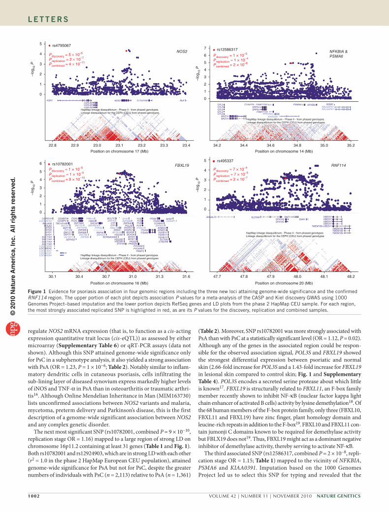
One of the associated SNPs (rs4795067, combined P = 4 × 10⁻¹¹, replicationstage OR = 1.19) mapped to an intron of NOS2 (Table 1). As shown in Figure 1, the strongest imputed association signals from the CASPKiel GWAS clustered in a region of low linkage disequilibrium (LD) containing no other genes. NOS2 encodes iNOS (inducible nitric oxide synthase), which is expressed by a subset of CD11c positive, CD1cnegative, TNFα (tumor necrosis factorα)producing inflammatory dendritic cells that is markedly expanded (30fold) in psoriatic lesions15. Consistent with this, in lesional psoriatic skin we found a 1.54fold overexpression of NOS2 mRNA by microarray analysis (Supplementary Table 4) and a marked 16fold overexpression by QRTPCR (Fig. 2 and Supplementary Table 5). However, neither rs4795067 nor any other SNP in this region was found to significantly
table 1 loci with strongest evidence of association with psoriasis in the combined sampleDiscovery samples
(1,831 cases, 2,546 controls)aReplication samples
(4,064 cases, 4,685 controls)a
SNPb Setc Chr.Pos (Mb)
Alleles risk/
nonrisk
FrequencydOR
(meta) P (meta)
FrequencydOR
(meta) P (meta)Combined P (meta)
Notable nearby genes (relative position)eCase Control Case Control
rs4795067 B 17 23.13 G/A 0.397 0.354 1.20 5 × 10⁻⁵ 0.389 0.349 1.19 2 × 10⁻⁷ 4 × 10⁻¹¹ NOS2 (intronic)rs10782001 B 16 30.85 G/A 0.394 0.347 1.22 1 × 10⁻⁵ 0.402 0.368 1.16 1 × 10⁻⁵ 9 × 10⁻¹⁰ FBXL19 (intronic)rs12924903 A 16 30.84 A/G 0.393 0.343 1.24 1 × 10⁻⁴ 0.384 0.349 1.16 2 × 10⁻⁶ 1 × 10⁻⁹ FBXL19 (−6.9 kb)rs12586317 C 14 34.75 T/C 0.783 0.742 1.25 1 × 10⁻⁵ 0.777 0.751 1.15 1 × 10⁻⁴ 2 × 10⁻⁸ NFKBIA, PSMA6rs1008953 B 20 43.41 C/T 0.809 0.770 1.27 1 × 10⁻⁵ 0.809 0.787 1.14 8 × 10⁻⁴ 1 × 10⁻⁷ SDC4 (−3.7 kb)
rs495337 C 20 47.96 G/A 0.610 0.573 1.17 7 × 10⁻⁴ 0.616 0.570 1.21 7 × 10⁻⁵ 2 × 10⁻⁷ RNF114 (silent)
rs12580100 B 12 54.73 A/G 0.898 0.868 1.33 2 × 10⁻⁶ 0.913 0.896 1.17 1 × 10⁻² 1 × 10⁻⁶ RPS26 (−1.2 kb)Chr., chromosome. aThe number of cases and controls that were genotyped and passed quality control for at least one of the 91 SNPs in the three replication sets. The actual numbers of cases and controls typed for each SNP varies and and can be determined from the genotype counts in supplementary table 2. bSNPs attaining genome-wide significance and their notable nearby genes are indicated in bold. cReplication set for the SNP (see main text and supplementary table 1 for more details). dFrequency of the risk allele. ePosition of each SNP relative to notable nearby genes is given. +/− indicates whether the SNP is upstream (−) or downstream (+) of the transcription start site. SNPs within the gene are labeled as ‘intronic’ or ‘silent’.
table 2 Association of strongest replicated loci with psoriatic arthritis (PsA) and cutaneous psoriasis (PsC)PsA versus control
(1,747 PsA, 7,231 controls)aPsC versus control
(3,394 PsC, 7,231 controls)aPsA versus PsC
(1,747 PsA, 3,394 PsC)a
SNP Chr. Pos (Mb)
Alleles risk/
nonrisk Frequenciesb OR P Frequenciesb OR P Frequenciesb OR PNotable nearby
genes
rs4795067 17 23.13 G/A 0.389/0.341 1.23 1 × 10⁻⁶ 0.391/0.348 1.20 1 × 10⁻⁸ 0.396/0.382 1.06 0.25 NOS2rs10782001 16 30.85 G/A 0.412/0.358 1.26 4 × 10⁻⁸ 0.388/0.353 1.16 2 × 10⁻⁶ 0.412/0.385 1.12 0.022 FBXL19rs12924903 16 30.84 A/G 0.412/0.352 1.29 4 × 10⁻⁹ 0.380/0.347 1.15 4 × 10⁻⁵ 0.403/0.378 1.11 0.058 FBXL19rs12586317 14 34.75 T/C 0.786/0.751 1.22 6 × 10⁻⁵ 0.778/0.747 1.19 1 × 10⁻⁶ 0.785/0.780 1.03 0.60 NFKBIArs495337 20 47.96 G/A 0.615/0.573 1.20 2 × 10⁻³ 0.614/0.572 1.19 1 × 10⁻⁶ 0.615/0.618 0.99 0.86 RNF114rs1008953 20 43.41 C/T 0.803/0.772 1.21 2 × 10⁻⁴ 0.792/0.769 1.15 2 × 10⁻⁴ 0.803/0.795 1.05 0.37 SDC4rs12580100 12 54.73 A/G 0.882/0.867 1.15 0.029 0.893/0.866 1.29 1 × 10⁻⁶ 0.888/0.893 0.95 0.54 RPS26
Chr., chromosome. aThe number of cases and controls for the specified phenotype comparison that were genotyped and passed quality control for at least one of the 91 SNPs in the three replication sets. The actual numbers of cases and controls typed for each SNP varies and can be determined from the genotype counts in supplementary table 2. bFrequencies of the PsV risk allele for the two phenotypes being compared. Allele frequencies for the same phenotype (PsA, PsC or control) and the same SNP often differ among comparisons because of differing number of qualifying cohorts (for example, some cohorts have PsA but no PsC cases so qualify for PsA versus control but not PsA versus PsC or PsC versus control comparisons) and because variations in the proportions of each phenotype per cohort alter the relative cohort weights in the meta-analyses.
© 2
010
Nat
ure
Am
eric
a, In
c. A
ll ri
gh
ts r
eser
ved
.
1002 VOLUME 42 | NUMBER 11 | NOVEMBER 2010 Nature GeNetics
l e t t e r s
regulate NOS2 mRNA expression (that is, to function as a cisacting expression quantitative trait locus (ciseQTL)) as assessed by either microarray (Supplementary Table 6) or qRTPCR assays (data not shown). Although this SNP attained genomewide significance only for PsC in a subphenotype analysis, it also yielded a strong association with PsA (OR = 1.23, P = 1 × 10⁻⁶; Table 2). Notably similar to inflammatory dendritic cells in cutaneous psoriasis, cells infiltrating the sublining layer of diseased synovium express markedly higher levels of iNOS and TNFα in PsA than in osteoarthritis or traumatic arthritis16. Although Online Mendelian Inheritance in Man (MIM163730) lists unconfirmed associations between NOS2 variants and malaria, mycetoma, preterm delivery and Parkinson’s disease, this is the first description of a genomewide significant association between NOS2 and any complex genetic disorder.
The next most significant SNP (rs10782001, combined P = 9 × 10⁻¹⁰, replication stage OR = 1.16) mapped to a large region of strong LD on chromosome 16p11.2 containing at least 31 genes (Table 1 and Fig. 1). Both rs10782001 and rs12924903, which are in strong LD with each other (r2 = 1.0 in the phase 2 HapMap European CEU population), attained genomewide significance for PsA but not for PsC, despite the greater numbers of individuals with PsC (n = 2,113) relative to PsA (n = 1,361)
(Table 2). Moreover, SNP rs10782001 was more strongly associated with PsA than with PsC at a statistically significant level (OR = 1.12, P = 0.02). Although any of the genes in the associated region could be responsible for the observed association signal, POL3S and FBXL19 showed the strongest differential expression between psoriatic and normal skin (2.66fold increase for POL3S and a 1.43fold increase for FBXL19 in lesional skin compared to control skin; Fig. 1 and Supplementary Table 4). POL3S encodes a secreted serine protease about which little is known17. FBXL19 is structurally related to FBXL11, an Fbox family member recently shown to inhibit NFκB (nuclear factor kappa light chain enhancer of activated B cells) activity by lysine demethylation18. Of the 68 human members of the Fbox protein family, only three (FBXL10, FBXL11 and FBXL19) have zinc finger, plant homology domain and leucinerich repeats in addition to the Fbox19. FBXL10 and FBXL11 contain jumonji C domains known to be required for demethylase activity but FBLX19 does not19. Thus, FBXL19 might act as a dominant negative inhibitor of demethylase activity, thereby serving to activate NFκB.
The third associated SNP (rs12586317, combined P = 2 × 10⁻⁸, replication stage OR = 1.15; Table 1) mapped to the vicinity of NFKBIA, PSMA6 and KIAA0391. Imputation based on the 1000 Genomes Project led us to select this SNP for typing and revealed that the
rs4795067
rs10782001
Preplication = 1 × 10⁻⁵
Pcombined = 9 × 10⁻¹⁰
Pdiscovery = 5 × 10⁻⁵
Preplication = 2 × 10⁻⁷
Pcombined = 4 × 10–11
Pdiscovery = 1 × 10⁻⁵
rs12586317
Pdiscovery = 1 × 10⁻⁵
Preplication = 1 × 10⁻⁴
Pcombined = 2 × 10⁻⁸
rs495337
Preplication = 7 × 10⁻⁵
Pcombined = 2 × 10⁻⁷
Pdiscovery = 7 × 10⁻⁴
5
4
3
2
1
0
5
6
7
4
3
2
1
0
22.8 22.9
KSR1LGALS9LGALS9LGALS9
CORO1A
LOC388242
SULT1A3SULT1A4
SULT1A4
SULT1A3
TBC1D10B
ZNF771ZNF771
SEPHS2
BCL7C
CTF1CTF1
ORAI3
ITGALITGAL
ZNF768
ZNF688
NCRNA00095
SRCAPFBRS
HSD3B7STX1B
HSD3B7
HSD3B7
ZNF646
ITGAMITGAM
FUSFUSFUSFUS
C16orf58
AHSP
SLC5A2
TGFB1l1TGFB1l1TGFB1l1
B4GALT5SNAl1
TMEM189-UBE2V1TMEM189TMEM189TMEM189
UBE2V1UBE2V1UBE2V1UBE2V1RNF114
SPATA2SPATA2
SLC9A8
ARMC5ARMC5
COX6A2ITGAD
ITGAX
MYST1MYST1
PRSS36
TRIM72
PYCARDPYCARD
PRSS8
BCKDKBCKDK
VKORC1VKORC1POL3S
RNF40ZNF688
SEPT1
CD2BP2CD2BP2
GIYD1GIYD1GIYD2GIYD2
BOLA2BOLA2B
NOS2 C17orf108 NLK
CFL2CFL2CFL2CFL2CFL2
KIAA0391
RALGAPA1RALGAPA1
PPP2R3C
LOC613038
LOC613037LOC595101LOC440354
LOC606724
MYLPF
ZNF48
ZNF689
PHKG2
SETD1A
FBXL19
ZNF668
PYDC1ZNF843
CSDAP1STX4
MIR762
C16orf93
SNORA30
PRR14
ZNF785 ZNF629
ZNF764ZNF747
DCTPP1
SRP54SRP54
FAM177A1 NFKBIA INSM2PSMA6
FAM177A1
C14orf19
BAZ1ABAZ1A
23.0
HapMap linkage disequilibrium - Phase II - from phased genotypesLinkage disequilibrium for the CEPH (CEU) from phased genotypes
HapMap linkage disequilibrium - Phase II - from phased genotypesLinkage disequilibrium for the CEPH (CEU) from phased genotypes
HapMap linkage disequilibrium - Phase II - from phased genotypesLinkage disequilibrium for the CEPH (CEU) from phased genotypes
HapMap linkage disequilibrium - Phase II - from phased genotypesLinkage disequilibrium for the CEPH (CEU) from phased genotypes
23.1 23.2 23.3 23.4 34.2
47.7 47.8 47.9 48.0 48.1 48.230.1 30.4 30.7 31.0 31.3 31.6
34.4 34.6 34.8 35.0 35.2
Position on chromosome 17 (Mb)
Position on chromosome 16 (Mb)
Position on chromosome 14 (Mb)
Position on chromosome 20 (Mb)
NOS2
FBXL19 RNF114
NFKBIA &PSMA6
–log
10 P
6
5
4
3
2
1
0
5
4
3
2
1
0
–log
10 P
–log
10 P
–log
10 P
Figure 1 Evidence for psoriasis association in four genomic regions including the three new loci attaining genome-wide significance and the confirmed RNF114 region. The upper portion of each plot depicts association P values for a meta-analysis of the CASP and Kiel discovery GWAS using 1000 Genomes Project–based imputation and the lower portion depicts RefSeq genes and LD plots from the phase 2 HapMap CEU sample. For each region, the most strongly associated replicated SNP is highlighted in red, as are its P values for the discovery, replication and combined samples.©
201
0 N
atu
re A
mer
ica,
Inc.
All
rig
hts
res
erve
d.

Nature GeNetics VOLUME 42 | NUMBER 11 | NOVEMBER 2010 1003
l e t t e r s
associated region extended to encompass NFKBIA, an observation that was not evident from HapMapbased imputation (Fig. 1 and data not shown). Of these three genes, only PSMA6 is overexpressed in psoriatic lesions (Fig. 2 and Supplementary Table 4). NFKBIA and PSMA6 are both attractive candidate genes for psoriasis susceptibility, as NFKBIA encodes IκBα, an inhibitor of NFκB signaling, and PSMA6 encodes a proteosomal subunit involved in MHC class I antigen processing. Polymorphisms in both genes (MIM164008 and MIM602855) have been suggestively associated with susceptibility to various disorders including myocardial infarction, Graves’ disease and inflammatory bowel disease. However, this is the first demonstration of a genomewide significant association in this region for any complex genetic disorder.
Although they did not attain genomewide significance, SNPs rs1008953 (combined P = 1 × 10⁻⁷, replication stage OR = 1.14) and rs12580100 (combined P = 1 × 10⁻⁶, replication stage OR = 1.17) were also of interest. rs1008953 resides 3.7 kb upstream of SDC4, which encodes syndecan4, a cellsurface heparin sulfate proteoglycan that inhibits Tcell activation20. Expression of SDC4 mRNA was markedly reduced in lesional psoriatic skin relative to both uninvolved and normal skin (Supplementary Tables 4 and 5). However, there are several other genes under the association peak, and several SNPs in the region function as ciseQTLs for SYS1, DBNDD2, and PIGT expression (Supplementary Table 6 and Supplementary Fig. 2).
rs12580100 resides 1.2 kb upstream of RPS26, which resides in a region that has been implicated in susceptibility to type 1 diabetes21 and which encodes a ribosomal protein subunit whose expression is under cisacting genetic control in the liver22. rs12580100 lies only 300 kb from a confirmed association signal at rs2066808 near IL23A (ref. 1), but these association signals are at least partially independent because the two SNPs reside in different LD blocks (Supplementary Fig. 2) and show low pairwise LD (r2 = 0.23), and a logistic regression model including both SNPs yielded significant partial association for each (P = 0.016 for rs12580100 and P = 3.0 × 10⁻⁴ for rs2066808). Of note, we found that RPS26 is overexpressed in lesional psoriatic skin (Supplementary Tables 4 and 5) and confirm in skin that this region contains several highly significant eQTLs that clearly colocalize with the disease association signal for RPS26 but not for IL23A (Supplementary Table 6 and Supplementary Fig. 2).
Although also not reaching genomewide significance, rs495337 (combined P = 2 × 10⁻⁷, replication stage OR = 1.21) confirms previous reports of association between psoriasis and the SPATA2RNF114 region3. However, the 1000 Genomes Project–based imputation demonstrated that the region of association extends farther than previously reported and encompasses SLC9A8 and SNAI1 (Fig. 1). Of these four genes, RNF114 was the most strongly expressed in skin (Supplementary Table 4). None of the transcripts encoded by these four genes was increased in psoriatic lesions; one SPATA2 probe detected a 23% decrease (Supplementary Table 4). We were unable to confirm a previous report of a ciseQTL in this region3, either by microarray (Supplementary Table 6) or qRTPCR assays (data not shown); however, our gene expression sample was only about half the size of that used in the previous study.
Fine mapping studies are needed to identify causal variants in the three new susceptibility regions we have identified. Nevertheless, genes contained in these newly associated regions fit well with emerging concepts of psoriasis pathogenesis12. Given genetic, therapeutic and immunological results indicating that IL23 produced by dendritic cells promotes the survival and expansion of pathogenic Tcells in psoriasis12, it is noteworthy that one of our new regions contains NOS2, which is expressed by a massively expanded subset of TNFα producing inflammatory dendritic cells in psoriatic lesions15. Moreover, given that TNFAIP3 and TNIP1 regulate NFκB signaling and have been implicated in psoriasis susceptibility1, the fact that FBXL19 and NFKBIA regulate the same pathway further suggests the central importance of NFκB signaling in psoriasis. Finally, when combined with the profound response of PsA to TNFα blockers4 and prior genetic results implicating IL12B and TNIP1 in PsA susceptibility1, the identification of a new PsA susceptibility region containing FBXL19 suggests that this multicellular IL23/IL17/NFκB signaling circuit may also be of critical importance to the development of joint disease in individuals with psoriasis.
URLs. MACH software, http://www.sph.umich.edu/csg/abecasis/mach/; HapMap 2 CEU phased haplotypes, http://hapmap.ncbi.nlm.nih.gov/downloads/phasing/200607_phaseII/phased/; August 2009 release of 1000 Genome Project phased data, http://www.sph.umich.edu/csg/abecasis/mach/download/1000GSanger0908.html.
MeThOdsMethods and any associated references are available in the online version of the paper at http://www.nature.com/naturegenetics/.
Note: Supplementary information is available on the Nature Genetics website.
–5
–10
NOS2
P = 2 × 10–15
P = 8 × 10–15
–15
–20NN PN
Sample category
mR
NA
exp
ress
ion
PP
–4
–5
PSMA6
P = 4 × 10⁻¹⁰
P = 8 × 10–11
–7
–8NN PN
Sample category
mR
NA
exp
ress
ion
PP
–6
–1
–3
NFKBIA
–5
–6NN PN
Sample category
mR
NA
exp
ress
ion
PP
–2
–4
–10
–12
FBXL19P = 2 × 10⁻⁵
P = 2 × 10⁻⁴
–13
–15NN PN
Sample category
mR
NA
exp
ress
ion
PP
–11
–14
Figure 2 Expression data for notable candidate genes within the three psoriasis-associated regions. Log2-transformed levels of mRNA assayed by qRT-PCR are shown for normal (NN), uninvolved (PN) and lesional psoriatic (PP) skin. Figures include both individual measurements (jittered scatterplots) and box plots summarizing the distribution. Bars and significance levels are shown for all pairwise comparisons of mean mRNA levels that are significant at a nominal (P ≤ 0.05) level.
© 2
010
Nat
ure
Am
eric
a, In
c. A
ll ri
gh
ts r
eser
ved
.

1004 VOLUME 42 | NUMBER 11 | NOVEMBER 2010 Nature GeNetics
ACKNoWLEDGMENTSThe authors wish to thank the many psoriasis and PsA cases and normal controls who participated in this study and to acknowledge the key contributions of K. Callis Duffin, D. Goldgar and B. Jian Feng of the University of Utah and C. Helms of Washington University at St. Louis to the CASP study. This research was supported by grants R01AR42742, R01AR050511, R01AR054966, R01AR050266, R01HG002651 and U01HG005214 from the US National Institutes of Health, by the Ann Arbor Veterans Affairs Hospital, by the German Ministry of Education and Research through the National Genome Research Network (BMFT 01GS 0171/ BMBF NUWS23T10) and by the Krembil Foundation and the Canadian Institutes of Health Research.
AUTHoR CoNTRIBUTIoNSR.P.N., P.E.S. and J.T.E. performed SNP selection, data analysis and prepared the figures and tables. R.P.N., T.T., P.E.S., P.R. and E.E. performed genotyping. P.E.S., Y.L. and J.D. performed genotype imputation and association analyses, and P.E.S., J.E.G., and J.D. performed the expression analyses. G.R.A. helped with statistical analyses and interpretation of results. R.P.N., T.T., J.J.V., R.I., M.W., S.W., B.E., C.G., H.E.W., H.W.L., P.R., M.K., U.M. and D.D.G. coordinated subject recruitment and collected phenotype data. J.T.E., G.G.K. and A.M.B. contributed genotypes and phenotypes from the CASP discovery GWAS. J.T.E. and P.E.S. drafted the manuscript; R.P.N., G.R.A., E.E., M.W. and A.F. edited the manuscript; and J.T.E. planned and supervised the study. All authors approved the final draft.
CoMPETING FINANCIAL INTERESTSThe authors declare no competing financial interests.
Published online at http://www.nature.com/naturegenetics/. Reprints and permissions information is available online at http://npg.nature.com/reprintsandpermissions/.
1. Nair, R.P. et al. Genome-wide scan reveals association of psoriasis with IL-23 and NF-kappaB pathways. Nat. Genet. 41, 199–204 (2009).
2. Ellinghaus, E. et al. Genome-wide association study reveals association of psoriasis with TRAF3IP2. Nat. Genet. (in the press) (2010).
3. Capon, F. et al. Identification of ZNF313/RNF114 as a novel psoriasis susceptibility gene. Hum. Mol. Genet. 17, 1938–1945 (2008).
4. Nestle, F.O., Di Meglio, P., Qin, J.Z. & Nickoloff, B.J. Skin immune sentinels in health and disease. Nat. Rev. Immunol. 9, 679–691 (2009).
5. Gladman, D.D. Psoriatic arthritis. in Moderate to Severe Psoriasis (eds. Koo, J., Lee, C.S., Lebwohl, M., Weinstein, G.D. & Gottlieb, A.B.) 239–258 (Informa Health Care, New York, 2009).
6. Gelfand, J.M. et al. Risk of myocardial infarction in patients with psoriasis. J. Am. Med. Assoc. 296, 1735–1741 (2006).
7. Makredes, M., Robinson, D. Jr., Bala, M. & Kimball, A.B. The burden of autoimmune disease: a comparison of prevalence ratios in patients with psoriatic arthritis and psoriasis. J. Am. Acad. Dermatol. 61, 405–410 (2009).
8. Russell, T.J., Schultes, L.M. & Kuban, D.J. Histocompatibility (HL-A) antigens associated with psoriasis. N. Engl. J. Med. 287, 738–740 (1972).
9. Tiilikainen, A., Lassus, A., Karvonen, J., Vartiainen, P. & Julin, M. Psoriasis and HLA-Cw6. Br. J. Dermatol. 102, 179–184 (1980).
10. Nair, R.P. et al. Sequence and haplotype analysis supports HLA-C as the psoriasis susceptibility 1 gene. Am. J. Hum. Genet. 78, 827–851 (2006).
11. Hollox, E.J. et al. Psoriasis is associated with increased beta-defensin genomic copy number. Nat. Genet. 40, 23–25 (2008).
12. Elder, J.T. et al. Molecular dissection of psoriasis: integrating genetics and biology. J. Invest. Dermatol. 130, 1213–1236 (2009).
13. Li, Y., Willer, C., Sanna, S. & Abecasis, G. Genotype imputation. Annu. Rev. Genomics Hum. Genet. 10, 387–406 (2009).
14. Chandran, V., Schentag, C.T. & Gladman, D.D. Sensitivity and specificity of the CASPAR criteria for psoriatic arthritis in a family medicine clinic setting. J. Rheumatol. 35, 2069–2070, author reply 2070 (2008).
15. Zaba, L.C., Krueger, J.G. & Lowes, M.A. Resident and “inflammatory” dendritic cells in human skin. J. Invest. Dermatol. 129, 302–308 (2009).
16. Melchiorri, C. et al. Enhanced and coordinated in vivo expression of inflammatory cytokines and nitric oxide synthase by chondrocytes from patients with osteoarthritis. Arthritis Rheum. 41, 2165–2174 (1998).
17. Cal, S. et al. Identification and characterization of human polyserase-3, a novel protein with tandem serine-protease domains in the same polypeptide chain. BMC Biochem. 7, 9 (2006).
18. Lu, T. et al. Regulation of NF-kappaB by NSD1/FBXL11-dependent reversible lysine methylation of p65. Proc. Natl. Acad. Sci. USA 107, 46–51 (2010).
19. Jin, J. et al. Systematic analysis and nomenclature of mammalian F-box proteins. Genes Dev. 18, 2573–2580 (2004).
20. Chung, J.S., Bonkobara, M., Tomihari, M., Cruz, P.D. Jr. & Ariizumi, K. The DC-HIL/syndecan-4 pathway inhibits human allogeneic T-cell responses. Eur. J. Immunol. 39, 965–974 (2009).
21. Todd, J.A. et al. Robust associations of four new chromosome regions from genome-wide analyses of type 1 diabetes. Nat. Genet. 39, 857–864 (2007).
22. Schadt, E.E. et al. Mapping the genetic architecture of gene expression in human liver. PLoS Biol. 6, e107 (2008).
l e t t e r s©
201
0 N
atu
re A
mer
ica,
Inc.
All
rig
hts
res
erve
d.

Nature GeNeticsdoi:10.1038/ng.693
ONLINeMeThOdsGenotyping. We determined genotypes for the discovery phase of the Collaborative Association Study of Psoriasis (CASP) on a Perlegen platform1 and genotypes for the discovery phase of the Kiel GWAS on an Illumina HumanHap 550K array2. In Ann Arbor, followup genotyping was performed using either the TaqMan (Applied Biosystems) or the Sequenom MassArray (Sequenom) platforms. Additional followup genotyping was performed using the TaqMan platform in the samples from Kiel and the Sequenom platform in the samples from Newfoundland and Toronto.
Quality control filters. For the CASP and Kiel GWAS analyses, multiple quality control filters were applied to both the samples and the genotypes1,2. For the replication samples, we excluded SNPs with <95% genotyping call rates, with a Bonferonnicorrected Hardy Weinberg P < 0.05 or with cluster plots that did not show clear separation of the three genotype clusters. We also excluded samples with <95% genotyping call rate. For genotyping performed on the Sequenom platform, the sample filter was applied separately to each typing group (ranging from 15–23 SNPs in size). Sample and SNP filters were applied separately to each replication cohort.
Genotype imputation. We performed genotype imputation using a hidden Markov model algorithm implemented in MACH software version 1.0 as previously described13,23. For replication sets A and B, we used HapMap 2 CEU phased haplotypes24, which serve as a good reference for populations of European descent13,25. For replication set C, we used phased haplotypes from the August 2009 release of the 1000 Genomes Project as the reference. Parameters for the hidden Markov model for the 1000 Genomes Project imputation were first estimated using a subset of 250 randomly sampled individuals, and then all individuals were imputed based on those parameters. For both imputations, a dosage score (ranging from 0 to 2) was computed at each SNP for each individual, which is the expected number of copies of a given allele conditional on the genotypes of directly assayed SNPs and integrating overall possible configurations of the phased reference haplotypes. Imputed dosages for the two GWASs were analyzed separately, and the association test results were then combined by metaanalysis using the weighting scheme described below.
The predicted imputation quality of the 91 SNPs selected for replication was excellent; the median MACH r2 values were 0.9858 and 0.9688, respectively, for the HapMap and 1000 Genomes Project imputations of the CASP GWAS, and 0.9887 and 0.9841 for the two imputations of the Kiel GWAS. We also tested the accuracy of MACH’s predicted estimates of r2 between imputed and true genotypes. All 91 replication SNPs were genotyped for a large subset of the CASP GWAS samples (456 of 1,359 cases and 670 of 1,400 controls), and six of these SNPs yielding significance at or near genomewide levels were also typed for a substantial subset of the Kiel GWAS samples (443 of 472 cases). A strong correspondence was seen between the observed and predicted squared correlation of imputed and experimentally determined genotypes for both the CASP GWAS (Pearson r = 0.914 for HapMap and r = 0.937 for 1000 Genomes Project imputations) and the Kiel GWAS (r = 1.000 for HapMap and r = 0.999 for 1000 Genomes Project).
Meta-analysis of association. Evidence for association was combined across cohorts using a method adapted from published recommendations26. Signed z scores were calculated directly from association test statistics, with the sign of each z score indicating the direction of effect in that cohort. Z scores were then summed across multiple cohorts using weights equal to the square root of the effective sample size in each cohort divided by the sum of effective sample sizes in all cohorts, which ensures that the squared weights sum to one. The effective sample size (Ne) was determined for each combination of cohort and SNP by adjusting the raw sample size for asymmetry in the number of cases and controls and by scaling with the imputation uncertainty when applicable, as follows:
NeNaNu
Na Nur=
+×4 2MACH ˆ
where Na and Nu are the number of affected cases and unaffected controls typed or imputed for the given SNP in the given cohort, and MACH r̂2 is an
estimate of the squared Pearson correlation between the imputed genotype scores and the true genotypes for the given SNP. The first term on the righthand side of the equation calculates the symmetric casecontrol sample size yielding the same noncentrality parameter, and hence power, under the null hypothesis for an allelic association test of a biallelic marker, as does the given asymmetric casecontrol sample size. Under the alternative hypothesis of association, both the noncentrality parameter and Ne depend upon not only Na and Nu but also upon the genetic model (that is, the risk allele frequencies in cases and controls). However, for the effect sizes normally seen for complex disease loci (OR < 2.0), estimates of Ne under the null and the alternative differ only modestly for a wide variety of genetic models, total sample sizes and Na to Nu sample allocations. The second term on the righthand side of the equation downweights the contribution of a GWAS when a particular SNP was poorly imputed.
Estimates of odds ratios and allele frequencies were summed across multiple cohorts using the square of the weights described above. Odds ratios were first log transformed and then back transformed after summation across cohorts.
Quantile-quantile plots and genomic control for meta-analysis of CASP and Kiel GWAS. Quantilequantile plots and genomic control inflation factors for the P values from a metaanalysis of the CASP and Kiel GWAS were restricted to those SNPs with a MACH r2 imputation quality of at least 0.3 for both studies. The genomic control variance inflation factor (λ) was computed using the bounded median method27. Quantilequantile plots and genomic control λ values were determined both for all reliably imputed SNPs and after exclusion of SNPs within nine previously published regions of genomewide significant association with psoriasis. For the MHC, the excluded interval was the 7.7Mb extended region previously defined28. For regions harboring candidate loci IL12B, IL23R, IL23A, IL13, TNIP1 and TNFAIP3, boundaries of the excluded intervals were chosen as ± 500 kb relative to the most strongly associated SNP as previously determined1. Boundaries were also chosen to be ± 500 kb relative to the most strongly associated SNP in the RNF114 region3 and ± 500 kb relative to the 32 kb deletion spanning the LCE3B and LCE3C genes on chromosome 129. The published psoriasis locus that maps to a highly variable copy number variation near the βdefensin cluster on chromosome 8 was not excluded because there are no known SNPs in strong LD with this copy number variation30.
For the 1000 Genomes Project–based imputations, a 210 kb region on chromosome 17p11.2 with multiple SNPs exhibiting strong but spurious association was also excluded for one of the quantilequantile plots. These spurious associations are an artifact of a single SNP in the Kiel GWAS (rs4889730), which passed all quality control filters but was nevertheless unreliably typed as determined by post hoc inspection of the Illumina genotyping cluster plot. Removal of this SNP from the Kiel GWAS before reimputation resulted in loss of all evidence of association (data not shown). Notably, this region showed no evidence of association in the CASP GWAS, nor could observed associations in the Kiel GWAS be replicated for rs1975974 and rs17052344 in the Michigan sample (Supplementary Table 3) or for rs17052344 in the German sample2.
Measurement and analysis of gene expression. Six millimeter punch biopsies of normal skin (of unaffected individuals (74 subjects), as well as uninvolved skin and/or involved lesional skin of affected individuals (66 subjects), were obtained at the University of Michigan Department of Dermatology as previously described31. Involved skin biopsies were taken from psoriasis plaques, and uninvolved and normal skin were sampled from the buttocks at least 10 cm away from the nearest plaque. Study subjects did not use any systemic antipsoriatic treatments for 2 weeks prior to biopsy or topical antipsoriatic treatments for 1 week before biopsy. Informed consent was obtained from all subjects under protocols approved by the Institutional Review Board of the University of Michigan Medical School and the study was conducted according to the Declaration of Helsinki Principles. RNA from each biopsy was isolated using the RNeasy kit (Qiagen).
Samples for 64 normal skin biopsies and 58 paired affected and uninvolved skin biopsies were run on Affymetrix U133 Plus 2.0 arrays according to the manufacturer’s protocol. The raw data were processed using the Robust Multichip Average (RMA) method, adjusting RMA expression values
© 2
010
Nat
ure
Am
eric
a, In
c. A
ll ri
gh
ts r
eser
ved
.
Nature GeNetics doi:10.1038/ng.693
to account for batch and gender effects31. Gene expression was further assessed by qRTPCR, as described previously31, for 39 normal skin, 38 uninvolved skin and 37 lesional skin biopsies. These qRTPCR samples partially overlapped the microarray sample set (29 normal, 33 uninvolved and 29 affected skin samples common to both).
Two sample t tests were used to compare mean mRNA expression in skin from normal controls with that in lesional or nonlesional skin from individuals with psoriasis. For comparisons of mean expression in involved and uninvolved skin from affected individuals, paired t tests were used for microarray assays, as lesional and nonlesional skin was always sampled in pairs from each individual, but twosample t tests were used for qRTPCR assays because many of the affected individuals were biopsied for only lesional or nonlesional skin but not both. Log2transformed mRNA levels were used for all t tests.
For microarray data, we tested for cisacting expression quantitative trait loci (ciseQTL) separately in normal, uninvolved and involved skin. We used the score test in MERLIN (fastassoc option) to test cis associations between log2transformed mRNA levels of each probe (transcript) and SNPs in its cis candidate region, mapping from 1 Mb upstream of the transcription start site to 1 Mb downstream of the transcription end site. For genotyped SNPs, which were extracted from the CASP GWAS after quality control filtering, the observed number of copies of one allele was modeled. For imputed SNPs, which used HapMap2 CEU phased haplotypes as a reference, the dosage of one allele was modeled. Fiftyseven normal skin samples and fiftythree pairs of uninvolved and involved skin samples qualified for eQTL analysis, having both microarray expression profiling and genotype data. To increase power, a metaanalysis across independent skin samples (normal and uninvolved skin or normal and involved skin) was performed by combining sample size–weighted signed
z scores for the individual skin types. Significance thresholds of 9 × 10⁻⁷, 1.0 × 10⁻⁵ and 2.8 × 10⁻⁵, corresponding to false discovery rates of approximately 0.01, 0.05 and 0.10 were used to assess evidence for ciseQTLs.
For ciseQTL analyses using qRTPCR data, we used linear regression of mRNA levels against dosage of the risk allele. Combined analysis across independent skin types was performed by including skin type as an independent indicator variable in the regression model. Thirtyseven normal skin, thirtyseven uninvolved skin and 35 lesional skin samples qualified for analysis and had both qRTPCR expression and genotype data.
23. Scott, L.J. et al. A genome-wide association study of type 2 diabetes in Finns detects multiple susceptibility variants. Science 316, 1341–1345 (2007).
24. Frazer, K.A. et al. A second generation human haplotype map of over 3.1 million SNPs. Nature 449, 851–861 (2007).
25. Huang, L. et al. Genotype-imputation accuracy across worldwide human populations. Am. J. Hum. Genet. 84, 235–250 (2009).
26. de Bakker, P.I. et al. Practical aspects of imputation-driven meta-analysis of genome-wide association studies. Hum. Mol. Genet. 17, R122–R128 (2008).
27. Devlin, B., Roeder, K. & Wasserman, L. Genomic control, a new approach to genetic-based association studies. Theor. Popul. Biol. 60, 155–166 (2001).
28. Horton, R. et al. Gene map of the extended human MHC. Nat. Rev. Genet. 5, 889–899 (2004).
29. de Cid, R. et al. Deletion of the late cornified envelope LCE3B and LCE3C genes as a susceptibility factor for psoriasis. Nat. Genet. 41, 211–215 (2009).
30. Abu Bakar, S., Hollox, E.J. & Armour, J.A. Allelic recombination between distinct genomic locations generates copy number diversity in human beta-defensins. Proc. Natl. Acad. Sci. USA 106, 853–858 (2009).
31. Gudjonsson, J.E. et al. Global gene expression analysis reveals evidence for decreased lipid biosynthesis and increased innate immunity in uninvolved psoriatic skin. J. Invest. Dermatol. 129, 2795–2804 (2009).
© 2
010
Nat
ure
Am
eric
a, In
c. A
ll ri
gh
ts r
eser
ved
.