European genome-wide association study identifies SLC14A1 as a new urinary bladder cancer...
14
European genome-wide association study identifies SLC14A1 as a new urinary bladder cancer susceptibility gene Thorunn Rafnar 1 , Sita H. Vermeulen 2,3 , Patrick Sulem 1 , Gudmar Thorleifsson 1 , Katja K. Aben 2,6 , J. AlfredWitjes 4 , Anne J.Grotenhuis 2 , Gerald W.Verhaegh 4 , Christina A.Hulsbergen-van de Kaa 5 , Soren Besenbacher 7 , Daniel Gudbjartsson 1 , Simon N. Stacey 1 , Julius Gudmundsson 1 , Hrefna Johannsdottir 1 , Hjordis Bjarnason 1 , Carlo Zanon 1 , Hafdis Helgadottir 1 , Jon Gunnlaugur Jonasson 8,9 , Laufey Tryggvadottir 8,9 , Eirikur Jonsson 10 , Gudmundur Geirsson 10 , Sigfus Nikulasson 11 , Vigdis Petursdottir 11 , D. Timothy Bishop 12 , Sei Chung-Sak 13 , Ananya Choudhury 14 , Faye Elliott 12 , Jennifer H. Barrett 12 , Margaret A. Knowles 13 , Petra J. de Verdier 15 , Charlotta Ryk 15 , Annika Lindblom 16 , Peter Rudnai 17 , Eugene Gurzau 18 , Kvetoslava Koppova 19 , Paolo Vineis 20,21 , Silvia Polidoro 20 , Simonetta Guarrera 20 , Carlotta Sacerdote 20,22 , Angeles Panadero 23 , Jose ´ I. Sanz-Velez 24 , Manuel Sanchez 25 , Gabriel Valdivia 25 , Maria D. Garcia-Prats 24 , Jan G. Hengstler 26 , Silvia Selinski 26 , Holger Gerullis 27 , Daniel Ovsiannikov 28 , Abdolaziz Khezri 29 , Alireza Aminsharifi 30 , Mahyar Malekzadeh 29 , Leonard H. van den Berg 31 , Roel A. Ophoff 32,33 , Jan H. Veldink 31 , Maurice P. Zeegers 34,36 , Eliane Kellen 37 , Jacopo Fostinelli 38 , Daniele Andreoli 38 , Cecilia Arici 38 , Stefano Porru 38 , Frank Buntinx 35,39 , Abbas Ghaderi 29 , Klaus Golka 26 , Jose ´ I. Mayordomo 25 , Giuseppe Matullo 20,40 , Rajiv Kumar 41 , Gunnar Steineck 42 , Anne E. Kiltie 43 , Augustine Kong 1 , Unnur Thorsteinsdottir 1,9 , Kari Stefansson 1,9, { and Lambertus A. Kiemeney 2,4,6, ∗, { 1 deCODE Genetics, Sturlugata 8, 101 Reykjavik, Iceland, 2 Department of Epidemiology, Biostatistics and HTA, 3 Department of Genetics, 4 Department of Urology and 5 Department of Pathology, Radboud University Nijmegen Medical Centre, PO Box 9101, 6500 Nijmegen, The Netherlands, 6 Integraal Kankercentrum Nederland, PO Box 1281, 6501 BG Nijmegen, The Netherlands, 7 Bioinformatics Research Center, Aarhus University, 8000 Aarhus C, Denmark, 8 Icelandic Cancer Registry, Skogarhlid 8, 105 Reykjavik, Iceland, 9 Faculty of Medicine, University of Iceland, 101 Reykjavik, Iceland, 10 Department of Urology and 11 Department of Pathology, Landspitali-University Hospital, 101 Reykjavik, Iceland, 12 Section of Epidemiology and Biostatistics and 13 Section of Experimental Oncology, Leeds Institute of Molecular Medicine, St James’s University Hospital, Beckett Street, LS9 7TF Leeds, UK, 14 Christie Hospital National Health Service Foundation Trust, Wilmslow Road, M20 4BX Manchester, UK, 15 Urology Laboratory M1:02, Department of Molecular Medicine and Surgery and 16 Department of Molecular Medicine and Surgery L1:00, Karolinska Institutet, 171 76 Stockholm, Sweden, 17 National Institute of Environmental Health, Josef Fodor Nation Center of Public Health, Nagyvarad ter 2, H-1450 Budapest, Hungary, 18 Environmental Health Centre, Department of Health, Cetatti 23 A, 3400 Cluj-Napoca, Romania, 19 State Health Institute, Cesta K. Nemocnici 1, SK-975 56 Banska Bystrica, Slovakia, 20 Human Genetics Foundation—HuGeF, Via Nizza 52, 10126 Torino, Italy, 21 Department of Epidemiology and Public Health, Imperial College, Norfolk Place W2 1PG, London, UK, 22 Centre for Cancer † These authors contributed equally to this work. ∗ To whom correspondence should be addressed. Tel: +31 243613745; Fax: +31 243613505; Email: [email protected] # The Author 2011. Published by Oxford University Press. All rights reserved. For Permissions, please email: [email protected] Human Molecular Genetics, 2011, Vol. 20, No. 21 4268–4281 doi:10.1093/hmg/ddr303 Advance Access published on July 12, 2011
Transcript of European genome-wide association study identifies SLC14A1 as a new urinary bladder cancer...

European genome-wide association study identifiesSLC14A1 as a new urinary bladder cancersusceptibility gene
Thorunn Rafnar1, Sita H. Vermeulen2,3, Patrick Sulem1, Gudmar Thorleifsson1, Katja K. Aben2,6,
J. AlfredWitjes4, Anne J.Grotenhuis2, Gerald W.Verhaegh4, Christina A.Hulsbergen-van de Kaa5,
Soren Besenbacher7, Daniel Gudbjartsson1, Simon N. Stacey1, Julius Gudmundsson1,
Hrefna Johannsdottir1, Hjordis Bjarnason1, Carlo Zanon1, Hafdis Helgadottir1,
Jon Gunnlaugur Jonasson8,9, Laufey Tryggvadottir8,9, Eirikur Jonsson10,
Gudmundur Geirsson10, Sigfus Nikulasson11, Vigdis Petursdottir11, D. Timothy Bishop12,
Sei Chung-Sak13, Ananya Choudhury14, Faye Elliott12, Jennifer H. Barrett12,
Margaret A.Knowles13, Petra J.de Verdier15, CharlottaRyk15, AnnikaLindblom16, PeterRudnai17,
Eugene Gurzau18, Kvetoslava Koppova19, Paolo Vineis20,21, Silvia Polidoro20,
Simonetta Guarrera20, Carlotta Sacerdote20,22, Angeles Panadero23, Jose I. Sanz-Velez24,
Manuel Sanchez25, Gabriel Valdivia25, Maria D. Garcia-Prats24, Jan G. Hengstler26,
Silvia Selinski26, Holger Gerullis27, Daniel Ovsiannikov28, Abdolaziz Khezri29,
Alireza Aminsharifi30, Mahyar Malekzadeh29, Leonard H. van den Berg31, Roel A. Ophoff32,33,
Jan H.Veldink31, Maurice P.Zeegers34,36, ElianeKellen37, JacopoFostinelli38, DanieleAndreoli38,
Cecilia Arici38, Stefano Porru38, Frank Buntinx35,39, Abbas Ghaderi29, Klaus Golka26,
Jose I. Mayordomo25, Giuseppe Matullo20,40, Rajiv Kumar41, Gunnar Steineck42, Anne E. Kiltie43,
Augustine Kong1, Unnur Thorsteinsdottir1,9, Kari Stefansson1,9,{ and
Lambertus A. Kiemeney2,4,6,∗,{
1deCODE Genetics, Sturlugata 8, 101 Reykjavik, Iceland, 2Department of Epidemiology, Biostatistics and HTA,3Department of Genetics, 4Department of Urology and 5Department of Pathology, Radboud University Nijmegen
Medical Centre, PO Box 9101, 6500 Nijmegen, The Netherlands, 6Integraal Kankercentrum Nederland, PO Box 1281,
6501 BG Nijmegen, The Netherlands, 7Bioinformatics Research Center, Aarhus University, 8000 Aarhus C, Denmark,8Icelandic Cancer Registry, Skogarhlid 8, 105 Reykjavik, Iceland, 9Faculty of Medicine, University of Iceland,
101 Reykjavik, Iceland, 10Department of Urology and 11Department of Pathology, Landspitali-University Hospital,
101 Reykjavik, Iceland, 12Section of Epidemiology and Biostatistics and 13Section of Experimental Oncology,
Leeds Institute of Molecular Medicine, St James’s University Hospital, Beckett Street, LS9 7TF Leeds, UK, 14Christie
Hospital National Health Service Foundation Trust, Wilmslow Road, M20 4BX Manchester, UK, 15Urology Laboratory
M1:02, Department of Molecular Medicine and Surgery and 16Department of Molecular Medicine and Surgery L1:00,
Karolinska Institutet, 171 76 Stockholm, Sweden, 17National Institute of Environmental Health, Josef Fodor Nation
Center of Public Health, Nagyvarad ter 2, H-1450 Budapest, Hungary, 18Environmental Health Centre, Department of
Health, Cetatti 23 A, 3400 Cluj-Napoca, Romania, 19State Health Institute, Cesta K. Nemocnici 1, SK-975 56 Banska
Bystrica, Slovakia, 20Human Genetics Foundation—HuGeF, Via Nizza 52, 10126 Torino, Italy, 21Department of
Epidemiology and Public Health, Imperial College, Norfolk Place W2 1PG, London, UK, 22Centre for Cancer
†These authors contributed equally to this work.
∗To whom correspondence should be addressed. Tel: +31 243613745; Fax: +31 243613505; Email: [email protected]
# The Author 2011. Published by Oxford University Press. All rights reserved.For Permissions, please email: [email protected]
Human Molecular Genetics, 2011, Vol. 20, No. 21 4268–4281doi:10.1093/hmg/ddr303Advance Access published on July 12, 2011

Epidemiology and Prevention (CPO Piemonte), Via Santena 19, 10126 Torino, Italy, 23Ciudad de Coria Hospital,
Avenida Cervantes 75, 10800 Coria, Spain, 24San Jorge University Hospital, Avenida Martınez de Velasco 36, 22004
Huesca, Spain, 25University of Zaragoza, Avenida San Juan Bosco 15, 50009 Zaragoza, Spain, 26Leibniz Research
Centre for Working Environment and Human Factors, Ardeystraße 67, D-44139 Dortmund, Germany, 27Department of
Urology, Lukasklinik Neuss, Preussenstr. 64, D-41464 Neuss, Germany, 28Department of Urology, St.-Josefs-Hospital
Dortmund-Horde, Wilhelm-Schmidt-Str. 4, D-44263 Dortmund, Germany, 29Shiraz Institute for Cancer Research,
Shiraz and 30Department of Urology, Shiraz University of Medical Sciences, PO Box 71345–3119, Shiraz, Iran,31Department of Neurology and 32Department of Medical Genetics, Rudolf Magnus Institute of Neuroscience,
University Medical Center Utrecht, 3584 CX Utrecht, The Netherlands, 33UCLA Center for Neurobehavioral Genetics,
90095-1761 Los Angeles, CA, USA, 34Department of Complex Genetics, Cluster of Genetics and Cell Biology,
Nutrition and Toxicology Research Institute and 35Department of General Practice, Maastricht University, 6200 MD,
Maastricht, The Netherlands, 36Unit of Genetic Epidemiology, Department of Public Health and Epidemiology,
University of Birmingham, B15 2TT Birmingham, UK, 37Leuven University Centre for Cancer Prevention,
Kapucijnenvoer 33, B3000 Leuven, Belgium, 38Section of Occupational Medicine and Industrial Hygiene,
Department of Experimental and Applied Medicine, University of Brescia, 1-25125 Brescia, Italy, 39Department of
General Practice, Catholic University of Leuven, Kapucijnenvoer 33, B3000 Leuven, Belgium, 40Department of
Genetics, Biology and Biochemistry, University of Torino, Via Santena 19, 10126 Torino, Italy, 41Division of Molecular
Genetic Epidemiology, German Cancer Research Centre, Im Neuenheimer Feld 580, D-69120 Heidelberg, Germany,42Division of Clinical Cancer Epidemiology, University of Gothenburg, Sahlgrenska University Hospital, SE - 413 45
Gothenburg, Sweden and 43Department of Oncology, Gray Institute for Radiation Oncology and Biology, University of
Oxford, Old Road Campus Research Building, Off Roosevelt Drive, OX3 7DQ Oxford, UK
Received May 11, 2011; Revised and Accepted July 7, 2011
Three genome-wide association studies in Europe and the USA have reported eight urinary bladder cancer(UBC) susceptibility loci. Using extended case and control series and 1000 Genomes imputations of5 340 737 single-nucleotide polymorphisms (SNPs), we searched for additional loci in the European GWAS.The discovery sample set consisted of 1631 cases and 3822 controls from the Netherlands and 603 casesand 37 781 controls from Iceland. For follow-up, we used 3790 cases and 7507 controls from 13 samplesets of European and Iranian ancestry. Based on the discovery analysis, we followed up signals inthe urea transporter (UT) gene SLC14A. The strongest signal at this locus was represented by a SNP inintron 3, rs17674580, that reached genome-wide significance in the overall analysis of the discovery andfollow-up groups: odds ratio 5 1.17, P 5 7.6 3 10211. SLC14A1 codes for UTs that define the Kidd bloodgroup and are crucial for the maintenance of a constant urea concentration gradient in the renal medullaand, through this, the kidney’s ability to concentrate urine. It is speculated that rs17674580, or othersequence variants in LD with it, indirectly modifies UBC risk by affecting urine production. If confirmed,this would support the ‘urogenous contact hypothesis’ that urine production and voiding frequencymodify the risk of UBC.
INTRODUCTION
Globally each year, almost 400 000 new patients are diagnosedwith urinary bladder cancer (UBC) and more than 150 000patients die from the disease (1,2). Most patients with UBCare treated with conservative surgery but they experience anextremely high risk of frequent recurrences. Because of that,bladder cancer is the most expensive cancer in many Westerncommunities (3). Both in the USA and western Europe, 1 inevery 25 men and 1 in 80 women will develop bladder cancersometime during life (1). This 3:1 male/female ratio is largelyexplained by historical differences in smoking habits and
occupational exposure to carcinogens, the most important riskfactors for UBC. Bladder cancer has historically not beenperceived as a disease with a strong genetic background, eventhough high-risk UBC families have been identified (4). Therisk of UBC is increased almost 2-fold for first-degree relativesof UBC cases but this clustering may largely be explained bylow-penetrance genetic polymorphisms (5–8). Candidate geneassociation studies have consistently shown that NAT2 slowacetylator and GSTM1 null genotypes increase UBC risk (9).Dozens of other suggestions from such studies have not beenreplicated. Recently, three genome-wide association studies(GWAS) have identified eight additional UBC risk loci
Human Molecular Genetics, 2011, Vol. 20, No. 21 4269
(10–13) (Table 1). All of these loci have been extensivelyreplicated (12).
Here, we report on a new UBC susceptibility locus discov-ered from an extension of the European UBC GWAS.
RESULTS
To search for variants that affect the risk of UBC, we imputed1000 Genomes project’s single-nucleotide polymorphisms(SNPs) into our two discovery data sets, composed of 603 Ice-landic cases and 37 781 Icelandic controls and 1631 Dutchcases and 3822 Dutch controls, genotyped on the Human-Hap300 or HumanCNV370-duo BeadChips. For the Icelandicdata set, the imputation was done with version 3 of the1000 Genomes data set, whereas version 2 was used for theDutch data set (see Materials and Methods). After excludingSNPs that failed quality control in either of the data sets andSNPs with minor allele frequency ,0.01, 5 340 737 SNPspresent in both data sets were included in a combined analysisof the two sets. After excluding SNPs that showed associationswith significant heterogeneity between the two populationsand a single SNP, rs10094872, located at the previouslyreported bladder cancer locus at 8q24 (10), no variantsreached genome-wide significance, here defined conserva-tively as P , 1 × 1028 (¼0.05/5 million SNPs).
Assessing missense variants in protein-coding genes, wenoted that among the top variants were two SNPs located inthe solute carrier family 14, member 1 (SLC14A1 or UT-B)gene, a member of the SLC14A family of urea transporters(UTs) crucial to the kidney’s ability to concentrate urine.The gene is also expressed on red blood cells where differentisoforms, defined by two alleles Jka and Jkb, form the Kiddblood group system (14). The G allele of marker rs1058396(rs1058396[G], SLC14A1 D280N) showed an odds ratio(OR) of 1.16 and a P-value of 3.4 × 1025, whereasrs11877062[C] (SLC14A1 R4W) had an OR of 1.15 and aP-value of 9.8 × 1025. The SNP that showed the strongerassociation with UBC, rs1058396[G], causes the amino acidchange D280N which defines the two alleles of the Kiddblood group system (15). Since the two missense variantsare highly correlated (r2¼ 0.88 based on the 566
chromosomes in the 1000 Genomes version 3 training set),subsequent follow-up studies focused on rs1058396(D280N). Before proceeding to such follow-up studies,however, we scrutinized the association results betweenUBC and all tested SNPs in a 0.5 Mb region centered onrs1058396 (Fig. 1). Apart from rs1058396 (D280N) andrs11877062 (R4W), a total of 35 markers had P-valueslower than 1.0 × 1024 (Supplementary Material, Table S1)and of those, 18 were strongly correlated with the codingSNPs (r2. 0.8). For follow-up, we also selected the SNPrs17674580 as a representative of a group of 10 correlatedSNPs that did not correlate well with rs1058396 (r2 ≈ 0.5).rs17674580 is located in an intron of SLC14A1, �12 kb cen-tromeric to rs1058396 (D280N) (Fig. 1). The LD block whereboth rs17674580 and rs1058396 (D280N) are located containsonly the SLC14A1 gene (Fig. 1). After adjusting for eitherrs1058396 or rs17674580, only one of the other 35 SNPsassociated with UBC with nominal significance and noneafter adjusting for the number of SNPs (SupplementaryMaterial, Table S1).
We genotyped both rs17674580 and rs1058396 (D280N) in13 additional UBC case–control sample sets from Iceland,Italy, the UK, Spain, Sweden, Belgium, Germany, EasternEurope and Iran (Table 2). Both SNPs replicated in thefollow-up groups combined and both reached GW significancein the overall analysis of the discovery and follow-up groupswith an OR of 1.17 and a P-value of 7.6 × 10211 forrs17674580[T] and an OR of 1.14 and a P-value of 2.9 ×1029 for rs1058396[G]. We did not observe significant hetero-geneity of the ORs between studies [Phet ¼ 0.10 and 0.54,I2 ¼ 33.0 and 0 for rs17674580 and rs1058396 (D280N),respectively]. Relative to the non-carriers, the ORs for hetero-zygous and homozygous carriers of the rs17674580[T] wereestimated to be 1.17 and 1.37, respectively.
We examined the joint effect of rs17674580[T] andrs1058396[G] using samples directly genotyped by a single-track assay for both SNPs (Table 3). After adjusting forrs1058396[G] (D280N), the association of rs17674580[T]remained significant (OR ¼ 1.1, 95% CI 1.03–1.18,P ¼ 0.0061), whereas the association between rs1058396[G](D280N) and UBC almost disappeared after adjustment forrs17674580[T].
Table 1. Extensively replicated UBC susceptibility loci
Locus Gene region SNP Risk allelea Allelic OR Risk allele frequency Reference Replicated by
8q24.21 MYC rs9642880 T 1.22 0.45 (10) (12,13,39,50,51)3q28 TP63 rs710521 A 1.19 0.73 (10) (12,13,37)5p15.33 TERT rs2736098 A 1.12b 0.26 (52) (53)5p15.33 CLPTM1L rs401681 C 1.07b 0.54 (52) (12,53)8q24.3 PSCA rs2294008 T 1.15 0.46 (13) (12,54)4p16.3 TACC3-FGFR3 rs798766 T 1.24 0.19 (11) (12,55)22q13.1 CBX6, APOBEC3A rs1014971 T 1.14 0.62 (12) (12) included data from (11,13)19q12 CCNE1 rs8102137 C 1.13 0.33 (12) (12) included data from (11,13)2q37.1 UGT1A rs11892031 A 1.19 0.92 (12) (12) included data from (11,13)8p22 NAT2 rs1495741 A 1.15 0.80 c (12)1p13.3 GSTM1 — Null 1.47 0.51 c (12)
aFor GSTM1, these do not refer to the risk allele but to the risk genotype.bReported ORs are mutually adjusted for rs401681 and rs2736098.cThese susceptibility loci have frequently been reported by candidate gene association studies, using different assays.
4270 Human Molecular Genetics, 2011, Vol. 20, No. 21

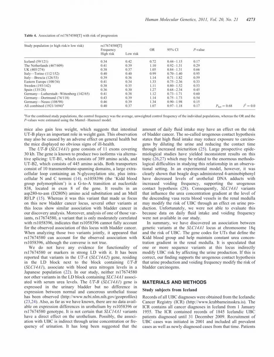
We have previously reported a UBC risk variant at theFGFR3/TACC3 locus at 4p16 that shows significantly strongerassociation with UBC with a predicted ‘low risk’ of pro-gression (tumors confined to the bladder mucosa and notpoorly differentiated) than with UBC with a predicted ‘highrisk’ of progression (tumor invasion in or beyond the laminapropria or poorly differentiated). Using the same classificationof cases, we found that the frequency of rs17674580[T] did notdiffer between patients with tumors of different aggressiveness(combined OR high versus low risk ¼ 1.07, P ¼ 0.17,Table 4). By regressing on the age at diagnosis for 4750cases, we found that rs17674580[T] did not associate withage at diagnosis of UBC (effect ¼ 20.25 years per allele;P ¼ 0.27, Phet ¼ 0.33).
Since smoking is a strong risk factor for UBC and variantsin the sequence of the genome that affect smokingbehavior have been identified, we tested whether eitherrs17674580[T] or rs1058396[G] (D280N) was associatedwith smoking behavior, using genotypes and information onsmoking from 17 617 Icelanders who have a history ofsmoking (16). Neither rs17674580[T] nor rs1058396[G](D280N) associated with smoking quantity as measured bynumber of cigarettes smoked per day [P ¼ 0.94 and 0.88 for
rs17674580[T] and rs1058396[G] (D280N), respectively].Finally, we tested for association between rs17674580[T]and serum urea in an Icelandic sample set that had ureameasurements (n ¼ 3641) (17). No significant associationbetween blood urea and rs17674580 was observed (P ¼ 0.68).
DISCUSSION
Urea is produced in the liver as the main nitrogenous endproduct of protein catabolism. Depending on the diet, �20–30 g of urea is produced per day and passed into the blood.It is filtered in the kidney and either excreted or absorbedand concentrated in the renal medulla in order to negate theosmotic effect of urea in the urine and thereby maintainbody fluid balance (18). Specific proteins, called facilitativeUTs, mediate urea flux across cellular membranes, necessaryfor the process of concentrating urine and salvaging nitrogen.These proteins are derived from two distinct genes, SoluteCarrier Family 14, member 1; SLC14A1 (or UT-B) andSLC14A2 (or UT-A). The genes occur in tandem on chromo-some 18q12.1–q21.1 and are probably the result of dupli-cation of a primordial UT gene (19). UT-A and UT-B
Figure 1. A schematic view of the structure and association results in the UBC-associated region on chromosome 18q12.3. (A) Estimated recombination rates(saRR) in cM/Mb from the HapMap (release 22) Phase II data. (B) Location of known genes in the region. (C) Schematic view of the association with bladdercancer for all SNPs tested in the region for the initial scan (Iceland and the Netherlands). The y-axis indicates the –log 10 P-value. Red dots indicate SNPsdirectly genotyped in both discovery series; blue dots indicate SNPs imputed in both discovery series; green dots indicate SNPs directly genotyped in the Ice-landic series but imputed in the Dutch series.
Human Molecular Genetics, 2011, Vol. 20, No. 21 4271

transporters have a similar basic structure and function butthere are also a number of significant differences. WhereasUT-A transporters are mainly found in the kidney, UT-Bexpression is mainly found on the erythrocyte plasma mem-brane and, to a lesser extent, in the descending vasa recta ofthe kidney, in the brain, ear, testis, intestine and urinarybladder (18). Unlike UT-A, UT-Bs transport water in additionto urea, have a higher transport rate, are inhibited by mercurialcompounds and are less acutely regulated (14).
Until now, no overt pathologies associated with defects inurea transport are known. Several mutations in the UT-B
gene lead to expression of defective UT-B proteins and alack of urea transport in erythrocytes. People with suchUT-B mutations have red blood cells that are devoid of theKidd blood group antigen (i.e. JKnull) but further than thatthere is no clear phenotypic effect. Only when JKnull individ-uals are dehydrated, they appear to have a mild (�20%)reduction in maximal urinary concentrating capacity (20). Inagreement with this finding, studies in UT-B knockout miceshow a 45-fold reduction in red blood cell urea permeability,50% increase in urine output and 30% decrease in urineosmolality compared with WT mice (21). The UT-B null
Table 2. Association of rs1058396[G] and rs17674580[T] on 18q12.3 with UBC
Study population (n cases/n controls) rs1058396[G] rs17674580[T]Frequency OR 95% CI P-value Info Frequency OR 95% CI P-value InfoCases Controls Cases Controls
Discovery groups (GWA)Iceland (603/37 781)a 0.531 0.487 1.20 1.06–1.34 2.5 × 10²³ 0.986 0.381 0.334 1.24 1.10–1.40 5.0 × 1024 0.992The Netherlands (1631/3822)a 0.548 0.515 1.14 1.05–1.24 1.6 × 10²³ 1000 0.392 0.366 1.13 1.03–1.23 6.8 × 10²³ 0.987
Follow-up groupsIceland II (178/2055)b 0.55 0.48 1.29 1.02–1.63 0.031 0.41 0.36 1.23 0.98–1.54 0.07Iceland III (350/3500)b 0.50 0.48 1.18 1.05–1.34 0.0071 0.36 0.34 1.26 1.11–1.43 3.1 × 1024
UK (724/535) 0.56 0.55 1.04 0.89–1.22 0.65 0.38 0.38 1.01 0.85–1.19 0.93Italy—Torino (328/389) 0.51 0.51 1.00 0.81–1.23 0.97 0.40 0.35 1.22 0.98–1.53 0.072Italy—Brescia (181/192) 0.52 0.46 1.26 0.95–1.68 0.11 0.38 0.34 1.21 0.89–1.64 0.22Belgium (191/378) 0.48 0.52 0.87 0.68–1.21 0.27 0.32 0.38 0.78 0.60–1.02 0.064Eastern Europe (213/526) 0.52 0.48 1.19 0.94–1.49 0.15 0.37 0.34 1.17 0.92–1.48 0.20Sweden (343/1264) 0.52 0.51 1.07 0.90–1.27 0.46 0.36 0.34 1.12 0.94–1.34 0.20Spain (238/881) 0.56 0.52 1.20 0.97–1.48 0.085 0.37 0.35 1.07 0.87–1.32 0.52Iran (269/246) 0.57 0.54 1.09 0.85–1.39 0.51 0.36 0.33 1.15 0.88–1.49 0.31Germany—LW (213/198) 0.55 0.50 1.23 0.93–1.62 0.14 0.40 0.33 1.35 1.01–1.80 0.044Germany—Dortmund (196/239) 0.55 0.49 1.29 0.99–1.69 0.059 0.41 0.32 1.45 1.09–1.92 9.5 × 10²³
Germany—Neuss (216/104) 0.56 0.50 1.29 0.92–1.79 0.14 0.42 0.33 1.45 1.03–2.06 0.035GWA (2234/41 603)c 1.16 1.08–1.24 3.4 × 1025 1.17 1.08–1.25 4.9 × 1025
Follow-up groups (3790/7507)c 1.13 1.07–1.20 2.5 × 1025 1.16 1.10–1.23 5.8 × 1027
All combined (6024/49 110)c 1.14 1.09–1.19 2.9 × 1029 1.17 1.11–1.22 7.6 × 10211
All P-values shown are two-sided. Shown are the corresponding numbers of cases and controls (n), allelic frequencies of variants in affected and controlindividuals, the allelic odds-ratio (OR) with P-values based on the multiplicative model.aResults presented for Iceland and the Netherlands were individually adjusted by the method of genomic control (see Materials and Methods).bIceland II consists of cases and controls genotyped by single-SNP assay, Iceland III consists of cases and controls genotyped in silico, using genotype informationon relatives.cFor the combined study populations, the control frequency was the average, unweighted control frequency of the individual populations, whereas the OR and theP-values were estimated using the Mantel–Haenszel model.
Table 3. Joint analysis of rs17674580[T] and rs1058396[G] in UBC
Population rs17674580[T] adjusted for rs1058396[G] rs1058396[G] adjusted for rs17674580[T]OR 95% CI P-value OR 95% CI P-value
The Netherlands 1.04 0.92–1.18 0.52 1.11 0.98–1.25 0.094Iceland 1.17 0.99–1.37 0.063 1.08 0.92–1.27 0.36Belgium 0.80 0.55–1.17 0.25 1.00 0.70–1.43 1.00Germany—Lutherstadt–Wittenberg 1.34 0.88–2.02 0.17 0.99 0.67–1.49 0.98Germany—Dortmund 1.42 0.95–2.13 0.08 1.03 0.70–1.52 0.89Eastern Europe 1.17 0.82–1.67 0.39 1.04 0.73–1.47 0.83Iceland II 1.13 0.79–1.62 0.52 1.19 0.83–1.70 0.34Iran 1.14 0.81–1.61 0.46 0.99 0.71–1.38 0.97Italy—Brescia 0.99 0.63–1.55 0.97 1.30 0.85–1.99 0.23Italy—Torino 1.33 0.97–1.83 0.07 0.85 0.63–1.16 0.30Germany—Neuss 1.39 0.85–2.27 0.19 1.07 0.67–1.71 0.77Spain 1.02 0.75–1.38 0.91 1.15 0.85–1.54 0.36Sweden 1.18 0.91–1.54 0.20 0.94 0.73–1.21 0.63UK 0.96 0.77–1.21 0.76 1.07 0.86–1.34 0.53Combined 1.1 1.03–1.18 0.0061 1.06 1.00–1.14 0.069
Phet ¼ 0.62, I2 ¼ 0.0 Phet ¼ 0.97, I2 ¼ 0.0
4272 Human Molecular Genetics, 2011, Vol. 20, No. 21
mice also gain less weight, which suggests that intestinalUT-B plays an important role in weight gain. This observationmay also be caused by an adverse effect on general health butthe mice displayed no obvious signs of ill-health.
The UT-B (SLC14A1) gene consists of 11 exons covering30 kb. The gene is known to produce two isoforms via alterna-tive splicing: UT-B1, which consists of 389 amino acids, andUT-B2, which consists of 445 amino acids. Both transportersconsist of 10 transmembrane-spanning domains, a large extra-cellular loop containing an N-glycosylation site, plus intra-cellular N and C termini (14). rs1058396 (the ‘Kidd bloodgroup polymorphism’) is a G-to-A transition at nucleotide838, located in exon 8 of the gene. It results in anasp280-to-asn (D280N) amino acid substitution and an MnlIRFLP (15). Whereas it was this variant that made us focuson this new bladder cancer locus, several other variants atthis locus show similar association with bladder cancer inour discovery analysis. Moreover, analysis of one of those var-iants, rs17674580, a variant that is only moderately correlatedwith rs1058396, indicates that rs1058396 cannot fully accountfor the observed association of this locus with bladder cancer.When analyzing those two variants jointly, it appeared thatrs17674580 can account for the observed association withrs1058396, although the converse is not true.
We do not have any evidence for functionality ofrs17674580 or markers in strong LD with it. It has beenreported that variants in the UT-A (SLC14A2) gene, residingin the LD block next to the block containing UT-B(SLC14A1), associate with blood urea nitrogen levels in aJapanese population (22). In our study, neither rs17674580nor other variants in the LD block containing SLC14A1 associ-ated with serum urea levels. The UT-B (SLC14A1) gene isexpressed in the urinary bladder but no difference inexpression between normal and cancerous urothelial tissuehas been observed (http://www.ncbi.nlm.nih.gov/geoprofiles)(23,24). Also, as far as we have known, there are no data avail-able on expression differences in urothelium by rs1058396 orrs17674580 genotype. It is not certain that SLC14A1 variantshave a direct effect on the urothelium. Possibly, the associ-ation with UBC is indirect through urine concentration or fre-quency of urination. It has long been suggested that the
amount of daily fluid intake may have an effect on the riskof bladder cancer. The so-called urogenous contact hypothesisstates that high fluid intake may reduce exposure to carcino-gens by diluting the urine and reducing the contact timethrough increased micturition (25). Large prospective epide-miological studies have yielded inconsistent results on thistopic (26,27) which may be related to the enormous methodo-logical difficulties in studying this relationship in an observa-tional setting. In an experimental model, however, it wasclearly shown that beagle dogs administered 4-aminobiphenylhave decreased levels of urothelial DNA adducts withincreased voiding frequency, supporting the urogenouscontact hypothesis (28). Consequently, SLC14A1 variantsthat influence the urea concentration gradient at the level ofthe descending vasa recta blood vessels in the renal medullamay modify the risk of UBC through an effect on urine pro-duction. Unfortunately, we were not able to evaluate thisbecause data on daily fluid intake and voiding frequencywere not available in our study.
In summary, we have discovered an association betweengenetic variants at the SLC14A1 locus at chromosome 18qand the risk of UBC. The gene codes for UTs that define theKidd blood group and help maintain constant urea concen-tration gradient in the renal medulla. It is speculated thatone or more sequence variants at this locus indirectlymodify UBC risk by affecting the urine production. If this iscorrect, our finding supports the urogenous contact hypothesisthat urine production and voiding frequency modify the risk ofbladder carcinogens.
MATERIALS AND METHODS
Study subjects from Iceland
Records of all UBC diagnoses were obtained from the IcelandicCancer Registry (ICR) (http://www.krabbameinsskra.is). TheICR contains all cancer diagnoses in Iceland from 1 January1955. The ICR contained records of 1845 Icelandic UBCpatients diagnosed until 31 December 2009. Recruitment ofUBC cases was initiated in 2001 and included all prevalentcases as well as newly diagnosed cases from that time. Patients
Table 4. Association of rs17674580[T] with risk of progression
Study population (n high risk/n low risk) rs17674580[T]Frequency OR 95% CI P-valueHigh risk Low risk
Iceland (59/121) 0.34 0.42 0.72 0.44–1.15 0.17The Netherlands (467/609) 0.41 0.39 1.10 0.92–1.31 0.29UK (405/274) 0.38 0.37 1.05 0.84–1.31 0.66Italy—Torino (112/152) 0.40 0.40 0.99 0.70–1.40 0.95Italy—Brescia (126/53) 0.39 0.36 1.14 0.71–1.82 0.59Eastern Europe (100/34) 0.41 0.34 1.33 0.75–2.36 0.33Sweden (193/142) 0.38 0.35 1.11 0.80–1.52 0.53Spain (135/28) 0.36 0.30 1.27 0.68–2.34 0.45Germany—Lutherstadt–Wittenberg (142/65) 0.41 0.38 1.12 0.73–1.71 0.60Germany—Dortmund (74/118) 0.43 0.39 1.14 0.75–1.73 0.54Germany—Neuss (108/98) 0.46 0.39 1.34 0.90–1.98 0.15All combined (1921/1694)a 0.40 0.37 1.07 0.97–1.18 0.17 Phet ¼ 0.68 I2 ¼ 0.0
aFor the combined study populations, the control frequency was the average, unweighted control frequency of the individual populations, whereas the OR and theP-values were estimated using the Mantel–Haenszel model.
Human Molecular Genetics, 2011, Vol. 20, No. 21 4273

were recruited by trained nurses on behalf of the patients’ treat-ing physicians, through special recruitment clinics. Participantsin the study donated a blood sample and answered a lifestylequestionnaire. A total of 603 patients (77% males; diagnosedfrom December 1964 to December 2009) were included in agenome-wide SNP genotyping effort, using the Infinium IIassay method and either the Sentrix HumanHap 300 orHumanCNV370-duo BeadChip (Illumina). The median age atdiagnosis for all consenting cases was 67.8 years (range 20–95 years) compared with 68.5 years for all UBC patients inthe ICR. The 37 781 controls (41% males; mean age 61years; SD ¼ 21) used in this study consisted of individualsfrom other ongoing GWAS at deCODE and represent .15%of the adult population of Iceland. No individual diseasegroup is represented by .10% of the total control group.Cancer patients (prostate, breast, colorectal and lung) were ana-lyzed separately, and the frequency of the sequence variantsstudied did not differ from other controls. Samples from pros-tate, breast, colorectal and lung cancer patients as well as indi-viduals used for the analysis of smoking variables come fromother ongoing project at deCODE Genetics. The study wasapproved by the Data Protection Authority of Iceland and theNational Bioethics Committee. Written informed consent wasobtained from all patients, relatives and controls. Personal iden-tifiers associated with medical information and blood sampleswere encrypted with a third-party encryption system forwhich the Data Protection Authority maintains the code.
In addition to the chip-genotyped case–control series thatwas used for the discovery phase of the study (Iceland I),we selected two follow-up (replication) case–control samplesets. The first of these (Iceland II) consists of 178 UBCcases and 2055 controls who were recruited after 2007 inthe same way as the group ‘Iceland I’ but who were not whole-genome genotyped. This group was genotyped usingsingle-SNP assays. The second follow-up group (Iceland III)was generated using information on 950 UBC cases withoutDNA who had close relatives that had been genotyped.These cases were assigned in silico genotypes, correspondingto an effective sample size of approximately 350 cases and3500 controls who were also in silico genotyped [see thesection In silico genotyping (Iceland III)].
Study subjects from the Netherlands
The Dutch patients were recruited for the Nijmegen BladderCancer Study (NBCS: http://dceg.cancer.gov/icbc/membership.html). The NBCS identified patients through thepopulation-based regional cancer registry held by the Compre-hensive Cancer Centre East, Nijmegen, that serves a region of1.3 million inhabitants in the eastern part of the Netherlands(www.ikcnet.nl). Patients diagnosed between 1995 and 2006under the age of 75 years were selected and their vital statusand current addresses updated through the hospital infor-mation systems of the seven community hospitals and one uni-versity hospital (Radboud University Nijmegen MedicalCentre, RUNMC) that are covered by the cancer registry.All patients still alive on 1 August 2007 were invited to thestudy by the Comprehensive Cancer Center on behalf of thepatients’ treating physicians. A second group of patients, diag-nosed between 2007 and 2008, was invited in 2009. In case of
consent, patients were sent a lifestyle questionnaire to fill outand blood samples were collected by Thrombosis Servicecenters which hold offices in all the communities in theregion. In total, 1940 patients were invited to participate. Ofall the invitees, 1275 gave informed consent (66%): 1185filled out the questionnaire (61%) and 1219 (63%) provideda blood sample. The number of participating patients wasincreased with a non-overlapping series of 439 bladdercancer patients who were recruited previously for a study ongene–environment interactions in three hospitals (RUNMC,Canisius Wilhelmina Hospital, Nijmegen, and Streekzieken-huis Midden-Twente, Hengelo, the Netherlands). Ultimately,completed questionnaires and blood samples were availablefor 1429 and 1691 patients, respectively. All the patientsselected for the analyses (n ¼ 1631) were of self-reportedEuropean descent. The median age at diagnosis was 62(range 25–93) years. Eighty-two percent of the participantswere males. Data on tumor stage and grade were obtainedthrough the cancer registry.
The control group (n ¼ 3822) came from different sources. Atotal of 1918 cancer-free control individuals (46% males) wererecruited within a project entitled ‘Nijmegen biomedical study’(NBS). The details of this study were reported previously (29).Briefly, this is a population-based survey conducted by theDepartment of Epidemiology and Biostatistics and theDepartment of Clinical Chemistry of the RUNMC, in which9371 individuals participated from a total of 22 500 age- andsex-stratified, randomly selected inhabitants of Nijmegen.Control individuals from the NBS were invited to participatein a study on gene–environment interactions in multifactorialdiseases such as cancer. They were all of self-reported Euro-pean descent and fully informed about the goals and the pro-cedures of the study. The study protocols of the NBCS andthe NBS were approved by the Institutional Review Board ofthe RUNMC and all study subjects gave written informedconsent. The NBS control group was extended with a Dutchcontrol group provided by collaborators from the Universityof Utrecht that had been chip-genotyped in conjunction withother studies and have been described in a previous publication(30). Briefly, these controls were recruited from two sources: (i)613 control individuals were unrelated, healthy volunteers whoaccompanied non-amyotrophic lateral sclerosis (ALS) patientsto the University Medical Center Utrecht neurology outpatientclinic, (ii) 1358 individuals were included from a GWAS onschizophrenia. All were of Dutch descent, with at least threeout of four grandparents of Dutch ancestry. Of the 3881 chip-typed controls, 3822 passed all quality control filters andwere used for analysis.
Study subjects from the UK
Details of the Leeds Bladder Cancer Study have been reportedpreviously (31). In brief, patients from the urology departmentof St James’s University Hospital, Leeds, were recruited fromAugust 2002 to March 2006. All patients undergoing cysto-scopy or transurethral resection of a bladder tumor who hadpreviously been found, or were subsequently shown, to haveurothelial cell carcinoma of the bladder were included. Exclu-sion criterion was significant mental impairment or a bloodtransfusion in the past month. All non-Caucasians were
4274 Human Molecular Genetics, 2011, Vol. 20, No. 21

excluded from the study, leaving 764 patients. The median ageat diagnosis of the patients was 73 years (range 30–101).Seventy-one percent of the patients were male and 36% of allthe patients had tumors with low risk of progression (pTaG1/2). The controls were recruited from the otolaryngology outpa-tients and ophthalmology inpatient and outpatient departmentsat St James’s Hospital, Leeds, from August 2002 to March2006. All controls of appropriate age for frequency-matchingwith the cases were approached and recruited. As for thecases, exclusion criteria for the controls were significantmental impairment or a blood transfusion in the past month.Also, controls were excluded if they had symptoms suggestiveof bladder cancer, such as hematuria; 2.8% of the controls werenon-Caucasian, leaving 530 Caucasian controls for the study,and 71% of the controls were male. Data were collected by ahealth questionnaire on smoking habits and smoking history(non-, ex- or current smoker, smoking dose in pack-years),occupational exposure history (to plastics, rubber, laboratories,printing, dyes and paints, diesel fumes), family history ofbladder cancer, ethnicity and place of birth and places ofbirth of parents. The participation rate of cases was �99%,and that among the controls was �80%. Ethical approval forthe study was obtained from Leeds (East) Local ResearchEthics Committee, project number 02/192.
Study subjects from Torino, Italy
The sources of cases for the Torino bladder cancer study weretwo urology departments of the main hospital in Torino, theSan Giovanni Battista Hospital (32). Cases were all Caucasianmen, aged 40–75 years (median 63 years) and living in theTorino metropolitan area. They were newly diagnosedbetween 1994 and 2006 with a histologically confirmed, inva-sive or in situ, bladder cancer. Of all the patients with infor-mation on stage and grade, 56% were at low risk ofprogression (pTaG1/2). The sources of controls are urology,medical and surgical departments of the same hospital inTorino. All controls are Caucasian men resident in theTorino metropolitan area. They were diagnosed and treatedbetween 1994 and 2006 for benign diseases (such as prostatichyperplasia, cystitis, hernias, heart failure, asthma and benignear diseases). Controls with cancer, liver or renal diseases andsmoking-related conditions were excluded. The median age ofthe controls was 57 years (range 40–74). Data were collectedby a professional interviewer who used a structured question-naire to interview both cases and controls face-to-face. Datacollected included demographics (age, sex, ethnicity, regionand education) and smoking. For cases, additional data werecollected on tumor histology, tumor site, size, stage, gradeand treatment of the primary tumor. The participation rateswere 90% for cases and 75% for controls, resulting in 328cases and 389 controls. Ethical approval for the study wasobtained from Comitato Etico Interaziendale, A.O.U. SanGiovanni Battista/C.T.O./Maria Adelaide.
Study subjects from Brescia, Italy
The Brescia bladder cancer study is a hospital-basedcase–control study. The study was reported in detail pre-viously (33). In short, the catchment area of the cases and
controls was the Province of Brescia, a highly industrializedarea in Northern Italy (mainly metal and mechanical industry,construction, transport, textiles) but also with relevant agricul-tural areas. Cases and controls were enrolled in 1997–2000from the two main city hospitals. The total number of eligiblesubjects was 216 cases and 220 controls. The participation rate(enrolled/eligible) was 93% (n ¼ 201) for cases and 97% (n ¼214) for controls. Only males were included. All cases andcontrols had Italian nationality and were of Caucasian ethni-city. All cases had to be residents of the Province ofBrescia, aged between 20 and 80, and newly diagnosed withhistologically confirmed bladder cancer. The median age ofthe patients was 63 years (range 22–80); 29% of all thepatients with known stage and grade had tumors of low riskof progression (pTaG1/2). Controls were patients admittedfor various urological non-neoplastic diseases and werefrequency-matched to cases on age, hospital and period ofadmission. The study was formally approved by the ethicalcommittee of the hospital where the majority of subjectswere recruited. A written informed consent was obtainedfrom all participants. Data were collected from clinicalcharts (tumor histology, site, grade, stage, treatments, etc.)and by means of face-to-face interviews during hospitaladmission, using a standardized semi-structured questionnaire.The questionnaire included data on demographics (age, ethni-city, region, education, residence, etc.) and smoking. ISCOand ISIC codes and expert assessments were used for occu-pational coding. Blood samples were collected from casesand controls for genotyping and DNA adduct analyses.
Study subjects from Belgium
The Belgian study has been reported in detail (34). In brief,cases were selected from the Limburg Cancer Registry(LIKAR) and were approached through urologists andgeneral practitioners. All cases were diagnosed with histologi-cally confirmed urothelial cell carcinoma of the bladderbetween 1999 and 2004, and were Caucasian inhabitants ofthe Belgian province of Limburg. The median age of thepatients was 68 years, and 86% of all the patients weremales. For the recruitment of controls, a request was madeto the ‘Kruispuntbank’ of the social security for simplerandom sampling, stratified by municipality and socio-economic status, among all citizens .50 years of age of theprovince. The median age of the controls was 64 years; 59%of the controls were males. Three trained interviewersvisited cases and controls at home. Information was collectedthrough a structured interview and a standardized food fre-quency questionnaire. In addition, biological samples werecollected. Data collected included medical history, lifetimesmoking history, family history of bladder cancer and a life-time occupational history. Informed consent was obtainedfrom all participants and the study was approved by theethical review board of the Medical School of the CatholicUniversity of Leuven, Belgium.
Study subjects from Eastern Europe
The details of this study have been described previously (35).Cases and controls were recruited as part of a study designed
Human Molecular Genetics, 2011, Vol. 20, No. 21 4275

to evaluate the risk of various cancers due to environmentalarsenic exposure in Hungary, Romania and Slovakia between2002 and 2004. The recruitment was carried out in the countiesof Bacs, Bekes, Csongrad and Jasz-Nagykun-Szolnok inHungary; Bihor and Arad in Romania; and Banska Bystricaand Nitra in Slovakia. The cases (n ¼ 214) and controls(n ¼ 533) selected were of Hungarian, Romanian and Slovaknationalities. Bladder cancer patients were invited on the basisof histopathological examinations by pathologists. Hospital-based controls were included in the study, subject to fulfillmentof a set of criteria. All general hospitals in the study areas wereinvolved in the process of control recruitment. The controls werefrequency-matched with cases for age, gender, country of resi-dence and ethnicity. Controls included general surgery, orthope-dic and trauma patients aged 30–79 years. Patients withmalignant tumors, diabetes and cardiovascular diseases wereexcluded as controls. The median age of the bladder cancerpatients was 65 years (range 36–90). Eighty-three percent ofthe patients were males. The median age for the controls was61 years (range 28–83). Fifty-one percent of the controls weremales. The participation rates among cases and controls were�70%. Of all the patients with known stage and grade infor-mation, 28% had a low-risk tumor (pTaG1/2). Clinicians tookvenous blood and other biological samples from cases and con-trols after consent forms had been signed. Cases and controlsrecruited to the study were interviewed by trained personneland completed a general lifestyle questionnaire. Ethnic back-ground for cases and controls was recorded along with othercharacteristics of the study population. Local ethical boardsapproved the study plan and design.
Study subjects from Sweden
The Swedish patients came from a population-based study ofUBC patients diagnosed in the Stockholm region in1995–1996 (36). Blood samples from 352 patients were avail-able out of a collection of 538 patients with primary urothelialcarcinoma of the bladder. The average age at onset for thesepatients was 69 years (range 32–97 years) and 67% of thepatients were males. Clinical data, including age at onset,grade and stage of tumor, were prospectively obtained fromhospitals and urology units in the region. The controlsamples came from blood donors in the Stockholm regionand were from cancer-free individuals of both genders. Theregional ethical committee approved of the study and all par-ticipants gave informed consent.
Study subjects from Spain
The Spanish study patients were recruited from the urologyand oncology departments of Zaragoza Hospital between Sep-tember 2007 and June 2009. A total of 246 patients with his-tologically proven urothelial cell carcinoma of the bladderwere enrolled (response 77%). Clinical information includingage at onset, grade and stage was obtained from medicalrecords. The median age at diagnosis for the patients was 65years (range 27–94) and 87% were males. The 890 Spanishcontrol individuals were part of a larger collection of controlsamples obtained from individuals who had attended the Uni-versity Hospital in Zaragoza, Spain, for diseases other than
cancer between November 2001 and May 2007. The controlswere of both genders and median age was 52 years (range11–87). Controls were questioned to rule out prior cancersbefore drawing the blood sample. All patients and controlswere of self-reported European descent. Study protocolswere approved by the Institutional Review Board of ZaragozaUniversity Hospital. All subjects gave written informedconsent.
Study subjects from Germany
The study subjects from Germany came from three differentstudies.
(i) The Neuss bladder cancer study. Details of the bladdercancer cases of this study have been published previously(37). The ongoing case–control series consists of 217bladder cancer cases and 105 controls from the Depart-ment of Urology, Lukasklinik Neuss, Germany, located�20 km from the Ruhr area. The median age at diagnosiswas 72.9 (range 26.1–93.4) years. Seventy-eight percentof the participants were males. Data on tumor stage andgrade were obtained through the cancer registry.Forty-five percent of the patients had a low-risk tumor(pTaG1/2). The 105 control individuals (64% males)(median age 42.4, range 18.0–89.0) were cancer-free.Data were collected from June 2009 to July 2010. Thelocal ethics committee approved the study plan anddesign.
(ii) The Dortmund bladder cancer study. Details of thebladder cancer cases of this study have been publishedpreviously (37). The case–control series consists of 197patients with a confirmed bladder cancer from theDepartment of Urology, St.-Josefs-Hospital Dortmund-Horde, located in the Ruhr area, an area of former coal,iron and steel industries and 240 controls from thesame Department of Urology, admitted for treatment ofbenign urological diseases, enrolled from July 2009 toJuly 2010. The median age at diagnosis was 71.2(range 35.0–89.2) years. Seventy-five percent of the par-ticipants were males, and 60% of the patients had tumorswith low risk of progression (pTaG1/2). The 240 controlindividuals (77% males) were cancer-free and frequency-matched for age with the cases (median age 70.7, range21.7–100). The local ethics committee approved thestudy plan and design.
(iii) The Lutherstadt Wittenberg (LW) study. Details of thebladder cancer cases of this study have been reported pre-viously (37–39). In brief, 221 patients with a confirmedbladder cancer from the Department of Urology, PaulGerhardt Foundation, Lutherstadt Wittenberg, Germany,were included. Patients were enrolled from December1995 to January 1999. Exclusion criterion was amissing written informed consent into the study. Themedian age of the patients was 65 years (range 20–91);86% of the patients were males. A total of 214 controlswere from the same Department of Urology, but wereadmitted for treatment of benign urological diseases.Exclusion criterion was a malignant disease in themedical history or a missing written informed consent.
4276 Human Molecular Genetics, 2011, Vol. 20, No. 21

The median age of the controls was 68 years (range 29–91); 84% of the controls were males. Data were collectedfrom July 2000 to May 2005. All cases and controls wereCaucasians, which was confirmed by questionnaire-baseddocumentation of nationality. Cases and controls werematched for age. Data collected in cases and controlsinclude age, gender, a complete documentation of occu-pational activities performed at least for 6 months, docu-mentation of work places with known bladder cancer riskover the entire working life, exposures to known or sus-pected occupational bladder carcinogens, lifetimesmoking habits, family history of bladder cancer,numbers of urinary infections treated by drugs duringthe previous 10 years, place of birth and places of resi-dency for .10 years. For bladder cancer cases, data ontumor staging, grading and treatment were taken fromthe records. First diagnosis of bladder cancer wasrecorded from July 1979 to January 1999. The localethics committee approved the study plan and design.
Study subjects from Iran
Samples from Iranian bladder cancer patients were obtainedfrom the Biobank of Shiraz Institute for Cancer Research(http://icr.ir/groups/biobank.html). The majority of thepatients were residents of the province of Fars (of whichShiraz is the capital city) in the south of Iran. The people ofFars are Persian with little racial heterogeneity. However,since hospitals affiliated with Shiraz University of MedicalSciences are referral hospitals for large parts of southernIran, inclusion of patients from other ethnic groups cannotbe ruled out. The DNA collection of all cancer patients inthe Biobank of Shiraz Institute for Cancer Research is anongoing process that started in 1999. For the present study,267 patients (229 males and 38 females) with histologicallyverified bladder cancer were available. The mean ages at diag-nosis were 63.6 and 60.1 years, respectively. For 3% of allcases, age could not be determined. The majority of thepatients had urothelial cell carcinoma (n ¼ 239; 89.5%); theremaining had squamous cell carcinoma, adenocarcinoma,rhabdomyosarcoma or unspecified/unknown morphology.The control group consisted of 222 male and 25 femalehealthy Iranian subjects without apparent cancer or auto-immune diseases and a negative first-degree family historyof cancer. The controls were recruited among healthy blooddonors at the Fars blood transfusion center. The mean ageswere 54.0 years for males and 53.5 years for females. DNAof the controls was also obtained from the Biobank ofShiraz Institute for Cancer Research. Informed consent wasobtained from all patients and controls. The biobanking pro-cedures have been approved by Shiraz University of MedicalSciences Ethics Committee.
Illumina genome-wide genotyping
The Dutch and Icelandic case and control samples were assayedwith the Illumina HumanHap300 or HumanHapCNV370 beadchips (Illumina, San Diego, CA, USA), containing 317 503and 370 404 haplotype tagging SNPs derived from phase I ofthe International HapMap project, respectively. SNPs were
excluded if they had: (i) a yield ,95% in cases or controls;(ii) a minor allele frequency ,1% in the population; or (iii)showed significant deviation from Hardy–Weinberg equili-brium in the controls (P , 0.001). Any samples with a callrate ,98% were excluded from the analysis.
SNP imputations
For the Dutch data set, we used 292 650 autosomal SNPspresent on both chip types to impute an additional 7 543 837ungenotyped SNPs using the IMPUTE v2.1 software (40,41)and a training set consisting of the combined 1000 Genomeslow-coverage pilot haplotypes (released June 2010, 120chromosomes) and the HapMap3 haplotypes (released Febru-ary 2009, 1920 chromosomes) downloaded from http://mathgen.stats.ox.ac.uk/impute/impute_v2.html#filtered_1kg_hm3_haps. Individuals with ,96% yield for the 292 650 SNPsand SNPs with minor allele frequency ,0.01, which werenot in Hardy–Weinberg equilibrium (P , 1025) or with adifferent frequency for the two chip types used (P , 1025),were excluded from the imputation. For the Icelandic dataset, we used 289 572 autosomal SNPs to impute an additional11 283 069 ungenotyped SNPs using as training set the 566phased European haplotypes of the August 2010 release ofthe 1000 Genomes project, downloaded from http://mathgen.stats.ox.ac.uk/impute/impute_v2.html#August_haps. Theimputation method used for the Icelandic data set has beendescribed previously (42). In short, it is an adaption of themethodology as used in the IMPUTE software but takingadvantage of the long-range phasing of the whole Icelandicdata set. As for the Dutch data set, individuals with lowyield and SNPs that failed quality control were excludedprior to imputation.
Genome-wide analysis
For both the Icelandic and the Dutch data set, we usedSNPTEST v2.1.1 (41) to test each SNP for association withbladder cancer, assuming additive genetic effect and usingmissing data likelihood score test (the method score option)to account for imputation uncertainty. Individuals with geno-type yield ,98% and SNPs with information (proper_info),0.4 in the analysis of either data set were excluded. Whencombining the two data sets, we restricted the analysis toSNPs that were present in both data sets and that had MAF.1%. As the August 2010 release of the 1000 Genomes ismapped to NCBI Genome build 37, the location of thoseSNPs were mapped to build 36 using the UCSC LiftOvertool (http://genome.ucsc.edu/cgi-bin/hgLiftOver). A total of5 340 737 SNPs that passed quality criteria and that wereidentified in both data sets were included when the resultsfor the two data sets were combined.
Prior to combining the two data sets, the results wereadjusted using the method of genomic control by dividingthe x2 statistics by 1.071 for the Dutch data set and by1.011 for the Icelandic data set. For the combined analysis,we assumed a fixed effect model implemented in theprogram METAL (http://www.sph.umich.edu/csg/abecasis/Metal/) and weighted the contribution of each data set withthe corresponding standard error. Heterogeneity in the effect
Human Molecular Genetics, 2011, Vol. 20, No. 21 4277

estimate for each SNP was calculated by including the corre-sponding option in METAL.
Single-SNP genotyping
Single-SNP genotyping for all samples was carried out atdeCODE Genetics in Reykjavik, Iceland, applying the sameplatform to all populations studied. All single-SNP genotypingwas carried out using the Centaurus (Nanogen) platform (43).The quality of each Centaurus SNP assay was evaluated by gen-otyping each assay on the CEU samples and comparing theresults with the HapMap data (44). All assays had mismatchrate ,0.5%. Additionally, all markers were re-genotyped on.10% of samples typed with the Illumina platform, resultingin an observed mismatch in ,0.5% of samples.
In silico genotyping (Iceland III)
In addition to imputing rs1058396 and rs17674580 into chip-genotyped individuals, we also performed a second imputationstep where genotypes were imputed into relatives of chip-genotyped individuals, creating in silico genotypes. Theinputs into the second imputation step are the fully phased(in particular, every allele has been assigned a parent oforigin) imputed and chip-typed genotypes of the availablechip-typed individuals. The algorithm used to perform thesecond imputation step consists of:
(i) For each ungenotyped individual (the proband), find allchip-genotyped individuals within two meioses of theindividual. The six possible types of two-meioses rela-tives of the proband are (ignoring more complicatedrelationships due to pedigree loops) parents, full andhalf siblings, grandparents, children and grandchildren.If all pedigree paths from the proband to a genotypedrelative go through other genotyped relatives, then thatrelative is excluded. For example, if a parent of theproband is genotyped, then the proband’s grandparentsthrough that parent are excluded. If the number ofmeioses in the pedigree around the proband exceeds athreshold (we used 12), then relatives are removedfrom the pedigree until the number of meioses fallsbelow 12, in order to reduce computational complexity.
(ii) At every point in the genome, calculate the probabilityfor each genotyped relative sharing with the probandbased on the autosomal SNPs used for phasing. A multi-point algorithm, based on the hidden Markov-modelLander–Green multipoint linkage algorithm using fastFourier transforms, is used to calculate these sharingprobabilities (10,45,46). First, single-point sharing prob-abilities are calculated by dividing the genome into0.5 cM bins and using the haplotypes over these bins asalleles. Haplotypes that are the same, except that theycan differ for one SNP at most, are treated as identical.When the haplotypes in the pedigree are incompatibleover a bin, then a uniform probability distribution wasused for that bin. The most common causes for suchincompatibilities are recombinations within the pedigree,phasing errors and genotyping errors. Note that since theinput genotypes are fully phased, the single-point
information is substantially more informative than forunphased genotyped persons. In particular one haplotypeof the parent of a genotyped child is always known. Thesingle-point distributions are then convolved using themultipoint algorithm to obtain multipoint sharing prob-abilities at the center of each bin. Genetic distanceswere obtained from the most recent version of thedeCODE genetic map (47).
(iii) Based on the sharing probabilities at the center of eachbin, all the SNPs from the whole-genome genotypingare imputed into the proband. To impute the genotypeof the paternal allele of an SNP located at x, flanked bybins with centers at xleft and xright, starting with the leftbin, going through all possible sharing patterns n, let Inbe the set of haplotypes of genotyped individuals thatshare identically by descent within the pedigree withthe proband’s paternal haplotype given the sharingpattern n and P(n) be the probability of n at the leftbin—this is the output from step (ii) above—and let ei
be the expected allele count of the SNP for haplotype i.Then ev =
∑i[Iv[i
/∑
i[Iv1 is the expected allele countof the paternal haplotype of the proband given n and anoverall estimate of the allele count given the sharing dis-tribution at the left bin is obtained from eleft =
∑vP v( )ev
.If In is empty, then no relative shares with the proband’spaternal haplotype given n and thus there is no infor-mation about the allele count. We therefore store theprobability that some genotyped relative shared the pro-band’s paternal haplotype, Oleft =
∑v,Iv=f P(V ), and an
expected allele count, conditional on the proband’spaternal haplotype being shared by at least one genotypedrelative: Cleft =
∑v,Iv=f P(v)ev/
∑v,Iv=f P(v). In the
same way, calculate Oright and Cright. Linear interpolationis then used to get an estimate at the SNP from the twoflanking bins:
O = Oleft +x − xleft
xright − xleft
(Oright − Oleft),
C = Cleft +x − xleft
xright − xleft
(Cright − Cleft).
If u is an estimate of the population frequency of the SNP, thenOc + (1 – O)u is an estimate of the allele count for the pro-band’s paternal haplotype. Similarly, an expected allelecount can be obtained for the proband’s maternal haplotype.
When performing case–control analysis with the in silicogenotypes, we use logistic regression to test for associationbetween SNPs and disease, treating disease status as theresponse and expected genotype counts from imputation orallele counts from direct genotyping as covariates. Testingwas performed using the likelihood ratio statistic. Whentesting for association based on the in silico genotypes, con-trols were matched to cases based on the informativeness ofthe imputed genotypes, such that for each case C, controlsof matching informativeness were chosen. Failing to matchcases and controls will lead to a highly inflated genomiccontrol factor, and in some cases may lead to spurious false-positive findings. The informativeness of each of the
4278 Human Molecular Genetics, 2011, Vol. 20, No. 21

imputation of each one of an individual’s haplotypes was esti-mated by taking the average of
a(e, u) =e − u
1 − ue ≥ u
u− e
ue , u
⎧⎪⎨⎪⎩
over all SNPs imputed for the individual, where e is theexpected allele count for the haplotype at the SNP and u isthe population frequency of the SNP. Note that a(u,u) ¼ 0and a(0,u) ¼ a(1,u) ¼ 1. The mean informativeness valuescluster into groups corresponding to the most common pedi-gree configurations used in the imputation, such as imputingfrom parent into child or from child into parent. Based onthis clustering of imputation informativeness, we divided thehaplotypes of individuals into 7 groups of varying informa-tiveness, which created 27 groups of individuals of similarimputation informativeness; 7 groups of individuals withboth haplotypes having similar informativeness, 21 groupsof individuals with the two haplotypes having different infor-mativeness, minus the one group of individuals with neitherhaplotype being imputed well. Within each group, we calcu-late the ratio of the number of controls and the number ofcases, and choose the largest integer C that was less thanthis ratio in all the groups. For example, if in one groupthere are 10.3 times as many controls as cases and if in allother groups this ratio was greater, then we would setC ¼ 10 and within each group randomly select 10 times asmany controls as there are cases.
Association analysis
For association analysis of the replication data sets, we used astandard likelihood ratio statistic, implemented in the NEMOsoftware (48) to calculate two-sided P-values for each individ-ual allele, assuming a multiplicative model for UBC risk, i.e.the relative risk from carrying two copies of the high-riskallele (compared with none) is assumed to be the square ofthe relative risk from carrying one copy. Results from multiplecase–control groups were combined using a Mantel–Haenszelmodel in which the groups were allowed to have differentpopulation frequencies for alleles and genotypes but wereassumed to have common ORs. Stratified analyses were con-ducted by smoking and by UBC aggressiveness. For thelatter, all patients for whom detailed histology informationwas available were classified with regard to risk of pro-gression, based on stage and grade information. Patientswith ‘low risk of progression’ were defined as having TNMstage pTa in combination with WHO 1973 differentiationgrade 1 or 2 or WHO/ISUP 2004 low grade. All othertumors were classified as ‘high risk of progression’ (stagepTis or ≥ pT1 or WHO 1973 grade 3 or WHO/ISUP 2004high grade).
Heterogeneity calculations
Heterogeneity was tested by comparing the null hypothesis ofthe effect being the same in all populations to the alternativehypothesis of each population having a different effect using
a likelihood ratio test. I2 lies between 0 and 100% anddescribes the proportion of total variation in study estimatesthat is due to heterogeneity (49).
SUPPLEMENTARY MATERIAL
Supplementary Material is available at HMG online.
ACKNOWLEDGEMENTS
We thank the individuals who participated in the study andwhose contribution made this work possible. We also thankthe personnel at all the recruitment centers. We acknowledgethe cancer registries in Iceland and the Netherlands for assist-ance in the ascertainment of the Icelandic and Dutch UBCpatients.
Conflict of Interest statement. The authors who are affiliatedwith deCODE genetics are all employees of deCODE, a bio-technology company that provides genetic testing services,and some own stocks or stock options in the company.
FUNDING
This work was supported by the following funding agencies.Collection of samples and data in Iceland and the Netherlandswas funded in part by the European Commission (POLY-GENE: LSHC-CT-2005) (grant number 018827) and aresearch investment grant of the Radboud University Nijme-gen Medical Centre (RUNMC). Control samples from the Uni-versity of Utrecht were genotyped with generous support fromthe ‘Prinses Beatrix Fonds’, VSB Fonds, H. Kersten andM. Kersten (Kersten Foundation), The Netherlands ALS Foun-dation, J.R. van Dijk and the Adessium Foundation. The con-trols from the Dutch Schizophrenia GWA study weregenotyped with the support of the National Institute ofMental Health (NIH/NIMH MH078075). The Leeds BladderCancer Study was funded by Cancer Research UK and York-shire Cancer Research. Torino Bladder Cancer Case ControlStudy was supported by a grant to ECNIS (EnvironmentalCancer Risk, Nutrition and Individual Susceptibility), anetwork of excellence operating within the European Union6th Framework Program, Priority 5: ‘Food Quality andSafety’ (Contract No. 513943); by a grant of the Compagniadi San Paolo—Human Genetics Foundation (HuGeF), theItalian Association for Cancer Research, Italy and the Pied-mont Region Progetti di Ricerca Sanitaria Finalizzata. TheBelgian bladder cancer study was funded by the Flemish gov-ernment and by the health authorities of the Belgian provinceof Limburg. The Swedish study was funded by the SwedishCancer Society and the Swedish Research Council. TheIranian study was supported by a grant from Shiraz Institutefor Cancer Research, Shiraz, Iran (grant numberICR-87-502). C.Z. and S.B. are funded by a European Com-mission 7th Framework Programme FP7-MC-IAPP (grantagreement number 218071 CancerGene). J.I.M. is funded byRed Tematica de Investigacion Cooperativa en Cancer (grantnumber RD06/0020/1054).
Human Molecular Genetics, 2011, Vol. 20, No. 21 4279

REFERENCES
1. Ploeg, M., Aben, K.K. and Kiemeney, L.A. (2009) The present and futureburden of urinary bladder cancer in the world. World J. Urol., 27, 289–293.
2. Ferlay, J., Shin, H.R., Bray, F., Forman, D., Mathers, C. and Parkin, D.M.(2010) Estimates of worldwide burden of cancer in 2008: GLOBOCAN2008 Int. J. Cancer, 127, 2893–2917.
3. Sievert, K.D., Amend, B., Nagele, U., Schilling, D., Bedke, J., Horstmann,M., Hennenlotter, J., Kruck, S. and Stenzl, A. (2009) Economic aspects ofbladder cancer: what are the benefits and costs? World J. Urol.,27, 295–300.
4. Mueller, C.M., Caporaso, N. and Greene, M.H. (2008) Familial andgenetic risk of transitional cell carcinoma of the urinary tract. Urol.
Oncol., 26, 451–464.5. Aben, K.K., Witjes, J.A., Schoenberg, M.P., Hulsbergen-van de Kaa, C.,
Verbeek, A.L. and Kiemeney, L.A. (2002) Familial aggregation ofurothelial cell carcinoma. Int. J. Cancer, 98, 274–278.
6. Aben, K.K., Baglietto, L., Baffoe-Bonnie, A., Coebergh, J.W.,Bailey-Wilson, J.E., Trink, B., Verbeek, A.L., Schoenberg, M.P., Alfred,W.J. and Kiemeney, L.A. (2006) Segregation analysis of urothelial cellcarcinoma. Eur. J. Cancer, 42, 1428–1433.
7. Murta-Nascimento, C., Silverman, D.T., Kogevinas, M., Garcia-Closas,M., Rothman, N., Tardon, A., Garcia-Closas, R., Serra, C., Carrato, A.,Villanueva, C. et al. (2007) Risk of bladder cancer associated with familyhistory of cancer: do low-penetrance polymorphisms account for theincrease in risk? Cancer Epidemiol. Biomarkers Prev., 16, 1595–1600.
8. Amundadottir, L.T., Thorvaldsson, S., Gudbjartsson, D.F., Sulem, P.,Kristjansson, K., Arnason, S., Gulcher, J.R., Bjornsson, J., Kong, A.,Thorsteinsdottir, U. and Stefansson, K. (2004) Cancer as a complexphenotype: pattern of cancer distribution within and beyond the nuclearfamily. PLoS Med., 1, e65.
9. Garcia-Closas, M., Malats, N., Silverman, D., Dosemeci, M., Kogevinas,M., Hein, D.W., Tardon, A., Serra, C., Carrato, A., Garcia-Closas, R. et al.(2005) NAT2 slow acetylation, GSTM1 null genotype, and risk of bladdercancer: results from the Spanish Bladder Cancer Study and meta-analyses.Lancet, 366, 649–659.
10. Kiemeney, L.A., Thorlacius, S., Sulem, P., Geller, F., Aben, K.K., Stacey,S.N., Gudmundsson, J., Jakobsdottir, M., Bergthorsson, J.T., Sigurdsson,A. et al. (2008) Sequence variant on 8q24 confers susceptibility to urinarybladder cancer. Nat. Genet., 40, 1307–1312.
11. Kiemeney, L.A., Sulem, P., Besenbacher, S., Vermeulen, S.H.,Sigurdsson, A., Thorleifsson, G., Gudbjartsson, D.F., Stacey, S.N.,Gudmundsson, J., Zanon, C. et al. (2010) A sequence variant at 4p16.3confers susceptibility to urinary bladder cancer. Nat. Genet., 42, 415–419.
12. Rothman, N., Garcia-Closas, M., Chatterjee, N., Malats, N., Wu, X.,Figueroa, J.D., Real, F.X., Van Den, B.D., Matullo, G., Baris, D. et al.(2010) A multi-stage genome-wide association study of bladder canceridentifies multiple susceptibility loci. Nat. Genet., 42, 978–984.
13. Wu, X., Ye, Y., Kiemeney, L.A., Sulem, P., Rafnar, T., Matullo, G.,Seminara, D., Yoshida, T., Saeki, N., Andrew, A.S. et al. (2009) Geneticvariation in the prostate stem cell antigen gene PSCA conferssusceptibility to urinary bladder cancer. Nat. Genet., 41, 991–995.
14. Stewart, G. (2011) The emerging physiological roles of the SLC14Afamily of urea transporters. Br. J. Pharmacol., 41, 991–995.
15. Olives, B., Merriman, M., Bailly, P., Bain, S., Barnett, A., Todd, J.,Cartron, J.P. and Merriman, T. (1997) The molecular basis of the Kiddblood group polymorphism and its lack of association with type 1 diabetessusceptibility. Hum. Mol. Genet., 6, 1017–1020.
16. Thorgeirsson, T.E., Geller, F., Sulem, P., Rafnar, T., Wiste, A.,Magnusson, K.P., Manolescu, A., Thorleifsson, G., Stefansson, H.,Ingason, A. et al. (2008) A variant associated with nicotine dependence,lung cancer and peripheral arterial disease. Nature, 452, 638–642.
17. Gudbjartsson, D.F., Holm, H., Indridason, O.S., Thorleifsson, G.,Edvardsson, V., Sulem, P., de Vegt, F., d’Ancona, F.C., den Heijer, M.,Wetzels, J.F. et al. (2010) Association of variants at UMOD with chronickidney disease and kidney stones—role of age and comorbid diseases.PLoS Genet., 6, e1001039.
18. Smith, C.P. (2009) Mammalian urea transporters. Exp. Physiol.,94, 180–185.
19. Fenton, R.A., Hewitt, J.E., Howorth, A., Cottingham, C.A. and Smith,C.P. (1999) The murine urea transporter genes Slc14a1 and Slc14a2 occurin tandem on chromosome 18. Cytogenet. Cell Genet., 87, 95–96.
20. Sands, J.M., Gargus, J.J., Frohlich, O., Gunn, R.B. and Kokko, J.P. (1992)Urinary concentrating ability in patients with Jk(a-b-) blood type who lackcarrier-mediated urea transport. J. Am. Soc. Nephrol., 2, 1689–1696.
21. Yang, B., Bankir, L., Gillespie, A., Epstein, C.J. and Verkman, A.S.(2002) Urea-selective concentrating defect in transgenic mice lacking ureatransporter UT-B. J. Biol. Chem., 277, 10633–10637.
22. Kamatani, Y., Matsuda, K., Okada, Y., Kubo, M., Hosono, N., Daigo, Y.,Nakamura, Y. and Kamatani, N. (2010) Genome-wide association studyof hematological and biochemical traits in a Japanese population. Nat.Genet., 42, 210–215.
23. Dyrskjot, L., Thykjaer, T., Kruhoffer, M., Jensen, J.L., Marcussen, N.,Hamilton-Dutoit, S., Wolf, H. and Orntoft, T.F. (2003) Identifying distinctclasses of bladder carcinoma using microarrays. Nat. Genet., 33, 90–96.
24. Dyrskjot, L., Kruhoffer, M., Thykjaer, T., Marcussen, N., Jensen, J.L.,Moller, K. and Orntoft, T.F. (2004) Gene expression in the urinarybladder: a common carcinoma in situ gene expression signature existsdisregarding histopathological classification. Cancer Res.,64, 4040–4048.
25. Braver, D.J., Modan, M., Chetrit, A., Lusky, A. and Braf, Z. (1987)Drinking, micturition habits, and urine concentration as potential riskfactors in urinary bladder cancer. J. Natl Cancer Inst., 78, 437–440.
26. Michaud, D.S., Spiegelman, D., Clinton, S.K., Rimm, E.B., Curhan, G.C.,Willett, W.C. and Giovannucci, E.L. (1999) Fluid intake and the risk ofbladder cancer in men. N. Engl. J. Med., 340, 1390–1397.
27. Ros, M.M., Bas Bueno-de-Mesquita, H.B., Buchner, F.L., Aben, K.K.,Kampman, E., Egevad, L., Overvad, K., Tjonneland, A., Roswall, N.,Clavel-Chapelon, F. et al. (2011) Fluid intake and the risk of urothelialcell carcinomas in the European Prospective Investigation into Cancer andNutrition (EPIC). Int. J. Cancer, 128, 2695–2708.
28. Kadlubar, F.F., Dooley, K.L., Teitel, C.H., Roberts, D.W., Benson, R.W.,Butler, M.A., Bailey, J.R., Young, J.F., Skipper, P.W. and Tannenbaum,S.R. (1991) Frequency of urination and its effects on metabolism,pharmacokinetics, blood hemoglobin adduct formation, and liver andurinary bladder DNA adduct levels in beagle dogs given the carcinogen4-aminobiphenyl. Cancer Res., 51, 4371–4377.
29. Wetzels, J.F., Kiemeney, L.A., Swinkels, D.W., Willems, H.L. and denHeijer, M. (2007) Age- and gender-specific reference values of estimatedGFR in Caucasians: the Nijmegen Biomedical Study. Kidney Int., 72,632–637.
30. van Es, M.A., Veldink, J.H., Saris, C.G., Blauw, H.M., van Vught, P.W.,Birve, A., Lemmens, R., Schelhaas, H.J., Groen, E.J., Huisman, M.H.et al. (2009) Genome-wide association study identifies 19p13.3(UNC13A) and 9p21.2 as susceptibility loci for sporadic amyotrophiclateral sclerosis. Nat. Genet., 41, 1083–1087.
31. Sak, S.C., Barrett, J.H., Paul, A.B., Bishop, D.T. and Kiltie, A.E. (2005)The polyAT, intronic IVS11–6 and Lys939Gln XPC polymorphisms arenot associated with transitional cell carcinoma of the bladder.Br. J. Cancer, 92, 2262–2265.
32. Matullo, G., Guarrera, S., Sacerdote, C., Polidoro, S., Davico, L.,Gamberini, S., Karagas, M., Casetta, G., Rolle, L., Piazza, A. and Vineis,P. (2005) Polymorphisms/haplotypes in DNA repair genes and smoking: abladder cancer case-control study. Cancer Epidemiol. Biomarkers Prev.,14, 2569–2578.
33. Shen, M., Hung, R.J., Brennan, P., Malaveille, C., Donato, F., Placidi, D.,Carta, A., Hautefeuille, A., Boffetta, P. and Porru, S. (2003)Polymorphisms of the DNA repair genes XRCC1, XRCC3, XPD,interaction with environmental exposures, and bladder cancer risk in acase-control study in northern Italy. Cancer Epidemiol. Biomarkers Prev.,12, 1234–1240.
34. Kellen, E., Zeegers, M., Paulussen, A., Van Dongen, M. and Buntinx, F.(2006) Fruit consumption reduces the effect of smoking on bladder cancerrisk. The Belgian case control study on bladder cancer. Int. J. Cancer,118, 2572–2578.
35. Thirumaran, R.K., Bermejo, J.L., Rudnai, P., Gurzau, E., Koppova, K.,Goessler, W., Vahter, M., Leonardi, G.S., Clemens, F., Fletcher, T. et al.
(2006) Single nucleotide polymorphisms in DNA repair genes and basalcell carcinoma of skin. Carcinogenesis, 27, 1676–1681.
36. Larsson, P., Wijkstrom, H., Thorstenson, A., Adolfsson, J., Norming, U.,Wiklund, P., Onelov, E. and Steineck, G. (2003) A population-based studyof 538 patients with newly detected urinary bladder neoplasms followedduring 5 years. Scand. J. Urol. Nephrol., 37, 195–201.
37. Lehmann, M.L., Selinski, S., Blaszkewicz, M., Orlich, M., Ovsiannikov,D., Moormann, O., Guballa, C., Kress, A., Truss, M.C., Gerullis, H. et al.
4280 Human Molecular Genetics, 2011, Vol. 20, No. 21
(2010) Rs710521[A] on chromosome 3q28 close to TP63 is associatedwith increased urinary bladder cancer risk. Arch. Toxicol., 84, 967–978.
38. Golka, K., Schmidt, T., Seidel, T., Dietrich, H., Roemer, H.C., Lohlein,D., Reckwitz, T., Sokeland, J., Weistenhofer, W., Blaszkewicz, M. andSelinski, S. (2008) The influence of polymorphisms of glutathioneS-transferases M1 and M3 on the development of human urothelial cancer.J. Toxicol. Environ. Health A, 71, 881–886.
39. Golka, K., Hermes, M., Selinski, S., Blaszkewicz, M., Bolt, H.M., Roth,G., Dietrich, H., Prager, H.M., Ickstadt, K. and Hengstler, J.G. (2009)Susceptibility to urinary bladder cancer: relevance of rs9642880[T],GSTM1 0/0 and occupational exposure. Pharmacogenet. Genomics, 19,903–906.
40. Howie, B.N., Donnelly, P. and Marchini, J. (2009) A flexible and accurategenotype imputation method for the next generation of genome-wideassociation studies. PLoS Genet., 5, e1000529.
41. Marchini, J., Howie, B., Myers, S., McVean, G. and Donnelly, P. (2007) Anew multipoint method for genome-wide association studies byimputation of genotypes. Nat. Genet., 39, 906–913.
42. Kong, A., Steinthorsdottir, V., Masson, G., Thorleifsson, G., Sulem, P.,Besenbacher, S., Jonasdottir, A., Sigurdsson, A., Kristinsson, K.T.,Jonasdottir, A. et al. (2009) Parental origin of sequence variantsassociated with complex diseases. Nature, 462, 868–874.
43. Kutyavin, I.V., Milesi, D., Belousov, Y., Podyminogin, M., Vorobiev, A.,Gorn, V., Lukhtanov, E.A., Vermeulen, N.M. and Mahoney, W. (2006) Anovel endonuclease IV post-PCR genotyping system. Nucleic Acids Res.,34, e128.
44. International Hapmap Consortium (2005) A haplotype map of the humangenome. Nature, 437, 1299–1320.
45. Kruglyak, L. and Lander, E.S. (1998) Faster multipoint linkage analysisusing Fourier transforms. J. Comput. Biol., 5, 1–7.
46. Lander, E.S. and Green, P. (1987) Construction of multilocus geneticlinkage maps in humans. Proc. Natl Acad. Sci. USA, 84, 2363–2367.
47. Kong, A., Thorleifsson, G., Gudbjartsson, D.F., Masson, G., Sigurdsson,A., Jonasdottir, A., Walters, G.B., Jonasdottir, A., Gylfason, A.,
Kristinsson, K.T. et al. (2010) Fine-scale recombination rate differencesbetween sexes, populations and individuals. Nature, 467, 1099–1103.
48. Gretarsdottir, S., Thorleifsson, G., Reynisdottir, S.T., Manolescu, A.,Jonsdottir, S., Jonsdottir, T., Gudmundsdottir, T., Bjarnadottir, S.M.,Einarsson, O.B., Gudjonsdottir, H.M. et al. (2003) The gene encodingphosphodiesterase 4D confers risk of ischemic stroke. Nat. Genet., 35,131–138.
49. Higgins, J.P. and Thompson, S.G. (2002) Quantifying heterogeneity in ameta-analysis. Stat. Med., 21, 1539–1558.
50. Cortessis, V.K., Yuan, J.M., Van Den, B.D., Jiang, X., Gago-Dominguez,M., Stern, M.C., Castelao, J.E., Xiang, Y.B., Gao, Y.T., Pike, M.C. andConti, D.V. (2010) Risk of urinary bladder cancer is associated with 8q24variant rs9642880[T] in multiple racial/ethnic groups: results from the LosAngeles-Shanghai case-control study. Cancer Epidemiol. Biomarkers
Prev., 19, 3150–3156.
51. Wang, M., Wang, M., Zhang, W., Yuan, L., Fu, G., Wei, Q. and Zhang, Z.(2009) Common genetic variants on 8q24 contribute to susceptibility tobladder cancer in a Chinese population. Carcinogenesis, 30, 991–996.
52. Rafnar, T., Sulem, P., Stacey, S.N., Geller, F., Gudmundsson, J.,Sigurdsson, A., Jakobsdottir, M., Helgadottir, H., Thorlacius, S., Aben,K.K. et al. (2009) Sequence variants at the TERT-CLPTM1L locusassociate with many cancer types. Nat. Genet., 41, 221–227.
53. Gago-Dominguez, M., Jiang, X., Conti, D.V., Castelao, J.E., Stern, M.C.,Cortessis, V.K., Pike, M.C., Xiang, Y.B., Gao, Y.T., Yuan, J.M. and VanDen Berg, D.J. (2011) Genetic variations on chromosomes 5p15 and15q25 and bladder cancer risk: findings from the Los Angeles-Shanghaibladder case-control study. Carcinogenesis, 32, 197–202.
54. Wang, S., Tang, J., Wang, M., Yuan, L. and Zhang, Z. (2010) Geneticvariation in PSCA and bladder cancer susceptibility in a Chinesepopulation. Carcinogenesis, 31, 621–624.
55. Wang, M., Chu, H., Yan, F., Qin, C., Li, P., Yuan, L., Yin, C., Xu, J. andZhang, Z. (2011) Chromosome 4p16.3 variant modify bladder cancer riskin a Chinese population. Carcinogenesis, 32, 872–875.
Human Molecular Genetics, 2011, Vol. 20, No. 21 4281