Genome-Wide Scan Identifies TNIP1, PSORS1C1, and RHOB as Novel Risk Loci for Systemic Sclerosis
13
Genome-Wide Scan Identifies TNIP1, PSORS1C1, and RHOB As Novel Risk Loci for Systemic Sclerosis Yannick Allanore 1,2 *, Mohamad Saad 3 , Philippe Dieude ´ 4 , Je ´ro ˆ me Avouac 1,2 , Jorg H. W. Distler 5 , Philippe Amouyel 6 , Marco Matucci-Cerinic 7 , Gabriella Riemekasten 8 , Paolo Airo 9 , Inga Melchers 10 , Eric Hachulla 11 , Daniele Cusi 12,13 , H.-Erich Wichmann 14,15 , Julien Wipff 1,2 , Jean-Charles Lambert 6 , Nicolas Hunzelmann 16 , Kiet Tiev 17 , Paola Caramaschi 18 , Elisabeth Diot 19 , Otylia Kowal-Bielecka 20 , Gabriele Valentini 21 , Luc Mouthon 22 , La ´ szlo ´ Czirja ´k 23 , Nemanja Damjanov 24 , Erika Salvi 12,13 , Costanza Conti 25 , Martina Mu ¨ ller 26,27 , Ulf Mu ¨ ller-Ladner 28 , Valeria Riccieri 29 , Barbara Ruiz 2 , Jean-Luc Cracowski 30 , Luc Letenneur 31,32 , Anne Marie Dupuy 33 , Oliver Meyer 4 , Andre ´ Kahan 1 , Arnold Munnich 2 , Catherine Boileau 2,34 , Maria Martinez 3 1 Universite ´ Paris Descartes, Rhumatologie A, INSERM, U1016, Ho ˆ pital Cochin, APHP, Paris, France, 2 INSERM, U781, Universite ´ Paris Descartes, Ho ˆ pital Necker, Paris, France, 3 INSERM, U563, CHU Purpan, Universite ´ Paul Sabatier, Toulouse, France, 4 Universite ´ Paris Diderot, Rhumatologie, INSERM, U699, Ho ˆ pital Bichat Claude-Bernard, Paris, France, 5 Department for Internal Medicine 3 and Institute for Clinical Immunology Friedrich-Alexander-University Erlangen-Nuremberg, Nuremberg, Germany, 6 INSERM, U744, Institut Pasteur de Lille, Universite ´ de Lille Nord, Lille, France, 7 Department of Biomedicine, Division of Rheumatology AOUC, Denothe Centre, University of Florence, Florence, Italy, 8 Department of Rheumatology and Clinical Immunology, Charite ´ University Hospital, Berlin, Germany, 9 Rheumatology and Clinical Immunology, Spedali Civili, Brescia, Italy, 10 Clinical Research Unit for Rheumatology, University Medical Center, Freiburg, Germany, 11 Lille II University, Internal Medecine Department, Lille, France, 12 University of Milano, Department of Medicine, Surgery, and Dentistry San Paolo School of Medicine, 13 Genomics and Bioinformatics Platform, Fondazione Filarete, Milan, Italy, 14 Institute of Epidemiology I, Helmholtz Zentrum Mu ¨ nchen – German Research Center for Environmental Health, Neuherberg, Germany, 15 Institute of Medical Informatics, Biometry, and Epidemiology, Chair of Epidemiology, Ludwig-Maximilians-Universita ¨t and Klinikum Grosshadern, Munich, Germany, 16 Department of Dermatology, University of Cologne, Ko ¨ ln, Germany, 17 Universite ´ Pierre et Marie Curie, Service de Me ´decine Interne, Ho ˆ pital Saint Antoine, Paris, France, 18 Rheumatology Unit, University of Verona, Verona, Italy, 19 INSERM, U618, IFR 135, CHU Bretonneau, Tours, France, 20 Department of Rheumatology and Internal Medicine, Medical University of Bialystok, Bialystok, Poland, 21 Department of Clinical and Experimental Medicine, Rheumatology Unit, Second University of Naples, Naples Italy, 22 Universite ´ Paris Descartes, Me ´decine Interne, Ho ˆ pital Cochin, APHP, Paris, France, 23 Department of Immunology and Rheumatology, University of Pe ´cs, Pe ´ cs, Hungary, 24 Institute of Rheumatology, School of Medicine, University of Belgrade, Belgrade, Serbia, 25 Kos Genetic SRL, Milano, Italy, 26 Institute of Genetic Epidemiology, Helmholtz Zentrum Mu ¨ nchen – German Research Center for Environmental Health, Neuherberg, Germany, and, 27 Institute of Medical Informatics, Biometry and Epidemiology, Chair of Epidemiology and Chair of Genetic Epidemiology, Ludwig-Maximilians-Universita ¨t and Department of Medicine I, University Hospital Grosshadern, Ludwig-Maximilians-Universita ¨t, Munich Germany, 28 University of Giessen, Department of Rheumatology and Clinical Immunology Kerckhoff-Klinik, Bad Nauheim, Germany, 29 Division of Rheumatology, Department of Internal Medicine and Medical Specialities, University ‘‘Sapienza,’’ Rome, Italy, 30 INSERM, CIC3, CHU Grenoble, France, 31 INSERM, U897, Bordeaux, France, 32 Universite ´ Bordeaux Segalen, Bordeaux, France, 33 INSERM, U888, Ho ˆ pital de la Colombie `re, Montpellier, France, 34 Universite ´ Versailles-SQY, Laboratoire de Biochimie, d’Hormonologie et de Ge ´ne ´tique Mole ´culaire, Ho ˆ pital Ambroise Pare ´, AP-HP, Boulogne, France Abstract Systemic sclerosis (SSc) is an orphan, complex, inflammatory disease affecting the immune system and connective tissue. SSc stands out as a severely incapacitating and life-threatening inflammatory rheumatic disease, with a largely unknown pathogenesis. We have designed a two-stage genome-wide association study of SSc using case-control samples from France, Italy, Germany, and Northern Europe. The initial genome-wide scan was conducted in a French post quality-control sample of 564 cases and 1,776 controls, using almost 500 K SNPs. Two SNPs from the MHC region, together with the 6 loci outside MHC having at least one SNP with a P,10 25 were selected for follow-up analysis. These markers were genotyped in a post-QC replication sample of 1,682 SSc cases and 3,926 controls. The three top SNPs are in strong linkage disequilibrium and located on 6p21, in the HLA-DQB1 gene: rs9275224, P = 9.18 6 10 28 , OR = 0.69, 95% CI [0.60–0.79]; rs6457617, P = 1.14 6 10 27 and rs9275245, P = 1.39 6 10 27 . Within the MHC region, the next most associated SNP (rs3130573, P = 1.86 6 10 25 , OR = 1.36 [1.18–1.56]) is located in the PSORS1C1 gene. Outside the MHC region, our GWAS analysis revealed 7 top SNPs (P,10 25 ) that spanned 6 independent genomic regions. Follow-up of the 17 top SNPs in an independent sample of 1,682 SSc and 3,926 controls showed associations at PSORS1C1 (overall P = 5.70 6 10 210 , OR:1.25), TNIP1 (P = 4.68 6 10 29 , OR:1.31), and RHOB loci (P = 3.17 6 10 26 , OR:1.21). Because of its biological relevance, and previous reports of genetic association at this locus with connective tissue disorders, we investigated TNIP1 expression. A markedly reduced expression of the TNIP1 gene and also its protein product was observed both in lesional skin tissue and cultured dermal fibroblasts from SSc patients. Furthermore, TNIP1 showed in vitro inhibitory effects on inflammatory cytokine-induced collagen production. The genetic signal of association with TNIP1 variants, together with tissular and cellular investigations, suggests that this pathway has a critical role in regulating autoimmunity and SSc pathogenesis. PLoS Genetics | www.plosgenetics.org 1 July 2011 | Volume 7 | Issue 7 | e1002091
Transcript of Genome-Wide Scan Identifies TNIP1, PSORS1C1, and RHOB as Novel Risk Loci for Systemic Sclerosis

Genome-Wide Scan Identifies TNIP1, PSORS1C1, andRHOB As Novel Risk Loci for Systemic SclerosisYannick Allanore1,2*, Mohamad Saad3, Philippe Dieude4, Jerome Avouac1,2, Jorg H. W. Distler5, Philippe
Amouyel6, Marco Matucci-Cerinic7, Gabriella Riemekasten8, Paolo Airo9, Inga Melchers10, Eric
Hachulla11, Daniele Cusi12,13, H.-Erich Wichmann14,15, Julien Wipff1,2, Jean-Charles Lambert6, Nicolas
Hunzelmann16, Kiet Tiev17, Paola Caramaschi18, Elisabeth Diot19, Otylia Kowal-Bielecka20, Gabriele
Valentini21, Luc Mouthon22, Laszlo Czirjak23, Nemanja Damjanov24, Erika Salvi12,13, Costanza Conti25,
Martina Muller26,27, Ulf Muller-Ladner28, Valeria Riccieri29, Barbara Ruiz2, Jean-Luc Cracowski30, Luc
Letenneur31,32, Anne Marie Dupuy33, Oliver Meyer4, Andre Kahan1, Arnold Munnich2, Catherine
Boileau2,34, Maria Martinez3
1 Universite Paris Descartes, Rhumatologie A, INSERM, U1016, Hopital Cochin, APHP, Paris, France, 2 INSERM, U781, Universite Paris Descartes, Hopital Necker, Paris,
France, 3 INSERM, U563, CHU Purpan, Universite Paul Sabatier, Toulouse, France, 4 Universite Paris Diderot, Rhumatologie, INSERM, U699, Hopital Bichat Claude-Bernard,
Paris, France, 5 Department for Internal Medicine 3 and Institute for Clinical Immunology Friedrich-Alexander-University Erlangen-Nuremberg, Nuremberg, Germany,
6 INSERM, U744, Institut Pasteur de Lille, Universite de Lille Nord, Lille, France, 7 Department of Biomedicine, Division of Rheumatology AOUC, Denothe Centre, University
of Florence, Florence, Italy, 8 Department of Rheumatology and Clinical Immunology, Charite University Hospital, Berlin, Germany, 9 Rheumatology and Clinical
Immunology, Spedali Civili, Brescia, Italy, 10 Clinical Research Unit for Rheumatology, University Medical Center, Freiburg, Germany, 11 Lille II University, Internal
Medecine Department, Lille, France, 12 University of Milano, Department of Medicine, Surgery, and Dentistry San Paolo School of Medicine, 13 Genomics and
Bioinformatics Platform, Fondazione Filarete, Milan, Italy, 14 Institute of Epidemiology I, Helmholtz Zentrum Munchen – German Research Center for Environmental
Health, Neuherberg, Germany, 15 Institute of Medical Informatics, Biometry, and Epidemiology, Chair of Epidemiology, Ludwig-Maximilians-Universitat and Klinikum
Grosshadern, Munich, Germany, 16 Department of Dermatology, University of Cologne, Koln, Germany, 17 Universite Pierre et Marie Curie, Service de Medecine Interne,
Hopital Saint Antoine, Paris, France, 18 Rheumatology Unit, University of Verona, Verona, Italy, 19 INSERM, U618, IFR 135, CHU Bretonneau, Tours, France, 20 Department
of Rheumatology and Internal Medicine, Medical University of Bialystok, Bialystok, Poland, 21 Department of Clinical and Experimental Medicine, Rheumatology Unit,
Second University of Naples, Naples Italy, 22 Universite Paris Descartes, Medecine Interne, Hopital Cochin, APHP, Paris, France, 23 Department of Immunology and
Rheumatology, University of Pecs, Pecs, Hungary, 24 Institute of Rheumatology, School of Medicine, University of Belgrade, Belgrade, Serbia, 25 Kos Genetic SRL, Milano,
Italy, 26 Institute of Genetic Epidemiology, Helmholtz Zentrum Munchen – German Research Center for Environmental Health, Neuherberg, Germany, and, 27 Institute of
Medical Informatics, Biometry and Epidemiology, Chair of Epidemiology and Chair of Genetic Epidemiology, Ludwig-Maximilians-Universitat and Department of Medicine
I, University Hospital Grosshadern, Ludwig-Maximilians-Universitat, Munich Germany, 28 University of Giessen, Department of Rheumatology and Clinical Immunology
Kerckhoff-Klinik, Bad Nauheim, Germany, 29 Division of Rheumatology, Department of Internal Medicine and Medical Specialities, University ‘‘Sapienza,’’ Rome, Italy,
30 INSERM, CIC3, CHU Grenoble, France, 31 INSERM, U897, Bordeaux, France, 32 Universite Bordeaux Segalen, Bordeaux, France, 33 INSERM, U888, Hopital de la
Colombiere, Montpellier, France, 34 Universite Versailles-SQY, Laboratoire de Biochimie, d’Hormonologie et de Genetique Moleculaire, Hopital Ambroise Pare, AP-HP,
Boulogne, France
Abstract
Systemic sclerosis (SSc) is an orphan, complex, inflammatory disease affecting the immune system and connective tissue.SSc stands out as a severely incapacitating and life-threatening inflammatory rheumatic disease, with a largely unknownpathogenesis. We have designed a two-stage genome-wide association study of SSc using case-control samples fromFrance, Italy, Germany, and Northern Europe. The initial genome-wide scan was conducted in a French post quality-controlsample of 564 cases and 1,776 controls, using almost 500 K SNPs. Two SNPs from the MHC region, together with the 6 locioutside MHC having at least one SNP with a P,1025 were selected for follow-up analysis. These markers were genotyped ina post-QC replication sample of 1,682 SSc cases and 3,926 controls. The three top SNPs are in strong linkage disequilibriumand located on 6p21, in the HLA-DQB1 gene: rs9275224, P = 9.1861028, OR = 0.69, 95% CI [0.60–0.79]; rs6457617,P = 1.1461027 and rs9275245, P = 1.3961027. Within the MHC region, the next most associated SNP (rs3130573,P = 1.8661025, OR = 1.36 [1.18–1.56]) is located in the PSORS1C1 gene. Outside the MHC region, our GWAS analysis revealed7 top SNPs (P,1025) that spanned 6 independent genomic regions. Follow-up of the 17 top SNPs in an independentsample of 1,682 SSc and 3,926 controls showed associations at PSORS1C1 (overall P = 5.70610210, OR:1.25), TNIP1(P = 4.6861029, OR:1.31), and RHOB loci (P = 3.1761026, OR:1.21). Because of its biological relevance, and previous reports ofgenetic association at this locus with connective tissue disorders, we investigated TNIP1 expression. A markedly reducedexpression of the TNIP1 gene and also its protein product was observed both in lesional skin tissue and cultured dermalfibroblasts from SSc patients. Furthermore, TNIP1 showed in vitro inhibitory effects on inflammatory cytokine-inducedcollagen production. The genetic signal of association with TNIP1 variants, together with tissular and cellular investigations,suggests that this pathway has a critical role in regulating autoimmunity and SSc pathogenesis.
PLoS Genetics | www.plosgenetics.org 1 July 2011 | Volume 7 | Issue 7 | e1002091

Citation: Allanore Y, Saad M, Dieude P, Avouac J, Distler JHW, et al. (2011) Genome-Wide Scan Identifies TNIP1, PSORS1C1, and RHOB As Novel Risk Loci forSystemic Sclerosis. PLoS Genet 7(7): e1002091. doi:10.1371/journal.pgen.1002091
Editor: Mark I. McCarthy, University of Oxford, United Kingdom
Received October 28, 2010; Accepted April 5, 2011; Published July 7, 2011; 11 February 2011
Copyright: � 2011 Allanore et al. This is an open-access article distributed under the terms of the Creative Commons Attribution License, which permitsunrestricted use, distribution, and reproduction in any medium, provided the original author and source are credited.
Funding: This project was funded by Agence Nationale pour la Recherche (Project ANR-08-GENO-016-1) and supported by research grants from SERVIERresearch group, SANOFI-AVENTIS, Association des Sclerodermies de France, and Groupe Francais de Recherche sur la Sclerodermie. The KORA (Cooperative healthresearch in the Region of Augsburg) studies were financed by the Helmholtz Zentrum Munchen, German Research Center for Environmental Health, Neuherberg,Germany, and supported by grants from the German Federal Ministry of Education and Research (BMBF) and by the State of Bavaria. Part of this work wasfinanced by the German National Genome Research Network (NGFN). This research was supported within the Munich Center of Health Sciences (MC Health) aspart of LMUinnovativ. HYPERGENES (European Network for Genetic-Epidemiological Studies) is funded by EU within the FP7: HEALTH-F4-2007-201550. Thesample considered in the present study has been collected by the Milano group in collaboration with the Center of Transfusion Medicine, Cellular Therapy, andCryobiology, Foundation Ca’ Granda Ospedale Maggiore Policlinico, and Fondazione Filarete. The funders had no role in study design, data collection and analysis,decision to publish, or preparation of the manuscript.
Competing Interests: The authors have declared that no competing interests exist.
* E-mail: [email protected]
Introduction
Systemic sclerosis (MIM181750) is a connective tissue disease
characterized by generalized microangiopathy, severe immuno-
logic alterations and massive deposits of matrix components in the
connective tissue. Being an orphan disease, SSc presents a major
medical challenge and is recognized as the most severe connective
tissue disorder with high risk of premature deaths [1]. Epidemi-
ological data on SSc vary in different parts of the world and
depend on selection criteria for the study population. Inasmuch,
the prevalence of the disease fluctuates across global regions and
population-based studies result in higher prevalence than do
hospital records-based studies. In North America, the prevalence
of SSc has been reported as 0.7–2.8 per 10,000 in a Canadian
study, whereas in the U.S. figures of 2.6 per 10,000 versus 7.5 per
10,000 were reported by medical records - versus population-
based studies, respectively. In Europe, a prevalence of 1.6 per
10,000 was reported in Denmark, 3.5 per 10,000 in Estonia, 1.58
per 10,000 adults (95% confidence interval, 129–187) in Seine-
Saint-Denis in France [2–4]. The risk of SSc is increased among
first-degree relatives of patients, compared to the general
population. In a study of 703 families in the US, including 11
multiplex SSc families, the familial relative risk in first-degree
relatives was about 13, with a 1.6% recurrence rate, compared to
0.026% in the general population [5]. The sibling risk ratio was
about 15 (ranging from 10 to 27 across cohorts). The only twin
study reported to date included 42 twin pairs [6]. The data showed
a similar concordance rate in monozygotic twins (4.2%, n = 24)
and dizygotic twins (5.6%) (NS) and an overall cross-sectional
concordance rate of 4.7%. However, concordance for the presence
of antinuclear antibodies was significantly higher in the monozy-
gotic twins (90%) than in the dizygotic twins (40%) suggesting that
genetics may be important for the auto-immune part of the
disease.
The aetiology of SSc is still unclear but some key steps have
been described, in particular early endothelial damage and
dysregulation of the immune system with abnormal autoantibody
production [7]. At the cellular level, early events include
endothelial injury and perivascular inflammation with the release
of a large array of inflammatory mediators [8,9]. In the advanced
stage, a progressive activation of fibroblasts in the skin and in
internal organs leads to hyperproduction of collagen and
irreversible tissue fibrosis [9]. Epidemiological investigations
indicate that SSc follows a pattern of multifactorial inheritance
[10]. Previous candidate-gene association studies have only
identified a handful of SSc risk loci, most contributing to the
genetic susceptibility of other autoimmune diseases [9–16]. So far,
two genome-wide association studies of SSc have been conducted
[17,18]. The studies differ according to the ancestry of the studied
population (Korean vs US/European) and the genome-wide
association data: map density (,440 K vs 280 K SNPs) and
sample size (,700 vs ,7300 subjects). They provided evidence of
association with known MHC loci, but only one ‘new’ locus was
identified at CD247 in the US/European dataset, variants at
CD247 being known to contribute to the susceptibility of systemic
lupus erythematosus [18].
The diagnosis of SSc is based on recognized clinical criteria
established decades ago however, these do not include specific
autoantibodies or recent tools for assessment of the disease [19,20].
Therefore, phenotypic heterogeneity is a concern for SSc and
genetic heterogeneity is also highly probable with regards to
data obtained in other connective tissue disorders. Given these
considerations, and previous findings in other autoimmune
diseases, it is apparent that additional risk variants for SSc remain
to be discovered. Therefore, to identify further common variants
that contribute to SSc risk in the European population, we
conducted a two-stage GWAS, in two case-control samples (total
.8,800 subjects).
Results/Discussion
We established a collaborative consortium including groups
from 4 European countries (France, Italy, Germany and Eastern-
Europe) from which we were able to draw upon a combined
sample of over 8,800 subjects (before quality control) and
conducted a two-stage genome-wide association study. In stage
1, we genotyped 1,185 samples on Illumina Human610-Quad
BeadChip and genotypes obtained using the same chip from 2,003
control subjects were made available to us from the 3C study
[21,22]. After stringent quality control, we finally tested for
association in stage-1, 489,814 autosomal SNPs in 2,340 subjects
(564 cases and 1,776 controls) (Table 1). We tested for association
between each SNP and SSc using the logistic regression
association test, assuming additive genetic effects. The quantile-
quantile plot and estimation of the genomic inflation factor
(l= 1.035) indicated minimal overall inflation (Figure 1A). The
genome-wide logistic association results are presented in Figure 1B.
Table S1 provides details for all SNPs with P,1024, including one
SNP exceeding P,1027, the Bonferroni threshold for genome-
wide significance. The three top SNPs were located on 6p21, in
the HLA-DQB1 gene: rs9275224, P = 9.1861028, OR = 0.69,
95%CI[0.60–0.79]; rs6457617, P = 1.1461027 and rs9275245,
P = 1.3961027 (Figure 1B and Table 2). Several associated SNPs
in HLA-DQB1 have already been reported but rs6457617 was also
Genome-Wide Scan in Systemic Sclerosis
PLoS Genetics | www.plosgenetics.org 2 July 2011 | Volume 7 | Issue 7 | e1002091
identified as the most associated SNP in the previous US/
European GWAS study [18]. Of note, the three SNPs in HLA-
DQB1 are in strong LD (r2.0.97). Within the MHC region, the
next most associated SNP (rs3130573, P = 1.8661025, OR =
1.36[1.18–1.56]) is located in the psoriasis susceptibility 1
candidate 1 (PSORS1C1) gene (Table 2), a candidate gene for
psoriasis [23]. Conditional analyses of susceptibility variants within
MHC showed that there were two independent association signals
at rs6457617 (HLA-DQB1) and at rs3130573 (PSORS1C1). Indeed,
the association at PSORS1C1 remained significant (P,2.161025)
after controlling for the association at HLA-DQB1 and the
association at HLA-DQB1 remained also significant (P,1.561027) after controlling for the association at PSORS1C1 (Table
S2).
Outside the MHC region, our GWAS analysis revealed 7 top
SNPs (P,1025) that spanned 6 independent genomic regions
(Figure 1B and Table S1). Conditional analyses of each of them on
HLA-DQB1 showed no significant drop in the association signals
(Table S3). The 6 loci having at least one SNP with a P,1025
were selected for follow-up analysis. Within each locus we selected
the SNPs with the strongest (P,1024) association signals to be
genotyped in a post-QC replication sample of 1,682 SSc cases and
3,926 controls (Table 1). To this list we added two top SNPs in
HLA-DQB1 and the SNP in PSORS1C1. Finally, we further
included 4 SNPs at the two known loci (STAT4 and TNPO3-IRF5)
and at the newly identified locus (CD247) by Radstake et al [18].
Out of a total set of 21 SNPs submitted for replication, 20 passed
the quality-control analyses.
Stratified association analyses in stage 2 data (Table 2),
confirmed the strong association for HLA-DQB1 (rs6457617,
P = 1.35610228) at 6p21.3 and also with the PSORS1C1 variant
(rs3130573, P = 4.9861023) at 6p21.1. Of the 6 remaining loci
selected in stage 1, only 2 were replicated with nominal P,
5% and with same direction of effect. They mapped at 2p24
(rs342070, P = 0.026; rs13021401, P = 0.024) and 5q33 (rs379
2783, P = 4.1461025; rs2233287, P = 4.3861023; rs4958881, P =
2.0961023). None of the replicated SNPs showed evidence for
heterogeneity of effects among the 4 geographical origins (Breslow-
day P.0.10). As expected, ORs estimated in the discovery tended
to be higher than those obtained in the replication stage data.
Afterwards, association signals from joint analyses of the 2 datasets
(Table 2) consistently showed highly significant association for
HLA-DQB1 (P = 2.33610237), PSORS1C1 (P = 5.70610210) and
TNIP1 (P = 4.6861029), and also showed some evidence of
association for RHOB (P = 3.1761026). All populations showed
same direction of effects (Figure 2). Finally, we also replicated
association signals at IRF5 (P = 3.4961025; combined-P = 4.1361027), at STAT4 (P = 1.9610210; combined-P = 2.26610213) and
at the recently identified new SSc risk locus, CD247 (P =
2.9061025; combined-P = 1.3061026) (Table 2). In our combined
data, the locus-specific PAR estimates were 24% for HLA-DQB1,
Author Summary
Systemic sclerosis (SSc) is a connective tissue diseasecharacterized by generalized microangiopathy, severeimmunologic alterations, and massive deposits of matrixcomponents in the connective tissue. Epidemiologicalinvestigations indicate that SSc follows a pattern ofmultifactorial inheritance; however, only a few loci havebeen replicated in multiple studies. We undertook a two-stage genome-wide association study of SSc involvingover 8,800 individuals of European ancestry. Combinedanalyses showed independent association at the knownHLA-DQB1 region and revealed associations at PSORS1C1,TNIP1, and RHOB loci, in agreement with a strong immunegenetic component. Because of its biological relevance,and previous reports of genetic association at this locuswith other connective tissue disorders, we investigatedTNIP1 expression. We observed a markedly reducedexpression of the gene and its protein product in SSc, aswell as its potential implication in control of extra-cellularmatrix synthesis, providing a new clue for a link betweeninflammation/immunity and fibrosis.
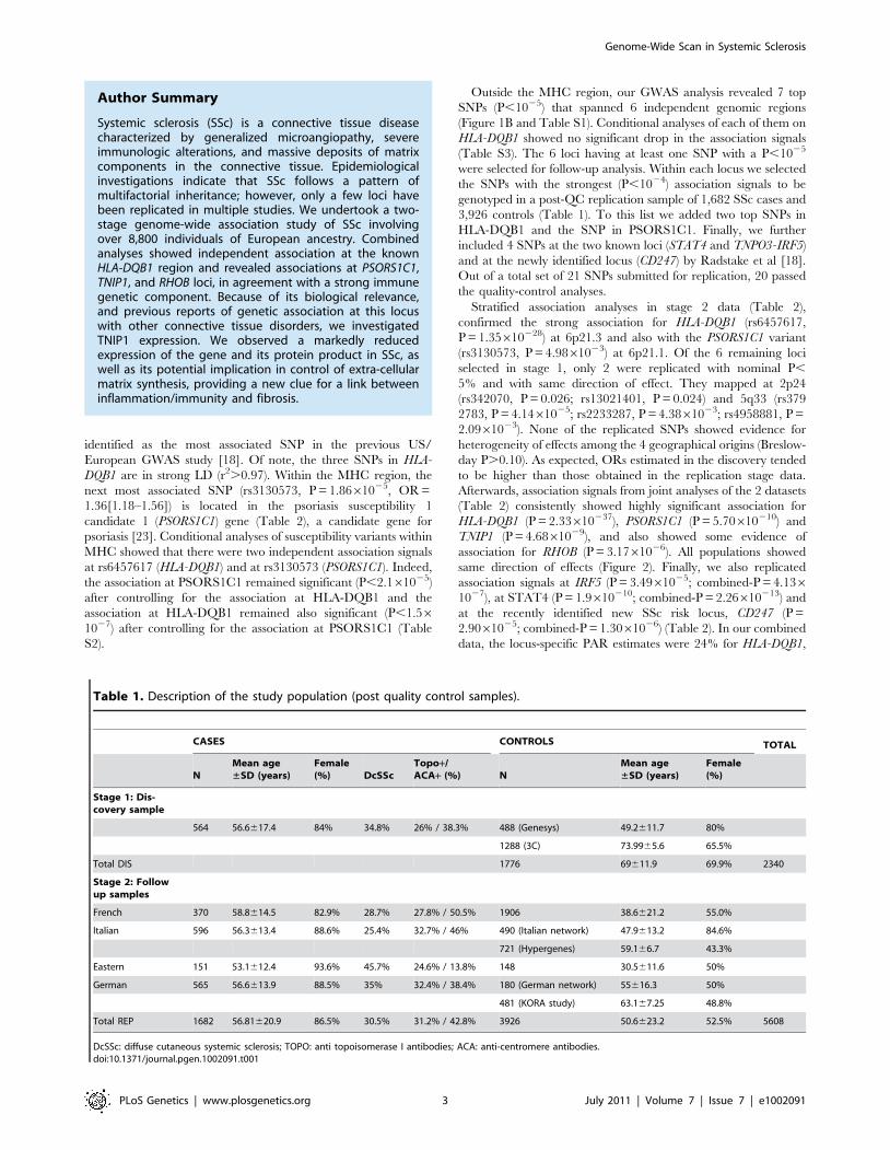
Table 1. Description of the study population (post quality control samples).
CASES CONTROLS TOTAL
NMean age±SD (years)
Female(%) DcSSc
Topo+/ACA+ (%) N
Mean age±SD (years)
Female(%)
Stage 1: Dis-covery sample
564 56.6617.4 84% 34.8% 26% / 38.3% 488 (Genesys) 49.2611.7 80%
1288 (3C) 73.9965.6 65.5%
Total DIS 1776 69611.9 69.9% 2340
Stage 2: Followup samples
French 370 58.8614.5 82.9% 28.7% 27.8% / 50.5% 1906 38.6621.2 55.0%
Italian 596 56.3613.4 88.6% 25.4% 32.7% / 46% 490 (Italian network) 47.9613.2 84.6%
721 (Hypergenes) 59.166.7 43.3%
Eastern 151 53.1612.4 93.6% 45.7% 24.6% / 13.8% 148 30.5611.6 50%
German 565 56.6613.9 88.5% 35% 32.4% / 38.4% 180 (German network) 55616.3 50%
481 (KORA study) 63.167.25 48.8%
Total REP 1682 56.81620.9 86.5% 30.5% 31.2% / 42.8% 3926 50.6623.2 52.5% 5608
DcSSc: diffuse cutaneous systemic sclerosis; TOPO: anti topoisomerase I antibodies; ACA: anti-centromere antibodies.doi:10.1371/journal.pgen.1002091.t001
Genome-Wide Scan in Systemic Sclerosis
PLoS Genetics | www.plosgenetics.org 3 July 2011 | Volume 7 | Issue 7 | e1002091

4% for TNIP1, 8% for PSORS1C1, 7% for CD247, 8% for STAT4
and 3% with IFR5/TNPO3. The combined PAR estimate
was 47.4%.
As secondary analyses, we assessed homogeneity of SNP’s effect
between sub-categories of SSc (cutaneous sub-types and auto-
antibodies). Case-only analyses revealed no significant evidence for
heterogeneous ORs between cutaneous sub-types of SSc patients
for any of the 5 replicated SNPs at 2p24 or 5q33 loci (Table S4A).
Indeed, similar association signals were obtained from case-
category association analyses (Table S4B). Altogether, the results
did not suggest that the association signals in the newly identified
5q33 locus were driven by a specific sub-type of SSc. Conversely,
for HLA-DQB1 and PSORS1C1 we found evidence of heterogene-
ity in OR estimates in positive vs negative ACA or TOPO auto-
antibody SSc patients (Table S4A). Yet, the association signals in
each of these sub-types of patients remained strong (Table S4B).
These results support the previously reported hypothesis that the
magnitude of the HLA-DQB1 effect on SSc susceptibility may
depend on auto-antibody status [11]. The GWAS stage had 78%
power to detect loci of the effect sizes observed in the discovery
sample for TNIP1 variants (OR = 1.50) at a significance of
P,1025. However, it is widely acknowledged that effect sizes of
Figure 1. Genome-wide association results from the discovery phase. (A) Quantile-quantile plot for test statistics (logistic regression test) for489,814 SNPs passing quality control. The plot shows a close match to the test statistics expected under the null distribution (l= 1.03). (B) Manhattanplot representing the P values across the genome. The 2log10 P of the logistic regression test (y axis) from 489,814 SNPs in 564 systemic sclerosispatients and 1,776 controls is plotted against its physical position (x-axis) on successive chromosomes. 90 SNPs with P,1024 lie above the bluehorizontal line and are listed in Table S1. Highly significant association was observed with SNPs within the MHC locus, including 1 SNPs that reachedthe conservative threshold for genome-wide significance (P,1027).doi:10.1371/journal.pgen.1002091.g001
Genome-Wide Scan in Systemic Sclerosis
PLoS Genetics | www.plosgenetics.org 4 July 2011 | Volume 7 | Issue 7 | e1002091

Ta
ble
2.
Ge
no
me
-wid
eas
soci
atio
nan
dre
plic
atio
nfo
rsy
ste
mic
scle
rosi
sri
skva
rian
ts.
Ch
r.(c
lose
stg
en
e)
Po
s.(b
p)
SN
PM
ino
r/M
ajo
rM
AF
Ca
ses/
Co
ntr
ols
PO
R9
5%
CI
MA
FC
ase
s/C
on
tro
ls$
PO
R9
5%
CI
**P
$P
OR
95
%C
I
MH
Clo
ci
6p
21
(PSO
RS1
C1
)3
12
14
24
7rs
31
30
57
3G
/A0
.39
1/
0.3
21
1.8
6E-
05
1.3
6(1
.18
–1
.56
)0
.41
6/0
.37
34
.98
E-0
31
.13
(1.0
4–
1.2
3)
4.8
E-0
15
.70
E-1
01
.25
(1.1
7–
1.3
5)
6p
21
(HLA
-DQ
B1
)3
27
67
85
6rs
92
75
22
4A
/G0
.40
5/
0.4
96
9.1
8E-
08
0.6
9(0
.6–
0.7
9)
&N
A-
--
--
--
6p
21
(HLA
-DQ
B1
)3
27
71
82
9rs
64
57
61
7C
/T0
.40
8/
0.4
98
1.1
4E-
07
0.6
9(0
.6–
0.7
9)
0.3
45
/0.4
63
1.3
5E-
28
0.6
1(0
.56
–0
.67
)1
.0E-
01
2.3
3E-
37
0.6
2(0
.58
–0
.67
)
No
nM
HC
loci
2p
24
(RH
OB
)2
05
48
95
2rs
34
20
70
C/T
0.2
93
/0
.22
65
.56
E-0
61
.42
(1.2
2–
1.6
5)
0.2
58
/0.2
35
2.6
1E-
02
1.1
2(1
.01
–1
.23
)1
.9E-
01
4.6
6E-
06
1.2
0(1
.11
–1
.30
)
20
55
20
00
rs1
30
21
40
1T
/C0
.28
9/
0.2
25
1.3
7E-
05
1.4
0(1
.2–
1.6
3)
0.2
57
/0.2
32
2.4
7E-
02
1.1
2(1
.01
–1
.24
)1
.3E-
01
3.1
7E-
06
1.2
1(1
.12
–1
.31
)
3p
25
(PP
AR
G/T
SEN
2)
12
46
83
47
rs9
85
56
22
T/C
0.1
45
/0
.09
61
.64
E-0
61
.66
(1.3
5–
2.0
5)
0.0
97
/0.1
09
9.8
6E-
01
1.0
0(0
.85
–1
.17
)7
.6E-
03
1.0
5E-
01
1.1
1(0
.94
–1
.17
)
12
23
46
16
rs3
10
74
6C
/T0
.12
1/
0.0
86
.15
E-0
51
.55
(1.2
5–
1.9
1)
0.0
74
/0.0
77
8.6
9E-
02
0.8
8(0
.77
–1
.02
)9
.6E-
01
4.2
2E-
01
1.0
5(0
.98
–1
.25
)
5q
33
(TN
IP1
)1
50
43
04
29
rs4
95
88
81
C/T
0.1
66
/0
.11
58
.26
E-0
61
.54
(1.2
8–
1.8
7)
0.1
51
/0.1
30
4.3
8E-
03
1.2
1(1
.06
–1
.38
)3
.2E-
01
5.7
9E-
06
1.2
9(1
.17
–1
.42
)
15
04
35
92
5rs
37
92
78
3G
/A0
.20
8/
0.1
52
1.1
4E-
05
1.4
7(1
.24
–1
.75
)0
.19
8/0
.16
62
.09
E-0
31
.21
(1.0
7–
1.3
6)
6.8
E-0
15
.73
E-0
71
.29
(1.2
0–
1.4
3)
15
04
20
29
0rs
22
33
28
7A
/G0
.13
9/
0.0
96
3.7
1E-
05
1.5
5(1
.26
–1
.91
)0
.12
1/0
.10
34
.14
E-0
51
.26
(1.1
3–
1.4
0)
6.8
E-0
14
.68
E-0
91
.31
(1.1
5–
1.4
3)
6p
16
-q1
6(A
SCC
3)
10
14
44
33
2rs
94
98
41
9A
/G0
.52
2/
0.4
46
7.7
1E–
06
1.3
7(1
.19
–1
.57
)0
.45
8/0
.47
51
.18
E-0
10
.93
(0.8
5–
1.0
2)
4.6
E-0
13
.15
E-0
11
.04
(0.9
7–
1.1
1)
10
14
45
69
9rs
69
19
74
5T
/C0
.52
2/
0.4
47
8.1
4E-
06
1.3
7(1
.19
–1
.57
)0
.46
1/0
.47
87
.47
E-0
20
.93
(0.8
5–
1.0
1)
4.0
E-0
13
.34
E-0
11
.04
(0.9
7–
1.1
1)
7p
12
-q2
1(S
EMA
3A
/HM
G1
7P
1)
84
16
60
13
rs4
32
92
28
C/A
0.3
05
/0
.23
96
.66
E-0
61
.42
(1.2
2–
1.6
5)
0.2
49
/0.2
48
5.0
8E-
01
0.9
7(0
.87
–1
.07
)3
.4E-
01
2.0
5E-
01
1.0
6(1
.04
–1
.20
)
83
97
69
40
rs1
02
95
41
T/C
0.2
88
/0
.22
72
.37
E-0
51
.39
(1.2
–1
.63
)0
.22
3/0
.23
17
.93
E-0
11
.01
(0.9
2–
1.1
2)
9.6
E-0
11
.50
E-0
21
.11
(0.9
7–
1.1
5)
11
q2
5(O
PC
ML)
13
22
84
60
3rs
27
25
46
6G
/A0
.40
3/
0.3
28
4.6
0E-
06
1.3
9(1
.21
–1
.59
)0
.37
8/0
.37
55
.71
E-0
10
.98
(0.8
9–
1.0
6)
6.2
E-0
13
.55
E-0
31
.11
(1.0
4–
1.2
0)
13
22
87
03
3rs
27
25
43
7C
/T0
.40
4/
0.3
35
2.5
2E-
05
1.3
5(1
.17
–1
.54
)0
.39
5/0
.38
78
.33
E-0
10
.99
(0.9
1–
1.0
8)
5.4
E-0
11
.82
E-0
31
.12
(1.0
4–
1.2
0)
13
23
00
77
9rs
10
89
46
23
T/G
0.3
17
/0
.25
67
.75
E-0
51
.34
(1.1
6–
1.5
5)
0.2
75
/0.2
77
7.6
7E-
01
0.9
9(0
.90
–1
.08
)1
.4E-
01
5.8
3E-
02
1.0
8(1
.00
–1
.16
)
Pre
vio
usl
yre
po
rte
dw
ith
P,
10
27
1q
22
-23
(CD
24
7)
16
56
87
04
9rs
20
56
62
6G
/T0
.35
4/0
.39
31
.70
E-0
20
.84
(0.7
3–
0.9
7)
0.3
6/
0.4
2.9
0E-
05
0.8
2(0
.75
–0
.9)
3.8
E-0
11
.30
E-0
60
.83
(0.7
7–
0.8
9)
2q
32
(ST
AT
4)
19
16
11
00
3rs
38
21
23
6A
/G0
.22
7/0
.20
38
.70
E-0
21
.16
(0.9
8–
1.3
6)
0.2
4/
0.2
2.1
0E-
07
1.3
3(1
.2–
1.4
9)
8.0
E-0
12
.09
E-0
71
.27
(1.1
6–
1.3
9)
19
16
72
87
8rs
75
74
86
5T
/G0
.27
2/0
.21
92
.50
E-0
41
.33
(1.1
4–
1.5
5)
0.2
9/
0.2
21
.90
E-1
01
.40
(1.2
6–
1.5
6)
9.0
E-0
12
.26
E-1
31
.38
(1.2
7–
1.5
)
7q
32
(TN
PO
3-I
RF5
)1
28
38
14
19
rs1
04
88
63
1C
/T0
.12
4/0
.09
32
.50
E–0
31
.39
(1.1
2–
1.7
2)
0.1
4/
0.1
03
.49
E-0
51
.34
(1.1
7–
1.5
4)
4.5
E-0
14
.13
E-0
71
.35
(1.2
–1
.51
)
do
i:10
.13
71
/jo
urn
al.p
ge
n.1
00
20
91
.t0
02
Genome-Wide Scan in Systemic Sclerosis
PLoS Genetics | www.plosgenetics.org 5 July 2011 | Volume 7 | Issue 7 | e1002091

significant GWAS loci are overestimates of true effects and other
genes of lower effect sizes are unlikely to reach stringent significant
thresholds.
Our GWAS analysis revealed strong association with
PSORS1C1, which is ,1 Mb of HLA-DQB1. Notably, PSORS1C1
is known to be involved in autoimmune response [23]. In the
combined data, association with PSORS1C1 was highly significant
(P = 5.70610210) and remained significant after controlling for the
association at HLA-DQB1. Altogether, our results suggest that this
region is likely to contain more than one gene playing a role in the
pathogenesis of autoimmune disorders [23,24]. Fine mapping at
this locus is warranted to identify causal variants.
The three strongly associated SNPs at the 5q33 locus are
located within the TNFAIP3 interacting protein 1 (TNIP1) gene.
TNIP1 is a very interesting new candidate gene for SSc. The
protein encoded by this gene exerts a negative regulation of NF-
kappaB via two sequential activities: deubiquitination of Lys63-
based chains and synthesis of Lys48-based chains on the TNF
receptor-interacting protein and also inhibition of NF-KappaB
processing [25]. TNIP1 interacts with A20 (TNFAIP3) to
negatively regulate NF-kappaB. Several recent studies have
suggested that the activation of some inflammatory factors may
upregulate fibrotic mediators through Toll-like receptors (TLRs),
thereby contributing to SSc pathogenesis [8]. It has been shown
that TLR engagement leads to A20 induction in macrophages and
that TNIP1/A20 is essential for the termination of TLR-induced
NF-kappaB activity and proinflammatory cytokine production
[26]. Although interactions between TNIP1 and A20 are not well
known, A20 also acts as a deubiquitinating enzyme, suggesting a
molecular link between deubiquitinating activity and the regula-
tion of TLR signals [26]. Therefore, TNIP1 and A20 may play a
critical role in the regulation of downstream TLR signals, and this
issue will have to be addressed in SSc. Interestingly, variants at
TNIP1 have been shown to be implicated in systemic lupus
erythematosus susceptibility [27,28] and in psoriasis [29].
Furthermore, we have recently reported an association of one
TNFAIP3 variant with SSc [30]. In our stage-1 data, evidence of
association at TNFAIP3 was nominal (lowest P = 0.047) and no
pairwise interaction was found (P.0.06) between TNFAIP3 and
TNIP1 variants. Analysis of the LD structure across the TNIP1
gene revealed that the 3 strongly associated SNPs belong to the
same LD-block (Figure 3). No residual association signals were
observed when rs3792783 and each of the other 2 SNPs were
paired in conditional analyses. Therefore, any of them, or other
variants yet to be identified, could be the causal variant(s).
Interestingly, rs3792783 is located upstream from the transcription
start site in exon 2 (Figure 3). It is noteworthy that previously
reported lupus TNIP1 variants were located in the same LD-block
[27,28]. Because of the compelling evidence of the potential role of
NF-kappaB in autoimmune diseases and our raised new signal
association for SSc at TNIP1 (a negative regulator of this pathway)
we performed ex vivo and in vitro investigations to assess TNIP1
expression in SSc patients and healthy controls. For SSc patients,
the results showed a strikingly reduced expression of TNIP1 in skin
tissue (Figure 4A), and of both mRNA (Figure 4B) and protein
(Figure 4C) synthesis by cultured dermal fibroblasts. Addressing
the question of the potential link between the NF-kappaB pathway
and the fibrotic propensity that characterizes SSc, we next assessed
the influence of pro-inflammatory cytokines and TNIP1 on the
synthesis of extra-cellular matrix by dermal fibroblasts in culture.
Using cells from the skin of healthy controls (Figure 5) and SSc
patients (Figure 6), we showed that recombinant TNIP1 abrogated
collagen synthesis induced by inflammatory cytokines both at the
mRNA and protein levels. It must be acknowledged that TNIP1 is
described as an intra-cellular protein whereas we used recombi-
nant protein added to cell supernatant in these experiments. The
observed effects may be related to different hypotheses. TNIP1 has
been described as a nuclear shuttling protein and it could have a
chaperon-like activity, highly interacting with other protein that
could result in engulfment of TNIP1 through interaction with a
cell surface protein. Such intra-cellular effects of extra-cellular
proteins has been shown also for the S100 family of proteins that
have no leader sequence and for clusterin for which it is postulated
that the protein could be taken up by interacting with either a yet
unidentified receptor or by a mechanism related to their
chaperon-like activity [31]. More work is needed to determine
which of these hypotheses has to be retained and to investigate
more in depth soluble TNIP1 In this first attempt to explore
TNIP1 functional disturbances, we could not investigate a
relationship between specific TNIP variants and in vitro or in vivo
changes; this will need to be addressed ideally after the
identification of the causal variant and using a much larger
sample size. Nevertheless, our results raise a potential relationship
between inflammation and fibrosis and open a new and highly
Figure 2. Forest plots showing odds ratios and confidence intervals of the HLA-DQB1, PSORS1C1, TNIP1, and RHOB associations in thevarious populations studied in stage-1 and stage-2 data.doi:10.1371/journal.pgen.1002091.g002
Genome-Wide Scan in Systemic Sclerosis
PLoS Genetics | www.plosgenetics.org 6 July 2011 | Volume 7 | Issue 7 | e1002091

relevant field of investigation in SSc pathogenesis and in fibrotic
disorders.
Our next most associated SNPs at 2p24 are in strong LD
(r2 = 0.98) and map ,30 kb from RHOB. RHOB is the Ras
homolog gene family member B that regulates protein signalling
and intracellular protein trafficking. RhoB is essential for activity
of farnesyltransferase inhibitors and also statins that are two strong
potential future drugs in SSc [9,32]. To our knowledge,
association to RHOB has never been reported so far. The signal
for association was weaker at this locus and therefore will need to
be confirmed in other samples and more investigations are
warranted to assess RHOB implication in this disease.
rs3763009
rs9324672
rs4958876
rs2233299
rs11747787
rs42
9243
9
rs2233297
rs7713223
rs7713567
rs888989
rs2112635
rs871269
rs17
1116
95
rs12516176
rs1422673
rs2233287
rs17728260
rs1422674
rs3792789
rs4958881
rs3792785
rs3792783
rs10036748
rs91
8498
1 2 3 4 5 6 7 8 9 10 11 12 13 14 15 16 17 18 19 20 21 22 23 24
96
99
98
96
95
44
98
91
33
21
15
85
90
87
80
39
83
72
76
77
81
98
94
77
88
77
88
82
82
11
33
67
34
34
24
44
37
60
76
7
98
0
43
87
38
50
85
78
4
2
31
82
35
57
30
5
97
95
96
56
39
90
63
37
77
83
69
20
74
67
71
34
23
20
99
73
63
81
82
54
70
14
24
83
88
79
26
80
68
75
22
44
99
2
97
36
51
84
27
59
90
86
12
23
46
86
51
65
52
36
98
65
93
67
73
92
99
70
88
95
99
88
87
19
36
97
91
49
74
93
79
90
91
45
49
14
0
35
87
93
81
35
85
77
78
32
41
44
87
34
20
94
90
8
80
92
82
90
86
97
75
74
39
93
98
80
13
72
79
68
29
68
98
90
4
98
74
68
90
97
97
96
10
49
91
98
75
79
87
90
78
86
83
85
42
51
91
27
64
27
36
3
99
82
75
41
76
85
74
97
58
97
98
97
97
97
98
98
97
96
98
98
97
96
97
40
51
90
98
99
6
99
99 2
99
Block 1 (14 kb) Block 2 (1 kb) Block 3 (0 kb) Block 4 (3 kb) Block 5 (18 kb)
rs2233287
rs4958881 rs3792783
chr 5
Entrez genesNM_006058
TNIP1: Nef-associated factor 1
150400k 150410k 150420k 150430k
Figure 3. Association and linkage disequilibrium patterns at the TNIP1 gene. (A) Association of SNPs in TNIP1: 2log10 P of the logisticregression test for association (y axis) in the GWAS stage of SNPs is plotted against their physical position. The continuous line corresponds toP,1025, the minimum P value of the top 7 SNPs identified in stage 1. The three SNPs that were followed and replicated in stage 2 are highlighted bya blue circle. Positions are given as NCBI build. (B) Linkage disequilibrim patterns at the TNIP1gene: pairwise LD (D9) are indicated by color gradients:D9$0.80, red; 0.5#D9,0.8, pink; 0.2#D9,0.5, light pink; D9,0.2, white. The 3 SNPs are in strong LD (r2 = 0.57/0.72 between rs3792783 andrs2233287/rs495881). Intron and exon structure of the TNIP1 gene are taken from the UCSC Genome Browser.doi:10.1371/journal.pgen.1002091.g003
Genome-Wide Scan in Systemic Sclerosis
PLoS Genetics | www.plosgenetics.org 7 July 2011 | Volume 7 | Issue 7 | e1002091

In conclusion, we have conducted a large genome-wide
association study of SSc and identified two new SSc-risk loci,
PSORS1C1 and TNIP1. We also confirmed the association of SSc
with variants at STAT4, IRF5 and CD247, in the European
population. We also found compelling evidence of association to a
putative new SSc risk locus on 2p24, close to the RHOB gene.
None of the newly identified 3 loci have been previously reported
associated to SSc. The TNIP1 variants identified do not have
precise functional implications; however, their localization within
a regulatory region strongly suggests an impact on transcription of
the gene. This is supported by our ex vivo and in vitro investigations.
Altogether, our results are consistent with a reduced inhibition of
NF-kappaB, therefore favoring inflammatory/immune responses
and potentially contributing to the overproduction of extra-cellular
matrix. This raises a new clue for a link between inflammation and
SSc that could also be of importance in other fibrotic disorders.
Material and Methods
Study populationsStage-1 included 654 SSc patients and 531 controls recruited
through the French GENESYS project [11,13,30] and 2,003
Figure 4. Decreased TNIP1 expression in SSc patients. (A) Expression of the TNIP1 protein was decreased ex vivo in SSc lesional skin tissuecompared to controls (arrows indicate TNIP-1 positive cells). Shown are representative sections of the 5 patients and controls included in the analysis.(B) Consistent with these findings, a 1.7-fold decrease of TNIP1 mRNA levels was observed in dermal fibroblasts from SSc patients (* indicates a P-value = 0.001 vs. controls). These results were confirmed at the protein level (C).doi:10.1371/journal.pgen.1002091.g004
Genome-Wide Scan in Systemic Sclerosis
PLoS Genetics | www.plosgenetics.org 8 July 2011 | Volume 7 | Issue 7 | e1002091

Genome-Wide Scan in Systemic Sclerosis
PLoS Genetics | www.plosgenetics.org 9 July 2011 | Volume 7 | Issue 7 | e1002091

controls from the French Three-City (3C) cohort [21,22]. The
stage-2 data included an independent collection of 4,492 samples
(pre quality controls) from several University Hospitals in France,
Italy, Germany and Eastern Europe. It also included 721 Italian
controls recruited through nationwide efforts by HYPERGENE
consortium and 481 Illumina HumanHap550 for the KORA S4
study [33], recruited in the city of Augsburg, Southern Germany.
In both stage 1 and stage 2 samples, SSc patients fulfilled ACR
criteria [34] and were classified in cutaneous subsets according to
LeRoy’s criteria [19]. Table 1 shows the main characteristics of
the post-QC SSc patients and controls.
Ethics statementAll participants gave written informed consent, and approval
was obtained from the relevant local ethical committees.
Genotyping and quality control analysesStage 1. French DNA samples from GENESYS and 3C were
genotyped at Integragen and at the Centre National de
Genotypage (Evry, France), respectively, with Illumina
Human610-Quad BeadChip. Data were subjected to standard
quality control procedures using tools implemented in PLINK
version 1.07 [35]. Markers were removed if they had a genotype-
missing rate .0.03 or a minor allele frequency (MAF),0.05 or a
Hardy-Weinberg P, = 1025. Samples were removed on low
(,98%) call rate, inconsistencies between reported gender and
genotype-determined gender and/or genetic relatedness (identity-
by-descent estimate .0.12). Applying these QC filters led to the
removal of 791 subjects (56 cases, 735 controls). To detect
individuals of non-European ancestry, we computed genome-wide
average identity-by-state (IBS) distance with PLINK using a
thinned map of 55,193 SNPs. To this end, we removed SNPs in
extensive regions of LD (Chr.2, Chr.5, Chr.6, Chr.8, Chr.11) [36],
and excluded SNPs if any pair within a 1000-SNPs window had
r2.0.2. Our data were then merged with genotypes at the same
SNPs from 381 unrelated European (CEU), Yoruban (YRI) and
Asian (CHB and JPT) samples from the HapMap project.
Classical multi-dimensional scaling analysis was applied on the
resulting matrix of IBS distances and the first two dimensions were
extracted and plotted against each other. The HapMap data were
clearly separated into three distinct clusters according to ancestry.
Fifty seven of our stage-1 subjects did not cluster within the
European group and were excluded from further analyses.
Stage 2. Follow-up analysis was conducted for the set of
17 SNPs that were identified in stage I and for 4 SNPs at
STAT4, IRF5/TNPO3 and CD247 loci. De novo genotyping
was performed by a competitive allele-specific PCR system
(Kbioscience, Hoddeston, UK) [11,13,30]. The additional set of
Italian and German control samples were previously genotyped
using Illumina 1MQuad or Human610Quad bead chip. One
SNP (rs9275224 in HLA) failed genotyping. Accuracy of
genotyping was assessed using quality control procedures
similar to those applied to our stage 1 data; following the
quality control analyses, our stage 2 data consisted of 20 SNPs
genotyped in a total of 1,682 cases and 3,926 controls (Table 1).
Statistical association analysesAssociation analysis of the genotype data was conducted with
PLINK (v1.07) software [35]. All reported P values are two sided.
In stage 1, we applied logistic regression assuming an additive
genetic model. The quantile-quantile plot was used to evaluate
overall significance of the genome-wide association results and the
potential impact of residual population substructure. A conserva-
tive genome-wide significance threshold of 0.05/489,918 =
1.0261027 was used.
Stage 2 association and combined analyses were carried out
with the Mantel-Haenszel test to control for differences between
geographical groups. A Breslow-Day test was performed to assess
the heterogeneity of effects in different populations. In the
replication analysis, P values,0.05 and direction of effect as
observed in the stage-1 data, were considered to indicate statistical
significance.
Secondary statistical analyses were conducted to assess inde-
pendency of multiple association signals within and between loci
and homogeneity of effects between subgroups of SSc patients.
Case-only association analyses were conducted using the three
main clinical variables (Table 1). The LD structure of the
identified loci was analyzed using Haploview 4.1 [37] and LD
blocks delimited using the D9-based confidence interval method
[38]. The locus-specific Population attributable risk (PAR) was
calculated for each of the 6 replicated loci (HLA-DQB1, TNIP1,
PSORS1C1, STAT4, IFR5/TNPO3 and CD247) according to
the following formula: PAR = RAF6(OR-1)/(RAF6(OR-1)+1),
where RAF is the frequency of the associated allele in the controls,
and OR is the odds ratio associated with the risk allele. The
combined PAR was computed as 12Pj(12PARj).
Histologic and cytologic investigationsFibroblast cultures were prepared by outgrowth cultures from
lesional skin biopsy specimens of eleven SSc patients and from
twelve healthy controls matched for age and sex. The median age
of SSc patients was 49 years old (range: 22–67 years) and their
median disease duration was 7 years (range: 1–17 years); seven had
the limited cutaneous subset and four the diffuse. Immunohisto-
chemistry was performed on paraffin-embedded skin sections from
5 SSc patients and 5 controls using mouse anti-human TNIP1
antibodies (eBioscience, Frankfurt, Germany). Total RNA, issued
from cultured dermal fibroblasts, isolation and reverse transcrip-
tion into complementary DNA were performed as previously
described [39]. Gene expression was quantified by SYBR Green
real-time PCR, with a specific primer pair available upon request.
Protein assessment was performed on western blots, as previously
described [40] using mouse anti-human TNIP1 antibodies
(eBioscience, CA, USA). In selected experiments, dermal fibro-
blasts from healthy control subjects and patients with SSc were
treated for 24 hours with recombinant TNIP1 (2 mg/ml, Abnova,
Tapei City, Taiwan) in the presence or not of the following
proinflammatory cytokines: TNFa (20 ng/ml, R&D systems,
Abingdon, UK), IL1b (1 mg/ml, Immunotools, Friesoythe,
Germany) or IL6 (1 mg/ml, Immunotools). mRNA levels of
human a1(I) and a2(I) procollagen were quantified by quantitative
Figure 5. TNIP1 abrogates the profibrotic effects of proinflammatory cytokines on collagen synthesis by healthy fibroblatsts. (A,B)Healthy dermal fibroblasts treated with recombinant Il1b or Il6 and incubated 24 hours with TNIP1 displayed decreased mRNA levels for (A) COL1A1(1.6 and 1.4-fold reduction, P = 0.01 and 0.03, respectively) and (B) COL1A2 (1.7 and 1.6-fold reduction, P = 0.02 and 0.03, respectively). No significanteffect was observed in cells treated with recombinant TNFa. (C) Collagen content in cell culture supernatants treated with Il1b or Il6 was also reducedupon treatment with TNIP1 (1.5 and 1.8-fold reduction, P = 0.02 and 0.03, respectively). * indicates a P,0.05 versus healthy control fibroblasts treatedwith recombinant IL1b. ** indicates a P,0.05 versus healthy control fibroblasts treated with recombinant IL6.doi:10.1371/journal.pgen.1002091.g005
Genome-Wide Scan in Systemic Sclerosis
PLoS Genetics | www.plosgenetics.org 10 July 2011 | Volume 7 | Issue 7 | e1002091

Genome-Wide Scan in Systemic Sclerosis
PLoS Genetics | www.plosgenetics.org 11 July 2011 | Volume 7 | Issue 7 | e1002091

real-time PCR, specific primers are available upon request. The
collagen content in cell culture supernatants was analyzed with the
SirCol collagen assay (Biocolor, Belfast, UK) [41]. Comparisons
were performed using Student’s T test.
Supporting Information
Table S1 GWAS results for the most associated (P,1024) SNPs.
(DOC)
Table S2 Results of conditional logistic regression analysis for 28
SNPs (P,1023) in the MHC region.
(DOC)
Table S3 Results of Conditional logistic regression analysis for
top 7 SNPs outside the MHC region in GWAS data.
(DOC)
Table S4 Association results in the combined (stage-1 and stage-
2) data for the replicated SNPs by sub-type of SSc patients.
(DOC)
Acknowledgments
The EULAR Scleroderma Trials and Research group (EUSTAR)
facilitated the DNA collection.
Centers that contributed to the recruitment of SSc patients: Carpentier
PH (Grenoble, France), Sibilia J (Strasbourg, France), Amoura Z, Frances
C (Paris, France), Cosnes A (Creteil, France), and Bichat Paris CRB (Dr
Benessiano and Pr Grandchamp) for the controls.
Author Contributions
Conceived and designed the experiments: Y Allanore, M Saad, P Dieude, J
Avouac, JHW Distler, C Boileau, M Martinez. Performed the experiments:
Y Allanore, M Saad, P Dieude, J Avouac, C Boileau, M Martinez.
Analyzed the data: Y Allanore, M Saad, P Dieude, J Avouac, JHW Distler,
C Boileau, M Martinez. Wrote the paper: Y Allanore, M Saad, P Dieude, J
Avouac, C Boileau, M Martinez. Revision of the manuscript: Y Allanore,
M Saad, P Dieude, J Avouac, JHW Distler, P Amouyel, M Matucci-
Cerinic, G Riemekasten, P Airo, I Melchers, E Hachulla, D Cusi, H-E
Wichmann, J Wipff, J-C Lambert, N Hunzelmann, K Tiev, P Caramaschi,
E Diot, O Kowal-Bielecka, G Valentini, L Mouthon, L Czirjak, N
Damjanov, E Salvi, C Conti, M Muller, U Muller-Ladner, V Riccieri, B
Ruiz, J-L Cracowski, L Letenneur, A-M Dupuy, O Meyer, A Kahan, A
Munnich, C Boileau, M Martinez. Contribution to sampling (biomaterial
and clinical data): Y Allanore, P Dieude, J Avouac, JHW Distler, P
Amouyel, M Matucci-Cerinic, G Riemekasten, P Airo, I Melchers, E
Hachulla, D Cusi, H-E Wichmann, J Wipff, J-C Lambert, N Hunzelmann,
K Tiev, P Caramaschi, E Diot, O Kowal-Bielecka, G Valentini, L
Mouthon, L Czirjak, N Damjanov, E Salvi, C Conti, M Muller, U Muller-
Ladner, V Riccieri, B Ruiz, J-L Cracowski, L Letenneur, A-M Dupuy, O
Meyer, A Kahan, A Munnich.
References
1. Valentini G, Black C (2002) Systemic sclerosis. Best Pract Res Clin Rheumatol
16: 807–16.
2. Thompson AE, Pope JE. Increased prevalence of scleroderma in southwestern
Ontario: a cluster analysis. J Rheumatol 29: 1867–73.
3. Le Guern V, Mahr A, Mouthon L, Jeanneret D, Carzon M, et al. (2004)
Prevalence of systemic sclerosis in a French multi-ethnic county. Rheumatology
(Oxford) 2004;43: 1129–37.
4. Czirjak L, Kiss CG, Lovei C, Suto G, Varju C, et al. (2005) Survey of Raynaud’s
phenomenon and systemic sclerosis based on a representative study of 10,000
south-Transdanubian Hungarian inhabitants. Clin Exp Rheumatol 23: 801–8.
5. Arnett FC, Cho M, Chatterjee S, Aguilar MB, Reveille JD, et al. (2001) Familial
occurrence frequencies and relative risks for systemic sclerosis (scleroderma) in
three United States cohorts. Arthritis Rheum 44: 1359–62.
6. Feghali-Bostwick C, Medsger TA, Jr., Wright TM (2003) Analysis of systemic
sclerosis in twins reveals low concordance for disease and high concordance for
the presence of antinuclear antibodies. Arthritis Rheum 48: 1956–6.
7. Gabrielli A, Avvedimento EV, Krieg T (2009) Scleroderma. N Engl J Med 360:
1989–2003.
8. Lafyatis R, York M (2009) Innate immunity and inflammation in systemic
sclerosis. Curr Opin Rheumatol 21: 617–22.
9. Varga J, Abraham D (2007) Systemic sclerosis: a prototypic multisystem fibrotic
disorder. J Clin Invest 117: 557–67.
10. Allanore Y, Dieude P, Boileau C (2010) Updating the genetics of Systemic
Sclerosis. Curr Opin Rheumatol 22: 665–70.
11. Dieude P, Guedj M, Wipff J, Avouac J, Fajardy I, et al. (2009) Association
between the IRF5 rs2004640 functional polymorphism and systemic sclerosis: a
new perspective for pulmonary fibrosis. Arthritis Rheum 60: 225–33.
12. Ito I, Kawaguchi Y, Kawasaki A, Hasegawa M, Ohashi J, et al. (2009)
Association of a functional polymorphism in the IRF5 region with systemic
sclerosis in a Japanese population. Arthritis Rheum 60: 1845–50.
13. Dieude P, Guedj M, Wipff J, Ruiz B, Hachulla E, et al. (2009) STAT4 is a
genetic risk factor for systemic sclerosis having additive effects with IRF5 on
disease susceptibility and related pulmonary fibrosis. Arthritis Rheum 60:
2472–9.
14. Rueda B, Broen J, Simeon C, Hesselstrand R, Diaz B, et al. (2009) The STAT4
gene influences the genetic predisposition to systemic sclerosis phenotype. Hum
Mol Genet 18: 2071–7.
15. Gourh P, Agarwal SK, Divecha D, Assassi S, Paz G, et al. (2009) Polymorphisms
in TBX21 and STAT4 increase the risk of systemic sclerosis: evidence of possible
gene-gene interaction and alterations in Th1/Th2 cytokines. Arthritis Rheum
60: 3794–806.
16. Arnett FC, Gourh P, Shete S, Ahn CW, Honey RE, et al. (2010) Major
histocompatibility complex (MHC) class II alleles, haplotypes, and epitopes
which confer susceptibility or protection in the fibrosing autoimmune disease
systemic sclerosis: analyses in 1300 Caucasian, African-American and Hispanic
cases and 1000 controls. Ann Rheum Dis 69: 822–7.
17. Zhou X, Lee JE, Arnett FC, Xiong M, Park MY, et al. (2009) HLA-DPB1
and DPB2 are genetic loci for systemic sclerosis: a genome-wide association
study in Koreans with replication in North Americans. Arthritis Rheum 60:
3807–14.
18. Radstake TR, Gorlova O, Rueda B, Martin JE, Alizadeh BZ, et al. (2010)
Genomewide association study of systemic sclerosis identifies CD247 as a new
susceptibility locus. Nat Genet 42: 426–9.
19. LeRoy EC, Black C, Fleischmajer R, Jablonska S, Krieg T, et al. (1988)
Scleroderma (systemic sclerosis): classification, subsets and pathogenesis.
J Rheumatol 15: 202–205.
20. Wollheim FA (2005) Classification of systemic sclerosis. Visions and reality.
Rheumatology 44: 1212–6.
21. Lambert JC, Heath S, Even G, Campion D, Sleegers K, et al. (2009)
Genomewide association study identifies variants at CLU and CR1 associated
with Alzheimer’s disease. Nat Genet 41: 1094–1099.
22. C Study Group (2003) Vascular factors and risk of dementia: design of the
Three-City Study and baseline characteristics of the study population.
Neuroepidemiology 22: 316–25.
23. Fan X, Yang S, Huang W, Wang ZM, Sun LD, et al. (2008) Fine mapping of the
psoriasis susceptibility locus PSORS1 supports HLA-C as the susceptibility gene
in the Han Chinese population. PLoS Genet 4: e1000038. doi:10.1371/
journal.pgen.1000038.
24. Reich K, Huffmeier U, Konig IR, Lascorz J, Lohmann J, et al. (2007) TNF
polymorphisms in psoriasis: association of psoriatic arthritis with the promoter
polymorphism TNF*-857 independent of the PSORS1 risk allele. Arthritis
Rheum 56: 2056–64.
25. Wertz IE, O’Rourke KM, Zhou H, Eby M, Aravind L, et al. (2004) De-
ubiquitination and ubiquitin ligase domains of A20 downregulate NF-kappaB
signalling. Nature 430: 694–699.
26. Boone DL, Turer EE, Lee EG, Ahmad RC, Wheeler MT, et al. (2004) The
ubiquitin-modifying enzyme A20 is required for termination of Toll-like receptor
responses. Nat Immunol 5: 1052–1060.
Figure 6. TNIP1 abrogates the profibrotic effects of proinflammatory cytokines on collagen synthesis by SSc fibroblatsts. (A,B) SScdermal fibroblasts treated with recombinant TNFa or IL1b and incubated 24 hours with TNIP1 displayed decreased mRNA levels for (A) COL1A1 (1.6and 1.5-fold reduction, P = 0.02 and 0.04, respectively) and (B) COL1A2 (1.7 and 1.8-fold reduction, P = 0.02 and 0.009, respectively). No significanteffect was observed in cells treated with recombinant IL6. (C) Collagen content in cell culture supernatants treated with TNFa or IL1b was alsoreduced upon treatment with TNIP1 (1.8 and 2.3-fold reduction, P = 0.04 and 0.03, respectively). * indicates a P,0.05 vs. SSc fibroblasts treated withrecombinant TNFa. ** indicates a P,0.05 vs. SSc fibroblasts treated with recombinant IL1 b.doi:10.1371/journal.pgen.1002091.g006
Genome-Wide Scan in Systemic Sclerosis
PLoS Genetics | www.plosgenetics.org 12 July 2011 | Volume 7 | Issue 7 | e1002091

27. Gateva V, Sandling JK, Hom G, Taylor KE, Chung SA, et al. (2009) A large-
scale replication study identifies TNIP1, PRDM1, JAZF1, UHRF1BP1 andIL10 as risk loci for systemic lupus erythematosus. 41: 228–233.
28. Han JW, Zheng HF, Cui Y, Sun LD, Ye DQ, et al. (2009) Genomewide
association study in a Chinese Han population identifies nine new susceptibilityloci for systemic lupus erythematosus. Nat Genet 41: 1234–1237.
29. Nair RP, Duffin KC, Helms C, Ding J, Stuart PE, et al. (2009) Genomewidescan reveals association of psoriasis with IL-23 and NF-kappaB pathways. Nat
Genet 41: 199–204.
30. Dieude P, Guedj M, Wipff J, Ruiz B, Riemekasten G, et al. (2010) Association ofthe TNFAIP3 rs5029939 variant with systemic sclerosis in the European
Caucasian population. Ann Rheum Dis 69: 1958–64.31. Falgarone G, Chiocchia G (2009) Clusterin: A multifacet protein at the
crossroad of inflammation and autoimmunity. Adv Cancer Res 104: 139–70.32. Gabrielli A, et al. (2007) Stimulatory autoantibodies to the PDGF receptor: a
link to fibrosis in scleroderma and a pathway for novel therapeutic targets.
Autoimmun Rev 7: 121–6.33. Wichmann HE, Gieger C, Illig T (2005) KORA-gen-resource for population
genetics, controls and a broad spectrum of disease phenotypes. Gesundheitswe-sen 67, Suppl. 1: S26–S30.
34. Anonymous (1980) Subcommittee for Scleroderma Criteria of the American
Rheumatism Association Diagnostic and Therapeutic Criteria Committee:
Preliminary criteria for the classification of systemic sclerosis (scleroderma).
Arthritis Rheum 23: 581–90.35. Purcell S, Neale B, Todd-Brown K, Thomas L, Ferreira MA, et al. (2007)
PLINK: a tool set for whole-genome association and population-based linkage
analyses. Am J Hum Genet 81: 559–575.36. Price AL, Weale ME, Patterson N, Myers SR, Need AC, et al. (2008) Long-
range LD can confound genome scans in admixed populations. Am J HumGenet 83: 132–135.
37. Barrett JC, Fry B, Maller J, Daly MJ (2005) Haploview: analysis and
visualization of LD and haplotype maps. Bioinformatics 21: 263–265.38. Gabriel SB, Schaffner SF, Nguyen H, Moore JM, Roy J, et al. (2002) The
structure of haplotype blocks in the human genome. Science 296: 2225–2229.39. Distler JH, Jungel A, Huber LC, Schulze-Horsel U, Zwerina J, et al. (2007)
Imatinib mesylate reduces production of extracellular matrix and preventsdevelopment of experimental dermal fibrosis. Arthritis Rheum 56: 311–322.
40. Avouac J, Wipff J, Goldman O, Ruiz B, Couraud PO, et al. (2008) Angiogenesis
in systemic sclerosis: impaired expression of vascular endothelial growth factorreceptor 1 in endothelial progenitor-derived cells under hypoxic conditions.
Arthritis Rheum 58: 3550–3561.41. Reich N, Maurer B, Akhmetshina A, Venalis P, Dees C, et al. (2010) The
transcription factor Fra-2 regulates the production of extracellular matrix in
systemic sclerosis. Arthritis Rheum 62: 280–290.
Genome-Wide Scan in Systemic Sclerosis
PLoS Genetics | www.plosgenetics.org 13 July 2011 | Volume 7 | Issue 7 | e1002091