Preserving White Hegemony: Skilled Migration, ‘Asians’ and Middle-Class Assimilation
Upload
independentCategory
view
0download
0
Nature GeNetics VOLUME 43 | NUMBER 4 | APRIL 2011 339
Genome-wide association studies have identified 11 common variants convincingly associated with coronary artery disease (CAD)1–7, a modest number considering the apparent heritability of CAD8. All of these variants have been discovered in European populations. We report a meta-analysis of four large genome-wide association studies of CAD, with ~575,000 genotyped SNPs in a discovery dataset comprising 15,420 individuals with CAD (cases) (8,424 Europeans and 6,996 South Asians) and 15,062 controls. There was little evidence for ancestry-specific associations, supporting the use of combined analyses. Replication in an independent sample of 21,408 cases and 19,185 controls identified five loci newly associated with CAD (P < 5 × 10⁻⁸ in the combined discovery and replication analysis): LIPA on 10q23, PDGFD on 11q22, ADAMTS7-MORF4L1 on 15q25, a gene rich locus on 7q22 and KIAA1462 on 10p11. The CAD-associated SNP in the PDGFD locus showed tissue-specific cis expression quantitative trait locus effects. These findings implicate new pathways for CAD susceptibility.
Genome-wide association studies (GWAS) in CAD have detected common variants with odds ratios of 1.1–1.3 which in aggregate explain only a small proportion of the predicted genetic risk. We hypothesized that the discovery of new susceptibility loci of smaller effect sizes (and, hence, identification of new CAD-related pathways) would be aided by conducting much larger studies in addition to an emphasis on early onset CAD and clearly defined clinical endpoints.
For the discovery stage, 8,424 cases of European ancestry were recruited by the Precocious Coronary Artery Disease (PROCARDIS) study and Heart Protection Study (HPS), and 6,996 cases of South Asian ancestry (chiefly from Pakistan and India) were recruited by the Pakistan Risk of Myocardial Infarction Study (PROMIS) and London Life Sciences Prospective Population (LOLIPOP) study. All studies recruited controls, or supplemented their data with genotypes from common controls, from within the same self-reported ethnic or
linguistic groups from which cases were recruited (Fig. 1). Overall, 81% of the cases had a prior history of myocardial infarction and the remainder had confirmed diagnoses of symptomatic CAD (angina or coronary artery revascularization), with an average age at first event under 60 years (Supplementary Table 1).
All individuals were typed using whole-genome Illumina BeadChips, allowing for a meta-analysis of actual genotypes rather than imputed data. This enabled analysis of low frequency variants (1–5%), which have typically been excluded from GWAS either due to sample size or because imputation has been required to combine data from different genotyping platforms.
As there is population substructure within India and Pakistan9, principal component analysis10 was used in the PROMIS and LOLIPOP studies to identify ancestry informative principal compo-nents (Online Methods and Supplementary Fig. 1), which were then used to adjust for population substructure in regression analyses.
Genotypes were tested for association with CAD in the four discovery studies (Online Methods and Supplementary Table 2). Association tests for 574,919 SNPs were entered into pre-specified fixed-effects meta-analyses with study-level correction for genomic control; the meta-analysis groups were: (i) all four studies combined; (ii) the two European studies; and (iii) the two South Asian studies. The genomic control parameters
A genome-wide association study in Europeans and South Asians identifies five new loci for coronary artery disease
*A full list of authors and affiliations appears at the end of the paper.
Received 22 July 2010; accepted 10 February 2011; published online 6 March 2011; doi:10.1038/ng.782
Discovery Replication
Cases CasesControls Controls
PROCARDIS
PROCARDIS TDTs
THISEAS
SHEEP/SCARF
ISIS
COROGENE
GISSI-P
AMC-PAS
TOTAL
TOTAL
4,255 4,098
59 SNPsselected
forreplication
4,381b 7,692d
2,887c
5,720a
2,704
1,971
1,533
1,285
452
879406
597
979
1,579
1,434
1,893
1,1431,143
19,18521,408
1,267 1,100
1,7282,092
9,248
2,172
3,696
15,06215,420
2,741LOLIPOP
PROMIS
PROMIS
INTERHEART
HPS (GWAS)
HPS (non-GWAS)
European studies European studies
South Asian studies
South Asian studiesFigure 1 Studies contributing to the discovery and replication meta-analyses. aIncludes 2,133 cases who are either full or half siblings of another case. bIncludes 2,697 controls from the National Blood Service. cIncludes 2,887 controls from the 1958 British Birth cohort. dIncludes 5,157 controls from the UK Twins study and 2,535 additional independent PROCARDIS controls not used in the discovery analysis.
The Coronary Artery Disease (C4D) Genetics Consortium*
l e t t e r s©
201
1 N
atu
re A
mer
ica,
Inc.
All
rig
hts
res
erve
d.

340 VOLUME 43 | NUMBER 4 | APRIL 2011 Nature GeNetics
l e t t e r s
(λGC) after the study-level genomic control correction were 1.03 for the four studies combined, 1.01 for the European studies only and 1.01 for the South Asian studies only; a further meta-analysis–level genomic control correction was applied to each meta-analysis. Heterogeneity was tested between the four discovery GWAS and between the European and South Asian meta-analyses. Manhattan plots are shown in Figure 2 for the meta-analysis of all studies combined and in Supplementary Figure 2 for the ancestry-specific meta-analyses, and the quantile-quantile plots are shown in Supplementary Figure 3.
We confirmed the power and representative nature of our dis-covery-stage studies with data supporting the relevance of 11 known CAD susceptibility loci with comparable effect sizes to those reported previously (Table 1). We saw directionally consistent effects in the European and South Asian populations for all 11 loci.
We selected 59 SNPs from 50 loci that showed potential new asso-ciations from the meta-analysis of the European and South Asian studies (41 SNPs; P < 1.0 × 10⁻⁴), the European only meta-analysis (8 SNPs; P < 3.0 × 10⁻⁵), the South Asian only meta-analysis (6 SNPS; P < 3.0 × 10⁻⁵) and three loci with strong biological plausibility but only suggestive P values (4 SNPs). These SNPs were tested in ten replication studies involving a total of 21,408 CAD cases and 19,185 controls largely by de novo genotyping (Fig. 1). The meta-analysis
of the replication association results is shown in Supplementary Table 3. Five SNPs in the newly associated loci achieved the pre-specified threshold for replication (P < 8.5 × 10⁻⁴; which is P < 0.05 after Bonferroni correction for 59 independent tests), and each also achieved conventional genome-wide significance (P < 5.0 × 10⁻⁸), with P values ranging from 2.8 × 10⁻¹³ to 3.9 × 10⁻⁸ for the combined dis-covery and replication meta-analysis (Fig. 3). Apparent heterogeneity between the European and South Asian effect for rs4380028 in the ADAMTS7-MORF4L1 locus in the discovery meta-analysis was not supported by the independent replication. We observed no evidence of ancestry-specific heterogeneity for any of the other previously uni-dentified loci in either the discovery or replication meta-analyses.
In addition to the five newly associated loci, rs9349379, located in an intron of PHACTR1, was significantly associated in the replication alone (P = 9.9 × 10⁻¹⁰) and in the combined discovery and replication (P = 8.7 × 10⁻²⁶) meta-analyses, and rs17114046, in an intron of PPAP2B, showed consistent support in the discovery and replication meta-analyses but fell outside the pre-determined significance level in both the replication alone (P = 1.1 × 10⁻³) and in the combined discovery and replication (P = 2.5 × 10⁻⁷) meta- analyses. It is plausible that other SNPs in the replication study that had suggestive associations have a real effect on CAD; a quantile-quantile plot (Supplementary Fig. 3) of the replication study results shows an overdispersion of the test statistic.
The regional association plots for each confirmed locus are shown in Figure 4. We performed analyses conditioning on the replicated SNP at each new locus in all four discovery studies, and the meta-analysis of these results revealed no evidence of additional independent asso-ciations with CAD.
We investigated associations between the expression levels of all genes within 200 kb of each of the confirmed risk SNPs in tissue samples of aortic media and adventitia, mammary artery, carotid plaque, liver, adipose tissue, transformed lymphoblastoid cell lines and skin. The Bonferroni-adjusted significance threshold was P < 3.1 × 10⁻⁴ (P < 0.05 over 163 tests). Estimates of genotype effect on gene expression are reported in Supplementary Table 4 for the most significantly associated gene in each search window and, when different from the risk SNP, the most significant expression quanti-tative trait locus (eQTL) SNP.
None of these loci has any previously reported associations with established CAD risk factors (lipids, blood pressure, glucometabolic traits or body mass index). rs4380028 in the ADAMTS7-MORF4L1
table 1 evidence in the discovery studies for 11 previously reported common variants associated with CAD in GWAs
Gene or locus SNPRisk Allele
C4D odds ratio (95% CI)
European studies South Asian studies All studies
PROCARDIS HPS PROMIS LOLIPOP Odds ratio (95% CI) P
9p21 rs4977574 G 1.27 (1.19–1.35) 1.22 (1.13–1.32) 1.16 (1.09–1.24) 1.16 (1.08–1.25) 1.20 (1.16–1.25) 1.62 × 10⁻²⁵
CELSR2-PSRC1-SORT1 rs646776 T 1.32 (1.22–1.42) 1.08 (0.98–1.18) 1.09 (1.01–1.17) 1.07 (0.99–1.17) 1.14 (1.09–1.19) 6.05 × 10⁻¹⁰
PHACTR1 rs1332844a T 1.17 (1.09–1.25) 0.99 (0.92–1.07) 1.15 (1.07–1.23) 1.09 (1.01–1.18) 1.11 (1.07–1.15) 5.82 × 10⁻⁸
WDR12 rs6725887 C 1.12 (1.02–1.23) 1.14 (1.03–1.28) 1.13 (0.96–1.33) 0.99 (0.81–1.20) 1.11 (1.05–1.19) 6.19 × 10⁻⁴
SLC5A3-MRPS6-KCNE2 rs7278204b G 1.21 (1.11–1.33) 1.04 (0.93–1.16) 0.97 (0.86–1.09) 1.07 (0.94–1.23) 1.09 (1.03–1.15) 3.13 × 10⁻³
MRAS rs1199338c C 1.13 (1.03–1.23) 1.07 (0.96–1.18) 1.07 (0.97–1.18) 1.01 (0.90–1.13) 1.08 (1.02–1.13) 4.34 × 10⁻³
LDLR rs2228671d C 1.16 (1.05–1.28) 0.99 (0.89–1.12) 1.06 (0.94–1.20) 1.13 (0.98–1.30) 1.09 (1.02–1.15) 5.88 × 10⁻³
CXCL12 rs1746048 C 1.10 (1.00–1.21) 1.06 (0.94–1.19) 1.05 (0.99–1.13) 1.03 (0.96–1.12) 1.06 (1.01–1.10) 8.52 × 10⁻³
MIA3 rs17011666e A 1.15 (1.02–1.29) NA NA 1.07 (0.99–1.15) 1.09 (1.02–1.16) 1.13 × 10⁻²
SH2B3 rs3184504 T NA 1.05 (0.97–1.13) 1.06 (0.96–1.16) 1.04 (0.93–1.16) 1.05 (1.00–1.11) 6.61 × 10⁻²
PCSK9 rs11206510 T 1.13 (1.04–1.23) 0.97 (0.88–1.07) 1.03 (0.90–1.17) 1.02 (0.88–1.18) 1.05 (1.00–1.11) 7.00 × 10⁻²
Odds ratios per risk allele and 95% CIs for 11 SNPs previously associated with CAD1,3–7,19 are reported in the discovery studies separately and overall.aTagging published SNP (r2 = 0.90) rs12526453 (risk allele C). bTagging published SNP (r2 = 0.85) rs9982601 (risk allele T). cTagging published SNP (r2 = 1) rs9818870 (risk allele T). dTagging published SNP (r2 = 0.73) rs1122608 (risk allele G). eTagging published SNP (r2 = 0.57) rs17465637 (risk allele C). NA, not available.
10
01 2 3 4 5 6 7
Chromosomes
CE
LSR
2-S
OR
T1
PH
AC
TR1
CX
CL1
2LI
PA
KIA
A14
62
7q22
9p21
PD
GF
D
AD
AM
TS7
8 9 10 11 12 13 14 16 18 20 22 X
–log
10 P
5
Figure 2 Genome-wide Manhattan plot of P values for all studies (European and South Asian). The −log10 P for 574,919 SNPs from the meta-analysis of the PROCARDIS, HPS, PROMIS and LOLIPOP studies. The y axis is truncated at −log10 P of 12; rs9349379 at the PHACTR1 locus (P = 5.8 × 10⁻¹⁹) and 15 SNPs at the 9p21 locus (7.9 × 10⁻¹³ > P > 1.3 × 10⁻²⁵) exceed the truncation. SNP associations with CAD that exceeded the genome-wide significance threshold (P < 5.0 × 10⁻⁸) are shown in magenta; P values between P = 4.5 × 10⁻⁵ and P = 5.0 × 10⁻⁸ are shown in blue. The locations of the new replicated loci are annotated in black and previously reported CAD loci (table 1) with P < 4.0 × 10⁻⁵ in the meta-analysis of all studies together are annotated in gray.
© 2
011
Nat
ure
Am
eric
a, In
c. A
ll ri
gh
ts r
eser
ved
.

Nature GeNetics VOLUME 43 | NUMBER 4 | APRIL 2011 341
l e t t e r s
locus is ~200 kb downstream of a robust QTL for cigarettes smoked per day, but a conditional analysis for cigarettes smoked per day showed no attenuation of the CAD risk (P = 0.38).
Inspection of the regional association plots of each locus (Fig. 4), reinforced by our own or previously published eQTL data, suggests that specific genes can be implicated at two of the new loci (rs1412444 and rs974819).
rs1412444 is in an intron of the LIPA, the lysosomal acid lipase gene. The risk allele of this SNP has been strongly linked with increased
expression level of LIPA mRNA in circulating monocytes11, was the lead eQTL SNP for LIPA in our data and had suggestive association with increased expression in liver (P < 1.6 × 10⁻³; Supplementary Table 4). Loss-of-function alleles in LIPA cause a Mendelian choles-terol ester storage disorder with hypercholesterolaemia and increased atherosclerosis, but rs1412444 is, at most, very weakly associated with LDL cholesterol levels11. This suggests that risk is mediated not by interfering with hepatic cholesterol ester hydrolysis (and thereby LDL receptor downregulation) but rather by some other mechanism.
SNPGene or locus
T
1.08 (1.05–1.12) 1.10 (1.07–1.14) 1.09 (1.07–1.12)
1.07 (1.04–1.09)
1.07 (1.05–1.10)
1.08 (1.05–1.11)
1.07 (1.04–1.09)
1.07 (1.04–1.11)
1.08 (1.04–1.11)
1.07 (1.03–1.11)
1.05 (1.02–1.09)
0.32
0.88
0.59
0.99
0.9 1 1.25 1.25Odds ratio (95% CI)
1.1 0.9 1
0.84
0.66
0.40
0.80
0.26
Odds ratio (95% CI)1.1
1.09 (1.05–1.13)
1.07 (1.03–1.11)
1.10 (1.05–1.15)
1.08 (1.05–1.12) 8.78 × 10⁻⁶
3.65 × 10⁻⁴
6.11 × 10⁻⁴ 3.87 × 10⁻⁸
3.12 × 10⁻⁸
3.71 × 10⁻⁹
2.41 × 10⁻⁹
2.76 × 10⁻¹³
2.81 × 10⁻⁶
4.72 × 10⁻⁵
4.52 × 10⁻⁹
1.33 × 10⁻⁵
3.32 × 10–4* 3 × 10–3
1.0 × 10⁻⁵
1.5 × 10⁻⁵
T
C
C
C
EuropeanS AsianAll
EuropeanS AsianAll
EuropeanS AsianAll
EuropeanS AsianAll
EuropeanS AsianAll
0.420.340.38
0.750.840.80
0.600.700.65
0.290.350.32
0.340.510.42
8,412/7,2506,993/7,788
15,405/15,038
17,224/15,1201,878/1,555
19,102/16,675
16,651/14,8451,571/1,362
18,222/16,207
17,968/16,3143,192/2,678
21,160/18,992
16,858/14,8372,057/1,702
18,915/16,539
17,800/16,0762,073/1,707
19,873/17,783
8,413/7,2536,996/7,790
15,409/15,043
8,413/7,2536,994/7,792
15,407/15,045
8,416/7,2546,987/7,789
15,403/15,043
8,417/7,2536,989/7,784
15,406/15,037
Riskallele
Allelefreq
Cases/controls
Cases/controls
Discovery Discovery + replication
Odds ratio(95% CI)
Odds ratio(95% CI)
Replication
Odds ratio(95% CI)
EthnicPhet
EthnicPhet
P P P
rs1412444LIPA
rs974819PDGFD
rs109535417q22
rs2505083KIAA1462
rs4380028ADAMTS7-MORF4L1
Ethnicgroup
8 80
60
40
20
0
6
4
rs1412444P = 1.02 × 10⁻⁵
2
Recom
bination rate (cM/M
b)
0
91,000Chromosome 10 position (kb)
91,20090,800
FAS CH25H
SLC16A12IFIT5
IFIT1IFIT3
IFIT2LIPA
Obs
erve
d –l
og10
P
a
rs974819P = 1.15 × 10⁻⁵
80
60
40
20
0
Recom
bination rate (cM/M
b)
8
6
4
2
0
Chromosome 11 position (kb)103,000 103,400103,200
PDGFD
Obs
erve
d –l
og10
P
b
rs10953541P = 1.33 × 10⁻⁵
80
60
40
20
0
Recom
bination rate (cM/M
b)
Chromosome 7 position (kb)107,000106,700106,400
PIK3CG PRKAR2B CBLL1SLC26A3
SLC26A4BCAP29
DUS4LGPR22COG5
HBP1
8
6
4
2
0Obs
erve
d –l
og10
P
d
rs4380028P = 3.61 × 10⁻⁶
80
60
40
20
0
Recom
bination rate (cM/M
b)
Chromosome 15 position (kb)77,20076,90076,600
IREB2 PSMA4CHRNA5
ADAMTS7 MORF4L1
RASGRF1
mir18CTSH
CHRNB4CHRNA3
8
6
4
2
0Obs
erve
d –l
og10
P
c
rs2505083P = 8.78 × 10⁻⁶
80
60
40
20
0
Recom
bination rate (cM/M
b)
Chromosome 10 position (kb)30,70030,50030,300
KIAA1462 MTPAP
8
6
4
2
0Obs
erve
d –l
og10
P
e
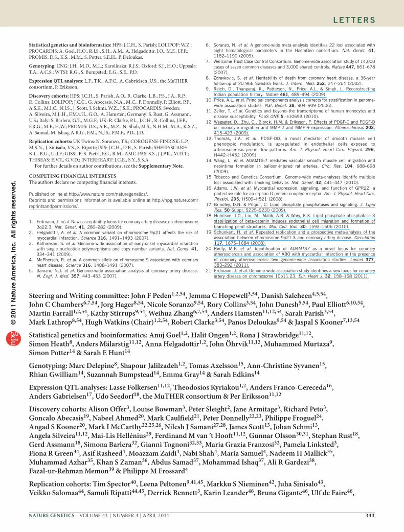
local LD structure on a secondary y axis. The most significant lead SNP (red diamond) at each locus is annotated with its discovery P value, and flanking SNPs are color-coded to represent the pairwise r2 measure of LD with the lead SNP: blue, r2 ≥ 0.8; green, 0.5 ≤ r2 < 0.8; orange, 0.2 ≤ r2 ≤ 0.5; gray, r2 < 0.2. a,b and d,e report r2 values calculated from HapMap2 CEU reference samples, and c reports r2 values calculated from HapMap2 GIH reference samples. Green bars represent RefSeq genes in the region. All positions are on NCBI build 36. The purple star in b represents significant eQTL association in aortic media. The black arrowhead in c represents the South Asian only meta-analysis P value.
Figure 4 Regional plots for significant associations with CAD. Regional association plots of the discovery meta-analysis from which each replicated SNP was selected; that is, the European and South Asian discovery meta-analysis (a,b and d,e) and South Asian meta-analysis (c). Each panel shows SNPs plotted by position on the chromosome against –log10 P, with estimated recombination rates from HapMap release 27 (CEU, YRI and JPT+CHB populations) plotted in light blue to reflect the
Figure 3 Newly identified loci and variants associated with CAD in European, South Asian and all studies. Odds ratios per copy of the risk allele are indicated by squares (size inversely proportional to the variance), with horizontal lines indicating 95% CIs. Odds ratios and 95% CIs for all participants are indicated by diamonds. Allele frequencies (allele freq) are given for the risk allele. P values for heterogeneity between European and South Asian (S Asian) results are reported (ethnic Phet). *The South Asian only discovery P value for rs4380028 was P = 3.6 × 10⁻⁶.
© 2
011
Nat
ure
Am
eric
a, In
c. A
ll ri
gh
ts r
eser
ved
.

342 VOLUME 43 | NUMBER 4 | APRIL 2011 Nature GeNetics
l e t t e r s
The nearest gene to rs974819 is PDGFD, 117 kb downstream in an adjacent block of linkage disequilibrium (LD) (Fig. 4). We found a significant PDGFD eQTL for rs974819 in aortic media (P < 2.3 × 10⁻⁷), with suggestive associations in aortic adventitia (P < 7.7 × 10⁻⁴) and mammary artery (P < 7.2 × 10⁻⁴) (Supplementary Table 4). In all three tissues, the risk allele was associated with increased expression, and PDGFD was one of the top 10% most highly expressed transcripts. Platelet-derived growth factor D, encoded by PDGFD, is expressed in several cell types in atherosclerotic plaques and is predicted to stimulate atherosclerosis by influencing matrix metalloproteinase activity and monocyte migration12 and by inhibiting smooth muscle cell gene expression13.
rs4380028 is 7.6 kb upstream of ADAMTS7. ADAMTS7 is a metallo-proteinase that accumulates in carotid artery neointima after injury and is upregulated by platelet-derived growth factor14. The CAD association at the ADAMTS7 locus appears to be independent of the adjacent QTL for cigarette smoking, which has been attributed to the CHRNA5-CHRNA3-CHRNB4 nicotinic acid receptor cluster15. The signal at rs4380028 therefore appears new but cannot be attributed to a specific gene.
At the 7q22 locus, the lead SNP, rs10953541, is within an intron of BCAP29, which encodes B-cell receptor-associated protein 29, but this SNP is also in strong LD with five other protein coding genes (PRKAR2B, HBP1, COG5, GPR22 and DUS4L). We observed no unique eQTL effect. One promising biological candidate is GPR22, which encodes a G-protein–coupled receptor expressed in coronary arteries and heart16.
rs2505083 is in an intron of KIAA1462, a widely expressed and evo-lutionarily conserved gene. The function of the 1,359-amino-acid pro-tein encoded by KIAA1462 is largely unknown, with no recognizable functional domains and little homology to other protein families.
The genetic evidence for rs17114046 in PPAP2B is further sup-ported by the association of the risk allele, with a suggestive (P < 3.9 × 10⁻³) 30% increase in PPAP2B expression in atherosclerotic plaque (Supplementary Table 4). PPAP2B encodes phosphatidic acid phos-phatase type 2B, a membrane glycoprotein that hydrolyzes bioactive lipids involved in signaling17. It is expressed in the adherens junctions of endothelial cells and is believed to influence endothelial cell adhe-sion and migration and vasculogenesis18.
In summary, five new loci passed both the pre-specified signi-ficance threshold for the stand-alone replication and the conventional threshold for genome-wide significance in the combined discovery and replication data, yielding a substantial increase in the number of confirmed susceptibility loci for CAD. We did not find any suscep-tibility variants with material differences in effect size or allele fre-quency between South Asians and Europeans. We note, however, the potential limitation that current genome-wide arrays may not capture all important variants in South Asians. Nevertheless, all of the known and new variants were significantly associated with CAD risk in both the European and South Asian populations in the current study, indi-cating the importance of the genes associated with CAD beyond the European ancestry groups in which they were first defined.
As is seen in the present study, the effect sizes of previously uni-dentified CAD-associated genes discovered by GWAS have become progressively smaller, suggesting that there may not be large-effect common variants remaining to be discovered, but rather that a large number of common variants of small effect may contribute to CAD risk. Reliable detection of differences of only 5–10% in the per-allele risk of CAD has previously been difficult. However, the availability of large-scale GWAS carried out in populations of different ances-try, and our demonstration that results from such populations
can be informatively combined in genetic discovery, suggests that even broader collaborations would identify additional variants that influence CAD risk. Greater understanding of the genetic variants underlying CAD, and particularly the pathways involved, may lead to development of new therapeutic approaches to help address the world’s leading cause of death.
URLs. TRANSMIT software, http://www-gene.cimr.cam.ac.uk/clayton/software/transmit.txt; METAL software, http://www.sph.umich.edu/csg/abecasis/Metal/.
Note added in proof: Since this manuscript was submitted ADAMTS7 (ref. 20) and KIAA1462 (ref. 21) have been independently reported as loci for coronary artery disease.
METhoDSMethods and any associated references are available in the online version of the paper at http://www.nature.com/naturegenetics/.
Note: Supplementary information is available on the Nature Genetics website.
ACknowleDGmenTsWe are grateful to all of the study participants in the studies contributing to these meta-analyses and to all laboratory staff and former colleagues who have contributed to these studies over many years. This study makes use of data generated by the Wellcome Trust Case-Control Consortium (WTCCC); a full list of the investigators who contributed to the generation of the data is available from www.wtccc.org.uk. This study also makes use of data generated by the UK Twins study, The Twin Research Unit, King’s College London, UK.
The PROCARDIS study was supported by the European Community Sixth Framework Program (LSHM-CT-2007-037273), AstraZeneca, the British Heart Foundation, the Oxford British Heart Foundation Centre of Research Excellence, the Wellcome Trust (075491/Z/04), the Swedish Research Council, the Knut and Alice Wallenberg Foundation, the Swedish Heart-Lung Foundation, the Torsten and Ragnar Söderberg Foundation, the Strategic Cardiovascular Program of Karolinska Institutet and Stockholm County Council, the Foundation for Strategic Research and the Stockholm County Council (560283).
The Heart Protection Study (ISRCTN48489393) was funded by the UK Medical Research Council, the British Heart Foundation, Merck & Co and Roche Vitamins Ltd. Genotyping and analysis was supported by a grant to Oxford University and the Centre National de Génotypage (CNG) from Merck & Co and the Oxford BHF Centre of Research Excellence.
The PROMIS study was funded by unrestricted grants to investigators at the University of Cambridge, UK and at the Centre for Non-Communicable Diseases, Pakistan. Genotyping was funded by the Wellcome Trust.
The LOLIPOP study is supported by the National Institute for Health Research Comprehensive Biomedical Research Centre Imperial College Healthcare NHS Trust, Ealing Hospital NHS Trust, the British Heart Foundation (SP/04/002), the Medical Research Council (G0700931, G0601966), the Wellcome Trust (084723/Z/08/Z) and the National Institute for Health Research (RP-PG-0407-10371). P.E. is a National Institute for Health Research Senior Investigator. This work was facilitated by Barts and The London National Institute for Health Biomedical Research Unit. We thank the participants and research staff who made the study possible.
The COROGENE-FINRISK study was supported in part by the Aarno Koskelo Foundation and the Finnish Foundation for Cardiovascular Research.
The Biobank of Karolinska Carotid Endarterectomies (BiKE) and Advanced Study of Aortic Pathology (ASAP) eQTL study was supported by the Swedish Heart-Lung Foundation, the Swedish Research Council, the European Commission (FAD, Health-F22008-200647), (AtheroRemo HEALTH-2007-A-201668), DASTI (Danish Agency for Science, Technology and Innovation) and a donation from F. Lundberg.
The Multiple Tissue Human Expression Resource (MuTHER) study was supported by the Wellcome Trust (081917/Z/07/Z).
N. Soranzo is supported by the Wellcome Trust (Core Grant Number 091746/Z/10/Z).
AUTHoR ConTRIBUTIonsSteering and writing committee: J.F.P., J.C.H., D.S., J.C.C., J.H., N. Soranzo, R. Collins, J.D., P. Elliott, M.F., K.S., W.Z., A. Hamsten, S. Parish, M.L., H.W. (Chair), R. Clarke, P. Deloukas, J.S.K.
Corresponding authors: H.W., D.S., R. Collins, J.S.K.
Analysis committee: J.C.H., W.Z., N. Soranzo, J.F.P., D.S., J.C.C., S. Parish, M.F. (Chair).
© 2
011
Nat
ure
Am
eric
a, In
c. A
ll ri
gh
ts r
eser
ved
.
Nature GeNetics VOLUME 43 | NUMBER 4 | APRIL 2011 343
l e t t e r s
Statistical genetics and bioinformatics: HPS: J.C.H., S. Parish; LOLIPOP: W.Z.; PROCARDIS: A. Goel, H.O., R.J.S., S.H., A.M., A. Helgadottir, J.O., M.F., J.F.P.; PROMIS: D.S., K.S., M.M., S. Potter, S.E.H., P. Deloukas.
Genotyping: CNG: J.H., M.D., M.L.; Karolinska: R.J.S.; Oxford: S.J., H.O.; Uppsala: T.A., A.C.S.; WTSI: R.G., S. Bumpsted, E.G., S.E., P.D.
Expression QTL analyses: L.F., T.K., A.F.C., A. Gabrielsen, U.S., the MuTHER consortium, P. Eriksson.
Discovery cohorts: HPS: J.C.H., S. Parish, A.O., R. Clarke, L.B., P.S., J.A., R.P., R. Collins; LOLIPOP: J.C.C., G. Abecasis, N.A., M.C., P. Donnelly, P. Elliott, P.F., A.S.K., M.I.C., N.J.S., J. Scott, J. Sehmi, W.Z., J.S.K.; PROCARDIS: Sweden: A. Silveira, M.L.H., F.M.v.H., G.O., A. Hamsten; Germany: S. Rust, G. Assmann, U.S.; Italy: S. Barlera, G.T., M.G.F.; UK: R. Clarke, P.L., J.C.H., R. Collins, J.F.P., F.R.G., M.F., H.W.; PROMIS: D.S., A.R., M.Z., N. Shah, M.S., N.H.M., M.A., K.S.Z., A. Samad, M. Ishaq, A.R.G., F.M., N.J.S., P.M.F., P.D., J.D.
Replication cohorts: UK Twins: N. Soranzo, T.S.; COROGENE-FINRISK: L.P., M.S.N., J. Sinisalo, V.S., S. Ripatti; ISIS: J.C.H., D.B., S. Parish; SHEEP/SCARF: K.L., B.G., U.d.F.; GISSI-P: S. Pietri, F.G., R.M.; AMC-PAS: S.S., J.J.P.K., M.D.T.; THISEAS: E.V.T., G.V.D.; INTERHEART: J.C.E., S.Y., S.S.A.
For further details on author contributions, see the Supplementary Note.
ComPeTInG FInAnCIAl InTeResTsThe authors declare no competing financial interests.
Published online at http://www.nature.com/naturegenetics/. Reprints and permissions information is available online at http://npg.nature.com/reprintsandpermissions/.
1. Erdmann, J. et al. New susceptibility locus for coronary artery disease on chromosome 3q22.3. Nat. Genet. 41, 280–282 (2009).
2. Helgadottir, A. et al. A common variant on chromosome 9p21 affects the risk of myocardial infarction. Science 316, 1491–1493 (2007).
3. Kathiresan, S. et al. Genome-wide association of early-onset myocardial infarction with single nucleotide polymorphisms and copy number variants. Nat. Genet. 41, 334–341 (2009).
4. McPherson, R. et al. A common allele on chromosome 9 associated with coronary heart disease. Science 316, 1488–1491 (2007).
5. Samani, N.J. et al. Genome-wide association analysis of coronary artery disease. N. Engl. J. Med. 357, 443–453 (2007).
6. Soranzo, N. et al. A genome-wide meta-analysis identifies 22 loci associated with eight hematological parameters in the HaemGen consortium. Nat. Genet. 41, 1182–1190 (2009).
7. Wellcome Trust Case Control Consortium. Genome-wide association study of 14,000 cases of seven common diseases and 3,000 shared controls. Nature 447, 661–678 (2007).
8. Zdravkovic, S. et al. Heritability of death from coronary heart disease: a 36-year follow-up of 20 966 Swedish twins. J. Intern. Med. 252, 247–254 (2002).
9. Reich, D., Thangaraj, K., Patterson, N., Price, A.L. & Singh, L. Reconstructing Indian population history. Nature 461, 489–494 (2009).
10. Price, A.L. et al. Principal components analysis corrects for stratification in genome-wide association studies. Nat. Genet. 38, 904–909 (2006).
11. Zeller, T. et al. Genetics and beyond–the transcriptome of human monocytes and disease susceptibility. PLoS ONE 5, e10693 (2010).
12. Wagsater, D., Zhu, C., Bjorck, H.M. & Eriksson, P. Effects of PDGF-C and PDGF-D on monocyte migration and MMP-2 and MMP-9 expression. Atherosclerosis 202, 415–423 (2009).
13. Thomas, J.A. et al. PDGF-DD, a novel mediator of smooth muscle cell phenotypic modulation, is upregulated in endothelial cells exposed to atherosclerosis-prone flow patterns. Am. J. Physiol. Heart Circ. Physiol. 296, H442–H452 (2009).
14. Wang, L. et al. ADAMTS-7 mediates vascular smooth muscle cell migration and neointima formation in balloon-injured rat arteries. Circ. Res. 104, 688–698 (2009).
15. Tobacco and Genetics Consortium. Genome-wide meta-analyses identify multiple loci associated with smoking behavior. Nat. Genet. 42, 441–447 (2010).
16. Adams, J.W. et al. Myocardial expression, signaling, and function of GPR22: a protective role for an orphan G protein-coupled receptor. Am. J. Physiol. Heart Circ. Physiol. 295, H509–H521 (2008).
17. Brindley, D.N. & Pilquil, C. Lipid phosphate phosphatases and signaling. J. Lipid Res. 50 Suppl, S225–S230 (2009).
18. Humtsoe, J.O., Liu, M., Malik, A.B. & Wary, K.K. Lipid phosphate phosphatase 3 stabilization of beta-catenin induces endothelial cell migration and formation of branching point structures. Mol. Cell. Biol. 30, 1593–1606 (2010).
19. Schunkert, H. et al. Repeated replication and a prospective meta-analysis of the association between chromosome 9p21.3 and coronary artery disease. Circulation 117, 1675–1684 (2008).
20. Reilly, M.P. et al. Identification of ADAMTS7 as a novel locus for coronary atherosclerosis and association of ABO with myocardial infarction in the presence of coronary atherosclerosis: two genome-wide association studies. Lancet 377, 383–292 (2011).
21. Erdmann, J. et al. Genome-wide association study identifies a new locus for coronary artery disease on chromosome 10p11.23. Eur. Heart J. 32, 158–168 (2011).
steering and writing committee: John F Peden1,2,54, Jemma C Hopewell3,54, Danish saleheen4,5,54, John C Chambers6,7,54, Jorg Hager8,54, nicole soranzo9,54, Rory Collins3,54, John Danesh5,54, Paul elliott6,10,54, martin Farrall1,2,54, kathy stirrups9,54, weihua Zhang6,7,54, Anders Hamsten11,12,54, sarah Parish3,54, mark lathrop8,54, Hugh watkins (Chair)1,2,54, Robert Clarke3,54, Panos Deloukas9,54 & Jaspal s kooner7,13,54
statistical genetics and bioinformatics: Anuj Goel1,2, Halit ongen1,2, Rona J strawbridge11,12, simon Heath8, Anders mälarstig11,12, Anna Helgadottir1,2, John Öhrvik11,12, muhammed murtaza9, simon Potter14 & sarah e Hunt14
Genotyping: marc Delepine8, shapour Jalilzadeh1,2, Tomas Axelsson15, Ann-Christine syvanen15, Rhian Gwilliam14, suzannah Bumpstead14, emma Gray14 & sarah edkins14
expression QTl analyses: lasse Folkersen11,12, Theodosios kyriakou1,2, Anders Franco-Cereceda16, Anders Gabrielsen17, Udo seedorf18, the muTHeR consortium & Per eriksson11,12
Discovery cohorts: Alison offer3, louise Bowman3, Peter sleight2, Jane Armitage3, Richard Peto3, Goncalo Abecasis19, nabeel Ahmed20, mark Caulfield21, Peter Donnelly22,23, Philippe Froguel24, Angad s kooner20, mark I mcCarthy22,25,26, nilesh J samani27,28, James scott13, Joban sehmi13, Angela silveira11,12, mai-lis Hellénius29, Ferdinand m van ’t Hooft11,12, Gunnar olsson30,31, stephan Rust18, Gerd Assmann18, simona Barlera32, Gianni Tognoni32,33, maria Grazia Franzosi32, Pamela linksted3, Fiona R Green34, Asif Rasheed4, moazzam Zaidi4, nabi shah4, maria samuel4, nadeem H mallick35, muhammad Azhar35, khan s Zaman36, Abdus samad37, mohammad Ishaq37, Ali R Gardezi38, Fazal-ur-Rehman memon39 & Philippe m Frossard4
Replication cohorts: Tim spector40, leena Peltonen9,41,45, markku s nieminen42, Juha sinisalo43, Veikko salomaa44, samuli Ripatti44,45, Derrick Bennett3, karin leander46, Bruna Gigante46, Ulf de Faire46,
© 2
011
Nat
ure
Am
eric
a, In
c. A
ll ri
gh
ts r
eser
ved
.

344 VOLUME 43 | NUMBER 4 | APRIL 2011 Nature GeNetics
1Department of Cardiovascular Medicine, The Wellcome Trust Centre for Human Genetics, University of Oxford, Oxford, UK. 2Department of Cardiovascular Medicine, University of Oxford, John Radcliffe Hospital, Headington, Oxford, UK. 3Clinical Trial Service Unit, University of Oxford, Oxford, UK. 4Center for Non-Communicable Diseases Pakistan, Karachi, Pakistan. 5Department of Public Health and Primary Care, University of Cambridge, Strangeways Research Laboratory, Cambridge, UK. 6Epidemiology and Biostatistics, Imperial College London, Norfolk Place, London, UK. 7Cardiology, Ealing Hospital National Health Service (NHS) Trust, Middlesex, UK. 8Commissariat à l'Energie Atomique (CEA) Genomics Institute-Centre National de Génotypage, Evry Cedex, France. 9Human Genetics, Wellcome Trust Sanger Institute, Wellcome Trust Genome Campus, Hinxton, UK. 10Medical Research Council-Health Protection Agency (MRC-HPA) Centre for Environment and Health, Imperial College London, London, UK. 11Atherosclerosis Research Unit, Department of Medicine, Karolinska Institutet, Karolinska University Hospital, Stockholm, Sweden. 12Center for Molecular Medicine, Karolinska University Hospital, Stockholm, Sweden. 13National Heart and Lung Institute, Imperial College London, London, UK. 14Wellcome Trust Sanger Institute, Wellcome Trust Genome Campus, Hinxton, UK. 15Department of Medical Sciences, Molecular Medicine, Uppsala University, Uppsala, Sweden. 16Thoracic Surgery Unit, Department of Molecular Medicine and Surgery, Karolinska Institutet, Karolinska University Hospital, Stockholm, Sweden. 17Cardiology Unit, Department of Medicine, Karolinska Institutet, Karolinska University Hospital, Stockholm, Sweden. 18Gesellschaft für Arterioskleroseforschung e.V., Leibniz-Institut für Arterioskleroseforschung an der Universität Münster (LIFA), Münster, Germany. 19Department of Biostatistics, University of Michigan, Ann Arbor, Michigan, USA. 20Cardiology, Ealing Hospital NHS Trust, Middlesex, UK. 21William Harvey Research Institute, Barts and The London School of Medicine and Dentistry, Queen Mary University of London, London, UK. 22Wellcome Trust Centre for Human Genetics, University of Oxford, Oxford, UK. 23Department of Statistics, Oxford University, Oxford, UK. 24Genomic Medicine, Imperial College London, London, UK. 25Oxford Centre for Diabetes, Endocrinology and Metabolism, Oxford University, Oxford, UK. 26Oxford National Institute of Health Research (NIHR) Biomedical Research Centre, Churchill Hospital, Headington, UK. 27Department of Cardiovascular Sciences, University of Leicester, Glenfield Hospital, Leicester, UK. 28Leicester NIHR Biomedical Research Unit in Cardiovascular Disease, Glenfield Hospital, Leicester, UK. 29Cardiology Unit, Department of Medicine, Karolinska Institutet, Karolinska University Hospital, Stockholm, Sweden. 30Cardiovascular Drug Research at the Department of Medicine, Solna, Sweden. 31Cardiovascular and Gastrointestinal Innovative Medicines, Global Research and Development, AstraZeneca, Sweden. 32Department of Cardiovascular Research, Istituto Mario Negri, Milano, Italy. 33Consorzio Mario Negri Sud, Santa Maria Imbaro (Chieti), Italy. 34Biochemical Sciences Division, Faculty of Health and Medical Sciences, University of Surrey, Guilford, UK. 35Department of Cardiology, Punjab Institute of Cardiology, Jail Road, Lahore, Pakistan. 36Department of Cardiology, National Institute of Cardiovascular Diseases, Karachi, Pakistan. 37Department of Cardiology, Karachi Institute of Heart Diseases, Federal B. Area, Karachi, Pakistan. 38Department of Cardiology, Ch. Pervaiz Elahi Institute Of Cardiology, Multan, Pakistan. 39Department of Cardiology, Red Crescent Institute of Cardiology, Latifabad, Hyderabad, Pakistan. 40Department of Twin Research and Genetic Epidemiology, Kings College London, London, UK. 41Department of Medical Genetics, University of Helsinki and the Helsinki University Central Hospital, Helsinki, Finland. 42Division of Cardiology, Helsinki University Central Hospital, Helsinki, Finland. 43University Central Hospital, Cardiovascular Laboratory, Helsinki, Finland. 44National Institute for Health and Welfare, Helsinki, Finland. 45Institute for Molecular Medicine Finland (FIMM), University of Helsinki, Helsinki, Finland. 46Cardiovascular Epidemiology Unit, Institute of Environmental Medicine, Karolinska Institutet, Stockholm, Sweden. 47Laboratory of Clinical Epidemiology of Cardiovascular Disease, Consorzio Mario Negri Sud, Santa Maria Imbaro, Italy. 48Vascular Medicine, Academic Medical Center, University of Amsterdam, Amsterdam, The Netherlands. 49Department of Dietetics-Nutrition, Harokopio University, Athens, Greece. 50McGill University Department of Medicine, Montreal, Quebec, Canada. 51McGill University Department of Human Genetics, Montreal, Quebec, Canada. 52Population Health Research Institute, Hamilton Health Sciences, McMaster University, Hamilton, Canada. 53Department of Medicine, McMaster University, Hamilton, Canada. 54These authors contributed equally to this work. Correspondence should be addressed to H.W. ([email protected]), D.S. ([email protected]), R.C. ([email protected]) or J.S.K. ([email protected]).
silvia Pietri32, Francesca Gori32, Roberto marchioli47, suthesh sivapalaratnam48, John J P kastelein48, mieke D Trip48, eirini V Theodoraki49, George V Dedoussis49, Jamie C engert50,51, salim Yusuf 52 & sonia s Anand52,53
l e t t e r s©
201
1 N
atu
re A
mer
ica,
Inc.
All
rig
hts
res
erve
d.

Nature GeNeticsdoi:10.1038/ng.782
oNLINE METhoDSDiscovery. Cases and controls in the discovery studies. The PROCARDIS study comprised individuals recruited from the UK, Italy, Sweden and Germany22. All cases had a diagnosis of CAD before age 66 and 80% had a sibling in whom CAD had been diagnosed before age 66. CAD was defined as clinically documented evidence of myocardial infarction (80%), coronary artery bypass graft (10%), acute coronary syndrome (6%), coronary angioplasty (1%) or stable angina (hospitalization for angina or documented obstructive coronary disease) (3%) before age 66. The mean age of onset of CAD was 53.2 years (standard deviation (s.d.), 7.2 years).
The Heart Protection Study (HPS) was a large UK-based cholesterol-lowering trial involving participants with a history of myocardial infarction, unstable or stable angina, coronary artery bypass graft or coronary angioplasty (as well as individuals with prior history of stroke or hypertension)23. Among these, 2,704 CAD cases were genotyped and compared with 2,887 controls from the UK 1958 British Birth Cohort. Of those individuals genotyped, the mean age of CAD onset was 58.8 years (s.d., 8.4 years), and 92% of CAD cases had a history of myocardial infarction, revascularization or hospitalization for angina.
The PROMIS study was a case-control study of myocardial infarction car-ried out among South Asians living in urban Pakistan24. The 4,253 cases had myocardial infarction, and the average age of disease onset was 53.8 years (s.d., 10.6 years). The 4,130 controls were matched to cases by sex and age and recruited in the same hospitals as the index cases. The major ethnic groups of the participants in the PROMIS study were as follows: Urdu (42%), Punjabi (28%), Pathan (8%) and Sindhi (8%).
The LOLIPOP study was a case-control study of CAD carried out among South Asians, a population category defined by having all four grandparents born on the Indian subcontinent, living in the UK25. The 2,741 cases had a his-tory of myocardial infarction, coronary artery revascularization (coronary artery bypass grafting or percutaneous coronary intervention) or angiographically confirmed coronary artery stenosis greater than 50%. The cases were compared with 3,696 controls, free from CAD, recruited from the same ethnic or linguistic groups. The mean age of entry into the study was 59.3 years (s.d., 9.7 years). For further details of the discovery cohorts, see the Supplementary Note.
Genotyping in the discovery studies. The genotyping platforms, centers and quality control parameters are summarized in Supplementary Table 2. Additional qual-ity control was done by testing for different allelic frequencies between centers, bead arrays, batches or BeadChips, and a further 3,883 SNPs were removed.
To identify ancestry outliers, principal component analysis using EIGENSOFT (3.0)10 was used to compare all samples with reference samples from the HapMap YRI, CHB, JPT and CEU panels. Samples with eigenvalues inconsistent with self-reported ancestry were removed. To detect and correct for population stratification, a panel of independent SNPs were used to gen-erate ancestry-informative principal components. South Asian samples were plotted by their self-reported ethnic or linguistic group and case or control status (Supplementary Fig. 1).
Ethics. All studies were collected under the approval of the appropriate ethics committee and all participants gave informed consent for each study.
Statistical analyses. Discovery genome-wide analyses. Association analyses of CAD were carried out after exclusion of SNPs that failed quality control. Analyses were performed using logistic models to estimate regression coef-ficients and their standard errors. Additive genetic effects were modeled by defining continuous variables with levels 0, 1 and 2 corresponding to geno-types AA, AB and BB. The test statistics for each study were inspected for over-dispersion, and the genomic control parameter (λGC), an estimate of the variance inflation, was between 1.03 to 1.08 (Supplementary Table 2).
Clustering and covariate adjustment. The multi-center PROCARDIS study included country of origin as a categorical main effect to model differences in SNP allele frequencies indirectly across the populations. The familial relatedness in the PROCARDIS study was taken into account using a robust (sandwich) estimator of the variance. The PROMIS and LOLIPOP studies included ances-try-informative principal components to absorb population stratification.
Discovery meta-analysis. A fixed-effects inverse variance-weighted meta-analysis as implemented by METAL (see URLs) was used to combine the indi-vidual studies concurrently as European studies only, South Asian studies only and all studies. The individual studies were subjected to study-level cor-rection for genomic control (using the sample-specific λGC described above). The test statistics for each meta-analysis were inspected for over-dispersion (Supplementary Fig. 3). The λGC of the three meta-analyses were calculated (λGC = 1.01 Europeans only, λGC = 1.01 South Asians only and λGC = 1.03 for all studies), and an additional genomic control correction was applied using these λGC factors (double genomic control correction). Heterogeneity between the European and the South Asian studies and between all studies was assessed using the Cochran’s Q statistic.
Replication. Cases and controls in the replication studies. Independent replica-tion was sought in a total of 21,408 CAD cases and 19,185 ancestry-matched controls obtained from eight European and two South Asian studies. A total of 18,049 CAD cases and 16,357 controls of European ancestry were included from HPS23, COROGENE-FINRISK26, International Study of Infarct Survival (ISIS)27, Stockholm Heart Epidemiology Programme and Stockholm Coronary Artery Risk Factor study SHEEP/SCARF28,29, Precocious Coronary Artery Disease Transmission Disequilibrium Test (PROCARDIS TDT) cohort30, Gruppo Italiano per lo Studio della Sopravvivenza nell’Infarto Miocardico Prevenzion (GISSI-P)31, Academic Medical Center Amsterdam Premature Atherosclerosis Study (AMC-PAS)32, The Hellenic study of Interactions between SNPs & Eating in Atherosclerosis Susceptibility (THISEAS)33, and 3,359 cases and 2,828 controls of South Asian ancestry from the PROMIS24 and INTERHEART34 studies.
Selection of replication SNPs. SNPs were ranked by the association P value in the discovery meta-analysis. The genotype clusters for all SNPs with an asso-ciation with CAD of P < 1.0 × 10⁻⁴ were manually checked, and 13 SNPs were removed. With the exception of rs9349379 at the PHACTR1 locus, SNPs that were located within a locus that had been previously reported to be associated with CAD (Table 1), or SNPs in strong LD (r2 > 0.5) with the most signifi-cant SNP at each locus, were removed. All remaining SNPs with P < 3.0 × 10⁻⁵ in either the European studies only or South Asian studies or with P < 1.0 × 10⁻⁴ in the all-studies meta-analysis were considered for replication. When a SNP could not be multiplexed, alternative tagging SNPs (r2 > 0.8) were considered.
Genotyping in the replication studies. Genotyping of the replication samples was performed by primer extension and MALDI-TOF mass spectrometry using Sequenom iPLEX technology as two multiplexes containing 59 replica-tion SNPs and three gender-specific polymorphisms. At the Centre National de Génotypage (CNG), the assay for rs9349379 was replaced by a Kaspar SNP assay supplied by KBioscience Ltd. The PROMIS replication genotypes included data from 1,005 Illumina Human660 arrays which were not available during the discovery GWAS. The COROGENE replication genotypes were obtained in silico from Illumina Human670 GWAS data.
The allelic intensities of each SNP assay were plotted and, where neces-sary, genotypes were manually called. SNPs with genotype calls that deviated from Hardy-Weinberg equilibrium with P < 8.5 × 10⁻⁴ or had minor allele frequency <1% were excluded. The call rate cutoff for SNPs was empirically established in each study. SNP genotypes that passed quality control were used to determine a study-specific sample rate cutoff. Details of replication geno-typing and related quality control steps are summarized in Supplementary Table 2. The PROCARDIS TDT study was a family based collection, and an additional quality control step was applied whereby families with greater than three Mendelian misinheritances were excluded.
Statistical analyses. Replication analyses. Logistic regression models were used to perform association analyses of CAD and to estimate the per-allele effect and standard error. The family based transmission disequilibrium test was used to estimate the association in the PROCARDIS TDT samples. This test is robust to the presence of population structure and was performed using TRANSMIT (see URLs) written by David Clayton.
© 2
011
Nat
ure
Am
eric
a, In
c. A
ll ri
gh
ts r
eser
ved
.

Nature GeNetics doi:10.1038/ng.782
Replication meta-analysis. A fixed-effects inverse-variance–weighted meta-analysis was used to combine the results for each SNP across all replication studies with available data. Heterogeneity between all studies and between the European and the South Asian studies was assessed using Cochran’s Q statistic.
Conditional analysis. Conditional analyses were performed in each of the dis-covery studies. The position of the recombination hotspots flanking each of the SNPs selected for replication was determined, and those SNPs that were located between the two flanking hotspots were tested for association with CAD with the equivalent model used in the discovery analysis but condition-ing on the genotype of the SNP chosen for replication.
eQTL analyses. Tissue samples for gene expression biobanks were obtained as described previously35. Briefly, tissue biopsies were taken from patients undergoing carotid endartectomy (plaque n = 117) or valve surgery (liver n = 152, aorta media n = 117, aorta adventitia n = 103 and mammary artery n = 88). Extracted RNA was hybridized to Affymetrix HG-U133 plus 2.0 micro-arrays (plaque) or Affymetrix ST 1.0 Exon arrays (liver, aorta and mammary artery), and obtained scans were robust multichip average (RMA) normal-ized and log2 transformed. DNA was extracted from circulating blood cells and hybridized to Illumina Human610w-Quad Beadarrays. In the MuTHER study, RNA levels were measured in lymphoblastoid cell lines (n = 826), skin (n = 705) and fat biopsies (n = 825) from 850 well-phenotyped female twins (1/3 MZ and 2/3 DZ) from the TwinsUK resource using Illumina’s whole-genome expression array HumanHT-12 version 3 as previously described36. Genotyping was performed in parallel using Illumina’s whole-genome arrays. Associations between genotype and expression of genes were assessed using an additive linear model.
23. Heart Protection Study Collaborative Group. MRC/BHF Heart Protection Study of cholesterol lowering with simvastatin in 20,536 high-risk individuals: a randomised placebo-controlled trial. Lancet 360, 7–22 (2002).
24. Saleheen, D. et al. The Pakistan Risk of Myocardial Infarction Study: a resource for the study of genetic, lifestyle and other determinants of myocardial infarction in South Asia. Eur. J. Epidemiol. 24, 329–338 (2009).
25. Chambers, J.C. et al. Common genetic variation near MC4R is associated with waist circumference and insulin resistance. Nat. Genet. 40, 716–718 (2008).
26. Ripatti, S. et al. A multilocus genetic risk score for coronary heart disease: case-control and prospective cohort analyses. Lancet 376, 1393–1400 (2010).
27. Clarke, R. et al. Lymphotoxin-alpha gene and risk of myocardial infarction in 6,928 cases and 2,712 controls in the ISIS case-control study. PLoS Genet. 2, e107 (2006).
28. Reuterwall, C. et al. Higher relative, but lower absolute risks of myocardial infarction in women than in men: analysis of some major risk factors in the SHEEP study. The SHEEP Study Group. J. Intern. Med. 246, 161–174 (1999).
29. Samnegard, A. et al. Serum matrix metalloproteinase-3 concentration is influenced by MMP-3 -1612 5A/6A promoter genotype and associated with myocardial infarction. J. Intern. Med. 258, 411–419 (2005).
30. PROCARDIS Consortium. A trio family study showing association of the lymphotoxin-alpha N26 (804A) allele with coronary artery disease. Eur. J. Hum. Genet. 12, 770–774 (2004).
31. GISSI-Prevenzione Investigators. Dietary supplementation with n-3 polyunsaturated fatty acids and vitamin E after myocardial infarction: results of the GISSI-Prevenzione trial. Gruppo Italiano per lo Studio della Sopravvivenza nell’Infarto miocardico. Lancet 354, 447–455 (1999).
32. Trip, M.D. et al. Frequent mutation in the ABCC6 gene (R1141X) is associated with a strong increase in the prevalence of coronary artery disease. Circulation 106, 773–775 (2002).
33. Theodoraki, E.V. et al. Fibrinogen beta variants confer protection against coronary artery disease in a Greek case-control study. BMC Med. Genet. 11, 28 (2010).
34. Mente, A. et al. Metabolic syndrome and risk of acute myocardial infarction a case-control study of 26,903 subjects from 52 countries. J. Am. Coll. Cardiol. 55, 2390–2398 (2010).
35. Folkersen, L. et al. Association of genetic risk variants with expression of proximal genes identifies novel susceptibility genes for cardiovascular disease. Circ. Cardiovasc. Genet. 3, 365–373 (2010).
36. Stranger, B.E. et al. Population genomics of human gene expression. Nat. Genet. 39, 1217–1224 (2007).
22. Broadbent, H.M. et al. Susceptibility to coronary artery disease and diabetes is encoded by distinct, tightly linked SNPs in the ANRIL locus on chromosome 9p. Hum. Mol. Genet. 17, 806–814 (2008).
© 2
011
Nat
ure
Am
eric
a, In
c. A
ll ri
gh
ts r
eser
ved
.
Copyright © 2022 FDOKUMEN