Magnetic and Morphological Properties of Ferrofluid-Impregnated Hydroxyapatite/Collagen Scaffolds
Upload
independentCategory
view
1download
0
Fabrication of silver nanocomposite films impregnatedwith curcumin for superior antibacterial applications
K. Varaprasad • K. Vimala • S. Ravindra •
N. Narayana Reddy • G. Venkata Subba Reddy •
K. Mohana Raju
Received: 8 January 2011 / Accepted: 30 May 2011 / Published online: 18 June 2011
� Springer Science+Business Media, LLC 2011
Abstract Silver nanocomposite films are found to be
very effective material for anti-bacterial application. In the
present work, sodium carboxylmethyl cellulose silver
nanocomposite films (SCMC SNCF) were tried for anti-
bacterial applications. To enhance their applicability novel
film-silver nanoparticle-curcumin composites have been
developed. SCMC SNCF are developed from sodium car-
boxylmethyl cellulose (SCMC), N,N1-methylenebisacryla-
mide (MBA) and silver nitrate solution. These films were
characterized by FTIR, UV–visible, XRD, TGA, DSC and
TEM techniques. The formed silver nanoparticles have an
average particle size of *15 nm as observed by trans-
mission electron microscopy (TEM). Curcumin loading
into SCMC SNCF is achieved by diffusion mechanism.
The UV–Visible analysis indicated that higher encapsula-
tion of curcumin in the films with higher SCMC content.
Further, it was observed that the presence of silver nano-
particles in the films enhanced the encapsulation of cur-
cumin indicating an interaction between them. Moreover,
the antibacterial activity showed that the SCMC films
generated with silver nanoparticles have a synergistic
effect in the antimicrobial activity against Escherichia coli
(E. coli). In order improve the healing efficacy as anti-
bacterial agents, curcumin loaded with SCMC SNCFs were
developed which showed significant inhibition of E. coli
growth than the silver nanoparticles and curcumin alone
film. Therefore, the present study clearly provides novel
antimicrobial films which are potentially useful in pre-
venting/treating infections.
1 Introduction
Polysaccharides are the most popular polymeric materials to
prepare nanoparticles for wound dressing [1, 2] and drug
delivery applications [3–5] as they are highly stable, safe, non-
toxic, hydrophilic and biodegradable materials which can be
easily modified chemically. Therefore, in recent years, a large
number of studies have been conducted on polysaccharides
for their potential application as nanoparticle drug delivery
systems as well as antibacterial applications [6–8].
Recently, silver-based nanostructure materials have
gained much attention to control infections. The use of
silver nanoparticles (AgNPs) has exhibited improved
antibacterial properties than bulk silver due to high surface
area and high fraction of surface atoms, leading to incor-
porating more NPs inside the bacteria and promoting its
efficacy in a sustained manner. Water soluble polymer
based biomaterials are capable of the combined antibac-
terial properties of AgNPs having no toxicity [9]. Basing
on this, numerous polymers have been employed to prepare
polymer-silver nanocomposites [10]. The combination of
silver nanoparticles with water soluble biopolymers will
produce new antimicrobials. Among the various biopoly-
mers, Carboxymethyl cellulose is widely used in drug
delivery and wound dressing applications. The reason is its
biocompatibility, and biodegradable nature with enormous
metal complexation capacity [11]. In particular, sodium
carboxymethyl cellulose (SCMC) is currently used in oral,
pharmaceutical formulations and as tablet binder as well as
K. Varaprasad (&) � K. Vimala � S. Ravindra �N. Narayana Reddy � K. Mohana Raju
Synthetic Polymer Laboratory, Department of Polymer Science
and Technology, Sri Krishnadevaraya University, Anantapur
515055, India
e-mail: [email protected]; [email protected]
G. Venkata Subba Reddy
Department of Microbiology, Sri Krishnadevaraya University,
Anantapur, Andhra Pradesh 515055, India
123
J Mater Sci: Mater Med (2011) 22:1863–1872
DOI 10.1007/s10856-011-4369-5

metal stabilization properties which enhanced the bio-
medical applications [10, 12]. For these applications, it is
important to have good stability of nanoparticles in films
Vimala et al. [13] developed polysaccharide silver nano-
particle films for antibacterial applications. Cedric Chau-
vierre et al. [14] used the based on polysaccharide based
poly(alkylcyanoacrylates) nanoparticle templates for drug
delivery applications. We recently reported the studies on
chitosen based silver nanoparticle films which possessed
superior antibacterial activity [15].
The objective of this study was to improve the swelling
and mechanical properties as well as improved wound
dressing properties of film by generating of silver nano-
particles as shown in Scheme 1. The developed SCMC
silver nanoparticles composite films were analyzed by of
UV–Vis, fourier transform infrared (FTIR) spectrophoto-
metric, thermogravimetric analysis (TGA), differential
scanning calorimetry (DSC) and transmission electron
microscopy (TEM) techniques. Curcumin (CM), a hydro-
phobic polyphenolic compound derived from the rhizome
of the herb curcuma longa, possesses a wide range of
biological activity including wound healing, anti-bacterial,
anti-oxidant, anti-inflammatory and anti-cancer properties
[16]. Hence, this compound was incorporated into SCMC
SNCF to improve significantly the therapeutic antibacterial
efficacy of the film. The effect of AgNPs and curcumin on
the antibacterial activity of the films was studied.
2 Materials
Sodium carboxymethyl cellulose (SCMC), N,N1-methyl-
enebisacrylamide (MBA), ammonium persulfate (APS),
silver nitrate (AgNO3) were obtained from Merck (Mum-
bai, India) and used as received.
2.1 Preparation of SCMC films
One gram of SCMC powder was dissolved in 100 ml of dis-
tilled water and stirred for 6 h. To this solution additionally
2.59 mM of 1% MBA and 21.91 mM of APS were added for
strong network. The reactant solution was transferred imme-
diately onto a Teflon sheet covered glass plate (dimensions:
100 mm length 9 100 mm width 9 3 mm height) and dried
at 25�C for 12 h. Finally this film was cut into the required
length and wreath for further studies. The film codes and the
corresponding feed composition are listed in Table 1.
2.2 Preparation of SCMC nanocomposite films
The procedure of synthesis was described in Scheme 1. One
gram of SCMC was dissolved in 100 ml distilled water and
stirred for 6 h. To this, AgNO3 solution (200 mg/10 ml
distilled water), 2.59 mM of MBA solution and 21.91 mM
of APS solution were added at 25�C. This solution was kept
in the sunlight for 1 h. The colorless solution started turning
to red, then brown and brownish indicating the formation of
AgNPs. The solution was then poured onto Teflon covered
glass plates and dried as explained earlier. Finally, the dried
film was cut into the required size for further studies.
2.3 Swelling studies
Dried films were swollen in (100 ml) phosphate buffer (pH
7.4) solution at 25�C. The weight of swollen films was
measured at equilibrium swelling after removing the
Scheme 1 Schematic diagram
of formation of SCMC silver
nanocomposite films and
curcumin encapsulated SCMC
silver nanocomposite films
1864 J Mater Sci: Mater Med (2011) 22:1863–1872
123

surface solution with filter paper. Swelling ratio (Q) was
calculated as follows: Q = We/Wd, where We is the weight
of the swollen film at equilibrium and Wd is the dry weight
of the film.
2.4 Characterization
2.4.1 FTIR spectroscopy
To record the FTIR spectra of films, the samples were
completely dried in an oven at 40�C for 6 h. The spectra
was recorded between 500 and 4000 cm-1 on a MB3000
Model, ABB company (Hoizon software) FTIR spectrom-
eter (Quebec, Canada) using the KBr disk method.
2.5 Tensile test
The tensile parameters of the SCMC and SCMC SNCF
samples were determined using the INSTRON 3369 Uni-
versal Testing Machine (Buckinghamshire, England). The
specimens with dimensions of length 100 mm and width
10 mm were used. A gauge length of 50 mm was main-
tained for all the samples. The tensile parameters-maxi-
mum stress, modulus and % elongation at break were
determined at a crosshead speed of 5 mm/min using a
10 kg load cell. In each case, three samples were used and
the average value reported.
2.6 UV–Vis spectrophotometer
UV–Vis absorption spectra of the samples were recorded
on a Shimadzu 160A model UV–Vis spectrophotometer in
the range of 200–700 nm. For this study, 100 mg of silver
nanocomposite film was dispersed in 10 ml of distilled
water and allowed for 1 day to extract all silver nanopar-
ticles into aqueous phase and these solutions were used for
absorption spectra.
2.6.1 Transmission electron microscopy (TEM)
The size of the AgNPs in film network was determined
using a Technai F12 TEM (Tokyo, Japan) microscope. For
this study, the samples were prepared by placing a drop of
aqueous solution of SCMC SNCF on carbon-coated copper
grid and subsequently drying in air, before transferring
them to the microscope operated at an accelerated voltage
of 120 kV.
2.7 X-ray diffraction (XRD)
The X-ray diffraction method was used to identify the
formation of nanoparticles in the films. These measure-
ments were carriedout for dried and finely grounded
samples on a Rikagu diffractometer (Cu radiation, k =
0.1546 nm) running at 40 kV and 40 mA.
Table 1 Feed composition, mechanical properties and % of encapsulation efficiency and release kinetics parameters of hydrogels
Film code Composition of components Swelling data of films
SCMC (g) MBA (mM) APS (mM) Swelling
ratio (g/g)
SCMC-SNCF
swelling ratio (g/g)
SCMC1 1 2.59 21.91 4.12 3.12
SCMC2 1.5 2.59 21.91 5.24 3.86
SCMC3 2 2.59 21.91 7.14 5.34
SCMC4 3 2.59 21.91 10.47 6.45
Mechanical properties of SCMC and SCMC SNCFs
Maximum stress (MPa) Young’s modulus (MPa) Elongation at break (%)
SCMC4 SCMC4 SNCF SCMC4 SCCMC4 SNCF SCMC4 SCMC4 SNCF
15.86 18.45 1496.00 1813.64 2.159 2.967
% of encapsulation efficiency and release kinetics parameters of different formulations at 25�C
Film code % of encapsulation
efficiency
n k (102) R2 % of curcumin cumulative
release at ambient
temperatures at 30 min
% of curcumin cumulative releases
at ambient temperatures
at there end time
SCMC4-CM 46.25 0.86 0.4764 0.877 8.13 100
SCMC1-CM 31.53 0.98 0.5917 0.9712 4.13 100
SCMC4 SNCF-CM 71.25 1.38 1.70 0.99 1.97 90.73
SCMC1 SNCF-CM 43.98 1.62 2.37 0.98 1.08 74.74
J Mater Sci: Mater Med (2011) 22:1863–1872 1865
123
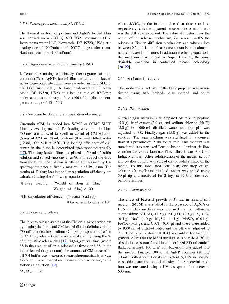
2.7.1 Thermogravimetric analysis (TGA)
The thermal analysis of pristine and AgNPs loaded films
was carried on a SDT Q 600 TGA instrument (T.A.
Instruments-water LLC, Newcastle, DE 19720, USA) at a
heating rate of 10�C/min in 40–700�C range under a con-
stant nitrogen flow (100 ml/min).
2.7.2 Differential scanning calorimetry (DSC)
Differential scanning calorimetry thermograms of pure
curcumin(CM), AgNPs loaded film and curcumin loaded
silver nanocomposite films were recorded using a SDT Q
600 DSC instrument (T.A. Instruments-water LLC, New-
castle, DE 19720, USA) at a heating rate of 10�C/min
under a constant nitrogen flow (100 ml/min)in the tem-
perature range of 40–450�C.
2.8 Curcumin loading and encapsulation efficiency
Curcumin (CM) is loaded into SCMC or SCMC SNCF
films by swelling method. For loading curcumin, the films
(50 mg) are allowed to swell in 20 ml of CM solution
(5 mg of CM in 20 ml, acetone (8 ml)—distilled water
(12 ml)) for 24 h at 25�C. The loading efficiency of cur-
cumin in the films is determined spectrophotometrically
[17]. The drug-loaded films are placed in 50 ml of buffer
solution and stirred vigorously for 96 h to extract the drug
from the films. The solution is filtered and assayed by UV
spectrophotometer at fixed k max value of 491.2 nm. The
results of % drug loading and encapsulation efficiency are
calculated using the following equations.
% Drug loading ¼ðWeight of drug in film=
Weight of filmÞ � 100
% Encapsulation efficiency¼ð% actual loading=
% theoretical loading�100
2.9 In vitro drug release
The in vitro release studies of the CM drug were carried out
by placing the dried and CM loaded film in definite volume
(50 ml) of releasing medium (7.4 pH phosphate buffer) at
37�C. Drug release kinetics were analyzed by using the %
of cumulative release data [18] (Mt/Mo) versus time (where
Mt is the amount of drug released at time t and Mo is the
initial loaded drug amount), the amount of CM released in
pH 7.4 buffer was measured spectrophotometrically at kmax
492.2 nm. Experimental results were fitted according to the
following equation [19].
Mt=M1 ¼ ktn
where Mt/M? is the faction released at time t and ?respectively, k is the apparent releases rate constant, and
n is the diffusion exponent. The value of n determines the
nature of the release mechanism, i.e. when n = 0.5 the
release is Fickian diffusion mechanism and when n lies
between 0.5 and 1, the release mechanism is anomalous in
nature or Case II in nature. In addition if n being equal to 1,
the mechanism is coined as Super Case II, the most
desirable condition in controlled release technology
[20–22].
2.10 Antibacterial activity
The antibacterial activity of the films prepared was inves-
tigated using two methods—disc method and count
method.
2.10.1 Disc method
Nutrient agar medium was prepared by mixing peptone
(5.0 g), beef extract (3.0 g), and sodium chloride (NaCl)
(5.0 g) in 1000 ml distilled water and the pH was
adjusted to 7.0. Finally, agar (15.0 g) was added to the
solution. The agar medium was sterilized in a conical
flask at a pressure of 15 lbs for 30 min. This medium was
transferred into sterilized Petri dishes in a laminar air flow
chamber (Microfilt Laminar Flow Ultra Clean Air Unit,
India, Mumbai). After solidification of the media, E. coli
and bacillus culture was spread on the solid surface of the
media. To this inoculated Petri dish, one drop of gel
solution (20 mg/10 ml distilled water) was added using
50-ll tip and incubated for 2 days at 37�C in the incu-
bation chamber.
2.10.2 Count method
The effect of bacterial growth of E. coli in mineral salt
medium (MSM) was studied in the presence of AgNPs or
HSNCs. This medium was prepared by the following
composition: NH4NO3 (1.5 g), KH2PO4 (2.5 g), K2HPO4
(0.5 g), NaCl (1.0 g), MgSO4 (1.5 g), MnSO4 (0.01 g),
FeSO4 (0.05 g), and CaCl2 (0.05 g) and these were added
to 1000 ml of distilled water and the pH was adjusted to
7.0. Then, yeast extract (0.01%) was added for bacterial
growth. After that the MSM medium was sterilized, 50 ml
of solution was transferred into a sterilized 250-ml conical
flask. Afterward, 100 ll E. coli bacterium was added into
the media. Finally, 100 ll of AgNP solution (20 mg/
10 ml distilled water) or its equivalent AgNPs suspension
was added, and the optical density of the bacterial med-
ium was measured using a UV–vis spectrophotometer at
600 nm.
1866 J Mater Sci: Mater Med (2011) 22:1863–1872
123
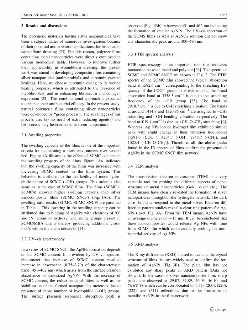
3 Results and discussions
The polymeric materials having silver nanoparticles have
been a subject matter of numerous investigations because
of their potential use in several applications, for instance, in
wound/burn dressing [23]. For this reason, polymer films
containing metal nanoparticles were directly employed in
various biomedical fields. However, to improve further
their applicability in wound/burn dressing, the present
work was aimed in developing composite films containing
silver nanoparticles (antimicrobial), and curcumin (wound
healing). Here, we choose curcumin owing to its wound
healing property, which is attributed to the presence of
myofibroblast, and in enhancing fibronectin and collagen
expression [23]. This combinational approach is expected
to enhance their antibacterial efficacy. In the present study,
natural polymeric films containing silver nanoparticles
were developed by ‘‘green process’’. The advantages of this
process are: (a) no need of extra reducing agent(s) and
(b) process may be conducted at room temperature.
3.1 Swelling properties
The swelling capacity of the films is one of the important
criteria for maintaining a moist environment over wound
bed. Figure 1A illustrates the effect of SCMC content on
the swelling property of the films. Figure 1Aa. indicates
that the swelling capacity of the films was increased with
increasing SCMC content in the films system. This
behavior is attributed to the availability of more hydro-
philic nature of SCMC (–OH) groups. This order is also
same as in the case of SCMC films. The films (SCMC1-
SCMC4) showed higher swelling capacity than silver
nanocomposite films (SCMC SNCF) (Fig. 1Ab). The
swelling ratio results (SCMC, SCMC SNCF) are parented
in Table 1. This lowering in the swelling capacity can be
attributed due to binding of AgNPs with electrons of ‘O’
and ‘N’ atoms of hydroxyl and amine groups present in
SCMC/MBA chains thereby producing additional cross-
link’s within the chain networks [14].
3.2 UV–vis spectroscopy
In a series of SCMC SNCF, the AgNPs formation depends
on the SCMC content. It is evident by UV–vis spectro-
photometer that increase of SCMC content resulted
increase in absorbance (0.75–2.79) of the characteristic
band (451–462 nm) which arises from the surface plasmon
absorbance of nanosized AgNPs. With the increase of
SCMC content, the reduction capabilities as well as the
stabilization of the formed nanoparticles increases due to
presence of more number of hydrophilic (–OH) groups.
The surface plasmon resonance absorption peak is
observed (Fig. 1Bb) in between 451 and 462 nm indicating
the formation of smaller AgNPs. The UV–vis spectrum of
the SCMS films as well as AgNO3 solution did not show
any characteristic peak around 400–470 nm.
3.3 FTIR spectral analysis
FTIR spectroscopy is an important tool that indicates
interaction between metal and polymer [24]. The spectra of
SCMC and SCMC SNCF are shown in Fig. 2. The FTIR
spectra of the SCMC film showed the typical absorption
band at 1582.6 cm-1 corresponding to the stretching fre-
quency of the COO- group. It is evident that the broad
absorption band at 3338.3 cm-1 is due to the stretching
frequency of the –OH group [25]. The band at
2918.7 cm-1 is due to C–H stretching vibration. The bands
at around 1414.7 and 1320.85 cm-1 are assigned to –CH2
scissoring and –OH bending vibration, respectively. The
band at1019.4 cm-1 is due to[CH–O–CH2 stretching [9].
Whereas, Ag NPs loaded hydrogel film exhibited similar
peak with slight change in their vibration frequencies
[1591.6 (COO-), 3254.7 (–OH), 2945.7 (–CH2), and
1025.4 (–CH–O–CH2)]. Therefore, all the above peaks
found in the IR spectra of films confirm the presence of
AgNPs in the SCMC SNCP film network.
3.4 TEM analysis
The transmission electron microscope (TEM) is a very
versatile tool for probing the different aspects of nano-
structure of metal nanoparticles (Gold, silver etc.). The
TEM images have clearly revealed the formation of silver
nanoparticles throughout the hydrogels network. The dark
core should correspond to the metal silver. Electron dif-
fraction pattern studies reveal a clear ring pattern for Ag-
NPs (inset, Fig. 3A). From the TEM image, AgNPs have
an average diameter of *15 nm. It can be concluded that
these nanocomposites would release Ag NPs with time
from SCMS film which can eventually prolong the anti-
bacterial activity of Ag NPs.
3.5 XRD analysis
The X-ray diffraction (XRD) is used to evaluate the crystal
structure of films that are widely used to confirm the for-
mation of AgNPs (Fig. 3B). The plain film has not
exhibited any sharp peaks in XRD pattern (Data not
shown). In the case of silver nanocomposite film, sharp
peaks are observed at 29.07, 31.89, 46.03, 56.10, and
76.63� O–, which can be corroborated to (111), (200), (220),
(222), and (311) reflections, due to the formation of
metallic AgNPs in the film network.
J Mater Sci: Mater Med (2011) 22:1863–1872 1867
123
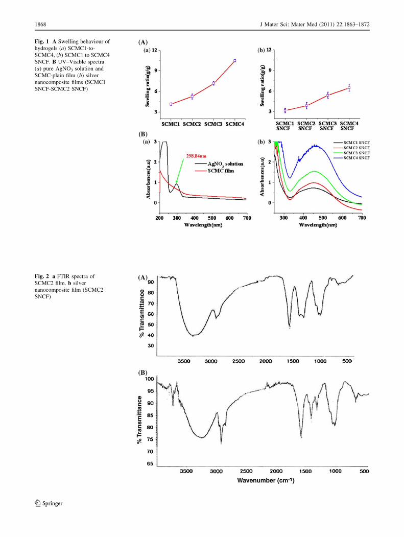
Fig. 1 A Swelling behaviour of
hydrogels (a) SCMC1-to-
SCMC4, (b) SCMC1 to SCMC4
SNCF. B UV–Visible spectra
(a) pure AgNO3 solution and
SCMC-plain film (b) silver
nanocomposite films (SCMC1
SNCF-SCMC2 SNCF)
Fig. 2 a FTIR spectra of
SCMC2 film. b silver
nanocomposite film (SCMC2
SNCF)
1868 J Mater Sci: Mater Med (2011) 22:1863–1872
123
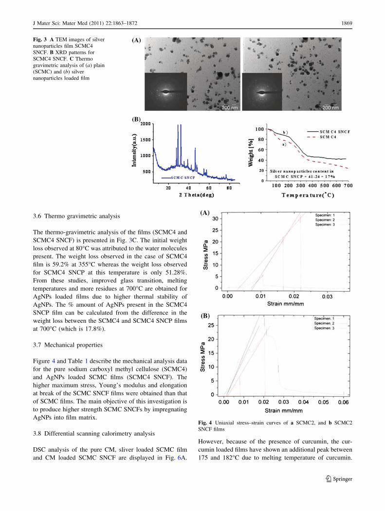
3.6 Thermo gravimetric analysis
The thermo-gravimetric analysis of the films (SCMC4 and
SCMC4 SNCF) is presented in Fig. 3C. The initial weight
loss observed at 80�C was attributed to the water molecules
present. The weight loss observed in the case of SCMC4
film is 59.2% at 355�C whereas the weight loss observed
for SCMC4 SNCP at this temperature is only 51.28%.
From these studies, improved glass transition, melting
temperatures and more residues at 700�C are obtained for
AgNPs loaded films due to higher thermal stability of
AgNPs. The % amount of AgNPs present in the SCMC4
SNCP film can be calculated from the difference in the
weight loss between the SCMC4 and SCMC4 SNCP films
at 700�C (which is 17.8%).
3.7 Mechanical properties
Figure 4 and Table 1 describe the mechanical analysis data
for the pure sodium carboxyl methyl cellulose (SCMC4)
and AgNPs loaded SCMC films (SCMC4 SNCF). The
higher maximum stress, Young’s modulus and elongation
at break of the SCMC SNCF films were obtained than that
of SCMC films. The main objective of this investigation is
to produce higher strength SCMC SNCFs by impregnating
AgNPs into film matrix.
3.8 Differential scanning calorimetry analysis
DSC analysis of the pure CM, sliver loaded SCMC film
and CM loaded SCMC SNCF are displayed in Fig. 6A.
However, because of the presence of curcumin, the cur-
cumin loaded films have shown an additional peak between
175 and 182�C due to melting temperature of curcumin.
Fig. 3 A TEM images of silver
nanoparticles film SCMC4
SNCF. B XRD patterns for
SCMC4 SNCF. C Thermo
gravimetric analysis of (a) plain
(SCMC) and (b) silver
nanoparticles loaded film
Fig. 4 Uniaxial stress–strain curves of a SCMC2, and b SCMC2
SNCF films
J Mater Sci: Mater Med (2011) 22:1863–1872 1869
123
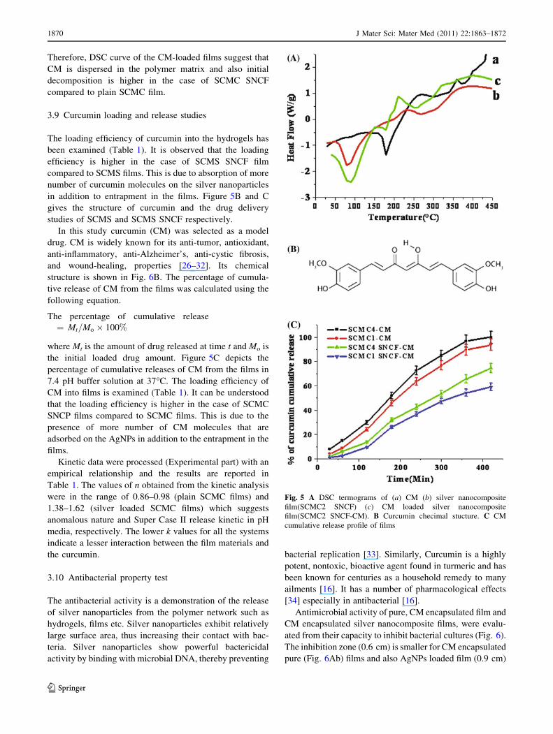
Therefore, DSC curve of the CM-loaded films suggest that
CM is dispersed in the polymer matrix and also initial
decomposition is higher in the case of SCMC SNCF
compared to plain SCMC film.
3.9 Curcumin loading and release studies
The loading efficiency of curcumin into the hydrogels has
been examined (Table 1). It is observed that the loading
efficiency is higher in the case of SCMS SNCF film
compared to SCMS films. This is due to absorption of more
number of curcumin molecules on the silver nanoparticles
in addition to entrapment in the films. Figure 5B and C
gives the structure of curcumin and the drug delivery
studies of SCMS and SCMS SNCF respectively.
In this study curcumin (CM) was selected as a model
drug. CM is widely known for its anti-tumor, antioxidant,
anti-inflammatory, anti-Alzheimer’s, anti-cystic fibrosis,
and wound-healing, properties [26–32]. Its chemical
structure is shown in Fig. 6B. The percentage of cumula-
tive release of CM from the films was calculated using the
following equation.
The percentage of cumulative release
¼ Mt=Mo � 100%
where Mt is the amount of drug released at time t and Mo is
the initial loaded drug amount. Figure 5C depicts the
percentage of cumulative releases of CM from the films in
7.4 pH buffer solution at 37�C. The loading efficiency of
CM into films is examined (Table 1). It can be understood
that the loading efficiency is higher in the case of SCMC
SNCP films compared to SCMC films. This is due to the
presence of more number of CM molecules that are
adsorbed on the AgNPs in addition to the entrapment in the
films.
Kinetic data were processed (Experimental part) with an
empirical relationship and the results are reported in
Table 1. The values of n obtained from the kinetic analysis
were in the range of 0.86–0.98 (plain SCMC films) and
1.38–1.62 (silver loaded SCMC films) which suggests
anomalous nature and Super Case II release kinetic in pH
media, respectively. The lower k values for all the systems
indicate a lesser interaction between the film materials and
the curcumin.
3.10 Antibacterial property test
The antibacterial activity is a demonstration of the release
of silver nanoparticles from the polymer network such as
hydrogels, films etc. Silver nanoparticles exhibit relatively
large surface area, thus increasing their contact with bac-
teria. Silver nanoparticles show powerful bactericidal
activity by binding with microbial DNA, thereby preventing
bacterial replication [33]. Similarly, Curcumin is a highly
potent, nontoxic, bioactive agent found in turmeric and has
been known for centuries as a household remedy to many
ailments [16]. It has a number of pharmacological effects
[34] especially in antibacterial [16].
Antimicrobial activity of pure, CM encapsulated film and
CM encapsulated silver nanocomposite films, were evalu-
ated from their capacity to inhibit bacterial cultures (Fig. 6).
The inhibition zone (0.6 cm) is smaller for CM encapsulated
pure (Fig. 6Ab) films and also AgNPs loaded film (0.9 cm)
Fig. 5 A DSC termograms of (a) CM (b) silver nanocomposite
film(SCMC2 SNCF) (c) CM loaded silver nanocomposite
film(SCMC2 SNCF-CM). B Curcumin checimal stucture. C CM
cumulative release profile of films
1870 J Mater Sci: Mater Med (2011) 22:1863–1872
123

system (Fig. 6Bb) compared with CM encapsulated silver
nanocomposite (1.25 cm) film (Fig. 6Bc) due to the fact that
the AgNPs as well as CM combination highly inhibited
bacteria. The same phenomenon was observed in mineral salt
medium (Fig. 6C). The growth rate or killing kinetics of the
E. coli has been performed in mineral salt medium (MSM)
and the results are determined by culture turbidity (OD)
measurements. Because of the presence of more number of
silver nanoparticles and curcumin, SCMC SNCF-CM com-
posite showed 86% inhibition growth while other film
composites showed only 25% inhibition growth of E. coli
(Fig. 6C). The reason for this is two fold are the curcumin
suppresses the growth of bacteria and second the release of
silver nanoparticles from the films networks. Over all the
addition of CM to AgNPs enhanced the antibacterial activity.
The nanocomposites release Ag nanoparticles as well as
curcumin with time which can eventually promote their
antibacterial application. It can be inferred from the study of
the curcumin loaded sodium carboxylmethyl cellulose–sil-
ver nanocomposite films could be used for antimicrobial and
biomedical applications.
4 Conclusion
In this work we successfully obtained curcumin nano-
composite systems based on polysaccharides and silver
nanoparticles. These composites were developed and
characterized by spectral, thermal, X-ray diffraction, and
electronic microscopic studies. The developed silver
nanocomposite films exhibited fairly good mechanical
strength and superior antimicrobial properties. Further, the
current work demonstrates a promising method to combine
silver nanocomposites with a natural compound (curcumin)
in developing novel antimicrobial agents. These agents
may find potential applications in antimicrobial packaging
materials and wound/burns dressing.
References
1. Han Y-S, Lee S-H, Choi KH, Park I. Preparation and character-
ization of chitosan-clay nanocomposites with antimicrobial
activity J Phys Chem Solids 2010;71:464–7.
Fig. 6 A Antibacterial activity
of (a) Plain (SCMC3) film,
(b) curcumin loaded (SCMC3)
film and (c) curcumin loaded
silver nanocomposite (SCMC3)
film against E. coli.B Antibacterial activity of
(a) plain (SCMC4) film,
(b) silver nanocomposite
(SCMC4 SNCF) film and
(c) curcumin loaded silver
nanocomposite (CM-SCMC4
SNCF) film against E. coli.C Semi-quantitative inhibition
effects of SCMC based films
against E. coli
J Mater Sci: Mater Med (2011) 22:1863–1872 1871
123

2. Kiuchi H, Kai W, Inoue Y. Preparation and characterization of
poly(ethylene glycol) crosslinked films. J Appl Polym Sci.
2008;107:3823–30.
3. Uhrich KE, Cannizzaro SM, Langer RS. Polymeric systems for
controlled drug release. Chem Rev. 1999;99:3181.
4. Ranney DF. Biomimetic transport and rational drug delivery.
Biochem Pharmacol. 2000;59:105–14.
5. Soppimath KS, Aminabhavi TM, Kulkarni AR. Biodegradable
polymeric nanoparticles as drug delivery devices. J Control
Release. 2001;70:1–20.
6. Lemarchand C, Gref R, Couvreur P. Polysacchride-decorated
nanopartiecles. J Eur Pharm Biopharm. 2004;58:327–41.
7. Rubinstein A. Natural polysaccharides as targeting tools of drugs
to the human colon. Drug Dev Res. 2000;50:435–9.
8. Sinha VR, Kumria R. Polysaccharides in colon-specific drug
delivery. Int J Pharm. 2001;224:19–38.
9. Ma J, Xu Y, Fan B, Liang B. Preparation and characterization of
sodium carboxymethylcellulose/poly(N-isopropylacrylamide)/clay
semi-IPN nanocomposite hydrogels. J Eur Polym. 2007;43:2221.
10. Nadagouda MN, Varma RS. Systhesis of thermally stable carboxy-
methyl cellulose/metal biodegradable nanocomposites for potentia
biological applications. Biomacromolecules. 2007;8:2762.
11. Choi Y, Simonsen J. Cellulose nanocrystal-filled carboxymethyl
cellulose nanocomposites. J Nanosci Nanotech. 2006;6:633–9.
12. Ramirez Rigo MV, Allemandi DA, Manzo RH. Drug Deliv. 2009;
16:108–15.
13. Chauvierre C, Manchanda R, Labarre D, Vauthier C, Marden
MC, Leclerc L. Swellable drug—polyelectrolyte matrices of
drug-carboxymethylcellulose complexes. Biomaterials 2010;31:
6069–74.
14. Vimala K, Samba Sivudu K, Murali Mohan Y, Sreedhar B,
Mohana Raju K. Controlled silver nanoparticles synthesis in
semi-hydrogel networks of poly(acrylamide) and carbohydrates:
a rational methodology for antibacterial application. Carbohyd
Polym. 2009;75:47–463.
15. Vimala K, Murali Mohana Y, Samba Sivudua K, Varaprasada K,
Ravindraa S, Narayana Reddya N, Padmab Y, Sreedharc B,
Mohana Rajua K. Fabrication of porous chitosan films impreg-
nated with silver nanoparticles: a facile approach for superior
antibacterial application. Colloid Surf B: Biointerfaces. 2010;76:
248–58.
16. Bhawana, Basniwal RK, Buttar HS, Jain VK, Jain N. Curcumin
nanoparticles: preparation, characterization, and antimicrobial
study. J Agric Food Chem. 2011;59:2056–61.
17. Suwantong O, Opanasopit P, Ruktanonchai U, Supaphol P.
Electrospun cellulose acetate fiber mats containing curcumin and
release characteristic of the herbal substance. Polymer. 2007;48:
7546–57.
18. Ekici S, Saraydin D. Interpenetrating polymeric network hydro-
gels for potential gastrointestinal drug release. Polym Int. 2007;
56:137.
19. Ritger PL, Peppas NA. A simple equation for description of
solute release, II: Fickian and anomalous release from swellable
devices. Control Release. 1987;5:37–42.
20. Lotfipour F, Nokhodchi A, Saeedi M, Norouzi-Sani S, Sharbafi J,
Siahi-Shadbad MR. II Farmaco, The effect of hydrophilic and
lipophilic polymers and fillers on the release rate of a tenolol
from HPMC matrices. IL Farmaco. 2004;59:819–25.
21. Ranga Rao KV, Padmalatha Devi K, Buri P. Cellulose matrices
for zero order release of soluble drugs. Drug Dev Ind Pharm.
1998;14:2299–320.
22. Talukdar MM, Kinget R. Swelling and drug release behaviour of
xanthan gum matrix tablets. Int J Pharm. 1995;120:63–72.
23. Shahidi S, Rashidi A, Ghoranneviss M, Anvari A, Rahimi MK,
Bameni Moghaddam M, Wiener J. Cellulose. 2010;17:627.
24. Varaprasad K, Murali Mohan Y, Ravindra S, Narayana Reddy N,
Vimala K, Monika K, Sreedhar B, Mohana Raju K. Hydrogel–
silver nanoparticle composites: a new generation of antimicro-
bials. J Appl Polym Sci. 2010;115:1199.
25. Biswal DR, Singh RP. Characterization of carboxymethyl cellu-
lose and polyacrylamide graft copolymer. Carbohydr Polym.
2004;57:379–87.
26. Sharma RA, Gescher AJ, Steward WP. Curcumin: the story so
far. J Eur Cancer. 2005;41:1955–68.
27. Jayaprakasha GK, Jagan L, Rao M, Sakariah KK. Trends Food
Sci Tech. 2005;15:533.
28. Jayaprakasha GK, Rao LJ, Sakariah KK. Antioxidant activities of
curcumin, demethoxycurcumin and bisdemethoxycurcumin. Food
Chem. 2006;98:720.
29. Maheshwari RK, Singh AK, Gaddipati J, Srimal RC. Multiple
biological activities of curcumin: a short review. Life Sci.
2006;78:2081.
30. Yang FS, Lim GP, Begum AN, Ubeda OJ, Simmons MR,
Ambegaokar SS, Chen PP, Kayed R, Glabe CG, Frautschy SA,
Cole GM. Curcumin inhibits formation of amyloid beta oligo-
mers and fibrils, binds plaques, and reduces amyloid in vivo.
J Biol Chem. 2005;280:5892.
31. Gopinath D, Ahmed MR, Gomathi K, Chitra K, Sehgal PK, Ja-
yakumar R. Dermal wound healing processes with curcumin
incorporated collagen films. Biomaterials. 2004;25:1911.
32. Jagetia GC, Rajanikant GK. Curcumin treatment enhances the
repair and regeneration of wounds in mice exposed to hemibody
gamma-irradiation. Plast Reconstr Surg. 2005;115:515.
33. Aggor FS, Ahmed EM, El-Aref AT, Asem MA. Synthesis and
characterization of poly(Acrylamide-co-Acrylic acid) hydrogel
containing silver nanoparticles for antimicrobial applications.
J Am Sci. 2010;12:6.
34. Kaewnopparat N, Kaewnopparat S, Jangwang A, Maneenaun D,
Chuchome T, Panichayupakaranant P. Increased solubility, dis-
solution and physicochemical studies of curcumin-poly-
vinylpyrrolidone K-30 solid dispersions. World Academy Sci,
Eng Technol. 2009;55:229–34.
1872 J Mater Sci: Mater Med (2011) 22:1863–1872
123
Copyright © 2022 FDOKUMEN