Extracellular matrix production by nucleus pulposus and bone marrow stem cells in response to...
10
Extracellular matrix production by nucleus pulposus and bone marrow stem cells in response to altered oxygen and glucose microenvironments Syeda M. Naqvi 1,2 and Conor T. Buckley 1,2 1 Trinity Centre for Bioengineering, Trinity Biomedical Sciences Institute, Trinity College Dublin, Dublin, Ireland 2 Department of Mechanical Engineering, School of Engineering, Trinity College Dublin, Dublin, Ireland Abstract Bone marrow (BM) stem cells may be an ideal source of cells for intervertebral disc (IVD) regeneration. However, the harsh biochemical microenvironment of the IVD may significantly influence the biological and metabolic vitality of injected stem cells and impair their repair potential. This study investigated the viability and production of key matrix proteins by nucleus pulposus (NP) and BM stem cells cultured in the typical biochemical microenvironment of the IVD consisting of altered oxygen and glucose concentrations. Culture- expanded NP cells and BM stem cells were encapsulated in 1.5% alginate and ionically crosslinked to form cylindrical hydrogel constructs. Hydrogel constructs were maintained under different glucose concentrations (1, 5 and 25 mM) and external oxygen concentrations (5 and 20%). Cell viability was measured using the Live/ Deadâ assay and the production of sulphated glycosaminoglycans (sGAG), and collagen was quantified biochemically and histologically. For BM stem cells, IVD-like micro-environmental conditions (5 mM glucose and 5% oxygen) increased the accumulation of sGAG and collagen. In contrast, low glucose conditions (1 mM glucose) combined with 5% external oxygen concentration promoted cell death, inhibiting proliferation and the accumulation of sGAG and collagen. NP-encapsulated alginate constructs were relatively insensitive to oxygen concentration or glucose condition in that they accumulated similar amounts of sGAG under all conditions. Under IVD-like microenvironmental conditions, NP cells were found to have a lower glucose consumption rate compared with BM cells and may in fact be more suitable to adapt and sustain the harsh microenvironmental conditions. Considering the highly specialised microenvironment of the central NP, these results indicate that IVD-like concentrations of low glucose and low oxygen are critical and influential for the survival and biological behaviour of stem cells. Such findings may promote and accelerate the translational research of stem cells for the treatment of IVD degeneration. Key words: bone marrow; glucose; intervertebral disc; metabolism; microenvironment; nucleus pulposus; oxygen; stem cells. Introduction Low back pain (LBP) is a significant epidemiological prob- lem and economic burden worldwide (Hoy et al. 2010). It is established that the primary cause of LBP is degeneration of the intervertebral disc (IVD), characterised by decreased extracellular matrix (ECM) synthesis and increased cell death (Deyo & Weinstein, 2001). IVD degeneration initiates within the nucleus pulposus (NP) and progresses with attrition of the annulus fibrosus (AF), which leads to eventual impair- ment of the IVD. Healthy NP tissue contains randomly organised collagen types II and VI, embedded in a highly hydrated gel-like matrix rich in proteoglycans (PGs), with aggrecan being predominantly abundant (Inoue, 1981). Other proteogly- cans such as biglycan, decorin and fibromodulin are also present (Singh et al. 2009). The high osmotic pressure within the NP, provided by the proteoglycans, is important in maintaining tissue hydration and resisting compressive forces during normal motion and activities. As degenera- tion progresses, the proteoglycan content of the NP dimin- ishes resulting in decreased osmotic pressure with a concomitant loss of hydration and reduction in disc height, thereby impairing the mechanical functionality of the IVD. Correspondence Conor T. Buckley, Trinity Centre for Bioengineering, Trinity Biomedi- cal Sciences Institute, Trinity College Dublin, Dublin, Ireland. T: + 353 1 896 2061; F: + 353 1 679 5554; E: [email protected] Accepted for publication 3 March 2015 © 2015 Anatomical Society J. Anat. (2015) doi: 10.1111/joa.12305 Journal of Anatomy
Transcript of Extracellular matrix production by nucleus pulposus and bone marrow stem cells in response to...
Extracellular matrix production by nucleus pulposusand bone marrow stem cells in response to alteredoxygen and glucose microenvironmentsSyeda M. Naqvi1,2 and Conor T. Buckley1,2
1Trinity Centre for Bioengineering, Trinity Biomedical Sciences Institute, Trinity College Dublin, Dublin, Ireland2Department of Mechanical Engineering, School of Engineering, Trinity College Dublin, Dublin, Ireland
Abstract
Bone marrow (BM) stem cells may be an ideal source of cells for intervertebral disc (IVD) regeneration.
However, the harsh biochemical microenvironment of the IVD may significantly influence the biological and
metabolic vitality of injected stem cells and impair their repair potential. This study investigated the viability
and production of key matrix proteins by nucleus pulposus (NP) and BM stem cells cultured in the typical
biochemical microenvironment of the IVD consisting of altered oxygen and glucose concentrations. Culture-
expanded NP cells and BM stem cells were encapsulated in 1.5% alginate and ionically crosslinked to form
cylindrical hydrogel constructs. Hydrogel constructs were maintained under different glucose concentrations (1,
5 and 25 mM) and external oxygen concentrations (5 and 20%). Cell viability was measured using the Live/
Dead� assay and the production of sulphated glycosaminoglycans (sGAG), and collagen was quantified
biochemically and histologically. For BM stem cells, IVD-like micro-environmental conditions (5 mM glucose and
5% oxygen) increased the accumulation of sGAG and collagen. In contrast, low glucose conditions (1 mM
glucose) combined with 5% external oxygen concentration promoted cell death, inhibiting proliferation and
the accumulation of sGAG and collagen. NP-encapsulated alginate constructs were relatively insensitive to
oxygen concentration or glucose condition in that they accumulated similar amounts of sGAG under all
conditions. Under IVD-like microenvironmental conditions, NP cells were found to have a lower glucose
consumption rate compared with BM cells and may in fact be more suitable to adapt and sustain the harsh
microenvironmental conditions. Considering the highly specialised microenvironment of the central NP, these
results indicate that IVD-like concentrations of low glucose and low oxygen are critical and influential for the
survival and biological behaviour of stem cells. Such findings may promote and accelerate the translational
research of stem cells for the treatment of IVD degeneration.
Key words: bone marrow; glucose; intervertebral disc; metabolism; microenvironment; nucleus pulposus;
oxygen; stem cells.
Introduction
Low back pain (LBP) is a significant epidemiological prob-
lem and economic burden worldwide (Hoy et al. 2010). It is
established that the primary cause of LBP is degeneration
of the intervertebral disc (IVD), characterised by decreased
extracellular matrix (ECM) synthesis and increased cell death
(Deyo & Weinstein, 2001). IVD degeneration initiates within
the nucleus pulposus (NP) and progresses with attrition of
the annulus fibrosus (AF), which leads to eventual impair-
ment of the IVD.
Healthy NP tissue contains randomly organised collagen
types II and VI, embedded in a highly hydrated gel-like
matrix rich in proteoglycans (PGs), with aggrecan being
predominantly abundant (Inoue, 1981). Other proteogly-
cans such as biglycan, decorin and fibromodulin are also
present (Singh et al. 2009). The high osmotic pressure
within the NP, provided by the proteoglycans, is important
in maintaining tissue hydration and resisting compressive
forces during normal motion and activities. As degenera-
tion progresses, the proteoglycan content of the NP dimin-
ishes resulting in decreased osmotic pressure with a
concomitant loss of hydration and reduction in disc height,
thereby impairing the mechanical functionality of the IVD.
Correspondence
Conor T. Buckley, Trinity Centre for Bioengineering, Trinity Biomedi-
cal Sciences Institute, Trinity College Dublin, Dublin, Ireland.
T: + 353 1 896 2061; F: + 353 1 679 5554; E: [email protected]
Accepted for publication 3 March 2015
© 2015 Anatomical Society
J. Anat. (2015) doi: 10.1111/joa.12305
Journal of Anatomy
The synthesis and composition of collagens also vary with
progressive degeneration; with increased collagen type I
produced in the NP, leading to a fibrotic transformation
of the NP tissue and a progressive inability to identify a
clear demarcation between the NP and AF tissues. Con-
comitant with matrix degradation and reduced disc height
is often an in-growth of blood vessels and nerves into the
normally avascular and aneural tissue (Freemont et al.
1997).
Cell-based therapies targeted to regenerate the NP region
may prevent progressive degeneration. Autologous disc cell
transplantation (ADCT) is a therapy that involves harvesting
NP tissue from the patient, isolating and expanding cells to
required numbers and injecting the expanded cells into the
central NP region of an early-stage degenerated IVD (Ho-
haus et al. 2008). However, limitations with this approach
include the low yield of healthy NP cells obtainable from
degenerated discs and the limited expansion capability of
NP cells (Hiyama et al. 2008; Xia et al. 2013). This has moti-
vated the exploration of stem cells due to their propensity
to proliferate and their ability to form multiple tissue types
(Caplan, 1991).
BM stem cells possess significant potential and perhaps
provide a clinically feasible source of cells to promote the
repair of NP tissue. The rationale and benefits to transplant-
ing stem cells into the IVD are twofold; first, transplanted
stem cells may stimulate endogenous NP cells, and secondly,
the resident host NP cells may promote differentiation of
the transplanted stem cells towards a nucleus pulposus phe-
notype (Richardson et al. 2006; Miyamoto et al. 2010). In
vivo studies have shown that implantation of stem cells into
experimentally induced degenerate animal discs leads to
improved disc height and accumulation of proteoglycans
(Sakai et al. 2003; Crevensten et al. 2004; Risbud et al.
2004). Furthermore, a human clinical study performed by
Orozco et al. injected autologous bone marrow stem cells
into the nucleus pulposus of 10 patients diagnosed with
lumbar disc degeneration. Results indicated that pain, dis-
ability and quality of life improved over the 12-month trial
(Orozco et al. 2011).
However, the regenerative potential of BM stem cells
may be limited by the harsh microenvironment within the
disc, characterised by low oxygen, low glucose and low pH
conditions (Bartels et al. 1998; Urban, 2002; Grunhagen
et al. 2006). In the central nucleus pulposus the oxygen con-
centration ranges from 5% to as low as 1% (Mwale et al.
2011), the pH ranges from 7.1 to as low 6.5 (Urban, 2002),
and the glucose concentration ranges from 5 mM to lower
levels (Bibby et al. 2005) as the degeneration transgresses
from mildly degenerated to a severely degenerated state.
NP cells have been shown to be well adapted to this harsh
microenvironment (Risbud et al. 2006) but this biochemical
microenvironment may negatively influence the biological
and metabolic vitality of stem cells and impair their regen-
erative potential. Therefore, understanding how stem cells
respond to limited nutrient availability is a key factor for
clinical translation.
Numerous studies have focused on cell growth and sur-
vival (Johnson et al. 2008; Stephan et al. 2011). Stephan
et al. (2011) cultured bovine NP cells in alginate beads
under zero glucose or high glucose conditions and demon-
strated that NP cell proliferation and survival are influenced
by the availability of glucose. The absence of glucose
resulted in more apoptotic and senescent cells. Interest-
ingly, Johnson et al. (2008) cultured bovine NP cells encap-
sulated in alginate gels under similar conditions and
observed that glucose deprivation leads to a minimal
increase in cell proliferation. Mwale et al. (2011) also cul-
tured bovine NP cells encapsulated in alginate beads under
different oxygen concentrations and found that low oxy-
gen levels increased the expression of aggrecan mRNA lev-
els but, interestingly, this was not reflected in GAG release.
Also, Stoyanov et al. (2011) cultured BM stem cells in algi-
nate beads under low and high oxygen concentrations and
observed that hypoxia increased aggrecan and collagen
gene expression. Although these studies describe the influ-
ence of glucose and oxygen on NP cell and BM stem cell
growth and survival, little is known of the effect on the
capacity of these cells to produce NP-like matrix. Further
experimentation is required to address ECM synthesis,
which is of major importance to the functioning of the disc.
Furthermore, the same studies have investigated the effects
of oxygen (Risbud et al. 2006; Mwale et al. 2011; Stoyanov
et al. 2011; Yang et al. 2013) or glucose (Li et al. 2007;
Wuertz et al. 2008; Deorosan & Nauman, 2011; Stephan
et al. 2011; Liang et al. 2012) independently, which has
resulted in several contradictions in the literature and con-
firms the need to study the effect of a combination of envi-
ronmental factors that more likely reflects the situation as it
exists in vivo.
The objective of this study was to investigate how micro-
environmental conditions may affect subsequent matrix
production of porcine NP and BM stem cells encapsulated
in 3D alginate hydrogels cultured in three different glucose
(1, 5 and 25 mM) media at two different oxygen concentra-
tions (5 and 20%).
Methods
Nucleus pulposus and bone marrow stem cell
isolation and culture
NP cells were harvested from the intervertebral discs (IVDs) of por-
cine donors (n = 2, 3–4 months, 20–30 kg) within 3 h of sacrifice as
previously described (Naqvi & Buckley, 2015). NP tissue was isolated
and enzymatically digested in 2.5 mg mL�1 pronase solution for
1 h followed by 3 h in 0.5 mg mL�1 collagenase solution at 37 °C.
Digested tissue/cell suspension was passed through a 100-lm cell
strainer to remove tissue debris followed by 70- and 40-lm cell
strainers to separate notochordal cells (NC) from the desired nucleus
pulposus cells (NP) as previously described (Spillekom et al. 2014).
© 2015 Anatomical Society
Matrix production by nucleus pulposus and bone marrow stem cells, S. M. Naqvi and C. T. Buckley2
Cells were washed three times by repeated centrifugation (650 g
for 5 min), plated at a density of 5 9 103 cells cm�2 and cultured
to passage 2 in T-175 cm2 flasks with low-glucose Dulbecco’s
modified Eagle’s medium (LG-DMEM, 1 mg mL�1D-glucose),
supplemented with 10% fetal bovine serum (FBS), 100 U mL�1 peni-
cillin, 100 lg mL�1 streptomycin, 0.25 lg mL�1 amphotericin B,
5 ng mL�1 fibroblast growth factor-2 (FGF-2; PeproTech, UK).
Donor matched bone marrow (BM) was isolated from the femora
and plated at 10 9 106 cells in T-75 cm2 flasks to allow for colony
formation (P0) in supplemented LG-DMEM. After P0, cells were re-
plated at 5 9 103 cells cm�2 and expanded to P2 in a humidified
atmosphere at 37 °C and 5% CO2. The differentiation capacity of
BM cells from donors was assessed as previously described (Vinardell
et al. 2011). In all cases, BM stem cells demonstrated successful dif-
ferentiation towards the osteogenic, adipogenic and chondrogenic
lineages.
Alginate hydrogel encapsulation and culture
Expanded cells (NP and BM) were encapsulated in 1.5% alginate
(Pronova UP LVG, FMC NovaMatrix, Norway) at a seeding density of
4 9 106 cells mL�1 and ionically crosslinked with 100 mM calcium
chloride (CaCl2) for 30 min to form cylindrical constructs (diame-
ter = 5 mm; height = 3 mm). The geometric construct dimensions
used in this study were based on previous work from our laboratory
(Buckley et al. 2012). Constructs were maintained in 1, 5 or 25 mM
glucose medium consisting of DMEM supplemented with penicillin
(100 U mL�1)-streptomycin (100 lg mL�1) (both from GIBCO, Invi-
trogen, Ireland), 0.25 lg mL�1 amphotericin B, 100 lg mL�1 sodium
pyruvate, 40 lg mL�1L-proline, 1.5 mg mL�1 bovine serum albu-
min, 4.7 lg mL�1 linoleic acid, 19 insulin–transferrin–selenium,
50 lg mL�1L-ascorbic acid-2-phosphate, 100 nM dexamethasone (all
Sigma-Aldrich, Ireland) and 10 ng mL�1 transforming growth factor
(TGF)-b3 (PeproTech, UK). Constructs were cultured in standard 24-
well plates with one construct per well with 2 mL of supplemented
medium in hypoxic (5% oxygen) or normoxic (20% oxygen)
conditions. Constructs were assessed at days 0 and 21 in terms of
cell viability (n = 1), biochemical content (DNA, sulphated-glycos-
aminoglycan (sGAG) and collagen content) (n = 3), histologically
and immunohistochemically (n = 2). This study was performed twice
with independent donors in each case. Results were reproducible
for all conditions investigated.
Cell viability
Cell viability was assessed using a LIVE/DEAD� Viability/Cytotoxicity
Assay Kit (Invitrogen, Bio-science, Ireland). Constructs were removed
from culture, sectioned, rinsed with phosphate-buffered saline
(PBS) and incubated for 1 h at 37 °C in live/dead solution contain-
ing 2 lM calcein AM, 4 lM ethidium homodimer-1 (EthD-1). After
incubation, segments were again washed with PBS and imaged
with an Olympus FV-1000 Point- Scanning Confocal Microscope
(Southend-on-Sea, UK) at 515 and 615 nm channels and analysed
using FV10-ASW 2.0 VIEWER software.
Quantitative biochemical analysis
Samples were digested with papain (125 lg mL�1) in 0.1 M sodium
acetate, 5 mM L-cysteine HCl, and 0.05 M EDTA (Sigma-Aldrich,
Ireland) at 60 °C under constant agitation for 18 h. DNA content
was quantified using the Hoechst 33258 dye-based DNA QF Kit
(Sigma-Aldrich, Ireland). Proteoglycan content was quantified using
the dimethylmethylene blue (DMMB) dye-binding assay (Blyscan,
Biocolor Ltd, Northern Ireland) with a chondroitin sulphate stan-
dard. Total collagen content was determined by measuring the
hydroxyproline content. Briefly, samples were hydrolysed at 110 °C
for 18 h in concentrated hydrochloric acid (HCl) (38%) and assayed
using a chloramine-T assay (Kafienah & Sims, 2004), at a hydroxy-
proline-to-collagen ratio of 1 : 7.69 (Ignat’eva et al. 2007).
Glucose concentrations in media samples from day 18 to day 21
were quantitatively measured using a glucose meter (Accu-Chek
Aviva glucose meter, Roche Diagnostics Ltd, UK). Samples of culture
media (1, 5 and 25 mM) served as controls. Cellular consumption
rates were determined by normalising to cell number and time.
Histology and immunohistochemistry
Constructs were fixed in 4% paraformaldehyde (PFA) overnight,
dehydrated in ethanol, embedded in paraffin wax, and sectioned
at a thickness of 8 lm. Sections were stained for glycosaminogly-
cans (GAGs) using aldehyde fuchsin and 1% alcian blue 8GX
(Sigma-Aldrich, Ireland) in 0.1 M HCl (Simmons et al. 2004) and
picro-sirius red to assess for collagen deposition. The deposition of
collagen types I and II was identified through immunohistochemis-
try. Briefly, sections were rinsed with PBS before treatment with
chondroitinase ABC in a humidified environment at 37 °C. Slides
were rinsed with PBS and non-specific sites were blocked with goat
serum. Sections were incubated for 1 h at 4 °C with the primary
antibody; mouse monoclonal collagen type I antibody (1 : 200;
1 mg mL�1) or mouse monoclonal anti-collagen type II (1 : 80;
1 mg mL�1). After washing in PBS, the peroxidase activity of the
sections was quenched and the sections incubated for 1 h in the
secondary antibody; anti-mouse IgG biotin antibody produced in
goats (1 : 133; 2.1 mg mL�1). Colour was developed using the Vec-
tastain ABC reagent followed by exposure to peroxidase DAB sub-
strate kit. Positive and negative controls of porcine ligament and
cartilage were included for each batch.
Statistical analyses
Statistical analyses were performed using GRAPHPAD PRISM (version 4)
software. Two-way ANOVA was used for analysis of variance with
Bonferroni post-tests to compare groups. Numerical and graphical
results are displayed as mean � standard deviation. Significance
was accepted at a level of P < 0.05.
Results
Viability of nucleus pulposus and bone marrow stem
cells in IVD-like microenvironmental conditions
For BM constructs, DNA content increased from day 0 for
both oxygen concentrations irrespective of glucose condi-
tion (Fig. 1A). A similar result was obtained for NP con-
structs maintained in 5 mM glucose under normoxic
conditions (20% oxygen). These results were confirmed
through confocal imaging of live and dead cells (Fig. 1B,
C). Interestingly, a core of dead cells was observed in all
BM constructs irrespective of the culture condition,
although this core effect was more pronounced in the
© 2015 Anatomical Society
Matrix production by nucleus pulposus and bone marrow stem cells, S. M. Naqvi and C. T. Buckley 3
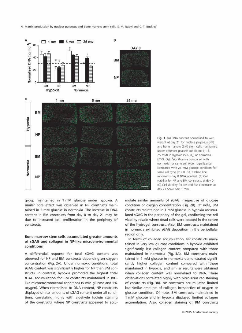
group maintained in 1 mM glucose under hypoxia. A
similar core effect was observed in NP constructs main-
tained in 5 mM glucose in normoxia. The increase in DNA
content in BM constructs from day 0 to day 21 may be
due to increased cell proliferation in the periphery of
constructs.
Bone marrow stem cells accumulated greater amounts
of sGAG and collagen in NP-like microenvironmental
conditions
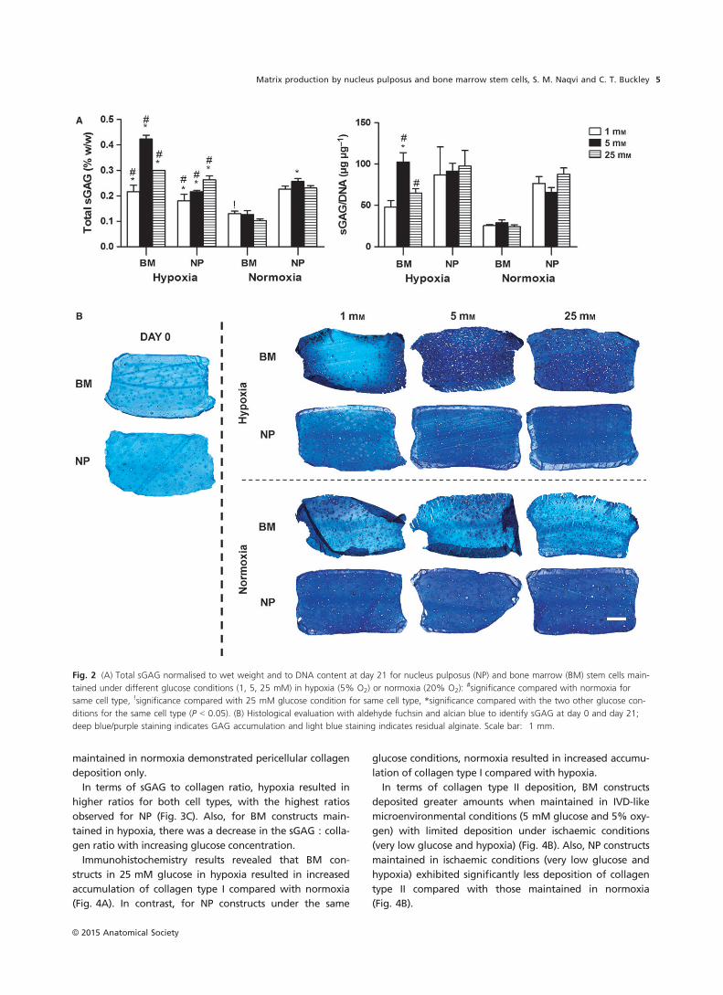
A differential response for total sGAG content was
observed for NP and BM constructs depending on oxygen
concentration (Fig. 2A). Under normoxic conditions, total
sGAG content was significantly higher for NP than BM con-
structs. In contrast, hypoxia promoted the highest total
sGAG accumulation for BM constructs maintained in IVD-
like microenvironmental conditions (5 mM glucose and 5%
oxygen). When normalised to DNA content, NP constructs
displayed similar amounts of sGAG content under all condi-
tions, correlating highly with aldehyde fuchsin staining
of the constructs, where NP constructs appeared to accu-
mulate similar amounts of sGAG irrespective of glucose
condition or oxygen concentration (Fig. 2B). Of note, BM
constructs maintained in 1 mM glucose in hypoxia accumu-
lated sGAG in the periphery of the gel, confirming the cell
viability results where dead cells were located in the centre
of the hydrogel construct. Also, BM constructs maintained
in normoxia exhibited sGAG deposition in the pericellular
region only.
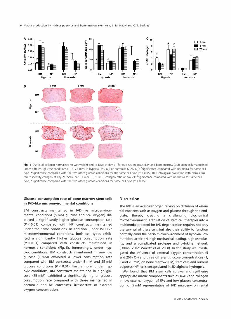
In terms of collagen accumulation, NP constructs main-
tained in very low glucose conditions in hypoxia exhibited
significantly less collagen content compared with those
maintained in normoxia (Fig. 3A). BM constructs main-
tained in 1 mM glucose in normoxia demonstrated signifi-
cantly higher collagen content compared with those
maintained in hypoxia, and similar results were obtained
when collagen content was normalised to DNA. These
observations correlated highly with picro-sirius red staining
of constructs (Fig. 3B). NP constructs accumulated limited
but similar amounts of collagen irrespective of oxygen or
glucose condition. Of note, BM constructs maintained in
1 mM glucose and in hypoxia displayed limited collagen
accumulation. Also, collagen staining of BM constructs
A B
C
Fig. 1 (A) DNA content normalised to wet
weight at day 21 for nucleus pulposus (NP)
and bone marrow (BM) stem cells maintained
under different glucose conditions (1, 5,
25 mM) in hypoxia (5% O2) or normoxia
(20% O2):#significance compared with
normoxia for same cell type. !significance
compared with 25 mM glucose condition for
same cell type (P < 0.05), dashed line
represents day 0 DNA content. (B) Cell
viability for NP and BM constructs at day 0
(C) Cell viability for NP and BM constructs at
day 21 Scale bar: 1 mm.
© 2015 Anatomical Society
Matrix production by nucleus pulposus and bone marrow stem cells, S. M. Naqvi and C. T. Buckley4
maintained in normoxia demonstrated pericellular collagen
deposition only.
In terms of sGAG to collagen ratio, hypoxia resulted in
higher ratios for both cell types, with the highest ratios
observed for NP (Fig. 3C). Also, for BM constructs main-
tained in hypoxia, there was a decrease in the sGAG : colla-
gen ratio with increasing glucose concentration.
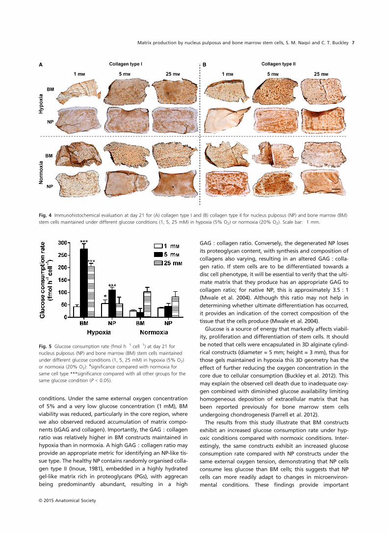
Immunohistochemistry results revealed that BM con-
structs in 25 mM glucose in hypoxia resulted in increased
accumulation of collagen type I compared with normoxia
(Fig. 4A). In contrast, for NP constructs under the same
glucose conditions, normoxia resulted in increased accumu-
lation of collagen type I compared with hypoxia.
In terms of collagen type II deposition, BM constructs
deposited greater amounts when maintained in IVD-like
microenvironmental conditions (5 mM glucose and 5% oxy-
gen) with limited deposition under ischaemic conditions
(very low glucose and hypoxia) (Fig. 4B). Also, NP constructs
maintained in ischaemic conditions (very low glucose and
hypoxia) exhibited significantly less deposition of collagen
type II compared with those maintained in normoxia
(Fig. 4B).
A
B
Fig. 2 (A) Total sGAG normalised to wet weight and to DNA content at day 21 for nucleus pulposus (NP) and bone marrow (BM) stem cells main-
tained under different glucose conditions (1, 5, 25 mM) in hypoxia (5% O2) or normoxia (20% O2):#significance compared with normoxia for
same cell type, !significance compared with 25 mM glucose condition for same cell type, *significance compared with the two other glucose con-
ditions for the same cell type (P < 0.05). (B) Histological evaluation with aldehyde fuchsin and alcian blue to identify sGAG at day 0 and day 21;
deep blue/purple staining indicates GAG accumulation and light blue staining indicates residual alginate. Scale bar: 1 mm.
© 2015 Anatomical Society
Matrix production by nucleus pulposus and bone marrow stem cells, S. M. Naqvi and C. T. Buckley 5
Glucose consumption rate of bone marrow stem cells
in IVD-like microenvironmental conditions
BM constructs maintained in IVD-like microenviron-
mental conditions (5 mM glucose and 5% oxygen) dis-
played a significantly higher glucose consumption rate
(P < 0.01) compared with NP constructs maintained
under the same conditions. In addition, under IVD-like
microenvironmental conditions, both cell types exhib-
ited a significantly higher glucose consumption rate
(P < 0.01) compared with constructs maintained in
normoxic conditions (Fig. 5). Interestingly, under hyp-
oxic conditions, BM constructs maintained in very low
glucose (1 mM) exhibited a lower consumption rate
compared with BM constructs under 5 mM and 25 mM
glucose conditions (P < 0.01). Furthermore, under hyp-
oxic conditions, BM constructs maintained in high glu-
cose (25 mM) exhibited a significantly higher glucose
consumption rate compared with those maintained in
normoxia and NP constructs, irrespective of external
oxygen concentration.
Discussion
The IVD is an avascular organ relying on diffusion of essen-
tial nutrients such as oxygen and glucose through the end-
plate, thereby creating a challenging biochemical
microenvironment. Translation of stem cell therapies into a
multimodal protocol for IVD degeneration requires not only
the survival of these cells but also their ability to function
normally amid the harsh microenvironment of hypoxia, low
nutrition, acidic pH, high mechanical loading, high osmolar-
ity, and a complicated protease and cytokine network
(Urban, 2002; Wuertz et al. 2008). In this study we investi-
gated the influence of external oxygen concentration (5
and 20% O2) and three different glucose concentrations (1,
5 and 20 mM) on bone marrow (BM) stem cells and nucleus
pulposus (NP) cells encapsulated in 3D alginate hydrogels.
We found that BM stem cells survive and synthesise
appropriate matrix components such as sGAG and collagen
in low external oxygen of 5% and low glucose concentra-
tion of 5 mM representative of IVD microenvironmental
A C
B
Fig. 3 (A) Total collagen normalised to wet weight and to DNA at day 21 for nucleus pulposus (NP) and bone marrow (BM) stem cells maintained
under different glucose conditions (1, 5, 25 mM) in hypoxia (5% O2) or normoxia (20% O2):#significance compared with normoxia for same cell
type, *significance compared with the two other glucose conditions for the same cell type (P < 0.05). (B) Histological evaluation with picro-sirius
red to identify collagen at day 21. Scale bar: 1 mm. (C) sGAG : collagen ratio at day 21: #significance compared with normoxia for same cell
type, *significance compared with the two other glucose conditions for same cell type (P < 0.05).
© 2015 Anatomical Society
Matrix production by nucleus pulposus and bone marrow stem cells, S. M. Naqvi and C. T. Buckley6
conditions. Under the same external oxygen concentration
of 5% and a very low glucose concentration (1 mM), BM
viability was reduced, particularly in the core region, where
we also observed reduced accumulation of matrix compo-
nents (sGAG and collagen). Importantly, the GAG : collagen
ratio was relatively higher in BM constructs maintained in
hypoxia than in normoxia. A high GAG : collagen ratio may
provide an appropriate metric for identifying an NP-like tis-
sue type. The healthy NP contains randomly organised colla-
gen type II (Inoue, 1981), embedded in a highly hydrated
gel-like matrix rich in proteoglycans (PGs), with aggrecan
being predominantly abundant, resulting in a high
GAG : collagen ratio. Conversely, the degenerated NP loses
its proteoglycan content, with synthesis and composition of
collagens also varying, resulting in an altered GAG : colla-
gen ratio. If stem cells are to be differentiated towards a
disc cell phenotype, it will be essential to verify that the ulti-
mate matrix that they produce has an appropriate GAG to
collagen ratio; for native NP, this is approximately 3.5 : 1
(Mwale et al. 2004). Although this ratio may not help in
determining whether ultimate differentiation has occurred,
it provides an indication of the correct composition of the
tissue that the cells produce (Mwale et al. 2004).
Glucose is a source of energy that markedly affects viabil-
ity, proliferation and differentiation of stem cells. It should
be noted that cells were encapsulated in 3D alginate cylind-
rical constructs (diameter = 5 mm; height = 3 mm), thus for
those gels maintained in hypoxia this 3D geometry has the
effect of further reducing the oxygen concentration in the
core due to cellular consumption (Buckley et al. 2012). This
may explain the observed cell death due to inadequate oxy-
gen combined with diminished glucose availability limiting
homogeneous deposition of extracellular matrix that has
been reported previously for bone marrow stem cells
undergoing chondrogenesis (Farrell et al. 2012).
The results from this study illustrate that BM constructs
exhibit an increased glucose consumption rate under hyp-
oxic conditions compared with normoxic conditions. Inter-
estingly, the same constructs exhibit an increased glucose
consumption rate compared with NP constructs under the
same external oxygen tension, demonstrating that NP cells
consume less glucose than BM cells; this suggests that NP
cells can more readily adapt to changes in microenviron-
mental conditions. These findings provide important
A B
Fig. 4 Immunohistochemical evaluation at day 21 for (A) collagen type I and (B) collagen type II for nucleus pulposus (NP) and bone marrow (BM)
stem cells maintained under different glucose conditions (1, 5, 25 mM) in hypoxia (5% O2) or normoxia (20% O2). Scale bar: 1 mm.
Fig. 5 Glucose consumption rate (fmol h�1 cell�1) at day 21 for
nucleus pulposus (NP) and bone marrow (BM) stem cells maintained
under different glucose conditions (1, 5, 25 mM) in hypoxia (5% O2)
or normoxia (20% O2):#significance compared with normoxia for
same cell type ***significance compared with all other groups for the
same glucose condition (P < 0.05).
© 2015 Anatomical Society
Matrix production by nucleus pulposus and bone marrow stem cells, S. M. Naqvi and C. T. Buckley 7
insights in the development of clinical cell-based therapies
in determining the suitability of specific cell types for tar-
geted regeneration.
Indeed, it has been reported previously that glucose
uptake is increased during hypoxia and that stem cells are
known to possess the ability to adapt their oxygen con-
sumption rate to changes in the oxygen environment (Patt-
appa et al. 2013). Deschepper et al. (2010) previously
demonstrated that stem cells can remain viable when main-
tained in severe hypoxic conditions (i.e. 0.2% O2) but not in
the absence of glucose. This correlates well with the results
from this study, where BM-encapsulated gels maintained in
hypoxia with sufficient glucose (5 and 25 mM) demon-
strated higher cell viability, sGAG and collagen accumula-
tion. Importantly, the reduced cell viability was not evident
in BM-encapsulated alginate hydrogels maintained under
the same very low glucose concentration (1 mM glucose)
and an external oxygen concentration of 20%. Under these
conditions, cells appeared to remain viable but only depos-
ited matrix pericellularly. Furthermore, this was observed
for all BM-encapsulated alginate hydrogels maintained
under 20% oxygen conditions. Interestingly, the therapeu-
tic potential of stem cells is commonly investigated under
20% O2 (normoxia) conditions in vitro, whereas the typical
physiological oxygen concentration in human ranges from
4 to 7% (Packer & Fuehr, 1977; Kofoed et al. 1985) and falls
to 1% in some pathological ischaemic tissues, as well as in
the degenerated IVD (Bartels et al. 1998). Numerous studies
have investigated the influence of hypoxia and have found
that BM stem cells proliferated more rapidly, exhibited
greater colony forming unit (CFU) formation ability (Gray-
son et al. 2006, 2007), and maintained better ‘stemness’ in
hypoxia through the down-regulation of E2A-p21 by HIF-
1a-Twist pathway (Tsai et al. 2011). Furthermore, previous
studies have demonstrated that glucose is a significant fac-
tor in the metabolic response of mesenchymal stem cells
(Deorosan & Nauman, 2011) and that a low oxygen envi-
ronment enhances GAG synthesis in pellets and hydrogels
(Sheehy et al. 2012). Risbud et al. (2004) found that 2% O2
and 10 ng mL�1 TGF-b could stimulate rat BM stem cell dif-
ferentiation to acquire phenotypes similar to that of NP
cells.
In contrast, NP-encapsulated alginate hydrogels main-
tained under the same very low glucose concentration
(1 mM glucose) and an external oxygen concentration of
5% did not exhibit reduced cell viability. In fact, NP cells
remained relatively insensitive to external microenviron-
mental conditions such that similar amounts of sGAG and
collagen were deposited homogeneously throughout. This
may be due to reduced glucose and oxygen consumption
rates, as NP cells naturally reside in a microenvironment
with limited nutrient availability. The cell-specific response
observed in this study may thus be a function of metabolic
activity. It is plausible that at a particular oxygen concentra-
tion and glucose concentration, NP cells and stem cells
possess altering metabolic demands. Agrawal et al. (2007)
indicated that oxygen-independent stabilisation of HIF-1a,
a transcription factor that regulates oxidative metabolism,
in NP cells is a metabolic adaptation to a unique microenvi-
ronment. Furthermore, it should be noted that these experi-
ments were performed using a cell density of
4 9 106 cells mL�1, which is the typical cell density of native
nucleus pulposus tissue. Higher seeding densities that are
typically used in tissue engineering investigations would
exacerbate the nutrient demands, resulting in limited
matrix formation. This is an important consideration for IVD
regeneration strategies regarding the optimal number of
cells that can be injected into the intervertebral disc to elicit
a therapeutic response and formation of new tissue. The
success of any cell-based strategy will therefore be depen-
dent on the state of degeneration and more importantly
the microenvironment of the disc that can maintain the via-
bility and support the function of injected cells.
Among several studies that have investigated the effects
of IVD-like culture conditions on stem cell survival and dif-
ferentiation, Wuertz et al. (2008) demonstrated that com-
bining low glucose with high osmolarity (485 mOsm) and
low pH (6.8) is detrimental to the differentiation of stem
cells, with decreased cellular proliferation and collagen
and sGAG expression suggesting that the beneficial effects
of IVD-like low-glucose culture are not sufficient for pro-
motion of stem cell differentiation when other environ-
mental factors are considered. Of note, Wuertz et al.
(2008) did not examine the effect of hypoxia, which is
known to be a potent regulator of matrix production. Fur-
thermore, it is crucial to determine the response of stem
cells to pro-inflammatory cytokines to elucidate fully how
these cells may respond post implantation in a degenerate
IVD niche. Culture of stem cells in the presence of inter-
leukin (IL)-1b significantly decreases culture pellet size,
and cells produce an ECM with atypical mechanical
strength and decreased expression of matrix molecules
(Felka et al. 2009).
Considering the highly specialised microenvironment of
the central NP, these results indicate that IVD-like low glu-
cose and low oxygen are critical and influential for the sur-
vival and biological behaviour of BM stem cells. In this
study, for BM constructs, glucose effects were only evident
under hypoxic conditions, suggesting that low oxygen is an
important regulator of matrix production. Furthermore, NP
cells and BM stem cells respond differentially to varying
environmental conditions due to altered metabolic activity.
Under IVD-like microenvironmental conditions, NP cells
were found to have a lower glucose consumption rate
compared with BM cells and may in fact be more suitable
to survive the harsh microenvironment that exists within
the IVD. Such findings may promote and accelerate the
development of clinical therapies in demonstrating the suit-
ability of different cell types for targeted regeneration of
the IVD.
© 2015 Anatomical Society
Matrix production by nucleus pulposus and bone marrow stem cells, S. M. Naqvi and C. T. Buckley8
Acknowledgements
This work was supported by the Graduate Research Education Pro-
gramme in Engineering (GREP-Eng), PRTLI Cycle 5 funded pro-
gramme. PRTLI is 50% co-funded under the European Regional
Development Fund. The authors have no conflict of interest to
declare.
References
Agrawal A, Guttapalli A, Narayan S, et al. (2007) Normoxic stabil-
ization of HIF-1alpha drives glycolytic metabolism and regulates
aggrecan gene expression in nucleus pulposus cells of the rat
intervertebral disk. Am J Physiol Cell Physiol 293, C621–C631.
Bartels EM, Fairbank JC, Winlove CP, et al. (1998) Oxygen and
lactate concentrations measured in vivo in the intervertebral
discs of patients with scoliosis and back pain. Spine (Phila Pa
1976) 23, 1–7; discussion 8.
Bibby SR, Jones DA, Ripley RM, et al. (2005) Metabolism of the
intervertebral disc: effects of low levels of oxygen, glucose,
and pH on rates of energy metabolism of bovine nucleus
pulposus cells. Spine (Phila Pa 1976), 30, 487–496.
Buckley CT, Meyer EG, Kelly DJ (2012) The influence of construct
scale on the composition and functional properties of cartilag-
inous tissues engineered using bone marrow-derived mesen-
chymal stem cells. Tissue Eng Part A 18, 382–396.
Caplan AI (1991) Mesenchymal stem cells. J Orthop Res 9, 641–
650.
Crevensten G, Walsh AJ, Ananthakrishnan D, et al. (2004) Inter-
vertebral disc cell therapy for regeneration: mesenchymal
stem cell implantation in rat intervertebral discs. Ann Biomed
Eng 32, 430–434.
Deorosan B, Nauman EA (2011) The role of glucose, serum, and
three-dimensional cell culture on the metabolism of bone
marrow-derived mesenchymal stem cells. Stem Cells Int 2011,
429187.
Deschepper M, Oudina K, David B, et al. (2010) Survival and
function of mesenchymal stem cells (MSCs) depend on glucose
to overcome exposure to long-term, severe and continuous
hypoxia. J Cell Mol Med 15, 1505–1514.
Deyo RA, Weinstein JN (2001) Low back pain. N Engl J Med 344,
363–370.
Farrell MJ, Comeau ES, Mauck RL (2012) Mesenchymal stem cells
produce functional cartilage matrix in three-dimensional cul-
ture in regions of optimal nutrient supply. Eur Cell Mater 23,
425–440.
Felka T, Schafer R, Schewe B, et al. (2009) Hypoxia reduces the
inhibitory effect of IL-1beta on chondrogenic differentiation of
FCS-free expanded MSC. Osteoarthritis Cartilage 17, 1368–1376.
Freemont AJ, Peacock TE, Goupille P, et al. (1997) Nerve
ingrowth into diseased intervertebral disc in chronic back
pain. Lancet 350, 178–181.
Grayson WL, Zhao F, Izadpanah R, et al. (2006) Effects of
hypoxia on human mesenchymal stem cell expansion and plas-
ticity in 3D constructs. J Cell Physiol 207, 331–339.
Grayson WL, Zhao F, Bunnell B, et al. (2007) Hypoxia enhances
proliferation and tissue formation of human mesenchymal
stem cells. Biochem Biophys Res Commun 358, 948–953.
Grunhagen T, Wilde G, Soukane DM, et al. (2006) Nutrient sup-
ply and intervertebral disc metabolism. J Bone Joint Surg Am
88(Suppl 2), 30–35.
Hiyama A, Mochida J, Iwashina T, et al. (2008) Transplantation
of mesenchymal stem cells in a canine disc degeneration
model. J Orthop Res 26, 589–600.
Hohaus C, Ganey TM, Minkus Y, et al. (2008) Cell transplanta-
tion in lumbar spine disc degeneration disease. Eur Spine J 17
(Suppl 4), 492–503.
Hoy D, Brooks P, Blyth F, et al. (2010) The epidemiology of low
back pain. Best Pract Res Clin Rheumatol 24, 769–781.
Ignat’eva NY, Danilov NA, Averkiev SV, et al. (2007) Determina-
tion of hydroxyproline in tissues and the evaluation of the
collagen content of the tissues. J Anal Chem 62, 51–57.
Inoue H (1981) Three-dimensional architecture of lumbar inter-
vertebral discs. Spine (Phila Pa 1976), 6, 139–146.
Johnson WE, Stephan S, Roberts S (2008) The influence of
serum, glucose and oxygen on intervertebral disc cell growth
in vitro: implications for degenerative disc disease. Arthritis
Res Ther 10, R46.
Kafienah W, Sims TJ (2004) Biochemical methods for the analysis
of tissue-engineered cartilage. Methods Mol Biol 238, 217–230.
Kofoed H, Sjontoft E, Siemssen SO, et al. (1985) Bone marrow
circulation after osteotomy. Blood flow, pO2, pCO2, and pres-
sure studied in dogs. Acta Orthop Scand 56, 400–403.
Li YM, Schilling T, Benisch P, et al. (2007) Effects of high glucose
on mesenchymal stem cell proliferation and differentiation.
Biochem Biophys Res Commun 363, 209–215.
Liang C, Li H, Tao Y, et al. (2012) Responses of human adipose-
derived mesenchymal stem cells to chemical microenvironment
of the intervertebral disc. J Transl Med 10, 49.
Miyamoto T, Muneta T, Tabuchi T, et al. (2010) Intradiscal trans-
plantation of synovial mesenchymal stem cells prevents inter-
vertebral disc degeneration through suppression of matrix
metalloproteinase-related genes in nucleus pulposus cells in
rabbits. Arthritis Res Ther 12, R206.
Mwale F, Roughley P, Antoniou J (2004) Distinction between
the extracellular matrix of the nucleus pulposus and hyaline
cartilage: a requisite for tissue engineering of intervertebral
disc. Eur Cell Mater 8, 58–63.
Mwale F, Ciobanu I, Giannitsios D, et al. (2011) Effect of oxygen
levels on proteoglycan synthesis by intervertebral disc cells.
Spine (Phila Pa 1976), 36, E131–E138.
Naqvi SM, Buckley CT (2015) Differential response of encapsu-
lated nucleus pulposus and bone marrow stem cells in isola-
tion and coculture in alginate and chitosan hydrogels. Tissue
Eng Part A, 21, 288–299.
Orozco L, Soler R, Morera C, et al. (2011) Intervertebral disc
repair by autologous mesenchymal bone marrow cells: a pilot
study. Transplantation 92, 822–828.
Packer L, Fuehr K (1977) Low oxygen concentration extends the
lifespan of cultured human diploid cells. Nature 267, 423–425.
Pattappa G, Thorpe SD, Jegard NC, et al. (2013) Continuous and
uninterrupted oxygen tension influences the colony formation
and oxidative metabolism of human mesenchymal stem cells.
Tissue Eng Part C Methods 19, 68–79.
Richardson SM, Walker RV, Parker S, et al. (2006) Intervertebral
disc cell-mediated mesenchymal stem cell differentiation. Stem
Cells 24, 707–716.
Risbud MV, Albert TJ, Guttapalli A, et al. (2004) Differentiation
of mesenchymal stem cells towards a nucleus pulposus-like
phenotype in vitro: implications for cell-based transplantation
therapy. Spine (Phila Pa 1976) 29, 2627–2632.
Risbud MV, Guttapalli A, Stokes DG, et al. (2006) Nucleus
pulposus cells express HIF-1 alpha under normoxic culture
© 2015 Anatomical Society
Matrix production by nucleus pulposus and bone marrow stem cells, S. M. Naqvi and C. T. Buckley 9
conditions: a metabolic adaptation to the intervertebral disc
microenvironment. J Cell Biochem 98, 152–159.
Sakai D, Mochida J, Yamamoto Y, et al. (2003) Transplantation
of mesenchymal stem cells embedded in Atelocollagen gel to
the intervertebral disc: a potential therapeutic model for disc
degeneration. Biomaterials 24, 3531–3541.
Sheehy EJ, Buckley CT, Kelly DJ (2012) Oxygen tension regulates
the osteogenic, chondrogenic and endochondral phenotype
of bone marrow derived mesenchymal stem cells. Biochem
Biophys Res Commun 417, 305–310.
Simmons CA, Alsberg E, Hsiong S, et al. (2004) Dual growth fac-
tor delivery and controlled scaffold degradation enhance
in vivo bone formation by transplanted bone marrow stromal
cells. Bone 35, 562–569.
Singh K, Masuda K, Thonar EJ, et al. (2009) Age-related changes
in the extracellular matrix of nucleus pulposus and anulus fi-
brosus of human intervertebral disc. Spine (Phila Pa 1976), 34,
10–16.
Spillekom S, Smolders LA, Grinwis GC, et al. (2014) Increased
osmolarity and cell clustering preserve canine notochordal cell
phenotype in culture. Tissue Eng Part C Methods 20, 8.
Stephan S, Johnson WE, Roberts S (2011) The influence of
nutrient supply and cell density on the growth and survival of
intervertebral disc cells in 3D culture. Eur Cell Mater 22, 97–
108.
Stoyanov JV, Gantenbein-Ritter B, Bertolo A, et al. (2011)
Role of hypoxia and growth and differentiation factor-5 on
differentiation of human mesenchymal stem cells towards
intervertebral nucleus pulposus-like cells. Eur Cell Mater 21,
533–547.
Tsai CC, Chen YJ, Yew TL, et al. (2011) Hypoxia inhibits senes-
cence and maintains mesenchymal stem cell properties
through down-regulation of E2A-p21 by HIF-TWIST. Blood
117, 459–469.
Urban JP (2002) The role of the physicochemical environment in
determining disc cell behaviour. Biochem Soc Trans 30, 858–
864.
Vinardell T, Buckley CT, Thorpe SD, et al. (2011) Composition-
function relations of cartilaginous tissues engineered from
chondrocytes and mesenchymal stem cells isolated from bone
marrow and infrapatellar fat pad. J Tissue Eng Regen Med 5,
673–683.
Wuertz K, Godburn K, Neidlinger-Wilke C, et al. (2008) Behavior
of mesenchymal stem cells in the chemical microenvironment
of the intervertebral disc. Spine (Phila Pa 1976), 33, 1843–
1849.
Xia XP, Chen HL, Cheng HB (2013) Prevalence of adjacent seg-
ment degeneration after spine surgery: a systematic review
and meta-analysis. Spine (Phila Pa 1976), 38, 597–608.
Yang SH, Hu MH, Sun YH, et al. (2013) Differential phenotypic
behaviors of human degenerative nucleus pulposus cells
under normoxic and hypoxic conditions: influence of oxygen
concentration during isolation, expansion, and cultivation.
Spine J 13, 1590–1596.
© 2015 Anatomical Society
Matrix production by nucleus pulposus and bone marrow stem cells, S. M. Naqvi and C. T. Buckley10