Genetic and epigenetic regulation in nuclear microenvironments for biological control in cancer
15
Genetic and Epigenetic Regulation in Nuclear Microenvironments for Biological Control in Cancer Gary S. Stein 1,* , Sayyed K. Zaidi 1 , Janet L. Stein 1 , Jane B. Lian 1 , Andre van Wijnen 1 , Martin Montecino 2 , Daniel W. Young 3 , Amjad Javed 1 , Jitesh Pratap 1 , Je-Yong Choi 4 , Syed A. Ali 1 , Sandhya Pande 1 , and Mohammad Q. Hassan 1 1 Department of Cell Biology, University of Massachusetts Medical School, Worcester, MA 01655 2 Universidad de Concepcion Facultad de Ciencias Biologicas - Departmento de Biologia Molecular Barrio Universitario s/n Concepcion, Concepcion 4079100 Chile 3 Wolf, Greenfield & Sacks, P.D., Boston, MA 02210-2206 4 Dept of Biochemistry, School of Medicine, Kyungpook National University, Daegu 700-422, Korea Abstract The regulatory machinery that governs genetic and epigenetic control of gene expression is compartmentalized in nuclear microenvironments. Temporal and spatial parameters of regulatory complex organization and assembly are functionally linked to biological control and are compromised with the onset and progression of tumorigenesis providing a novel platform for cancer diagnosis and treatment. Keywords nuclear microenvironments; epigenetic control; transcription; nuclear matrix; chromatin For the past century control of gene expression has been a dominant theme in fundamental biological and clinically relevant research. A platform for discovery has been provided by advances in nucleic acid, protein and carbohydrate biochemistry, structural biology and microscopy. The capabilities to synthesize and sequence nucleic acids and proteins have been instrumental. The pathway to understanding gene expression and the molecular basis of pathologies was initiated by the seminal discovery that DNA is the genetic material (Avery et al., 1944). Subsequent breakthroughs that have been pivotal in elucidating gene regulatory mechanisms include resolving the double helical structure of DNA (Watson and Crick, 1953), defining parameters of transcription, replication, recombination and repair, mapping signaling pathways that transduce and integrate regulatory cues within and between cells and linking genes with cancer and a broad spectrum of diseases. The accrual of insight into biological control from encyclopedic initiatives in genomics, proteomics and bioinformatics has been spectacular. Yet, there is additionally a requirement for resolution of mechanisms that support the organization, assembly, and integration of gene regulatory information into physiologically responsive networks. And, the mechanisms that epigenetically support cell fate and lineage commitment must be further defined. * Corresponding Author: Department of Cell Biology, University of Massachusetts Medical School, 55 Lake Avenue North, Worcester, MA 01655-0002; tel: 508-856-5625; fax: 508-856-6800; [email protected]. NIH Public Access Author Manuscript J Cell Biochem. Author manuscript; available in PMC 2009 August 15. Published in final edited form as: J Cell Biochem. 2008 August 15; 104(6): 2016–2026. doi:10.1002/jcb.21813. NIH-PA Author Manuscript NIH-PA Author Manuscript NIH-PA Author Manuscript
-
Upload
independent -
Category
Documents
-
view
0 -
download
0
Transcript of Genetic and epigenetic regulation in nuclear microenvironments for biological control in cancer

Genetic and Epigenetic Regulation in NuclearMicroenvironments for Biological Control in Cancer
Gary S. Stein1,*, Sayyed K. Zaidi1, Janet L. Stein1, Jane B. Lian1, Andre van Wijnen1, MartinMontecino2, Daniel W. Young3, Amjad Javed1, Jitesh Pratap1, Je-Yong Choi4, Syed A. Ali1,Sandhya Pande1, and Mohammad Q. Hassan1
1 Department of Cell Biology, University of Massachusetts Medical School, Worcester, MA 016552 Universidad de Concepcion Facultad de Ciencias Biologicas - Departmento de BiologiaMolecular Barrio Universitario s/n Concepcion, Concepcion 4079100 Chile3 Wolf, Greenfield & Sacks, P.D., Boston, MA 02210-22064 Dept of Biochemistry, School of Medicine, Kyungpook National University, Daegu 700-422,Korea
AbstractThe regulatory machinery that governs genetic and epigenetic control of gene expression iscompartmentalized in nuclear microenvironments. Temporal and spatial parameters of regulatorycomplex organization and assembly are functionally linked to biological control and arecompromised with the onset and progression of tumorigenesis providing a novel platform forcancer diagnosis and treatment.
Keywordsnuclear microenvironments; epigenetic control; transcription; nuclear matrix; chromatin
For the past century control of gene expression has been a dominant theme in fundamentalbiological and clinically relevant research. A platform for discovery has been provided byadvances in nucleic acid, protein and carbohydrate biochemistry, structural biology andmicroscopy. The capabilities to synthesize and sequence nucleic acids and proteins havebeen instrumental. The pathway to understanding gene expression and the molecular basis ofpathologies was initiated by the seminal discovery that DNA is the genetic material (Averyet al., 1944). Subsequent breakthroughs that have been pivotal in elucidating gene regulatorymechanisms include resolving the double helical structure of DNA (Watson and Crick,1953), defining parameters of transcription, replication, recombination and repair, mappingsignaling pathways that transduce and integrate regulatory cues within and between cellsand linking genes with cancer and a broad spectrum of diseases. The accrual of insight intobiological control from encyclopedic initiatives in genomics, proteomics and bioinformaticshas been spectacular. Yet, there is additionally a requirement for resolution of mechanismsthat support the organization, assembly, and integration of gene regulatory information intophysiologically responsive networks. And, the mechanisms that epigenetically support cellfate and lineage commitment must be further defined.
*Corresponding Author: Department of Cell Biology, University of Massachusetts Medical School, 55 Lake Avenue North, Worcester,MA 01655-0002; tel: 508-856-5625; fax: 508-856-6800; [email protected].
NIH Public AccessAuthor ManuscriptJ Cell Biochem. Author manuscript; available in PMC 2009 August 15.
Published in final edited form as:J Cell Biochem. 2008 August 15; 104(6): 2016–2026. doi:10.1002/jcb.21813.
NIH
-PA Author Manuscript
NIH
-PA Author Manuscript
NIH
-PA Author Manuscript
Both genetic and epigenetic control mediate the regulation of proliferation, cell growth andphenotype, as well as establish and sustain the properties of normal and tumor cells.Recognizing the scope of mechanisms that are associated with genetic and epigeneticcontrol we will focus on the Runx family of transcription factors to illustrate involvement ofregulation at both levels. Temporal and spatial configuration of regulatory machinery forcompartmentalization of genetic and epigenetic control in nuclear microenvironments willbe considered within the context of gene activation and suppression.
I: Gene Expression Within the Three Dimensional Context of NuclearArchitectureA. Multiple levels of nuclear organization contribute to combinatorial control of geneexpression
Fidelity of gene expression necessitates integrating a broad spectrum of regulatory signalsthat govern proliferation, differentiation and maintenance of cell and tissue phenotypes. Toaccommodate the requirements for short term and sustained expression of cell growth andtissue-specific genes, it is necessary to identify and functionally characterize the promoterregulatory elements as well as cohorts of protein/DNA and protein/protein interactions thatdetermine the extent to which genes are transcribed. However, it is becoming increasinglyevident that the catalogue of regulatory elements and proteins is insufficient to supporttranscriptional control in the nucleus of intact cells in an in vivo environment. Rather, generegulatory mechanisms must be understood in relation to the subnuclear organization ofnucleic acids and regulatory proteins.
There is growing appreciation that transcriptional control requires multiple levels of nuclearorganization. It is essential to package 2.5 yards of DNA as chromatin within the limitedconfines of the nucleus. Gene promoter elements must be rendered competent for protein/DNA and protein/protein interactions in a manner that permits binding and functionalactivities of primary transcription factors as well as co-activators and co-repressors. Lessunderstood but pivotally relevant to physiologic control is the localization of the regulatorymachinery for gene expression, replication, and repair at subnuclear sites where themacromolecular complexes that support DNA and RNA synthesis are localized (Reviewedin Taatjes DJ et al., 2004; Isogai and Tjian, 2003; Zaidi et al., 2005; Zaidi et al., 2007;Misteli, 2004; Wei et al., 1998; Htun et al., 1996; DeFranco, 2002; Handwerger and Gall,2006; Kosak and Groudine, 2004; Cremer et al., 2006; Shav-Tal et al., 2006; Branco andPombo, 2007; Cai et al., 2003; Bissell et al., 1999).
While the mechanisms that govern gene expression remain to be formally defined, there isgrowing awareness that the fidelity of gene expression necessitates the coordination oftranscription factor metabolism and the spatial organization of genes and regulatory proteinswithin the three dimensional context of nuclear architecture. The components of nuclearorganization include the sequence of gene regulatory elements, chromatin structure andhigher order organization of the transcriptional regulatory machinery in subnuclear domains.All of these parameters involve mechanisms that include transcription factor synthesis,nuclear import and retention (reviewed in Henderson, 2003; Yashiroda and Yoshida, 2003;Kau et al., 2004), post translational modification of factors, and directing factors tosubnuclear sites that support the organization (Zeng et al., 1997; Zeng et al., 1998) andassembly of regulatory machinery for gene expression. Remodeling of chromatin andnucleosome organization to accommodate requirements for protein/DNA and protein/proteininteractions, at promoter elements are essential modifications for both activation andsuppression of genes and physiological control of transcription (Reviewed in Drobic et al.,2006; Singh et al., 2000; Carorozza et al., 2003; de la Serna et al., 2006; Nakemura et al.,
Stein et al. Page 2
J Cell Biochem. Author manuscript; available in PMC 2009 August 15.
NIH
-PA Author Manuscript
NIH
-PA Author Manuscript
NIH
-PA Author Manuscript
2002). This is a key component of epigenetic control that mediates competency for geneactivation or suppression and conveys phenotype and lineage commitment to progeny cellsduring mitotic division. The reconfiguration of gene promoters and assembly of specializedsubnuclear domains reflect the orchestration of both regulated and regulatory mechanisms.There are analogous and complex regulatory requirements for processing of gene transcripts.Here it has been similarly demonstrated that the regulatory components of splicing andexport of messenger RNA to the cytoplasm are dependent on the architectural organizationof nucleic acids and regulatory proteins (Spector, 2001; Shopland et al., 2002; Smith et al.,1999). There is growing evidence that the focal localization of regulatory machinery innuclear microenvironments supports the integration of regulatory signals in a manner thatfacilitates competency for physiological responsiveness (Reviewed in Taatjes et al., 2004;Zaidi et al., 2005; Zaidi et al., 2007; Misteli, 2004; Kosak and Groudine, 2004; Shav-Tal etal., 2006). The biological relevance of nuclear microenvironments is reflected by thepunctate subnuclear localization of factors that mediate transcription, processing of genetranscripts, DNA replication and DNA repair at discreet domains and retention of regulatoryfactors with target gene promoters during mitosis to epigenetically maintain phenotype inprogeny cells (Figure 1).
From a biological perspective, each parameter of factor metabolism requires stringentcontrol and must be linked to structure-function interrelationships that mediate transcriptionand processing of gene transcripts. However, rather than representing regulatory obstacles,the complexities of nuclear biochemistry and morphology provide the required specificityfor physiological responsiveness to a broad spectrum of signaling pathways to modulatetranscription under diverse circumstances. Equally important, evidence is accruing thatmodifications in nuclear architecture and nuclear structure-function interrelationshipsaccompany and appear to be causally related to compromised gene expression underpathological conditions, particularly in cancer, providing a platform for novel dimensions todiagnosis and treatment.
B. Organization and assembly of regulatory machinery in nuclear microenvironmentsRunx transcription factors provide a paradigm for the focal organization and assembly oftranscriptional regulatory machinery in nuclear microenvironments. These lineage-specificmaster regulatory proteins (Speck and Gilliland, 2002; Galindo et al., 2005; Barseguian etal., 2002; McNeil et al., 1999; Ito et al., 2005; Durst and Hiebert, 2004; Huang et al., 2008;Lian et al., 2004; Westendorf and Hiebert, 1999) control hematopoietic (Runx1), osteogenic(Runx2), and gastrointestinal/neural (Runx3) differentiation at two levels of nuclearorganization. Activity is mediated by interactions with multiple sites of target genepromoters where they strategically provide scaffolds for the recruitment and integration ofregulatory signals (e.g., TGFβ, SRC), as well as the recruitment of histone modifyingenzymes and chromatin remodeling factors (e.g., HATs, HDAC, SWI/SNF) to influencepromoter accessibility and placement of a broad spectrum of coregulatory proteins thatcontribute to transcriptional activation and suppression. Relevance for promoter localizationof Runx transcription factors has been provided by loss or decline of biological activitywhen promoter binding sites of target genes are mutated or when functional domains of theRunx transcription factors are selectively mutated (Gutierrez et al., 2004). Gene expressionwithin the three dimensional context of nuclear architecture is additionally supported by theorganization of Runx regulatory machinery in punctate intranuclear domains (Zeng et al.,1997; Zeng et al., 1998; Zaidi et al., 2001). Here the necessity for fidelity of location withinthe nucleus is supported by the identification of a Runx-specific intranuclear targeting signalthat is required for the execution of regulatory signals, Runx-dependent histonemodifications and chromatin remodeling and differentiation both in vitro and in vivo (Figure2) (Gutierrez et al., 2004; Choi et al., 2001; Gutierrez et al., 2007; Javed et al., 1999).
Stein et al. Page 3
J Cell Biochem. Author manuscript; available in PMC 2009 August 15.
NIH
-PA Author Manuscript
NIH
-PA Author Manuscript
NIH
-PA Author Manuscript

Beyond the pivotal role for intranuclear organization of Runx regulatory complexes tosupport differentiation and development (e.g., osteogenesis and myeloid differentiation),there is a requirement for subnuclear localization of Runx proteins to initiate and sustaintransformation and tumor progression. Localization of Runx2 within the nucleus is requiredfor metastatic breast cancer and prostate cancer cells to form osteolytic lesions in bone(Javed et al., 2005) and competency for Runx1 intranuclear trafficking is necessary formyeloid differentiation and mutations that prevent intranuclear localization of Runx1 inmyeloid progenitor cells results in a leukemic phenotype (Vradii et al., 2005).
Despite the compelling evidence for a focal organization of regulatory machinery within thenucleus to support biological activity, as illustrated by Runx regulatory complexes, there arekey parameters of control that are essential to be clarified. The model for focal organizationof factors to establish threshold concentrations for interactions with coregulatory proteinsand target genes remains to be formally demonstrated. Rate limiting constituents ofregulatory complex formation must be determined. It is essential to discriminate betweencolocalization and functional interactions. Determinants for the turnover and modificationsof components in regulatory complexes should be identified and characterized. The extent towhich targeting and retention are the definitive determinants for focal formation andstability of regulatory domains is open ended. The involvement of intranuclear traffickingand dynamic self assembly in the organization and turnover of regulatory sites for geneexpression should be further explored.
Checkpoints that monitor the subnuclear distribution of regulatory factors and the sortingsteps that ensure structural and functional fidelity of nuclear domains must be definedbiochemically and mechanistically. However, there is growing support for informationalcontent to organization of nuclear domains that is illustrated by the subnuclear organizationof Runx regulatory machinery.
Recently, mathematical algorithms designated intranuclear informatics, have beendeveloped to identify and assign unique quantitative signatures that define regulatory proteinlocalization within the nucleus (Young et al., 2004). Quantitative parameters that can beassessed include nuclear size and variability in domain number, size, spatial randomness,and radial positioning.
The significance and implications of intranuclear informatics can be shown by three distinctbiological examples. Regulatory proteins with different activities can be subjected tointranuclear informatics analysis, which assigns each protein a unique architecturalsignature. The overlap between the architectural signatures of different proteins is oftencorrelated to their functional overlap. Alternatively, the subnuclear organization of theprotein domain can be linked with subnuclear targeting, biological function and disease. Forexample, Runx2, and its subnuclear targeting defective mutant (mSTD) show distinctarchitectural signatures, indicating that the biological activity of a protein can be defined andquantified as subnuclear organization. Finally, the data can be used to define functionalconservation. For example, this technique can be used to show that the post-mitoticrestoration of the spatially ordered Runx subnuclear organization is functionally conserved.From the signatures that reflect regulatory protein localization within the nucleus andmodifications that are associated with physiological responsiveness, transformation andtumorigenesis, a quantitative basis is provided for defining phenotype and detection/diagnosis of disease. It is also realistic to incorporate such signatures in strategies for noveldimensions to therapy.
Stein et al. Page 4
J Cell Biochem. Author manuscript; available in PMC 2009 August 15.
NIH
-PA Author Manuscript
NIH
-PA Author Manuscript
NIH
-PA Author Manuscript
C. Focal organization of transcription factors within the nucleus support regulatorynetworks for physiological responsiveness
The biological significance of focally organized regulatory complexes in nuclearmicroenvironments may reflect defined nuclear domains where threshold concentrations ofregulatory factors for optimal formation of macromolecular complexes reside. Thecomplexity of nuclear organization and nuclear structure-gene expression relationshipsensures biological responsiveness. Each architecture-linked regulatory parameter isvulnerable to perturbations that can compromise control of cell growth, proliferation, anddifferentiation. However, each of these parameters is a potential target for therapy. Anadjuvant therapeutic approach might be based on changes in radio- and chemo-sensitivity asa consequence of hypothermia-induced changes in the composition, assembly, andarchitectural organization of regulatory machinery within the cancer cell nucleus (Coffee etal., 2006). Challenges include: (i) methods of quantitative analysis that reproducibly capturesubtle differences in subnuclear protein localization between normal and cancer cells; and(ii) development of small molecule inhibitors that specifically and selectively targetcomponents of nuclear organization that are perturbed during tumorigenesis.
These challenges can in part be overcome by an integrated biological approach. Theheterogeneity of interactions that are supported by Runx transcription factors as scaffoldingproteins serves as a basis for mechanisms that can accommodate diverse parameters ofbiological control. Architectural signatures that are derived from mathematical algorithmssuch as intranuclear informatics have the potential to discriminate between intranuclearlocalization of proteins in normal and cancer cells. Intranuclear informatics can be combinedwith proteomics (changes in protein-DNA and protein-protein interactions) and genomics(altered gene expression profiles) to develop a novel platform for identification and targetingof perturbed regulatory pathways in cancer cells (Figure 3). The convergence and integrationof signaling networks in nuclear microenvironments provides an architecturally-basedoption for selectively targeting cancer-related changes in control of transcription,replication, and repair.
II. Architectural Parameters of Epigenetic ControlA. Runx transcription factors contribute to epigenetic regulation
Runx proteins illustrate a key parameter of epigenetic control that supports physiologicalresponsiveness. The location of Runx transcription factors at proximal and upstream sites oftargeted gene promoters supports the placement of histone-modifying and chromatinremodeling factors at regulatory domains which control basal and enhancer-mediatedactivity (Gutierrez et al., 2007; Javed et al., 1999; Gutierrez et al., 2004). Serving asscaffolds for assembling cohorts of regulatory factors that reconfigure chromatinorganization and selectively modulate accessibility of promoter sequences to regulatorysignals and proteins, an important component of biological control is provided that is basedon a signature which does not depend on DNA sequences (Figure 2). This is an example ofepigenetic regulatory information that establishes promoter landscape as architecturallyassembled regulatory cues that can be conveyed to progeny cells during cell division. Froma biological perspective such “epigenetic signatures” can sustain gene expression thatestablishes and ensures the persistence of phenotypes during development and tissueremodeling. A basis is also provided to support transformation and tumor progression in amanner where the tumor phenotype is retained as the cell population expands and thedisease progresses.
There has been an evolution in our appreciation for the informational content of epigeneticcontrol. Initial approaches focused on the chromatin organization of candidate genes and thelocalization of enzymology for histone modifications in the proximity of sequences where
Stein et al. Page 5
J Cell Biochem. Author manuscript; available in PMC 2009 August 15.
NIH
-PA Author Manuscript
NIH
-PA Author Manuscript
NIH
-PA Author Manuscript
chromatin structure supports a phenotype. Runx transcription factor interactions with basal,tissue defining and upstream enhancer sequences of the bone specific osteocalcin geneprovide scaffolds for the placement of HATs and HDACs (Westendorf et al., 2002; Yang etal, 2007). This mechanism supports epigenetic control by a master regulatory factor that isrequired for skeletogenesis and bone remodeling. Similarly, it is a requirement for Runx-mediated epigenetic control of skeletal genes in metastatic breast cancer and prostate cancercells that are functionally linked to formation of osteolytic or osteoblastic lesions in bone(Barnes et al., 2004; Barnes et al., 2003; Pratap et al., 2005).
Recently, genome-wide profiling strategies have been developed that permit a globalassessment of parameters for chromatin organization (Liu et al., 2005; Janjova et al., 2008).These global approaches provide complex but instructive signatures for epigeneticparameters of genome structure and organization. At the level of individual genes, thearchitectural context in which specific genes are embedded is revealed. Epigenetic control isnot restricted to histone and chromatin signatures. DNA methylation is an additional, welldocumented, component of epigenetic regulatory mechanisms (Yoo and Jones, 2006). Aswith histone modifications, genome-wide profiling has enhanced understanding ofepigenetic control that is functionally linked to biological regulation as well as to a broadspectrum of diseases that include cancer. Beyond the insight into regulatory mechanismsthat are supported by histone modifications and DNA methylation, these components ofepigenetic control serve as a basis for tumor diagnosis. Equally as relevant, HDACinhibitors and DNA methylation inhibitors are being effectively used for cancerchemotherapy (Marks et al., 2004; Yoo and Jones, 2006).
B. Mitotic retention and segregation of transcriptional regulatory machineryPost-mitotic gene expression necessitates restoration of nuclear organization. Regulatorycomplexes must be assembled in progeny cells as they emerge from cell division. There isan immediate and stringent requirement for expression of cell cycle, cell growth, andphenotypic genes. Using the focal nuclear organization of Runx transcription factors as aparadigm, immunofluorescence microscopy has directly shown that Runx transcriptionfactors are focally retained on mitotic chromosomes and partitioned to progeny cells (Zaidiet al., 2003; Young et al., 2007a; Young et al., 2007b; Ali et al., 2008). The symmetricallocalization of Runx transcription factors on mitotic chromosomes (Figure 4) andconfirmation by chromatin immunoprecipitation analysis, indicate that Runx transcriptionfactors remain associated with target genes as cells progress to mitosis (Young et al., 2007a;Young et al., 2007b).
Consequently the regulatory machinery for Runx control of gene expression remains inplace during cell division rendering genes competent to reinitiate a program of transcriptionpost-mitotically. The key question is the extent to which mitotic retention and segregation ofregulatory proteins is a general regulatory mechanism. Several lines of evidence from geneexpression profiling studies indicate mitotic retention of Runx transcription factors withmore than 30 target gene promoters that are components of mechanisms which supportmultiple parameters of biological control (Young et al., 2007b). Association of regulatoryfactors that include SP1 (He and Davie, 2006), C/EBP, TBP, and TTF2 (Jiang et al., 2004;Segil et al., 1996; Tang et al., 2003) with chromosomes and/or genes during mitosisestablishes the generality of this mechanism as a component of epigenetic control beyondhistone modifications and DNA methylation.
Despite the compelling evidence for mitotic retention of transcription factors as a parameterof epigenetic control, there are numerous fundamental questions that must be resolved. Howis association of transcription factors with target genes compatible with the global repressionof genes during mitosis? Are transcription factors alone or transcription factors that are
Stein et al. Page 6
J Cell Biochem. Author manuscript; available in PMC 2009 August 15.
NIH
-PA Author Manuscript
NIH
-PA Author Manuscript
NIH
-PA Author Manuscript
complexed with cohorts of co-regulatory proteins retained at target genes and conveyed toprogeny cells? Are unique mechanisms in place to support association of transcriptionfactors with target genes that are compatible with conformational properties of genes that areassociated with chromatin condensation and decondensation during the entry and exit frommitosis? Are gene-associated regulatory proteins determinants for formation of interphasechromosomal territories? Resolution of these questions should reveal additional dimensionsto nuclear structure – gene expression relationships that relate to epigenetic control.
C. Epigenetic control coordinates regulation of proliferation, cell growth, and phenotypeSeveral lines of evidence support association of transcription factors and co-regulatoryproteins with RNA polymerase I and RNA polymerase II target genes during mitosis (Zaidiet al., 2003; Young et al., 2007a; Young et al., 2007b). Involvement in epigenetic control ofgene expression for cell fate and lineage commitment is suggested by mitotic retention oftissue-specific regulatory proteins with promoters that are functionally linked to theestablishment and maintenance of cell phenotype (Young et al., 2007b; Ali et al., 2008). Inaddition to mitotic retention of phenotypic genes, regulatory factors remain associated withgenes that encode key components of signaling pathways, cell cycle control, and growthcontrol (Young et al., 2007b). Occupancy of ribosomal gene promoters with key regulatoryfactors indicates that a major component of the regulatory machinery for protein synthesis ispoised for resumption of expression when cells emerge from mitosis.
Recent results implicate phenotypic transcription factors in epigenetically mediatingcoordinate regulation of proliferation, cell cycle, and growth control. The Runx2 skeletaltranscription factor associates with promoters of genes that support tissue-specific geneexpression and expression of cell cycle regulatory genes that are transcribed by RNApolymerase II (Zaidi et al., 2003). In addition, Runx2 controls DNA polymerase I-mediatedribosomal gene transcription (Young et al., 2007a). During mitosis Runx2 resides at largediscrete foci at nucleolar organizing regions where the ribosomal genes are located. TheRunx2-UBF foci transition to nucleoli at sites of ribosomal RNA synthesis during interphase(Figure4). Functional studies directly establish Runx control of ribosomal gene transcriptionand protein synthesis (Young et al., 2007a). Similarly, the hematopoietic Runx1 andgastrointestinal/neural Runx3 transcription factors co-localize with ribosomal genes duringmitosis and interphase to regulate protein synthesis. A similar mechanism is operative forcontrol of ribosomal genes by MyoD during myogenesis and by C/EBP during adipogenesis(Ali et al., 2008).
Interrelationships between epigenetic control of tissue-specific genes, cell cycle and growthcontrol appear to be operative in sustaining the transformed phenotype. The translocation offusion protein AML/ETO associates with ribosomal genes during interphase and mitosis andcontributes ribosomal gene expression and regulation of protein synthesis. Taken together,these findings are consistent with a critical molecular link between cell fate, proliferationand growth control.
III. Nuclear Structure – Gene Expression RelationshipsThe compartmentalization of regulatory machinery for gene expression is becomingincreasingly evident. The focal organization of nucleic acids and regulatory proteins thatsupport RNA polymerase I and RNA polymerase II-mediated transcription during interphaseand mitosis are consistent with an architectural basis for contributions of genetic andepigenetic control to lineage-specific coordination of cell cycle and, cell growth, andphenotype regulation in nuclear microenvironments. Temporal and spatial organization ofregulatory machinery in nuclear microenvironments conveys insight into mechanisms thatsupport biological control. Equally relevant, a basis is provided for novel dimensions to
Stein et al. Page 7
J Cell Biochem. Author manuscript; available in PMC 2009 August 15.
NIH
-PA Author Manuscript
NIH
-PA Author Manuscript
NIH
-PA Author Manuscript

understanding parameters of nuclear organization that are compromised in tumors can serveas a platform for diagnosis and therapy.
AcknowledgmentsThis work was supported by grants from the National Institutes of Health (CA82834, AR48818). The authors thankPatricia Jamieson for editorial assistance with the preparation of the manuscript.
Grants: This work was supported by grants from the National Institutes of Health (CA82834, AR48818).
ReferencesAli SA, Zaidi SK, Dacwag CS, Salma N, Young DW, Shakoori AR, Montecino MA, Lian JB, van
Wijnen AJ, Imbalzano AN, Stein GS, Stein JL. Phenotypic transcription factors epigeneticallymediate cell growth control. Proc Natl Acad Sci USA. 2008 in press.
Avery OT, MacLeod CM, McCarty M. Studies on the chemical nature of the substance inducingtransformation of pneumococcal types: induction of transformation by a desoxyribonucleic acidfraction isolated from pneumococcus Type III. Journal of Experimental Medicine. 1944; 79(2):137–158. [PubMed: 19871359]
Barnes GL, Hebert KE, Kamal M, Javed A, Einhorn TA, Lian JB, Stein GS, Gerstenfeld LC. Fidelityof Runx2 activity in breast cancer cells is required for the generation of metastases-associatedosteolytic disease. Cancer Res. 2004; 64:4506–4513. [PubMed: 15231660]
Barnes GL, Javed A, Waller SM, Kamal MH, Hebert KE, Hassan MQ, Bellahcene A, Van Wijnen AJ,Young MF, Lian JB, Stein GS, Gerstenfeld LC. Osteoblast-related transcription factors Runx2(Cbfa1/AML3) and MSX2 mediate the expression of bone sialoprotein in human metastatic breastcancer cells. Cancer Res. 2003; 63:2631–2637. [PubMed: 12750290]
Barseguian K, Lutterbach B, Hiebert SW, Nickerson J, Lian JB, Stein JL, van Wijnen AJ, Stein GS.Multiple subnuclear targeting signals of the leukemia-related AML1/ETO and ETO repressorproteins. Proc Natl Acad Sci USA. 2002; 99:15434–15439. [PubMed: 12427969]
Bissell MJ, Weaver VM, Lelièvre SA, Wang F, Petersen OW, Schmeichel KL. Tissue structure,nuclear organization, and gene expression in normal and malignant breast. Cancer Res. 1999;59:1757–1763s. [PubMed: 10197593]
Branco MR, Pombo A. Chromosome organization: new facts, new models. Trends Cell Biol. 2007;17:127–134. [PubMed: 17197184]
Cai S, Han HJ, Kohwi-Shigematsu T. Tissue-specific nuclear architecture and gene expressionregulated by SATB1. Nature Genet. 2003; 34:42–51. [PubMed: 12692553]
Carorozza MJ, Utley RT, Workman JL, Cote J. The diverse functions of histone acetyltransferasecomplexes. Trends Genet. 2003; 19:321–329. [PubMed: 12801725]
Choi JY, Pratap J, Javed A, Zaidi SK, Xing L, Balint E, Dalamangas S, Boyce B, van Wijnen AJ, LianJB, Stein JL, Jones SN, Stein GS. Subnuclear targeting of Runx/Cbfa/AML factors is essential fortissue-specific differentiation during embryonic development. Proc Natl Acad Sci USA. 2001;98:8650–8655. [PubMed: 11438701]
Coffee DS, Getzenberg RH, DeWeese TL. Hyperthermic biology and cancer therapies: a hypothesisfor the “Lance Armstrong effect”. JAMA. 2006; 296:445–448. [PubMed: 16868303]
Cremer T, Cremer M, Dietzel S, Müller S, Solovei I, Fakan S. Chromosome territories – a functionalnuclear landscape. Curr Opin Cell Biol. 2006; 18:307–316. [PubMed: 16687245]
de la Serna I, Ohkawa Y, Imbalzano AN. Chromatin remodeling in mammalian differentiation: lessonsfrom ATP-dependent remodellers. Nature Rev Genet. 2006; 7:461–473. [PubMed: 16708073]
DeFranco DB. Navigating steroid hormone receptors through the nuclear compartment. MolEndocrinol. 2002; 16:1449–1455. [PubMed: 12089341]
Drobic B, Dunn KL, Espino PS, Davie JR. Abnormalities of chromatin in tumor cells. EXS. 2006;96:25–47. [PubMed: 16383013]
Durst KL, Hiebert SW. Role of RUNX family members in transcriptional repression and genesilencing. Oncogene. 2004; 23:4220–4224. [PubMed: 15156176]
Stein et al. Page 8
J Cell Biochem. Author manuscript; available in PMC 2009 August 15.
NIH
-PA Author Manuscript
NIH
-PA Author Manuscript
NIH
-PA Author Manuscript
Galindo M, Pratap J, Young DW, Hovhannisyan H, Im HJ, Choi JY, Lian JB, Stein JL, Stein GS, vanWijnen AJ. The bone-specific expression of RUNX2 oscillates during the cell cycle to support aG1 related anti-proliferative function in osteoblasts. J Biol Chem. 2005; 280:20274–20285.[PubMed: 15781466]
Gutiérrez J, Paredes R, Cruzat F, Hill DA, van Wijnen AJ, Lian JB, Stein GS, Stein JL, ImbalzanoAN, Montecino M. Chromatin remodeling by SWI/SNF results in nucleosome mobilization topreferential positions in the rat osteocalcin gene promoter. J Biol Chem. 2007; 282:9445–9457.[PubMed: 17272279]
Gutierrez S, Liu J, Javed A, Montecino M, Stein GS, Lian JB, Stein JL. The vitamin D responseelement in the distal osteocalcin promoter contributes to chromatin organization of the proximalregulatory domain. J Biol Chem. 2004; 279:43581–43588. [PubMed: 15299011]
Hajkova P, Ancelin K, Waldmann T, Lacoste N, Lange UC, Cesari F, Lee C, Almouzni G, SchneiderR, Surani MA. Chromatin dynamics during epigenetic reprogramming in the mouse germ line.Nature. 2008 Mar 19. Epub ahead of print.
Handwerger KE, Gall JG. Subnuclear organelles: new insights into form and function. Trends CellBiol. 2006; 16:19–26. [PubMed: 16325406]
He S, Davie JR. Sp1 and Sp3 foci distribution throughout mitosis. J Cell Sci. 2006; 119:1063–1070.[PubMed: 16492704]
Henderson B. Nuclear transport as a target for cancer therapies. Drug Disc Today. 2003; 8:249.Htun H, Barsony j, Renyi I, Gould DL, Hager GL. Visualization of glucocorticoid receptor
translocation and intranuclear organization in living cells with a green fluorescent protein chimera.Proc Natl Acad Sci USA. 1996; 93:4845–4850. [PubMed: 8643491]
Huang G, Zhang P, Hirai H, Elf S, Yan X, Chen Z, Koschmieder S, Okuno Y, Dayaram T, GrowneyJD, Shivdasani RA, Gilliland DG, Speck NA, Nimer SD, Tenen DG. PU.1 is a major downstreamtarget of AML1(RUNX1) in adult mouse hematopoiesis. Nat Genet. 2008; 40:51–60. [Erratum inNat Genet 2008 40:255.]. [PubMed: 17994017]
Isogai Y, Tjian R. Targeting genes and transcription factors to segregated nuclear compartments. CurrOpin Cell Biol. 2003; 15:296–303. [PubMed: 12787771]
Ito K, Liu Q, Salto-Tellez M, Yano T, Tada K, Ida H, Huang C, Shah N, Inoue M, Rajnakova A,Hiong KC, Peh BK, Han HC, Ito T, Teh M, Yeoh KG, Ito Y. RUNX3, a novel tumor suppressor,is frequently inactivated in gastric cancer by protein mislocalization. Cancer Res. 2005; 65:7743–7750. [PubMed: 16140942]
Javed A, Barnes GL, Pratap J, Antkowiak T, Gerstenfeld LC, van Wijnen AJ, Stein JL, Lian JB, SteinGS. Impaired intranuclear trafficking of Runx2 (AML3/CBFA1) transcription factors in breastcancer cells inhibits osteolysis in vivo. Proc Natl Acad Sci USA. 2005; 102:1454–1459. [PubMed:15665096]
Javed A, Gutierrez S, Montecino M, van Wijnen AJ, Stein JL, Stein GS, Lian JB. Multiple Cbfa/AMLsites in the rat osteocalcin promoter are required for basal and vitamin D-responsive transcriptionand contribute to chromatin organization. Mol Cell Biol. 1999; 19:7491–7500. [PubMed:10523637]
Jiang Y, Liu M, Spencer CA, Price DH. Involvement of transcription termination factor 2 in mitoticrepression of transcription elongation. Mol Cell. 2004; 14:375–385. [PubMed: 15125840]
Kau TR, Way JC, Silver PA. Nuclear transport and cancer: from mechanism to intervention. NatureRev Cancer. 2004; 4:106–117. [PubMed: 14732865]
Kosak ST, Groudine M. Gene order and dynamic domains. Science. 2004; 306:644–647. [PubMed:15499009]
Lian JB, Javed A, Zaidi SK, Lengner C, Montecino M, van Wijnen AJ, Stein JL, Stein GS. Regulatorycontrols for osteoblast growth and differentiation: role of Runx/Cbfa/AML factors. Crit RevEukaryot Gene Expr. 2004; 14:1–41. [PubMed: 15104525]
Liu CL, Kaplan T, Kim M, Buratowski S, Schreiber SL, Friedman N, Rando OJ. Single-nucleosomemapping of histone modifications in S. cerevisiae. PLoS Biol. 2005; 3(10):e328. [PubMed:16122352]
Marks PA, Richon VM, Miller T, Kelly WK. Histone deacetylase inhibitors. Adv Cancer Res. 2004;91:137–168. [PubMed: 15327890]
Stein et al. Page 9
J Cell Biochem. Author manuscript; available in PMC 2009 August 15.
NIH
-PA Author Manuscript
NIH
-PA Author Manuscript
NIH
-PA Author Manuscript
McNeil S, Zeng C, Harrington KS, Hiebert S, Lian JB, Stein JL, van Wijnen AJ, Stein GS. The t(8;21)chromosomal translocation in acute myelogenous leukemia modifies intranuclear targeting of theAML1/CBFalpha2 transcription factor. Proc Natl Acad Sci USA. 1999; 96:14882–14887.[PubMed: 10611307]
Misteli T. Spatial positioning; a new dimension in genome function. Cell. 2004; 119:153–156.[PubMed: 15479633]
Nakamura T, Mori T, Tada S, Krajewski W, Rozovskaia T, Wassell R, Dubois G, Mazo A, Croce CM,Canaani E. ALL-1 is a histone methyltransferase that assembles a supercomplex of proteinsinvolved in transcriptional regulation. Mol Cell. 2002; 10:1119–1128. [PubMed: 12453419]
Pratap J, Javed A, Languino LR, van Wijnen AJ, Stein JL, Stein GS, Lian JB. The Runx2 osteogenictranscription factor regulates matrix metalloproteinase 9 in bone metastatic cancer cells andcontrols cell invasion. Mol Cell Biol. 2005; 25:8581–8591. [PubMed: 16166639]
Segil N, Guermah M, Hoffmann A, Roeder RG, Heintz N. Mitotic regulation of TFIID: inhibition ofactivator-dependent transcription and changes in subcellular localization. Genes Dev. 1996;10:2389–2400. [PubMed: 8843192]
Shav-Tal Y, Darzacq X, Singer RH. Gene expression within a dynamic nuclear landscape. EMBO J.2006; 25:3469–3479. [PubMed: 16900099]
Shopland LS, Johnson CV, Lawrence JB. Evidence that all SC-35 domains contain mRNAs and thattranscripts can be structurally constrained within these domains. J Struct Biol. 2002; 140:131–139.[PubMed: 12490161]
Singh H, Sekinger EA, Gross DS. Chromatin and cancer: causes and consequences. J Cell Biochem(Suppl). 2000; 35:61–68. [PubMed: 11389533]
Smith KP, Moen PT, Wydner KL, Coleman JR, Lawrence JB. Processing of endogenous pre-mRNAsin association with SC-35 domains is gene specific. J Cell Biol. 1999; 144:617–629. [PubMed:10037785]
Speck NA, Gilliland DG. Core-binding factors in haematopoiesis and leukaemia. Nature Rev Cancer.2002; 2:502–513. [PubMed: 12094236]
Spector DL. Nuclear domains. J Cell Sci. 2001; 114:2891–2893. [PubMed: 11686292]Taatjes DJ, Marr MT, Tjian R. Regulatory diversity among metazoan co-activator complex. Nature
Rev Mol Cell Biol. 2004; 5:403–410. [PubMed: 15122353]Tang QQ, Otto TC, Lane MD. CCAAT/enhancer-binding protein beta is required for mitotic clonal
expansion during adipogenesis. Proc Natl Acad Sci USA 2003. 2003; 100:850–855.Vradii D, Zaidi SK, Lian JB, van Wijnen AJ, Stein JL, Stein GS. Point mutation in AML1 disrupts
subnuclear targeting, prevents myeloid differentiation, and effects a transformation-likephenotype. Proc Natl Acad Sci USA. 2005; 102:7174–7179. [PubMed: 15870195]
Watson JD, Crick FH. Molecular structure of nucleic acids; a structure for deoxyribose nucleic acid.Nature. 1953; 171(4356):737–738. [PubMed: 13054692]
Wei X, Samarabandu J, Devdhar RS, Siegel AJ, Acharya R, Berezney R. Segregation of transcriptionand replication sites into higher order domains. Science. 1998; 281:1502–1506. [PubMed:9727975]
Westendorf JJ, Hiebert SW. Mammalian runt-domain proteins and their roles in hematopoiesis,osteogenesis, and leukemia. J Cell Biochem. 1999; 32–33(Suppl):51–58.
Westendorf JJ, Zaidi SK, Cascino JE, Kahler R, van Wijnen AJ, Lian JB, Yoshida M, Stein GS, Li X.Runx2 (Cbfa1, AML-3) interacts with histone deacetylase 6 and represses the p21(CIP1/WAF1)promoter. Mol Cell Biol. 2002; 22:7982–7992. [PubMed: 12391164]
Yang G, Thompson MA, Brandt SJ, Hiebert SW. Histone deacetylase inhibitors induce thedegradation of the t(8;21) fusion oncoprotein. Oncogene. 2007; 26:91–101. [PubMed: 16799637]
Yashiroda Y, Yoshida M. Nucleo-cytoplasmic transport of proteins as a target for therapeutic drugs.Curr Med Chem. 2003; 10:741–748. [PubMed: 12678777]
Yoo CB, Jones PA. Epigenetic therapy of cancer: past, present and future. Nature Rev Drug Disc.2006; 5:37–50.
Young DW, Hassan MQ, Pratap J, Galindo M, Zaidi SK, Lee SH, Yang X, Xie R, Javed A,Underwood JM, Furcinitti P, Imbalzano AN, Penman S, Nickerson JA, Montecino MA, Lian JB,
Stein et al. Page 10
J Cell Biochem. Author manuscript; available in PMC 2009 August 15.
NIH
-PA Author Manuscript
NIH
-PA Author Manuscript
NIH
-PA Author Manuscript

Stein JL, van Wijnen AJ, Stein GS. Mitotic occupancy and lineage-specific transcriptional controlof rRNA genes by Runx2. Nature. 2007a; 445:442–446. [PubMed: 17251981]
Young DW, Hassan MQ, Yang XQ, Galindo M, Javed A, Zaidi SK, Furcinitti P, Lapointe D,Montecino M, Lian JB, Stein JL, van Wijnen AJ, Stein GS. Mitotic retention of gene expressionpatterns by the cell fate-determining transcription factor Runx2. Proc Natl Acad Sci USA. 2007b;104:3189–3194. [PubMed: 17360627]
Young DW, Zaidi SK, Furcinitti PS, Javed A, van Wijnen AJ, Stein JL, Lian JB, Stein GS.Quantitative signature for architectural organization of regulatory factors using intranuclearinformatics. J Cell Sci. 2004; 117:4889–4896. [PubMed: 15367579]
Zaidi SK, Javed A, Choi JY, van Wijnen AJ, Stein JL, Lian JB, Stein GS. A specific targeting signaldirects Runx2/Cbfa1 to subnuclear domains and contributes to transactivation of the osteocalcingene. J Cell Sci. 2001; 114:3093–3102. [PubMed: 11590236]
Zaidi SK, Young DW, Choi JY, Pratap J, Javed A, Montecino M, Stein JL, van Wijnen AJ, Lian JB,Stein GS. The dynamic organization of gene-regulatory machinery in nuclear microenvironments.EMBO Rep. 2005; 6:128–133. [PubMed: 15689940]
Zaidi SK, Young DW, Javed A, Pratap J, Montecino M, van Wijnen A, Lian JB, Stein JL, Stein GS.Nuclear microenvironments in biological control and cancer. Nature Rev Cancer. 2007; 7(6):454–463. [PubMed: 17522714]
Zaidi SK, Young DW, Pockwinse SM, Javed A, Lian JB, Stein JL, van Wijnen AJ, Stein GS. Mitoticpartitioning and selective reorganization of tissue-specific transcription factors in progeny cells.Proc Natl Acad Sci U S A. 2003; 100:14852–14857. [PubMed: 14657346]
Zeng C, McNeil S, Pockwinse S, Nickerson J, Shopland L, Lawrence JB, Penman S, Hiebert S, LianJB, van Wijnen AJ, Stein JL, Stein GS. Intranuclear targeting of AML/CBFalpha regulatoryfactors to nuclear matrix-associated transcriptional domains. Proc Natl Acad Sci USA. 1998;95:1585–1589. [PubMed: 9465059]
Zeng C, van Wijnen AJ, Stein JL, Meyers S, Sun W, Shopland L, Lawrence JB, Penman S, Lian JB,Stein GS, Hiebert SW. Identification of a nuclear matrix targeting signal in the leukemia and bone-related AML/CBFα transcription factors. Proc Natl Acad Sci USA. 1997; 94:6746–6751.[PubMed: 9192636]
Stein et al. Page 11
J Cell Biochem. Author manuscript; available in PMC 2009 August 15.
NIH
-PA Author Manuscript
NIH
-PA Author Manuscript
NIH
-PA Author Manuscript
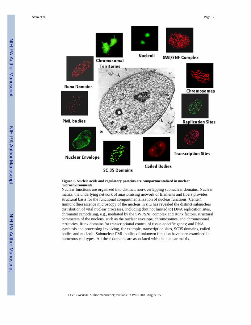
Figure 1. Nucleic acids and regulatory proteins are compartmentalized in nuclearmicroenvironmentsNuclear functions are organized into distinct, non-overlapping subnuclear domains. Nuclearmatrix, the underlying network of anastomising network of filaments and fibers providesstructural basis for the functional compartmentalization of nuclear functions (Center).Immunofluorescence microscopy of the nucleus in situ has revealed the distinct subnucleardistribution of vital nuclear processes, including (but not limited to) DNA replication sites,chromatin remodeling, e.g., mediated by the SWI/SNF complex and Runx factors, structuralparameters of the nucleus, such as the nuclear envelope, chromosomes, and chromosomalterritories, Runx domains for transcriptional control of tissue-specific genes; and RNAsynthesis and processing involving, for example, transcription sites, SC35 domains, coiledbodies and nucleoli. Subnuclear PML bodies of unknown function have been examined innumerous cell types. All these domains are associated with the nuclear matrix.
Stein et al. Page 12
J Cell Biochem. Author manuscript; available in PMC 2009 August 15.
NIH
-PA Author Manuscript
NIH
-PA Author Manuscript
NIH
-PA Author Manuscript

Figure 2. Positive and negative control of Runx responsive genes: Rat osteocalcin as a paradigmfor tissue specific transcriptional controlRunx transcription factors are multifunctional proteins that can synergize with differentsequences-specific DNA-binding proteins (e.g., AP1, C/EBP) and associate with positive(e.g., Smads, p300) or negative (e.g., groucho/TLE, HDACs) gene regulatory cofactorsdepending on the promoter context of bone-tissue specific genes. Possible interactionsaccommodating the positive (Top) and negative (Bottom) regulation of transcription areshown. Depending on the cell type, coregulatory proteins may modulate transcriptionallevels of the gene rather than completely repress the gene as illustrated.
Stein et al. Page 13
J Cell Biochem. Author manuscript; available in PMC 2009 August 15.
NIH
-PA Author Manuscript
NIH
-PA Author Manuscript
NIH
-PA Author Manuscript
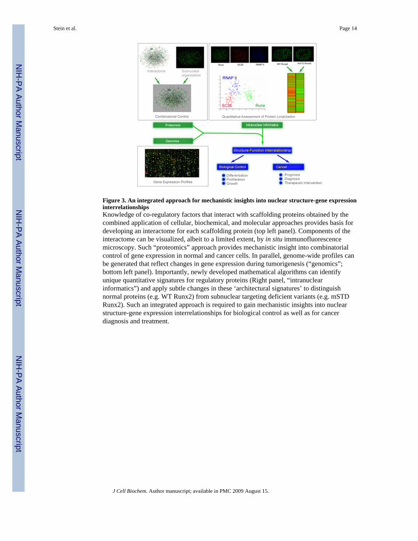
Figure 3. An integrated approach for mechanistic insights into nuclear structure-gene expressioninterrelationshipsKnowledge of co-regulatory factors that interact with scaffolding proteins obtained by thecombined application of cellular, biochemical, and molecular approaches provides basis fordeveloping an interactome for each scaffolding protein (top left panel). Components of theinteractome can be visualized, albeit to a limited extent, by in situ immunofluorescencemicroscopy. Such “proteomics” approach provides mechanistic insight into combinatorialcontrol of gene expression in normal and cancer cells. In parallel, genome-wide profiles canbe generated that reflect changes in gene expression during tumorigenesis (“genomics”;bottom left panel). Importantly, newly developed mathematical algorithms can identifyunique quantitative signatures for regulatory proteins (Right panel, “intranuclearinformatics”) and apply subtle changes in these ‘architectural signatures’ to distinguishnormal proteins (e.g. WT Runx2) from subnuclear targeting deficient variants (e.g. mSTDRunx2). Such an integrated approach is required to gain mechanistic insights into nuclearstructure-gene expression interrelationships for biological control as well as for cancerdiagnosis and treatment.
Stein et al. Page 14
J Cell Biochem. Author manuscript; available in PMC 2009 August 15.
NIH
-PA Author Manuscript
NIH
-PA Author Manuscript
NIH
-PA Author Manuscript

Figure 4. Phenotypic transcription factors associate with mitotic chromosomes to epigeneticallyconvey necessary regulatory information to progeny cells for lineage maintenance andcommitmentPhenotypic transcription factors that include Runx transcription factors are associated withmitotic chromosomes during cell division. This sequence-specific association of regulatoryproteins with their target genes that regulate cell growth, proliferation, and differentiationbookmarks these genes for expression in the G1 phase of the cell cycle. Shown here areactively dividing human osteosarcoma SAOS-2 cells, immunostained with Tubulin (red) toidentify mitotic spindle and Runx2 (green), which is a master regulator of bone formation.Cells are counterstained with DAPI (blue) to visualize mitotic chromosomes. Top panelsshow all phases of mitosis, while the bottom panel shows an enlarged human acrocentricchromosome exhibiting two large Runx foci (indicated by arrowhead).
Stein et al. Page 15
J Cell Biochem. Author manuscript; available in PMC 2009 August 15.
NIH
-PA Author Manuscript
NIH
-PA Author Manuscript
NIH
-PA Author Manuscript