Biological Psychology - Vision
8
BİOLOGİCAL PSYCHOLOGY CH.6 VISION 1. Sensory receptor A specialized neuron that detects a particular category of physical events. 2. Sensory transduction The process by which sensory stimuli are transduced into slow, graded receptor potential. 3. Receptor potential A slow, graded electrical potential produced by a receptor cell in response to a physical stimulus. Extra Notes: B We need to change physical energy such as light into electrical energy, action potential. B Sensory receptors in eyes convert light into electrical energy. This process is called sensory transduction. ?We receive information about the environment from sensory receptors. Stimuli impinge on the receptors and alter their membrane potential. This process is known as sensory transduction because sensory events are transduced (“transferred”) into changes in the cells’ membrane potential. These electrical changes are called receptor potentials. These potentials affect the release of neurotransmitters and hence modify the pattern of firing in neurons with which sensory receptors from synapses. Ultimately the information reaches the brain. ?Light is a narrow band of the spectrum of electromagnetic radiation. ?Electromagnetic radiation with a wavelength of between 380 and 760 nanomete (one billion of a meter) is visible to us ?The range of wavelengts, we call light. ?The perceived color of light is determined by three dimensions: Hue, Brightness, Saturation. 4. Hue (renk tonu) One of the perceptual dimensions of color; the dominant wavelength. 5. Brightness (parlaklık) One of the perceptual dimensions of color; intensity. 6. Saturation (doygunluk, canlılık) One of the perceptual dimensions of color; purity. ?Gözler kafa tasının ön kısmında bulunan kemik boşluğu (orbita) içerisinde asılı bulunur. Göz akı (sclera) adı verilen en dış tabakası sert yapıdadır ve beyaz renklidir. Gözler altı adet ekstraoküler kas aracılığı ile göz akına tutunur ve hareket ederler. Normalde göz küremizin arka kısmını ve bu kasları göremeyiz, çünkü bu kasların gözle olan bağlantıları conjunctiva ile gizlenmektedir. ?The white outer layer of most of the eye, the sclera, is opaque and does not permit entry of light. However, the cornea, the outer layer at the front of the eye, is transparent and admits light. The amount of light that enters is regulated by the size of the pupil, which is an opening in the iris, the pigmented ring of the muscles situated behind the cornea. (İris, korneanın arkasında bulunan pigmentli kas halkalarından oluşur) The lens, situated immediately behind the iris, consists of a series of transparent, onion? like layers. Its shape can be altered by contraction of the ciliary muscles, a set of muscle fibers attached to the outer edge of the lens. Pupilla: göz bebeği/irisin ortasındaki açıklık 7. Saccadic movement The rapid, jerky movement of the eyes used in scanning a visual scene. (Bir görüntünün taranması sırasında, gözlerin kullandığı hızlı titreme hareketi) 8. Pursuit movement The movement that the eyes make to maintain an image of a moving object on the fovea. (Foveada hareket eden bir objenin görüntüsünü korumak üzere gözlerin yaptığı hareket) 9. Accommodation Changes in the thickness of the lens of the eye, accomplished by the ciliary muscles, that focus images of near or distant objects on the retina. (Uzak ya da yakındaki cisim görüntülerinin ciliary kaslarının kasılarak –böylece lensin şekli değiştirilerekB retina üzerine odaklanması) ?Işık lensi geçtikten sonra, şeffaf ve jelatinsi bir madde olan vitreous humor (glassy liquid=camsı sıvı) ile dolu olan gözün ana kısmına doğru geçiş yapar. Light then falls on the retina, the interior lining of the back of the eye. In the retina are located the receptor cells, the rods and cones, collectively known as photoreceptors. 10. Retina The neural tissue and photoreceptive cells located on the inner surface of the posterior portion of the eye. (Gözün arka iç kısmını döşeyen ışığa duyarlı hücrelerden oluşan sinir tabakası). 11. Rod One of the receptor cells of the retina; sensitive to light of low intensity. Although rods do not detect different colors and provide vision of poor activity, they are more sensitive to light. In a very dimly lighted environment, we use our rod vision; therefore, in dim light we are colorBblind and lack of foveal vision. ?The human retina contains approximately 120 million rods and 6 million codes. Although they are greatly outnumbered by rods, cones provide us with most of the information about our environment. 12. Cone One of the receptor cells of the retina; maximally sensitive to one of three different wavelengths of light and hence encodes color division. Cones are responsible for our daytime vision. They provide us with information about small features in the environment and thus are the source of vision of the highest sharpness, or acuity (keskin görmenin kaynağıdır). Cones are also responsible for color vision – our ability to discriminate light of different wavelengths. 13. Photoreceptor A specialized structure or cell that is sensitive to light. In vertebrate animals, the photoreceptors are the rods and cones of the eye's retina. Photoreceptor is a nerve ending, cell, or group of cells specialized to sense or receive light. It transduces photic energy into electrical potentials. 14. Fovea or central region of the retina, meditates our most accurate vision, contains only cones. Extra Notes: ?Rons and Cones exists in the retina and rods receive light and dark, and cones receive colors. ?Fovea is the specific region of the retina, includes amounts of cones. That allows to see a lot of detail. Cones give a lot of detail vision. 15. Optic disk The location of the exit point from the retina of the fibers of the ganglion cells that form the optic nerve; responsible for the blind spot. (Optik disk bölgesinde fotoreseptör olmadığı için kör nokta oluşur.) Optik disk: Ganglion hücre aksonlarının birleşerek optik siniri oluşturduğu ve gözü terk ettiği bölge. 16. Bipolar cell A bipolar neuron located in the middle layer of the retina, conveying information from the photoreceptors to the ganglion cells. (Fotoreseptörlerden aldığı bilgiyi ganglion hücrelerine iletir). Bipolar cells are neurons whose two arms connect the shallowest and deepest layers of the retina. These neurons form synapses with the ganglion cells
Transcript of Biological Psychology - Vision

BİOLOGİCAL(PSYCHOLOGY(CH.6((((VISION((1. Sensory(receptor(A!specialized!neuron!that!detects!a!particular!
category!of!physical!events.!!2. Sensory(transduction!The!process!by!which!sensory!stimuli!are!
transduced!into!slow,!graded!receptor!potential.!3. Receptor(potential(A!slow,!graded!electrical!potential!produced!by!
a!receptor!cell!in!response!to!a!physical!stimulus.!!!Extra!Notes:!!B!We!need!to!change!physical!energy!such!as!light!into!electrical!energy,!action!potential.!!B!Sensory!receptors!in!eyes!convert!light!into!electrical!energy.!This!process!is!called!sensory(transduction.!!!?We'receive'information'about'the'environment'from'sensory!receptors.'Stimuli'impinge'on'the'receptors'and'alter'their'membrane'potential.'This'process'is'known'as'sensory!transduction'because'sensory'events'are'transduced'(“transferred”)'into'changes'in'the'cells’'membrane'potential.'These'electrical'changes'are'called'receptor!potentials.'These'potentials'affect'the'release'of'neurotransmitters'and'hence'modify'the'pattern'of'firing'in'neurons'with'which'sensory'receptors'from'synapses.'Ultimately'the'information'reaches'the'brain.'''?Light'is'a'narrow'band'of'the'spectrum'of'electromagnetic'radiation.''?Electromagnetic'radiation'with'a'wavelength'of'between'380'and'760'nanomete'(one'billion'of'a'meter)'is'visible'to'us'?The'range'of'wavelengts,'we'call'light.''?The'perceived'color'of'light'is'determined'by'three'dimensions:'Hue,'Brightness,'Saturation.''4. Hue((renk!tonu)!One!of!the!perceptual!dimensions!of!color;!the!
dominant!wavelength.!!5. Brightness((parlaklık)!One!of!the!perceptual!dimensions!of!color;!
intensity.!!6. Saturation((doygunluk,!canlılık)!One!of!the!perceptual!dimensions!
of!color;!purity.!!!?Gözler'kafa'tasının'ön'kısmında'bulunan'kemik'boşluğu'(orbita)'içerisinde'asılı'bulunur.'Göz'akı'(sclera)'adı'verilen'en'dış'tabakası'sert'yapıdadır've'beyaz'renklidir.'Gözler'altı'adet'ekstraoküler'kas'aracılığı'ile'göz'akına'tutunur've'hareket'ederler.'Normalde'göz'küremizin'arka'kısmını've'bu'kasları'göremeyiz,'çünkü'bu'kasların'gözle'olan'bağlantıları'conjunctiva'ile'gizlenmektedir.'''?The'white'outer'layer'of'most'of'the'eye,'the'sclera,'is'opaque'and'does'not'permit'entry'of'light.'However,'the'cornea,'the'outer'layer'at'the'front'of'the'eye,'is'transparent'and'admits'light.'The'amount'of'light'that'enters'is'regulated'by'the'size'of'the'pupil,'which'is'an'opening'in'the'iris,'the'pigmented'ring'of'the'muscles'situated'behind'the'cornea.'(İris,'korneanın'arkasında'bulunan'pigmentli'kas'halkalarından'oluşur)'The'lens,'situated'immediately'behind'the'iris,'consists'of'a'series'of'transparent,'onion?like'layers.'Its'shape'can'be'altered'by'contraction'of'the'ciliary'muscles,'a'set'of'muscle'fibers'attached'to'the'outer'edge'of'the'lens.''Pupilla:'göz'bebeği/irisin'ortasındaki'açıklık''''
7. Saccadic(movement!The!rapid,!jerky!movement!of!the!eyes!used!in!scanning!a!visual!scene.!(Bir!görüntünün!taranması!sırasında,!gözlerin!kullandığı!hızlı!titreme!hareketi)!
8. Pursuit(movement(The!movement!that!the!eyes!make!to!maintain!an!image!of!a!moving!object!on!the!fovea.!(Foveada!hareket!eden!bir!objenin!görüntüsünü!korumak!üzere!gözlerin!yaptığı!hareket)!
9. Accommodation(Changes!in!the!thickness!of!the!lens!of!the!eye,!accomplished!by!the!ciliary!muscles,!that!focus!images!of!near!or!distant!objects!on!the!retina.!(Uzak!ya!da!yakındaki!cisim!görüntülerinin!ciliary!kaslarının!kasılarak!–böylece!lensin!şekli!değiştirilerekB!retina!üzerine!odaklanması)!
?Işık'lensi'geçtikten'sonra,'şeffaf've'jelatinsi'bir'madde'olan'vitreous'humor'(glassy'liquid=camsı'sıvı)'ile'dolu'olan'gözün'ana'kısmına'doğru'geçiş'yapar.'Light'then'falls'on'the'retina,'the'interior'lining'of'the'back'of'the'eye.'In'the'retina'are'located'the'receptor'cells,'the'rods'and'cones,'collectively'known'as'photoreceptors.''10. Retina!The!neural!tissue!and!photoreceptive!cells!located!on!the!
inner!surface!of!the!posterior!portion!of!the!eye.!(Gözün!arka!iç!kısmını!döşeyen!ışığa!duyarlı!hücrelerden!oluşan!sinir!tabakası).!
11. Rod(One!of!the!receptor!cells!of!the!retina;!sensitive!to!light!of!low!intensity.!!Although!rods!do!not!detect!different!colors!and!provide!vision!of!poor!activity,!they!are!more!sensitive!to!light.!In!a!very!dimly!lighted!environment,!we!use!our!rod!vision;!therefore,!in!dim!light!we!are!colorBblind!and!lack!of!foveal!vision.!!?The'human'retina'contains'approximately'120'million'rods'and'6'million'codes.'Although'they'are'greatly'outnumbered'by'rods,'cones'provide'us'with'most'of'the'information'about'our'environment.!!
12. Cone!One!of!the!receptor!cells!of!the!retina;!maximally!sensitive!to!one!of!three!different!wavelengths!of!light!and!hence!encodes!color!division.!!Cones!are!responsible!for!our!daytime!vision.!They!provide!us!with!information!about!small!features!in!the!environment!and!thus!are!the!source!of!vision!of!the!highest!sharpness,!or!acuity'(keskin!görmenin!kaynağıdır).!Cones!are!also!responsible!for!color!vision!–our!ability!to!discriminate!light!of!different!wavelengths.!!
!!13. Photoreceptor!A!specialized!structure!or!cell!that!is!sensitive!to!
light.!In!vertebrate!animals,!the!photoreceptors!are!the!rods!and!cones!of!the!eye's!retina.!Photoreceptor!is!a!nerve!ending,!cell,!or!group!of!cells!specialized!to!sense!or!receive!light.!It!transduces!photic!energy!into!electrical!potentials. !
14. Fovea!or!central!region!of!the!retina,!meditates!our!most!accurate!vision,!contains!only!cones.!!
Extra'Notes:'?Rons'and'Cones'exists'in'the'retina'and'rods'receive'light'and'dark,'and'cones'receive'colors.'?Fovea'is'the'specific'region'of'the'retina,'includes'amounts'of'cones.'That'allows'to'see'a'lot'of'detail.'Cones'give'a'lot'of'detail'vision.'15. Optic(disk!The!location!of!the!exit!point!from!the!retina!of!the!
fibers!of!the!ganglion!cells!that!form!the!optic!nerve;!responsible!for!the!blind'spot.!(Optik!disk!bölgesinde!fotoreseptör!olmadığı!için!kör!nokta!oluşur.)!!Optik!disk:!Ganglion!hücre!aksonlarının!birleşerek!optik!siniri!oluşturduğu!ve!gözü!terk!ettiği!bölge.!!
16. Bipolar(cell!A!bipolar!neuron!located!in!the!middle!layer!of!the!retina,!conveying!information!from!the!photoreceptors!to!the!ganglion!cells.!(Fotoreseptörlerden!aldığı!bilgiyi!ganglion!hücrelerine!iletir).!Bipolar!cells!are!neurons!whose!two!arms!connect!the!shallowest!and!deepest!layers!of!the!retina.!These!neurons!form!synapses!with!the!ganglion!cells!
17. Ganglion(cell!A!neuron!located!in!the!retina!that!receives!visual!information!from!bipolar!cells;!its!axons!give!rise!to!the!optic!nerve.!(Görsel!mesajı!bipolar!hücreden!alarak!beyne!ileten!sinir!hücresi;!aksonları!optic!siniri!oluşturur.)!
18. Horizontal(cell!A!neuron!in!the!retina!that!interconnects!adjacent!photoreceptors!and!the!outer!processes!of!the!bipolar!cells.!
19. Amacrine(cell(A!neuron!in!the!retina!that!interconnects!adjacent!ganglion!cells!and!the!inner!processes!of!the!bipolar!cells.!!
!20. Lamella(A!layer!of!membrane!containing!photopigments;!found!in!
rods!and!cones!of!the!retina.!!?The'nature'of'transduction'of'visual'information:'The'first'step'in'the'chain'of'events'that'leads'to'visual'perception'involves'a'special'chemical'called'a'photopigment.'Photopigments'are'special'molecules'embedded'in'the'membrane'of'the'lamellae.'A'single'of'human'rod'contains'approximately'tenmillionofthem'21. Photopigment(A!protein!dye!bonded!to!retinal,!a!substance!derived!
from!vitamin!A;!responsible!for!transduction!of!visual!information.!The!molecules!consist!of!two!parts:!an!opsin!(a!protein)!and!retinal!(lipid).!!
22. Opsin!A!class!of!protein!that,!together!with!retinal,!constitutes!the!photopigments.!(Bir!çeşit!proteindir,!retinal!ile!fotopigmentleri!oluşturur)!
23. Retinal(A!chemical!synthesized!from!vitamin!A;!joins!with!an!opsin!to!form!a!photopigment.!(Vitamin!A!ürünüdür,!opsinle!beraber!fotopigmentleri!oluşturur)!Retinal'is'synthesized'from'vitamin'A,'which'explains'why'carrots,'which'are'rich'in'this'vitamin,'are'said'to'be'good'for'your'eyesight.!!
24. Rhodopsin!There!are!several!forms!of!opsin;!for!example,!the!photopigment!of!human!rods,!rhodopsin!is!a!particular!opsin!found!in!rods!and!consists!of!rod'opsin!plus!retinal.!!
?Rodopsinin'bir'molekülü'ışığa'maruz'kaldığında,'iki'bileşenine'ayrılır:'rod'opsin've'retinal.'Bu'olay'meydana'geldiğinde'opsinin'gün'rengi'uçuk'sarıya'dönüşür;'bu'yüzden'ışığın'fotosigmenti'beyazlaşır.'The'splitting'of'the'photopigment'produces'the'receptor'potential:'a'change'in'the'membrane'potential'of'the'photoreceptor.'The'receptor'potential'affects'the'release'of'neurotransmitter'by'the'photoreceptor,'which'alters'the'firing'rate'of'the'bipolar'cells'with'which'the'photoreceptors'communicates(bu'da,'fotoreseptör'iletişiminde'bipolar'hücrelerin'tetiklenme'oranını'düzenler).'This'information'is'passed'on'to'the'ganglion'cells.'''25. Dorsal(lateral(geniculate(nucleus((LGN)!Retinal!ganglion!
hücrelerinin!aksonları,!bilgiyi!beynin!kalan!kısmına!getirirler.!Bunlar!optik!sinir!boyunca!ilerler!ve!talamusun!dorsal!lateral!geniculate!nucleusa!(LGN)!ulaşırlar.!!Dorsal!lateral!geniculate!nucleus!is!a!group!of!cell!bodies!within!the!lateral!geniculate!body!of!the!thalamus!and!receives!inputs!from!the!retina!and!projects!to!the!primary!visual!cortex.!
!
26. Magnocellular(layer!One!of!the!inner!two!layers!of!neurons!in!the!dorsal!lateral!geniculate!nucleus!and!transmits!information!necessary!for!the!perception!of!form,!movement,!depth,!and!small!differences!in!brightness!to!the!primary!cortex.!(primer!görme!korteksine!şekil,!hareket,!yoğunluk!ve!parlaklıkla!ilgili!küçük!değişiklilklerin!algılanması!için!gerekli!bilgiyi!aktarır)!
27. Parvocellular(layer!One!of!the!four!outer!layers!of!neurons!in!the!dorsal!lateral!geniculate!nucleus!and!transmits!information!necessary!for!the!perception!of!color!and!fine!details!to!the!primary!visual!cortex.!(primer!görme!korteksine!rengin!ve!en!ince!detayların!algılanması!için!gerekli!bilgiyi!aktarır)!
28. Konicellular(layer(One!of!the!sublayers!of!neurons!in!the!dorsal!lateral!geniculate!nucleus!found!ventral!to!each!of!the!magnocellular!and!parvocellular!layers;!transmits!information!from!shortBwavelength!(“blue”)!cones!to!the!primary!visual!cortex.!(primer!görme!korteksine!kısa!dalgaboyundaki!(mavi)!konlardan!bilgiyi!aktarır)!!Lateral!geniculate!nucleus!contains!six!layers!of!neurons,!each!of!which!receives!input!from!the!only!one!eye.!Layers!1,!4,!6!of!the!lateral!geniculate!nucleus!receive!input!from!the!contralateral!eye,!and!2,!3,!5!receive!input!from!the!ipsilateral!layer.!!Layers!1!and!2!are!the!Magnocellular!layers;!layers!3,!4,!5,!6!are!the!parvocellular!layers.!The!koniocellular!sublayers!are!found!ventral!to!each!of!the!parvocellular!and!Magnocellular!layers.!!!
?The'neurons'in'the'dorsal'lateral'geniculate'nucleus'send'their'axons'through'a'pathway'known'as'the'optic'radiations'to'the'primary'visual'cortex'–the'region'surrounding'the!calcarine!fissure.'29. Calcarine(fissure(A!horizontal!fissure!on!the!inner!surface!of!the!
posterior!cerebral!cortex;!the!location!of!the!primary!visual!cortex.!30. Striate(cortex!The!primary!visual!cortex!is!often!called!the!striate!
cortex!because!it!contains!a!darkstaining!layer!(striation)!of!cells.!!!
Anatomy(of(the(Visual(System(31. Optic(chiasm(A!crossBshaped!connection!between!the!optic!nerves!
(optic!sinirler!arasındaki!çapraz!yapılı!bir!baağlantıdır),!located!below!the!base!of!the!brain!(beyin!tabanının!altında),!just!anterior!to!the!pituitary!gland!(hipofizin!önündedir).!!
!!!!!!!

Coding(of(Visual(Information(32. Receptive(field!That!portion!of!the!visual!field!in!which!the!
presentation!of!visual!stimuli!will!produce!an!alternation!in!the!firing!rate!of!a!particular!neuron.!Receptive(field!as!an!"area!in!which!stimulation!leads!to!response!of!a!particular!sensory!neuron”.!The!receptive(field(of!a!neuron!in!the!visual!system!is!the!part!of!the!visual!field!that!neuron!"see"—that!is,!the!part!in!which!light!must!fall!for!the!neuron!to!be!stimulated.!
!
!
!?'Central'Versus'Peripheral'Acuity:'Ganglion'cells'in'the'fovea'receive'input'from'a'smaller'number'of'photoreceptors'than'in'the'periphery'and'hence'provide'more'acute'visual'information.'
Extra'Notes:'?Over'sixty'years'ago,'Hartline'(1938)'discovered'that'the'frog'retina'contained'three'types'of'ganglion'cells.'ON'cells'responded'with'an'excitatory'burst'when'the'retina'was'illuminated,'OFF'cells'responded'when'the'light'was'turned'off,'and'ON/OFF'cells'respond'briefly'when'the'light'went'on'and'again'when'it'went'off.'?'There'are'two'types'of'retinal'ganglion'cells:'"on?center"'and'"off?center".'An'on?center'cell'is'stimulated'when'the'center'of'its'receptive'field'is'exposed'to'light,'and'is'inhibited'when'the'surround'is'exposed'to'light.'Off?center'cells'have'just'the'opposite'reaction'?The'two'major'categories'of'ganglion'cells'(ON'and'OFF)'and'the'organization'of'their'receptive'fields'into'contrasting'center'and'surround'provide'useful'information'to'the'rest'of'the'visual'system.'
'(
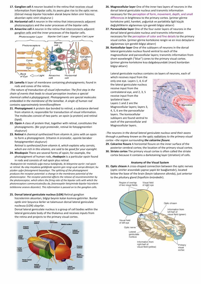
Coding(of(Color(Objects!in!our!environment!selectively!absorb!some!wavelengths!of!light!and!reflect!others,!which,!to!our!eyes,!gives!them!different!colors.!
The!retinas!of!humans,!apes,!Old!World!monkeys,!and!one!species!of!New!World!monkey!contain!three!different!types!of!cones,!which!provides!them!(and!us)!with!the!most!elaborate!form!of!color!vision.!!!TRICHROMACY!!!!!!!!!!!!!!!!!!!!!!!!!!!!!!!!!!!!!!!!!!!!!!!!TETRACHROMACY!
!BTetrachromacy!birds!can!see!red,!green,!blue!and!an!ultraviolet!light.!!!?In'1802'Thomas'Young,'a'British'physicist'and'physician,'proposed'that'the'eye'detected'different'colors'because'it'contained'three'types'of'receptors,'each'sensitive'to'a'single'hue.'His'theory'was'referred'to'as'the'trichromatic'(three?color)'theory.'?Physiological'investigations'of'retinal'photoreceptors'in'higher'primates'have'found'that'Young'was'right:'Three'different'types'of'photoreceptors'(three'different'types'of'cones)'are'responsible'for'color'vision.'?Investigators'have'studied'the'absorption'characteristics'of'individual'photoreceptors,'determining'the'amount'of'light'of'different'wavelengths'that'is'absorbed'by'the'photopigments.'?These'characteristics'are'controlled'by'the'particular'opsin'a'photoreceptor'contains;'different'opsins'absorb'particular'wavelengths'more'readily.'!BTrichromatic!theory:!Trichromatic!vision:!Every!color!we!see!in!the!world!they!are!the!combination!of!these!3!colors.!.!3!types!of!cones!(receptors)!.!Cones!get!activated!depending!on!the!wavelength!.!"Color!mixing"!.!Blue/Green/Red!Red!cones!only!fire!if!there!is!red!light.!!Green!cones!only!fire!if!there!is!green!light.!Blue!cones!only!fire!if!there!is!blue!light.!Opponents!process!theory:!!.!Two!type!of!ganglion!cells:!YellowBblue!and!greenBred!.!As!one!cell!increases!its!firing!rate!to!yellow!light!in!the!center,!it!will!decrease!its!firing!rate!to!blue!light'''?'For'convenience,'the'short?,'medium?,'and'long?wavelength'cones'are'traditionally'called'"blue",'"green",'and'"red"'cones,'respectively.'?The'peak'sensitivities'of'the'three'types'of'cones'are'approximately'420'nm'(blue?violet),'530'nm'(green),'abd'560'nm'(yellow?green).''!?Genetic'defects'in'color'vision'appear'to'result'from'anomalies'in'one'or'more'of'the'three'types'of'cones.'33. Protanopia(An!inherited!form!of!defective!color!vision!in!which!red!
and!green!hues!are!confused;!“red”!cones!are!filled!“green”!cone!opsin.!People!with!protanopia!confuse(red!and!green.!They!see!the!world!in!shades!of!yellow!and!blue;!both.!
34. Deuteranopia(An!inherited!form!of!defective!color!vision!in!which!red!and!green!hues!are!confused;!“green”!cones!are!filled!“red”!cone!opsin.!People!with!deuteranopia(("secondBcolor!defect")!also!confuse!red!and!green!and!also!have!normal!visual!acuity.!Their!"green"!cones!appear!to!be!filled!with!"red"!con!opsin.!
35. Tritanopia(An!inherited!form!of!defective!color!vision!in!which!hues!with!short!wavelengths!are!confused.!“Blue”!cones!are!either!lacking!or!faulty.!("thirdBcolor!defect")!This!disorder!involves!a!faulty!gene!that!is!not!located!on!an!X!chromosome;!thus,!it!is!equally!prevalent!in!males!and!females.!People!with!tritanopia!have!difficulty!with!hues!of!short!wavelengths!and!see!the!world!in!greens!and!reds.!
?'At'the'level'of'the'retinal'ganglion'cell,'the'three?color'code'gets'translated'into'an'opponent?color'system.'Thus,'the'retina'contains'two'kinds'of'color?sensitive'ganglion'cells:'red?green'cells'and'yellow?blue'cells.'Some'color?sensitive'ganglion'cells'respond'in'a'center?surround'fashion.''
'BReceptive!Fields!of!ColorBSensitive!Ganglion!Cells:!When!a!portion!of!the!receptive!field!is!illuminated!with!the!color!shown,!the!cell’s!rate!of!firing!increases.!When!a!portion!is!illuminated!with!the!complementary!color,!the!cell’s!rate!of!firing!decreases.!
Color'='quality,'Wavelength'='quantity''quality'is'not'equal'to'quantity''Color'is'not'in'nature.'We'interpret'it'in'our'mind''?For'example,'red'light'excites'"red"'cones,'which'causes'the'excitation'of'red?green'ganglion'cells.''Green'light'excites'"green"'cones,'which'causes'the'inhibition'of'red?green'cells.'But'consider'the'effect'of'mediate'between'red'and'green,'it'will'stimulate'both'"red"'and'"green"'cones'about'equally.'Yellow?blue'ganglion'cells'are'excited'by'both'"red"'and'"green"'cones,'so'their'rate'of'firing'increases.'However,'red?green'ganglion'cells'are'excited'by'red'and'inhibited'by'green,'so'their'firing'rate'does'not'change.''
''
''
''''''''
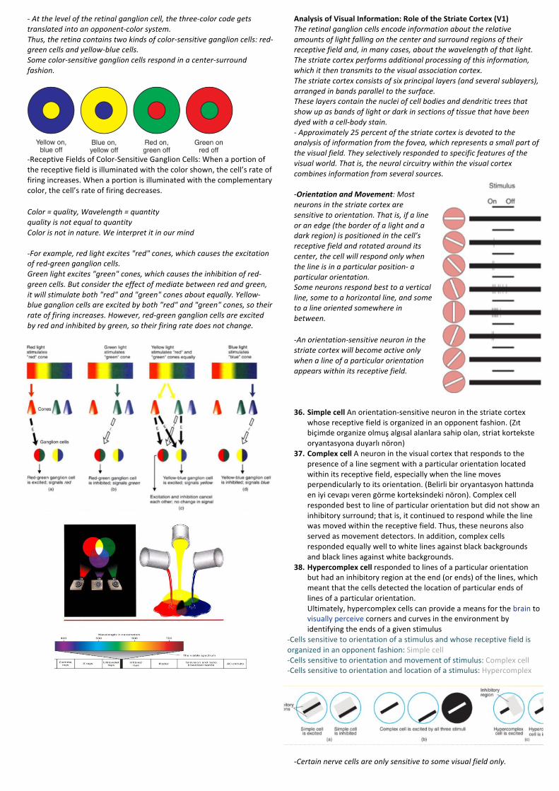
Analysis(of(Visual(Information:(Role(of(the(Striate(Cortex((V1)(The'retinal'ganglion'cells'encode'information'about'the'relative'amounts'of'light'falling'on'the'center'and'surround'regions'of'their'receptive'field'and,'in'many'cases,'about'the'wavelength'of'that'light.'The'striate'cortex'performs'additional'processing'of'this'information,'which'it'then'transmits'to'the'visual'association'cortex.'The'striate'cortex'consists'of'six'principal'layers'(and'several'sublayers),'arranged'in'bands'parallel'to'the'surface.'These'layers'contain'the'nuclei'of'cell'bodies'and'dendritic'trees'that'show'up'as'bands'of'light'or'dark'in'sections'of'tissue'that'have'been'dyed'with'a'cell?body'stain.'?'Approximately'25'percent'of'the'striate'cortex'is'devoted'to'the'analysis'of'information'from'the'fovea,'which'represents'a'small'part'of'the'visual'field.'They'selectively'responded'to'specific'features'of'the'visual'world.'That'is,'the'neural'circuitry'within'the'visual'cortex'combines'information'from'several'sources.''?Orientation!and!Movement:'Most'neurons'in'the'striate'cortex'are'sensitive'to'orientation.'That'is,'if'a'line'or'an'edge'(the'border'of'a'light'and'a'dark'region)'is'positioned'in'the'cell’s'receptive'field'and'rotated'around'its'center,'the'cell'will'respond'only'when'the'line'is'in'a'particular'position?'a'particular'orientation.''Some'neurons'respond'best'to'a'vertical'line,'some'to'a'horizontal'line,'and'some'to'a'line'oriented'somewhere'in'between.'
?An'orientation?sensitive'neuron'in'the'striate'cortex'will'become'active'only'when'a'line'of'a'particular'orientation'appears'within'its'receptive'field.''!!!36. Simple(cell!An!orientationBsensitive!neuron!in!the!striate!cortex!
whose!receptive!field!is!organized!in!an!opponent!fashion.!(Zıt!biçimde!organize!olmuş!algısal!alanlara!sahip!olan,!striat!kortekste!oryantasyona!duyarlı!nöron)!
37. Complex(cell!A!neuron!in!the!visual!cortex!that!responds!to!the!presence!of!a!line!segment!with!a!particular!orientation!located!within!its!receptive!field,!especially!when!the!line!moves!perpendicularly!to!its!orientation.!(Belirli!bir!oryantasyon!hattında!en!iyi!cevapı!veren!görme!korteksindeki!nöron).!Complex!cell!responded!best!to!line!of!particular!orientation!but!did!not!show!an!inhibitory!surround;!that!is,!it!continued!to!respond!while!the!line!was!moved!within!the!receptive!field.!Thus,!these!neurons!also!served!as!movement!detectors.!In!addition,!complex!cells!responded!equally!well!to!white!lines!against!black!backgrounds!and!black!lines!against!white!backgrounds.!!
38. Hypercomplex(cell!responded!to!lines!of!a!particular!orientation!but!had!an!inhibitory!region!at!the!end!(or!ends)!of!the!lines,!which!meant!that!the!cells!detected!the!location!of!particular!ends!of!lines!of!a!particular!orientation.!!Ultimately,!hypercomplex!cells!can!provide!a!means!for!the!brain!to!visually!perceive!corners!and!curves!in!the!environment!by!identifying!the!ends!of!a!given!stimulus!
BCells!sensitive!to!orientation!of!a!stimulus!and!whose!receptive!field!is!organized!in!an!opponent!fashion:!Simple!cell!BCells!sensitive!to!orientation!and!movement!of!stimulus:!Complex!cell!BCells!sensitive!to!orientation!and!location!of!a!stimulus:!Hypercomplex!!
''?Certain'nerve'cells'are'only'sensitive'to'some'visual'field'only.'
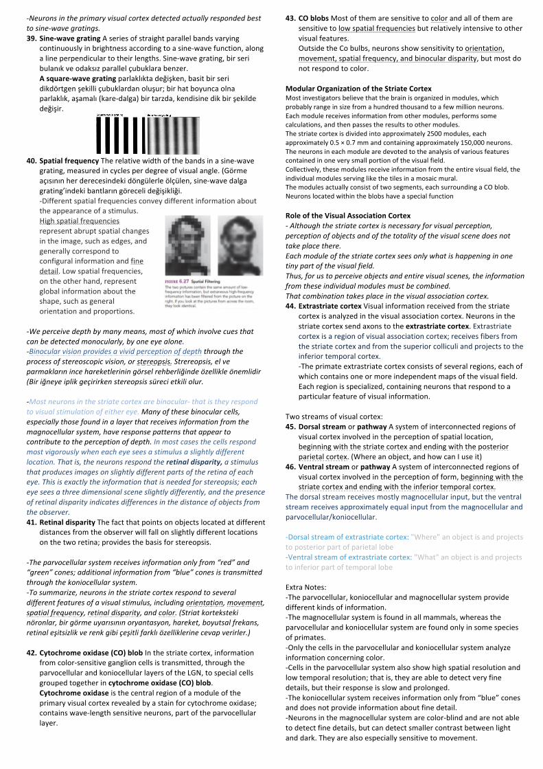
?Neurons'in'the'primary'visual'cortex'detected'actually'responded'best'to'sine?wave'gratings.'39. Sine]wave(grating(A!series!of!straight!parallel!bands!varying!
continuously!in!brightness!according!to!a!sineBwave!function,!along!a!line!perpendicular!to!their!lengths.!SineBwave!grating,!bir!seri!bulanık!ve!odaksız!parallel!çubuklara!benzer.!A(square]wave(grating!parlaklıkta!değişken,!basit!bir!seri!dikdörtgen!şekilli!çubuklardan!oluşur;!bir!hat!boyunca!olna!parlaklık,!aşamalı!(kareBdalga)!bir!tarzda,!kendisine!dik!bir!şekilde!değişir.!!
!40. Spatial(frequency(The!relative!width!of!the!bands!in!a!sineBwave!
grating,!measured!in!cycles!per!degree!of!visual!angle.!(Görme!açısının!her!derecesindeki!döngülerle!ölçülen,!sineBwave!dalga!grating’indeki!bantların!göreceli!değişikliği.!BDifferent!spatial!frequencies!convey!different!information!about!the!appearance!of!a!stimulus.!High!spatial!frequencies!represent!abrupt!spatial!changes!in!the!image,!such!as!edges,!and!generally!correspond!to!configural!information!and!fine!detail.!Low!spatial!frequencies,!on!the!other!hand,!represent!global!information!about!the!shape,!such!as!general!orientation!and!proportions.!
!?We'perceive'depth'by'many'means,'most'of'which'involve'cues'that'can'be'detected'monocularly,'by'one'eye'alone.''?Binocular'vision'provides'a'vivid'perception'of'depth'through'the'process'of'stereoscopic'vision,'or'stereopsis.'Strereopsis,'el've'parmakların'ince'hareketlerinin'görsel'rehberliğinde'özellikle'önemlidir'(Bir'iğneye'iplik'geçirirken'stereopsis'süreci'etkili'olur.'''?Most'neurons'in'the'striate'cortex'are'binocular?'that'is'they'respond'to'visual'stimulation'of'either'eye.'Many'of'these'binocular'cells,'especially'those'found'in'a'layer'that'receives'information'from'the'magnocellular'system,'have'response'patterns'that'appear'to'contribute'to'the'perception'of'depth.'In'most'cases'the'cells'respond'most'vigorously'when'each'eye'sees'a'stimulus'a'slightly'different'location.'That'is,'the'neurons'respond'the'retinal!disparity,!a'stimulus'that'produces'images'on'slightly'different'parts'of'the'retina'of'each'eye.'This'is'exactly'the'information'that'is'needed'for'stereopsis;'each'eye'sees'a'three'dimensional'scene'slightly'differently,'and'the'presence'of'retinal'disparity'indicates'differences'in'the'distance'of'objects'from'the'observer.''41. Retinal(disparity(The!fact!that!points!on!objects!located!at!different!
distances!from!the!observer!will!fall!on!slightly!different!locations!on!the!two!retina;!provides!the!basis!for!stereopsis.!!
?The'parvocellular'system'receives'information'only'from'“red”'and'“green”'cones;'additional'information'from'“blue”'cones'is'transmitted'through'the'koniocellular'system.''?To'summarize,'neurons'in'the'striate'cortex'respond'to'several'different'features'of'a'visual'stimulus,'including'orientation,'movement,'spatial'frequency,'retinal'disparity,'and'color.'(Striat'korteksteki'nöronlar,'bir'görme'uyarısının'oryantasyon,'hareket,'boyutsal'frekans,'retinal'eşitsizlik've'renk'gibi'çeşitli'farklı'özelliklerine'cevap'verirler.)'(42. Cytochrome(oxidase((CO)(blob!In!the!striate!cortex,!information!
from!colorBsensitive!ganglion!cells!is!transmitted,!through!the!parvocellular!and!koniocellular!layers!of!the!LGN,!to!special!cells!grouped!together!in!cytochrome(oxidase((CO)(blob.!!Cytochrome(oxidase!is!the!central!region!of!a!module!of!the!primary!visual!cortex!revealed!by!a!stain!for!cytochrome!oxidase;!contains!waveBlength!sensitive!neurons,!part!of!the!parvocellular!layer.!!
!
43. CO(blobs(Most!of!them!are!sensitive!to!color!and!all!of!them!are!sensitive!to!low!spatial!frequencies!but!relatively!intensive!to!other!visual!features.!!Outside!the!Co!bulbs,!neurons!show!sensitivity!to!orientation,!movement,!spatial!frequency,!and!binocular!disparity,!but!most!do!not!respond!to!color.!!
!Modular(Organization(of(the(Striate(Cortex(Most!investigators!believe!that!the!brain!is!organized!in!modules,!which!probably!range!in!size!from!a!hundred!thousand!to!a!few!million!neurons.!Each!module!receives!information!from!other!modules,!performs!some!calculations,!and!then!passes!the!results!to!other!modules.!The!striate!cortex!is!divided!into!approximately!2500!modules,!each!approximately!0.5!×!0.7!mm!and!containing!approximately!150,000!neurons.!The!neurons!in!each!module!are!devoted!to!the!analysis!of!various!features!contained!in!one!very!small!portion!of!the!visual!field.!Collectively,!these!modules!receive!information!from!the!entire!visual!field,!the!individual!modules!serving!like!the!tiles!in!a!mosaic!mural.!The!modules!actually!consist!of!two!segments,!each!surrounding!a!CO!blob.!Neurons!located!within!the!blobs!have!a!special!function!'Role(of(the(Visual(Association(Cortex((?'Although'the'striate'cortex'is'necessary'for'visual'perception,'perception'of'objects'and'of'the'totality'of'the'visual'scene'does'not'take'place'there.'Each'module'of'the'striate'cortex'sees'only'what'is'happening'in'one'tiny'part'of'the'visual'field.''Thus,'for'us'to'perceive'objects'and'entire'visual'scenes,'the'information'from'these'individual'modules'must'be'combined.'That'combination'takes'place'in'the'visual'association'cortex.'44. Extrastriate(cortex(Visual!information!received!from!the!striate!
cortex!is!analyzed!in!the!visual!association!cortex.!Neurons!in!the!striate!cortex!send!axons!to!the!extrastriate(cortex.!Extrastriate!cortex!is!a!region!of!visual!association!cortex;!receives!fibers!from!the!striate!cortex!and!from!the!superior!colliculi!and!projects!to!the!inferior!temporal!cortex.!BThe!primate!extrastriate!cortex!consists!of!several!regions,!each!of!which!contains!one!or!more!independent!maps!of!the!visual!field.!!Each!region!is!specialized,!containing!neurons!that!respond!to!a!particular!feature!of!visual!information.!!
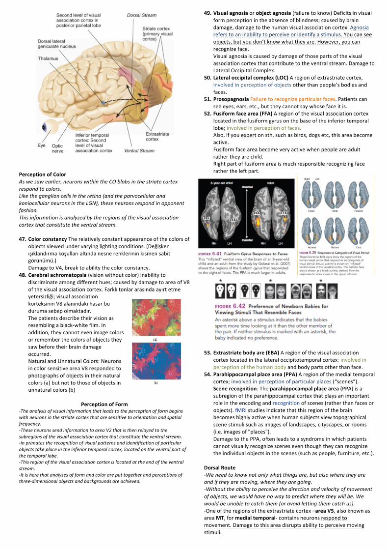
!Two!streams!of!visual!cortex:!!45. Dorsal(stream(or!pathway(A!system!of!interconnected!regions!of!
visual!cortex!involved!in!the!perception!of!spatial!location,!beginning!with!the!striate!cortex!and!ending!with!the!posterior!parietal!cortex.!(Where!an!object,!and!how!can!I!use!it)!
46. Ventral(stream!or!pathway(A!system!of!interconnected!regions!of!visual!cortex!involved!in!the!perception!of!form,!beginning!with!the!striate!cortex!and!ending!with!the!inferior!temporal!cortex.!!
The!dorsal!stream!receives!mostly!magnocellular!input,!but!the!ventral!stream!receives!approximately!equal!input!from!the!magnocellular!and!parvocellular/koniocellular.!!!BDorsal!stream!of!extrastriate!cortex:!"Where"!an!object!is!and!projects!to!posterior!part!of!parietal!lobe!BVentral!stream!of!extrastriate!cortex:!"What"!an!object!is!and!projects!to!inferior!part!of!temporal!lobe!!Extra!Notes:!!BThe!parvocellular,!koniocellular!and!magnocellular!system!provide!different!kinds!of!information.!!BThe!magnocellular!system!is!found!in!all!mammals,!whereas!the!parvocellular!and!koniocellular!system!are!found!only!in!some!species!of!primates.!BOnly!the!cells!in!the!parvocellular!and!koniocellular!system!analyze!information!concerning!color.!!BCells!in!the!parvocellular!system!also!show!high!spatial!resolution!and!low!temporal!resolution;!that!is,!they!are!able!to!detect!very!fine!details,!but!their!response!is!slow!and!prolonged.!!BThe!koniocellular!system!receives!information!only!from!“blue”!cones!and!does!not!provide!information!about!fine!detail.!!BNeurons!in!the!magnocellular!system!are!colorBblind!and!are!not!able!to!detect!fine!details,!but!can!detect!smaller!contrast!between!light!and!dark.!They!are!also!especially!sensitive!to!movement.!!
!
Perception(of(Color((As'we'saw'earlier,'neurons'within'the'CO'blobs'in'the'striate'cortex'respond'to'colors.'Like'the'ganglion'cells'in'the'retina'(and'the'parvocellular'and'koniocellular'neurons'in'the'LGN),'these'neurons'respond'in'opponent'fashion.''This'information'is'analyzed'by'the'regions'of'the'visual'association'cortex'that'constitute'the'ventral'stream.''47. Color(constancy(The!relatively!constant!appearance!of!the!colors!of!
objects!viewed!under!varying!lighting!conditions.!(Değişken!ışıklandırma!koşulları!altında!nesne!renklerinin!kısmen!sabit!görünümü.)!Damage!to!V4,!break!to!ability!the!color!constancy.!!
48. Cerebral(achromatopsia((vision!without!color)(Inability!to!discriminate!among!different!hues;!caused!by!damage!to!area!of!V8!of!the!visual!association!cortex.!Farklı!tonlar!arasında!ayırt!etme!yetersizliği;!visual!association!korteksinin!V8!alanındaki!hasar!bu!duruma!sebep!olmaktadır.!!The!patients!describe!their!vision!as!resembling!a!blackBwhite!film.!In!addition,!they!cannot!even!image!colors!or!remember!the!colors!of!objects!they!saw!before!their!brain!damage!occurred.!!Natural!and!Unnatural!Colors:!Neurons!in!color!sensitive!area!V8!responded!to!photographs!of!objects!in!their!natural!colors!(a)!but!not!to!those!of!objects!in!unnatural!colors!(b)!!
Perception(of(Form(?The'analysis'of'visual'information'that'leads'to'the'perception'of'form'begins'with'neurons'in'the'striate'cortex'that'are'sensitive'to'orientation'and'spatial'frequency.'?These'neurons'send'information'to'area'V2'that'is'then'relayed'to'the'subregions'of'the'visual'association'cortex'that'constitute'the'ventral'stream.'?In'primates'the'recognition'of'visual'patterns'and'identification'of'particular'objects'take'place'in'the'inferior'temporal'cortex,'located'on'the'ventral'part'of'the'temporal'lobe.'?This'region'of'the'visual'association'cortex'is'located'at'the'end'of'the'ventral'stream.'?It'is'here'that'analyses'of'form'and'color'are'put'together'and'perceptions'of'three?dimensional'objects'and'backgrounds'are'achieved.'''''''
49. Visual(agnosia(or!object(agnosia((failure!to!know)!Deficits!in!visual!form!perception!in!the!absence!of!blindness;!caused!by!brain!damage,!damage!to!the!human!visual!association!cortex.!Agnosia!refers!to!an!inability!to!perceive!or!identify!a!stimulus.!You!can!see!objects,!but!you!don’t!know!what!they!are.!However,!you!can!recognize!face.!!Visual!agnosia!is!caused!by!damage!of!those!parts!of!the!visual!association!cortex!that!contribute!to!the!ventral!stream.!Damage!to!Lateral!Occipital!Complex.!!
50. Lateral(occipital(complex((LOC)!A!region!of!extrastriate!cortex,!involved!in!perception!of!objects!other!than!people’s!bodies!and!faces.!!!
51. Prosopagnosia(Failure!to!recognize!particular!faces.!Patients!can!see!eyes,!ears,!etc.,!but!they!cannot!say!whose!face!it!is.!!
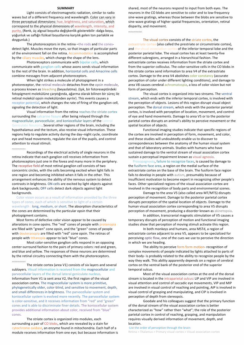
52. Fusiform(face(area((FFA)(A!region!of!the!visual!association!cortex!located!in!the!fusiform!gyrus!on!the!base!of!the!inferior!temporal!lobe;!involved!in!perception!of!faces.!!Also,!if!you!expert!on!sth,!such!as!birds,!dogs!etc,!this!area!become!active.!Fusiform!face!area!become!very!active!when!people!are!adult!rather!they!are!child.!!Right!part!of!fusiform!area!is!much!responsible!recognizing!face!rather!the!left!part.!!
!
!!53. Extrastriate(body(are((EBA)!A!region!of!the!visual!association!
cortex!located!in!the!lateral!occipitotemporal!cortex;!involved!in!perception!of!the!human!body!and!body!parts!other!than!face.!!
54. Parahippocampal(place(area((PPA)(A!region!of!the!medial!temporal!cortex;!involved!in!perception!of!particular!places!(“scenes”).!!Scene(recognition:!The!parahippocampal(place(area!(PPA)!is!a!subregion!of!the!parahippocampal!cortex!that!plays!an!important!role!in!the!encoding!and!recognition!of!scenes!(rather!than!faces!or!objects).!fMRI!studies!indicate!that!this!region!of!the!brain!becomes!highly!active!when!human!subjects!view!topographical!scene!stimuli!such!as!images!of!landscapes,!cityscapes,!or!rooms!(i.e.!images!of!"places").!Damage!to!the!PPA,!often!leads!to!a!syndrome!in!which!patients!cannot!visually!recognize!scenes!even!though!they!can!recognize!the!individual!objects!in!the!scenes!(such!as!people,!furniture,!etc.).!!
!Dorsal(Route((?We'need'to'know'not'only'what'things'are,'but'also'where'they'are'and'if'they'are'moving,'where'they'are'going.''?Without'the'ability'to'perceive'the'direction'and'velocity'of'movement'of'objects,'we'would'have'no'way'to'predict'where'they'will'be.'We'would'be'unable'to'catch'them'(or'avoid'letting'them'catch'us).!BOne!of!the!regions!of!the!extrastriate!cortex!–area(V5,!also!known!as!area!MT,!for!medial(temporalB!contains!neurons!respond!to!movement.!Damage!to!this!area!disrupts!ability!to!perceive!moving!stimuli.!

BA!region!adjacent!area!V5,!is!area(V5a!is!called!MST,!for!medial(superior(temporal,!receives!information!about!movement!from!V5!and!performs!a!further!analysis.!MTS!neurons!respond!to!complex!patterns!of!movement,!including!radial,!circular,!and!spiral!motion.!One!important!function!of!this!region!appears!to!be!analysis!of!optic(flow.!As!we!move!around!in!our!environment!or!as!objects!in!our!environment!move!in!relation!to!us,!the!sizes,!shapes,!and!locations!of!environmental!features!on!our!retinas!change.!!55. Optic(flow!The!complex!motion!of!points!in!the!visual!field!caused!
by!relative!movement!between!the!observer!and!environment;!provides!information!about!the!relative!distance!of!objects!from!the!observes!and!of!the!relative!direction!of!movement.!!Optik!akış!ismi!verilen!görsel!bilgi!akışı,!bir!nesnenin!hareketine!bakarak!mesafeleri!algılamamızı!sağlamaktadır.!
56. Akinetopsia(Inability!to!perceive!movement,!caused!by!damage!to!area!V5!(also!called!MTS)!of!the!visual!association!cortex.!!
57. Intraparietal(sulcus((IP)(The!end!of!the!dorsal!stream!of!the!visual!association!cortex;!involved!perception!of!location,!visual!attention,!and!control!of!eye!and!hand!movements.!Intraparietal!sulcus!have!5!regions:!AIP,!LIP,!VIP,!CIP,!MIP.!(anterior,!lateral,!ventral,!caudal,!medial)!!Anterior!intreparietal!region!says!that!how!to!grasp!objects.!!Ventral!intraparietal!region!is!responsible!to!point!things.!(Birşeyi!işaret!etmek)!Lateral!intraparietal!region!controls!saccadic!eye!movement.!!Caudal!intraparietal!region!is!responsible!for!seeing!depth!of!things.!Medial!intraparietal!region!is!responsible!to!reach!something.!!
!!Extra!Notes:!!BVisual!association!cortex!also!receives!information!from!superior!colliculi!in!brain!stem.!!BDorsal!route!analyze!information!faster,!and!dorsal!route!is!more!important!then!ventral!because!if!you!even!don’t!know!what!something!is,!if!you!know!where!it!is,!you!can!escape!from!it.!!BObjects’!bigness,!thickness!or!etc.,!are!analyzed!by!ventral!route.!Dorsal!route!say!how!can!you!use!it.!!(2.5!years!old!children!experiment!(scale!error)!!!BThe!importance!of!the!visual!system!is!shown!by!the!fact!that!approximately!25%!of!our!cerebral!cortex!is!devoted!to!this!sense!modality!and!by!the!many!discoveries!being!made!by!the!laboratories!that!are!busy!discovering!interesting!things!about!vision.!!BEyes,!Thalamus,!V1!and!Visual!Association!Cortex!all!play!an!important!role!in!analysis!of!visual!information.!!BDorsal!stream!in!Visual!Association!Cortex!specializes!in!analyzing!where!things!are!in!relation!to!us!or!how!to!react!to!them!while!the!ventral!stream!specializes!in!analyzing!what!things!are.!
!
(((((((
(((((((((((((((((((((((((((((((((((((((((((((((((((((((((((((((((((((((((
SUMMARY(! Light!consists!of!electromagnetic!radiation,!similar!to!radio!waves!but!of!a!different!frequency!and!wavelength.!Color!can!vary!in!three!perceptual!dimensions:!hue,!brightness,!and!saturation,!which!correspond!to!the!physical!dimensions!of!wavelength,!intensity,!and!purity.!(Renk,!üç!algısal!boyutta!değişkenlik!gösterebilir:!dalga!boyu,!yoğunluk!ve!saflığn!fiziksel!boyutlarına!karşılık!gelen!ton!parlaklık!ve!doygunluk.)!! The!photoreceptors!in!the!retina!–the!rods!and!the!conesB!detect!light.!Muscles!move!the!eyes,!so!that!images!of!particular!parts!of!the!environment!fall!on!the!retina.!Accommodation!is!accomplished!by!the!ciliary!muscles,!which!change!the!shape!of!the!lens.!!! Photoreceptors!communicate!with!bipolar!cells,!which!communicate!with!ganglion!cells!whose!axons!sends!visual!information!to!the!rest!of!the!brain.!In!addition,!horizontal!cells!and!Amacrine!cells!combine!messages!from!adjacent!photoreceptors.!!! When!light!strikes!a!molecule!of!photopigment!in!a!photoreceptor,!the!retinal!molecule!detaches!from!the!opsin!molecule,!a!process!known!as!bleaching!(beyazlatma).!(Işık,!bir!fotoreseptördeki!fotopigment!molekülüne!çarptığında,!ağarma!olarak!bilinen!bir!süreç!ile!retinal!molekül!opsin!molekülünden!ayrılır).!This!events!causes!a!receptor!potential,!which!changes!the!rate!of!firing!of!the!ganglion!cell,!signaling!the!detection!of!light.!!! Visual!information!from!the!retina!reaches!the!striate!cortex!surrounding!the!calcarine!fissure!after!being!relayed!through!the!magnocellular,!parvocellular,!and!koniocellular!layers!of!the!Lateral!Geniculate!Neurons.!Several!other!regions!of!the!brain,!including!the!hypothalamus!and!the!tectum,!also!receive!visual!information.!These!regions!help!to!regulate!activity!during!the!day–night!cycle,!coordinate!eye!and!head!movements,!regulate!the!size!of!the!pupils,!and!control!attention!to!visual!stimuli.!…………….!! ! Recordings!of!the!electrical!activity!of!single!neurons!in!the!retina!indicate!that!each!ganglion!cell!receives!information!from!photoreceptorsBjust!one!in!the!fovea!and!many!more!in!the!periphery.!The!receptive!field!of!most!retinal!ganglion!cell!consists!of!two!concentric!circles,!with!the!cells!becoming!excited!when!light!falls!in!one!region!and!becoming!inhibited!when!it!falls!in!the!other.!This!arrangement!enhances!the!ability!of!the!nervous!system!to!detect!contrasts!in!brightness.!ON!cells!are!excited!by!light!objects!against!dark!backgrounds;!OFF!cells!detect!dark!objects!against!light!backgrounds.!! Color!vision!occurs!as!a!result!of!information!provided!by!the!three!types!of!cones,!each!of!which!is!sensitive!to!light!of!a!certain!wavelength!:!long,!medium,!or!short.!The!absorption!characteristics!of!the!cones!are!determined!by!the!particular!opsin!that!their!photopigment!contains.!!! Most!forms!of!defective!color!vision!appear!to!be!caused!by!alterations!in!cone!opsins.!The!"red"!cones!of!people!with!protanopia!are!filled!with!"green"!cone!opsin,!and!the!"green"!cones!of!people!with!deuteranopia!are!filled!with!"red"!cone!opsin.!The!retinas!of!people!with!tritanopia!appear!to!lack!"blue"!cones.!
Most!colorBsensitive!ganglion!cells!respond!in!an!opposing!centerBsurround!fashion!to!the!pairs!of!primary!colors:!red!and!green,!and!blue!and!yellow.!The!responses!of!these!neurons!are!determined!by!the!retinal!circuitry!connecting!them!with!the!photoreceptors.!…………….!! ! The!striate!cortex!(area!V1)!consists!of!six!layers!and!several!sublayers.!Visual!information!is!received!from!the!magnocellular!and!parvocellular!layers!of!the!dorsal!lateral!geniculate!nucleus.!Information!from!V1!ia!sent!area!V2,!the!first!region!of!the!visual!association!cortex.!The!magnocellular!system!is!more!primitive,!phylogenetically!older,!colorBblind,!and!sensitive!to!movement,!depth,!and!small!differences!in!brightness.!The!parvocellular!system!and!koniocellular!system!is!evolved!more!recently.!The!parvocellular!system!is!colorBsensitive,!and!it!receives!information!from!“red”!and!“green”!cones!and!is!able!to!discriminate!finer!details.!The!koniocellular!system!provides!additional!information!about!color,!received!from!“blue”!cones.!!! ! The!striate!cortex!is!organized!into!modules,!each!surrounding!a!pair!of!CO!blobs,!which!are!revealed!by!a!stain!for!cytochrome!oxidase,!an!enzyme!found!in!mitochondria.!Each!half!of!a!module!receives!information!from!one!eye;!but!because!information!is!
shared,!most!of!the!neurons!respond!to!input!from!both!eyes.!The!neurons!in!the!CO!blobs!are!sensitive!to!color!and!to!lowBfrequency!sineBwave!gratings,!whereas!those!between!the!blobs!are!sensitive!to!sineBwave!gratings!of!higher!spatial!frequencies,!orientation,!retinal!disparity,!and!movement.!…………….!!! ! The!visual!cortex!consists!of!the!striate!cortex,!the!extrastriate!cortex!(also!called!the!prestriate!or!circumstriate!cortex),!and!the!visual!association!cortex!of!the!inferior!temporal!lobe!and!the!posterior!parietal!lobe.!The!visual!cortex!has!at!least!twentyBfive!different!subregions,!arranged!in!a!hierarchical!fashion.!The!extrastriate!cortex!receives!information!from!the!striate!cortex!and!from!the!superior!colliculus.!The!colorBsensitive!cells!in!the!CO!blobs!in!the!striate!cortex!send!information!to!area!V4!of!the!extrastriate!cortex.!Damage!to!the!area!V4!abolishes!color!constancy!(accurate!perception!of!color!under!different!lighting!conditions),!and!damage!to!area!V8!causes!cerebral!achromatospia,!a!loss!of!color!vision!but!not!form!of!perception.!!! ! The!visual!cortex!is!organized!into!two!streams.!The!ventral!stream,!which!ends!with!the!inferior!temporal!cortex,!is!involved!with!the!perception!of!objects.!Lesions!of!this!region!disrupt!visual!object!perception.!The!dorsal!stream,!which!ends!with!the!posterior!parietal!cortex,!is!involved!with!perception!of!location,!movement,!and!control!of!eye!and!hand!movements.!Damage!to!area!V5!or!to!the!posterior!parietal!cortex!disrupts!an!animal’s!ability!to!perceive!movement!or!the!spatial!location!of!objects.!! ! Functional!imaging!studies!indicate!that!specific!regions!of!the!cortex!are!involved!in!perception!of!form,!movement,!and!color,!and!these!studies!will!undoubtedly!enable!us!to!discover!to!correspondences!between!the!anatomy!of!the!human!visual!system!and!that!of!laboratory!animals.!Studies!with!humans!who!have!sustained!damage!to!the!ventral!stream!of!visual!association!cortex!sustain!a!perceptual!impairment!known!as!visual!agnosia.!! Prosopagnosia,!failure!to!recognize!faces,!is!caused!by!damage!to!the!fusiform!face!area,!a!region!on!the!medial!surface!of!the!extrastriate!cortex!on!the!base!of!the!brain.!The!fusiform!face!region!fails!to!develop!in!people!with!autism,!presumably!because!of!insufficient!motivation!to!become!expert!in!recognizing!other!people’s!faces.!Other!specialized!regions!of!the!visual!association!cortex!are!involved!in!the!recognition!of!body!parts!and!environmental!scenes.!!
Damage!to!the!area!V5!(also!called!area!MT/MTS)!disrupts!perception!of!movement.!Damage!to!the!posterior!parietal!cortex!disrupts!perception!of!the!spatial!location!of!objects.!Damage!to!the!human!visual!association!cortex!corresponding!to!area!V5!disrupts!perception!of!movement,!producing!a!disorder!known!as!akinetopsia.!!
In!addition,!transcranial!magnatic!stimulation!of!V5!causes!a!temporary!disrupts!of!perception!of!motion!and!functional!imaging!studies!show!that!perception!of!moving!stimuli!activate!this!region.!!
In!both!monkeys!and!humans,!area!MSTd,!a!region!of!extrastriate!cortex!adjacent!to!area!V5,!appears!to!be!specialized!for!perceiving!optic!flow,!one!of!the!cues!we!use!to!perceive!the!direction!in!which!we!are!heading.!!
The!ability!to!perceive!form!form!motionB!recognition!of!complex!movements!of!people!indicated!by!lights!attached!to!parts!of!their!bodyB!is!probably!related!to!the!ability!to!recognize!people!by!the!way!they!walk.!This!ability!apparently!depends!on!a!region!of!cerebral!cortex!on!the!ventral!bank!of!the!posterior!end!of!the!superior!temporal!sulcus.!!
Most!of!the!visual!association!cortex!at!the!end!of!the!dorsal!stream!is!located!in!the!intraparietal!sulcus:!LIP!and!VIP!are!involved!in!visual!attention!and!control!of!saccadic!eye!movements,!VIP!and!MIP!are!involved!in!visual!control!of!reaching!and!pointing,!AIP!is!involved!in!visual!control!of!grasping!and!manipulating,!and!CIP!is!involved!in!perception!of!depth!from!stereopsis.!!
Goodale!and!his!colleagues!suggest!that!the!primary!function!of!the!dorsal!stream!of!the!visual!association!cortex!is!better!characterized!as!“how”!rather!then!“what”;!the!role!of!the!posterior!parietal!cortex!in!control!of!reaching,!grasping,!and!manipulation!requires!visually!derived!information!of!movement,!depth,!and!location.!!BList!the!order!of!perception!through!the!brain:!Retina!>!Thalamus!>!Primary!visual!cortex!>!Visual!association!cortex!