Gi protein coupling to adenosine A1-A2A receptor heteromers in human brain caudate nucleus
Upload
independentCategory
view
3download
0
Shape abnormalities of caudate nucleus in schizotypalpersonality disorder
James J. Levitta,b,*, Martin Stynerd, Marc Niethammerd, Sylvain Bouixb, Min-Seong Kooe,Martina M. Voglmaiera,f, Chandlee C. Dickeya,b, Margaret A. Niznikiewicza,b, Ron Kikinisc,W. McCarley Roberta, and Martha E. Shentona,b,ca Laboratory of Neuroscience, Department of Psychiatry, Veterans Affairs Boston HealthcareSystem, Brockton Division, Harvard Medical School, Brockton, MA, USAb Psychiatry Neuroimaging Laboratory, Department of Psychiatry, Brigham and Women’s Hospital,Harvard Medical School, Boston, MA, USAc Surgical Planning Laboratory, Magnetic Resonance Imaging Division, Department of Radiology,Brigham and Women’s Hospital, Harvard Medical School, Boston, MA, USAd Department of Computer Science and Department of Psychiatry, University of North Carolina,Chapel Hill, NC, USAe College of Medicine, Kwandong University, Seoul, Koreaf Department of Psychiatry, Cambridge Health Alliance, Cambridge, MA, USA
AbstractBackground—Previously, we reported abnormal volume and global shape in the caudate nucleusin schizotypal personality disorder (SPD). Here, we use a new shape measure which importantlypermits local in addition to global shape analysis, as well as local correlations with behavioralmeasures.
Methods—Thirty-two female and 15 male SPDs, and 29 female and 14 male normal controls(NCLs), underwent brain magnetic resonance imaging (MRI). We assessed caudate shape measuresusing spherical harmonic-point distribution model (SPHARM-PDM) methodology.
Results—We found more pronounced global shape differences in the right caudate in male andfemale SPD, compared with NCLs. Local shape differences, principally in the caudate head, survivedstatistical correction on the right. Also, we performed correlations between local surfacedeformations with clinical measures and found significant correlations between local shape deflateddeformations in the anterior medial surface of the caudate with verbal learning capacity in femaleSPD.
*Corresponding author. Department of Psychiatry-116A, VA Boston Healthcare System, Brockton Division, Harvard Medical School,940 Belmont Street, Brockton, MA 02301, USA. [email protected] (J.J. Levitt).Conflict of interestAll of the authors reported no biomedical financial interests or potential conflicts of interest.ContributorsDrs. Levitt, Shenton, McCarley and Neithammer were involved in the design of this study and contributed to the writing of the manuscript.Dr. Styner designed the shape analysis tools and contributed to the writing of the paper. Dr. Neithammer performed the shape data andcorrelation analyses and contributed to the writing of the manuscript. Dr. Bouix contributed technical expertise to the shape data analysis.Dr. Koo manually traced the female caudate data. Dr. Voglmaier was involved in diagnosing subjects and neuropsychological datacollection. Dr. Dickey was involved in diagnosing subjects. Dr. Niznikiewicz was involved in the recruitment of subjects. Dr. Kikiniswas involved in technical support for MRI imaging processing. Dr. Levitt wrote the first draft of the manuscript and manually traced themale caudate data. All authors approved the manuscript for submission.
NIH Public AccessAuthor ManuscriptSchizophr Res. Author manuscript; available in PMC 2010 May 1.
Published in final edited form as:Schizophr Res. 2009 May ; 110(1-3): 127–139. doi:10.1016/j.schres.2008.11.012.
NIH
-PA Author Manuscript
NIH
-PA Author Manuscript
NIH
-PA Author Manuscript

Conclusions—Using SPHARM-PDM methodology, we found both global and local caudate shapeabnormalities in male and female SPD, particularly right-sided, and largely restricted to limbic andcognitive anterior caudate. The most important and novel findings were bilateral statisticallysignificant correlations between local surface deflations in the anterior medial surface of the head ofthe caudate and verbal learning capacity in female SPD. By extension, these local caudate correlationfindings implicate the ventromedial prefrontal cortex (vmPFC), which innervates that area of thecaudate, and demonstrate the utility of local shape analysis to investigate the relationship betweenspecific subcortical and cortical brain structures in neuropsychiatric conditions.
KeywordsSchizotypal personality disorder; Caudate nucleus; Striatum; MRI; Shape; Spherical harmonics
1. IntroductionOne of the key circuits in the brain, which connects prefrontal cortical and subcortical regions,is the frontal-striatal-thalamic (FST) circuit. Here, we explore in SPD subjects a key subcorticalcomponent of this circuit, the caudate nucleus, using shape analyses to provide both global andlocal information. Abnormalities in any of the core components of the FST circuit mayfunctionally “disconnect” the overall circuit, resulting in behavioral syndromes that resemblesyndromes resulting from pathology to the prefrontal cortex, itself (e.g., Bhatia and Marsden,1994; Calabresi et al., 1997; Cummings, 1993; Levitt et al., 2002). The caudate is one of thesecore components, which if abnormal, can disrupt the flow of information through the FSTloops.
Haber et al., (2000), derived from their work in adult macaques, proposed that the striatum canbe divided into 3 functional subregions based on the afferent inputs received from the frontalcortex and from the substantia nigra. Furthermore, these frontal projections to the striatum arearranged in a ventromedial to dorsolateral gradient from limbic to cognitive to motor functions.The dorsolateral striatum (the motor region) receives afferent input from motor cortex,primarily from premotor, motor and cingulate motor cortices, the central striatum (the cognitiveregion) receives afferent input from association (cognitive) cortex, primarily dorsolateralprefrontal cortex, and the ventromedial striatum (the limbic region including the nucleusaccumbens) receives afferent input from limbic cortex, primarily from orbital and medialprefrontal cortex. Given the functional heterogeneity of the striatum, a more local and refinedlevel of analysis, such as that provided by our surface based shape analysis, may uncover localcaudate group differences. By extension, this may implicate inter-connected specific frontal-subcortial subloop circuits innervating local caudate areas.
We have previously studied the caudate nucleus in SPD in order to avoid the confoundingeffects of medication and chronicity as these subjects are not overtly psychotic and, hence, donot require neuroleptic treatment or recurrent hospitalizations. Moreover, genetic studiessupport that SPD is part of the schizophrenia spectrum, (Kendler et al.,1993; Siever et al.,1993) suggesting that pathology found in SPD will be relevant for understanding pathology inschizophrenia. Furthermore, the study of SPD has importance as a way to understand betterthe pathophysiology of schizophrenia, itself. Siever and Davis (2004) have comprehensivelyreviewed the ways in which understanding both the shared neurobiological liability of SPDwith schizophrenia and the factors protecting SPD subjects from developing features of chronicschizophrenia, including frank psychosis, can help to understand further the pathophysiologyof schizophrenia and lead to possible preventive measures. In particular, these authors proposethat subjects with SPD, compared with schizophrenics, may have more “frontal reservecapacity” together with reduced subcortical responsiveness of dopamine activity, the latter ofwhich, in turn, makes SPD subjects less vulnerable to the effects of hypofrontality, or what
Levitt et al. Page 2
Schizophr Res. Author manuscript; available in PMC 2010 May 1.
NIH
-PA Author Manuscript
NIH
-PA Author Manuscript
NIH
-PA Author Manuscript
they label as “frontal hypodopaminergia”. Such “frontal hypodopaminergia”, the authorsreview, in rodent studies, can lead to subcortical dopamine upregulation. For example, basedon research in rodents, it has been hypothesized that decreased prefrontal cortical dopaminelevels may result in disinhibition of dopamine neurons that project to the striatum (Akil et al.,2003; Weinberger, 1987). A possible consequence of such neurobiological differences betweenSPD and schizophrenia, as suggested by Siever and Davis (2004), is that SPD subjects are lessprone to developing the frank psychosis and severe deficits in social and cognitive functioningof chronic schizophrenia.
In two prior volumetric studies in neuroleptic-naive male (Levitt et al., 2002) and female (Kooet al., 2006) SPD subjects, compared with matched controls, we found a significant bilateralreduction in caudate nucleus volume. Furthermore, we previously assessed the global shapeof the head of the caudate nucleus in male SPD and found a difference between SPD malesand controls, lateralized to the right, with correlations in SPD males between global shape andpoorer neuropsychological performance (Levitt et al., 2004). Our current surface based shapemethod (Gerig and Styner, 2001; Shenton et al., 2002; Styner et al., 2004, 2005, 2006) addsthe crucial advantage of permitting local as well as a global shape analysis and, furthermore,allows for performing local clinical correlations. We also note that there has been a negativefinding for a change in caudate volume in SPD. More specifically, Shihabuddin et al.,(2001), found diminished putamen volume, but not caudate volume, in SPD compared tocontrols. In this study, however, their method for measuring striatal structures differed fromour own. In this report, which also examined striatal glucose metabolic rate, the putamen andcaudate were measured only on 2 MRI slices, “corresponding to dorsal and ventral levels” andnormalized for whole brain volume. Furthermore, in a pilot study by Seidman et al., (1997),using 11 controls and 6 relatives, findings were suggestive of a reduction in putamen, but notcaudate volume, in non-psychotic siblings of schizophrenic patients.
In this study, assessing local and global caudate shape in neuroleptic-naive male and femaleSPD subjects, in whom we have shown bilateral reductions of caudate nucleus volume, wehypothesized the following: (1) local shape surface deflation will be non-uniformly distributedwith more pronounced deformation in limbic and cognitive anterior subregions, compared withthe motor subregion, of the caudate nucleus in both male and female SPDs; (2) convergentwith our prior shape analyses in a male sample, we predict that we will find more local rightlateralized shape abnormalities in male SPDs; and, (3) correlations between local caudate shapedeformation and clinical measures also will be non-uniformly distributed and will showstronger relationships with anterior regions of the caudate.
2. Methods2.1. Subjects
Both male and female SPD subjects underwent MRI scanning on a 1.5 Tesla General Electricscanner (GE Medical Systems, Milwaukee, WI). The subjects consisted of 32 female and 15male SPD subjects, together with 29 female NCLs and 14 male NCLs, respectively. Subjectcharacteristics, the recruitment and diagnostic methods employed by us are described in greaterdetail in previous studies from our group (Dickey et al., 2000; Koo et al., 2006; Levitt et al.,2004). In brief, SPD and NCLs were interviewed by either a research psychologist orpsychiatrist using the SCID-P (First et al.,1995) and the personality disorders version of theSCID (SCID-II) (First et al., 1997). Inclusion criteria were: 1) age between 18–55 years; 2)right-handedness; 2) English as the primary language; 3) no history of neurological disorder(including head trauma with loss of consciousness greater than 2 min); 4) no history of ECT,no drug or alcohol dependence ever or abuse in the last year; and 5) no history of current orpast use of neuroleptic medications; 6) no current use of psychotropic medications and no use
Levitt et al. Page 3
Schizophr Res. Author manuscript; available in PMC 2010 May 1.
NIH
-PA Author Manuscript
NIH
-PA Author Manuscript
NIH
-PA Author Manuscript
of medications that might affect MRI such as steroids. Interrater reliability for the diagnosisof SPD, as previously reported, was high (kappa=0.89, n=25).
NCLs were required not to have a personal or family history (in first degree relatives) of majormental illness or a personal history of personality disorder. There were no significant groupdifferences in demographic characteristics between male SPD and male control subjects inmean age (38.5 SD 11.0 years vs. 38.0 years SD 10.5 years), number of years of educationcompleted, estimated IQ, or parental socioeconomic status, although personal socioeconomicstatus did differ (p<0.01). Similarly, there were no significant group differences indemographic characteristics between female SPD and female control subjects in mean age(30.0 SD 9.2 years vs. 32.0 years SD 10.5 years), number of years of education completed(p=0.07), estimated IQ, or parental socioeconomic status, although as with the male subjects,personal socioeconomic status did differ (p<0.01).
Written informed consent was obtained from all male and female subjects, who were the samesubjects used in our prior MRI caudate volumetric studies, after they received a completedescription of the studies.
2.2. Measures of psychopathologyThe cognitive and clinical tests that we previously used and which revealed significant globalcaudate volume behavior correlations for female SPD subjects were: 1) the California VerbalLearning Test, CVLT (Delis et al., 1987), a measure of verbal learning capacity, 2) theWisconsin Card Sorting Test (WCST) (Heaton, 1981), a measure of concept formation and setshifting; and, 3) a modified Structured Interview for Schizotypy (SIS; Kendler et al., 1989),appropriate for assessing subtle SPD symptoms. For male SPD subjects the cognitive tests wepreviously used and which revealed significant global caudate volume behavior correlationswere: 1) a delayed alternation spatial working memory task; and, 2) the Controlled Oral WordAssociation Test, a verbal fluency working memory task (Levitt et al., 2002). In this earliermale SPD study, we used the SANS and SAPS (Andreasen, 1981; Andreasen, 1984) to assessclinical symptoms, which did not yield any significant caudate volume clinical correlations.Here, we reassessed these same correlations but, instead, performed the within groupcorrelations locally, based upon local shape deformation. In order to reduce the probability oftype I errors, we restricted our local shape analyses to just those prior global caudate volumebehavior correlations which we had previously found to be significant in our studies of globalvolume of the caudate in male and female SPD.
3. MRI methods3.1. Image acquisition and post-processing
Images were acquired using a 1.5 Tesla General Electric Scanner (GE Medical Systems,Milwaukee WI). The methodology employed in this study has been described previously indetail (Dickey et al., 2000; Gerig et al., 1992a). Briefly, images were acquired using two pulsesequences. A 3D Fourier Transform Spoiled Gradient-Recalled Acquisition (SPGR) sequencewas used to delineate caudate ROIs yielding a series of contiguous 1.5-mm coronal imagesthroughout the brain. The parameters used were: echo time (TE)=5 ms, repetition time (TR)=35 ms, one repetition, nutation angle=45°, field of view=24 cm, acquisitionmatrix=256×256×124, voxel dimensions=0.9375×0.9375×1.5 mm. Additionally, an axialseries of double-echo (proton density and T2 weighted) images was acquired. The imagingparameters used were: echo time (TE)=30 and 80 ms, repetition time (TR)=3000 ms, field ofview=24 cm, acquisition matrix=256×256×108, voxel dimensions=0.9375×0.9375×3.0 mm.An anisotropic diffusion filter was applied to each set of scans to reduce noise prior to furtherprocessing steps (Gerig et al., 1992b). To obtain intracranial contents (ICC), the double echo
Levitt et al. Page 4
Schizophr Res. Author manuscript; available in PMC 2010 May 1.
NIH
-PA Author Manuscript
NIH
-PA Author Manuscript
NIH
-PA Author Manuscript
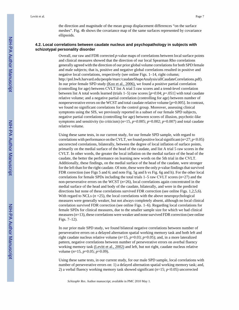
images were reformatted and registered to the 1.5 mm SGPR images and the resulting imagewas used to obtain ICC volumes using an iterative expectation-maximization segmentationprotocol (Wells et al., 1996). MRI caudate nucleus manual tracing measurements were allperformed blind to diagnostic status.
3.2. Anatomical landmarks for the caudate nucleusThe caudate nucleus was measured manually, bilaterally, using our medical image editingsoftware, 3D-slicer (http://www.slicer.org), where we used 3 orthogonal views, as previouslydescribed in detail (Levitt et al., 2002). Briefly, the caudate head, body and tail were measuredincluding where the tail bordered the lateral aspect of the atrium of the lateral ventricles. Wherethe caudate and putamen are joined anteriorly, we separated them by drawing a vertical linefrom the most ventral point of the internal capsule down to the external capsule, includingmuch of the nucleus accumbens, and forming the inferior-lateral bound of the caudate nucleus.Inter-rater reliability, as previously reported, was high for left and right caudate nucleus (rI >0.98) for male SPD subjects (Levitt et al., 2002) as it was for left (ri = 0.95) and right (ri =0.94)caudate nucleus in female SPD subjects (Koo et al., 2006).
3.3. MR shape analysisOur shape analysis of the manual caudate segmentation runs automatically using the UNCshape analysis tools1 and is applied to all segmented datasets in the same manner. A detaileddescription of all steps of the shape description and the statistical analysis is available in Styneret al., (2006). In the first step of the processing, the caudate segmentations are adapted to fillany interior holes, followed by the application of a morphological closing operation and aminimal smoothing operation. The boundary surfaces of the processed segmentations arerepresented on the sphere in terms of spherical harmonic-based shape descriptions(Brechbuhler et al.,1995), which continuously describe the surfaces by sets of coefficientsweighting the spherical harmonic basis functions. The surface correspondence betweendifferent surfaces is established by parameter-space rotation based on the first-order expansionof spherical harmonics. This parametric spherical harmonic-based shape description is thenuniformly sampled into a set of 1002 surface points, called SPHARM-PDM. Each surface isthen spatially aligned to an average caudate template using rigid Procrustes alignment(Bookstein, 1997) and the size is corrected for head size variations by normalization usingintracranial contents (ICC) volume. Unlike the original segmentations, the resulting SPHARM-PDM surfaces are aligned, scaling normalized, smoothed and uniformly sampled surfaces witha point-to-point correspondence, for 1002 points, defined across surfaces of different subjects.
3.4. Statistical analysesIn order to compare the shape of the caudate between SPD and control groups, we computelocally a non-parametric statistical test that compares the local surface coordinates for groupmean differences at 1002 discrete surface locations. The group difference metric between thegroups of surface coordinates is given by the standard robust Hotelling T2 two sample metric.This caudate shape analysis involves computing 1002 hypothesis tests, one per surface location,and a correction for the multiple testing problem is necessary, as an uncorrected analysis isoverly optimistic. Our local shape analysis employs permutation tests for the computation ofthe raw uncorrected p-values. Since a correction for multiple comparisons with strong controlover experiment-wise type 1 errors (Pantazis et al., 2004; Styner et al., 2004) is overlypessimistic, we apply an alternative false discovery rate (FDR) based correction (Genovese etal., 2002), which results in a less conservative estimate of false-negatives. The statisticalanalysis provides visualizations of the group test’s local effect size via mean difference
1The UNC shape tools are publicly available at http://www.ia.unc.edu/dev/download/shapeAnalysis.
Levitt et al. Page 5
Schizophr Res. Author manuscript; available in PMC 2010 May 1.
NIH
-PA Author Manuscript
NIH
-PA Author Manuscript
NIH
-PA Author Manuscript

magnitude displacement maps (see Fig. 3). Significance maps displaying the local statisticalp-values, both raw and corrected for multiple comparisons are also generated (see Figs. 1 and2). In addition to the local statistical shape analysis, we also compute an overall, global shapedifference that summarizes the group differences across the surface via averaging. The clinicalcorrelations were performed locally based on the rank ordering resulting from the projectionof the corresponding surface points onto the line passing through the corresponding point ofthe average shape for all subjects in the direction perpendicular to the average surface. Therank increases in the outward direction along the surface normal. A positive correlation withrespect to a chosen measure indicates that an increase in the measure coincides with structuralinflation, and a negative correlation with respect to a chosen measure indicates that an increasein the measure coincides with structural deflation. Correlations were performed using aSpearman’s rank order statistic. As the local correlations were performed for 1002 surfacepoints this necessitated a correction for multiple comparisons. We used the FDR approach,with a false discovery rate of 5%, which is less conservative than applying a Bonferronicorrection (Genovese et al., 2002).
4. Results4.1. Global and local shape analyses
We applied our shape analysis methodology to manually segmented caudate nuclei for bothmale and female data sets. All reported results were corrected for ICC volume. Here we presentresults based upon both raw and FDR corrected p value maps together with corresponding localdisplacement maps.
With regard to our global shape measures, which capture the average of the mean surface localdifferences across the whole surface, we found in male subjects a significant global surfacedeflation on the right (p=0.02) but not the left (p=0.30). Alternatively, in females, we foundsignificant global surface deflation differences for both right (p=0.0042) and left (p=0.044)caudate nuclei, but more pronounced on the right.
Overall, with respect to local analyses, we found in both male and female samples: 1) morepronounced group differences in the right, versus the left, caudate nucleus, using both our rawand FDR corrected statistical tests; 2) that group differences, bilaterally, were mainly anterior,in the head of the caudate; and 3) that no group differences survived FDR correction on theleft side.
Specifically, local analyses in our female sample, based upon 28 SPD and 25 NCLs, usingFDR corrected statistics, showed significant differences in the dorsal and ventral aspects of theright (Fig. 1g,h), but not left (Fig. 1c,d), head of the caudate nucleus, as revealed in our p valuesurface maps in Fig. 1. Further, the local displacement maps in our female subjects (Fig. 3a–d), support that these areas of significant group difference, using FDR corrected statistics, wereareas where there was local caudate deflation in SPD versus NCL subjects (Fig. 3c,d).
In our male sample, local analyses, based upon 15 SPD and 14 NCLS, using FDR correctedstatistics, showed small islets of significant group difference on the right head of the caudateas revealed in our p value surface maps in Fig. 2g,h. No group differences were revealed, usingFDR corrected statistics, for left caudate (Fig. 2c,d). Once again, the local displacement mapsin our male subjects (Fig. 3e,f,g,h) support that areas of significant difference, using FDRcorrected statistics, where there was local deflation in SPD versus NCLs (Fig. 3g,h).
To further demonstrate our methodology, we present a visualization (see Fig. 4a) of the meandisplacement difference, in millimeters (mm), between the mean shape, or surface, in the rightmedial caudate of female SPD versus female controls with a superimposed vectormap showing
Levitt et al. Page 6
Schizophr Res. Author manuscript; available in PMC 2010 May 1.
NIH
-PA Author Manuscript
NIH
-PA Author Manuscript
NIH
-PA Author Manuscript
the direction and magnitude of the mean group displacement differences “on the surfacemeshes”. Fig. 4b shows the covariance map of the same surfaces represented by covarianceellipsoids.
4.2. Local correlations between caudate nucleus and psychopathology in subjects withschizotypal personality disorder
Overall, our raw and FDR corrected p-value maps of correlations between local surface pointsand clinical measures showed that the direction of our local Spearman Rho correlationsgenerally agreed with the direction of our prior global volume correlations for both SPD femaleand male subjects; that is, positive and negative global correlations resulted in positive andnegative local correlations, respectively (see online Figs. 1–14, right column;http://pnl.bwh.harvard.edu/people/marc/caudateShapeAnalysis/allCaudateCorrelations.pdf).In our prior female SPD study (Koo et al., 2006), we found a positive partial correlation(controlling for age) between CVLT list A trial 5 raw scores and a trend-level correlationbetween list A total words learned (trials 1–5) raw scores [p=0.04; p=.051] with total caudaterelative volume; and a negative partial correlation (controlling for age) between number ofnonperseverative errors on the WCST and total caudate relative volume [p=0.005]. In contrast,we found no significant correlations for the control group. Moreover, assessing clinicalsymptoms using the SIS, we previously reported in a subset of our female SPD subjects,negative partial correlations (controlling for age) between scores of illusion, psychotic-likesymptoms and sensitivity (to criticism) (n=15, p=0.005; p=0.002; p=0.007) and total caudaterelative volume.
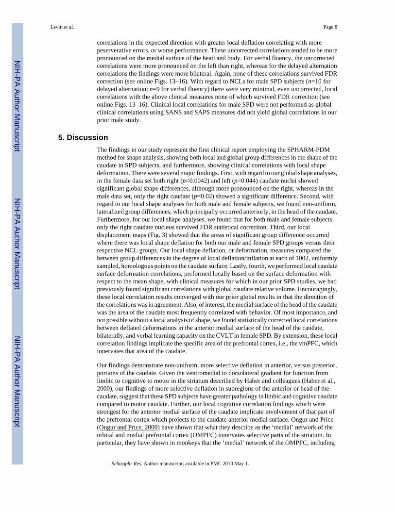
Using these same tests, in our current study, for our female SPD sample, with regard tocorrelations with performance on the CVLT, we found positive local significant (n=27; p<0.05)uncorrected correlations, bilaterally, between the degree of local inflation of surface points,primarily on the medial surface of the head of the caudate, and list A trial 5 raw scores in theCVLT. In other words, the greater the local inflation on the medial surface of the head of thecaudate, the better the performance on learning new words on the 5th trial in the CVLT.Additionally, these findings, on the medial surface of the head of the caudate, were strongerfor the left than for the right caudate. Of note, these were the only p-value findings that survivedFDR correction (see Figs 5 and 6; and note Fig. 5g and h vs Fig. 6g and h). For the other localcorrelations for female SPDs including the total trials 1–5 raw CVLT scores (n=27) and thenon-perseverative errors on the WCST (n=26), local correlations again concentrated in themedial surface of the head and body of the caudate, bilaterally, and were in the predicteddirections but none of these correlations survived FDR correction (see online Figs. 1,2,5,6).With regard to NCLs (n =25), the local correlations with the above neuropsychologicalmeasures were generally weaker, but not always completely absent, although no local clinicalcorrelation survived FDR correction (see online Figs. 1–6). Regarding local correlations forfemale SPDs for clinical measures, due to the smaller sample size for which we had clinicalmeasures (n=13), these correlations were weaker and none survived FDR correction (see onlineFigs. 7–12).
In our prior male SPD study, we found bilateral negative correlations between number ofperseverative errors on a delayed alternation spatial working memory task and both left andright caudate nucleus relative volume (n=15, p=0.03; p=0.05); and, in a more lateralizedpattern, negative correlations between number of perseverative errors on averbal fluencyworking memory task (Levitt et al., 2002) and left, but not right, caudate nucleus relativevolume (n=15, p=0.05; p=0.09).
Using these same tests, in our current study, for our male SPD sample, local correlations withnumber of perseverative errors on: 1) a delayed alternation spatial working memory task; and,2) a verbal fluency working memory task showed significant (n=15; p<0.05) uncorrected
Levitt et al. Page 7
Schizophr Res. Author manuscript; available in PMC 2010 May 1.
NIH
-PA Author Manuscript
NIH
-PA Author Manuscript
NIH
-PA Author Manuscript
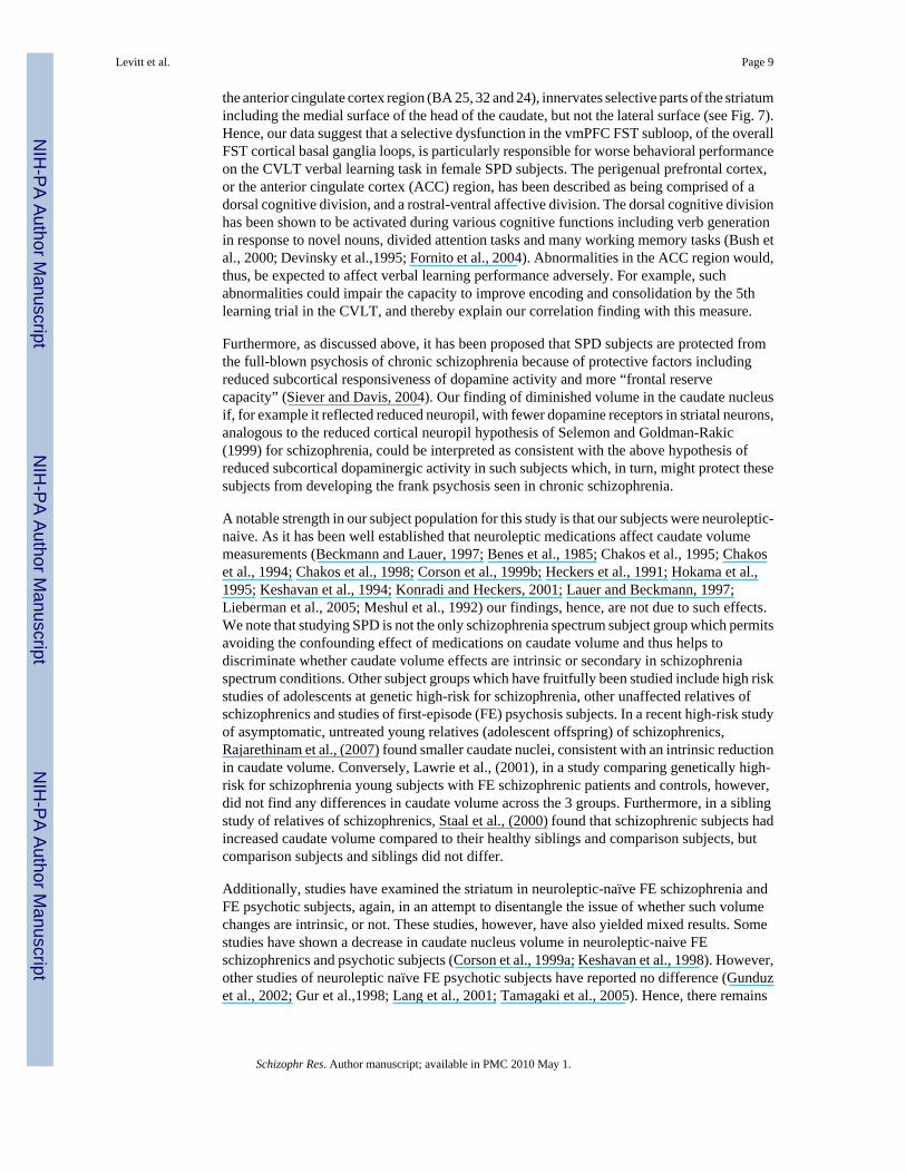
correlations in the expected direction with greater local deflation correlating with morepeserverative errors, or worse performance. These uncorrected correlations tended to be morepronounced on the medial surface of the head and body. For verbal fluency, the uncorrectedcorrelations were more pronounced on the left than right, whereas for the delayed alternationcorrelations the findings were more bilateral. Again, none of these correlations survived FDRcorrection (see online Figs. 13–16). With regard to NCLs for male SPD subjects (n=10 fordelayed alternation; n=9 for verbal fluency) there were very minimal, even uncorrected, localcorrelations with the above clinical measures none of which survived FDR correction (seeonline Figs. 13–16). Clinical local correlations for male SPD were not performed as globalclinical correlations using SANS and SAPS measures did not yield global correlations in ourprior male study.
5. DiscussionThe findings in our study represent the first clinical report employing the SPHARM-PDMmethod for shape analysis, showing both local and global group differences in the shape of thecaudate in SPD subjects, and furthermore, showing clinical correlations with local shapedeformation. There were several major findings. First, with regard to our global shape analyses,in the female data set both right (p=0.0042) and left (p=0.044) caudate nuclei showedsignificant global shape differences, although more pronounced on the right, whereas in themale data set, only the right caudate (p=0.02) showed a significant difference. Second, withregard to our local shape analyses for both male and female subjects, we found non-uniform,lateralized group differences, which principally occurred anteriorly, in the head of the caudate.Furthermore, for our local shape analyses, we found that for both male and female subjectsonly the right caudate nucleus survived FDR statistical correction. Third, our localdisplacement maps (Fig. 3) showed that the areas of significant group difference occurredwhere there was local shape deflation for both our male and female SPD groups versus theirrespective NCL groups. Our local shape deflation, or deformation, measures compared thebetween group differences in the degree of local deflation/inflation at each of 1002, uniformlysampled, homologous points on the caudate surface. Lastly, fourth, we performed local caudatesurface deformation correlations, performed locally based on the surface deformation withrespect to the mean shape, with clinical measures for which in our prior SPD studies, we hadpreviously found significant correlations with global caudate relative volume. Encouragingly,these local correlation results converged with our prior global results in that the direction ofthe correlations was in agreement. Also, of interest, the medial surface of the head of the caudatewas the area of the caudate most frequently correlated with behavior. Of most importance, andnot possible without a local analysis of shape, we found statistically corrected local correlationsbetween deflated deformations in the anterior medial surface of the head of the caudate,bilaterally, and verbal learning capacity on the CVLT in female SPD. By extension, these localcorrelation findings implicate the specific area of the prefrontal cortex, i.e., the vmPFC, whichinnervates that area of the caudate.
Our findings demonstrate non-uniform, more selective deflation in anterior, versus posterior,portions of the caudate. Given the ventromedial to dorsolateral gradient for function fromlimbic to cognitive to motor in the striatum described by Haber and colleagues (Haber et al.,2000), our findings of more selective deflation in subregions of the anterior or head of thecaudate, suggest that these SPD subjects have greater pathology in limbic and cognitive caudatecompared to motor caudate. Further, our local cognitive correlation findings which werestrongest for the anterior medial surface of the caudate implicate involvement of that part ofthe prefrontal cortex which projects to the caudate anterior medial surface. Ongur and Price(Ongur and Price, 2000) have shown that what they describe as the ‘medial’ network of theorbital and medial prefrontal cortex (OMPFC) innervates selective parts of the striatum. Inparticular, they have shown in monkeys that the ‘medial’ network of the OMPFC, including
Levitt et al. Page 8
Schizophr Res. Author manuscript; available in PMC 2010 May 1.
NIH
-PA Author Manuscript
NIH
-PA Author Manuscript
NIH
-PA Author Manuscript
the anterior cingulate cortex region (BA 25, 32 and 24), innervates selective parts of the striatumincluding the medial surface of the head of the caudate, but not the lateral surface (see Fig. 7).Hence, our data suggest that a selective dysfunction in the vmPFC FST subloop, of the overallFST cortical basal ganglia loops, is particularly responsible for worse behavioral performanceon the CVLT verbal learning task in female SPD subjects. The perigenual prefrontal cortex,or the anterior cingulate cortex (ACC) region, has been described as being comprised of adorsal cognitive division, and a rostral-ventral affective division. The dorsal cognitive divisionhas been shown to be activated during various cognitive functions including verb generationin response to novel nouns, divided attention tasks and many working memory tasks (Bush etal., 2000; Devinsky et al.,1995; Fornito et al., 2004). Abnormalities in the ACC region would,thus, be expected to affect verbal learning performance adversely. For example, suchabnormalities could impair the capacity to improve encoding and consolidation by the 5thlearning trial in the CVLT, and thereby explain our correlation finding with this measure.
Furthermore, as discussed above, it has been proposed that SPD subjects are protected fromthe full-blown psychosis of chronic schizophrenia because of protective factors includingreduced subcortical responsiveness of dopamine activity and more “frontal reservecapacity” (Siever and Davis, 2004). Our finding of diminished volume in the caudate nucleusif, for example it reflected reduced neuropil, with fewer dopamine receptors in striatal neurons,analogous to the reduced cortical neuropil hypothesis of Selemon and Goldman-Rakic(1999) for schizophrenia, could be interpreted as consistent with the above hypothesis ofreduced subcortical dopaminergic activity in such subjects which, in turn, might protect thesesubjects from developing the frank psychosis seen in chronic schizophrenia.
A notable strength in our subject population for this study is that our subjects were neuroleptic-naive. As it has been well established that neuroleptic medications affect caudate volumemeasurements (Beckmann and Lauer, 1997; Benes et al., 1985; Chakos et al., 1995; Chakoset al., 1994; Chakos et al., 1998; Corson et al., 1999b; Heckers et al., 1991; Hokama et al.,1995; Keshavan et al., 1994; Konradi and Heckers, 2001; Lauer and Beckmann, 1997;Lieberman et al., 2005; Meshul et al., 1992) our findings, hence, are not due to such effects.We note that studying SPD is not the only schizophrenia spectrum subject group which permitsavoiding the confounding effect of medications on caudate volume and thus helps todiscriminate whether caudate volume effects are intrinsic or secondary in schizophreniaspectrum conditions. Other subject groups which have fruitfully been studied include high riskstudies of adolescents at genetic high-risk for schizophrenia, other unaffected relatives ofschizophrenics and studies of first-episode (FE) psychosis subjects. In a recent high-risk studyof asymptomatic, untreated young relatives (adolescent offspring) of schizophrenics,Rajarethinam et al., (2007) found smaller caudate nuclei, consistent with an intrinsic reductionin caudate volume. Conversely, Lawrie et al., (2001), in a study comparing genetically high-risk for schizophrenia young subjects with FE schizophrenic patients and controls, however,did not find any differences in caudate volume across the 3 groups. Furthermore, in a siblingstudy of relatives of schizophrenics, Staal et al., (2000) found that schizophrenic subjects hadincreased caudate volume compared to their healthy siblings and comparison subjects, butcomparison subjects and siblings did not differ.
Additionally, studies have examined the striatum in neuroleptic-naïve FE schizophrenia andFE psychotic subjects, again, in an attempt to disentangle the issue of whether such volumechanges are intrinsic, or not. These studies, however, have also yielded mixed results. Somestudies have shown a decrease in caudate nucleus volume in neuroleptic-naive FEschizophrenics and psychotic subjects (Corson et al., 1999a; Keshavan et al., 1998). However,other studies of neuroleptic naïve FE psychotic subjects have reported no difference (Gunduzet al., 2002; Gur et al.,1998; Lang et al., 2001; Tamagaki et al., 2005). Hence, there remains
Levitt et al. Page 9
Schizophr Res. Author manuscript; available in PMC 2010 May 1.
NIH
-PA Author Manuscript
NIH
-PA Author Manuscript
NIH
-PA Author Manuscript

controversy over the finding of decreased volume in the striatum in FE schizophrenia andpsychotic subjects.
The above studies of global caudate nucleus volume in schizophrenia related conditions revealmixed findings in neuroleptic-naive subjects. We believe that a more refined morphometricanalysis such as provided by our SPHARM-PDM shape analysis provides the advantage ofhelping to clarify the above discrepant findings by permitting an analysis of local shapedifferences.
Further, our data allow for a comparison based on gender. Although, in normals, females havebeen reported to have relatively larger caudate volume (Filipek et al., 1994; Goldstein et al.,2001; Murphy et al., 1996), based upon our prior volume and our current shape data, it appearsthat both males and female SPD subjects are similar in the distribution of their morphometricdeficits. Compared with their respective male and female controls, both male and female SPDsubjects have bilateral caudate volume reductions together with local shape deflationabnormalities in the caudate, which are more pronounced on the right, and more pronouncedanteriorly.
A potential limitation of our study is the smaller sample size of the male SPD subjects. Hence,the clinical correlations with local shape deformation not surviving FDR correction, in ourmale sample, might represent a type 2 error. In support of this, when we compared the relativeeffect sizes for group shape difference in our male and female samples, as revealed by the localdisplacement maps, we found that the magnitude of millimeter displacement for both groupswas very similar (see Fig. 3).
In summary, using SHPARM-PDM global and local shape analyses in neuroleptic-naïve maleand female SPD subjects with bilateral caudate nucleus volume reductions, we report, both,global caudate shape group differences together with subregional, local shape deflationabnormalities in the caudate, a core component of FST circuits in the brain. Our data suggestthat non-uniformly distributed shape abnormalities in the caudate nucleus in SPD are diseaserelated and occur in both males and females. Furthermore, we show that local deflationabnormalities for both male and female SPD subjects occur particularly on the right side andprincipally in more anterior subregions, and that local clinical correlations both converge withglobal correlations and, further, specify that the strongest source of these correlations derivefrom the anterior medial surface of the head of the caudate. This latter finding of anterior medialcaudate involvement is consistent with abnormalities in limbic and cognitive subregions of thecaudate nucleus and, by extension, implicates the vmPFC, which innervates that area of thecaudate. This, in turn, suggests an abnormality in the vmPFC FST subbloop of the corticalbasal ganglia loops in SPD, and demonstrates the utility of local shape analysis to investigatethe relationship between specific subcortical and cortical brain structures in neuropsychiatricconditions.
AcknowledgmentsRole for funding source
Our sponsors, recognized in the acknowledgments, served no role in the study design; in the collection, analysis andinterpretation of data; in the writing of the report; or in the decision to submit the paper for publication.
This work was supported in part by Department of Veteran Affairs Awards (Merits, JJL, MES, RWM; ResearchEnhancement Award Program, RWM, MES), and National Institute of Health grants K05MH070047 andR01MH50740 (MES) and R01 MH 40799 and R01 MH 052807 (RWM), Grant U54 EB005149 (RK, GG, MES, MS,MK), P50MH080272 (RWM MES, MK,SB).
Levitt et al. Page 10
Schizophr Res. Author manuscript; available in PMC 2010 May 1.
NIH
-PA Author Manuscript
NIH
-PA Author Manuscript
NIH
-PA Author Manuscript

ReferencesAkil M, Kolachana BS, Rothmond DA, Hyde TM, Weinberger DR, Kleinman JE. Catechol-O-
methyltransferase genotype and dopamine regulation in the human brain. J Neurosci 2003;23:2008–2013. [PubMed: 12657658]
Andreasen, NC. Scale for the Assessment of Negative Symptoms (SANS). University of Iowa; IowaCity: 1981.
Andreasen, NC. Scale for the Assessment of Positive Symptoms (SAPS). University of Iowa; Iowa City:1984.
Beckmann H, Lauer M. The human striatum in schizophrenia. II Increased number of striatal neurons inschizophrenics. Psychiatry Res 1997;68:99–109. [PubMed: 9104757]
Benes FM, Paskevich PA, Davidson J, Domesick VB. The effects of haloperidol on synaptic patterns inthe rat striatum. Brain Res 1985;329:265–273. [PubMed: 3978446]
Bhatia KP, Marsden CD. The behavioural and motor consequences of focal lesions of the basal gangliain man. Brain 1994;117 (Pt 4):859–876. [PubMed: 7922471]
Bookstein FL. Shape and the information in medical images: a decade of the morphometric synthesis.Comput Vis Image Underst 1997;66:97–118.
Brechbuhler C, Gerig G, Kubler O. Parametrization of closed surfaces for 3-D shape description. ComputVis, Graphics, Image Process 1995;61:154–170.
Bush G, Luu P, Posner MI. Cognitive and emotional influences in anterior cingulate cortex. Trends CognSci 2000;4:215–222. [PubMed: 10827444]
Calabresi P, De Murtas M, Bernardi G. The neostriatum beyond the motor function: experimental andclinical evidence. Neuroscience 1997;78:39–60. [PubMed: 9135088]
Chakos MH, Lieberman JA, Alvir J, Bilder R, Ashtari M. Caudate nuclei volumes in schizophrenicpatients treated with typical antipsychotics or clozapine [letter]. Lancet 1995;345:456–457.[PubMed: 7853978]
Chakos MH, Lieberman JA, Bilder RM, Borenstein M, Lerner G, Bogerts B, et al. Increase in caudatenuclei volumes of first-episode schizophrenic patients taking antipsychotic drugs. Am J Psychiatry1994;151:1430–1436. [PubMed: 7916539]
Chakos MH, Shirakawa O, Lieberman J, Lee H, Bilder R, Tamminga CA. Striatal enlargement in ratschronically treated with neuroleptic. Biol Psychiatry 1998;44:675–684. [PubMed: 9798070]
Corson PW, Nopoulos P, Andreasen NC, Heckel D, Arndt S. Caudate size in first-episode neuroleptic-naive schizophrenic patients measured using an artificial neural network. Biol Psychiatry 1999a;46:712–720. [PubMed: 10472424]
Corson PW, Nopoulos P, Miller DD, Arndt S, Andreasen NC. Change in basal ganglia volume over 2years in patients with schizophrenia: typical versus atypical neuroleptics. Am J Psychiatry 1999b;156:1200–1204. [PubMed: 10450260]
Cummings JL. Frontal-subcortical circuits and human behavior. Arch Neurol 1993;50:873–880.[PubMed: 8352676]
Delis, DC.; Kramer, JH.; Kaplan, E.; Ober, BA. California Verbal Learning Test Manual–ResearchEdition. Psychological Corp; San Diego: 1987.
Devinsky O, Morrell MJ, Vogt BA. Contributions of anterior cingulate cortex to behaviour. Brain1995;118 (Pt 1):279–306. [PubMed: 7895011]
Dickey CC, Shenton ME, Hirayasu Y, Fischer I, Voglmaier MM, Niznikiewicz MA, et al. Large CSFvolume not attributable to ventricular volume in schizotypal personality disorder. Am J Psychiatry2000;157:48–54. [PubMed: 10618012]
Filipek PA, Richelme C, Kennedy DN, Caviness VS Jr. The young adult human brain: an MRI-basedmorphometric analysis. Cereb Cortex 1994;4:344–360. [PubMed: 7950308]
First, MB.; Gibbon, M.; Spitzer, RL.; Williams, JBW.; Benjamin, L. Structured Clinical Interview forDSM-IV Personality Disorders (SCID-II): Interview and Questionnaire. American Psychiatric Press;Washington, DC.: 1997.
First, MB.; Spitzer, RL.; Williams, JBW.; Gibbon, M. Stuctured Clinical Interview for DSM-IV-PatientEdition (SCID-P). American Psychiatric Press; Washington, DC: 1995.
Levitt et al. Page 11
Schizophr Res. Author manuscript; available in PMC 2010 May 1.
NIH
-PA Author Manuscript
NIH
-PA Author Manuscript
NIH
-PA Author Manuscript

Fornito A, Yucel M, Wood S, Stuart GW, Buchanan JA, Proffitt T, et al. Individual differences in anteriorcingulate/paracingulate morphology are related to executive functions in healthy males. Cereb Cortex2004;14:424–431. [PubMed: 15028646]
Genovese CR, Lazar NA, Nichols T. Thresholding of statistical maps in functional neuroimaging usingthe false discovery rate. Neuroimage 2002;15:870–878. [PubMed: 11906227]
Gerig G, Kubler O, Kikinis R, Jolesz F. Non-linear anisotropic filtering of MRI data. IEEE Trans MedImaging 1992a;11:221–232. [PubMed: 18218376]
Gerig G, Kubler O, Kikinis R, Jolesz FA. Non-linear anisotropic filtering of MRI data. IEEE Trans MedImaging 1992b;11:221–232. [PubMed: 18218376]
Gerig G, Styner M. Shape versus size: improved understanding of the morphology of brain structues.MICCAI 2001:24–32.
Goldstein JM, Seidman LJ, Horton NJ, Makris N, Kennedy DN, Caviness VS Jr, et al. Normal sexualdimorphism of the adult human brain assessed by in vivo magnetic resonance imaging. Cereb Cortex2001;11:490–497. [PubMed: 11375910]
Gunduz H, Wu H, Ashtari M, Bogerts B, Crandall D, Robinson DG, et al. Basal ganglia volumes in first-episode schizophrenia and healthy comparison subjects. Biol Psychiatry 2002;51:801–808.[PubMed: 12007454]
Gur RE, Maany V, Mozley PD, Swanson C, Bilker W, Gur RC. Subcortical MRI volumes in neuroleptic-naive and treated patients with schizophrenia. Am J Psychiatry 1998;155:1711–1717. [PubMed:9842780]
Haber SN, Fudge JL, McFarland NR. Striatonigrostriatal pathways in primates form an ascending spiralfrom the shell to the dorsolateral striatum. J Neurosci 2000;20:2369–2382. [PubMed: 10704511]
Heaton, RK. Psychological Assessment Resources. Odessa: 1981. Wisconsin Card Sorting Test Manual.Heckers S, Heinsen H, Heinsen Y, Beckmann H. Cortex, white matter, and basal ganglia in schizophrenia:
a volumetric postmortem study. Biol Psychiatry 1991;29:556–566. [PubMed: 2054430]Hokama H, Shenton ME, Nestor PG, Kikinis R, Levitt JJ, Metcalf D, et al. Caudate, putamen, and globus
pallidus volume in schizophrenia: a quantitative MRI study. Psychiatry Res 1995;61:209–229.[PubMed: 8748466]
Kendler KS, Lieberman JA, Walsh D. The Structured Interview for Schizotypy (SIS): a preliminaryreport. Schizophr Bull 1989;15:559–571. [PubMed: 2623438]
Kendler KS, McGuire M, Gruenberg AM, O’Hare A, Spellman M, Walsh D. The Roscommon FamilyStudy. I Methods, diagnosis of probands, and risk of schizophrenia in relatives. Arch Gen Psychiatry1993;50:527–540. [PubMed: 8317947]
Keshavan MS, Bagwell WW, Haas GL, Sweeney JA, Schooler NR, Pettegrew JW. Changes in caudatevolume with neuroleptic treatment [letter]. Lancet 1994;344:1434. [PubMed: 7968091]
Keshavan MS, Rosenberg D, Sweeney JA, Pettegrew JW. Decreased caudate volume in neuroleptic-naive psychotic patients. Am J Psychiatry 1998;155:774–778. [PubMed: 9619149]
Konradi C, Heckers S. Antipsychotic drugs and neuroplasticity: insights into the treatment andneurobiology of schizophrenia. Biol Psychiatry 2001;50:729–742. [PubMed: 11720691]
Koo MS, Levitt JJ, McCarley RW, Seidman LJ, Dickey CC, Niznikiewicz MA, et al. Reduction of caudatenucleus volumes in neuroleptic-naive female subjects with schizotypal personality disorder. BiolPsychiatry. 2006
Lang DJ, Kopala LC, Vandorpe RA, Rui Q, Smith GN, Goghari VM, et al. An MRI study of basal gangliavolumes in first-episode schizophrenia patients treated with risperidone. Am J Psychiatry2001;158:625–631. [PubMed: 11282699]
Lauer M, Beckmann H. The human striatum in schizophrenia. I Increase in overall relative striatal volumein schizophrenics. Psychiatry Res 1997;68:87–98. [PubMed: 9104756]
Lawrie SM, Whalley HC, Abukmeil SS, Kestelman JN, Donnelly L, Miller P, et al. Brain structure,genetic liability, and psychotic symptoms in subjects at high risk of developing schizophrenia. BiolPsychiatry 2001;49:811–823. [PubMed: 11343678]
Levitt JJ, McCarley RW, Dickey CC, Voglmaier MM, Niznikiewicz MA, Seidman LJ, et al. MRI studyof caudate nucleus volume and its cognitive correlates in neuroleptic-naive patients with schizotypalpersonality disorder. Am J Psychiatry 2002;159:1190–1197. [PubMed: 12091198]
Levitt et al. Page 12
Schizophr Res. Author manuscript; available in PMC 2010 May 1.
NIH
-PA Author Manuscript
NIH
-PA Author Manuscript
NIH
-PA Author Manuscript

Levitt JJ, Westin CF, Nestor PG, Estepar RS, Dickey CC, Voglmaier MM, et al. Shape of caudate nucleusand its cognitive correlates in neuroleptic-naive schizotypal personality disorder. Biol Psychiatry2004;55:177–184. [PubMed: 14732598]
Lieberman JA, Tollefson GD, Charles C, Zipursky R, Sharma T, Kahn RS, et al. Antipsychotic drugeffects on brain morphology in first-episode psychosis. Arch Gen Psychiatry 2005;62:361–370.[PubMed: 15809403]
Meshul CK, Janowsky A, Casey DE, Stallbaumer RK, Taylor B. Effect of haloperidol and clozapine onthe density of “perforated” synapses in caudate, nucleus accumbens, and medial prefrontal cortex.Psychopharmacology (Berl) 1992;106:45–52. [PubMed: 1531388]
Murphy DG, DeCarli C, McIntosh AR, Daly E, Mentis MJ, Pietrini P, et al. Sex differences in humanbrain morphometry and metabolism: an in vivo quantitative magnetic resonance imaging and positronemission tomography study on the effect of aging. Arch Gen Psychiatry 1996;53:585–594. [PubMed:8660125]
Ongur D, Price JL. The organization of networks within the orbital and medial prefrontal cortex of rats,monkeys and humans. Cereb Cortex 2000;10:206–219. [PubMed: 10731217]
Pantazis D, Leahy RM, Nichols TE, Styner M. Statistical surface-based morphometry using a non-parametric approach. IEEE Int Symp Biomed Imaging (ISBI) 2004:1283–1286.
Rajarethinam R, Upadhyaya A, Tsou P, Upadhyaya M, Keshavan MS. Caudate volume in offspring ofpatients with schizophrenia. Br J Psychiatry 2007;191:258–259. [PubMed: 17766768]
Seidman LJ, Faraone SV, Goldstein JM, Goodman JM, Kremen WS, Matsuda G, et al. Reducedsubcortical brain volumes in nonpsychotic siblings of schizophrenic patients: a pilot magneticresonance imaging study. Am J Med Genet 1997;74:507–514. [PubMed: 9342202]
Selemon LD, Goldman-Rakic PS. The reduced neuropil hypothesis: a circuit based model ofschizophrenia. Biol Psychiatry 1999;45:17–25. [PubMed: 9894571]
Shenton ME, Gerig G, McCarley RW, Szekely G, Kikinis R. Amygdala-hippocampal shape differencesin schizophrenia: the application of 3D shape models to volumetric MR data. Psychiatry Res2002;115:15–35. [PubMed: 12165365]
Shihabuddin L, Buchsbaum MS, Hazlett EA, Silverman J, New A, Brickman AM, et al. Striatal size andrelative glucose metabolic rate in schizotypal personality disorder and schizophrenia. Arch GenPsychiatry 2001;58:877–884. [PubMed: 11545672]
Siever LJ, Davis KL. The pathophysiology of schizophrenia disorders: perspectives from the spectrum.Am J Psychiatry 2004;161:398–413. [PubMed: 14992962]
Siever LJ, Kalus OF, Keefe RS. The boundaries of schizophrenia. Psychiatr Clin North Am 1993;16:217–244. [PubMed: 8332562]
Staal WG, Hulshoff Pol HE, Schnack HG, Hoogendoorn ML, Jellema K, Kahn RS. Structural brainabnormalities in patients with schizophrenia and their healthy siblings. Am J Psychiatry2000;157:416–421. [PubMed: 10698818]
Styner M, Lieberman J, Pantazis D, Gerig G. Boundary and medial shape analysis of the hippocampusin schizophrenia. Med Image Anal 2004;8:197–203. [PubMed: 15450215]
Styner M, Lieberman JA, McClure RK, Weinberger DR, Jones DW, Gerig G. Morphometric analysis oflateral ventricles in schizophrenia and healthy controls regarding genetic and disease-specific factors.Proc Natl Acad Sci U S A 2005;102:4872–4877. [PubMed: 15772166]
Styner M, Oguz I, Xu S, Brechbuhler C, Pantazis D, Levitt JJ, et al. Framework for the statistical shapeanalysis of brain structures using SPHARM-PDM. Insight J 2006:1–21.
Tamagaki C, Sedvall GC, Jonsson EG, Okugawa G, Hall H, Pauli S, et al. Altered white matter/graymatter proportions in the striatum of patients with schizophrenia: a volumetric MRI study. Am JPsychiatry 2005;162:2315–2321. [PubMed: 16330596]
Weinberger DR. Implications of normal brain development for the pathogenesis of schizophrenia. ArchGen Psychiatry 1987;44:660–669. [PubMed: 3606332]
Wells W, Kikinis R, Grimson W, Jolesz FA. Adaptive segmentation of MRI data. IEEE Trans MedImaging 1996;15:429–442. [PubMed: 18215925]
Levitt et al. Page 13
Schizophr Res. Author manuscript; available in PMC 2010 May 1.
NIH
-PA Author Manuscript
NIH
-PA Author Manuscript
NIH
-PA Author Manuscript
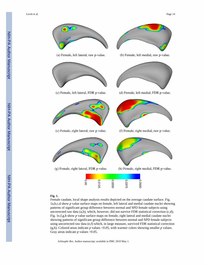
Fig. 1.Female caudate, local shape analysis results depicted on the average caudate surface. Fig.1a,b,c,d show p value surface maps on female, left lateral and medial caudate nuclei showingpatterns of significant group difference between normal and SPD female subjects usinguncorrected raw data (a,b), which, however, did not survive FDR statistical correction (c,d);Fig. 1e,f,g,h show p value surface maps on female, right lateral and medial caudate nucleishowing patterns of significant group difference between normal and SPD female subjectsusing uncorrected raw data (e,f) which, in large measure, survived FDR statistical correction(g,h). Colored areas indicate p values <0.05, with warmer colors showing smaller p values.Gray areas indicate p values >0.05.
Levitt et al. Page 14
Schizophr Res. Author manuscript; available in PMC 2010 May 1.
NIH
-PA Author Manuscript
NIH
-PA Author Manuscript
NIH
-PA Author Manuscript

Fig. 2.Male caudate, local shape analysis results depicted on the average caudate surface: Fig. 2a,b,c,dshow p value surface maps on male, left lateral and medial caudate nuclei showing patterns ofsignificant group difference between normal and SPD male subjects using uncorrected rawdata (a,b) which did not survive FDR statistical correction (c,d); Fig. 2e,f,g,h show p valuesurface maps on right male, lateral and medial caudate nuclei showing patterns of significantgroup difference between normal and SPD male subjects using uncorrected raw data (e,f)which, in small islets, survived FDR statistical correction (g,h). Colored areas indicate p values<0.05, with warmer colors showing smaller p values. Gray areas indicate p values >0.05. (For
Levitt et al. Page 15
Schizophr Res. Author manuscript; available in PMC 2010 May 1.
NIH
-PA Author Manuscript
NIH
-PA Author Manuscript
NIH
-PA Author Manuscript

interpretation of the references to colour in this figure legend, the reader is referred to the webversion of this article.)
Levitt et al. Page 16
Schizophr Res. Author manuscript; available in PMC 2010 May 1.
NIH
-PA Author Manuscript
NIH
-PA Author Manuscript
NIH
-PA Author Manuscript

Fig. 3.Local displacement maps in millimeters (mm) on female and male caudate nuclei depicted onthe average caudate surface: Fig. 3a,b,c,d show local displacement maps on female left andright lateral and medial caudate nuclei; Fig. 3e,f,g,h show local displacement maps on maleleft and right lateral and medial caudate nuclei. Colored areas indicate displacement in mm,with darker browns indicating negative mm displacement (i.e., surface deflation) for SPDsubjects compared to controls, and lighter colors indicating positive mm displacement (i.e.,surface inflation) for SPD compared to control subjects. (For interpretation of the referencesto colour in this figure legend, the reader is referred to the web version of this article.)
Levitt et al. Page 17
Schizophr Res. Author manuscript; available in PMC 2010 May 1.
NIH
-PA Author Manuscript
NIH
-PA Author Manuscript
NIH
-PA Author Manuscript
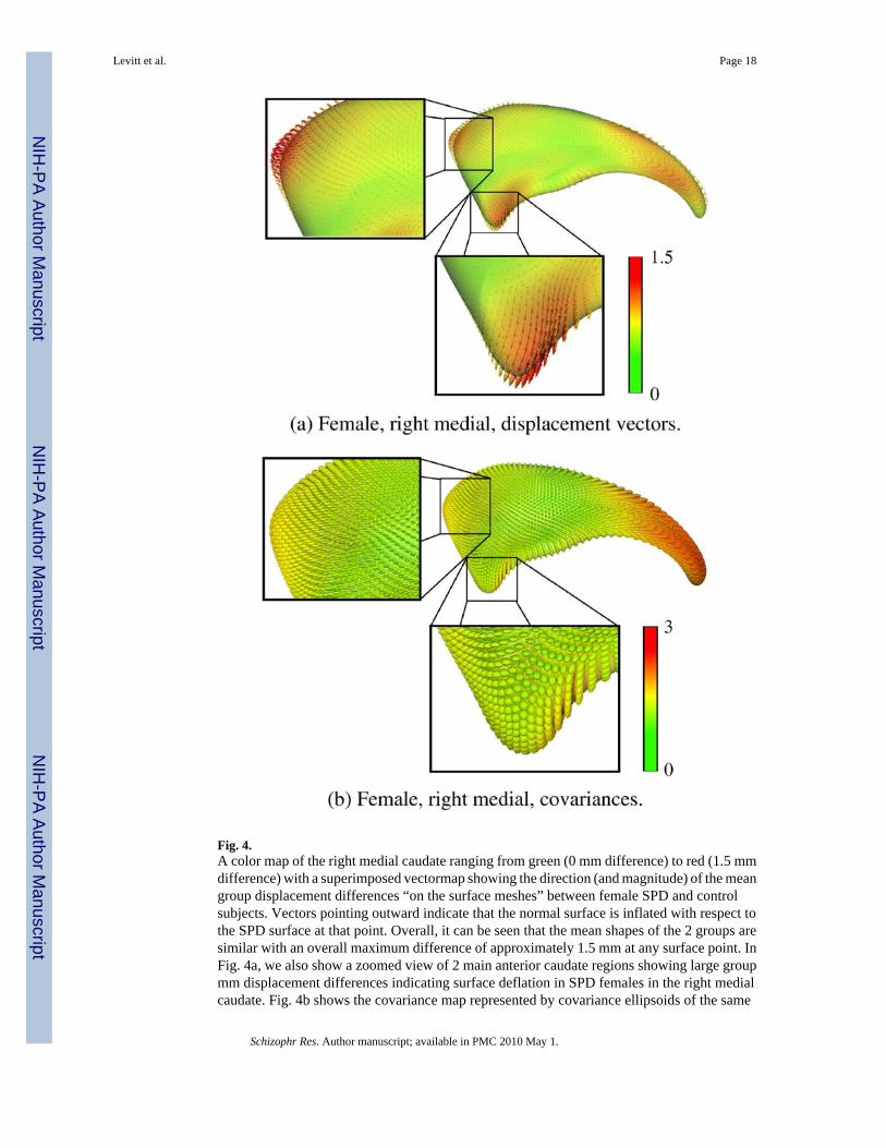
Fig. 4.A color map of the right medial caudate ranging from green (0 mm difference) to red (1.5 mmdifference) with a superimposed vectormap showing the direction (and magnitude) of the meangroup displacement differences “on the surface meshes” between female SPD and controlsubjects. Vectors pointing outward indicate that the normal surface is inflated with respect tothe SPD surface at that point. Overall, it can be seen that the mean shapes of the 2 groups aresimilar with an overall maximum difference of approximately 1.5 mm at any surface point. InFig. 4a, we also show a zoomed view of 2 main anterior caudate regions showing large groupmm displacement differences indicating surface deflation in SPD females in the right medialcaudate. Fig. 4b shows the covariance map represented by covariance ellipsoids of the same
Levitt et al. Page 18
Schizophr Res. Author manuscript; available in PMC 2010 May 1.
NIH
-PA Author Manuscript
NIH
-PA Author Manuscript
NIH
-PA Author Manuscript

right medial caudate surface for SPD compared with NCL subjects from green (0 mm2) to red(3 mm2); the larger the covariance ellipsoid, the greater the degree of covariance. Intuitively,one can appreciate that the 2 zoomed regions in Fig. 4a are significantly different (which theFDR corrected statistics indicated) as they represent areas both with large magnitude mmdisplacement differences (a more reddish hue) and relatively small covariance ellipsoids. (Forinterpretation of the references to colour in this figure legend, the reader is referred to the webversion of this article.)
Levitt et al. Page 19
Schizophr Res. Author manuscript; available in PMC 2010 May 1.
NIH
-PA Author Manuscript
NIH
-PA Author Manuscript
NIH
-PA Author Manuscript

Fig. 5.Local correlations between local surface deformation and verbal learning capacity, assessedusing the CVLT, in the left caudate in female SPD and NCL subjects, depicted on the averagecaudate surface. The left column shows the raw p-value, uncorrected for multiple tests. Themiddle column shows the p-value results after FDR correction for multiple comparisons. pvalues <0.05 are color coded; gray areas indicate p values >0.05. The right column shows thelocal Spearman’s Rho correlation coefficients color coded for values between −1.00 to +1.00.(For interpretation of the references to colour in this figure legend, the reader is referred to theweb version of this article.)
Levitt et al. Page 20
Schizophr Res. Author manuscript; available in PMC 2010 May 1.
NIH
-PA Author Manuscript
NIH
-PA Author Manuscript
NIH
-PA Author Manuscript

Fig. 6.Local correlations between local surface deformation and verbal learning capacity, assessedusing the CVLT, in the right caudate in female SPD and NCL subjects, depicted on the averagecaudate surface. The left column shows the raw p-value, uncorrected for multiple tests. Themiddle column shows the p-value results after FDR correction for multiple comparisons. pvalues <0.05 are color coded; gray areas indicate p values.>0.05. The right column shows thelocal Spearman’s Rho correlation coefficients color coded for values between −1.00 to +1.00.(For interpretation of the references to colour in this figure legend, the reader is referred to theweb version of this article.)
Levitt et al. Page 21
Schizophr Res. Author manuscript; available in PMC 2010 May 1.
NIH
-PA Author Manuscript
NIH
-PA Author Manuscript
NIH
-PA Author Manuscript
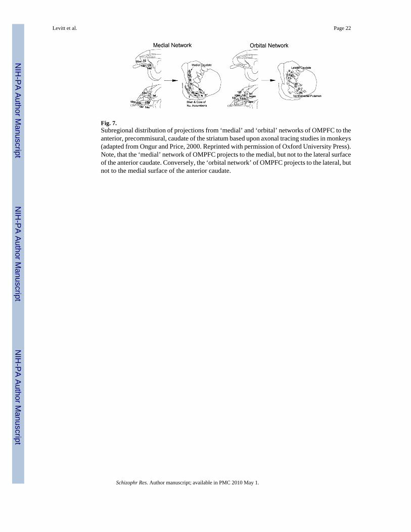
Fig. 7.Subregional distribution of projections from ‘medial’ and ‘orbital’ networks of OMPFC to theanterior, precommisural, caudate of the striatum based upon axonal tracing studies in monkeys(adapted from Ongur and Price, 2000. Reprinted with permission of Oxford University Press).Note, that the ‘medial’ network of OMPFC projects to the medial, but not to the lateral surfaceof the anterior caudate. Conversely, the ‘orbital network’ of OMPFC projects to the lateral, butnot to the medial surface of the anterior caudate.
Levitt et al. Page 22
Schizophr Res. Author manuscript; available in PMC 2010 May 1.
NIH
-PA Author Manuscript
NIH
-PA Author Manuscript
NIH
-PA Author Manuscript
Copyright © 2022 FDOKUMEN