Prions and the immune system: A trip through intestine, spleen, lymph nodes and nerves
EX VIVO EXPANSION OF MEMORY CD8 T CELLS FROM LYMPHNODES OR SPLEEN THROUGH IN VITRO CULTURE WITHINTERLEUKIN-7*
Christina Kittipatarin and Annette R. Khaled†Biomolecular Science Center, Burnett School of Biomedical Sciences, College of Medicine,University of Central Florida, Orlando, Florida 32826
AbstractInterleukin-7 (IL-7) increases lymphocyte numbers, a critical feature of immune reconstitution,through mechanisms that are still poorly understood. Part of the problem is that IL-7 is produced inlimited amounts by non-lymphoid cells, making in vivo studies of the cytokine’s activity a challenge.To overcome this, we developed an in vitro system by which lymphocytes from secondary immuneorgans could be cultured to produce IL-7 responsive cells. Using this method, we showed thatCD8hiCD44hi T cells accumulate in culture with IL-7 from a population of lymph node or spleniccells. These results were validated when a similar lymphocyte subset was found in mice expressinga constitutively active form of STAT5b, a key transducer of IL-7 signals. Interestingly, IL-7-expanded cells also up regulated the activation marker, CD69. The IL-7-derived CD44hiCD69hi cellswere not generated from naïve cells, but expanded from an existing population, since culture in IL-7of naïve lymphocytes from OT-1/Rag1-/- mice did not produce CD44hiCD69hi cells. Using the invitro culture system to study lymphocytes from mice deficient in the apoptotic protein, BIM, we wereable to attribute the expansion of CD8hiCD44hiCD69hi T cells to the proliferative and not survivalactivity of IL-7. The in vitro culture system provides an important new methodology to examine theactivities of this essential as well as immunotherapeutic cytokine.
KeywordsProliferation; Cytokine; Lymphocytes; Immunotherapy; Activation
INTRODUCTIONThe peripheral T cell pool in a healthy adult is maintained at a relatively constant level in termsof both overall size and subset composition by homeostatic mechanisms. The precise natureof these homeostatic mechanisms has not been unequivocally defined, but it is generallyaccepted that the cytokine, interleukin-7 (IL-7), is a critical growth factor. IL-7 has an essentialrole in early T cell development (Peschon et al., 1994;von-Freeden-Jeffry et al., 1995) and a
*This work was supported by an NIH/NIGMS Fellowship F31GM073565 (Kittipatarin) and an NIH/NCI Grant R01 CA109524 (Khaled).†CORRESPONDING AUTHOR: Annette R. Khaled, Ph.D., BioMolecular Science Center, Burnett School of Biomedical Sciences,University of Central Florida, 12722 Research Parkway, Orlando, FL 32826; Tel: 407-882-2254; fax 407-384-2062; email: E-mail:[email protected]'s Disclaimer: This is a PDF file of an unedited manuscript that has been accepted for publication. As a service to our customerswe are providing this early version of the manuscript. The manuscript will undergo copyediting, typesetting, and review of the resultingproof before it is published in its final citable form. Please note that during the production process errors may be discovered which couldaffect the content, and all legal disclaimers that apply to the journal pertain.
NIH Public AccessAuthor ManuscriptJ Immunol Methods. Author manuscript; available in PMC 2010 May 15.
Published in final edited form as:J Immunol Methods. 2009 May 15; 344(1): 45–57. doi:10.1016/j.jim.2009.03.001.
NIH
-PA Author Manuscript
NIH
-PA Author Manuscript
NIH
-PA Author Manuscript
mutation in the IL-7 receptor α chain is a main cause of severe combined immunodeficiency(SCID) syndrome in humans (Puel et al., 1998). Animal studies demonstrated that IL-7increases thymic output (Pido-Lopez et al., 2002;Fry et al., 2001) and maintains both the naïveand memory T cell populations (Tan et al., 2001;Schluns et al., 2000). Based on itsimmunorestorative properties, IL-7 is a candidate for use in immunotherapy and is currentlybeing tested in human clinical trials (Rosenberg et al., 2006;Sportes et al., 2008).
As an immunotherapeutic agent, IL-7 can augment T cell numbers by 1) increasing thymicoutput (Henson et al., 2004), 2) promoting T-cell survival (Kim et al., 1998;Khaled and Durum,2003), and 3) inducing cell cycling (Geiselhart et al., 2001;Kittipatarin et al., 2006). While theactivity of IL-7 as a survival factor promoting thymic development has been well documented(Maraskovsky et al., 1997;Khaled et al., 1999), its function as a proliferative factor remainspoorly understood. The replicative function of IL-7 has been mainly examined through invivo studies, and few in vitro systems have evaluated this activity of the cytokine. As example,primary T cells placed in single-cell suspensions die within a few days of standard culture(Tan et al., 2001) or poorly proliferate when these cultures are augmented with IL-7 alone(Armitage et al., 1990;Rathmell et al., 2001). In fact, many in vitro studies report that IL-7promotes proliferation when in the presence of additional signaling factors or co-stimulation(Grabstein et al., 1990;Costello et al., 1993;Tan et al., 2001). Hence, in the absence of otherstimuli, in vitro studies of IL-7 have revealed only its survival function, while it is the invivo studies that suggest a proliferative activity.
Although in vivo experiments have yielded findings, analysis of the data can be problematic.For example, it is difficult to attribute the proliferation observed in in vivo experiments solelyto IL-7. Contributions from other environmental components, such as co-stimulation orTCR:MHC signaling, must be taken into account. Moreover, most studies examining IL-7proliferation utilize the technique of adoptive transfer into immunodeficient mice. Because ofthe paucity of lymphocytes in these animals, signaling by various cytokines, self-peptide/MHCcomplexes, or foreign antigens may be atypical due to reduced T cell competition (Bourgeoisand Stockinger, 2006). Mice such as those lacking components of the IL-7 signaling pathwaymay also have abnormal lymphoid tissues, impacting lymphocyte trafficking (Kondrack et al.,2003;Singh and Schwartz, 2006). Finally, IL-7 treatment increases thymic output, making itdifficult to distinguish recent thymic emigrants (RTE) from preexisting (proliferating) naïveT-cells (Swainson et al., 2007;Sportes et al., 2008).
In order to study the proliferative activity of IL-7 independent of other factors and identifyspecific subsets of T cells that respond to IL-7 by proliferating, we developed an in vitro culturesystem for the ex vivo expansion of primary T cells. Previous in vitro systems failed to revealthe proliferative activity IL-7 for multiple reasons. The dose of IL-7 was not optimal (usually25 ng/ml or less). Cell concentrations varied but were usually low (~106 cells/ml). Lastly, theduration of culture with IL-7 rarely exceeded 7 days. The methodology we developed relieson using a higher density of T cells with a high concentration of IL-7 under extended cellculture conditions. Using this method, we were able to distinguish subsets that proliferated inresponse to IL-7 from those in which only survival was maintained. This is an importantdistinction that needs to be made to fully develop the therapeutic uses of IL-7; application ofIL-7 could result in the preferential expansion of a specific T cell subset, potentially leadingto a significant skewing of the T cell repertoire.
MATERIALS AND METHODSMice used and cell isolation techniques
C57BL/6 mice were purchased from Jackson Laboratory (Bar Harbor, Maine) and C57BL/6-Tg(OT-I)-RAG1tm1Mom mice were purchased from Taconic (Hudson, NY). Mice were housed
Kittipatarin and Khaled Page 2
J Immunol Methods. Author manuscript; available in PMC 2010 May 15.
NIH
-PA Author Manuscript
NIH
-PA Author Manuscript
NIH
-PA Author Manuscript
in the animal facility at the University of Central Florida and used at 2 -3 months of age.STAT5b-CA mice were generously provided by Dr. Michael Farrar (Department of LaboratoryMedicine and Pathology, University of Minnesota, Minneapolis, MN) and Bim deficient micewere a kind gift from Dr. Scott Durum (Laboratory of Molecular Immunoregulation, NationalCancer Institute-Frederick, Frederick, Maryland). Animal studies were reviewed and approvedby the Institutional Animal Care and Use Committee (IACUC) at the University of CentralFlorida.
Lymph node and spleen cells were isolated by gentle crushing of organs through a 70μM porefilter (BD Falcon) and pooled. Spleen cells were further treated with ACK lysis buffer (QualityBiological, Inc.) to lyse red blood cells (RBCs) and enriched for T cells by negative selectionwith a commercially available kit, the Mouse T-lymphocyte Enrichment kit (BD Biosciences).T cell enrichment was confirmed by analysis of surface markers by flow cytometry.
In vitro culture with IL-7Culture conditions, for the ex vivo expansion of IL-7-responsive cells, were established asfollows: Cells were maintained at a density of 3-5 × 10⁶ cells/ml in complete medium (RPMI1640 Medium (Invitrogen) supplemented with 10% Fetal Bovine serum (Hyclone), 2-βmercaptoethanol (1000X, Life Technologies), and 50 U/ml penicillin/streptomycin) in 24well plates. Recombinant human IL-7 (Peprotech) was added at the start of culture at aconcentration of 150 ng/ml and refreshed every other day. This concentration of IL-7 wasestablished experimentally after testing a dose range from 10-200 ng/ml for optimal effectsupon cell growth and viability. Cultures were maintained with or without IL-7 for >14 days oruntil noticeable cell death was apparent and confirmed by Trypan Blue exclusion staining.
Cell surface protein analysisSurface expression of membrane proteins expressed on T cells was assessed by flow cytometryusing the following fluorochrome-conjugated antibodies: PE-conjugated anti-CD4 (cloneGK1.5), PerCP-conjugated anti-CD8 (clone 53-6.7), FITC-conjugated anti-CD44 (IM7), andFITC-conjugated anti-CD69 (clone H1.2F3) (BD Biosciences). Cells were incubated withsaturating amounts of the appropriate antibodies for 20 minutes on ice, washed in buffer (PBS+ 0.1% bovine serum albumin (BSA)) and analyzed by flow cytometry on a FACSCalibur flowcytometer (BD Biosciences). Data was analyzed using FCS Express software (Ontario,Canada).
Proliferation assaysFor CFSE labeling, 5 × 10⁶ lymph node or splenic T cells were washed and resuspended in 2μM CFSE (Molecular Probes) staining solution (PBS supplemented with 0.1% BSA). After10 minutes, labeling was stopped and cells washed. The division status of cells was determinedby measuring CFSE fluorescence by flow cytometry at 48 hours after culture with or withoutIL-7. The generation number for the population was determined from a best fit of these datato a Gaussian curve using the Proliferation Add-on module for the FCS express software.Unstimulated, freshly-labeled CFSE-containing cells were used to verify the peakcorresponding to the undivided population. To measured DNA synthesis, 5 × 10⁶ cells werepulsed with BrdU (10μM) for 48 hours and BrdU incorporation was detected with acommercially available kit (BD Biosciences) according to manufacturer’s protocol. Briefly,cells were typed for surface markers CD4 or CD8 as described above, washed, fixed, andpermeabilized prior to incubation with a FITC-conjugated anti-BrdU antibody. The cells wereanalyzed by flow cytometry as described above.
Kittipatarin and Khaled Page 3
J Immunol Methods. Author manuscript; available in PMC 2010 May 15.
NIH
-PA Author Manuscript
NIH
-PA Author Manuscript
NIH
-PA Author Manuscript
RESULTSPhenotype of T cells from STAT5b-CA transgenic mice
In order to develop an in vitro culture system for the ex vivo expansion of IL-7 responsive Tcells, we first needed to identify the types of T cells that result from a constitutive IL-7 signal.Hence, we examined the distribution of T cells isolated from STAT5b-CA mice (Burchill etal., 2003). The signal transduced through the IL-7 receptor is typically mediated through theJAK/STAT pathway and expression of a constitutively activated STAT5 (STAT5b-CA) issufficient to promote IL-7 dependent growth in the absence of the cytokine (Burchill et al.,2003). We analyzed the phenotype of T cells isolated from the spleen (by negative selection)and lymph nodes from STAT5b-CA mice. Results are shown in Figure 1. In agreement withthe previous study (Burchill et al., 2003), we observed that the CD4:CD8 ratio of T cells eitherenriched from the spleen (Fig. 1A) or isolated from the lymph nodes (Fig. 1B) weresignificantly skewed towards CD8 T cells. Few CD4 T cells were detected (less than 16%).Moreover, the CD8 T cell subsets that was being generated by constitutive STAT5b signalingdid not up regulate expression of the activation marker, CD69, but displayed elevatedexpression of CD44 (Figs. 1A and 1B). This was most prominent in the T cells enriched fromthe spleen (Fig. 1A). Therefore, these results indicate that IL-7 promotes the expansion of aspecific subset of T cells, implicating that its replicative function may not extend to all T celltypes.
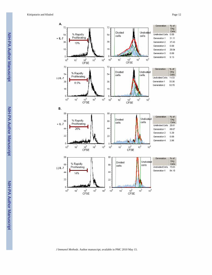
IL-7 promotes the proliferation of CD8 T cellsTo demonstrate that IL-7 can cause cell division under in vitro culture conditions, T cells wereenriched from the spleens of wild-type (WT) C57BL/6 mice and labeled with the intracellulardye, CFSE. Splenic T cells were cultured with or without IL-7 for 48 hours. Cell division wasassessed after two days of culture because beyond this significant cell death was observed inthe IL-7 withdrawn cells. Figure 2A shows that a greater percentage of T cells divided in thepresence of IL-7 compared to cells maintained in the absence of the cytokine. Forty-eight hoursof CFSE labeling revealed that there were two major proliferating populations: (1) A slowdividing population (that underwent 1-2 cell division cycles) and (2) A rapidly dividingpopulation (that underwent up to 4 more cell division cycles). The slow dividing populationwas IL-7 independent, and these were detected in both the presence and absence of the cytokine(Fig. 2A). In contrast, the rapidly dividing population was IL-7 dependent in that two fold morecells were detected in the presence of IL-7 (19%) compared to the absence (8.5%) and thesecells underwent up to four more cell division cycles (Fig. 2A). Note that undivided cells onlyremained in the cultures lacking IL-7. Similar results were observed with cells isolated fromlymph nodes, given that these cells, though not enriched for T cells like those from the spleen,still contained a major population of T cells (Fig. 2B).
We next determined whether the proliferation observed in in vitro cultures resembled thatobserved within the STAT5b-CA mice in terms of the preferential expansion of CD8 T cells.Since only a limited number of cell divisions can occur within 2 days, which may not be enoughto detect a skew in CD4:CD8 ratios, we extended the culture period with IL-7 to 7 days andmeasured DNA synthesis by BrdU incorporation. Splenic T cells were pulsed with BrdU forthe final 48 hours, and Figure 2C shows that 21% of the CD8 T cells as compared to 7% ofCD4 T cells incorporated BrdU. These results along with the CFSE findings (Fig. 2A) formedthe basis for development of the in vitro proliferative assay for the expansion of IL-7 responsivecells.
Ex vivo expansion of memory-phenotype CD8 T cells by IL-7To identify the type of cell expanded by IL-7, we developed an in vitro culture system basedon optimizing culture conditions and IL-7 dosage. Previously, we found that low doses of IL-7
Kittipatarin and Khaled Page 4
J Immunol Methods. Author manuscript; available in PMC 2010 May 15.
NIH
-PA Author Manuscript
NIH
-PA Author Manuscript
NIH
-PA Author Manuscript
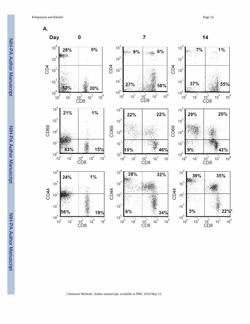
only supported survival while high doses promoted cell division (Kittipatarin et al., 2006).Moreover, IL-7 proliferative effects were detected in cells from lymphopenic or IL-7 transgenicmice (Bosco et al., 2005), which are in vivo conditions of higher than normal IL-7concentration. Using this information, we optimized the in vitro culture conditions and IL-7dosage and assessed the phenotype of cells from C57BL/6 WT mice following long-termculture with the cytokine. Starting with cells isolated from lymph nodes, we examined theCD4:CD8 ratio. The starting CD4:CD8 ratio was approximately 1:1 and by day 7 had shiftedto 1:6 and by day 14 to 1:8 (Figure 3A, top panels). Next the expression of activation andmemory markers was examined. We observed an increase in the expression of CD69 on theexpanding CD8 T cells that was sustained through 14 days of culture (Fig. 3A, middle panels).Similarly, the percentage of CD8hiCD44hi T cells increased upon in vitro culture with IL-7,becoming a predominant population by day 14 (Fig. 3A, bottom panels)
To rule out the contributions of additional factors possibly present in the lymph node cultures,we isolated T cells from the spleens of WT mice by negative selection. This enrichmenttypically yielded 95% or greater T cells (Fig. 3B, Day 0 data). Results from T cell cultureswere comparable to those observed with the lymph node cells. After 14 days of growth withIL-7, CD8 T cells, expressing the memory-phenotype marker, accumulated (Fig. 3B). Notethat a small population of CD8negCD44hi cells, observed in lymph node cultures (Fig. 3A),were not present in splenic T cell cultures, suggesting that these CD44hi lymph node cells werelikely not lymphocytes. Our results with splenic T cells were consistent with those observedwith the STAT5b-CA mice. We found that culture with IL-7 supported the expansion of CD8T cells with a memory phenotype (CD44hi). However, our results were also different since wedetected up regulation of the CD69 activation marker; an outcome not observed with T cellsfrom STAT5b-CA mice (Fig.1). Use of the in vitro culture system was successful in that wecould expand, in the absence of other signals, the type of T cells, CD8 memory, supported byIL-7. However, unlike results from in vivo studies (Burchill et al., 2003;Geiselhart et al.,2001), the ex vivo amplified T cells had an activated phenotype, as indicated by increased CD69expression, more typical of antigen-activated cells.
To determine whether the in vitro culture conditions with IL-7 were artificially inducing theexpression of CD69 and CD44, we isolated lymph node cells from OT-1/Rag1-/- mice. Thismouse line is homozygous for a transgene that encodes a TCR specific for ovalbumin (OVA)and is also deficient in rag1 (recombinase reactivating gene 1). These mice do not develop Tor B cells expressing endogenous receptors, while peripheral CD8 T cells express the TCR-OVA transgene. In the absence of OVA, most CD8 T cells from the OT-1/Rag1-/- mice are ofa naïve phenotype. Shown in Figure 4 are lymph node cells from OT-1/Rag1-/- mice culturedfor 14 days with IL-7. Unlike the findings with WT cells (Fig. 2), the OT-1/Rag1-/- cells didnot proliferate to IL-7, as indicated by the absence of BrdU incorporation after 14 days ofculture (Fig. 4A). These cells also did not substantially up regulate the expression of eitherCD69 or CD44 (Fig. 4B), as was observed with WT lymph node or splenic T cells (Fig. 3).We can therefore conclude from these findings that culture conditions with IL-7 do not resultin spontaneous proliferation or the induction of activation or memory markers. Further, we canestablish that the IL-7-driven accumulation of memory-phenotype CD8 T cells results not fromthe conversion of naïve cells to memory cells, but more likely from the expansion of an alreadyexisting population.
Use of the IL-7 in vitro culture system to differentiate survival from proliferative effectsTo demonstrate the usefulness of the IL-7 in vitro culture system, we applied our methodologyto determine whether the proliferative activity of IL-7 results as a default of its survivalfunction. T cells were enriched from the lymph nodes and spleen of mice which were deficientfor the apoptotic protein, Bim (Bim-/-). Bim has a role in the death of antigen-activated T cells
Kittipatarin and Khaled Page 5
J Immunol Methods. Author manuscript; available in PMC 2010 May 15.
NIH
-PA Author Manuscript
NIH
-PA Author Manuscript
NIH
-PA Author Manuscript
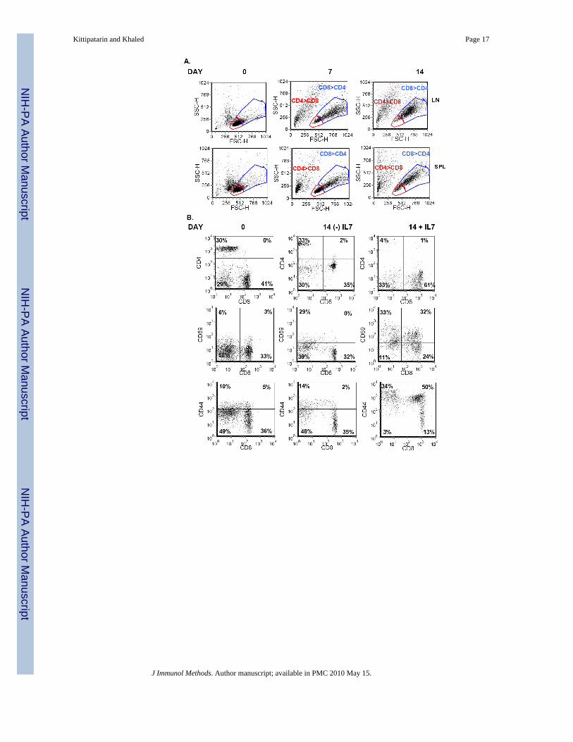
(Snow et al., 2008) and deficiency of Bim rescued the T cell depleted phenotype of IL-7receptor deficient mice (Pellegrini et al., 2004). Moreover, we observed that Bim-/- deficientT cells, unlike WT T cells, survived but did not proliferate in cultures lacking IL-7 (data notshown), suggesting that we could replace the survival activity of IL-7 by ablating Bim.Moreover, unlike WT T cells, we could culture Bim-/- T cells in the absence of IL-7 for extendedperiods of time. In Figure 5A, we show that the starting populations of Bim-/- T cells fromspleen or lymph nodes were predominantly small-sized cells with few larger blasts as indicatedby forward scatter (FSC) and side scatter (SSC) analysis. This trend was reversed uponextended culture with IL-7 in that by day 7, and later day 14, blast-sized cells became the majorpopulation, and subsequently, when phenotyped, we found that these cells were mainly CD8T cells (Fig. 5A).
Starting populations of Bim-/- lymph node cells revealed a significant number of CD4 T cells(30%), which dropped to 4% by day 14 in culture with IL-7 (Fig. 5B, top panel). Conversely,we observed that CD8 T cells from Bim-/- mice, cultured with IL-7, increased from 41% to61% by day 14 (Fig. 5B, top panel). Hence, IL-7 in vitro culture conditions selectively expandedCD8 T cells from a mixed population of cells isolated from Bim-/- mice in a manner similar tothat observed with cells from WT mice. No such trend was observed with Bim-/- lymph nodescells cultured without IL-7 (Fig. 5B, top panel), indicating that maintenance of survival throughBim deficiency was not sufficient to promote the expansion of CD8 T cells. This also showsthat there are no other factors present in IL-7 deprived lymph node cultures that could sustainthe growth of CD8 memory T cells. In fact, the few CD8 T cells remaining in the absence ofIL-7 also expressed low levels of CD4; a cell type not expanded in IL-7 containing cultures.Figure 5B (middle panel) showed that in the presence, but not absence of IL-7, there were moreCD8 T cells expressing high levels of CD69 (32%) after a 2 week culture of Bim-/- T cells.Surface staining also showed that the memory marker CD44 was up regulated only on CD8 Tcells cultured with IL-7, resulting in a significant accumulation of these cells by day 14 ofculture (Fig. 5B, bottom panel).
Next, we isolated T cells from the spleens of Bim-/- mice. Initially, the starting population ofBim-/- T cells enriched from the spleen was skewed toward a higher percentage of CD4 T cells(50%) compared to CD8 T cells (35%). Yet, by day 14 of culture in IL-7, the predominantpopulation was CD8 T cells, while in the absence of IL-7, CD4 T cells predominated (Fig. 4C,top panel). Recall that analysis of cell size by FSC/SSC showed that after 14 days of culturewith IL-7, the few remaining CD4 T cells were small sized and that the predominant blast-sized population was CD8 (Figure 4A, lower panels). In the absence of IL-7, few blast-sizedwere detected (data not shown). Similar to the results obtained from culture of WT T cells withIL-7 (Fig. 3), splenic T cells from Bim-/- mice expressed high levels of CD69 (26%) and CD44(42%) in contrast to cells cultured without IL-7 (1% and 3% respectively) (Figure 4C, middleand bottom panels).
In summary, our results show that in vitro culture with IL-7 results in the ex vivo expansion ofCD8 memory-phenotype T cells. This expansion is due not to the survival function of IL-7,since Bim deficiency, in the absence of IL-7, could not recapitulate the effects of IL-7. Rather,our in vitro culture has demonstrated that IL-7 has a separate proliferative function, resultingin the expansion of a specific subset of T cells – CD8 memory-phenotype cells – and that thisreplicative activity may have the hallmark of antigen-activated proliferation.
DISCUSSIONWe developed an in vitro culture system to specifically examine the proliferative activity ofIL-7. Results obtained from this approach were independent of other variables that could affectcell growth, such as other cytokines or MHC interactions. The proliferative activity of IL-7
Kittipatarin and Khaled Page 6
J Immunol Methods. Author manuscript; available in PMC 2010 May 15.
NIH
-PA Author Manuscript
NIH
-PA Author Manuscript
NIH
-PA Author Manuscript
has been demonstrated by in vivo studies (Schluns et al., 2000;Tan et al., 2001), however, thisactivity could not be attributed exclusively to the cytokine given the complexity of the invivo environment. Conversely in vitro studies that could more specifically study the functionof IL-7 resulted in conflicting findings. In some in vitro experiments, IL-7 induced proliferationof T cells (Cassese et al., 2007), while in others it did not (Rathmell et al., 2001). One reasonfor the different findings made by in vitro studies could be that varying culture conditions wereutilized. For example, we and others have shown that the concentration of IL-7 used in culturecan influence the experimental outcomes- high doses of IL-7 induce proliferation while lowdoses of IL-7 promote survival (Kittipatarin et al., 2006;Swainson et al., 2007). Another factorthat could affect in vitro results is the type of cell evaluated - naïve T cells may require multiplegrowth signals while memory T cells may not. Such variability in the in vitro assay conditionswould significantly affect the response of T cells to IL-7, creating the ambiguous reports ofthe cytokine’s proliferative function.
To address this problem, we developed an in vitro IL-7 culture method that would support theexpansion of T cells as has been observed under in vivo lymphopenic conditions. Features ofour sytem included an increased dose of IL-7 and extended culture periods. The concentrationof IL-7 (150 ng/ml) utilized in our in vitro cultures is higher than the levels of IL-7 typicallydetected in serum (0.3-8.4 pg/ml) (Sasson et al., 2006). We found that this higher dosage wasnecessary to support the active cell cycling of CD8 T cells under our in vitro conditions. Inprevious studies we reported that the induction of proliferation by IL-7 required a stronger IL-7dose (>50 ng/ml) than the maintenance of survival (<10 ng/ml) ((Kittipatarin et al., 2006). Itis possible that a strong proliferative stimulus (as we replicated in our culture system) couldbe delivered in vivo through synergy of IL-7 with additional signals such as TCR engagementof MHC/peptide as has been demonstrated with naïve T cells and CD4 memory T cells (Tanet al., 2001;Seddon et al., 2003). In this regard, IL-7 could function to lower the TCR activationthreshold. Although less well-documented in vivo, the IL-7 signal could be enhanced bysynergy with other cytokines to drive T cell proliferation. A recent report showed that IL-7could synergize with IL-6 or IL-21 in vitro to promote the proliferation of CD8 T cellsindependent of TCR signaling (Gagnon et al., 2008) with the resulting enhancement of IL-7dependent STAT5 activity. Keeping such possibilities in mind, in vivo experimentsdemonstrate that increasing IL-7 levels through the induction of lymphopenia (Napolitano etal., 2001;Schuler et al., 2004), expression of an IL-7 transgene (Kieper et al., 2002), or injectinghigh doses of IL-7 (relative to physiological doses) as has been done in human clinical trialslead to the expansion of T cells (Sportes et al., 2008). We found that using an elevated dose ofIL-7 in our in vitro experiments mimicked the replicative function of this cytokine observedin vivo.
One specific goal was to use the culture method to identify the subset of T cells that activelyproliferate to IL-7 in the absence of other signals. We observed that extended in vitro culturewith IL-7 selectively expanded CD8 T cells with a memory phenotype, such as has been seenwhen T cells proliferate in IL-7 transgenic hosts (Kieper et al., 2002). Our in vitro culture likelypromotes the specific expansion of memory-phenotype CD8 T cells for the following reasons:1) The accumulation of CD44hi CD8 T cells was not immediate but was detectable after 2weeks of culture in IL-7, suggesting that these cells expanded from a small existing populationand not from conversion of naïve cells to memory cells. This was confirmed when we observedthat naïve T cells from OT-1/Rag-/- mice did not generate CD44hi cells after culture in IL-7.2) After 14 days of culture, small populations of CD4 and CD44lo T cells still remained,indicating that non-specific up regulation of CD44 was not occurring. 3) Only a subset of Tcells, which were not naïve, rapidly proliferated to IL-7 as indicated by CFSE dilution andBrdU incorporation, demonstrating that IL-7 did not induce the proliferation of all culturedcells.
Kittipatarin and Khaled Page 7
J Immunol Methods. Author manuscript; available in PMC 2010 May 15.
NIH
-PA Author Manuscript
NIH
-PA Author Manuscript
NIH
-PA Author Manuscript
The proliferative activity of IL-7 has been characterized by others as non-activating in that theactivation marker CD69 was not up regulated (Geiselhart et al., 2001;Burchill et al., 2003). Incontrast, we observed that ex vivo expansion of T cells with IL-7 resulted in an increase inCD69 expression as well an increase in the percentage of CD8hi CD69hi T cells. This outcomeis not an artifact of in vitro culture conditions. We found that CD8 T cells from OT-1/Rag-/-
mice did not up regulate CD69. Moreover, T cells expressing CD69 have been observed indifferent tissues. In the bone marrow, CD8 T cells expressing CD69 have been found (Zhanget al., 2006), and in the gut, intestinal epithelial cells (IELs) retain CD69 expression long afterantigen has been cleared (Masopust et al., 2006). It follows that both the bone marrow and gutare anatomic sites where IL-7 production has been observed (Maeurer et al., 1997;Pillai et al.,2004).
What is unique with our IL-7 in vitro culture system is that we have revealed the presence ofa population of cells that only rely on an IL-7 signal to induce its proliferation. This is significantbecause the cell types expanded in vitro in our IL-7 assay are phenotypically similar, withexception of CD69 expression, to the T cell subset that accumulates in vivo in the lymphoidorgans of STAT5b-CA mice. Further, the expansion of memory CD8 T cells results not as adefault effect of IL-7’s survival function, but rather is a consequence of a distinct proliferativeactivity. This was shown by using the IL-7 in vitro system to culture Bim deficient T cells inthe presence or absence of the cytokine. These conclusions could not have been reached bytraditional adoptive transfer methods using immunodeficient mice. Therefore, the presentedmethod for the ex vivo expansion of IL-7 responsive cells incorporates features from in vivoand in vitro studies to provide a novel methodology to study the diverse activities of thisimportant cytokine.
References1. Armitage RJ, Namen AE, Sassenfeld HM, Grabstein KH. Regulation of human T cell proliferation by
IL-7. J Immunol 1990;144:938. [PubMed: 2295821]2. Bosco N, Agenes F, Ceredig R. Effects of increasing IL-7 availability on lymphocytes during and after
lymphopenia-induced proliferation. J Immunol 2005;175:162. [PubMed: 15972643]3. Bourgeois C, Stockinger B. T cell homeostasis in steady state and lymphopenic conditions. Immunol
Lett 2006;107:89. [PubMed: 16963128]4. Burchill MA, Goetz CA, Prlic M, O’Neil JJ, Harmon IR, Bensinger SJ, Turka LA, Brennan P, Jameson
SC, Farrar MA. Distinct effects of STAT5 activation on CD4+ and CD8+ T cell homeostasis:development of CD4+CD25+ regulatory T cells versus CD8+ memory T cells. J Immunol2003;171:5853. [PubMed: 14634095]
5. Cassese G, Parretta E, Pisapia L, Santoni A, Guardiola J, Di Rosa F. Bone marrow CD8 cells down-modulate membrane IL-7R{alpha} expression and exhibit increased STAT-5 and p38 MAPKphosphorylation in the organ environment. Blood. 2007
6. Costello R, Brailly H, Mallet F, Mawas C, Olive D. Interleukin-7 is a potent co-stimulus of the adhesionpathway involving CD2 and CD28 molecules. Immunology 1993;80:451. [PubMed: 7904590]
7. Fry TJ, Christensen BL, Komschlies KL, Gress RE, Mackall CL. Interleukin-7 restores immunity inathymic T-cell-depleted hosts. Blood 2001;97:1525. [PubMed: 11238086]
8. Gagnon J, Ramanathan S, Leblanc C, Cloutier A, McDonald PP, Ilangumaran S. IL-6 in synergy withIL-7 or IL-15 stimulates TCR-independent proliferation and functional differentiation of CD8+ Tlymphocytes. J Immunol 2008;180:7958. [PubMed: 18523259]
9. Geiselhart LA, Humphries CA, Gregorio TA, Mou S, Subleski J, Komschlies KL. IL-7 administrationalters the CD4:CD8 ratio, increases T cell numbers, and increases T cell function in the absence ofactivation. J Immunol 2001;166:3019. [PubMed: 11207251]
10. Grabstein KH, Namen AE, Shanebeck K, Voice RF, Reed SG, Widmer MB. Regulation of T cellproliferation by IL-7. J Immunol 1990;144:3015. [PubMed: 1969881]
Kittipatarin and Khaled Page 8
J Immunol Methods. Author manuscript; available in PMC 2010 May 15.
NIH
-PA Author Manuscript
NIH
-PA Author Manuscript
NIH
-PA Author Manuscript
11. Henson SM, Pido-Lopez J, Aspinall R. Reversal of thymic atrophy. Exp Gerontol 2004;39:673.[PubMed: 15050305]
12. Khaled AR, Durum SK. Death and Baxes: mechanisms of lymphotrophic cytokines. Immunol Rev2003;193:48. [PubMed: 12752670]
13. Khaled AR, Kim K, Hofmeister R, Muegge K, Durum SK. Withdrawal of IL-7 induces Baxtranslocation from cytosol to mitochondria through a rise in intracellular pH. Proc Natl Acad Sci US A 1999;96:14476. [PubMed: 10588730]
14. Kieper WC, Tan JT, Bondi-Boyd B, Gapin L, Sprent J, Ceredig R, Surh CD. Overexpression ofInterleukin (IL)-7 Leads to IL-15-independent Generation of Memory Phenotype CD8(+) T Cells. JExp Med 2002;195:1533. [PubMed: 12070281]
15. Kim K, Lee CK, Sayers TJ, Muegge K, Durum SK. The trophic action of IL-7 on pro-T cells: inhibitionof apoptosis of pro-T1, -T2 and -T3 cells correlates with Bcl-2 and Bax levels and is independent ofFas and p53 pathways. J Immunol 1998;160:5735. [PubMed: 9637482]
16. Kittipatarin C, Li WQ, Bulavin DV, Durum SK, Khaled AR. Cell Cycling Through Cdc25A:Transducer of Cytokine Proliferative Signals. Cell Cycle 2006;5
17. Kondrack RM, Harbertson J, Tan JT, McBreen ME, Surh CD, Bradley LM. Interleukin 7 regulatesthe survival and generation of memory CD4 cells. J Exp Med 2003;198:1797. [PubMed: 14662907]
18. Maeurer MJ, Walter W, Martin D, Zitvogel L, Elder E, Storkus W, Lotze MT. Interleukin-7 (IL-7)in colorectal cancer: IL-7 is produced by tissues from colorectal cancer and promotes preferentialexpansion of tumour infiltrating lymphocytes. Scand J Immunol 1997;45:182. [PubMed: 9042431]
19. Maraskovsky E, O’Reilly LA, Teepe M, Corcoran LM, Peschon JJ, Strasser A. Bcl-2 can rescue Tlymphocyte development in interleukin-7 receptor-deficient mice but not in mutant rag-1-/- mice.Cell 1997;89:1011. [PubMed: 9215624]
20. Masopust D, Vezys V, Wherry EJ, Barber DL, Ahmed R. Cutting edge: gut microenvironmentpromotes differentiation of a unique memory CD8 T cell population. J Immunol 2006;176:2079.[PubMed: 16455963]
21. Napolitano LA, Grant RM, Deeks SG, Schmidt D, De Rosa SC, Herzenberg LA, Herndier BG,Andersson J, McCune JM. Increased production of IL-7 accompanies HIV-1-mediated T-celldepletion: implications for T-cell homeostasis. Nat Med 2001;7:73. [PubMed: 11135619]
22. Pellegrini M, Bouillet P, Robati M, Belz GT, Davey GM, Strasser A. Loss of Bim increases T cellproduction and function in interleukin 7 receptor-deficient mice. J Exp Med 2004;200:1189.[PubMed: 15504823]
23. Peschon JJ, Morrissey PJ, Grabstein KH, Ramsdell FJ, Maraskovsky E, Gliniak BC, Park LS, ZieglerSF, Williams DE, Ware CB, et al. Early lymphocyte expansion is severely impaired in interleukin 7receptor-deficient mice. J Exp Med 1994;180:1955. [PubMed: 7964471]
24. Pido-Lopez J, Imami N, Andrew D, Aspinall R. Molecular quantitation of thymic output in mice andthe effect of IL-7. Eur J Immunol 2002;32:2827. [PubMed: 12355435]
25. Pillai M, Torok-Storb B, Iwata M. Expression and Function of IL-7 Receptors in Marrow StromalCells. Leuk Lymphoma 2004;45:2403. [PubMed: 15621752]
26. Puel A, Ziegler SF, Buckley RH, Leonard WJ. Defective IL7R expression in T(-)B(+)NK(+) severecombined immunodeficiency. Nat Genet 1998;20:394. [PubMed: 9843216]
27. Rathmell JC, Farkash EA, Gao W, Thompson CB. IL-7 enhances the survival and maintains the sizeof naive T cells. J Immunol 2001;167:6869. [PubMed: 11739504]
28. Rosenberg SA, Sportes C, Ahmadzadeh M, Fry TJ, Ngo LT, Schwarz SL, Stetler-Stevenson M,Morton KE, Mavroukakis SA, Morre M, Buffet R, Mackall CL, Gress RE. IL-7 administration tohumans leads to expansion of CD8+ and CD4+ cells but a relative decrease of CD4+ T-regulatorycells. J Immunother 2006;29:313. [PubMed: 16699374]
29. Sasson SC, Zaunders JJ, Kelleher AD. The IL-7/IL-7 receptor axis: understanding its central role inT-cell homeostasis and the challenges facing its utilization as a novel therapy. Curr Drug Targets2006;7:1571. [PubMed: 17168832]
30. Schluns KS, Kieper WC, Jameson SC, Lefrancois L. Interleukin-7 mediates the homeostasis of naiveand memory CD8 T cells in vivo. Nat Immunol 2000;1:426. [PubMed: 11062503]
Kittipatarin and Khaled Page 9
J Immunol Methods. Author manuscript; available in PMC 2010 May 15.
NIH
-PA Author Manuscript
NIH
-PA Author Manuscript
NIH
-PA Author Manuscript
31. Schuler T, Hammerling GJ, Arnold B. Cutting edge: IL-7-dependent homeostatic proliferation ofCD8+ T cells in neonatal mice allows the generation of long-lived natural memory T cells. J Immunol2004;172:15. [PubMed: 14688303]
32. Seddon B, Tomlinson P, Zamoyska R. Interleukin 7 and T cell receptor signals regulate homeostasisof CD4 memory cells. Nat Immunol 2003;4:680. [PubMed: 12808452]
33. Singh NJ, Schwartz RH. The lymphopenic mouse in immunology: from patron to pariah. Immunity2006;25:851. [PubMed: 17174925]
34. Snow AL, Oliveira JB, Zheng L, Dale JK, Fleisher TA, Lenardo MJ. Critical role for BIM in T cellreceptor restimulation-induced death. Biol Direct 2008;3:34. [PubMed: 18715501]
35. Sportes C, Hakim FT, Memon SA, Zhang H, Chua KS, Brown MR, Fleisher TA, Krumlauf MC, BabbRR, Chow CK, Fry TJ, Engels J, Buffet R, Morre M, Amato RJ, Venzon DJ, Korngold R, Pecora A,Gress RE, Mackall CL. Administration of rhIL-7 in humans increases in vivo TCR repertoire diversityby preferential expansion of naive T cell subsets. J Exp Med 2008;205:1701. [PubMed: 18573906]
36. Swainson L, Kinet S, Mongellaz C, Sourisseau M, Henriques T, Taylor N. IL-7-induced proliferationof recent thymic emigrants requires activation of the PI3K pathway. Blood 2007;109:1034. [PubMed:17023582]
37. Tan JT, Dudl E, LeRoy E, Murray R, Sprent J, Weinberg KI, Surh CD. IL-7 is critical for homeostaticproliferation and survival of naive T cells. Proc Natl Acad Sci U S A 2001;98:8732. [PubMed:11447288]
38. von-Freeden-Jeffry U, Vieira P, Lucian LA, McNeil T, Burdach SE, Murray R. Lymphopenia ininterleukin (IL)-7 gene-deleted mice identifies IL-7 as a nonredundant cytokine. J Exp Med1995;181:1519. [PubMed: 7699333]
39. Zhang X, Dong H, Lin W, Voss S, Hinkley L, Westergren M, Tian G, Berry D, Lewellen D, VileRG, Chen L, Farber DL, Strome SE. Human bone marrow: a reservoir for “enhanced effectormemory” CD8+ T cells with potent recall function. J Immunol 2006;177:6730. [PubMed: 17082586]
Kittipatarin and Khaled Page 10
J Immunol Methods. Author manuscript; available in PMC 2010 May 15.
NIH
-PA Author Manuscript
NIH
-PA Author Manuscript
NIH
-PA Author Manuscript

Figure 1. Phenotyping of T cells Isolated from STAT5b-CA MiceT cells isolated from spleen (A) and lymph nodes cells (B) of STAT5b-CA mice were analyzedfor changes in surface protein expression. Cells were stained with antibodies for CD4, CD8,CD69 and CD44 as described in Methods and analyzed by flow cytometry. FSC/ SSC gatingwas used to establish viable gates for lymphocytes. In each dot plot the numbers shown are thepercentages of cells in the indicated quadrant relative to total cells. Representative data fromone mouse is displayed
Kittipatarin and Khaled Page 11
J Immunol Methods. Author manuscript; available in PMC 2010 May 15.
NIH
-PA Author Manuscript
NIH
-PA Author Manuscript
NIH
-PA Author Manuscript
Kittipatarin and Khaled Page 12
J Immunol Methods. Author manuscript; available in PMC 2010 May 15.
NIH
-PA Author Manuscript
NIH
-PA Author Manuscript
NIH
-PA Author Manuscript

Figure 2. In Vitro Proliferation of T cells by IL-7(A-B) Splenic T-cells (A) and lymph nodes cells (B) of C57BL/6 mice were labeled with CFSEand placed in culture with or without IL-7 (150 ng/ml) for 48 hours. In the left panel are shownthe histograms displaying the fluorescence profile of the viable CFSE-labeled lymphocytes.The percent of rapidly proliferating cells is indicated. Shown in the right panels are the samehistograms subjected to analysis using FCS Express Proliferation software to determinegeneration time based on loss of CFSE due to cell division. Green lines indicate undivided(rightmost peak) and divided cells (leftmost peak). In the tables at the far right are displayedthe percent of cells in each generation. A representative experiment of three is shown. (C)Splenic T cells were cultured with IL-7 for 7 days and pulsed with BrdU for the final 48 hrsto determine the percentage of cells synthesizing DNA. Cells were stained with PerCP-conjugated antibodies for CD4 or CD8 and a FITC-conjugated anti-BrdU antibody to detectBrdU incorporation and analyzed by flow cytometry. Percentages shown in the dotplots aregated on viable cells (based on FSC/SSC) and CD4 or CD8 T cells that incorporated BrdU.Representative results from three experiments are shown.
Kittipatarin and Khaled Page 13
J Immunol Methods. Author manuscript; available in PMC 2010 May 15.
NIH
-PA Author Manuscript
NIH
-PA Author Manuscript
NIH
-PA Author Manuscript
Kittipatarin and Khaled Page 14
J Immunol Methods. Author manuscript; available in PMC 2010 May 15.
NIH
-PA Author Manuscript
NIH
-PA Author Manuscript
NIH
-PA Author Manuscript
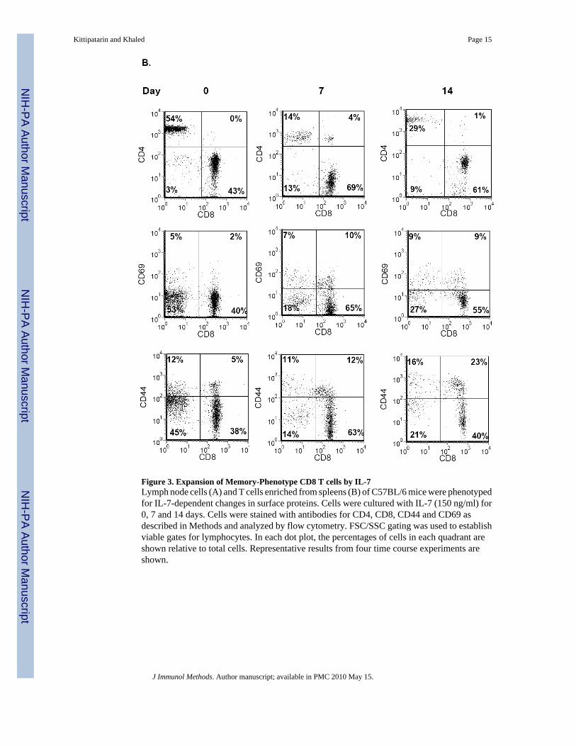
Figure 3. Expansion of Memory-Phenotype CD8 T cells by IL-7Lymph node cells (A) and T cells enriched from spleens (B) of C57BL/6 mice were phenotypedfor IL-7-dependent changes in surface proteins. Cells were cultured with IL-7 (150 ng/ml) for0, 7 and 14 days. Cells were stained with antibodies for CD4, CD8, CD44 and CD69 asdescribed in Methods and analyzed by flow cytometry. FSC/SSC gating was used to establishviable gates for lymphocytes. In each dot plot, the percentages of cells in each quadrant areshown relative to total cells. Representative results from four time course experiments areshown.
Kittipatarin and Khaled Page 15
J Immunol Methods. Author manuscript; available in PMC 2010 May 15.
NIH
-PA Author Manuscript
NIH
-PA Author Manuscript
NIH
-PA Author Manuscript
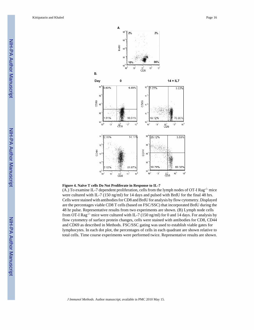
Figure 4. Naïve T cells Do Not Proliferate in Response to IL-7(A.) To examine IL-7 dependent proliferation, cells from the lymph nodes of OT-I Rag-/- micewere cultured with IL-7 (150 ng/ml) for 14 days and pulsed with BrdU for the final 48 hrs.Cells were stained with antibodies for CD8 and BrdU for analysis by flow cytometry. Displayedare the percentages viable CD8 T cells (based on FSC/SSC) that incorporated BrdU during the48 hr pulse. Representative results from two experiments are shown. (B) Lymph node cellsfrom OT-I Rag-/- mice were cultured with IL-7 (150 ng/ml) for 0 and 14 days. For analysis byflow cytometry of surface protein changes, cells were stained with antibodies for CD8, CD44and CD69 as described in Methods. FSC/SSC gating was used to establish viable gates forlymphocytes. In each dot plot, the percentages of cells in each quadrant are shown relative tototal cells. Time course experiments were performed twice. Representative results are shown.
Kittipatarin and Khaled Page 16
J Immunol Methods. Author manuscript; available in PMC 2010 May 15.
NIH
-PA Author Manuscript
NIH
-PA Author Manuscript
NIH
-PA Author Manuscript
Kittipatarin and Khaled Page 17
J Immunol Methods. Author manuscript; available in PMC 2010 May 15.
NIH
-PA Author Manuscript
NIH
-PA Author Manuscript
NIH
-PA Author Manuscript
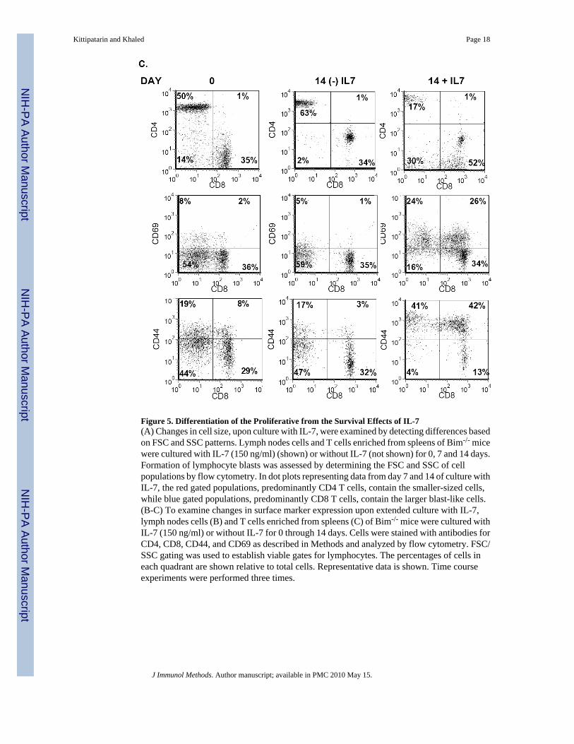
Figure 5. Differentiation of the Proliferative from the Survival Effects of IL-7(A) Changes in cell size, upon culture with IL-7, were examined by detecting differences basedon FSC and SSC patterns. Lymph nodes cells and T cells enriched from spleens of Bim-/- micewere cultured with IL-7 (150 ng/ml) (shown) or without IL-7 (not shown) for 0, 7 and 14 days.Formation of lymphocyte blasts was assessed by determining the FSC and SSC of cellpopulations by flow cytometry. In dot plots representing data from day 7 and 14 of culture withIL-7, the red gated populations, predominantly CD4 T cells, contain the smaller-sized cells,while blue gated populations, predominantly CD8 T cells, contain the larger blast-like cells.(B-C) To examine changes in surface marker expression upon extended culture with IL-7,lymph nodes cells (B) and T cells enriched from spleens (C) of Bim-/- mice were cultured withIL-7 (150 ng/ml) or without IL-7 for 0 through 14 days. Cells were stained with antibodies forCD4, CD8, CD44, and CD69 as described in Methods and analyzed by flow cytometry. FSC/SSC gating was used to establish viable gates for lymphocytes. The percentages of cells ineach quadrant are shown relative to total cells. Representative data is shown. Time courseexperiments were performed three times.
Kittipatarin and Khaled Page 18
J Immunol Methods. Author manuscript; available in PMC 2010 May 15.
NIH
-PA Author Manuscript
NIH
-PA Author Manuscript
NIH
-PA Author Manuscript
Copyright © 2022 FDOKUMEN