October-December 2013 | Volume 01 | Issue 03 Ultrasonographic Study of Cervical Lymph Nodes
10
International Journal of Scientific Study 20 October-December 2013 | Volume 01 | Issue 03 Original Article Ultrasonographic Study of Cervical Lymph Nodes Vijai Pratap 1 , Shruti Chandak 2 , Shalini Saraswat 3 , Omprakash 4 1 M.D., Associate Professor, Department of Radiology, Teerthanker Mahaveer Medical College & Research Centre, Moradabad, India. 2 M.D., Assistant Professor, Department of Radiology, Teerthanker Mahaveer Medical College & Research Centre, Moradabad, India. 3 D.N.B., Assistant Professor, Department of Radiology, Teerthanker Mahaveer Medical College & Research Centre, Moradabad, India. 4 M.D., Professor & Head of Department, Department of Radiology, Teerthanker Mahaveer Medical College & Research Centre, Moradabad, India. Introduction: An enlarged cervical lymph node is the most commonly encountered neck lump. Cervical lymph nodes are also common sites of involvement of lymphoma; tuberculous lymphadenitis; and other benign lymphadenitis. 1,2 In sonography examinations, cervical lymph nodes are usually classified into eight regions (Figure 1). Normal and reactive lymph nodes are usually found in submandibular fossa, temporal fossa, upper portion of neck, and posterior triangle of neck. High- resolution ultrasound is an ideal initial imaging investigation for most neck lumps. 3. The differential diagnosis of a neck mass depends on age of patient, location of the lesion, and its appearance on ultrasound. Several studies have shown that ultrasonography has higher sensitivity than palpation for the detection of enlarged lymph nodes in patients. 4-7 Ultrasonography provides valuable information with a high degree of diagnostic accuracy; and it is easily tolerated by patients and is cheaper and faster to perform than other methods. Because of these factors, ultrasonography is certainly the first radio-imaging modality that is in practice. Abstract Introduction: Cervical lymph nodes are common in patients with head and neck lumps. Cervical lymph nodes are common sites of involvement in lymphoma; tuberculous lymphadenitis; and other benign & malignant conditions of head and neck region. Sonography is a useful imaging tool in the assessment of cervical lymph nodes, which aids in the assessment of patient prognosis and helps in planning treatment. Aims & Objective: Aim of this study is to use non-ionising radio- imaging technique (US) to differentiate among various type of cervical lymph node pathologies. Material & Methods: This study was carried out in eighty patients (44 male, 36 females) of different age group, who attended daily Out Patient Department (OPD) of TeerthankarMahaveer Medical College, Moradabad. Subjects were selected using convenience sampling. Thorough physical examination of neck region was done and then the ultrasonography of cervical neck lumps was done by using ultrasound system present in Department of Radiology, TMMC& RC, Moradabad. Results: 10 cases of cervical adenopathy were diagnosed (five males & five females, having mostly tubercular, metastatic and lymphoma nodes with their special ultrasonographic appearances. A classical finding was that necrosis was present in 100% of cases. Conclusion: Ultrasound is a useful radio-imaging technique in assessment of cervical lymph nodes. Ultrasound is easily available less ionizing and having sensitivity of 95% approximately in cervical lumps diagnosis Key Words: Ultrasonography, L/T Ratio &Adenopathy.
Transcript of October-December 2013 | Volume 01 | Issue 03 Ultrasonographic Study of Cervical Lymph Nodes

International Journal of Scientific Study
20 October-December 2013 | Volume 01 | Issue 03
Original Article
Ultrasonographic Study of Cervical Lymph Nodes Vijai Pratap
1, Shruti Chandak
2, Shalini Saraswat
3, Omprakash
4
1 M.D., Associate Professor, Department of Radiology, Teerthanker Mahaveer Medical College & Research Centre,
Moradabad, India. 2 M.D., Assistant Professor, Department of Radiology, Teerthanker Mahaveer Medical College &
Research Centre, Moradabad, India. 3 D.N.B., Assistant Professor, Department of Radiology, Teerthanker Mahaveer
Medical College & Research Centre, Moradabad, India. 4 M.D., Professor & Head of Department, Department of
Radiology, Teerthanker Mahaveer Medical College & Research Centre, Moradabad, India.
Introduction: An enlarged cervical lymph node is the most
commonly encountered neck lump. Cervical lymph
nodes are also common sites of involvement of
lymphoma; tuberculous lymphadenitis; and other
benign lymphadenitis.1,2
In sonography
examinations, cervical lymph nodes are usually
classified into eight regions (Figure 1). Normal and
reactive lymph nodes are usually found in
submandibular fossa, temporal fossa, upper portion
of neck, and posterior triangle of neck. High-
resolution ultrasound is an ideal initial imaging
investigation for most neck lumps.3.The differential
diagnosis of a neck mass depends on age of patient,
location of the lesion, and its appearance on
ultrasound. Several studies have shown that
ultrasonography has higher sensitivity than
palpation for the detection of enlarged lymph nodes
in patients.4-7
Ultrasonography provides valuable
information with a high degree of diagnostic
accuracy; and it is easily tolerated by patients and
is cheaper and faster to perform than other
methods. Because of these factors, ultrasonography
is certainly the first radio-imaging modality that is
in practice.
Abstract Introduction: Cervical lymph nodes are common in patients with head and neck lumps. Cervical
lymph nodes are common sites of involvement in lymphoma; tuberculous lymphadenitis; and other
benign & malignant conditions of head and neck region. Sonography is a useful imaging tool in the
assessment of cervical lymph nodes, which aids in the assessment of patient prognosis and helps in
planning treatment.
Aims & Objective: Aim of this study is to use non-ionising radio- imaging technique (US) to
differentiate among various type of cervical lymph node pathologies.
Material & Methods: This study was carried out in eighty patients (44 male, 36 females) of different
age group, who attended daily Out Patient Department (OPD) of TeerthankarMahaveer Medical
College, Moradabad. Subjects were selected using convenience sampling. Thorough physical
examination of neck region was done and then the ultrasonography of cervical neck lumps was done
by using ultrasound system present in Department of Radiology, TMMC& RC, Moradabad.
Results: 10 cases of cervical adenopathy were diagnosed (five males & five females, having mostly
tubercular, metastatic and lymphoma nodes with their special ultrasonographic appearances. A
classical finding was that necrosis was present in 100% of cases.
Conclusion: Ultrasound is a useful radio-imaging technique in assessment of cervical lymph nodes.
Ultrasound is easily available less ionizing and having sensitivity of 95% approximately in cervical
lumps diagnosis
Key Words: Ultrasonography, L/T Ratio &Adenopathy.

International Journal of Scientific Study
21 October-December 2013 | Volume 01 | Issue 03
Original Article
Normal cervical lymph nodes are rarely visualized
by ultrasonography; however, hyperplastic lymph
nodes appear in many pathologies, and they are
visualized with high accuracy. 8-11
Benign as well
as malignant lymph nodes have typical
ultrasonographic morphologic characteristics.12,13
Internal architecture can also be useful in the
differential diagnosis of cervical lymph nodes. It is
thought that a hyperechoic hilum is a good
indicator of a benign lymph node3; however, the
absence of a hilum is more frequently a sign of
malignancy.14,15
No single criterion is an absolute
indicator for predicting malignant disease, and all
known criteria should be applied together. These
signs may point to a specific diagnosis/pathology.
The basic level of equipment required for head and
neck ultrasound is a modern system with a high-
frequency transducer (> 7.5 MHz).
Materials and Methods: This study was carried out in eighty patients of
different age group (0- 80 years), caste creed and
culture coming to attend daily Out Patients
Department of Teerthankar Mahaveer Medical
College & Research Centre, Moradabad. After
going through thorough physical examination of
neck region by concerned specialist, neck lumps
were inspected and palpated in Department of
Radiology. Ultrasonography of cervical neck
lumps was done by using ultrasound system.
A systematic examination protocol was made for
the evaluation of the neck. Consent from patient
was taken on prescribed format as issued by
Research committee of Teerthankar Mahaveer
University. Female patients were taken into
confidence as per normal routine, keeping one
female attendant with them. Ethical approval was
taken from ethics committee of Teerthanker
Mahaveer University, Moradabad. Subjects were
selected using convenience sampling. Sonography
of the neck begun with the examination of the
thyroidgland where the instrument was adjusted
and the frequency and gain wereoptimized. Then
the vascular sheath, salivary glands and lymph
node statuswere progressively evaluated.
TABLE 1:- AGE AND SEX DISTRIBUTION OF
PATIENTS.
Result:
Longitudinal / Transverse (L/T) ratio.
Lymph nodes were separated into 2 groups
according to their L/T ratio: oval (L/T ratio >2) and
round (L/T ratio <2). (L/T ratio >2) was present in
83% in tubercular nodes, 33% in metastatic nodes
and 33% in lymphoma nodes, while (L/T ratio <2)
was maximally present in metastatic lymph
nodes.83% lymph nodes were hypoechoic in
tubercular lymph nodes, 100% in metastatic lymph
nodes and 33% in lymphoma nodes. No
hyperechoic lymph nodes were seen in any kind of
cervical lymph nodes under study, while 17%
lymph nodes were seen isoechoic in tubercular
lymph nodes.17% lymph nodes were homogenous
in tubercular,67% in metastatic and 33% in
lymphoma nodes., while heterogenecity was seen
to the extent of 67% in tubercular and 33% in
metastatic lymph nodes. Lymphoma nodes didn’t
show any heterogenecity.
Age group
(In years) Male Female Total
0-10 4 2 6
11-20 2 6 8
21-30 8 16 24
31-40 6 6 12
41-50 10 4 14
51-60 6 2 8
61-70 6 - 6
71-80 2 - 2
Total 44 36 80

International Journal of Scientific Study
22 October-December 2013 | Volume 01 | Issue 03
Original Article
Calcification was seen to the extent of 17% in
tubercular lymph nodes.Necrosis was seen 100% in
tubercular lymph nodes and 33% in metastatic
lymph nodes. Thick regular rim enhancement to the
extent of 100% was seen in tubercular lymph
nodes, thin and focal (67%) in metastatic lymph
nodes and thin regular in lymphoma lymph nodes
to the extent of 33%.sharp border was present in
lymphoma lymph nodes and sharp border to the
extent of 67% in tubercular lymph nodes. Posterior
enhancement was seen in 67% of tubercular lymph
nodes and 33% in metastatic lymph nodes. Matting
was seen in 67% of tubercular lymph nodes.
Table:-2 Ultrasonic finding in 80 Masses in Head and Neck
Nature of lesion No. of Cases
Inflammatory
Abscess
Adenopathy
14
10
Benign neoplasm
Henangioma
10
Parathyroidadenoma 4 Epidermoid inclusion cyst 4 Lymphangioma 4 Plexiformneurofibroma 4 Chemodectoma 2
Malignant Neoplasms
Poorly differentiated carcinoma
Sq. Cell carcinoma
Lymphoma
10
4
6
Miscellaneous
Encehalocele
Parotitis
Normal lymph node
Thyroglossal duct remnant
2
2
2
2
Total 80
Adenopathy was noted in 10 cases while in 2 cases lymph node appearance was normal. Along with lymph
node enlargement other neck swellings were evaluated and differential diagnosis was made as per features,
clinical presentation & routine laboratory and radiological examinations

International Journal of Scientific Study
23 October-December 2013 | Volume 01 | Issue 03
Original Article
Table 3:- Age and Sex Distribution of Cases with Lymph Nodes Masses
Age group
(In years) Male Female
0-10 1 -
11-20 - 1
21-30 1 2
31-40 1 -
41-50 1 1
51-60 1 1
61-70 - -
Total 5 5
Distribution of adenopathy showed sexual difference and adenopathy was equal in both sexes with most
common among 21-30 yrs of age group (table-3)
Figure 1: Location of cervical lymph nodes

International Journal of Scientific Study
24 October-December 2013 | Volume 01 | Issue 03
Original Article
Table 4:- Distribution of Neck Masses According to the Nature of the Lesion
Nature of the lesion No. of cases Percentage of total cases
Inflammatory
Abscess
Adenopathy
2
14
20%
2.5%
17.5%
Developmental
Branchial Cyst
Lymphangioma
2
2
5%
2.5%
2.5%
Thyroid Masses
Benign
Malignant
16
8
30%
20%
10%
Mesenchymal
Lipoma
Sarcoma
4
4
10%
5%
5%
Neural
Schwannoma
Neurofibroma
2
2
5%
2.5%
2.5%
Vascular
Hemangioma
Carotid body tumor
2
2
5%
2.5%
2.5%
Bone
Osteoma
Metastasis
2
2
5%
2.5%
2.5%
Lymphnode Masses (non
inflammatory)
Lymphoma
Metastasis
2
6
10%
2.5%
7.5%
Salivary Gland Masses
Benign
Malignant
4
4
10%
5%
5%
Total 80 100%

International Journal of Scientific Study
25 October-December 2013 | Volume 01 | Issue 03
Original Article
Table:-5 Ultrasonographic Images of Lymph Nodes in Different Pathologies
Imaging Features Tubercular
Nodes
Metastatic Nodes Lymphoma Nodes
Shape
L/T > 2
L/T < 2
(83%)
(17%)
(33%)
(67%)
(33%)
-
Echogenecity
Hypoechoic
Hyperechoic
Isoechoic
(83%)
-
(17%)
(100%)
-
-
(33%)
-
-
Homogenecity
Homogeneous
Heterogeneous
(17%)
(67%)
(67%)
(33%)
(33%)
-
Calcification (17%) - -
Necrosis (100%) (33%) -
Rim Enhancement
Thick Irregular
Thin Regular
Thin with Focal
nodularity
(100%)
-
-
-
-
(67%)
-
(33%)
-
Nodal border
Sharp
Not Sharp
(33%)
(67%)
(67%)
(33%)
1(33%)
-
Posterior enhancement
on US
(67%) (33%) -
Surrounding Tissue
Fat plane blurring
Capsular spread,
Infiltration Adjacent
tissues
(33%)
-
-
(33%)
-
-
Matting (67%) - -

International Journal of Scientific Study
26 October-December 2013 | Volume 01 | Issue 03
Original Article
Figure 2: Typical Ultrasonographic Appearance of a Benign
Hyperplastic Lymph Node.
Figure 3: Typical Ultrasonographic
Appearance of a Malignant
Lymph Node.
Figure 4: Typical Ultrasonographic
Appearance of a Metastatic
Lymph Node.
International Journal of Scientific Study
27 October-December 2013 | Volume 01 | Issue 03
Original Article
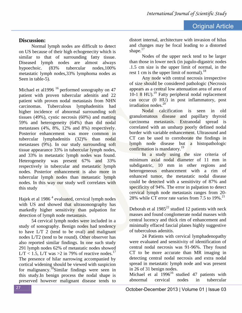
Discussion:
Normal lymph nodes are difficult to detect
on US because of their high echogenecity which is
similar to that of surrounding fatty tissue.
Diseased lymph nodes are almost always
hypoechoic. (83% tubercular nodes,100%
metastatic lymph nodes,33% lymphoma nodes as
Seen in table-5).
Michael et al1996 16
performed sonography on 47
patient with proven tubercular adenitis and 22
patient with proven nodal metastasis from NHN
carcinomas. Tuberculous lymphadenitis had
higher incidence of abnormal surrounding soft
tissues (49%), cystic necrosis (60%) and matting
59% and heterogeneity (64%) than did nodal
metastases (4%, 8%, 12% and 8%) respectively.
Posterior enhancement was more common in
tubercular lymphadenitis (32)% than nodal
metastases (9%). In our study surrounding soft
tissue appearance 33% in tubercular lymph nodes,
and 33% in metastatic lymph nodes was found.
Heterogeneity was present 67% and 33%
respectively in tubercular and metastatic lymph
nodes. Posterior enhancement is also more in
tubercular lymph nodes than metastatic lymph
nodes. In this way our study well correlates with
this study
Hajek et al 1986 4 evaluated, cervical lymph nodes
with US and showed that ultrasonongraphy has
markedly higher sensitivity than palpation for
detection of lymph node metastasis.
54 cervical lymph nodes were included in a
study of sonography. Benign nodes had tendency
to have L/T 2 (tend to be oval) and malignant
nodes L/T2 (tend to be round). Other observer has
also reported similar findings. In one such study
291 lymph nodes 62% of metastatic nodes showed
L/T < 1.5, L/T was >2 in 79% of reactive nodes.17
The presence of hilar narrowing accompanied by
cortical widening should be viewed with suspicion
for malignancy.18
Similar findings were seen in
this study.In benign process the nodal shape is
preserved however malignant disease tends to
distort internal, architecture with invasion of hilus
and changes may be focal leading to a distorted
shape.17
Nodes of the upper neck tend to be larger
than those in lower neck (in jugulo-digastric nodes
.1.5 cm size is the upper limit of normal, in the
rest 1 cm is the upper limit of normal).18
Any node with central necrosis irrespective
of size should be considered pathologic (Necrosis
appears as a central low attenuation area of area of
10-1 8 HU).19
Fatty peripheral nodal replacement
can occur (0 HU) in post inflammatory, post
irradiation nodes.20
Nodal calcification is seen in old
granulomatous disease and papillary thyroid
carcinoma metastasis. Extranodal spread is
correlated with an unsharp poorly defined nodal
border with variable enhancement. Ultrasound and
CT can be used to corroborate the findings in
lymph node disease but a histopathologic
confirmation is mandatory.19
In a study using the size criteria of
minimum axial nodal diameter of 11 mm in
subdigastric, 10 mm in other regions and
heterogeneous enhancement with a rim of
enhanced tumor, the metastatic nodal disease
could be detected with a sensitivity of 87% and
specificity of 94%. The error in palpation to detect
cervical lymph node metastasis ranges from 20-
28% while CT error rate varies from 7.5 to 19%.21
Deborah et al 198522
studied 12 patients with neck
masses and found conglomerate nodal masses with
central lucency and thick rim of enhancement and
minimally effaced fascial planes highly suggestive
of tuberculous adenitis.
24 Patients with cervical lymphadenopathy
were evaluated and sensitivity of identification of
central nodal necrosis was 91-96%. They found
CT to be more accurate than MR imaging in
detecting central nodal necrosis and extra nodal
spread in metastatic lymph node and was present
in 26 of 31 benign nodes.
Michael et al 199616
studied 47 patients with
abnormal cervical nodes in tubercular

International Journal of Scientific Study
28 October-December 2013 | Volume 01 | Issue 03
Original Article
lymphadenitis and metastasis from non head and
neck carcinoma. They found that the statistically
significant features of differential diagnosis were
lymph nodes longest diameter, echogenicity short
to long axis ratio, appearance of surrounding soft
tissue, intranodal necrosis, matting and posterior
enhancement.
US could correctly predict the etiology in 90%
cases of lymphadenopathy. Features that helped
differentiate between tuberculous lymphadenitis
and nodal metastatic from non head and neck
carcinoma were L/T ratio more than 2, thin
enhancing rims with focal nodularity and normal
surrounding tissues or capsule invasion with
infiltration into surrounding fat and adjacent
structures was associated with metastatic nodes.
Our observations were similar to those of.16,22
.
Conclusion: Ultrasound is a useful radio-imaging
technique in assessment of cervical lymph nodes.
Many features like L/T ratio, status of echogenic
hilus,echogenecitymicronodular
appearance,intranodal necrosis posterior
enhancement and calcification can very easily be
assessed without going into radio-imaging
techniques( like CT & MRI) which are costlier
and also potent source of radiation to the person
concerned. They should only be used when there
occurs a difficulty in assessing the features of
lesions by ultrasonography.
References: 1. Lee YY, Van Tassel P, Nauert C, North LB, Jing
BS. Lymphomas of the head and neck: CT
findings at initial presentation. AJR 1987; 149:575
–581
2. Reede DL, Bergeron RT. Cervical tuberculous
adenitis: CT manifestations. Radiology 1985;
154:701 –704
3. Ahuja AT. Lumps and bumps in the head and
neck. In: Ahuja AT, Evans RM, eds. Practical
head and neck ultrasound. London: Greenwich
Medical Media Limited, 2000:87-104.
4. Hajek PC, Salomonowitz E, Turk R, Tscholakoff
D, KumpanW, Czembirek H. Lymph nodes of the
neck: evaluation with US. Radiology 1986;
158:739–742.
5. Sutton RT, Reading CC, Charboneau WJ, James
EM, Grant CS, Hay ID. US-guided biopsy of neck
masses in postoperative management of patients
with thyroid cancer.Radiology 1988; 168:769–
772.
6. Baatenburg de Jong RJ, Rongen RJ, Verwoerd
CD, van Overhagen H, Lameris JS, Knegt P.
Ultrasound-guided fineneedle aspiration biopsy of
neck nodes. Arch Otolaryngol Head Neck Surg
1991; 117:402–404.
7. do Rosario PW, Fagundes TA, Maia FF, Franco
AC, Figueiredo MB, Purisch S. Sonography in the
diagnosis of cervical recurrence in patients with
differentiated thyroid carcinoma. J Ultrasound
Med 2004; 23:915–920.
8. Ahuja A, Ying M. An overview of neck node
sonography.InvestRadiol 2002; 37:333–342.
9. Bruneton JN, Normand F, Balu-Manestro C, et al.
Lymphomatous superficial lymph nodes: US
detection.Radiology 1987; 165:233–235.
10. Gritzmann N, Hollerweger A, Macheiner P,
Rettenbacher T. Sonography of soft tissue masses
of the neck. J Clin Ultrasound 2002; 30:356–373.
11. Gritzmann N. Sonography of the extrathyroidal
cervical soft tissues, the salivary glands and floor
of the mouth. Eur J Ultrasound 1994; 1:9–21.
12. Koischwitz D, Gritzmann N. Ultrasound of the
neck. RadiolClin North Am 2000; 38:1029–1045.
13. Steinkamp HJ, Mueffelmann M, Böck JC, Thiel T,
Kenzel P, Felix R. Differential diagnosis of lymph
node lesions: a semiquantitative approach with
colour Doppler ultrasound. Br JRadiol 1998;
71:828–833..
14. Sutton RT, Reading CC, Charboneau WJ, James
EM, Grant CS, Hay ID. US-guided biopsy of neck
masses in postoperative management of patients
with thyroid cancer.Radiology 1988; 168:769–
772.
International Journal of Scientific Study
29 October-December 2013 | Volume 01 | Issue 03
Original Article
15. Takeuchi Y, Suzuki H, Omura K, et al.
Differential diagnosis of cervical lymph nodes in
head and neck cancer by ultrasonography.
AurisNasus Larynx 1999; 26:331–336.
16. Michael Ying, Anil T Ahuja, Rhodri Evans et al.
Cervical Lymphadenopathy : Sonographic
Differentiation between tuberculous Nodes and
Nodal Metastases from Non-head and Neck
Carcinomas. J Clin Ultrasound Oct 1998; 26(13):
383-389.
17. Coit WE, Harnsberger HR, Osborn AG, et al.
Ranulas and their mimics: CT evaluation.
Radiology 1987; 136: 211-216
18. Miller MB, Rao VM, Tom BM, Cystic masses of
the head and neck :Petfalls in CT and MR
interpretation. AJR Sept 1992;159:601-607.
19. Harnsberger HR. Handbooks in Radiology. Head
and Neck Surgery. Chicago. New York 1990; 112-
137.
20. Zadvinskis DP, benson MT, Kerr HH, et al.
Congenital malformation of the cervicothoracic
lymphatic system. Radiographic 1992; 12: 1175-
1139.
21. Johnson TT : Abscesses and deep space infections
in head and neck. Infect DisClin North Am 1992;
6: 705-717.
22. Deborah L Reede, R. Thomas Bergeron: Cervical
Tuberculosis Adenitis: CT manifestations,
Radiology 1985; 154: 701-704
23. Miller WD, Furst IM, Sandor GKB et al. A
prospective blinded comparison of clinical
examination and computed tomography in deep
neck infection. Laryngoscope 1999; 109: 1873-
1879.
Corresponding Author Dr. Vijai Pratap, M.D. Associate Professor, Department of Radiology,
TeerthankerMahaveer Medical College &
Research Centre, Moradabad.
Email id: [email protected]