Prions and the immune system: A trip through intestine, spleen, lymph nodes and nerves
Upload
independentCategory
view
3download
0
Circulating B-Cell Chronic Lymphocytic Leukemia Cells DisplayImpaired Migration to Lymph Nodes and Bone Marrow
Tanja Nicole Hartmann,1,2Valentin Grabovsky,
2Wei Wang,
1Petra Desch,
1Gabriele Rubenzer,
1
Stefan Wollner,4Inbal Binsky,
2Alexandra Vallon-Eberhard,
2Anita Sapoznikov,
2Meike Burger,
4
Idit Shachar,2Michal Haran,
3Marek Honczarenko,
5Richard Greil,
1and Ronen Alon
2
1Laboratory for Immunological and Molecular Cancer Research, Third Medical Department, Salzburg University Hospital, Salzburg,Austria; 2Department of Immunology, Weizmann Institute of Science; 3Hematology Institute, Kaplan Medical Center, Rehovot, Israel;4Department of Internal Medicine, Freiburg University Clinic, Freiburg, Germany; and 5Biogen Idec, Inc., Cambridge, Massachusetts
Abstract
Homing to secondary lymphoid organs and bone marrow(BM) is a central aspect of leukemic pathophysiology. Weinvestigated the roles of the two major lymphocyte integrinsLFA-1 and VLA-4 on B-cell chronic lymphocytic leukemia(CLL) cells in these processes. We found that the majority ofCLL cells expressed significantly reduced LFA-1 due to low B2integrin transcripts. VLA-4 expression was heterogenous butunderwent rapid activation by the BM chemokine CXCL12.CLL cells failed to transmigrate across VCAM-1–expressing,ICAM-1–expressing, and CXCL12-expressing endothelium,whereas when LFA-1 expression was regained in subsets ofCLL cells, these lymphocytes rapidly transmigrated theendothelium. Furthermore, when injected into tail veins ofimmunodeficient mice, normal B cells rapidly homed to lymphnodes (LN) in a LFA-1–dependent manner, whereas CLL cellsdid not. Nevertheless, only residual CLL subsets could reenterBM, whereas both normal and CLL cells homed to the micespleen in an LFA-1–independent and VLA-4–independentmanner. Our results suggest that CLL cells have a reducedcapacity to adhere and transmigrate through multiplevascular endothelial beds and poorly home to lymphoidorgans other than spleen. Integrin blocking could thus be anefficient strategy to prevent circulating CLL cells fromreaching prosurvival niches in LNs and BM but not in spleen.[Cancer Res 2009;69(7):3121–30]
Introduction
B-cell chronic lymphocytic leukemia (CLL) is marked by theaccumulation of CD5+ B lymphocytes within the blood, bonemarrow (BM), and secondary lymphoid tissues but the molecularsignals that determine CLL trafficking to these organs are largelyunknown. Abnormalities in the expression and function of celladhesion molecules may account for the patterns of intranodalgrowth and hematogenous spread of the malignant cells (1, 2).Chemokine- and integrin-mediated adhesion and transendothelialmigration (TEM) are central aspects in trafficking and retention of
hematopoietic cells in the BM and lymphoid organs. To enter theseorgans, circulating cells need to arrest on specific endothelialbarriers, to locomote over the endothelial surface towardinterendothelial junctions, and to cross these junctions whileresisting disruptive shear forces (3, 4). Both firm adhesion andability to locomote and transmigrate across endothelial barriersdepend on the ability of circulating cells to establish dynamicadhesive interactions through their a4 integrins VLA-4 (a4h1) anda4h7 and the h2 integrins LFA-1 (aLh2) and Mac-1 (aMh2; ref. 5).During these interactions, the integrins undergo reversibleactivation by endothelial-presented chemokines (6).The major endothelial integrin ligands, chemokines, selectins,
and selectin ligands expressed on the main lymphoid organs areconserved between human and mice. Indeed, human stem cellsinteract with the murine BM vasculature and consequently canhome to and repopulate the BM of nonobese diabetic/severecombined immunodeficient (NOD/SCID) mice (7–9). This highconservation between the human and murine vasculature enablesthe comparison between normal and leukemic cell traffickingto the murine BM, and thus, adoptive transfer experimentsinto NOD/SCID mice have been used as a functional, preclinicalmodel for in vivo dissemination of human leukemic cells (10, 11).Human precursor-B acute lymphoblastic (ALL) cells adoptivelytransferred into these mice rapidly entered the murine BM in anintegrin-dependent manner (12). These early findings prompted usto use the NOD/SCID model to address the possible involvementof LFA-1 and VLA-4 in CLL cell trafficking to peripheral lymphnodes (LN), the BM, and the spleen using short-term homingassays.In the current study, we analyzed the migratory behavior of CLL
cells at the beginning of the disease and intended to excludechemotherapeutic influence. We therefore concentrated on CLLsamples of currently untreated patients of early and intermediatestages. We report a severely impaired in vivo homing of theseperipheral blood (PB) CLL cells to murine peripheral LNscompared with human B lymphocytes, which efficiently migratedinto these organs. This attenuated CLL cell trafficking to LNs wasthe result of reduced LFA-1 levels on the majority of the CLLsamples of our cohort. CLL cells also poorly interacted with variousICAM-1–expressing endothelial cells in vitro under shear flowconditions and displayed severely impaired in vitro TEM inresponse to stromal cell–derived factor-1 (CXCL12). CLL homingto BM was lower than that of normal B lymphocytes and dependedon functional VLA-4 expression, whereas spleen homing wasintegrin independent. In summary, our data suggest that PB CLLcells fail to mount integrin-mediated adhesions to multiplevascular endothelial beds, poorly extravasate through LNs andBM, and preferentially home to the spleen.
Note: Supplementary data for this article are available at Cancer Research Online(http://cancerres.aacrjournals.org/).
Requests for reprints: Tanja Nicole Hartmann, Laboratory for Immunological andMolecular Cancer Research, Third Medical Department, Salzburg University Hospital,Salzburg, Austria. Phone: 43-662-4482-1552; Fax: 43-662-4482-1570; E-mail:[email protected] or Ronen Alon, Department of Immunology, Weizmann Instituteof Science, Rehovot 76100, Israel. Phone: 972-8-9342482; Fax: 972-8-9344141; E-mail:[email protected].
I2009 American Association for Cancer Research.doi:10.1158/0008-5472.CAN-08-4136
www.aacrjournals.org 3121 Cancer Res 2009; 69: (7). April 1, 2009
Research Article

Materials and Methods
Patients and cell preparation. The study was approved by the localethics committees and conducted according to the Declaration of Helsinki.After informed consent, PB samples were obtained from 105 patientsfulfilling diagnostic and immunophenotypic criteria for common CLL atthe Kaplan Hospital (Rehovot, Israel), Freiburg University Hospital(Freiburg, Germany), and Salzburg University Hospital (Salzburg, Austria;Supplementary Table S1). Additional BM aspirates were obtained from 12CLL patients and 3 healthy donors. PB mononuclear cells (PBMC) of CLLor healthy donors were isolated via density gradient centrifugation andfreshly used or viably frozen in FCS plus 10% DMSO for storage in liquidnitrogen. Thawed cells were cultured overnight in RPMI 1640 with 10%FCS and antibiotics. No difference in the integrin expression or behavior ofthawed and cultured or fresh cells was observed (P = 0.458, n = 34). Forfunctional in vitro assays, B lymphocytes were enriched untouched(EasySep, StemCell Technologies). For RNA isolation, CLL cells werepurified by CD19+ selection (MACS, Miltenyi Biotec). Analysis of theimmunoglobulin heavy chain variable (IgVH) mutational status, ZAP-70,and CD38 expression and fluorescence in situ hybridization analysisof chromosomal abnormalities were routinely performed as described
(13, 14). ZAP-70 risk was assessed as T-cell/CLL expression ratio <2.5versus z2.5 as previously recommended as consensus of the optimizationof ZAP-70 evaluation procedures by multicenter efforts (15).
Endothelial cell culture. Human umbilical vein endothelial cells(HUVEC) were cultured as described (16). The endothelial-derived cellline ECV-304 (LS12), stably transfected with a-1,3-fucosyltransferase andN-acetylglucosamine 6-O-sulfotransferase and expressing functional sulfatedL-selectin ligands and ICAM (herein ECV-304-LL), was kindly provided byDr. R. Kannagi (Aichi Cancer Center, Nagoya, Japan). Cells were maintainedin RPMI 1640, 10% FCS, glutamine, and antibiotics.
Reagents and monoclonal antibodies. Recombinant human CXCL12was purchased from R&D Systems, and human serum albumin (HSA) wasfrom Calbiochem. Soluble purified seven-domain human VCAM-1 andthe anti-a4 HP1/2 monoclonal antibody (mAb) were kindly provided byDr. B. Pepinsky (Biogen). The h2 blocking antibody TS1/18 was providedby D. Staunton (ICOS). Phycoerythrin-conjugated anti-CD49d, anti-CD11a,FITC-conjugated anti-CD19 antibodies, and isotype controls were pur-chased from BD Biosciences. PC7-conjugated anti-CD5 antibody was fromBeckman Coulter.
Flow cytometric analysis. Cells were stained with a mixture offluorescence-labeled anti-CD19, anti-CD5, and anti–VLA-4 or anti–LFA-1
Figure 1. LFA-1 expression is decreased in CLLlymphocytes. A, left, representative fluorescencehistograms of normal B cells and CLL cells stainedwith anti–LFA-1 (black lines ) or the correspondingisotype control (tinted histograms ). CLL cells weredefined by gating on CD19+ CD5+ expression. Right,MFI ratios (MFIR ) of LFA-1 expression on Blymphocytes of 11 healthy donors and 105 CLLpatients. Data are presented in box-and-whiskerformat: the 25th and 75th percentile form the box,with the median marked as a line; the smallest andthe largest observation form whiskers; and outlinersare presented as circles. Significance levels areindicated in the figures. B, relative mRNA levels of aL(CD11a) and h2 (CD18) subunits were determinedfrom purified B and CLL cells by real-time quantitativePCR as described in Materials and Methods andnormalized to healthy B-cell levels. CD18 levels weredramatically reduced in CLL cells at n = 13. C, LFA-1expression (MFIR) of CLL cells on PB compared withBM samples of 12 CLL patients as determined byFACS. D, LFA-1 expression in clinical CD38 high-riskgroup compared with CD38 low-risk group, definedby the standard prognostic threshold defining CD38expression on <30% of the cells as low risk versus onz30% as high risk (49).
Cancer Research
Cancer Res 2009; 69: (7). April 1, 2009 3122 www.aacrjournals.org

mAbs and at least 5,000 CLL cells were analyzed by gating CD19+ CD5+ CLLcells using a FC-500 flow cytometer and CXP2-2 software (BeckmanCoulter) or using a FACSCalibur (BD Biosciences) compared with thecorresponding isotype controls.
Real-time PCR. Quantitative real-time PCR was performed using the7500 Real-Time PCR System (Applied Biosystems) using Taqman GeneExpression Assay numbers Hs01047127_m1, Hs01051751_m1, Hs01035609_m1,and Hs00559595_m1. Results were quantified based on the relativeexpression of the target genes versus a reference gene (18s rRNA) andnormalized to the expression levels of normal B lymphocytes.
In vitro shear flow assays. VCAM-1 was mixed in coating medium [PBS,20 mmol/L sodium bicarbonate (pH 8.5), and 2 Ag/mL HSA] and adsorbedovernight at 4jC, alone or with chemokine. HUVECs were tumor necrosisfactor-a (TNF-a) stimulated for 16 h (2 ng/mL, 5 units/mL; R&D Systems).B cells and CLL cells were suspended in HBSS, 2 mg/mL bovine serumalbumin, and 10 mmol/L HEPES (pH 7.4) and, at 37jC, perfused overendothelium or VCAM-1/chemokine–coated substrates, assembled as thelower wall of a flow chamber (260-Am gap) mounted on inverted phase-contrast microscope (Diaphot 300, Nikon). Cell perfusion was videotapedand all cellular interactions were tracked (17). Interactions were defined astransient if cells attached briefly (<2 s) to the substrate and as stable arrests
if remaining stationary for at least 3 s. Frequencies of adhesive categorieswere determined as percentages of cells flowing immediately over thesubstrates, as described (18). For transmigration assays, cells were perfusedover HUVECs at 0.75 dyn/cm2 for 1 min followed by 5 dyn/cm2 for 15 min.Motion analysis was done manually in high-power field (magnification,!20). Distinct categories (as percentages of originally accumulatedlymphocytes) were as follows: (a) lymphocytes that rolled away or detachedfrom the substrate during the shear application phase were considereddetaching; (b) lymphocytes that remained stationary throughout theshear application phase were considered arrested; (c) lymphocytes thatspread and migrated over the endothelium without crossing wereconsidered locomoting; and (d) lymphocytes that spread, migrated forvariable distances, and transmigrated through the ECs were consideredtransmigrating.
Mice. NOD/SCID mice were maintained under defined flora conditionsin individually ventilated, sterile microisolator cages at the WeizmannInstitute. All experiments were approved by the animal care committee ofthe Weizmann Institute. Human PBMCs (15–25 ! 106) of either healthydonors or CLL patients were injected into the tail vein of nonirradiatedmice. For in vivo blocking experiments, cells were first preincubated for30 min with 6 Ag/mL blocking anti-a4 (HP1/2) or anti-h2 (TS1/18) antibody
Figure 2. CLL cells fail to arrest on ICAM-expressingendothelium and cannot home to LNs due to reducedlevels of LFA-1. A, left, purified CLL cells or normal Bcells were perfused at 1.5 dyn/cm2 for 30 s overECV-304-LL cells, expressing ICAM, and overlaidwith CXCL12 where indicated. Arrested cells areexpressed in frequencies of flowing cells passingclose to the substrate. Data are expressed as themean F range of two fields in view and depictone experiment representative of three. Right,pretreatment with 10 Ag/mL LFA-1 blocking antibodyfor 5 min abolished CXCL12-induced arrests ofnormal B cells on ECV-304-LL cells. B, PBMCs(15–25 ! 106) of CLL patients or healthy donors wereinjected into NOD/SCID mice either untreated or afterblocking with anti-LFA (h2)–neutralizing antibodies.Human B cells or CLL cells in the LNs were detectedafter 3 h by flow cytometry using specific anti-humanCD19 and anti-CD45 antibodies as illustrated on thetop panels. Columns, mean of detected humanCD19+, CD45+ cells per 1 ! 106 million injected cells,and 1 ! 106 acquired LN cells from healthy donors(n = 3) or CLL patients (n = 8); bars, SD.
Impaired Trafficking of CLL Cells
www.aacrjournals.org 3123 Cancer Res 2009; 69: (7). April 1, 2009
and washed before the injection. Cells were recovered from LNs, BM, andspleen of the mice 3 h after transplantation and presence and number ofhuman B cells were detected by flow cytometry using human-specific anti-CD45 and anti-CD19 antibodies.
Statistical analysis. Statistical analysis was performed using Statisti-cal Package for the Social Sciences 14.0. All data were tested for normaldistribution and t test was performed in normally distributed data setsonly. PB-BM (data normally distributed) comparison was done withpaired t test. In nonnormally distributed data sets, as, for example,association of LFA-1/VLA-4 expression with CD38, ZAP-70, IgVH genemutations, and chromosomal aberrations, rank sum test (Mann-Whitney)was used.
Results
CLL cells do not home to LNs due to reduced LFA-1expression. LFA-1 has a predominant function in lymphocytetrafficking to LNs (19). LN infiltration with malignant Blymphocytes frequently occurs in progressive CLL but how efficientCLL cells reach LNs and other lymphoid organs remains elusive.We therefore first compared the expression and functionality ofthe major lymphocyte integrin, LFA-1 (aLh2, CD11a/CD18), inCLL and normal B lymphocytes. When LFA-1 expression on PBCLL cells derived from 105 patients was compared with that onB lymphocytes of 11 healthy age-matched persons, highly
significantly reduced LFA-1 expression was observed on CLL cells(Fig. 1A). This reduction did not result in decreased fractions ofLFA-1–positive cells but rather of a global reduction in the meanfluorescence intensity (MFI) of LFA-1 expression on all CLL cells.Quantitative real-time PCR indicated that this lowered surfaceLFA-1 expression was the result of reduced transcripts of the h2integrin subunit (CD18; Fig. 1B) rather than of the aL (CD11a)subunit of the LFA-1 heterodimer. Interestingly, reduced LFA-1expression on CLL cells was not restricted to the PB CLL poolbecause CLL cells derived from BM aspirates expressed similarlyreduced levels of LFA (Fig. 1C). Dissecting LFA-1 expression onhealthy BM B-cell types, we found lowest LFA-1 levels on pro-B andpre-B cells, comparable with those found in CLL cells, whereas atlater maturation stages of normal B lymphocytes LFA-1 expressionwas very high (Supplementary Fig. S1). Thus, CLL cells expresslower LFA-1 than PB and BM mature normal B lymphocytes butsimilar levels to those on pro-B-lymphocyte and pre-B-lymphocytematuration stages from normal donors. We next evaluated therelationship between LFA-1 expression and commonly usedprognostic parameters. LFA-1 expression was significantly higherin CD38 high-risk than low-risk groups (Fig. 1D) by trend higherin patients with clinically unfavorable chromosomal aberrationsbut not associated with any other tested risk parameter(Supplementary Table S2).
Figure 3. VLA-4 expression is decreasedin subsets of CLL lymphocytes. A, MFIRsof VLA-4 expression on B lymphocytes of11 healthy donors (defined by gatingCD19+ expression) and 105 CLL patients(defined by gating CD19+ CD5+). Data arepresented in box-and-whisker format.B, relative mRNA levels of a4 (CD49d) andh1 (CD29) subunits were determinedfrom purified CLL or B cells by real-timequantitative PCR as described in Materialsand Methods and are shown normalized tohealthy B-cell levels. CD49d levels weresignificantly reduced in CLL cells (n = 13).C, CLL cells of VLA-4+ patients wereperfused for 1 min at 0.75 dyn/cm2
over VCAM-1 coimmobilized withheat-inactivated (") or functional (+)CXCL12. Categories of interactions(tethers) are expressed as frequencies ofcells in direct contact with the substrate.Data are expressed as the mean F rangeof two fields in view and depict oneexperiment representative of experimentswith eight patients.
Cancer Research
Cancer Res 2009; 69: (7). April 1, 2009 3124 www.aacrjournals.org
To determine if the residual LFA-1 on CLL cells can be activatedby endothelial-displayed chemokines and support adhesiveness tothe key LFA-1 ligand, ICAM-1, we used an in vitro flow chamberassay to measure CLL interactions with a monolayer of ECV-304-LL(17). This cell line expresses ICAM-1 and L-selectin ligandsexpressed by LN high endothelial venules (HEV), but not theVLA-4 ligand VCAM-1 or the endothelial E-selectin or P-selectin,and serves as a model substrate for the analysis of LFA-1–dependent but VLA-4–independent arrest of lymphocytes undershear flow (17, 20). When overlaid with CXCL12, ECV-304-LL cellssupported robust LFA-1–mediated arrest of normal B lymphocytes(Fig. 2A, left) under physiologic shear flow. Importantly, all CXCL12-triggered B-lymphocyte arrest on the ECV-304-LL monolayer wasinhibited by LFA-1 blockage (Fig. 2A, right). In contrast, CLL cellsfailed to arrest on ECV-304-LL even upon CXCL12 activation(Fig. 2A, left). This failure was not the result of impaired CLLresponsiveness to CXCL12 because all CLL expressed high levels ofCXCR4 and migrated toward CXCL12 in chemotaxis assays (datanot shown). Furthermore, the residual LFA-1 on CLL cells couldstill undergo conformational activation by CXCL12, as evident fromthe induction of the h2 activation epitopes KIM127 and 327C inCLL cells incubated with the chemokine (Supplementary Fig. S2;ref. 21). Thus, the inability of CLL cells to arrest on endothelialICAM-1 in response to CXCL12 signals was the outcome of reducedLFA-1 expression rather than a defect of direct CXCL12 signaling tothe LFA-1 heterodimer.Lymphocyte entry into LNs is critically regulated by LFA-1 (22).
We next compared the capability of normal human B lymphocytesand CLL cells to home to LNs of NOD/SCID mice in short-termadoptive assays. PBMCs of either healthy donors or CLL patientswere injected into the tail veins of the mice and human Blymphocytes were identified 3 hours after injection in explantedperipheral and mesenteric nodes (Fig. 2B). Whereas normal Blymphocytes were easily detected in both types of LNs, CLL cellscould be hardly detected. As expected, functional blocking LFA-1
on the normal B lymphocytes almost completely abrogated theirLN homing (Fig. 2B). Collectively, these data suggest a majorextravasation defect of CLL cells into LNs.Subgroups of CLL cells express functional and chemokine-
responsive VLA-4 but require LFA-1 to migrate throughactivated endothelium under shear flow conditions. We nextanalyzed VLA-4 expression on CLL cells by flow cytometry. VLA-4(a4h1, CD49d/CD29) was found to be the exclusive member of thea4 integrin subfamily expressed by CLL cells. The second familymember, the a4h7 integrin, a counterreceptor to the gut-enrichedendothelial ligand MadCAM-1 (23), was essentially undetectable,and therefore, all subsequent a4 staining results were attributedto a4h1 (data not shown). We found that VLA-4 expression wasvariable on CLL cells and significantly reduced compared withnormal B cells (Fig. 3A). Reduced VLA-4 expression was due to amajor reduction in a4 subunit transcripts as determined by real-time PCR (Fig. 3B). VLA-4 (CD49d) was expressed heterogeneouslyin CLL subgroups and variable patterns of VLA-4 fluorescenceintensity clearly corresponded to different percentages of VLA-4–positive (VLA-4+) CLL subpopulations as previously described (24).Comparing VLA-4 and LFA-1 expression, we also found lowestVLA-4 levels on CLL cells deficient in LFA-1 expression. Highlysignificant associations of VLA-4 and IgVH status, CD38 expres-sion, and cytogenetic aberrations were detected, but no significantassociations between VLA-4 and ZAP-70 could be established(Table 1).Immobilized CXCL12 rapidly activates VLA-4 on lymphocytes
tethered to the chemokine in the presence of purified VCAM-1 (18).We tested if VLA-4 expressed on CLL cells is able to undergochemokine-induced activation and support CLL arrest on VCAM-1under shear stress conditions. VLA-4 on CLL cells perfused over aVCAM-1–presenting substrate underwent significant activation bysurface-bound CXCL12 (Fig. 3C). CXCL12-induced arrests onVCAM-1 were highly susceptible to CXCR4 blocking withAMD3100 (data not shown). VLA-4–negative (VLA-4") CLL
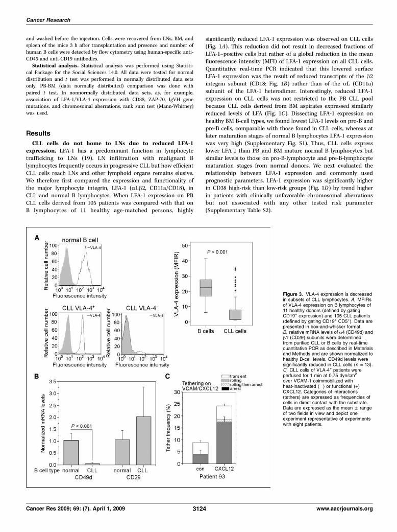
Table 1. Expression of VLA-4 in different prognostic risk groups
Risk parameters P
TotalMedian VLA-4 expression on CLL cells (MFIR) 1.3 (n = 105)CD38 B-CLL cell risk Low High 0.0004Median VLA-4 1.13 7.02
n = 62 n = 18ZAP-70 risk Low High n.s.Median VLA-4 3.12 2.44
n = 33 n = 48IgVH mutation status Mutated Unmutated 0.001Median VLA-4 1.05 4.48
n = 46 n = 26Cytogenetic aberrations Favorable Unfavorable 0.01Median VLA-4 1.05 5.61
n = 31 n = 16
NOTE: Expression of VLA-4 in different prognostic risk groups. VLA-4 expression was compared in different favorable or unfavorable clinical risk groupsas defined by the following standard prognostic thresholds: CD38 (expression on <30% versusz30% of CLL cells), ZAP-70 (NKT/B ratio >2.5 versus V2.5),IgVH mutational status (>2% as mutated versus V 2% regarded unmutated), and cytogenetics [favorable (normal, del13q) versus unfavorable(tri12, del11q, del17p)].Abbreviation: n.s., not significant.
Impaired Trafficking of CLL Cells
www.aacrjournals.org 3125 Cancer Res 2009; 69: (7). April 1, 2009

samples were not able to interact with a VCAM-1–presentingsurface and did not respond to any CXCL12 signals, suggesting thatCXCL12-CXCR4 interactions on their own are not adhesive (datanot shown).Interaction of VLA-4 (a4, CD49d) with VCAM-1 was implicated
in B-cell adhesion to activated endothelium under shear stress (25).We therefore compared VLA-4+ and VLA-4" CLL cell accumulation
on TNF-a–activated HUVECs, a prototype for cytokine-activatedvascular endothelium expressing high levels of E-selectin, VCAM-1,and ICAM-1 (26), using normal B cells as reference lymphocytes.TNF-a–activated HUVECs supported normal B-lymphocyterolling, but only small numbers of lymphocytes could arrest onthe endothelial monolayer unless encountering a potent integrinstimulatory chemokine, such as CXCL12 (Fig. 4A, left). Accordingly,
Figure 4. CLL cells require LFA-1 to transmigrateacross activated endothelium under shear flow.A, left, normal B-cell accumulation on TNF-a–stimulatedHUVEC cells. Purified B cells were perfused over theHUVEC monolayer at 0.75 dyn/cm2 for 60 s. Whereindicated, HUVECs were preoverlaid with CXCL12.Categories of interactions were determined in frequenciesof interacting cells within the cell flux in direct contactwith the monolayer and are expressed as themean F range of two fields in view. Right, B cellsaccumulated at 0.75 dyn/cm2 for 60 s on HUVECs alone oroverlaid with CXCL12 were subjected to physiologicshear stress (5 dyn/cm2) for 15 min. Shown are relativenumbers of each migratory phenotype. B, left, VLA-4+ CLLcell accumulation on TNF-a–stimulated HUVEC cellswith and without CXCL12 overlay; right, effects ofCXCL12 on the adhesive and migratory phenotype of theaccumulated CLL cells when subjected to physiologicshear stress after the accumulation phase. Examplepatient 105, Supplementary Table S1, representativefor five tested patients. C, left, effects of CXCL12 onthe accumulation of LFA-1 and VLA-4+ CLL cells onTNF-a–stimulated HUVEC; right, effects of CXCL12 onthe adhesive and migratory phenotypes of the initiallyaccumulated CLL cells shown in the left panel. Examplepatient 74, Supplementary Table S1. D, accumulation ofVLA-4" LFA-1 low CLL cells on TNF-a–stimulated HUVECcells. Example patient 95, Supplementary Table S1,representative for three tested patients.
Cancer Research
Cancer Res 2009; 69: (7). April 1, 2009 3126 www.aacrjournals.org

occasionally arrested B lymphocytes readily detached on applica-tion of physiologic shear flow and did not transmigrate theendothelium unless encountering CXCL12 activation (Fig. 4A,right). CXCL12 stimulated not only B-lymphocyte arrest but alsoadhesion strengthening, locomotion (crawling) on the EC mono-layer, and TEM (Fig. 4A, right). In contrast to normal B cells, VLA-4+
LFA-1 low CLL cells accumulated on the HUVECs but exhibitedvery poor response to CXCL12 (Fig. 4B, left). Consequently, all ofthese arrested CLL cells detached from the endothelium and nonecould transmigrate under shear flow (Fig. 4B, right). Interestingly,a rare subset of VLA-4+ CLL cell samples, with exceptionally highLFA-1 expression (Supplementary Table S1, patient 74, see alsooutliners Fig. 1A), resisted detachment from the TNF-a–activatedHUVECs after initial arrest, and in response to CXCL12, asignificant fraction of these cells also successfully transmigratedthrough HUVECs (Fig. 4C). CLL cells, negative for VLA-4 and low inLFA-1, failed to transiently interact with the activated HUVECseven in the presence of CXCL12 (Fig. 4D). Taken together, theseresults suggest that VLA-4 on CLL cells, although responsive tochemokine signals, must cooperate with LFA-1 to establish optimal
adhesion strengthening and TEM capacity across inflamedendothelial barriers under shear flow.Functional VLA-4 is indispensable for residual CLL homing
to BM, whereas LFA-1 is not. Our in vitro experiments suggestedthat the loss of LFA-1 adhesiveness and the defective transmigra-tory capability of CLL cells across E-selectin–expressing, VCAM-1–expressing, and ICAM-1–expressing HUVECs may result inimpaired in vivo CLL crossing of BM endothelial cells, known toalso coexpress these adhesion molecules (27). We therefore nextinvestigated the capability of CLL cells and normal B lymphocytesto home to BM of NOD/SCID mice in short-term adoptive assays.Consistent with their in vitro properties, VLA-4+ CLL sampleshomed in significantly lower numbers to the murine BM thannormal B lymphocytes (P = 0.005; Fig. 5A) and VLA-4" CLLcompletely failed to reach the BM (P = 0.002; Fig. 5A). Consistentwith their in vitro properties, VLA-4+ CLL samples homed insignificantly lower numbers to the BM than normal B lymphocytes(P = 0.005; Fig. 5A) and VLA-4" CLL completely failed to reachthe BM (P = 0.002; Fig. 5A). Residual VLA-4+ BM homing of CLLcells could be blocked by anti-a4 antibody (P = 0.013), showing that
Figure 5. Functional VLA-4 expression isindispensable for the homing of normal and CLL cellsto the BM. A, PBMCs (15–25 ! 106) of either healthypersons or CLL patients were injected intoNOD/SCID mice, and after 3 h, human B cells or CLLcells were detected in the BM by flow cytometry usingspecific anti-human CD19 and anti-CD45 antibodies.Columns, mean of detected human CD19+,CD45+ cells per 106 acquired BM cells and 106
injected PBMCs either from healthy donors (n = 10)or from VLA-4+ (n = 7) or VLA-4" (n = 4) CLLsamples; bars, SD. B, PBMCs were treated withblocking LFA-1 or VLA-4 antibodies and injected intoNOD/SCID mice. Human cells were determined as inA. Columns, mean of experiments with three healthydonors and four VLA-4+ CLL patients; bars, SD.
Impaired Trafficking of CLL Cells
www.aacrjournals.org 3127 Cancer Res 2009; 69: (7). April 1, 2009
CLL migration across BM endothelial barriers is VLA-4 integrindependent (Fig. 5B).CLL cells home to the spleen in LFA-1–independent and
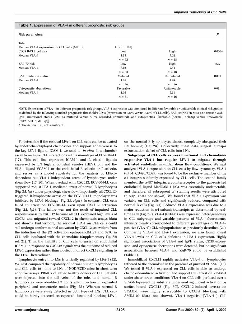
VLA-4–independent manner. Blood lymphocytes recirculatethrough the spleen (28). In light of their deficient LFA-1 functions,we next wished to assess the homing of CLL cells to the spleen.Notably, in contrast to the poor homing rates of VLA-4+ CLL cellsto BM, the lack of VLA-4" CLL cell homing to BM, and thecomplete failure of both VLA-4+ and VLA-4" CLL cells to home toLNs, CLL cells readily homed to the spleen. Relative spleen homingof CLL cells was by trend higher than that of normal B cells(Fig. 6A) without any difference related to LFA-1 or VLA-4expression. In absolute numbers, we recovered in average f8%(range, 2–25%) of the injected human B or CLL lymphocytes in LNs,BM, and spleen 3 hours after the injection. The highest absolutenumbers of human cells were thereby consistently observed in themurine spleens. After injection of normal B lymphocytes, 85% ofthe recovered B cells were located in the spleen, and after injectionof CLL lymphocytes, 98% of the recovered cells were located in thespleen, whereas only 2% and 0% of the recovered CLL cells enteredthe BM or LN, respectively, further showing the deficient CLLhoming capability to these organs. Interestingly, either pretreat-ment of normal B lymphocytes with VLA-4 or LFA-1 blockingantibodies or pretreatment of VLA-4+ CLL cells with anti–VLA-4blocking antibody did not alter their homing to the spleen (Fig. 6B).Thus, both normal and CLL lymphocyte migration to the spleenwas VLA-4 or LFA-1 integrin independent. These results collectivelysuggest that the defective ability of CLL to home to the LNs andthe BM is mirrored by enhanced retention in the circulation anda preferential tendency to enter the spleen.
Discussion
The TEM of lymphocytes and their localization in primary andsecondary lymphoid organs depend on multiple intercellularadherence mechanisms, primarily governed by integrins (29).Changes in expression or function of integrins on malignantlymphocytes can lead to different extravasation, homing, andretention properties of these cells and thereby dramatically alter
leukemic cell fate (30). Little is known about the capacity ofcirculating CLL cells to extravasate at distinct lymphoid organsand the specific roles of the two major lymphocyte integrins LFA-1and VLA-4 in this process. Furthermore, although VLA-4 wasrecently suggested as a novel prognostic marker in CLL (24, 31, 32),the specific functions of this integrin and of the second majorlymphocyte integrin, LFA-1, in CLL trafficking have been elusive.Importantly, our present results suggest that CLL traffickingthrough the BM, the spleen, and the LNs shares similar integrinusage with the trafficking of normal B lymphocytes through theseorgans. We find that normal B cells use VLA-4 but not LFA-1 toenter the BM, require LFA-1 to enter LNs, and enter the spleenindependently of either VLA-4 or LFA-1. Whereas CLL traffickingseems to be governed by the same molecular pathways, the lowLFA-1 and variable VLA-4 expression result in significantdeficiencies in entering lymphoid organs other than the spleen.Consequently, the majority of CLL samples tested by us whilefailing to enter LNs and poorly migrating to the BM preferentiallyhomed to the spleen despite inherently deficient LFA-1 or VLA-4expression and function.The severity of CLL is generally described using staging systems
devised by Rai and colleagues (33) and Binet and colleagues (34).To exclude chemotherapeutic influences on integrin expressionand signaling, and to study migratory behavior of CLL cells at thebeginning of the disease, our study concentrated on CLL samples ofcurrently untreated patients. Consequently, our cohort is mostlycomposed of early and intermediate stages (Rai 0, I, and II;Supplementary Table S1). The majority of these CLL samplesexpressed severely reduced LFA-1 levels and failed to home to LNsbut exhibited spleen tropism (19). Lymphocyte spleen homing doesnot necessarily require integrin activation (35). Our CLL homingdata are reminiscent of those of murine LFA-1 knockout mice,which exhibit impaired LN entry but high tropism toward spleen(19). In addition, VLA-4 blockage did not alter the spleen homing ofeither normal B cells or CLL cells in our experiments. Our resultsare furthermore highly consistent with a recently published CLLxenograft model in which highest absolute numbers of humancells were recovered in murine spleens (36). These recovered CLLcells displayed an activated and proliferative phenotype with
Figure 6. CLL cells preferentially home to spleen. A, PBMCs (15–25 ! 106) of either healthy persons or CLL patients were injected into NOD/SCID mice, and after3 h, human B cells or CLL cells were detected in the spleen by flow cytometry using specific anti-human CD19 and anti-CD45 antibodies. Columns, mean of detectedhuman CD19+, CD45+ cells normalized to 106 acquired spleen cells and 106 PBMC cells from either healthy donors (n = 7) or CLL patients (n = 8); bars, SD. B, PBMCswere treated with blocking LFA-1 or VLA-4 antibodies and injected into NOD/SCID mice. Human cells were determined as in A. Columns, mean of three to fourexperiments; bars, SD.
Cancer Research
Cancer Res 2009; 69: (7). April 1, 2009 3128 www.aacrjournals.org

paraimmunoblast- or prolymphocytic-like morphology, thus indi-cating the spleen microenvironment to be favoring for CLL cells(36). It is therefore possible that in early and intermediate stagesof CLL, malignant B lymphocytes released from the BM fail toreturn to the BM or enter LNs and prefer integrin-independentpathways to enter and temporally accumulate in the spleen wherethey may gain survival and proliferation signals. Indeed, progres-sive spleen enlargement is a feature of Tcl-1 transgenic micedeveloping a CLL-like disorder resembling human B-CLL (37).Our results are in accordance with previous reports on low
integrin expression of CLL cells (38, 39). In addition, consistentwith our findings, a very early human study using 51Cr labelingindicated that CLL cells leave the circulation less rapidly thanlymphocytes from healthy persons (40). Furthermore, [3H]thymidine-labeled CLL cells were shown to survive in the circulation of a CLLpatient for many weeks without any evidence of extravasation (41).As we observe that CLL cases with high LFA-1 expression andtransmigratory capacity can enter LNs at low numbers (data notshown), we cannot exclude that small CLL subsets, underrepresentedin the circulating pools characterized by us, successfully home to LNs.As LN HEVs express VCAM-1 (42), VLA-4 might also contribute to avery low entry of CLL into LNs. In spite of the overall reduced entry ofCLL cells to LN, these cells may nevertheless accumulate in LNs dueto decreased egress through efferent lymphatics in light of reducedexpression or activity of egress receptors, a possibility that shouldbe addressed in the future. Another nonmutually exclusive possibilityis high proliferation of small CLL subsets within pseudofollicles(43). Interestingly, we find significantly higher LFA-1 expression inCD38 high-risk CLL samples. LFA-1 expression also defines theproliferative and/or progressive pool in multiple myeloma and acutemyeloid leukemia and LFA-1 antagonists are under evaluation (seeref. 30 for review). In addition, small lymphocytic leukemia cellsexpress high LFA-1 and are essentially compartmentalized withinLNs with few circulating cells (44). In contrast, in CLL, the circulatingpool may be disproportionally higher, probably due to their reducedLFA-1 and VLA-4 integrins obligatory for constitutive extravasationfrom blood vessels into LNs and BM. Interestingly, in a subgroup ofCLL patients with clinical lymphoadenopathy, autocrine vascularendothelial growth factor (VEGF) was argued to promote a cross-talkbetween VLA-4 and LFA-1 and thereby augment CLL TEM (45).Nevertheless, the role of VEGF in LFA-1 activation was shown in theabsence of shear flow (45), and so, its ability to activate CLL LFA-1within the vasculature should be further addressed.Our results emphasize the importance of VLA-4 expression and
function in CLL homing to BM. VLA-4 plays a key role not only inlymphocyte adhesion to and extravasation through endothelialbarriers expressing its key ligand, VCAM-1, but also in hemato-poietic cell retention to the BM stroma, which is a prerequisite forprogenitor cell proliferation (7). Only VLA-4+ CLL cells were able toresidually home to BM. Interestingly, pre-B-ALL cells also displaythe low LFA-1 expression (46) and use VLA-4 rather than LFA-1 tohome to the BM (12). This central role of VLA-4 in CLL-BMinteractions is clinically interesting because VLA-4–dependent CLL
adhesion to VCAM-1 or fibronectin at BM niches might contributeto cell adhesion–mediated drug resistance.Our study was mostly conducted on stages Rai 0, II, and II, and
within those groups, the majority of CLL cells expressed highlyreduced levels of both investigated integrins, VLA-4 and LFA-1.Furthermore, our data support recent observations achieved on axenoplant CLL model (36) in which human CLL cells were recoveredat low numbers from BM but at high numbers from spleen. BMrecovery, and possibly also LN recovery, may correlate with clinicaldisease activity. Very recently, three groups suggested VLA-4 (CD49d)as a novel independent prognostic marker for CLL predicting over-all survival (24, 31) and/or progressive disease (31, 32). The studiesdiffer due to cohort variations in their results about associations ofVLA-4 expression to several prognostic parameters, such as ZAP-70or IgVH mutational status, but unequivocally observe high asso-ciation of VLA-4 expression with CD38 (31). In complete accordanceto these recent reports, we observed highly significant higher VLA-4expression in CD38 high-risk groups compared with CD38 low-risk groups with nearly total loss of VLA-4 in the low-risk group.We also found significant higher VLA-4 expression in CLL samplesof patients with unmutated IgVH status and with unfavorablecytogenetic abnormalities (trisomy 12, deletion11q, deletion 17p).The low expression of both VLA-4 and LFA-1 in low-risk CLL
groups might explain the more favorable clinical course of thesepatients because it obviously restricts circulating CLL cells fromreentering the BM and from homing to the LNs, organs at whichthese cells are likely to encounter antiapoptotic factors. Our resultsalso highlight the possibility that VLA-4 and LFA-1 detection couldbe useful prognostic markers of CLL severity. More importantly,it can aid in evaluating the susceptibility of CLL cells to VLA-4 andLFA-1 blocking therapy. Anti–VLA-4 antibody treatment also causesCD34+ progenitor cell mobilization from the BM (47). Stem cellmobilization is generally associated with functional inactivationor down-regulation of VLA-4–VCAM-1 interactions withinthe BM niches (48). Therefore, anti–VLA-4 blocking therapycould be highly effective in releasing VLA-4+ CLL from the BM andpreventing their reentry to the BM. Together with their reducedlevels of LFA-1, these VLA-4–blocked CLL cells will be restrictedfrom emigration into supportive LN and BM niches, home to thespleen, andmay be rendered more susceptible to CLL chemotherapy.
Disclosure of Potential Conflicts of Interest
No potential conflicts of interest were disclosed.
Acknowledgments
Received 10/29/08; revised 1/11/09; accepted 1/15/09; published OnlineFirst 3/17/09.Grant support: Israel Science Foundation, Minerva Foundation of Germany,
Morros Foundation, and grants from the province of Salzburg, Austria. T.N. Hartmannwas funded by the Minerva Foundation and grants from the province of Salzburg.A. Vallon-Eberhard is supported by the Society of the Swiss Friends of the WeizmannInstitute of Science.
The costs of publication of this article were defrayed in part by the payment of pagecharges. This article must therefore be hereby marked advertisement in accordancewith 18 U.S.C. Section 1734 solely to indicate this fact.
References1. Stauder R, Hamader S, Fasching B, Kemmler G, ThalerJ, Huber H. Adhesion to high endothelial venules: amodel for dissemination mechanisms in non-Hodgkin’slymphoma. Blood 1993;82:262–7.
2. Caligaris-Cappio F, Riva M, Tesio L, Schena M, GaidanoG, Bergui L. Human normal CD5+ B lymphocytes can beinduced to differentiate to CD5" B lymphocytes withgerminal center cell features. Blood 1989;73:1259–63.
3. Alon R, Luscinskas FW. Crawling and INTEGRatingapical cues. Nat Immunol 2004;5:351–3.
4. Shimonaka M, Katagiri K, Nakayama T, et al. Rap1translates chemokine signals to integrin activation, cellpolarization, and motility across vascular endotheliumunder flow. J Cell Biol 2003;161:417–27.
5. Alon R, Feigelson S. From rolling to arrest on bloodvessels: leukocyte tap dancing on endothelial integrin
Impaired Trafficking of CLL Cells
www.aacrjournals.org 3129 Cancer Res 2009; 69: (7). April 1, 2009

Cancer Research
Cancer Res 2009; 69: (7). April 1, 2009 3130 www.aacrjournals.org
ligands and chemokines at sub-second contacts. SeminImmunol 2002;14:93–104.
6. Kinashi T. Intracellular signalling controlling integrinactivation in lymphocytes. Nat Rev Immunol 2005;5:546–59.
7. Peled A, Kollet O, Ponomaryov T, et al. The chemokineSDF-1 activates the integrins LFA-1, VLA-4, and VLA-5on immature human CD34(+) cells: role in trans-endothelial/stromal migration and engraftment ofNOD/SCID mice. Blood 2000;95:3289–96.
8. Peled A, Petit I, Kollet O, et al. Dependence of humanstem cell engraftment and repopulation of NOD/SCIDmice on CXCR4. Science 1999;283:845–8.
9. Imai K, Kobayashi M, Wang J, et al. Selective secretionof chemoattractants for haemopoietic progenitor cellsby bone marrow endothelial cells: a possible role inhoming of haemopoietic progenitor cells to bonemarrow. Br J Haematol 1999;106:905–11.
10. Kamel-Reid S, Letarte M, Sirard C, et al. A model ofhuman acute lymphoblastic leukemia in immune-deficient SCID mice. Science 1989;246:1597–600.
11. Uckun FM. Severe combined immunodeficientmouse models of human leukemia. Blood 1996;88:1135–46.
12. Spiegel A, Kollet O, Peled A, et al. Unique SDF-1-induced activation of human precursor-B ALL cells as aresult of altered CXCR4 expression and signaling. Blood2004;103:2900–7.
13. Dohner H, Stilgenbauer S, Benner A, et al. Genomicaberrations and survival in chronic lymphocytic leuke-mia. N Engl J Med 2000;343:1910–6.
14. Gryshchenko I, Hofbauer S, Stoecher M, et al. MDM2SNP309 is associated with poor outcome in B-cellchronic lymphocytic leukemia. J Clin Oncol 2008;26:2252–7.
15. Letestu R, Rawstron A, Ghia P, et al. Evaluation ofZAP-70 expression by flow cytometry in chroniclymphocytic leukemia: a multicentric internationalharmonization process. Cytometry B Clin Cytom 2006;70:309–14.
16. Peled A, Grabovsky V, Habler L, et al. The chemokineSDF-1 stimulates integrin-mediated arrest of CD34(+)cells on vascular endothelium under shear flow. J ClinInvest 1999;104:1199–211.
17. Shamri R, Grabovsky V, Feigelson SW, Dwir O, VanKooyk Y, Alon R. Chemokine stimulation of lymphocytea4 integrin avidity but not of leukocyte function-associated antigen-1 avidity to endothelial ligands undershear flow requires cholesterol membrane rafts. J BiolChem 2002;277:40027–35.
18. Grabovsky V, Feigelson S, Chen C, et al. Subsecondinduction of a4 integrin clustering by immobilizedchemokines stimulates leukocyte tethering and rollingon endothelial vascular cell adhesion molecule 1 underflow conditions. J Exp Med 2000;192:495–506.
19. Berlin-Rufenach C, Otto F, Mathies M, et al.Lymphocyte migration in lymphocyte function-associ-ated antigen (LFA)-1-deficient mice. J Exp Med 1999;189:1467–78.
20. Grabovsky V, Dwir O, Alon R. Endothelial chemo-kines destabilize L-selectin-mediated lymphocyte rollingwithout inducing selectin shedding. J Biol Chem 2002;277:20640–50.
21. Beals CR, Edwards AC, Gottschalk RJ, Kuijpers TW,Staunton DE. CD18 activation epitopes induced byleukocyte activation. J Immunol 2001;167:6113–22.
22. Warnock RA, Askari S, Butcher EC, von Andrian UH.Molecular mechanisms of lymphocyte homing toperipheral lymph nodes. J Exp Med 1998;187:205–16.
23. Berlin C, Bargatze RF, Campbell JJ, et al. a4 Integrinsmediate lymphocyte attachment and rolling underphysiologic flow. Cell 1995;80:413–22.
24. Shanafelt TD, Geyer SM, Bone ND, et al. CD49dexpression is an independent predictor of overallsurvival in patients with chronic lymphocytic leukae-mia: a prognostic parameter with therapeutic potential.Br J Haematol 2008;140:537–46.
25. Yago T, Tsukuda M, Tajima H, et al. Analysis of initialattachment of B cells to endothelial cells under flowconditions. J Immunol 1997;158:707–14.
26. Cinamon G, Shinder V, Shamri R, Alon R. Chemo-attractant signals and h2 integrin occupancy at apicalendothelial contacts combine with shear stress signalsto promote transendothelial neutrophil migration. JImmunol 2004;173:7282–91.
27. van Buul JD, Voermans C, van den Berg V, et al.Migration of human hematopoietic progenitor cellsacross bone marrow endothelium is regulated byvascular endothelial cadherin. J Immunol 2002;168:588–96.
28. Cyster JG. Lymphoid organ development and cellmigration. Immunol Rev 2003;195:5–14.
29. Ley K, Laudanna C, Cybulsky MI, Nourshargh S.Getting to the site of inflammation: the leukocyteadhesion cascade updated. Nat Rev Immunol 2007;7:678–89.
30. Schmidmaier R, Baumann P. Anti-adhesion evolvesto a promising therapeutic concept in oncology. CurrMed Chem 2008;15:978–90.
31. Gattei V, Bulian P, Del Principe MI, et al. Relevanceof CD49d protein expression as overall survival andprogressive disease prognosticator in chronic lympho-cytic leukemia. Blood 2008;111:865–73.
32. Rossi D, Zucchetto A, Rossi FM, et al. CD49dexpression is an independent risk factor of progressivedisease in early stage chronic lymphocytic leukemia.Haematologica 2008;93:1575–9.
33. Rai KR, Sawitsky A, Cronkite EP, Chanana AD, LevyRN, Pasternack BS. Clinical staging of chronic lympho-cytic leukemia. Blood 1975;46:219–34.
34. Binet JL, Auquier A, Dighiero G, et al. A newprognostic classification of chronic lymphocytic leuke-mia derived from a multivariate survival analysis.Cancer 1981;48:198–206.
35. Saito S, Kuwashima N, Koizumi H, et al. In vivofunction of homing receptors participating in lympho-cyte recirculation: transfer analysis in SCID mice.Pathobiology 1995;63:305–13.
36. Durig J, Ebeling P, Grabellus F, et al. A novel nonobesediabetic/severe combined immunodeficient xenograftmodel for chronic lymphocytic leukemia reflects im-portant clinical characteristics of the disease. CancerRes 2007;67:8653–61.
37. Bichi R, Shinton SA, Martin ES, et al. Human chroniclymphocytic leukemia modeled in mouse by targetedTCL1 expression. Proc Natl Acad Sci U S A 2002;99:6955–60.
38. Nadkarni JJ, Perambakam SM, Rathore VB, et al.Expression of adhesion molecules in B-cell chroniclymphocytic leukaemia: an analysis in lymphoidcompartments—peripheral blood, bone marrow andlymph node. Cancer Biother Radiopharm 1998;13:269–74.
39. Lucio PJ, Faria MT, Pinto AM, et al. Expression ofadhesion molecules in chronic B-cell lymphoprolifer-ative disorders. Haematologica 1998;83:104–11.
40. Bazerbashi MB, Reeve J, Chanarin I. Studies inchronic lymphocytic leukaemia. The kinetics of 51Cr-labelled lymphocytes. Scand J Haematol 1978;20:37–51.
41. Dormer P, Theml H, Lau B. Chronic lymphocyticleukemia: a proliferative or accumulative disorder? LeukRes 1983;7:1–10.
42. Till KJ, Lin K, Zuzel M, Cawley JC. The chemokinereceptor CCR7 and a4 integrin are important formigration of chronic lymphocytic leukemia cells intolymph nodes. Blood 2002;99:2977–84.
43. Patten PE, Buggins AG, Richards J, et al. CD38expression in chronic lymphocytic leukemia is regu-lated by the tumor microenvironment. Blood 2008;111:5173–81.
44. Angelopoulou MK, Kontopidou FN, Pangalis GA.Adhesion molecules in B-chronic lymphoproliferativedisorders. Semin Hematol 1999;36:178–97.
45. Till KJ, Spiller DG, Harris RJ, Chen H, Zuzel M,Cawley JC. CLL, but not normal, B cells are dependenton autocrine VEGF and a4h1 integrin for chemokine-induced motility on and through endothelium. Blood2005;105:4813–9.
46. Geijtenbeek TB, van Kooyk Y, van Vliet SJ, Renes MH,Raymakers RA, Figdor CG. High frequency of adhesiondefects in B-lineage acute lymphoblastic leukemia.Blood 1999;94:754–64.
47. Zohren F, Toutzaris D, Klarner V, Hartung HP,Kieseier B, Haas R. The monoclonal anti-VLA-4 antibodynatalizumab mobilizes CD34+ hematopoietic progenitorcells in humans. Blood 2008;111:3893–5.
48. Lichterfeld M, Martin S, Burkly L, Haas R, Kronen-wett R. Mobilization of CD34+ haematopoietic stem cellsis associated with a functional inactivation of theintegrin very late antigen 4. Br J Haematol 2000;110:71–81.
49. Hamblin TJ, Orchard JA, Ibbotson RE, et al. CD38expression and immunoglobulin variable regionmutations are independent prognostic variables inchronic lymphocytic leukemia, but CD38 expressionmay vary during the course of the disease. Blood2002;99:1023–9.
Copyright © 2022 FDOKUMEN