thee hi)tological lesion in lymph nodes in infectious ... - NCBI
Upload
independentCategory
view
0download
0
2005;65:2147-2156. Cancer Res Robert L. Ferris, Liqiang Xi, Siva Raja, et al. Cell Carcinoma of the Head and NeckMolecular Staging of Cervical Lymph Nodes in Squamous
Updated version
http://cancerres.aacrjournals.org/content/65/6/2147
Access the most recent version of this article at:
Material
Supplementary
http://cancerres.aacrjournals.org/content/suppl/2005/04/01/65.6.2147.DC1.html
Access the most recent supplemental material at:
Cited Articles
http://cancerres.aacrjournals.org/content/65/6/2147.full.html#ref-list-1
This article cites by 46 articles, 10 of which you can access for free at:
Citing articles
http://cancerres.aacrjournals.org/content/65/6/2147.full.html#related-urls
This article has been cited by 17 HighWire-hosted articles. Access the articles at:
E-mail alerts related to this article or journal.Sign up to receive free email-alerts
Subscriptions
Reprints and
To order reprints of this article or to subscribe to the journal, contact the AACR Publications
Permissions
To request permission to re-use all or part of this article, contact the AACR Publications
Research. on February 6, 2014. © 2005 American Association for Cancercancerres.aacrjournals.org Downloaded from
Research. on February 6, 2014. © 2005 American Association for Cancercancerres.aacrjournals.org Downloaded from
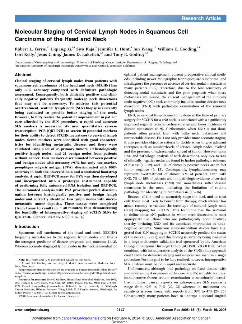
Molecular Staging of Cervical Lymph Nodes in Squamous Cell
Carcinoma of the Head and Neck
Robert L. Ferris,1,2Liqiang Xi,
2,3Siva Raja,
3Jennifer L. Hunt,
4Jun Wang,
1,2William E. Gooding,
2,5
Lori Kelly,2Jesus Ching,
6James D. Luketich,
2,3and Tony E. Godfrey
2,3
1Departments of Otolaryngology and Immunology, 2University of Pittsburgh Cancer Institute, Departments of 3Surgery, 4Pathology, and5Biostatistics, University of Pittsburgh, Pittsburgh, Pennsylvania; and 6Cepheid, Sunnyvale, California
Abstract
Clinical staging of cervical lymph nodes from patients withsquamous cell carcinoma of the head and neck (SCCHN) hasonly 50% accuracy compared with definitive pathologicassessment. Consequently, both clinically positive and clini-cally negative patients frequently undergo neck dissectionsthat may not be necessary. To address this potentialovertreatment, sentinel lymph node (SLN) biopsy is currentlybeing evaluated to provide better staging of the neck.However, to fully realize the potential improvement in patientcare afforded by the SLN procedure, a rapid and accurateSLN analysis is necessary. We used quantitative reversetranscription––PCR (QRT-PCR) to screen 40 potential markersfor their ability to detect SCCHN metastases to cervical lymphnodes. Seven markers were identified with good character-istics for identifying metastatic disease, and these werevalidated using a set of 26 primary tumors, 19 histologicallypositive lymph nodes, and 21 benign nodes from patientswithout cancer. Four markers discriminated between positiveand benign nodes with accuracy >>97% but only one marker,pemphigus vulgaris antigen (PVA), discriminated with 100%accuracy in both the observed data and a statistical bootstrapanalysis. A rapid QRT-PCR assay for PVA was then developedand incorporated into a prototype instrument capableof performing fully automated RNA isolation and QRT-PCR.The automated analysis with PVA provided perfect discrimi-nation between histologically positive and benign lymphnodes and correctly identified two lymph nodes with micro-metastatic tumor deposits. These assays were completed( from tissue to result) in ff30 minutes, thus demonstratingthe feasibility of intraoperative staging of SCCHN SLNs byQRT-PCR. (Cancer Res 2005; 65(6): 2147-56)
Introduction
Squamous cell carcinoma of the head and neck (SCCHN)frequently metastasizes to the regional lymph nodes and this isthe strongest predictor of disease prognosis and outcome (1, 2).Whereas accurate staging of lymph nodes in the neck is essential for
optimal patient management, current preoperative clinical meth-ods, including newer radiographic techniques, are suboptimal andmisdiagnose the presence or absence of cervical nodal metastasis inmany patients (3–5). Therefore, due to the low sensitivity ofdetecting nodal metastasis and the poor prognosis when thesemetastases are missed, the current management of the clinicallynode negative (cN0) neck commonly includes routine elective neckdissection (END) with pathologic examination of the removedlymph nodes.END, or cervical lymphadenectomy done at the time of primary
surgery for SCCHN for a cN0 neck, is associated with a significantlyimproved regional recurrence-free survival and lower incidence ofdistant metastases (6–9). Furthermore, when END is not done,patients often present later with bulky neck metastases andunresectable disease. END not only provides more accurate staging,it also provides objective criteria to decide when to give adjuvanttherapies, such as number/levels of cervical lymph nodes involvedand the presence of extracapsular spread of tumor. However, uponEND and pathologic analysis of neck dissections, only 25% to 30%of clinically negative necks are found to harbor pathologic evidenceof disease (10–12), and 15% of clinically positive necks are in facttumor negative (6, 13). Consequently, lymphadenectomy mayrepresent overtreatment of almost 50% of patients. Even withEND, 7% to 15% of patients with no pathologic evidence of cervicallymph node metastases (pN0) will nonetheless suffer diseaserecurrence in the neck, indicating the limitation of routinepathology for identifying micrometastasis (13–16).Because of the need to accurately stage the neck and to treat
only those most likely to benefit from therapy, much interest hasarisen recently to validate the technique of sentinel lymph node(SLN) mapping for SCCHN. This technique has the potentialto define those cN0 patients in whom neck dissection is mostappropriate (i.e., those who are pathologically node positive),thereby obviating END and its associated morbidities in node-negative patients. Numerous single-institution studies have sug-gested that SLN mapping in SCCHN accurately predicts the statusof the neck (5, 17–21), and this finding is currently being evaluatedin a large multicenter validation trial sponsored by the AmericanCollege of Surgeons Oncology Group (ACOSOG Z0360 trial). Whencombined with intraoperative analysis of the SLN(s), this approachcould allow for definitive staging and surgical treatment in a singleprocedure. For this goal to be fully realized, however, intraoperativeSLN analysis must be both rapid and accurate.Unfortunately, although final pathology on fixed tissues (with
immunostaining if necessary in the case of SLNs) is highly accurate,intraoperative frozen section examination is notoriously insensi-tive. In breast cancer, reports on intraoperative SLN sensitivityrange from 47% to 74% (22, 23) whereas in melanoma thesensitivity is even worse, with reports from 38% to 47% (24, 25).Consequently, many patients have to undergo a second surgical
Note: R.L. Ferris and L. Xi contributed equally to this work.L. Xi and T.E. Godfrey are currently at Mount Sinai School of Medicine, New
York, New York.Supplementary data for this article are available at Cancer Research Online (http://
cancerres.aacrjournals.org/) and at http://www.mssm.edu/labs/godfrt01/publications/supp.htm.
Requests for reprints: Tony E. Godfrey, Mount Sinai School of Medicine, Box 1079,One Gustave L. Levy Place, New York, NY 10029. Phone: 212-659-9082; Fax: 212-828-4221; E-mail: [email protected] or Robert L. Ferris, University of PittsburghCancer Institute, Hillman Research Wing 1.19d, 5117 Centre Avenue, Pittsburgh, PA15213. Phone: 412-623-7703; E-mail: [email protected].
I2005 American Association for Cancer Research.
www.aacrjournals.org 2147 Cancer Res 2005; 65: (6). March 15, 2005
Research Article
Research. on February 6, 2014. © 2005 American Association for Cancercancerres.aacrjournals.org Downloaded from
procedure to complete lymph node dissection after definitivepathologic assessment identifies microscopic metastatic diseasenot evident on intraoperative analysis. Our own work, and thatreported by others, suggests that low sensitivity may also be anissue for intraoperative SLN analysis in SCCHN (18, 21). Performinga second surgery on the neck is an undesirable scenario becausethis would likely increase complications and morbidity and coulddelay the use of adjuvant therapy. This is in addition to the extracost, discomfort, and psychological toll on the patient. Such issuesmay negatively affect the widespread acceptance of SLN biopsy inSCCHN.Reverse transcription–PCR (RT-PCR) has great theoretical
potential for the detection of small tumor deposits in lymphnodes. Studies in many tumor types including esophageal cancer,colon cancer, breast cancer, melanoma, and SCCHN have showedthe ability of an RT-PCR–based assay to detect histologically occultmicrometastases (7, 9, 26–29). Many of the earlier RT-PCR studiessuffered from poor specificity, but the introduction of real-timequantitative RT-PCR (QRT-PCR) has helped overcome this problemand provides the potential for objective and quantitative analysisof lymph nodes (30–33). Whereas we are also interested indetection of occult disease by QRT-PCR, we have recently focusedon application of this method to rapid, intraoperative analysis ofSLNs. We have shown that RNA isolation and QRT-PCR can be donein a time frame amenable to intraoperative analysis and we havealso developed multiplex PCR technology to allow incorporation ofnecessary controls into rapid QRT-PCR (34, 35). Unfortunately,these reports used manual RNA isolation and QRT-PCR setup, andthis is likely to be too labor intensive, prone to contamination, andvariable for true clinical use in an intraoperative setting. Thisconcern has recently been addressed, however, with the develop-ment of a fully automated and integrated RNA isolation andquantitative PCR instrument called the GeneXpert (Cepheid,Sunnyvale, CA).7 The GeneXpert is capable of performing RNAisolation from lysed tissue, reverse transcription, and quantitativePCR all in f30 minutes. Our goals in this study were to identifyappropriate QRT-PCR markers for detection of metastasis to lymphnodes and to use the GeneXpert to show the feasibility ofautomated analysis in an intraoperative time frame. When appliedto SLNs from patients with SCCHN, we show that this technologyhas the potential to accurately stage the neck and allow thesurgeon to provide optimal treatment in a single procedure.
Materials and Methods
PatientsA total of 26 primary tumors and 24 histologically tumor involved nodes
were harvested at surgery and immediately snap-frozen at �80jC until RNA
extraction was done. Of these specimens, eight sets of paired human head
and neck primary tumors and tumor-containing metastatic lymph nodes( from the same 8 patients with SCCHN) were included in this study. Benign
lymph nodes (n = 21) were obtained from patients undergoing surgery for
benign esophageal disorders (gastroesophageal reflux or achalasia).
Institutional review board–approved, written informed consent wasobtained from all patients donating specimens for this study, either
through the ENT Registry at the Department of Otolaryngology or the
Esophageal Risk Registry at the Division of Thoracic and Foregut Surgery,
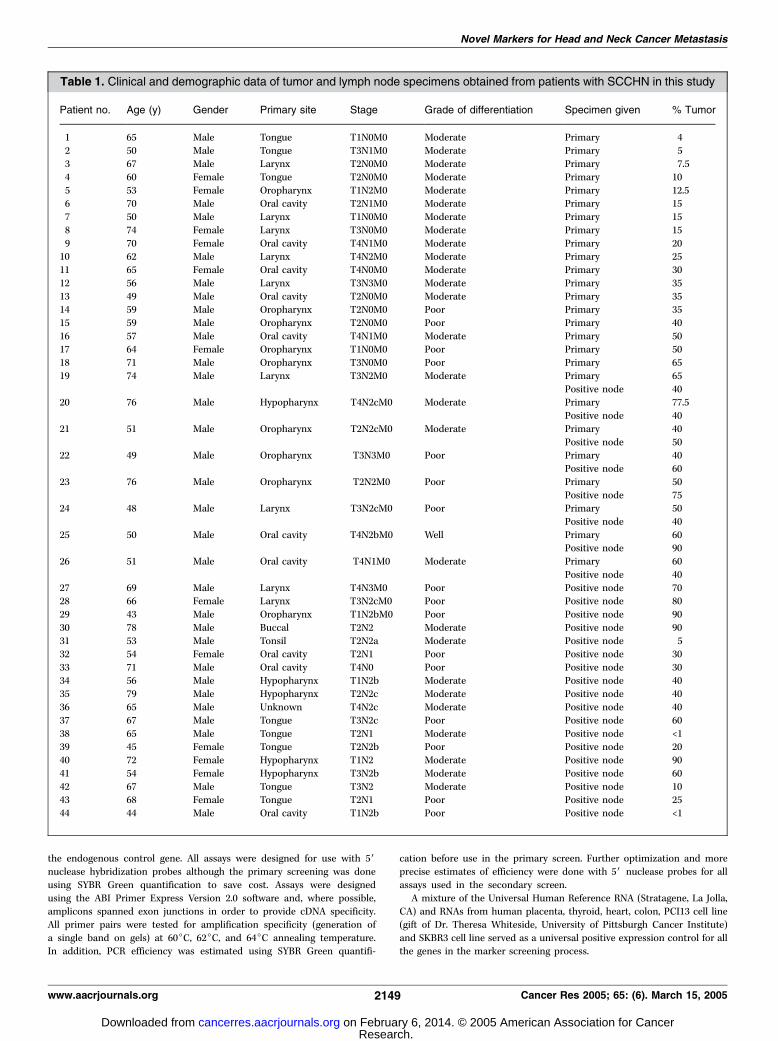
University of Pittsburgh. Clinical and demographic data for fresh tumor/metastasis specimens are shown in Table 1. These patients are similar to
historical patients with SCCHN in our practice, with tumors distributedthroughout all head and neck subsites. In the 44 patients studied,
histologically verified squamous cell carcinoma originated in one of the
following primary sites: oral cavity (18), oropharynx (10), larynx (10),
hypopharynx (5), and site undetermined (1).
Identification of Potential MarkersAn extensive literature and public database survey was conducted to
identify any potential markers. Resources for this survey included
PubMed, OMIM, UniGene (http://www.ncbi.nlm.nih.gov/), GeneCards
(http://bioinfo.weizmann.ac.il/cards), and CGAP (http://cgap.nci.nih.gov).Our survey criteria were somewhat flexible but the goal was to identify
genes with moderate to high expression in head and neck cancer and low
expression in normal lymph nodes. In addition, genes reported to be up-
regulated in head and neck cancer and genes with restricted tissuedistribution were considered potentially useful. Finally, genes reported to
be cancer specific, such as the cancer testis antigens and hTERT, were
evaluated.
Tissues and Pathologic EvaluationArchived tissue from all patients in this study was reviewed by
a specialized head and neck pathologist (J.L.H.), and the diagnosis was
reconfirmed histologically. Twenty 5-Am sections were cut from each
optimal cutting temperature compound (OCT)–embedded tissue for RNA
isolation. In addition, sections were cut and placed on slides for H&E andimmunohistochemistry analysis at the beginning, middle (between the 10th
and 11th sections for RNA), and end of the sections for RNA isolation. All
three H&E slides from each specimen underwent pathologic review to
confirm presence of tumor, percentage of tumor, and to identify thepresence of any contaminating tissues. All of the unstained slides were
stored at �20j. Immunohistochemistry evaluation was done using the
antibody pan keratin (AE1/AE3) mixture (DAKO, Carpinteria, CA), and
Vector Elite ABC kit and Vector AEC Chromagen (Vector Laboratories,Burlingame, CA). Immunohistochemistry was used as needed to confirm
the H&E histology.
Screening ApproachThe screening was conducted in two phases. All potential markers
entered the primary screening phase and expression was analyzed in 6primary tumors and 10 benign lymph nodes obtained from patients without
cancer ( five RNA pools with 2 lymph node RNAs per pool). Markers that
showed good characteristics for lymph node metastasis detection
(consistent, high expression in tumors and very low expression in benignnodes) passed into the secondary screening phase. The secondary screen
consisted of expression analysis on 26 primary tumors, 19 histologically
positive lymph nodes, and 21 benign lymph nodes without cancer.
RNA Isolation and cDNA SynthesisRNA was isolated using the RNeasy minikit (Qiagen, Valencia, CA)
essentially as described by the manufacturer. The only modification was that
we doubled the volume of lysis reagent and loaded the column in two steps.
This was found to provide better RNA yield and purity, probably because of
diluting out the OCT in the tissue sections. Reverse transcription was done in
100-AL reaction volumes with random hexamer priming and SuperScript II
(Invitrogen, Carlsbad, CA) reverse transcriptase (36). For the primary screen,
two reverse transcription reactionswere done, eachwith 1,000 ng of RNA. The
cDNAs were combined and quantitative PCR was done using the equivalent
of 20 ng RNA per reaction. For the secondary screen, the RNA input for
primary tumors and positive nodes was 1200 ng per reverse transcription
reaction and 20 ng per quantitative PCR reaction, but this was increased to
2000 ng per reverse transcription reaction and 80 ng per quantitative PCR
reaction for the benign nodes in order to improve sensitivity for detection of
low background expression.
Quantitative PCRAll quantitative PCR for marker screening was done on the ABI Prism
7700 Sequence Detection Instrument (Applied Biosystems, FosterCity, CA). Relative expression of the marker genes was calculated using
the DCT methods previously described (37) and with h-glucuronidase as7 Manuscript submitted for publication.
Cancer Research
Cancer Res 2005; 65: (6). March 15, 2005 2148 www.aacrjournals.org
Research. on February 6, 2014. © 2005 American Association for Cancercancerres.aacrjournals.org Downloaded from
the endogenous control gene. All assays were designed for use with 5Vnuclease hybridization probes although the primary screening was done
using SYBR Green quantification to save cost. Assays were designed
using the ABI Primer Express Version 2.0 software and, where possible,amplicons spanned exon junctions in order to provide cDNA specificity.
All primer pairs were tested for amplification specificity (generation of
a single band on gels) at 60jC, 62jC, and 64jC annealing temperature.In addition, PCR efficiency was estimated using SYBR Green quantifi-
cation before use in the primary screen. Further optimization and more
precise estimates of efficiency were done with 5V nuclease probes for all
assays used in the secondary screen.
A mixture of the Universal Human Reference RNA (Stratagene, La Jolla,CA) and RNAs from human placenta, thyroid, heart, colon, PCI13 cell line
(gift of Dr. Theresa Whiteside, University of Pittsburgh Cancer Institute)
and SKBR3 cell line served as a universal positive expression control for allthe genes in the marker screening process.
Table 1. Clinical and demographic data of tumor and lymph node specimens obtained from patients with SCCHN in this study
Patient no. Age (y) Gender Primary site Stage Grade of differentiation Specimen given % Tumor
1 65 Male Tongue T1N0M0 Moderate Primary 4
2 50 Male Tongue T3N1M0 Moderate Primary 53 67 Male Larynx T2N0M0 Moderate Primary 7.5
4 60 Female Tongue T2N0M0 Moderate Primary 10
5 53 Female Oropharynx T1N2M0 Moderate Primary 12.56 70 Male Oral cavity T2N1M0 Moderate Primary 15
7 50 Male Larynx T1N0M0 Moderate Primary 15
8 74 Female Larynx T3N0M0 Moderate Primary 15
9 70 Female Oral cavity T4N1M0 Moderate Primary 2010 62 Male Larynx T4N2M0 Moderate Primary 25
11 65 Female Oral cavity T4N0M0 Moderate Primary 30
12 56 Male Larynx T3N3M0 Moderate Primary 35
13 49 Male Oral cavity T2N0M0 Moderate Primary 3514 59 Male Oropharynx T2N0M0 Poor Primary 35
15 59 Male Oropharynx T2N0M0 Poor Primary 40
16 57 Male Oral cavity T4N1M0 Moderate Primary 5017 64 Female Oropharynx T1N0M0 Poor Primary 50
18 71 Male Oropharynx T3N0M0 Poor Primary 65
19 74 Male Larynx T3N2M0 Moderate Primary 65
Positive node 4020 76 Male Hypopharynx T4N2cM0 Moderate Primary 77.5
Positive node 40
21 51 Male Oropharynx T2N2cM0 Moderate Primary 40
Positive node 5022 49 Male Oropharynx T3N3M0 Poor Primary 40
Positive node 60
23 76 Male Oropharynx T2N2M0 Poor Primary 50
Positive node 7524 48 Male Larynx T3N2cM0 Poor Primary 50
Positive node 40
25 50 Male Oral cavity T4N2bM0 Well Primary 60Positive node 90
26 51 Male Oral cavity T4N1M0 Moderate Primary 60
Positive node 40
27 69 Male Larynx T4N3M0 Poor Positive node 7028 66 Female Larynx T3N2cM0 Poor Positive node 80
29 43 Male Oropharynx T1N2bM0 Poor Positive node 90
30 78 Male Buccal T2N2 Moderate Positive node 90
31 53 Male Tonsil T2N2a Moderate Positive node 532 54 Female Oral cavity T2N1 Poor Positive node 30
33 71 Male Oral cavity T4N0 Poor Positive node 30
34 56 Male Hypopharynx T1N2b Moderate Positive node 4035 79 Male Hypopharynx T2N2c Moderate Positive node 40
36 65 Male Unknown T4N2c Moderate Positive node 40
37 67 Male Tongue T3N2c Poor Positive node 60
38 65 Male Tongue T2N1 Moderate Positive node <139 45 Female Tongue T2N2b Poor Positive node 20
40 72 Female Hypopharynx T1N2 Moderate Positive node 90
41 54 Female Hypopharynx T3N2b Moderate Positive node 60
42 67 Male Tongue T3N2 Moderate Positive node 1043 68 Female Tongue T2N1 Poor Positive node 25
44 44 Male Oral cavity T1N2b Poor Positive node <1
Novel Markers for Head and Neck Cancer Metastasis
www.aacrjournals.org 2149 Cancer Res 2005; 65: (6). March 15, 2005
Research. on February 6, 2014. © 2005 American Association for Cancercancerres.aacrjournals.org Downloaded from
Quantification with SYBR Green (Primary Screen)For SYBR Green I–based quantitative PCR, each 50-AL reaction
contained 1� TaqMan buffer A (Applied Biosystems), 300 nmol/L each
deoxynucleotide triphosphate, 3.5 mmol/L MgCl2, 0.06 units/AL Amplitaq
Gold (Applied Biosystems), 0.25� SYBR Green I (Molecular Probes, Eugene,OR), and 200 nmol/L each primer. The amplification program consisted of
two stages with an initial 95jC Taq activation stage for 12 minutes followed
by 40 cycles of 95jC denaturation for 15 seconds, 60jC, 62jC, or 64jCanneal/extend for 60 seconds, and a 10-second data collection step at atemperature 2jC to 4jC below the Tm of the specific PCR product being
amplified (38). After amplification, a melting curve analysis was done by
collecting fluorescence data while increasing the temperature from 60jC to
95jC over 20 minutes.
Quantification with 5V Nuclease Probes (Secondary Screen)Probe-based quantitative PCR was done as described previously (26, 37).
Briefly, reactions were done with a probe concentration of 200 nmol/L and a
60-second anneal/extend phase at 60jC for h-GUS , CK19 , PTHrP, and
SCCA1/2 , 62jC for pemphigus vulgaris antigen (PVA), and 64jC forcarcinoembryonic antigen (CEA) and TACSTD1 . The sequences of primers
and probes (purchased from IDT, Coralville, IA) for genes evaluated in the
secondary screen are listed in Supplementary Table 1. The primer
sequences for markers used in the primary screen will be provided uponrequest.
Data AnalysisIn the primary screen, data from the melting curve were analyzed using
the ABI Prism 7700 Dissociation Curve Analysis 1.0 software (Applied
Biosystems). The first derivative of the melting cure was used to determinethe product Tm as well as to establish the presence of the specific product
in each sample. In general, samples were analyzed in duplicate PCR reac-
tions and the average Ct value was used in the expression analysis. However,
in the secondary screen triplicate runs were done for each individual benignnode and the lowest Ct value was used in the calculation of relative
expression to obtain the highest value of background expression for the
sample.
Statistical AnalysisGeneration of Prediction Rules. Six markers that passed the secondary
screen were evaluated individually and in combination with other markers.
The characteristics used to evaluate markers were sensitivity, specificity,
classification, accuracy and the area under the receiver operating
characteristic curve. For individual markers, a cutoff value was determinedthat maximized the classification accuracy (proportion of lymph nodes
correctly classified). In cases wherein classification accuracy was 100%, the
cutoff was set at the midpoint between the highest expressing negativenode and the lowest expressing positive node. Markers were also combined
into pairs for lymph node classification and a linear prediction rule was
generated for each pair. The rule was equivalent to the linear predictor that
equalized the fitted probabilities above and below the linear boundary.That is, points on the boundary line had a predicted probability midway
between the numerical scores assigned to positive and negative nodes. Forexample, if positive nodes were assigned a score of 2 and benign nodes a
score of 1, predicted scores >1.5 were classified as positive.
Internal Validation of Prediction Rules. Internal validation of
prediction rules was conducted by nonparametric bootstrap resamplingusing Efron and Tibshirani’s improved bootstrap method (39), in which 500
bootstrap samples of lymph nodes are selected from the pool of all positive
and negative nodes. The optimism in the original estimates of sensitivity,
specificity, and classification accuracy are then calculated as the differencebetween the bootstrap classification statistic applied to the original data
and applied to the bootstrap data. The average difference over all bootstrap
samples is computed and reported as the bias in the values derived from the
observed data and then subtracted from the original estimates to producethe bootstrap-validated estimates.
GeneXpert Analysis. Twenty-four 5-Am sections of OCT-embedded
tissue were sectioned into 800 AL of GeneXpert lysis buffer (Cepheid). Thelysis buffer was filtered through a 0.22-mm syringe filter (Osmonics Inc,
Westborough, MA), and loaded into a GeneXpert cartridge. The automated
processes of RNA isolation, reverse transcription, and QRT-PCR on the
GeneXpert are described elsewhere.7 Briefly, the filtered tissue lysate isplaced into a reservoir on the GeneXpert cartridge (Supplemental Fig. 1,
panel 5) and all necessary reagents for RNA isolation, reverse transcription
and quantitative PCR are placed in additional reservoirs. The cartridge is
placed in the GeneXpert (Supplemental Fig. 1, panel 6) and the remainingsteps are fully automated. The tissue lysate is first passed over an RNA
isolation resin, washed, and then RNA is eluted into a clean reservoir
(f6 minutes). Reverse transcription reagents are added and the mixture ispumped into the integrated PCR tube and heated for cDNA synthesis
(f5 minutes). The cDNA is then removed from the PCR tube and PCR
reagents (primers, probes, etc.) are added. The mixture is returned to the
PCR tube and thermal cycling is done (f20-25 minutes). Probefluorescence is monitored at each cycle and results are updated on the
monitor in real time. For this study, the PCR assay consisted of a multiplex
QRT-PCR for PVA and the endogenous control gene, h-glucuronidase. Thisassay was optimized to perform in a rapid, multiplex PCR protocol usingour previously published methods for rapid PCR and temperature-
controlled primer limiting (34, 35).
Results
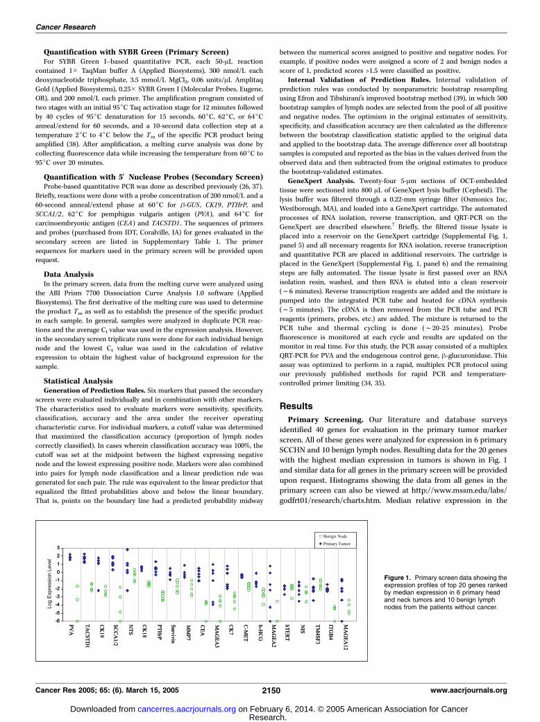
Primary Screening. Our literature and database surveysidentified 40 genes for evaluation in the primary tumor markerscreen. All of these genes were analyzed for expression in 6 primarySCCHN and 10 benign lymph nodes. Resulting data for the 20 geneswith the highest median expression in tumors is shown in Fig. 1and similar data for all genes in the primary screen will be providedupon request. Histograms showing the data from all genes in theprimary screen can also be viewed at http://www.mssm.edu/labs/godfrt01/research/charts.htm. Median relative expression in the
Figure 1. Primary screen data showing theexpression profiles of top 20 genes rankedby median expression in 6 primary headand neck tumors and 10 benign lymphnodes from the patients without cancer.
Cancer Research
Cancer Res 2005; 65: (6). March 15, 2005 2150 www.aacrjournals.org
Research. on February 6, 2014. © 2005 American Association for Cancercancerres.aacrjournals.org Downloaded from
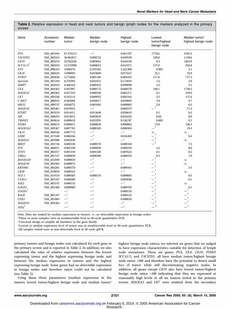
primary tumors and benign nodes was calculated for each gene inthe primary screen and is reported in Table 2. In addition, we alsocalculated the ratio of relative expression between the lowestexpressing tumor and the highest expressing benign node, andbetween the median expression in tumors and the highestexpressing benign node. Some genes had no detectable expressionin benign nodes and therefore ratios could not be calculated(see Table 2).Using these three parameters (median expression in the
tumors, lowest tumor/highest benign node and median tumor/
highest benign node ratios), we selected six genes that we judgedto have expression characteristics suitable for detection of lymphnode metastases. These six genes, PVA , CEA , CK19 , PTHrP,SCCA1/2 , and TACSTD1 , all have median tumor/highest benignnode ratios >500 and therefore have the potential to detect smallfoci of tumor while still discriminating negative nodes. Inaddition, all genes except CK19 also have lowest tumor/highestbenign node ratios >100 indicating that they are expressed atreasonably high levels in all six tumors tested in the primaryscreen. MAGEA3 and CK7 were omitted from the secondary
Table 2. Relative expression in head and neck tumors and benign lymph nodes for the markers analyzed in the primaryscreen
Gene Accession
number
Median
tumor
Median
benign node
Highest
benign node
Lowest
tumor/highest
benign node
Median tumor/
highest benign node
PVA NM_001944 67.476213 —* 0.021197 1770.6 3183.3
TACSTD1 NM_002354 56.962817 0.009753 0.049549 339.0 1149.6CK19 NM_002276 22.922226 0.003691 0.018136 0.3 1263.9
SCCA1/2c
NM_006919 13.372098 0.000014 0.015571 147.0 858.8
NTS NM_006183 4.880276 0.532185 2.321408 0.003 2.1
CK18 NM_000224 3.890994 0.033609 0.073557 22.1 52.9PTHrP NM_002820 3.175056 0.001186 0.004425 117.8 717.5
Survivin NM_001169 0.570393 0.022251 0.113834 1.2 5.0
MMP7 NM_002423 0.506164 0.009453 0.099098 2.5 5.1CEA NM_004363 0.467097 0.000175 0.000270 340.1 1730.3
MAGEA3 NM_005362 0.457354 0.000328 0.001271 0.1 359.9
CK7 NM_005556 0.435114 0.000953 0.003162 3.2 137.6
C-MET NM_000245 0.385698 0.020617 0.039692 5.9 9.7bHCG NM_000737 0.058275 0.001893 0.009005 2.0 6.5
MAGEA2 NM_005361 0.019915 —* 0.000279 —b
71.3
hTERT NM_003219 0.014411 0.012648 0.026645 0.1 0.5
NIS NM_000453 0.014012 0.003933 0.016232 0.02 0.9TM4SF3 NM_004616 0.009610 0.025295 0.136787 0.003 0.1
ITGB4 NM_000213 0.008011 0.000038 0.000063 74.8 126.3
MAGEA12 NM_005367 0.007785 0.000103 0.000404 —b
19.3
CK14 NM_000526 0.007772 —x —x 1 1LDHC NM_017448 0.006556 —* 0.016402 —
b0.4
MAGEA1 NM_004988 0.003538 —x —x —b 1
BRDT NM_001726 0.002530 0.000278 0.000350 —b
7.2STX NM_006011 0.001549 0.000030 0.000194 4.2 8.0
TITF1 NM_003317 0.001444 0.001320 0.007625 0.1 0.2
Villin1 NM_007127 0.000839 0.000288 0.000433 0.5 1.9
MAGEA10 NM_021048 0.000816 —x —x —b 1
MAGEA8 NM_005364 0.000675 —x —x —b 1
KRTHB1 NM_002281 0.000570 —* 0.000165 —b
3.5
CK20 NM_019010 0.000324 —x —x —b 1
SGY-1 NM_014419 0.000305 0.000134 0.000605 —b
0.5CTAG1 NM_001327 0.000206 —* 0.002036 —
b0.1
SSX2 NM_003147 0.000153 —x —x —b 1
GAGE1 NM_001468 0.000060 —* 0.000703 —b
0.1GAGEu
c—x —* 0.000126 —
b—b
BAGE NM_001187 —x —* 0.000152 —b
—b
CDX1 NM_001804 —x —* 0.000016 —b
—b
MAGEA4 NM_002362 —x —x —x —b
—b
SSXub —x —x —x —b —b
Note: Data are ranked by median expression in tumors. 1, no detectable expression in benign nodes.*Three or more samples were at nondetectable level in 40-cycle quantitative PCR.cUniversal design to amplify all members in the gene family.bLowest or median expression level of tumor was at nondetectable level in 40-cycle quantitative PCR.xAll samples tested were at non-detectable level in 40 cycle qPCR.
Novel Markers for Head and Neck Cancer Metastasis
www.aacrjournals.org 2151 Cancer Res 2005; 65: (6). March 15, 2005
Research. on February 6, 2014. © 2005 American Association for Cancercancerres.aacrjournals.org Downloaded from
screen due to a combination of low median tumor/highest benignnode expression ratios and relatively low median expression intumors. Similarly, CK18 was excluded based on a combination oflow expression ratios and ITGB4 was excluded due to very lowmedian expression in primary tumors. Finally, EGFR wasinadvertently omitted from the primary screen but was includedin the secondary screen based on its frequent use as a marker inSCCHN studies (40–42).Secondary Screen. Histologic evaluation of the 26 primary
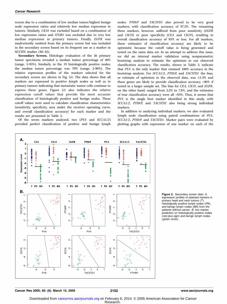
tumor specimens revealed a median tumor percentage of 40%(range, 5-95%). Similarly, in the 19 histologically positive nodes,the median tumor percentage was 70% (range, 2-90%). Therelative expression profiles of the markers selected for thesecondary screen are shown in Fig. 2A . The data shows that allmarkers are expressed in positive lymph nodes as well as inprimary tumors indicating that metastatic tumor cells continue toexpress these genes. Figure 2A also indicates the relativeexpression cutoff values that provide the most accurateclassification of histologically positive and benign nodes. Thesecutoff values were used to calculate classification characteristics(sensitivity, specificity, area under the receiver operating curve,and overall classification accuracy) for each marker and theresults are presented in Table 3.Of the seven markers analyzed, two (PVA and SCCA1/2)
provided perfect classification of positive and benign lymph
nodes. PTHrP and TACSTD1 also proved to be very goodmarkers, with classification accuracy of 97.5%. The remainingthree markers, however, suffered from poor sensitivity (EGFRand CK19) or poor specificity (CEA and CK19), resulting inoverall classification accuracy of 95% or less. For all markers,these estimates of classification accuracy are likely to beoptimistic because the cutoff value is being generated andtested on the same data set. In an attempt to address this issue,we did an internal marker validation using nonparametricbootstrap analysis to estimate the optimism in our observedclassification accuracy. The results, shown in Table 3, indicatethat PVA is the only marker that retained 100% accuracy in thebootstrap analysis. For SCCA1/2 , PTHrP, and TACSTD1 the bias,or estimate of optimism in the observed data, was V1.3% andthese genes are likely to provide classification accuracy >96% iftested in a larger sample set. The bias for CEA , CK19 , and EGFR ,on the other hand, ranged from 2.2% to 7.8%, and the estimatesof true classification accuracy were all <93%. Thus, it seems thatPVA is the single best marker identified in this study, withSCCA1/2 , PTHrP, and TACSTD1 also being strong individualmarkers.In addition to analyzing individual markers, we also evaluated
lymph node classification using paired combinations of PVA ,SCCA1/2 , PTHrP, and TACSTD1 . Marker pairs were evaluated byplotting graphs with expression of one marker on each of the
Figure 2. Secondary screen data: A,expression profiles of selected markers inprimary head and neck tumors (T ),histologically positive lymph nodes (PN ),and benign lymph nodes (BN ) from thepatients without cancer. B , two markerprediction on histologically positive nodes(red plus sign ) and benign lymph nodes(green circle ).
Cancer Research
Cancer Res 2005; 65: (6). March 15, 2005 2152 www.aacrjournals.org
Research. on February 6, 2014. © 2005 American Association for Cancercancerres.aacrjournals.org Downloaded from
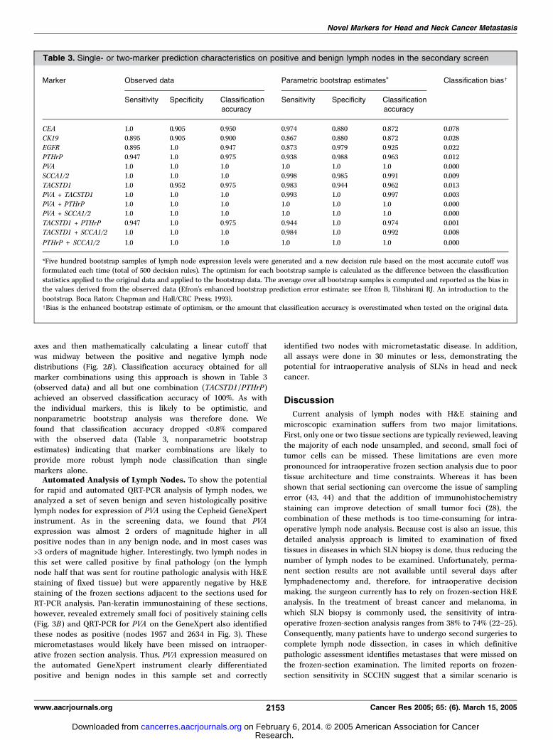
axes and then mathematically calculating a linear cutoff thatwas midway between the positive and negative lymph nodedistributions (Fig. 2B). Classification accuracy obtained for allmarker combinations using this approach is shown in Table 3(observed data) and all but one combination (TACSTD1/PTHrP)achieved an observed classification accuracy of 100%. As withthe individual markers, this is likely to be optimistic, andnonparametric bootstrap analysis was therefore done. Wefound that classification accuracy dropped <0.8% comparedwith the observed data (Table 3, nonparametric bootstrapestimates) indicating that marker combinations are likely toprovide more robust lymph node classification than singlemarkers alone.Automated Analysis of Lymph Nodes. To show the potential
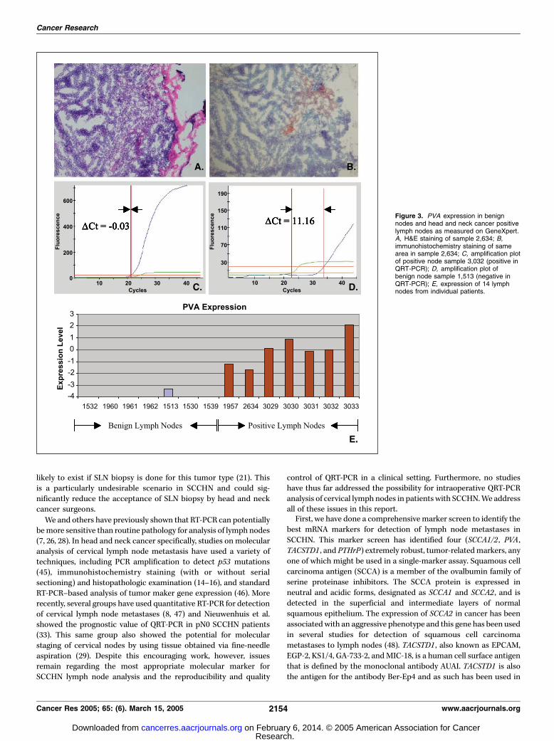
for rapid and automated QRT-PCR analysis of lymph nodes, weanalyzed a set of seven benign and seven histologically positivelymph nodes for expression of PVA using the Cepheid GeneXpertinstrument. As in the screening data, we found that PVAexpression was almost 2 orders of magnitude higher in allpositive nodes than in any benign node, and in most cases was>3 orders of magnitude higher. Interestingly, two lymph nodes inthis set were called positive by final pathology (on the lymphnode half that was sent for routine pathologic analysis with H&Estaining of fixed tissue) but were apparently negative by H&Estaining of the frozen sections adjacent to the sections used forRT-PCR analysis. Pan-keratin immunostaining of these sections,however, revealed extremely small foci of positively staining cells(Fig. 3B) and QRT-PCR for PVA on the GeneXpert also identifiedthese nodes as positive (nodes 1957 and 2634 in Fig. 3). Thesemicrometastases would likely have been missed on intraoper-ative frozen section analysis. Thus, PVA expression measured onthe automated GeneXpert instrument clearly differentiatedpositive and benign nodes in this sample set and correctly
identified two nodes with micrometastatic disease. In addition,all assays were done in 30 minutes or less, demonstrating thepotential for intraoperative analysis of SLNs in head and neckcancer.
Discussion
Current analysis of lymph nodes with H&E staining andmicroscopic examination suffers from two major limitations.First, only one or two tissue sections are typically reviewed, leavingthe majority of each node unsampled, and second, small foci oftumor cells can be missed. These limitations are even morepronounced for intraoperative frozen section analysis due to poortissue architecture and time constraints. Whereas it has beenshown that serial sectioning can overcome the issue of samplingerror (43, 44) and that the addition of immunohistochemistrystaining can improve detection of small tumor foci (28), thecombination of these methods is too time-consuming for intra-operative lymph node analysis. Because cost is also an issue, thisdetailed analysis approach is limited to examination of fixedtissues in diseases in which SLN biopsy is done, thus reducing thenumber of lymph nodes to be examined. Unfortunately, perma-nent section results are not available until several days afterlymphadenectomy and, therefore, for intraoperative decisionmaking, the surgeon currently has to rely on frozen-section H&Eanalysis. In the treatment of breast cancer and melanoma, inwhich SLN biopsy is commonly used, the sensitivity of intra-operative frozen-section analysis ranges from 38% to 74% (22–25).Consequently, many patients have to undergo second surgeries tocomplete lymph node dissection, in cases in which definitivepathologic assessment identifies metastases that were missed onthe frozen-section examination. The limited reports on frozen-section sensitivity in SCCHN suggest that a similar scenario is
Table 3. Single- or two-marker prediction characteristics on positive and benign lymph nodes in the secondary screen
Marker Observed data Parametric bootstrap estimates* Classification biasc
Sensitivity Specificity Classificationaccuracy
Sensitivity Specificity Classificationaccuracy
CEA 1.0 0.905 0.950 0.974 0.880 0.872 0.078CK19 0.895 0.905 0.900 0.867 0.880 0.872 0.028
EGFR 0.895 1.0 0.947 0.873 0.979 0.925 0.022
PTHrP 0.947 1.0 0.975 0.938 0.988 0.963 0.012
PVA 1.0 1.0 1.0 1.0 1.0 1.0 0.000SCCA1/2 1.0 1.0 1.0 0.998 0.985 0.991 0.009
TACSTD1 1.0 0.952 0.975 0.983 0.944 0.962 0.013
PVA + TACSTD1 1.0 1.0 1.0 0.993 1.0 0.997 0.003
PVA + PTHrP 1.0 1.0 1.0 1.0 1.0 1.0 0.000PVA + SCCA1/2 1.0 1.0 1.0 1.0 1.0 1.0 0.000
TACSTD1 + PTHrP 0.947 1.0 0.975 0.944 1.0 0.974 0.001
TACSTD1 + SCCA1/2 1.0 1.0 1.0 0.984 1.0 0.992 0.008
PTHrP + SCCA1/2 1.0 1.0 1.0 1.0 1.0 1.0 0.000
*Five hundred bootstrap samples of lymph node expression levels were generated and a new decision rule based on the most accurate cutoff wasformulated each time (total of 500 decision rules). The optimism for each bootstrap sample is calculated as the difference between the classification
statistics applied to the original data and applied to the bootstrap data. The average over all bootstrap samples is computed and reported as the bias in
the values derived from the observed data (Efron’s enhanced bootstrap prediction error estimate; see Efron B, Tibshirani RJ. An introduction to the
bootstrap. Boca Raton: Chapman and Hall/CRC Press; 1993).cBias is the enhanced bootstrap estimate of optimism, or the amount that classification accuracy is overestimated when tested on the original data.
Novel Markers for Head and Neck Cancer Metastasis
www.aacrjournals.org 2153 Cancer Res 2005; 65: (6). March 15, 2005
Research. on February 6, 2014. © 2005 American Association for Cancercancerres.aacrjournals.org Downloaded from
likely to exist if SLN biopsy is done for this tumor type (21). Thisis a particularly undesirable scenario in SCCHN and could sig-nificantly reduce the acceptance of SLN biopsy by head and neckcancer surgeons.
We and others have previously shown that RT-PCR can potentiallybemore sensitive than routine pathology for analysis of lymph nodes(7, 26, 28). In head and neck cancer specifically, studies on molecularanalysis of cervical lymph node metastasis have used a variety oftechniques, including PCR amplification to detect p53 mutations(45), immunohistochemistry staining (with or without serialsectioning) and histopathologic examination (14–16), and standardRT-PCR–based analysis of tumor maker gene expression (46). Morerecently, several groups have used quantitative RT-PCR for detectionof cervical lymph node metastases (8, 47) and Nieuwenhuis et al.showed the prognostic value of QRT-PCR in pN0 SCCHN patients(33). This same group also showed the potential for molecularstaging of cervical nodes by using tissue obtained via fine-needleaspiration (29). Despite this encouraging work, however, issuesremain regarding the most appropriate molecular marker forSCCHN lymph node analysis and the reproducibility and quality
control of QRT-PCR in a clinical setting. Furthermore, no studieshave thus far addressed the possibility for intraoperative QRT-PCRanalysis of cervical lymph nodes in patientswith SCCHN.We addressall of these issues in this report.First, we have done a comprehensive marker screen to identify the
best mRNA markers for detection of lymph node metastases inSCCHN. This marker screen has identified four (SCCA1/2 , PVA ,TACSTD1 , and PTHrP) extremely robust, tumor-relatedmarkers, anyone of which might be used in a single-marker assay. Squamous cellcarcinoma antigen (SCCA) is a member of the ovalbumin family ofserine proteinase inhibitors. The SCCA protein is expressed inneutral and acidic forms, designated as SCCA1 and SCCA2 , and isdetected in the superficial and intermediate layers of normalsquamous epithelium. The expression of SCCA2 in cancer has beenassociatedwith an aggressive phenotype and this gene has been usedin several studies for detection of squamous cell carcinomametastases to lymph nodes (48). TACSTD1 , also known as EPCAM,EGP-2, KS1/4, GA-733-2, andMIC-18, is a human cell surface antigenthat is defined by the monoclonal antibody AUAI. TACSTD1 is alsothe antigen for the antibody Ber-Ep4 and as such has been used in
Figure 3. PVA expression in benignnodes and head and neck cancer positivelymph nodes as measured on GeneXpert.A, H&E staining of sample 2,634; B,immunohistochemistry staining of samearea in sample 2,634; C, amplification plotof positive node sample 3,032 (positive inQRT-PCR); D, amplification plot ofbenign node sample 1,513 (negative inQRT-PCR); E, expression of 14 lymphnodes from individual patients.
Cancer Research
Cancer Res 2005; 65: (6). March 15, 2005 2154 www.aacrjournals.org
Research. on February 6, 2014. © 2005 American Association for Cancercancerres.aacrjournals.org Downloaded from
studies to detect lymph node micrometastases in a variety of tumortypes, including SCCHN (41, 49, 50). A recent study has also shownthat TACSTD1 is a good marker for RT-PCR–based identification oflymph node micrometastasis in non–small cell lung cancer (9).Parathyroid hormone–related protein (PTHrP, also known asPTHLH and HHM) regulates endochondral bone development andepithelial-mesenchymal interactions during the formation of themammary glands and teeth, and its expression in SCCHN mayrepresent dedifferentiation commonly seen in epithelial tumors. Thegene encoding PTHrP has been mapped to the short arm ofchromosome 12 and is known to contain 6 to 7 exons. Alternate RNAsplicing of this gene results in heterogeneity of the mRNA speciesencoded at the 3V ends. Pemphigus vulgaris antigen (PVA), alsoknown as desmoglein 3 (DSG-3), is a 130-kDa surface glycoproteinthat is the serologic target in the autoimmune skin diseasepemphigus vulgaris. PVA is a member of the desmoglein subfamilyof the desmosomal cadherins and the gene encoding this protein hasbeen mapped to the long arm of chromosome 18 and is known tocontain 15 exons. To our knowledge, neither PTHrP nor PVA havebeen used in prior studies to detect lymph node metastases inSCCHN or other tumor types.From our data it is clear that PVA and TACSTD1 are the best
individual markers studied. In all screening phases, PVA andTACSTD1 provided 100% discrimination between positive andbenign lymph nodes although PVA was the only individual markerto maintain 100% classification accuracy upon bootstrap analysis.Thus, it seems that a single marker QRT-PCR analysis for PVA couldbe adequate for staging of cervical lymph nodes in SCCHN.Whereas the marker screening phase of this study was done
using routine QRT-PCR techniques, we have previously publishedwork showing that it is possible to perform QRT-PCR assays in anintraoperative time frame (f25 minutes; refs. 34, 35). Given thepotential utility of an intraoperative SLN analysis in SCCHN, wetherefore applied our rapid, multiplex QRT-PCR techniquesto develop such an assay for PVA . This assay was incorporatedinto a completely automated RNA isolation and QRT-PCRinstrument (the GeneXpert) developed by Cepheid for moleculardiagnostic testing. The GeneXpert is an automated cartridgeprocessor with integrated real-time PCR capability. Each single-useGeneXpert cartridge consists of multiple reagent reservoirs,a syringe barrel, and a valve mechanism that allows transfer ofreagents between reservoirs. In addition, the cartridges used in thisstudy have a solid-phase matrix for nucleic acid purification andisolation. Finally, each cartridge has a PCR tube that can beaccessed from specific reagent reservoirs to allow loading ofreaction components into the PCR tube for reverse transcriptionand/or quantitative PCR. The GeneXpert instrument itselfinterfaces with the cartridge to control reagent movement andalso houses heat plates and optical excite/detect blocks to facilitatereal-time PCR. The details of RNA isolation and QRT-PCR using theGeneXpert are to be published elsewhere.
Although the speed of the GeneXpert allows for intraoperativeanalysis of SLNs there may also be additional clinical value if QRT-PCR eventually proves superior to routine pathology, as suggestedby Nieuwenhuis et al. (33). The detection of occult lymph nodedisease, and subsequent improved patient staging (28), could havesignificant consequences for the treatment of SCCHN. For example,patients with multiple positive nodes, or extracapsular extension oftumor, are often referred for radiation or chemoradiation therapy, toreduce the high risk of locoregional failure. Although QRT-PCRcannot distinguish extracapsular extension, it may well identifyadditional positive lymph nodes leading to upstaging, and moreappropriate adjuvant treatment. Furthermore, surgical approachand extent of lymph node dissection may also be determined by thelymph node status. Many surgeons advocate planned lymph nodedissection in N2 patients who undergo primary chemoradiationbecause the available data indicate that 20% to 30% of completeresponders (based on clinical modalities) may actually have residualtumor in the neck (3). Evaluation of suspicious postradiationcervical nodules could potentially be done via fine-needle aspirateand QRT-PCR, or by intraoperative QRT-PCR, allowing the surgeonto determine the necessary extent of resection before or during thesurgery. Thus, the detection of occult lymph node metastasis couldplay an important role in future staging and treatment options forpathologically node-positive as well as node-negative patients.In conclusion, we have showed that QRT-PCR can detect,
with high sensitivity and specificity, metastatic disease in lymphnodes of patients with SCCHN. In addition, we have showed thefeasibility of automated, intraoperative staging of cervical lymphnodes and the possibility that such an approach may eventuallyprove superior to conventional pathology. Staging of the cN0neck is currently a topic of intense interest in the head andneck oncologic community, with the goal that therapeuticsurgical and adjuvant treatment be administered to those mostlikely to benefit from it. Whereas the ACOSOG Z0360 trial ispowered to validate the multiple single-institution studies thatsuggest the utility of SLN mapping for staging the cN0 neck inSCCHN, it is unlikely that SLN biopsy will be widely acceptedwithout a rapid, accurate, and standardized method of stagingthe SLN(s). Our development of such an assay and identificationof discriminatory marker genes provides the pilot datanecessary for the incorporation of QRT-PCR into future clinicalstudies applying SLN mapping to clinical practice for patientswith this disease.
Acknowledgments
Received 10/19/2004; revised 12/14/2004; accepted 1/6/2005.Grant support: NIH/National Cancer Institute CA90665-01 research grant
(T.E. Godfrey), University of Pittsburgh Oral Cancer Center of Discovery pilot grant(R.L. Ferris and T.E. Godfrey), and a cooperative research and development agreementwith Cepheid (T.E. Godfrey).
The costs of publication of this article were defrayed in part by the payment of pagecharges. This article must therefore be hereby marked advertisement in accordancewith 18 U.S.C. Section 1734 solely to indicate this fact.
References1. Thekdi AA, Ferris RL. Diagnostic assessment oflaryngeal cancer. Otolaryngol Clin North Am2002;35:953–69.
2. Takes RP. Staging of the neck in patients with headand neck squamous cell cancer: imaging techniquesand biomarkers. Oral Oncol 2004;40:656–67.
3. Ferlito A, Rinaldo A, Devaney KO, et al. Prognosticsignificance of microscopic and macroscopic extrac-apsular spread from metastatic tumor in the cervicallymph nodes. Oral Oncol 2002;38:747–51.
4. Curtin HD, Ishwaran H, Mancuso AA, Dalley RW,Caudry DJ, McNeil BJ. Comparison of CT and MRimaging in staging of neck metastases. Radiology1998;207:123–30.
5. Hyde NC, Prvulovich E, Newman L, Waddington WA,Visvikis D, Ell P. A new approach to pre-treatmentassessment of the N0 neck in oral squamous cellcarcinoma: the role of sentinel node biopsy andpositron emission tomography. Oral Oncol 2003;39:350–60.
6. Duvvuri U, Simental AA Jr, Johnson JT, et al. Electiveneck dissection and survival in patients with squamous
Novel Markers for Head and Neck Cancer Metastasis
www.aacrjournals.org 2155 Cancer Res 2005; 65: (6). March 15, 2005
Research. on February 6, 2014. © 2005 American Association for Cancercancerres.aacrjournals.org Downloaded from

cell carcinoma of the oral cavity and oropharynx.Laryngoscope 2004;114:2228–34.
7. Liefers GJ, Cleton-Jansen AM, van de Velde CJ, et al.Micrometastases and survival in stage II colorectalcancer. N Engl J Med 1998;339:223–8.
8. Onishi A, Nakashiro K, Mihara M, et al. Basic andclinical studies on quantitative analysis of lymph nodemicrometastasis in oral cancer. Oncol Rep 2004;11:33–9.
9. Mitas M, Cole DJ, Hoover L, et al. Real-time reversetranscription-PCR detects KS1/4 mRNA in mediastinallymph nodes from patients with non-small cell lungcancer. Clin Chem 2003;49:312–5.
10. McGuirt WF, Johnson JT, Myers EN, Rothfield R,Wagner R. Floor of mouth carcinoma. The managementof the clinically negative neck. Arch Otolaryngol HeadNeck Surg 1995;121:278–82.
11. Alvi A, Johnson JT. Extracapsular spread in theclinically negative neck (N0): implications and outcome.Otolaryngol Head Neck Surg 1996;114:65–70.
12. Pitman KT, Johnson JT, Myers EN. Effectiveness ofselective neck dissection for management of theclinically negative neck. Arch Otolaryngol Head NeckSurg 1997;123:917–22.
13. Greenberg JS, el Naggar AK, Mo V, Roberts D,Myers JN.Disparity in pathologic and clinical lymph node stagingin oral tongue carcinoma. Implication for therapeuticdecision making. Cancer 2003;98:508–15.
14. Ambrosch P, Brinck U. Detection of nodal micro-metastases in head and neck cancer by serial sectioningand immunostaining. Oncology (Huntingt) 1996;10:1221–6.
15. Enepekides DJ, Sultanem K, Nguyen C, Shenouda G,Black MJ, Rochon L. Occult cervical metastases:immunoperoxidase analysis of the pathologicallynegative neck. Otolaryngol Head Neck Surg 1999;120:713–7.
16. Kocaturk S, Yilmazer D, Onal B, Erkam U, Urunal B.Do micrometastases detected with cytokeratin immu-noperoxidase reactivity affect the treatment approachto neck in supraglottic cancers? Otolaryngol Head NeckSurg 2003;128:407–11.
17. Shoaib T, Soutar DS, MacDonald DG, et al. Theaccuracy of head and neck carcinoma sentinel lymphnode biopsy in the clinically N0 neck. Cancer 2001;91:2077–83.
18. Rassekh CH, Johnson JT, Myers EN. Accuracy ofintraoperative staging of the NO neck in squamous cellcarcinoma. Laryngoscope 1995;105:1334–6.
19. Koch WM, Choti MA, Civelek AC, Eisele DW,Saunders JR. g Probe-directed biopsy of the sentinelnode in oral squamous cell carcinoma. Arch Otolar-yngol Head Neck Surg 1998;124:455–9.
20. Zitsch RP III, Todd DW, Renner GJ, Singh A.Intraoperative radiolymphoscintigraphy for detectionof occult nodal metastasis in patients with head andneck squamous cell carcinoma. Otolaryngol Head NeckSurg 2000;122:662–6.
21. Civantos FJ, Gomez C, Duque C, et al. Sentinelnode biopsy in oral cavity cancer: correlation withPET scan and immunohistochemistry. Head Neck2003;25:1–9.
22. Chao C, Wong SL, Ackermann D, et al. Utility ofintraoperative frozen section analysis of sentinellymph nodes in breast cancer. Am J Surg 2001;182:609–15.
23. Gulec SA, Su J, O’Leary JP, Stolier A. Clinical utility offrozen section in sentinel node biopsy in breast cancer.Am Surg 2001;67:529–32.
24. Tanis PJ, Boom RP, Koops HS, et al. Frozen sectioninvestigation of the sentinel node in malignant melano-ma and breast cancer. Ann Surg Oncol 2001;8:222–6.
25. Koopal SA, Tiebosch AT, Albertus PD, Plukker JT,Schraffordt KH, Hoekstra HJ. Frozen section analysis ofsentinel lymph nodes in melanoma patients. Cancer2000;89:1720–5.
26. Godfrey TE, Raja S, Finkelstein SD, Gooding WE,Kelly LA, Luketich JD. Prognostic value of quantitativereverse transcription-polymerase chain reaction inlymph node-negative esophageal cancer patients. ClinCancer Res 2001;7:4041–8.
27. Shivers SC, Wang X, Li W, et al. Molecular staging ofmalignant melanoma: correlation with clinical out-come. JAMA 1998;280:1410–5.
28. Bostick PJ, Morton DL, Turner RR, et al. Prognosticsignificance of occult metastases detected by sentinellymphadenectomy and reverse transcriptase-polymerasechain reaction in early-stage melanoma patients. J ClinOncol 1999;17:3238–44.
29. Nieuwenhuis EJ, Jaspars LH, Castelijns JA, et al.Quantitative molecular detection of minimal residualhead and neck cancer in lymph node aspirates. ClinCancer Res 2003;9:755–61.
30. Zehentner BK, Dillon DC, Jiang Y, et al. Application ofa multigene reverse transcription-PCR assay for detec-tion of mammaglobin and complementary transcribedgenes in breast cancer lymph nodes. Clin Chem2002;48:1225–31.
31. Xi L, Luketich JD, Raja S, et al. Molecular staging oflymph nodes from patients with esophageal adenocar-cinoma. Clin Cancer Res. In press 2005.
32. Mitas M, Mikhitarian K, Walters C, et al. Quantitativereal-time RT-PCR detection of breast cancer micro-metastasis using a multigene marker panel. Int J Cancer2001;93:162–71.
33. Nieuwenhuis EJ, Leemans CR, Kummer JA, et al.Assessment and clinical significance of micrometasta-ses in lymph nodes of head and neck cancer patientsdetected by E48 (Ly-6D) quantitative reverse transcrip-tion-polymerase chain reaction. Lab Invest 2003;83:1233–40.
34. Raja S, El Hefnawy T, Kelly LA, Chestney ML,Luketich JD, Godfrey TE. Temperature-controlledprimer limit for multiplexing of rapid, quantitativereverse transcription-PCR assays: application tointraoperative cancer diagnostics. Clin Chem 2002;48:1329–37.
35. Raja S, Luketich JD, Kelly LA, Gooding WE,Finkelstein SD, Godfrey TE. Rapid, quantitative reversetranscriptase-polymerase chain reaction: Application tointraoperative molecular detection of occult metastasesin esophageal cancer. J Thorac Cardiovasc Surg 2002;123:475–83.
36. Godfrey TE, Kim S-H, Chavira M, et al. QuantitativemRNA expression analysis from formalin-fixed, paraffin-embedded tissues using 5V nuclease quantitative RT-PCR. J Mol Diagn 2000;2:84–91.
37. Tassone F, Hagerman RJ, Taylor AK, Gane LW, GodfreyTE, Hagerman PJ. Elevated levels of FMR1 mRNA incarrier males: a new mechanism of involvement in thefragile-X syndrome. Am J Hum Genet 2000;66:6–15.
38. Morrison TB, Weis JJ, Wittwer CT. Quantification oflow-copy transcripts by continuous SYBR Green I mo-nitoring during amplification. Biotechniques 1998;24:954–8, 960, 962.
39. Efron B, Tibshirani RJ. An introduction to thebootstrap. Hardcover ed. Boca Raton: Chapman &Hall/CRC; 1994.
40. Takes RP, Baatenburg de Jong RJ, Schuuring E, et al.Markers for assessment of nodal metastasis in laryngealcarcinoma.ArchOtolaryngolHeadNeck Surg 1997;123:412–9.
41. Takes RP, Baatenburg de Jong RJ, Wijffels K, et al.Expression of genetic markers in lymph node metasta-ses compared with their primary tumours in head andneck cancer. J Pathol 2001;194:298–302.
42. Zeng Q, Smith DC, Suscovich TJ, Gooding WE,Trump DL, Grandis JR. Determination of intermediatebiomarker expression levels by quantitative reversetranscription-polymerase chain reaction in oral mucosaof cancer patients treated with liarozole. Clin CancerRes 2000;6:2245–51.
43. Turner RR, Ollila DW, Krasne DL, Giuliano AE.Histopathologic validation of the sentinel lymph nodehypothesis for breast carcinoma. Ann Surg 1997;226:271–6.
44. Cote RJ, Peterson HF, Chaiwun B, et al. Role ofimmunohistochemical detection of lymph-node metas-tases in management of breast cancer. InternationalBreast Cancer Study Group. Lancet 1999;354:896–900.
45. Brennan JA, Mao L, Hruban RH, et al. Molecularassessment of histopathological staging in squamous-cell carcinoma of the head and neck. N Engl J Med1995;332:429–35.
46. Hamakawa H, Fukizumi M, Bao Y, et al. Geneticdiagnosis of micrometastasis based on SCC antigenmRNA in cervical lymph nodes of head and neckcancer. Clin Exp Metastasis 1999;17:593–9.
47. Becker MT, Shores CG, Yu KK, Yarbrough WG.Molecular assay to detect metastatic head and necksquamous cell carcinoma. Arch Otolaryngol Head NeckSurg 2004;130:21–7.
48. Kano M, Shimada Y, Kaganoi J, et al. Detection oflymph node metastasis of oesophageal cancer by RT-nested PCR for SCC antigen gene mRNA. Br J Cancer2000;82:429–35.
49. Kubuschok B, Passlick B, Izbicki JR, Thetter O, PantelK. Disseminated tumor cells in lymph nodes as adeterminant for survival in surgically resected non-small-cell lung cancer. J Clin Oncol 1999;17:19–24.
50. Schroder CP, Ruiters MH, de Jong S, et al. Detection ofmicrometastatic breast cancer by means of real timequantitative RT-PCR and immunostaining in perioper-ative blood samples and sentinel nodes. Int J Cancer2003;106:611–8.
Cancer Research
Cancer Res 2005; 65: (6). March 15, 2005 2156 www.aacrjournals.org
Research. on February 6, 2014. © 2005 American Association for Cancercancerres.aacrjournals.org Downloaded from
Copyright © 2022 FDOKUMEN