Pyroptosis and adaptive immunity mechanisms are promptly engendered in mesenteric lymph-nodes during...
14
RESEARCH Open Access Pyroptosis and adaptive immunity mechanisms are promptly engendered in mesenteric lymph-nodes during pig infections with Salmonella enterica serovar Typhimurium Rodrigo Prado Martins 1 , Carmen Aguilar 1 , James E Graham 2 , Ana Carvajal 3 , Rocío Bautista 4 , M Gonzalo Claros 4 and Juan J Garrido 1* Abstract In this study, we explored the transcriptional response and the morphological changes occurring in porcine mesenteric lymph-nodes (MLN) along a time course of 1, 2 and 6 days post infection (dpi) with Salmonella Typhimurium. Additionally, we analysed the expression of some Salmonella effectors in tissue to complete our view of the processes triggered in these organs upon infection. The results indicate that besides dampening apoptosis, swine take advantage of the flagellin and prgJ expression by Salmonella Typhimuriun to induce pyroptosis in MLN, preventing bacterial dissemination. Furthermore, cross-presentation of Salmonella antigens was inferred as a mechanism that results in a rapid clearance of pathogen by cytotoxic T cells. In summary, although the Salmonella Typhimurium strain employed in this study was able to express some of its major virulence effectors in porcine MLN, a combination of early innate and adaptive immunity mechanisms might overcome virulence strategies employed by the pathogen, enabling the host to protect itself against bacterial spread beyond gut-associated lymph-nodes. Interestingly, we deduced that clathrin-mediated endocytosis could contribute to mechanisms of pathogen virulence and/or host defence in MLN of Salmonella infected swine. Taken together, our results are useful for a better understanding of the critical protective mechanisms against Salmonella that occur in porcine MLN to prevent the spread of infection beyond the intestine. Introduction Infections by Salmonella are a major health problem in the developing and developed world. In the European Union, despite the current decreasing trend of human cases, Salmonella persists as the main cause of food-borne out- breaks [1]. Pork is considered to be a significant source of Salmonella to humans next to eggs and poultry meat [2]. Indeed, according to the European food safety authority (EFSA), Salmonella enterica serovar Typhimurium (herein Salmonella Typhimurium) is the second serovar most frequently reported in human salmonellosis and infection by this pathogen is mostly associated with the consump- tion of contaminated pork [1]. Since the food industry and direct contact with infected animals represent the main sources of non-typhoid Salmonella [3], prevention of human salmonellosis depends significantly on decreasing the prevalence of infection in livestock hosts [4]. Salmonella Typhimurium infected pigs generally carry this serotype asymptomati- cally in the tonsils, intestines and gut-associated lymph- oid tissue, posing an important threat to animal and human health [5]. Epidemiological studies assert that Salmonella prevalence in slaughter swine lymph nodes varies widely at the country level, ranging from0 to 29% [2]. Although salmonellosis in pigs has been the subject of intensive research [5], a thorough knowledge of the pathogenesis of porcine infections with broadhost range Salmonella serotypes is still necessary. A combination of * Correspondence: [email protected] 1 Grupo de Genómica y Mejora Animal, Departamento de Genética, Facultad de Veterinaria, Universidad de Córdoba, Campus de Rabanales, Edificio Gregor Mendel C5, 14071, Córdoba, Spain Full list of author information is available at the end of the article VETERINARY RESEARCH © 2013 Martins et al.; licensee BioMed Central Ltd. This is an open access article distributed under the terms of the Creative Commons Attribution License (http://creativecommons.org/licenses/by/2.0), which permits unrestricted use, distribution, and reproduction in any medium, provided the original work is properly cited. Martins et al. Veterinary Research 2013, 44:120 http://www.veterinaryresearch.org/content/44/1/120
Transcript of Pyroptosis and adaptive immunity mechanisms are promptly engendered in mesenteric lymph-nodes during...

VETERINARY RESEARCHMartins et al. Veterinary Research 2013, 44:120http://www.veterinaryresearch.org/content/44/1/120
RESEARCH Open Access
Pyroptosis and adaptive immunity mechanismsare promptly engendered in mesentericlymph-nodes during pig infections withSalmonella enterica serovar TyphimuriumRodrigo Prado Martins1, Carmen Aguilar1, James E Graham2, Ana Carvajal3, Rocío Bautista4, M Gonzalo Claros4
and Juan J Garrido1*
Abstract
In this study, we explored the transcriptional response and the morphological changes occurring in porcinemesenteric lymph-nodes (MLN) along a time course of 1, 2 and 6 days post infection (dpi) with SalmonellaTyphimurium. Additionally, we analysed the expression of some Salmonella effectors in tissue to complete our viewof the processes triggered in these organs upon infection. The results indicate that besides dampening apoptosis,swine take advantage of the flagellin and prgJ expression by Salmonella Typhimuriun to induce pyroptosis in MLN,preventing bacterial dissemination. Furthermore, cross-presentation of Salmonella antigens was inferred as amechanism that results in a rapid clearance of pathogen by cytotoxic T cells. In summary, although the SalmonellaTyphimurium strain employed in this study was able to express some of its major virulence effectors in porcineMLN, a combination of early innate and adaptive immunity mechanisms might overcome virulence strategiesemployed by the pathogen, enabling the host to protect itself against bacterial spread beyond gut-associatedlymph-nodes. Interestingly, we deduced that clathrin-mediated endocytosis could contribute to mechanisms ofpathogen virulence and/or host defence in MLN of Salmonella infected swine. Taken together, our results are usefulfor a better understanding of the critical protective mechanisms against Salmonella that occur in porcine MLN toprevent the spread of infection beyond the intestine.
IntroductionInfections by Salmonella are a major health problem in thedeveloping and developed world. In the European Union,despite the current decreasing trend of human cases,Salmonella persists as the main cause of food-borne out-breaks [1]. Pork is considered to be a significant source ofSalmonella to humans next to eggs and poultry meat [2].Indeed, according to the European food safety authority(EFSA), Salmonella enterica serovar Typhimurium (hereinSalmonella Typhimurium) is the second serovar mostfrequently reported in human salmonellosis and infection
* Correspondence: [email protected] de Genómica y Mejora Animal, Departamento de Genética, Facultadde Veterinaria, Universidad de Córdoba, Campus de Rabanales, EdificioGregor Mendel C5, 14071, Córdoba, SpainFull list of author information is available at the end of the article
© 2013 Martins et al.; licensee BioMed CentralCommons Attribution License (http://creativecreproduction in any medium, provided the or
by this pathogen is mostly associated with the consump-tion of contaminated pork [1].Since the food industry and direct contact with infected
animals represent the main sources of non-typhoidSalmonella [3], prevention of human salmonellosisdepends significantly on decreasing the prevalence ofinfection in livestock hosts [4]. Salmonella Typhimuriuminfected pigs generally carry this serotype asymptomati-cally in the tonsils, intestines and gut-associated lymph-oid tissue, posing an important threat to animal andhuman health [5]. Epidemiological studies assert thatSalmonella prevalence in slaughter swine lymph nodesvaries widely at the country level, ranging from0 to 29%[2]. Although salmonellosis in pigs has been the subjectof intensive research [5], a thorough knowledge of thepathogenesis of porcine infections with broadhost rangeSalmonella serotypes is still necessary. A combination of
Ltd. This is an open access article distributed under the terms of the Creativeommons.org/licenses/by/2.0), which permits unrestricted use, distribution, andiginal work is properly cited.

Martins et al. Veterinary Research 2013, 44:120 Page 2 of 14http://www.veterinaryresearch.org/content/44/1/120
system-wide approaches and in vivo infection models isexpected to generate precise and novel data to analyzethe response to Salmonella infections in pigs [6]. Thus,whole-genome expression analysis has been used to ex-plore gene expression changes during infection of pigs bySalmonella, contributing to identify molecules and path-ways associated with the host response to infection [7,8].More recently, proteomic techniques have also beenemployed as a step towards a detailed understanding ofthe disease mechanisms [9,10]. However, despite this,there is a need to deepen understanding of the biologicalprocesses that control host-pathogen interaction andSalmonella persistence in porcine lymphatic tissue,which could provide new targets for treatment andcontrol of salmonellosis in this species. Therefore, theobjective of the current study was to explore the earlytranscriptional response of porcine mesenteric lymph-nodes (MLN) to Salmonella Typhimurim using a time-course analysis of an in vivo infection. In addition,the expression of some pathogen virulence effectors,as well as the morphological alterations associatedwith the presence of the bacteria in the tissue were alsoevaluated.
Materials and methodsExperimental infection and tissue samplingSixteen crossbred weaned piglets of approximately fourweeks of age, serologically and fecal-negative forSalmonella were used in an experimental infection de-scribed elsewhere [11]. Briefly, twelve piglets were orallyinfected with 108 cfu of a Salmonella Typhimurium pha-getype DT104 strain isolated from a naturally infectedpig [11], whereas the control group (4 animals) receivedsterile medium. Non-infected control pigs were necrop-sied prior to the experimental infection (0 day post-infection – dpi) and four randomly chosen infectedpiglets were necropsied at 1, 2 or 6 dpi. Samples ofMLN were collected from all experimental animals andimmediately frozen in liquid nitrogen for RNA and pro-tein isolation or fixed in 10% neutral buffered formalinfor histological processing. All procedures involving ani-mals were performed in accordance with the Europeanregulations regarding the protection of animals used forexperimental and other scientific purposes. Piglets werehoused in experimental isolation facilities of the Universityof Leon (Spain). Animal care and procedures were in ac-cordance with the guidelines of the Good ExperimentalPractices (GEP), under the supervision of the Ethical andAnimal Welfare Committee of the University of Leon(Spain).
RNA purificationAfter treatment with RNAlater-ICE (Ambion, Inc,Austin, TX, USA), MLN samples were soaked in RLT
buffer (Qiagen, Valencia, CA, USA) and disrupted in arotor-stator homogenizer. RNA was isolated using theAllPrep DNA/RNA/Protein Mini Kit (Qiagen), digestedwith the RNase-Free DNase Set (Qiagen) according to themanufacturer’s instructions and routinely precipitatedwith ethanol. RNA integrity was evaluated using theExperion RNA automated electrophoresis system(Bio-Rad, Hercules, CA, USA) before being quantified usinga ND-1000 spectrophotometer (Nanodrop Technologies,Wilminton, USA).
Microarray analysisGene expression analysis was carried out using theGeneChip Porcine Genome Array of the Affymetrix plat-form (Affymetrix Inc., Santa Clara, CA, USA) at theGenomics Unit of CABIMER (Andalusian Center forMolecular Biology and Regenerative Medicine, Seville,Spain). This chip contains 23 937 probe sets to interro-gate 23 256 transcripts in the pig, which represents 20201 genes. The One-Cycle Eukaryotic Target LabelingAssay was used to obtain biotinylated cRNA to be usedin the subsequent chip hybridization according tothe manufacturer’s instructions (Expression AnalysisTechnical Manual, Affymetrix). The biotinylated cRNAtargets were then cleaned up, fragmented, and hybrid-ized with the GeneChip Porcine Genome Array follow-ing Affymetrix recommended protocols. Chips werewashed, stained with a GeneChip Fluidics Station 450(Affymetrix) using the standard fluidics protocol andscanned with an Affymetrix GeneChip Scanner 3000(Affymetrix). Probe signal intensities were captured andprocessed with the GeneChip Operating Software 1.4.0.036(Affymetrix) and the resulting CEL files were reprocessedusing robust multi-array average normalization (RMA)[12]. Because the aim of analysis was to detect changes ingene expression along a time-course of infection, differen-tially expressed (DE) genes were accessed by the BATS(Bayesian Analysis of Time Series) software package [13],using default settings. A Bayes Factor (BF) value of 0.05was used as cutoff to rank significantly regulated tran-scripts. Since the Affymetrix Porcine GeneChip is not fullyannotated in all the features, it was re-annotated withBlast2GO [14] with a minimum E-value of 10-10 and aminimum similarity of 50%.
Systems biology analysisThe list of genes that showed significant changes in ex-pression was uploaded into Ingenuity Pathway Analysis(IPA, Ingenuity Systems Inc, Redwood City, CA, USA)[15] for bioinformatics analysis. Additionally, the DAVIDBioinformatic Database [16] was used applying the de-fault settings to refine some data from IPA analysis.Gene interaction networks were automatically generated,ranked by score and depicted on IPA as follows: each
Martins et al. Veterinary Research 2013, 44:120 Page 3 of 14http://www.veterinaryresearch.org/content/44/1/120
node in the network diagram represented a gene and itsrelationship with other molecules was represented by aline (solid and dotted lines represent direct and indirectassociation respectively). Nodes with a red backgroundwere input genes detected in this study while greynodes were molecules inserted by IPA based upon theIngenuity Knowledge Base to produce a highly connectednetwork. The score estimated the probability that acollection of genes equal to or greater than the numberin a network could be achieved by chance alone. Scoresof 3 or higher were considered to have a 99.9% confi-dence of not being generated by random chance alone.For statistical analysis of enriched functions/pathways, anIPA Knowledge Base was used as a reference set and theFisher’s exact test was employed to estimate the signifi-cance of association. P-values below 0.05 were consideredstatistically significant. For graphical representation ofthe canonical pathways, the ratio indicates the percentageof genes taking part in a pathway that could be found inan uploaded data set and –log(p-value) means the levelof confidence of association. The threshold line repre-sented a p-value of 0.05.
Relative gene expression analysis by qPCRReal-time quantitative PCR (qPCR) assays were per-formed as previously described [11]. Fold change valueswere calculated by the 2−ΔΔCq method [17] using beta-actin as the reference gene. Afterwards, data were stan-dardized as proposed by Willems et al. [18] and analyzedby Kruskal–Wallis and Mann–Whitney tests using thesoftware SPSS 15.0 for Windows (SPSS Inc, Chicago, IL,
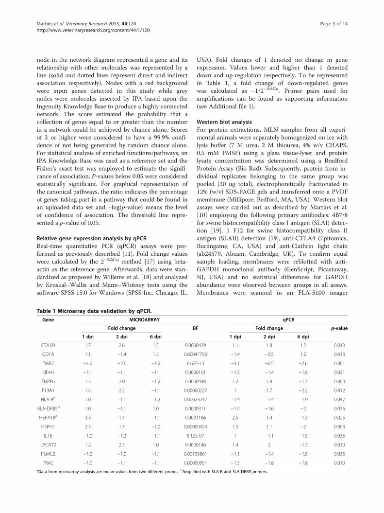
Table 1 Microarray data validation by qPCR.
Gene MICROARRAY
Fold change B
1 dpi 2 dpi 6 dpi
CD180 1.7 2.6 1.5 0.000
CD1A 1.1 −1.4 1.2 0.0004
DAB2 −1.2 −2.6 −1.2 6.62
EIF4H −1.1 −1.1 −1.1 0.000
ENPP6 1.3 2.0 −1.2 0.000
F13A1 1.4 2.2 −1.1 0.0000
HLA-Bb 1.0 −1.1 −1.2 0.0002
HLA-DRB5b 1.0 −1.1 1.0 0.000
HSPA1Ba 3.3 1.4 −1.1 0.000
HSPH1 2.3 1.7 −1.0 0.0000
IL16 −1.0 −1.2 −1.1 8.12
LPCAT2 1.2 2.3 1.0 0.000
PSMC2 −1.0 −1.0 −1.1 0.0010
TRAC −1.0 −1.1 −1.1 0.0000aData from microarray analysis are mean values from two different probes. bAmplifi
USA). Fold changes of 1 denoted no change in geneexpression. Values lower and higher than 1 denoteddown and up-regulation respectively. To be representedin Table 1, a fold change of down-regulated geneswas calculated as −1/2−ΔΔCq. Primer pairs used foramplifications can be found as supporting information(see Additional file 1).
Western blot analysisFor protein extractions, MLN samples from all experi-mental animals were separately homogenized on ice withlysis buffer (7 M urea, 2 M thiourea, 4% w/v CHAPS,0.5 mM PMSF) using a glass tissue-lyser and proteinlysate concentration was determined using a BradfordProtein Assay (Bio-Rad). Subsequently, protein from in-dividual replicates belonging to the same group waspooled (30 ug total), electrophoretically fractionated in12% (w/v) SDS-PAGE gels and transferred onto a PVDFmembrane (Millipore, Bedford, MA, USA). Western blotassays were carried out as described by Martins et al.[10] employing the following primary antibodies: 4B7/8for swine histocompatibility class I antigen (SLAI) detec-tion [19], 1 F12 for swine histocompatibility class IIantigen (SLAII) detection [19], anti-CTLA4 (Epitomics,Burlingame, CA, USA) and anti-Clathrin light chain(ab24579, Abcam, Cambridge, UK). To confirm equalsample loading, membranes were reblotted with anti-GAPDH monoclonal antibody (GenScript, Picastaway,NJ, USA) and no statistical differences for GAPDHabundance were observed between groups in all assays.Membranes were scanned in an FLA-5100 imager
qPCR
F Fold change p-value
1 dpi 2 dpi 6 dpi
0429 1.1 1.8 1.2 0.010
7793 −1.4 −2.5 1.2 0.013
E-13 −3.1 −6.5 −2.6 0.001
0101 −1.5 −1.4 −1.8 0.021
0448 1.2 1.8 −1.7 0.000
0227 1 1.7 −2.2 0.012
3747 −1.4 −1.4 −1.9 0.047
0311 −1.4 −1.6 −2 0.036
1166 2.5 1.4 −1.3 0.025
0424 1.5 1.1 −2 0.003
E-07 1 −1.1 −1.5 0.035
0146 1.4 2 −1.3 0.010
5861 −1.1 −1.4 −1.8 0.036
0951 −1.5 −1.8 −1.8 0.010
ed with SLA-B and SLA-DRB5 primers.

Martins et al. Veterinary Research 2013, 44:120 Page 4 of 14http://www.veterinaryresearch.org/content/44/1/120
(Fujifilm, Tokyo, Japan) and signal intensity was deter-mined using Multigauge software (Fujifilm, Tokyo,Japan) as previously described [10].
Histopathology, immunohistochemistry and confocalmicroscopy analysisParaffin sections (5 μm) of formalin fixed samples wereroutinely processed and stained with hematoxylin andeosin (H&E) to evaluate tissue morphology. For immu-nohistochemistry assays, a standard avidin-biotin perox-idase method was performed as described elsewhere [20]employing 1 F12 monoclonal antibody and a biotinylatedanti-mouse Ig (Dako, Barcelona, Spain) as a secondaryantibody. Immunofluorescence using confocal micros-copy was performed employing the anti-SLAII 1 F12monoclonal antibody, a rabbit polyclonal antibodyagainst the Salmonella somatic (O4, 5, 12) antigen[10] and a rabbit polyclonal antibody anti-SalmonellaTyphimurium flagellin [21]. Fluorescein isothiocyanate(FITC)-conjugated goat anti-rabbit IgG (Sigma-Aldrich,St. Louis, MO, USA) and Alexa Fluor 594 anti-mouseIgG (Life Technologies, Carlsbad, CA, USA) were usedas secondary antibodies. Immunostaining was performedas described by Robertson et al. [22]. Briefly, deparaffi-nized sections of formalin fixed MLN were blocked for30 min with 1% bovine serum albumin and 2% foetal calfserum in PBS. Then, sections were incubated overnightat 4 °C with primary antibodies, washed three timeswith PBS for 5 min and incubated for 1 h at 37 °C withfluorescent secondary antibodies. For negative controls,primary antibody was omitted. Finally, sections werewashed three times for 5 min in PBS containing1.43 μM 4′,6-diamidino-2-phenylindole (DAPI, LifeTechnologies). Samples were subsequently evaluated andimaged using an LSM 5 Exciter confocal microscope(Carl Zeiss, Jena, Germany).
Cell death analysisFormalin fixed MLN samples were evaluated for celldeath by terminal deoxynucleotidyl transferase dUTPnick end labeling (TUNEL), employing the TUNELApoptosis Detection Kit for Paraffin-embedded TissueSections (GenScript, Picastaway, NJ, USA) according tothe manufacturer’s instructions. Briefly, proteinase Ktreated samples were permeabilized with 0.1% TritonX-100 and 0.1% sodium citrate for 10 min and incubatedwith Blocking Solution II (GenScript) for 30 min. Subse-quently, tissues were covered with 50 μL of TUNELReaction Mixture (GenScript), incubated at 37 °C for 1 hin a dark humidified chamber and washed in PBS.Sections were examined in an LSM 5 Exciter confocalmicroscope (Carl Zeiss MicroImaging GmbH, Jena,Germany) using excitation wave 450–500 nm and emis-sion wave 515–565 nm (green). Fluorescence intensity
was quantified with the ImageJ software 1.46r [23] anddata were analyzed by ANOVA (p-value cutoff of 0.05)using SPSS 15.0 for Windows (SPSS Inc).
Selective capture of transcribed sequences (SCOTS)Selective capture of Salmonella transcripts from MLN ofpigs at 2 dpi was performed by the SCOTS method [24],following the procedure described by Sheikh et al. [25].Briefly, 5 μg of total RNA from infected MLN sampleswas converted into first strand cDNA by using randompriming and Superscript III reverse transcription (LifeTechnologies). Subsequently second strand cDNA wasproduced employing DNA polymerase I (Klenow frag-ment, Life Technologies). To create a correspondingin vitro Salmonella Typhimurium cDNA sample forcomparison, the same bacterial isolate employed in theexperimental infection was grown to early-log growthphase (OD600 = 0.3) and late-log growth phase (OD600 =0.8) in Luria Bertani (LB) broth. Afterwards, SalmonellaTyphimurium transcripts were selectively capturedfrom in vivo and in vitro double stranded cDNAby hybridization to sonicated biotinylated genomicSalmonella DNA, which was previously blocked withSalmonella ribosomal DNA fragments. MicrobialcDNA-genomic DNA hybrids were then captured bybinding to streptavidin-coated beads (Dynabeads M-280streptavidin, Invitrogen) and bacterial transcripts wereeluted by alkaline denaturation. Eluted bacterial cDNAwas then PCR-amplified with conserved primers andfinally purified using Qiagen PCR column purificationkit (Qiagen). After that, one round captured and purifiedcDNA from both in vitro and in vivo conditions werequantified by spectrophotometry and used as template(10 ng) for qPCR assays as described above. Primer pairsused for amplifications can be found as supporting infor-mation (see Additional file 2). Gene expression levelswere estimated employing gyrA as the reference gene.Since tissue from uninfected pigs was negative forSalmonella, those samples could not be used as referencefor fold change calculations of pathogen gene expresion.In addition, most screened genes showed Cq values infer-ior to those observed for gyrA in infected MLN. For thesereasons, gene expression levels were alternatively esti-mated as follows: gyrA Cq – target gene Cq. Highervalues meant higher expression levels and vice-versa.
ResultsTranscriptional changes in porcine MLN upon salmonellaTyphimurium infection and data validationMicroarray technology coupled to a Bayesian analysiswas employed to explore the transcriptional response ofporcine MLN to Salmonella Typhimurium along a timecourse of 1, 2 and 6 dpi. BATS, a method specifically de-signed for the analysis of time series microarray data

Martins et al. Veterinary Research 2013, 44:120 Page 5 of 14http://www.veterinaryresearch.org/content/44/1/120
[10], revealed significant changes in expression (BF <0.05) for 290 transcripts, representing 285 unique genes,as a result of the bacterial challenge (see Additionalfile 3). Then, to validate data, qPCR assays were per-formed on a panel of fourteen genes identified by BATSanalysis. As expected, all of them were confirmed to besignificantly regulated (p < 0.05) after infection (Table 1).Furthermore, an identical expression trend was observedfor most screened genes by qPCR and microarray analysis.
Biological interpretation of microarray dataTo translate microarray data into functional biologicalinformation, bioinformatics tools were employed to gainan insight into networks, functions and pathways associ-ated with the transcriptomic response of porcine MLNto Salmonella Typhimurium (see Additional file 4). IPAanalysis generated 17 gene interaction networks inte-grated by molecules associated with mechanisms such ascell-mediated immune response, cell-to-cell signalingand interaction, tissue morphology, cell movement andcell death. Networks 1 and 4 (Figure 1) revealed directrelationships between molecules taking part in five ofthe ten top enriched canonical pathways after infection(Figure 2). Moreover, network 4 demonstrated a centralrole for heat shock proteins and MHC encoding genesin the establishment of different mechanisms carried outin MLN in response to Salmonella Typhimurium. IPAalso ascertained the enrichment of biological functionsother than those identified by network analysis (Table 2).Thus, “Inflammatory disease” was the Ingenuity biofunc-tion more significantly related to the differentiallyexpressed genes, followed by “Protein synthesis” and“Antigen presentation”.
Modulation of immune response mechanismsWide transcriptomic data analysis by bioinformaticstools revealed an enrichment of distinct mechanismsleading to immune response activation in porcine MLNupon Salmonella Typhimurium infection and depictedconnections between them. As illustrated in Figure 3A,the association between “CTLA-4 signaling in cytotoxicT lymphocytes” and “Clathrin-mediated endocytosissignaling” pathways was established by the regulation ofshared genes. Then, we checked the abundance level ofcytotoxic T-lymphocyte-associated protein 4 (CTLA-4)and clathrin light chain A (CLTA) by Western blot, not-ing that CLTA was more abundant in infected animalswhereas CTLA4 showed reduced levels after infection(Figure 3B). Since changes in CTLA4 expression couldnot be detected by microarray analysis, we verifiedCTLA4 mRNA levels by qPCR (Figure 3C). In accord-ance with Western blot assays, CTLA4 was observed tobe significantly down-regulated in infected animals at 1and 6 dpi. Concerning CLTA, a similar trend towards
up-regulation was observed at mRNA (Figure 3D) andprotein levels.System biology analysis also revealed the involvement
of MHC encoding genes in many processes triggered inMLN in response to Salmonella Typhimurium. Thus,changes undergone by these molecules were evaluatedemploying different approaches. Firstly, Western blotanalysis demonstrated that major histocompatibility an-tigens class I (MHCI) and class II (MHCII) were moreabundant in tissue at 1 dpi (Figure 4A). Similarly, immu-nohistochemistry revealed higher MHCII expression atinitial stages of infection, with increased levels of thismolecule mainly detected in large irregularly-shapedmononuclear cells (Figure 4B-E). Then, confocal micros-copy analysis uncovered the presence of SalmonellaTyphimurium antigens in cells showing high levels ofMHCII (Figure 4H-K), suggesting a connection betweenthe increase of this receptor at the protein level and thepresence of pathogen in tissue. Curiously, MHCI andMHCII (annotated in data sets as HLA-B and HLA-DRB, respectively) were found to be down-regulated bymicroarray analysis. Since these results were confirmedby qPCR (Figure 4F-G), a divergence between transcrip-tomic and proteomic changes could be highlighted forthese molecules in infected MLN.
Tissue morphology and cell death“Cell death” was one of the most significantly alteredbiological functions after infection and integrated thehighest number of differentially expressed genes (n = 78).In order to find GO subcategories associated to genesimplicated in this function, a data set arranged into“Cell death” by IPA was loaded into the DAVIDBioinformatic Database. As expected, enriched termswere related to cell proliferation, differentiation anddeath (see Additional file 5). Among them, processes suchas “Negative regulation of apoptosis” and “Antiapoptosis”were found to be enriched all along the infection,suggesting an inhibition of apoptosis. To deepen andsharpen these results, TUNEL analysis followed byconfocal microscopy was performed to elucidate the celldeath mechanisms induced in MLN after SalmonellaTyphimurium infection. As shown in Figure 5A-D and 5I,DNA damage detected by TUNEL staining peaked at 1dpi and decreased at 2 and 6 dpi compared to controls.Afterwards, expression levels of CASP1 and CASP3, themain pyroptosis and apoptosis inducers respectively, werequantified by qPCR. CASP1 mRNA was significantly up-regulated at 2 dpi and down-regulated at 6 dpi (Figure 5J),whereas no significant changes were observed for CASP3(Figure 5K). Finally, lymph-node sections were H&E-stained to analyse the structural changes undergone by tis-sue as a consequence of infection (Figure 5E-H). Besidesthe loss of the typical lymph-node micro-architecture,

Network 1
Network 4
Figure 1 Gene network analysis by Ingenuity Pathway Analysis (IPA). Visual representation of networks 1 and 4. Red and grey nodes areinput and IPA-inserted molecules respectively. Colored lines highlight genes that take part in an enriched Canonical Pathway.
Martins et al. Veterinary Research 2013, 44:120 Page 6 of 14http://www.veterinaryresearch.org/content/44/1/120

Figure 2 Top 10 enriched canonical pathways. Blue bars andyellow squares denote –log(p-value) and ratio respectively.Threshold line represents a p-value of 0.05.
Martins et al. Veterinary Research 2013, 44:120 Page 7 of 14http://www.veterinaryresearch.org/content/44/1/120
phagocyte infiltration was the main alteration detectedafter infection, being observed mainly at 1 and 2 dpi.
Salmonella Typhimurium localization and gene expressionin vivoSalmonella Typhimurium was detected in MLN alongthe whole studied time course, with higher Salmonellalevels being observed at 2 dpi (data not shown).Confocal microscopy analysis revealed two distinct bac-terial populations according to location and labeling. Asshown in Figure 4H-K and Figure 6A, some bacteriawere labeled as spherical structures located in the peri-nuclear zone of mononuclear cells. In addition, regularbacilli shaped structures showing a different distributionwere also detected (Figure 6B). Although Z-stackconfocal images indicate that, most likely, this secondpopulation was found in an extracellular environment(see Additional file 6), further experiments will be neces-sary to clarify whether the pathogen is in the cytosolor outside the cell. Ultimately, expression of someSalmonella genes in vivo was also studied by the SCOTSapproach and compared to in vitro conditions. Type IIIsecretion systems (TTSS) encoded genes (sopB, avrA,sifA, sseL and prgJ) and spvB were found to be expressed
Table 2 Top five biological functions enriched in MLN ofpigs infected with Salmonella Typhimurium.
Annotations p-Valuea Input genes (n)
Inflammatory disease 4.64E-05 – 2.65E-02 13
Protein synthesis 6.52E-05 – 1.86E-02 32
Antigen presentation 1.8E-04 – 1.68E-02 5
Cell death 1.8E-04 – 2.67E-02 78
Cell-to-cell signalingand interaction
1.8E-04 – 2.67E-02 27
ap-values were calculated with the right-tailed Fisher’s Test.
by Salmonella Typhimurium in porcine MLN. Notably,lower expression of these genes was detected in vitrowhen compared to in vivo. The results regarding genescoding for flagella components and regulators showhigher expression levels in vitro for fliA, fliC whereasfljA mRNA was observed to be more expressed in vivo(Figure 6C).
DiscussionGut-associated lymphoid tissues have been proved to bean important niche for Salmonella during pig infections.Previous reports stated that Salmonella Typhimuriumcan be found in MLN of infected pigs from 2 h [5] up to6 weeks after oral inoculation [26] and sustain theseorgans as immune inductive sites during pig salmonel-losis [6,7,10,26,27]. For this reason, in this workwe aimed at dissecting host response mechanismsoccurring in porcine MLN upon interaction withSalmonella Typhimurium. Additionally, expression ofsome Salmonella virulence effectors was also analyzedin infected tissues attempting to integrate informationfrom both the host and pathogen.The systems biology analysis reported in this study
demonstrates the involvement of MHC molecules inseveral mechanisms triggered in swine MLN after bac-terial challenge. Intriguingly, both MHCI and MHCIIencoding genes were found to be down-regulated allalong the studied time course, in spite of the increasedlevels observed for these receptors at 1 dpi by Westernblot and microscopic analysis. We envisage that initially,antigen presenting cells bearing high levels of MHC mi-grate to MLN leading to an increase of these receptorsin tissue, as suggested by the detection of Salmonellaantigens in cells showing high levels of MHCII. Subse-quently, processes carried out in MLN might reduceMHC mRNA and protein expression levels at later timesof infection. Previous studies demonstrate that Salmonellainterferes with antigen presentation by reducing MHCIIsurface expression via a mechanism dependent on theSalmonella pathogenicity island (SPI)-2 encoded effectorSifA [28-30]. It is noteworthy that in this study we showthat sifA is expressed by Salmonella Typhimurium in por-cine MLN. Besides, it has been previously observed by us[21] and others [27] that pig infections with SalmonellaTyphimurium do not produce an up-regulation ofcytokines involved in T helper 1 (Th1) response in MLN,on the contrary to previous reports in mice [31]. Thesefindings could be related to the ability of pathogens tolimit antigen presentation to CD4 restricted T cells byreducing MHCII levels in infected cells. Salmonellaremoves mature MHCII complexes from the cell surfaceby enhancing their ubiquitination in a clathrin andAP2-dependent way [29]. Curiously, we identified the“Protein ubiquitination pathway” and “Clathrin-mediated

CLTACTLA4
GAPDH 364329kDa0 1 2 6
B
p<0.00
p=0.02
A
C D
p=0.005BF<0.00
Figure 3 Interaction between CTLA-4 signaling in cytotoxic T lymphocytes and Clathrin-mediated endocytosis signaling pathways. A:visual representation of CTLA-4 signaling in cytotoxic T lymphocyte pathway stressing the regulation of molecules taking part in Clathrin-mediatedendocytosis signaling. Red and grey nodes are input and IPA-inserted molecules respectively. B: Western blot analysis of CTLA4 and CLTA in MLN at1, 2 and 6 dpi. C: mRNA quantification of CTLA4 by qPCR. D: expression pattern of CLTA mRNA by microarray analysis.
Martins et al. Veterinary Research 2013, 44:120 Page 8 of 14http://www.veterinaryresearch.org/content/44/1/120
endocytosis signaling” as the most significantly affectedcanonical pathways upon infection. In spite of the re-ported use of clathrin-mediated endocytosis in bacterial-induced internalization, Salmonella is not able to employthis machinery to invade [32]. So, instead of promotingdirect entry of the pathogen to host cells, enrichment ofclathrin-mediated endocytosis could be related to themodulation of MHCII expression by Salmonella found intissue. Therefore, this evidence as a whole could suggest ahampering of processes mediated by MHCII in swineMLN following Salmonella infection.On the contrary, network analysis also associated
“Clathrin-mediated endocytosis signaling” to “CTLA4signaling in cytotoxic T lymphocytes’ pathway”, bringingto light a possible role of the former process in adaptiveimmunity triggering. CTLA4, an important negativeregulator of the T cell immune response [33], is
endocytosed via a clathrin and dynamin-dependent routein activated T-cells [34]. According to Johanns et al.[35], up-regulation of CTLA4 in regulatory T cells re-strains effector T cell activation at early infection timepoints and allows the increase of bacterial burden duringmurine salmonellosis. Similarly, Inoue et al. [33] statethat CTLA4-mediated Treg immunosuppression is crit-ical in preventing the host from eliminating invasivepathogens. Given that, CTLA4 down-regulation, concur-rent with clathrin up-regulation after the bacterial chal-lenge, could indicate the repression of a mechanism of Tcell inhibition in porcine MLN upon Salmonella Typhi-murium infection. However, since clathrin could be in-volved in the establishment of both host immunitymechanisms and virulence strategies evolved by thepathogen, a deeper investigation of processes mediatedby this molecule during infection is necessary and could

B C
D E
H I J K
45
kDa
36
25
MHCI
GAPDH
0 1 2 6
MHCII
A
p<0.00
p=0.02 p=0.036 p=0.047
GF
Figure 4 Analysis of MHC molecules in porcine MLN after Salmonella Typhimurium infection. A: Western blot analysis of MHC class I and IIat 1, 2 and 6 dpi. B-E: MHCII labeling in tissue at 0 (B), 1(C), 2 (D) and 6 (E) dpi by immunohistochemistry. Scale bar = 20 μm. F-G: mRNAquantification of SLA-DRB5 (MHCII) (F) and SLA-B (MHCI) (G) by qPCR. H-K: confocal analysis of Salmonella Typhimurium infected MLN demon-strate the presence of the pathogen in MHCII positive cells. Salmonella Typhimurium-FITC (H), MHCII-Alexa Fluor® 594 (I), DAPI (J) and merge (K).
Martins et al. Veterinary Research 2013, 44:120 Page 9 of 14http://www.veterinaryresearch.org/content/44/1/120
provide relevant knowledge on the pathogenesis of por-cine salmonellosis.Current results also pointed to the generation of adap-
tive immunity mechanisms in infected tissue at a shorttime after infection. High MHCI levels observed byWestern blot at 1 dpi reinforce our previous evidencethat Salmonella antigens are cross-presented in swineMLN at initial stages of infection [10]. Cross-presenta-tion is a mechanism that enables antigen presenting cells
to prime CD8+ T cells via their own MHCI molecules[36]. Interestingly, it has been reported that differentlyfrom MHCII, Salmonella is not able to reduce MHCIsurface expression of infected cells and consequentlyavoid early host cytotoxic response [28-30]. Therefore,cross-presentation might lead to an early SalmonellaTyphimurium clearance by cytotoxic T cells during por-cine infections, in agreement with the stimulation ofSalmonella-specific CD8 T cells readily observed after

E GF H
BA C D
p=0.02p<0.00 p=0.097
JI K
Figure 5 Cell death and histopathologic analysis of MLN from Salmonella Typhimurium infected pigs. A-D: Terminal deoxynucleotidyltransferase dUTP nick end labeling (TUNEL) analysis at 0 (A), 1(B), 2 (C) and 6 (D) dpi. Scale bar = 100 μm. E-H: H&E staining of at 0 (E), 1(F), 2 (G)and 6 (H) dpi. Scale bar = 50 μm. I: Quantification of TUNEL fluorescent labeling shows an increase of positive nuclei at 1 dpi and a decrease tolevels inferior than the controls at 2 and 6 dpi. J-K: mRNA quantification of CASP1 (J) and CASP3 (K) by qPCR.
Martins et al. Veterinary Research 2013, 44:120 Page 10 of 14http://www.veterinaryresearch.org/content/44/1/120
mice and human infections [37]. Additionally, CD180,an inducer of B cell proliferation, activation and differ-entiation [38], was uncovered to be up-regulated allalong infection. Taken together, our results indicate thatboth cellular and humoral immunity mechanisms areeffectively engendered in porcine MLN at a short timeafter infection with Salmonella Typhimurium. Thus, thedynamics of this protective response could be decisive inthe course of infection by this pathogen in pigs.Evidence of pyroptosis induction and apoptosis damp-
ening in infected MLN were disclosed in the currentstudy, supporting our previous reports [10]. Thus,microarray data mining detected an enrichment of pro-cesses such as “Negative regulation of apoptosis” and“Antiapoptosis” after the bacterial challenge, in additionto up-regulation of genes encoding for an inhibitor ofapoptosis proteins (IAP) like XIAP and PDCL3. Induc-tion of apoptosis has been asserted as a strategy thatfacilitates Salmonella cell-to-cell spread during systemicinfection [39]. Nevertheless, it has also been reportedthat AvrA, a Salmonella effector protein, prevents the
apoptotic elimination of host cell niche as a pathogenevasion mechanism [40]. Intriguingly, we observed in vivoexpression of spvB and sseL, both major SalmonellaTyphimurium apoptosis inducers, and the apoptosisinhibitor avrA, indicating that Salmonella appeared toexecute virulence mechanisms to modulate apoptosis inporcine MLN in its favor.As in apoptosis, pyroptotic cells show DNA fragmen-
tation, nuclear condensation and positive TUNEL stain-ing [41,42]. However, pyroptosis inherently results ininflammation due to caspase-1-mediated maturation ofpro-IL-1β and pro-IL-18 and release of the cytoplasmiccontent, whereas the apoptotic cell is considered to beimmunologically silent [43]. In the current study, in-crease of TUNEL positive labeling was observed in thetissue at 1 dpi, as well as infiltration of inflammatorycells and IL1β up-regulation. Therefore, we propose thatonce apoptosis is dampened, the infected cell undergoespyroptosis in swine MLN, producing pathogen dischargeto the extracellular milieu and clearance of bacteria byinnate mechanisms. In support of this, we previously

A B
C
Figure 6 Salmonella Typhimurium labeling and gene expression in porcine MLN. A-B: Different labeling profiles found for SalmonellaTyphimurium in porcine MLN. Scale bar = 10 μm. (A) Pathogen detection as spherical structures in the perinuclear zone of mononuclear cells.(B) Staining of bacilli shaped structures. C: Analysis of Salmonella Typhimurium gene expression by SCOTS in vivo and in vitro. Black dots and barsrespectively represent individual and mean expression values from analysis of cDNA from pig infected MLN. Triangles (early logarithmic phase)and squares (late logarithmic phase) denote gene expression data from Salmonella Typhimurium cultures. Higher values mean higher expressionlevels and vice-versa.
Martins et al. Veterinary Research 2013, 44:120 Page 11 of 14http://www.veterinaryresearch.org/content/44/1/120
reported an increase of phagocyte counts and mRNAlevels of pro-inflammatory genes upon infection withSalmonella Typhimurium and a significant reduction ofthe pathogen burden at 6 dpi [21].In line with this, an elegant study by Miao et al. [44]
stated that Salmonella Typhimurium is able to damperpyroptosis for its own advantage by avoiding flagellinexpression during infection of mice. Interestingly, wefound expression of Salmonella Typhimurium flagellacomponent (FliC) and regulators (FliA and FljA) in in-fected MLN. Additionally, flagella expression by infect-ing bacteria found in tissue was also corroborated bylabeling using a specific polyclonal antibody. Salmonellaenterica alternately expresses two different flagellar
filament proteins, FljB and FliC, in a process known asflagellar phase variation. In spite of the high homologylevel found between these proteins, their middle surfaceexposed sequences of amino acids are divergent, result-ing in distinct antigenicities [45]. It is noteworthy thatour results demonstrate higher expression levels for fliCand its regulator fliA in vitro than in vivo. On the con-trary, fljA, which is cotranscribed with fljB, was moreexpressed in Salmonella Typhimurium found in vivo.Moreover, this gene was notably less expressed than fliAand fliC in both early and late logarithmic phasecultures. Based on this, we deduced a skewing towardFliC flagellin expression by bacteria in vitro. Besides, wedrew the inference that a more heterogeneous flagellin

Martins et al. Veterinary Research 2013, 44:120 Page 12 of 14http://www.veterinaryresearch.org/content/44/1/120
expression is found in Salmonella Typhimurium repli-cating in vivo and that induction of flagellar phase vari-ation could be a strategy adopted by this pathogen tohinder pig immune response. Expression of prgJ was alsouncovered in swine MLN. Curiously, repression of thiseffector has been reported as a mechanism of pyroptosisinhibition in vivo [46]. Thus, it could be inferred thatexpression of flagellin and prgJ by SalmonellaTyphimuriumfound in tissue might enable pigs to use pyroptosis to clearbacteria in gut associated lymph-nodes, protecting itselffrom pathogen dissemination. Nevertheless, an issue thatshould be addressed by our assumption is why pathogenburden in tissue peaks after pyroptosis triggering. Miao andRajan [46] stated that in a single cell, pyroptosis only takesplace at late times of infection, following bacteria replica-tion. So, we inferred that increase of pathogen load at 2 dpimay be due to the release of replicated Salmonella fromcells dead by pyroptosis.Notably, the presence of TUNEL positive cells in
MLN was significantly reduced at 2 and 6 dpi, suggest-ing a decrease of cell death by apoptosis or pyroptosis.As with any physiological process, excessive pyroptosisis detrimental to the host [41]. So, modulation of thispathway by the host aiming to restore tissue integrityshould be expected. Actually, we observed up-regulationof MAP3K7 and TRAF7, both involved in NF-kB andsurvival pathway activation, at 2 and 6 dpi. However, evi-dence indicates that inhibition of caspase-dependentapoptosis primes cells towards programmed necrosis[47]. Since the mechanisms that dictate the cellular deci-sion to survive by activating NF-kB or to die throughapoptosis or necroptosis are still unclear [48] further re-search is necessary to clarify these results.In conclusion, the results provided led us to infer that
although the Salmonella Typhimurium strain employedin this study was able to express some of its major viru-lence effectors in porcine MLN, a combination of earlyhost triggered innate and adaptive immunity mecha-nisms might overcome virulence strategies employed bypathogens. Besides preventing apoptosis, swine appearto take advantage of flagellin and prgJ expressionby pathogens to induce pyroptosis in MLN. In thiscontext, pyroptosis might consist in a host protectivemechanism that prevents pathogen spread beyond gut-associated lymph-nodes. Furthermore, cross-presenta-tion of Salmonella antigens in MLN might result in arapid clearance of pathogens by cytotoxic T cells.Functional relevance was also shown by clathrin-mediated endocytosis that could contribute to mecha-nisms of pathogen virulence and/or host defence inMLN of Salmonella infected swine. Further analysis ofexamined mechanisms may support the discovery ofnovel strategies of host defense against Salmonella atthe intestinal level.
Additional files
Additional file 1: Primer pairs employed in host differentialexpression analysis by real-time quantitative PCR. Table showingsequences and accession numbers of primers used for differentialexpression analysis in porcine mesenteric lymph-nodes.
Additional file 2: Primer pairs employed in the analysis ofSalmonella Typhimurium virulence genes expressed in vivo. Tableshowing sequences and accession number of primers used forquantification of Salmonella Typhimurium transcripts expressed inporcine mesenteric lymph-nodes.
Additional file 3: Differentially expressed transcripts. Table showingchip ID, gene name, fold change and Bayes Factor value of transcriptsdifferentially expressed in swine mesenteric lymph-nodes.
Additional file 4: Ingenuity Pathway Analysis annotations. Excelspreadsheet describing networks, biofunctions and canonical pathwaysfound to be enriched in swine mesenteric lymph-nodes after SalmonellaTyphimurium infection.
Additional file 5: DAVID Bioinformatic Database analysis of genesinvolved in Cell Death. Excel spreadsheet describing Gene Ontologyterms associated to gene data set involved in Cell Death.
Additional file 6: Detection of Salmonella Typhimurium inmesenteric lymph-nodes of infected swine. Video showing confocalanalysis (Z-stack tool) of Salmonella Typhimurium labeled in mesentericlymph nodes of infected swine.
Competing interestsThe authors declare that they have no competing interests.
Authors’ contributionsRPM was responsible for the whole study, including lab work, data analysisand interpretation, as well as the writing of the manuscript. CA participatedin the confocal analysis. JEG collaborated with SCOTS analysis. AC performedthe experimental infection and collected the tissue samples. RB and MGCperformed microarray data processing. JJG conceived and designed theproject and participated in the interpretation and discussion of the results, aswell as in the writing of the manuscript. All authors read and approved thefinal manuscript.
AcknowledgementsWe thank Erena Ruiz Mora, Juana Molina and Reyes Alvarez for skillfultechnical assistance, Esther Peralbo for technical support in confocalmicroscopy analysis (IMIBIC) and Eloisa Andújar and Mónica Pérez fromthe Genomic Unit of CABIMER for their excellent array technical assistance.This work was supported by the Junta de Andalucía (P07-AGR-02672),the Spanish Ministry of Science and Innovation (AGL2008-00400 andAGL2011-28904) and EU funds through the SABRE project and EADGENEnetwork. RPM and CA are predoctoral researchers supported by the FPUResearch Program of the Spanish Ministry of Education and Science.
Author details1Grupo de Genómica y Mejora Animal, Departamento de Genética, Facultadde Veterinaria, Universidad de Córdoba, Campus de Rabanales, EdificioGregor Mendel C5, 14071, Córdoba, Spain. 2Department of Microbiology andImmunology, University of Louisville, School of Medicine, 40202, Louisville,KY, USA. 3Departamento de Sanidad Animal, Facultad de Veterinaria,Universidad de León, 24071, León, Spain. 4Plataforma Andaluza deBioinformática, Universidad de Málaga, Parque Tecnológico de Andalucía,29590, Málaga, Spain.
Received: 30 January 2013 Accepted: 25 November 2013Published: 5 December 2013
References1. EFSA: The European Union summary report on trends and sources of
zoonoses, zoonotic agents and food-borne outbreaks in 2009.EFSA J 2011, 9:2090.

Martins et al. Veterinary Research 2013, 44:120 Page 13 of 14http://www.veterinaryresearch.org/content/44/1/120
2. Baptista FM, Halasa T, Alban L, Nielsen LR: Modelling food safety andeconomic consequences of surveillance and control strategies forSalmonella in pigs and pork. Epidemiol Infect 2011, 139:754–764.
3. Sánchez-Vargas FM, Abu-El-Haija MA, Gómez-Duarte OG: Salmonellainfections: an update on epidemiology, management, and prevention.Travel Med Infect Dis 2011, 9:263–277.
4. Gopinath S, Carden S, Monack D: Shedding light on salmonella carriers.Trends Microbiol 2012, 20:320–327.
5. Boyen F, Haesebrouck F, Maes D, Van Immerseel F, Ducatelle R, Pasmans F:Non-typhoidal salmonella infections in pigs: a closer look atepidemiology, pathogenesis and control. Vet Microbiol 2008, 130:1–19.
6. Tuggle CK, Bearson SM, Uthe JJ, Huang TH, Couture OP, Wang YF, Kuhar D,Lunney JK, Honavar V: Methods for transcriptomic analyses of the porcinehost immune response: application to salmonella infection usingmicroarrays. Vet Immunol Immunopathol 2010, 138:280–291.
7. Wang Y, Couture OP, Qu L, Uthe JJ, Bearson SM, Kuhar D, Lunney J,Nettleton D, Dekkers JC, Tuggle CK: Analysis of porcine transcriptionalresponse to salmonella enterica serovar choleraesuis suggests noveltargets of NFkappaB are activated in the mesenteric lymph node.BMC Genomics 2008, 9:437.
8. Huang TH, Uthe JJ, Bearson SM, Demirkale CY, Nettleton D, Knetter S,Christian C, Ramer-Tait AE, Wannemuehler MJ, Tuggle CK: Distinct periph-eral blood RNA responses to salmonella in pigs differing in salmonellashedding levels: intersection of IFNG, TLR and miRNA pathways.PLoS One 2011, 6:e28768.
9. Collado-Romero M, Martins RP, Arce C, Moreno Á, Lucena C, Carvajal A,Garrido JJ: An in vivo proteomic study of the interaction betweensalmonella typhimurium and porcine ileum mucosa. J Proteomics 2012,75:2015–2026.
10. Martins RP, Collado-Romero M, Martínez-Gomáriz M, Carvajal A, Gil C,Lucena C, Moreno A, Garrido JJ: Proteomic analysis of porcine mesentericlymph-nodes alter salmonella typhimurium infection. J Proteomics 2012,75:4457–4470.
11. Collado-Romero M, Arce C, Ramirez-Boo M, Carvajal A, Garrido JJ: Quantita-tive analysis of the immune response upon salmonella typhimuriuminfection along the porcine intestinal gut. Vet Res 2010, 41:23.
12. Irizarry RA, Hobbs B, Collin F, Beazer-Barclay YD, Antonellis KJ, Scherf U,Speed TP: Exploration, normalization, and summaries of high densityoligonucleotide array probe level data. Biostatistics 2003, 4:249–264.
13. Angelini C, Cutillo L, De Canditiis D, Mutarelli M, Pensky M: BATS: aBayesian user-friendly software for analyzing time series microarrayexperiments. BMC Bioinformatics 2008, 9:415.
14. Conesa A, Götz S, García-Gómez JM, Terol J, Talón M, Robles M: Blast2GO: auniversal tool for annotation, visualization and analysis in functionalgenomics research. Bioinformatics 2005, 21:3674–3676.
15. Ingenuity Systems: Home [www.ingenuity.com]16. da Huang W, Sherman BT, Lempicki RA: Systematic and integrative
analysis of large gene lists using DAVID bioinformatics resources.Nat Protoc 2009, 4:44–57.
17. Livak KJ, Schmittgen TD: Analysis of relative gene expression data usingreal-time quantitative PCR and the 2−ΔΔCT method. Methods 2001,25:402–408.
18. Willems E, Leyns L, Vandesompele J: Standardization of real-time PCRgene expression data from independent biological replicates.Anal Biochem 2008, 379:127–129.
19. Bullido R, Gomez del Moral M, Alonso F, Ezquerra A, Zapata A, Sánchez C,Ortuño E, Alvarez B, Domínguez JJ: Monoclonal antibodies specific forporcine monocytes/macrophages: macrophage heterogeneity in the pigevidenced by the expression of surface antigens. Tissue Antigens 1997,49:403–413.
20. Yubero N, Jiménez-Marín A, Barbancho M, Garrido JJ: Two cDNAs codingfor the porcine CD51 (αV integrin subunit: cloning, expression analysis,adhesion assays and chromosomal localization. Gene 2011, 481:29–40.
21. Martins RP, Collado-Romero M, Arce C, Lucena C, Carvajal A, Garrido JJ:Exploring the immune response of porcine mesenteric lymph nodesto salmonella enterica serovar typhimurium: an analysis of transcrip-tional changes, morphological alterations and pathogen burden.Comp Immunol Microbiol Infect Dis 2013, 36:149–160.
22. Robertson D, Savage K, Reis-Filho JS, Isacke CM: Multiple immunofluorescencelabelling of formalin-fixed paraffin-embedded (FFPE) tissue.BMC Cell Biol 2008, 9:13–22.
23. Schneider CA, Rasband WS, Eliceiri KW: NIH Image to ImageJ: 25 years ofimage analysis. Nat Methods 2012, 9:671–675.
24. Graham JE, Clark-Curtiss JE: Identification of mycobacterium tuberculosisRNAs synthesized in response to phagocytosis by human macrophagesby selective capture of transcribed sequences (SCOTS). Proc Natl Acad SciUSA 1999, 96:11554–11559.
25. Sheikh A, Charles RC, Sharmeen N, Rollins SM, Harris JB, Bhuiyan MS,Arifuzzaman M, Khanam F, Bukka A, Kalsy A, Porwollik S, Leung DT, BrooksWA, LaRocque RC, Hohmann EL, Cravioto A, Logvinenko T, Calderwood SB,McClelland M, Graham JE, Qadri F, Ryan ET: In vivo expression ofsalmonella enterica serotype typhi genes in the blood of patients withtyphoid fever in Bangladesh. PLoS Negl Trop Dis 2011, 5:e1419.
26. Rostagno MH, Eicher SD, Lay DC Jr: Immunological, physiological, andbehavioral effects of salmonella enterica carriage and shedding inexperimentally infected finishing pigs. Foodborne Pathog Dis 2011, 8:623–630.
27. Uthe JJ, Royaee A, Lunney JK, Stabel TJ, Zhao SH, Tuggle CK, Bearson SM:Porcine differential gene expression in response to salmonella entericaserovars choleraesuis and typhimurium. Mol Immunol 2007, 44:2900–2914.
28. Van Parys A, Boyen F, Verbrugghe E, Leyman B, Bram F, Haesebrouck F,Pasmans F: Salmonella typhimurium induces SPI-1 and SPI-2 regulatedand strain dependent downregulation of MHC II expression on porcinealveolar macrophages. Vet Res 2012, 43:52.
29. Lapaque N, Hutchinson JL, Jones DC, Méresse S, Holden DW, Trowsdale J,Kelly AP: Salmonella regulates polyubiquitination and surface expressionof MHC class II antigens. Proc Natl Acad Sci USA 2009, 106:14052–14057.
30. Mitchell EK, Mastroeni P, Kelly AP, Trowsdale J: Inhibition of cell surfaceMHC class II expression by salmonella. Eur J Immunol 2004, 34:2559–2567.
31. Eckmann L, Kagnoff MF: Cytokines in host defense against salmonella.Microbes Infect 2001, 3:1191–1200.
32. Veiga E, Cossart P: The role of clathrin-dependent endocytosis in bacterialinternalization. Trends Cell Biol 2006, 16:499–504.
33. Inoue S, Bo L, Bian J, Unsinger J, Chang K, Hotchkiss RS: Dose-dependenteffect of anti-CTLA-4 on survival in sepsis. Shock 2011, 36:38–44.
34. Qureshi OS, Kaur S, Hou TZ, Jeffery LE, Poulter NS, Briggs Z, Kenefeck R,Willox AK, Royle SJ, Rappoport JZ, Sansom DM: Constitutive clathrin-mediated endocytosis of CTLA-4 persists during T cell activation.J Biol Chem 2012, 287:9429–9440.
35. Johanns TM, Ertelt JM, Rowe JH, Way SS: Regulatory T cell suppressivepotency dictates the balance between bacterial proliferation andclearance during persistent salmonella infection. PLoS Pathog 2010,6:e1001043.
36. Houde M, Bertholet S, Gagnon E, Brunet S, Goyette G, Laplante A, PrinciottaMF, Thibault P, Sacks D, Desjardins M: Phagosomes are competentorganelles for antigen cross-presentation. Nature 2003, 425:402–406.
37. Lee SJ, Dunmire S, McSorley SJ: MHC class-I-restricted CD8 T cells play aprotective role during primary salmonella infection. Immunol Lett 2012,148:138–143.
38. Chaplin JW, Kasahara S, Clark EA, Ledbetter JA: Anti-CD180 (RP105)activates B cells to rapidly produce polyclonal Ig via a T cell andMyD88-independent pathway. J Immunol 2011, 187:4199–4209.
39. Haimovich B, Venkatesan MM: Shigella and salmonella: death as a meansof survival. Microbes Infect 2006, 8:568–577.
40. Wu H, Jones RM, Neish AS: The salmonella effector AvrA mediatesbacterial intracellular survival during infection in vivo. Cell Microbiol2012, 14:28–39.
41. Labbé K, Saleh M: Pyroptosis: A Caspase-1-dependent programmed celldeath and a barrier to infection. In Progress in Inflammation Research: TheInflammasomes. Edited by Couillin I, Pétrilli V, Martinon F. Basel: SpringerBasel AG; 2011:17–36.
42. Duprez L, Wirawan E, Vanden Berghe T, Vandenabeele P: Major cell deathpathways at a glance. Microbes Infect 2009, 11:1050–1062.
43. Fink SL, Cookson BT: Apoptosis, pyroptosis, and necrosis: mechanisticdescription of dead and dying eukaryotic cells. Infect Immun 2005,73:1907–1916.
44. Miao EA, Leaf IA, Treuting PM, Mao DP, Dors M, Sarkar A, Warren SE,Wewers MD, Aderem A: Caspase-1-induced pyroptosis is an innateimmune effector mechanism against intracellular bacteria. Nat Immunol2010, 11:1136–1142.
45. Bonifield HR, Hughes KT: Flagellar phase variation in Salmonella entericais mediated by a posttranscriptional control mechanism. J Bacteriol 2003,185:3567–3574.

Martins et al. Veterinary Research 2013, 44:120 Page 14 of 14http://www.veterinaryresearch.org/content/44/1/120
46. Miao EA, Rajan JV: Salmonella and caspase-1: a complex interplay ofdetection and evasion. Front Microbiol 2011, 2:85.
47. Moquin D, Chan FK: The molecular regulation of programmed necroticcell injury. Trends Biochem Sci 2010, 35:434–441.
48. Christofferson DE, Yuan J: Necroptosis as an alternative form ofprogrammed cell death. Curr Opin Cell Biol 2010, 22:263–268.
doi:10.1186/1297-9716-44-120Cite this article as: Martins et al.: Pyroptosis and adaptive immunitymechanisms are promptly engendered in mesenteric lymph-nodesduring pig infections with Salmonella enterica serovar Typhimurium.Veterinary Research 2013 44:120.
Submit your next manuscript to BioMed Centraland take full advantage of:
• Convenient online submission
• Thorough peer review
• No space constraints or color figure charges
• Immediate publication on acceptance
• Inclusion in PubMed, CAS, Scopus and Google Scholar
• Research which is freely available for redistribution
Submit your manuscript at www.biomedcentral.com/submit