Brominated Furanones Inhibit Biofilm Formation by Salmonella enterica Serovar Typhimurium
11
Published Ahead of Print 12 September 2008. 10.1128/AEM.01262-08. 2008, 74(21):6639. DOI: Appl. Environ. Microbiol. Keersmaecker Vanderleyden, Dirk E. De Vos and Sigrid C. J. De De Coster, Tine L. Verhoeven, Kathleen Marchal, Jos David Edith Gellens, Jeremy Levin, Hui Zhao, Kim Hermans, Joost C. A. Janssens, Hans Steenackers, Stijn Robijns, Typhimurium Serovar Salmonella enterica Formation by Brominated Furanones Inhibit Biofilm http://aem.asm.org/content/74/21/6639 Updated information and services can be found at: These include: REFERENCES http://aem.asm.org/content/74/21/6639#ref-list-1 at: This article cites 66 articles, 28 of which can be accessed free CONTENT ALERTS more» articles cite this article), Receive: RSS Feeds, eTOCs, free email alerts (when new http://journals.asm.org/site/misc/reprints.xhtml Information about commercial reprint orders: http://journals.asm.org/site/subscriptions/ To subscribe to to another ASM Journal go to: on April 21, 2014 by guest http://aem.asm.org/ Downloaded from on April 21, 2014 by guest http://aem.asm.org/ Downloaded from
-
Upload
independent -
Category
Documents
-
view
4 -
download
0
Transcript of Brominated Furanones Inhibit Biofilm Formation by Salmonella enterica Serovar Typhimurium

Published Ahead of Print 12 September 2008. 10.1128/AEM.01262-08.
2008, 74(21):6639. DOI:Appl. Environ. Microbiol. KeersmaeckerVanderleyden, Dirk E. De Vos and Sigrid C. J. DeDe Coster, Tine L. Verhoeven, Kathleen Marchal, Jos
DavidEdith Gellens, Jeremy Levin, Hui Zhao, Kim Hermans, Joost C. A. Janssens, Hans Steenackers, Stijn Robijns, Typhimurium
SerovarSalmonella entericaFormation by Brominated Furanones Inhibit Biofilm
http://aem.asm.org/content/74/21/6639Updated information and services can be found at:
These include:
REFERENCEShttp://aem.asm.org/content/74/21/6639#ref-list-1at:
This article cites 66 articles, 28 of which can be accessed free
CONTENT ALERTS more»articles cite this article),
Receive: RSS Feeds, eTOCs, free email alerts (when new
http://journals.asm.org/site/misc/reprints.xhtmlInformation about commercial reprint orders: http://journals.asm.org/site/subscriptions/To subscribe to to another ASM Journal go to:
on April 21, 2014 by guest
http://aem.asm
.org/D
ownloaded from
on A
pril 21, 2014 by guesthttp://aem
.asm.org/
Dow
nloaded from

APPLIED AND ENVIRONMENTAL MICROBIOLOGY, Nov. 2008, p. 6639–6648 Vol. 74, No. 210099-2240/08/$08.00�0 doi:10.1128/AEM.01262-08Copyright © 2008, American Society for Microbiology. All Rights Reserved.
Brominated Furanones Inhibit Biofilm Formation bySalmonella enterica Serovar Typhimurium�
Joost C. A. Janssens,1,2 Hans Steenackers,1,2 Stijn Robijns,1 Edith Gellens,1 Jeremy Levin,2 Hui Zhao,1Kim Hermans,1 David De Coster,1 Tine L. Verhoeven,1 Kathleen Marchal,1 Jos Vanderleyden,1
Dirk E. De Vos,2 and Sigrid C. J. De Keersmaecker1*Centre of Microbial and Plant Genetics, Katholieke Universiteit Leuven, Kasteelpark Arenberg 20, B-3001 Leuven, Belgium,1 and
Centre for Surface Chemistry and Catalysis, Katholieke Universiteit Leuven, Kasteelpark Arenberg 23, B-3001 Leuven, Belgium2
Received 6 June 2008/Accepted 7 September 2008
Salmonella enterica serovar Typhimurium is a main cause of bacterial food-borne diseases. As Salmonella canform biofilms in which it is better protected against antimicrobial agents on a wide diversity of surfaces, it isof interest to explore ways to inhibit biofilm formation. Brominated furanones, originally extracted from themarine alga Delisea pulchra, are known to interfere with biofilm formation in several pathogens. In this study,we have synthesized a small focused library of brominated furanones and tested their activity against S. entericaserovar Typhimurium biofilm formation. We show that several furanones inhibit Salmonella biofilm formationat non-growth-inhibiting concentrations. The most interesting compounds are (Z)-4-bromo-5-(bromomethy-lene)-3-alkyl-2(5H)-furanones with chain lengths of two to six carbon atoms. A microarray study was per-formed to analyze the gene expression profiles of Salmonella in the presence of (Z)-4-bromo-5-(bromomethy-lene)-3-ethyl-2(5H)-furanone. The induced genes include genes that are involved in metabolism, stressresponse, and drug sensitivity. Most of the repressed genes are involved in metabolism, the type III secretionsystem, and flagellar biosynthesis. Follow-up experiments confirmed that this furanone interferes with thesynthesis of flagella by Salmonella. No evidence was found that furanones act on the currently known quorum-sensing systems in Salmonella. Interestingly, pretreatment with furanones rendered Salmonella biofilms moresusceptible to antibiotic treatment. Conclusively, this work demonstrates that particular brominated furanoneshave potential in the prevention of biofilm formation by Salmonella serovar Typhimurium.
Salmonella enterica is a worldwide major cause of bacterialfood-borne diseases. Nontyphoidal Salmonella serovars, suchas Salmonella enterica serovar Typhimurium, cause a localizedself-limiting gastroenteritis in humans (53). However, in im-munocompromised people, Salmonella infections are often fa-tal if they are not treated with antibiotics. While Salmonellainfections are in these cases most commonly treated usingfluoroquinolones (e.g., ciprofloxacin) and extended spectrumcephalosporins (e.g., cefotaxime), there are alarming reportsconcerning the development of resistance against these anti-microbials (7). In addition, Salmonella is capable of formingbiofilms on a variety of biotic and abiotic surfaces. These bio-films enable Salmonella to survive and spread in the environ-ment outside the host and show an even higher tolerance toantibiotics (49). This is of concern since, according to theNational Institutes of Health, in general approximately 80% ofpersistent bacterial infections in the United States are associ-ated with biofilms (47). Therefore, a strong need for the de-velopment of alternative strategies to combat the spread ofbacterial infections is arising (11, 59).
In recent years, halogenated furanones, a class of secondarymetabolites originally extracted from the red alga Delisea pul-chra, have been proven to hold great promise as antifoulingproducts and biofilm inhibitors (15, 16, 59). It has been shown
that natural brominated furanones and derivatives thereof neg-atively influence biofilm formation by several bacterial species,such as Pseudomonas aeruginosa (23, 24), Escherichia coli (56,58), Bacillus subtilis (57), Staphylococcus epidermidis (28), andStreptococcus spp. (37). In addition, brominated furanoneshave been reported to inhibit other forms of multicellularbehavior in gram-negative bacteria, such as swarming (20, 21,54, 58) and bioluminescence (12, 13, 40), without inhibiting thegrowth rate of these bacteria.
These forms of multicellular behavior (biofilm formation,swarming, and bioluminescence) have been shown for manybacterial species to be regulated by so-called “quorum-sens-ing” (QS) systems using different classes of small signal mole-cules (4, 8, 10, 27, 35, 38, 48, 50). In this type of bacterialcell-cell communication, each single bacterium produces asmall amount of one or more signal molecules, which aresubsequently released into the environment. When the totalamount of the signal molecule increases, the concentrationreaches a detection limit, thereby causing the activation orrepression of certain target genes. In this way, QS systemscoordinate gene expression, usually in a population-density-dependent manner (18, 72, 73). In gram-negative bacteria, thebest-studied QS systems use either N-acyl homoserine lactones(AHLs, produced by LuxI-type enzymes and detected byLuxR-type transcriptional activators [17, 61]) or the AI-2 classof molecules (produced by LuxS-type enzymes and detected bydifferent types of receptors [5, 46, 63]) as signal molecules. Thedetection of the signal molecules results in the activation orinhibition of several target genes, thereby regulating a number
* Corresponding author. Mailing address: Centre of Microbial andPlant Genetics, Katholieke Universiteit Leuven, Kasteelpark Arenberg20, B-3001 Leuven, Belgium. Phone: 32-16321631. Fax: 32-16321966.E-mail: [email protected].
� Published ahead of print on 12 September 2008.
6639
on April 21, 2014 by guest
http://aem.asm
.org/D
ownloaded from

of important biological functions, including those mentionedabove.
The pathogen of interest in this study, Salmonella serovarTyphimurium, has been shown to contain two putative QSsystems. First, Salmonella encodes a LuxR-type AHL receptor,SdiA (suppressor of cell division inhibition A), which has beenshown to respond to a broad range of AHLs and AHL ana-logues (2, 29, 45). Since Salmonella does not posses a luxIhomologue, it cannot produce its own AHLs and has thereforebeen hypothesized to use SdiA for the interception of AHLsignals produced by other species (1, 45, 62). In response toAHLs, SdiA activates two Salmonella-specific loci, srgE (sdiA-regulated gene E) and the rck (resistance to complement kill-ing) operon, but the exact function of SdiA in Salmonellaremains unclear (1, 62). Second, S. enterica serovar Typhi-murium also encodes a LuxS-type enzyme, which enables it tosynthesize (S)-4,5-dihydroxy-2,3-pentanedione (DPD), whichdiffuses out of the cell (63). The unstable DPD spontaneouslycyclizes to form the AI-2 group of signal molecules, and one ofthese compounds is detected by LsrB (LuxS regulated) andreinternalized by the bacteria via the Lsr ABC transporter (46,65, 66). Inside the cell, AI-2 activates the transcription of thelsrACDBFGE operon, of which the first four genes encode theLsr transport apparatus. Interestingly, a Salmonella LuxS mu-tant can no longer form biofilms on gallstones and polystyrene(14, 51), but we have previously shown that synthetic DPDcannot complement this biofilm defect (14). Therefore, theexact functions of both SdiA and LuxS as QS systems in Sal-monella remain unclear.
Since brominated furanones inhibit QS-regulated pheno-types in gram-negative bacteria, they were soon identified asQS inhibitors (13, 20, 39, 41, 56). This mode of action wasconfirmed for the activity of furanones on P. aeruginosa and E.coli by microarray analysis. It was shown that 80% of the P.aeruginosa genes repressed by a synthetic furanone were con-trolled by the AHL-mediated QS systems of this pathogen(25), while 79% of the E. coli genes that were repressed by anatural furanone were activated by AI-2 (56).
Since there have been no reports concerning the activity ofhalogenated furanones on Salmonella to date, we synthesized arange of brominated furanones and tested their activities onbiofilm formation by Salmonella serovar Typhimurium. Addi-tionally, we investigated the activities of combinations of furan-ones and antibiotics on Salmonella biofilms. Finally, we inves-tigated the effect of the furanones on the QS systems ofSalmonella and performed a microarray analysis to gain knowl-edge about the mode of action of these compounds.
MATERIALS AND METHODS
Bacterial strains, plasmids, and media. The bacterial strains used in this studywere E. coli DH5� (Gibco BRL), E. coli TOP10F� (Invitrogen), wild-type S. entericaserovar Typhimurium strain 14028 (American Type Culture Collection), the isogenicsdiA mutant S. enterica serovar Typhimurium BA612 (2), wild-type S. enterica sero-var Typhimurium SL1344 (26), and the isogenic luxS mutant S. enterica serovarTyphimurium CMPG5602 (14). The plasmids used were pJNS25 (PsrgE-luxCDABE;Tcr) (62), pFPV25.1 (PrpsM-gfpmut3; Apr) (70), pCMPG5623 ((PlsrA-gfpmut3; Apr)(S. De Keersmaecker, unpublished observations), pCMPG5638 (PlsrA-luxCDABE;Kmr) (14), pCMPG5836 (PsrgE-gfpmut3; Apr) (this study), and pCMPG5849(PrpsM-luxCDABE; Tcr) (this study). S. enterica serovar Typhimurium and E. coliwere grown with aeration at 37°C in Luria-Bertani (LB) medium (60) or on LBplates containing 1.5% agar (Invitrogen) unless stated otherwise. Tryptic soy brothdiluted 1/20 (TSB 1/20; BD Biosciences) was used for biofilm formation. Ampicillin,
kanamycin, and tetracycline were used at 100, 50, and 20 �g/ml, respectively, whenappropriate. Ciprofloxacin and cefotaxime were purchased from Fluka and Appli-chem, respectively, and used at concentrations specified in the text.
Synthesis of chemical compounds. Furanones Fur-1, Fur-2, and Fur-4 toFur-12 were synthesized as previously described (43) (Fig. 1). Furanone Fur-3was synthesized as reported by Kumar and Read (34), while the nonbrominatedfuranone Fur-12 was synthesized following the procedure of Gabriele et al. (19).All compounds were purified via preparative chromatography on Fluka silica gel60 (0.040 to 0.063 mm) for flash chromatography at atmospheric or low pressure.Whenever required, end products were further purified by high-performanceliquid chromatography on an Alltech Econosphere silica column (250 by 25 mm)or on a Phenomenex Gemini C18 column (250 by 10 mm) using ethyl acetate-hexane (0.05:99.95) or acetonitrile-water (67:33) as the eluent, respectively. Nu-clear magnetic resonance spectra were recorded on a Bruker AMX 300 at 300MHz (1H) and 75.5 MHz (13C). Gas chromatography-mass spectrometry analy-ses were performed using a gas chromatograph (Agilent Technologies 6890 N,HP-5MS column) coupled to an electron impact mass spectrometer (AgilentTechnologies 5973 network mass selective detector, with a 70-eV ionizationvoltage and 200°C ion source temperature). All furanones were stored as 50 mMstock solutions in ethanol at �20°C. 3O-C7-HSL [N-(3-oxoheptanoyl)-DL-homo-serine lactone] and 3O-C7-HTL [N-(3-oxoheptanoyl)-DL-homocysteine thiolac-tone] were synthesized as previously reported (29). These compounds werestored as dry powders at �20°C and used as dilutions from 10 mM stock solutionsin acetonitrile.
Plasmid construction. Standard protocols were used for buffer preparation,cloning, plasmid isolation, and E. coli competent cell preparation and transfor-mation (60). Salmonellae were transformed as previously described (52). Cloningsteps were performed using E. coli DH5� and TOP10F�.
To create pCMPG5836, the srgE promoter was cut from pJNS25 and cloned asan EcoRI fragment into the EcoRI site of pFPV25 upstream of gfpmut3. Theorientation of the promoter was determined by using NcoI. Dose-response ex-periments with 14028/pCMPG5836 and BA612/pCMPG5836 confirmed thatAHLs activate the expression of srgE in an SdiA-dependent fashion and that3O-C7-HTL activates SdiA at lower concentrations than 3O-C7-HSL, as previ-ously reported (29). To construct pCMPG5849, the promoter of rpsM was re-moved from pFPV25.1 by using EcoRI and XbaI and the fragment was bluntedwith Klenow polymerase. Subsequently, the fragment was cloned upstream ofluxCDABE in pJNS25, after removal of the srgE promoter from this plasmid byEcoRI and blunting. A plasmid with the promoter in the right orientation waseasily selected since colonies containing this plasmid were highly bioluminescent,which was detected with a charge-coupled device camera (Berthold Night Owl;PerkinElmer Life Science).
All constructs were verified by sequencing (ABI 3100-Avant genetic analyzer)and subsequently electroporated into Salmonella strains 14028 and BA612 usinga Bio-Rad gene pulser.
FIG. 1. Chemical structures of the synthesized compounds.
6640 JANSSENS ET AL. APPL. ENVIRON. MICROBIOL.
on April 21, 2014 by guest
http://aem.asm
.org/D
ownloaded from

MIC determination. MICs were generally determined according to previouslydescribed procedures (3). Briefly, colonies of a secondary subculture of S. en-terica serovar Typhimurium 14028 were taken from an LB agar plate and sus-pended into sterile distilled water until the density of a 0.5 McFarland standardwas reached. This suspension was subsequently diluted 1:100 in IsoSensitestbroth (Oxoid N.V.). Twofold-dilution series of the compounds were prepared in100-�l volumes of IsoSensitest broth in microtiter plates, and 100 �l of theinoculum was added. The plates were covered with Breathseal breathable sealingmembranes (Greiner Bio-One N.V.) and incubated for 20 h at 37°C with aera-tion. The MIC is defined as the lowest concentration of the compound at whichthere was no detectable growth of Salmonella.
Peg assay for biofilm formation. The peg assay experiments were essentiallyperformed as previously reported by De Keersmaecker et al. (14). Briefly, thedevice used for biofilm formation is a platform carrying 96 polystyrene pegs(Nunc no. 445497) that fits as a microtiter plate lid with a peg hanging into eachmicrotiter plate well (Nunc no. 269789). For biofilm formation, twofold serialdilutions of the compounds in 100 �l liquid TSB 1/20 broth per well wereprepared in the microtiter plate. Subsequently, an overnight culture of S. entericaserovar Typhimurium 14028 was diluted 1:50 into TSB 1/20 broth and 100 �l (ca.2 � 106 cells) was added to each well of the microtiter plates, resulting in a totalamount of 200 �l medium per well. The pegged lid was placed on the microtiterplate, and the plate was incubated for 48 h at 16°C without shaking. The biofilmsformed on the surface of the pegs, and after 24 h, the lid was transferred into anew plate with medium and the specific molecules used for testing. The opticaldensity at 600 nm (OD600) was measured for the planktonic cells in the first plateusing a VERSAmax microtiter plate reader (Molecular Devices), and thegrowth-retarding concentration (GRC) was determined as the concentration thatdecreases the OD600 of the planktonic cells by more than 30%. For quantifica-tion of biofilm formation, the pegs were washed once in 200 �l phosphate-buffered saline (PBS). The remaining attached bacteria were stained for 30 minwith 200 �l 0.1% (wt/vol) crystal violet in an isopropanol-methanol-PBS solution(1:1:18 [vol/vol]). Excess stain was rinsed off by placing the pegs in a 96-well platefilled with 200 �l distilled water per well. After the pegs were air dried (30min), the dye bound to the adherent cells was extracted with 30% glacial aceticacid. The OD570 of 135 �l of each well was measured using the VERSAmax.Typically, the OD570 values for untreated Salmonella biofilms (48 h old) andcontrols that did not contain bacteria were �2.0 and �0.06, respectively. The50% inhibitory concentration (IC50) of each compound was determined fromconcentration gradients in three independent experiments. These values wereverified in three subsequent experiments using six repeats of a single concentra-tion per experiment.
Determination of the number of viable biofilm cells. To calculate viable bio-film cell numbers, the following method was used. An overnight culture wasdiluted 1:100 into 5 ml TSB 1/20 (ca. 5.107 cells), and 60 �M Fur-8 (from a 50mM stock solution) or the corresponding volume of ethanol (6 �l) was added.The resulting solution was poured into a small petri dish and incubated at 16°Con a Gyrotory shaker (Unimax 1010; Heidolph) at 50 rpm. After 24 h, the biofilmthat had been formed on the bottom of the petri dish was gently washed with 5ml PBS to remove unattached bacteria. Subsequently, 1.2 ml LB broth was addedto the plate and all remaining cells were scraped off using a cell scraper (GreinerBio-One N.V.). The LB broth containing the biofilm cells was pipetted out of thepetri dish and vortexed, and 1/10 serial dilutions were prepared in PBS andplated onto LB agar plates. After overnight incubation at 37°C, colonies werecounted and the number of viable biofilm cells was expressed as CFU per plate.For the experiments with antibiotics, biofilms were first formed in the presenceof 60 �M Fur-8 or ethanol as described. After 24 h, the medium was replacedwith 5 ml TSB 1/20 containing the appropriate amount of antibiotic or solventand the petri dishes were incubated for an additional 24 h at 16°C with shaking.After this incubation period, the amount of viable cells present in the biofilm wasdetermined, as described above.
Epifluorescence microscopy. Biofilms of the Salmonella strain 14028/pFPV25.1(70), which constitutively expresses green fluorescent protein (GFP), were formed inthe presence of compounds or ethanol on small petri dishes as described above.After 24 h, the biofilms were gently rinsed with 5 ml 0.9% NaCl and subsequentlyvisualized using a Zeiss Axio Imager Z1 microscope with an EC Plan Neofluar (�40magnification/0.75 numerical aperture) objective. Pictures and Z-stacks were re-corded using an AxioCam MRm and the AxioVision software.
Transcriptome microarray analysis. An overnight culture of Salmonella sero-var Typhimurium 14028 was diluted 1:50 in 1 liter of TSB 1/20 broth (ca. 2.1010
cells) and incubated with shaking (200 rpm) at 16°C. The culture was split intotwo at an OD600 of 0.1 (early exponential growth in TSB 1/20), and 50 �M Fur-5was added to one of the cultures, while the corresponding amount of ethanol wasadded to the other. Samples for RNA isolation were retrieved at an OD of 0.12,
subsequently immediately transferred to 0.2 volume of an ethanol-phenol solu-tion (95:5 [vol/vol]), and finally stored at �80°C. Total RNA was isolated with aQiagen RNeasy minikit according to the manufacturer’s protocol. Contaminat-ing genomic DNA was removed from the RNA samples with Turbo DNA-free(Ambion). Removal of DNA was checked by PCR. Prior to labeling, the con-centration of total RNA was determined by measuring the A260 with a NanoDropspectrophotometer (ND-1000). RNA was labeled with Cy5 and Cy3 by reversetranscription (71). Hybridizations were performed in color flip on S. entericaserovar Typhimurium arrays containing 70-mer oligonucleotides representing allS. enterica serovar Typhimurium LT2 annotated genes (Operon) spotted induplicate on CodeLink activated slides (Amersham Biosciences), as previouslyreported (68). Data were Loess normalized with the LIMMA BioConductorpackage; no background correction was performed. Differentially expressedgenes were detected by significance analysis of microarrays (SAM) (69) by meansof the BioConductor siggenes package. The false discovery rate was set at 0.083.A d statistic for each gene representing a measure of differential expression (di)was calculated as ri/(si � s0), where i is 1, 2, . . . , p genes. The parameter si
represents the standard deviation, s0 represents a fixed factor, and ri representsa score which is the mean of the log ratios in the one-class case we applied.
Staining of flagella. An overnight culture of S. enterica serovar Typhimurium14028 was diluted in 200 �l of TSB 1/20 (ca. 2.106 cells) in a microtiter plate, inthe presence or absence of 50 �M Fur-5 or the corresponding amount of ethanol.The plate was covered with a breathable sealing membrane and incubated during4 h at 16°C with shaking (200 rpm). Subsequently, the flagella were stainedaccording to Kearns and Losick (31). The stain was prepared by mixing 10 partsmordant solution (2 g tannic acid, 10 ml 5% aqueous phenol, 10 ml saturatedaqueous AlKO8S2 · 12 H2O) with 1 part stain (12% crystal violet in ethanol).Three microliters of a sample was applied to a microscope slide and covered witha 22- by 40-mm coverslip. The slide was propped vertically, and 10 �l of dye wasapplied to the top edge of the coverslip to stain the sample by capillary action.Samples were observed by phase-contrast microscopy using a Zeiss Axio ImagerZ1 microscope with an EC Plan-Neofluar (�100 magnification/1.3 numericalaperture) objective, and pictures were recorded using an AxioCam MRm and theAxioVision software.
Swimming assay. The swimming assay was adapted from Kim and Surette(32). Each swimming plate contained 30 ml of TSB 1/20 with 0.25% agar and 50�M Fur-5 or the corresponding amount of ethanol. The plates were solidified for2 h at room temperature and inoculated with 3 �l of an overnight culture (ca.3.106 cells) by piercing the surface of the plate with the pipette tip. The plateswere incubated for 5 days at 16°C, the surface of the swimming colonies wasmeasured regularly.
QS reporter experiments. Competition experiments with Salmonella strainswere essentially performed as previously described (29). Briefly, threefold serialdilutions of the furanones were prepared in triplicate in microtiter plates (trans-parent plates for fluorescence reporters [Greiner Bio-one] and white plates[Cliniplatel Thermo Life Sciences] for luminescence reporters) in 100 �l liquidLB broth per well in the presence of 10 nM (reporter system pJNS25) or 40 nM(reporter system pCMPG5836) 3O-C7-HSL. Subsequently, an overnight cultureof S. enterica serovar Typhimurium 14028/pJNS25, BA612/pJNS25, 14028/pCMPG5849, 14028/pCMPG5836, BA612/pCMPG5836, or 14028/pFPV25.1 wasdiluted 1:50 into LB broth and 100 �l (ca. 2. 106 cells) was added to each well,resulting in a total amount of 200 �l medium per well and 5 nM or 20 nM3O-C7-HSL. The microtiter plates were covered with breathable sealing mem-branes and incubated with aeration for 6 h at 37°C or 16°C (experiments at 16°Cwere performed in TSB 1/20 instead of LB). Following this incubation period,the luminescence or the fluorescence and the OD600 were measured using acharge-coupled device camera (Berthold Night Owl; PerkinElmer Life Science),a Fluoroskan Ascent fluorimeter (Thermo Life Sciences), and the VERSAmax,respectively.
To study the influence of furanones on AI-2-mediated gene expression with thereporters SL1344/pCMPG5638, CMPG5602/pCMPG5638, SL1344/pCMPG5849,SL1344/pCMPG5623, CMPG5602/pCMPG5623, and SL1344/pFPV25.1, similar ex-periments were conducted in the absence of 3O-C7-HSL and the incubation periodwas 4 h instead of 6 h (14).
RESULTS
Synthesis and MIC determination of brominated furanones.In spite of their reported biological effects on numerous bac-terial species, brominated furanones are currently not com-mercially available. Therefore, studies of their biological activ-
VOL. 74, 2008 FURANONES INHIBIT SALMONELLA BIOFILM FORMATION 6641
on April 21, 2014 by guest
http://aem.asm
.org/D
ownloaded from

ity require a considerable synthetic effort. Since there arecurrently no reports concerning the activity of brominatedfuranones on Salmonella, it was decided to synthesize a smallfocused library of 11 brominated furanones (Fur-1 to Fur-11)(Fig. 1), as specified in Materials and Methods. These mole-cules differ in the numbers and positions of the bromine atomsand the lengths of the alkyl chain, ranging from 0 to 12 carbonatoms. The nonbrominated furanone Fur-12 (which lacks the
methylidene side chain as well) was synthesized to evaluate thenecessity of the bromine atom(s) for the activity of thefuranones. Of all compounds synthesized, only Fur-1 to Fur-3,Fur-6, and Fur-7 have been used previously in biological stud-ies (20, 24, 25, 30, 41). Since brominated furanones are knownto be very reactive molecules, we first determined the MIC ofthe furanones Fur-1 to Fur-12 in IsoSensitest broth on S.enterica serovar Typhimurium 14028. Table 1 shows that thefuranones without an alkyl chain (Fur-1 to Fur-3) exhibitedtoxic effects on Salmonella with MICs of 500 �M, while nogrowth inhibition was observed for the alkylated furanones atthe highest concentration tested (1 mM).
Brominated furanones inhibit Salmonella biofilm formation.Brominated furanones have been shown to interfere with bio-film formation in several bacterial species (59). The activity ofthe furanones on biofilm formation by S. enterica serovar Ty-phimurium 14028 was screened using a 96-well microtiter plateassay with polystyrene pegs and crystal violet staining as de-scribed in Materials and Methods. The planktonic growth inTSB 1/20 medium was monitored after 24 h. Table 1 lists theconcentrations of the furanones that were needed to inhibit thebiofilm formation by 50% (IC50s). Additionally, the GRCs aregiven. From these experiments, it can be concluded that sev-eral furanones inhibit Salmonella biofilm formation at concen-trations that do not influence the growth of planktonic cells.The furanones without an alkyl chain (Fur-1 to Fur-3) are themost active molecules regarding biofilm formation inhibition(IC50s of 10 to 15 �M) but also delay planktonic growth at lowconcentrations (GRCs of 30 to 40 �M). For the alkylatedfuranones Fur-5, Fur-6, and Fur-8, Fig. 2A to C show a dose-response effect (IC50s of 50, 100, and 60 �M, respectively) on
TABLE 1. MICs and GRCs of the compounds for S. entericaserovar Typhimurium 14028 and IC50s on biofilm formationa
Compound MIC (�M)b GRC (�M)c IC50 (�M)d
Fur-1 500 30 15 � 5Fur-2 500 40 15 � 4Fur-3 500 40 10 � 3Fur-4 � � �Fur-5 � 200 50 � 5Fur-6 � � 100 � 10Fur-7 � � �Fur-8 � 150 60 � 15Fur-9 � � �Fur-10 � � �Fur-11 � � �Fur-12 � � �
a The data are means � standard deviations from three independent experi-ments using concentration gradients. �, no activity was observed under theconditions used at a maximal concentration of 1 mM.
b The MIC is the concentration needed to inhibit growth after 24 h in Iso-Sensitest broth.
c The GRC is the concentration needed to decrease the OD600 by more than30% compared to that of a negative control after 24 h of incubation in TSB 1/20.Values are representative of three independent repeats.
d Concentration decreasing the amount of biofilm formed by 50% compared tothat of an untreated control.
FIG. 2. Inhibition of Salmonella biofilm formation by brominated furanones. (A to C) Biofilms were formed on polystyrene pegs in the presenceof different concentrations of Fur-5 (A), Fur-6 (B), and Fur-8 (C) for 48 h at 16°C. The biofilms were stained with crystal violet, and the amountof stain was measured (bars) and compared to that of a control that was treated with the corresponding amount of ethanol (100%). The black lineindicates 50% biofilm inhibition. After 24 h, the influence of the compounds on the growth of planktonic cells was determined by measuring theOD600 (squares) in the microtiter plate and comparing it to that of an ethanol control (100%). The data are the results of one experiment,representative of three independent repeats, and the error bars show standard deviations of six measurements. (D to F) Growth curves ofSalmonella in TSB 1/20 in the presence of brominated furanones. S. enterica serovar Typhimurium was grown in 200-�l volumes of TSB 1/20 inthe presence of furanone (gray diamonds) or solvent (black squares) at 16°C for 12 h in a microtiter plate. The OD600 was measured regularly.The furanones were 50 �M Fur-5 (D), 100 �M Fur-6 (E), and 60 �M Fur-8 (F). The data are the results of one experiment, representative of threeindependent repeats, and the error bars show standard deviations of four measurements. Means of biofilm formation that were found to besignificantly different from the control by the Tukey test are indicated (*, P 0.01; **, P 0.001).
6642 JANSSENS ET AL. APPL. ENVIRON. MICROBIOL.
on April 21, 2014 by guest
http://aem.asm
.org/D
ownloaded from
the amount of biofilm formed without influencing the growthof planktonic cells (Fig. 2D to F). Similarly, Fur-5, Fur-6, andFur-8 also inhibited biofilm formation by S. enterica serovarTyphimurium SL1344 (data not shown). Fur-5 and Fur-8 wereused for further study.
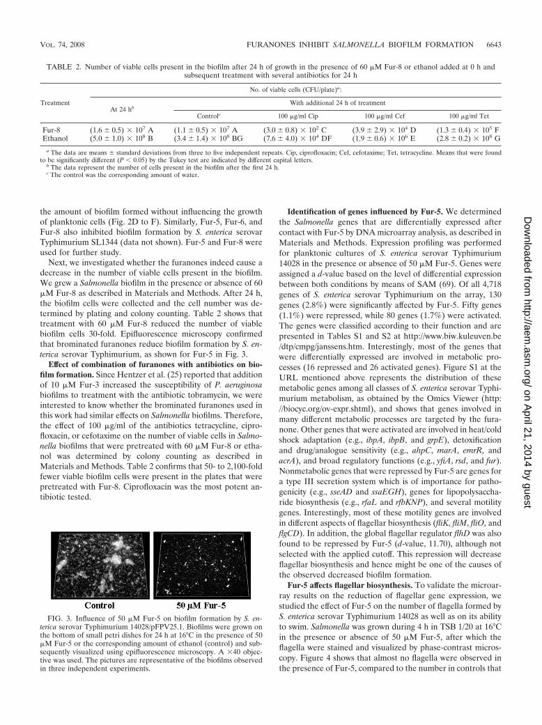
Next, we investigated whether the furanones indeed cause adecrease in the number of viable cells present in the biofilm.We grew a Salmonella biofilm in the presence or absence of 60�M Fur-8 as described in Materials and Methods. After 24 h,the biofilm cells were collected and the cell number was de-termined by plating and colony counting. Table 2 shows thattreatment with 60 �M Fur-8 reduced the number of viablebiofilm cells 30-fold. Epifluorescence microscopy confirmedthat brominated furanones reduce biofilm formation by S. en-terica serovar Typhimurium, as shown for Fur-5 in Fig. 3.
Effect of combination of furanones with antibiotics on bio-film formation. Since Hentzer et al. (25) reported that additionof 10 �M Fur-3 increased the susceptibility of P. aeruginosabiofilms to treatment with the antibiotic tobramycin, we wereinterested to know whether the brominated furanones used inthis work had similar effects on Salmonella biofilms. Therefore,the effect of 100 �g/ml of the antibiotics tetracycline, cipro-floxacin, or cefotaxime on the number of viable cells in Salmo-nella biofilms that were pretreated with 60 �M Fur-8 or etha-nol was determined by colony counting as described inMaterials and Methods. Table 2 confirms that 50- to 2,100-foldfewer viable biofilm cells were present in the plates that werepretreated with Fur-8. Ciprofloxacin was the most potent an-tibiotic tested.
Identification of genes influenced by Fur-5. We determinedthe Salmonella genes that are differentially expressed aftercontact with Fur-5 by DNA microarray analysis, as described inMaterials and Methods. Expression profiling was performedfor planktonic cultures of S. enterica serovar Typhimurium14028 in the presence or absence of 50 �M Fur-5. Genes wereassigned a d-value based on the level of differential expressionbetween both conditions by means of SAM (69). Of all 4,718genes of S. enterica serovar Typhimurium on the array, 130genes (2.8%) were significantly affected by Fur-5. Fifty genes(1.1%) were repressed, while 80 genes (1.7%) were activated.The genes were classified according to their function and arepresented in Tables S1 and S2 at http://www.biw.kuleuven.be/dtp/cmpg/janssens.htm. Interestingly, most of the genes thatwere differentially expressed are involved in metabolic pro-cesses (16 repressed and 26 activated genes). Figure S1 at theURL mentioned above represents the distribution of thesemetabolic genes among all classes of S. enterica serovar Typhi-murium metabolism, as obtained by the Omics Viewer (http://biocyc.org/ov-expr.shtml), and shows that genes involved inmany different metabolic processes are targeted by the fura-none. Other genes that were activated are involved in heat/coldshock adaptation (e.g., ibpA, ibpB, and grpE), detoxificationand drug/analogue sensitivity (e.g., ahpC, marA, emrR, andacrA), and broad regulatory functions (e.g., yfiA, rsd, and fur).Nonmetabolic genes that were repressed by Fur-5 are genes fora type III secretion system which is of importance for patho-genicity (e.g., sseAD and ssaEGH), genes for lipopolysaccha-ride biosynthesis (e.g., rfaL and rfbKNP), and several motilitygenes. Interestingly, most of these motility genes are involvedin different aspects of flagellar biosynthesis (fliK, fliM, fliO, andflgCD). In addition, the global flagellar regulator flhD was alsofound to be repressed by Fur-5 (d-value, 11.70), although notselected with the applied cutoff. This repression will decreaseflagellar biosynthesis and hence might be one of the causes ofthe observed decreased biofilm formation.
Fur-5 affects flagellar biosynthesis. To validate the microar-ray results on the reduction of flagellar gene expression, westudied the effect of Fur-5 on the number of flagella formed byS. enterica serovar Typhimurium 14028 as well as on its abilityto swim. Salmonella was grown during 4 h in TSB 1/20 at 16°Cin the presence or absence of 50 �M Fur-5, after which theflagella were stained and visualized by phase-contrast micros-copy. Figure 4 shows that almost no flagella were observed inthe presence of Fur-5, compared to the number in controls that
TABLE 2. Number of viable cells present in the biofilm after 24 h of growth in the presence of 60 �M Fur-8 or ethanol added at 0 h andsubsequent treatment with several antibiotics for 24 h
Treatment
No. of viable cells (CFU/plate)a:
At 24 hbWith additional 24 h of treatment
Controlc 100 �g/ml Cip 100 �g/ml Cef 100 �g/ml Tet
Fur-8 (1.6 � 0.5) � 107 A (1.1 � 0.5) � 107 A (3.0 � 0.8) � 102 C (3.9 � 2.9) � 104 D (1.3 � 0.4) � 105 FEthanol (5.0 � 1.0) � 108 B (3.4 � 1.4) � 108 BG (7.6 � 4.0) � 104 DF (1.9 � 0.6) � 106 E (2.8 � 0.2) � 108 G
a The data are means � standard deviations from three to five independent repeats. Cip, ciprofloxacin; Cef, cefotaxime; Tet, tetracycline. Means that were foundto be significantly different (P 0.05) by the Tukey test are indicated by different capital letters.
b The data represent the number of cells present in the biofilm after the first 24 h.c The control was the corresponding amount of water.
FIG. 3. Influence of 50 �M Fur-5 on biofilm formation by S. en-terica serovar Typhimurium 14028/pFPV25.1. Biofilms were grown onthe bottom of small petri dishes for 24 h at 16°C in the presence of 50�M Fur-5 or the corresponding amount of ethanol (control) and sub-sequently visualized using epifluorescence microscopy. A �40 objec-tive was used. The pictures are representative of the biofilms observedin three independent experiments.
VOL. 74, 2008 FURANONES INHIBIT SALMONELLA BIOFILM FORMATION 6643
on April 21, 2014 by guest
http://aem.asm
.org/D
ownloaded from

were treated with the corresponding amount of the solvent(ethanol). For each 10 cells counted, 1.0 � 0.7 cell containedflagella when grown in the presence of Fur-5, while 9 � 1 cellscontained flagella when grown in the absence of Fur-5 (P 0.001). Swimming assays were performed using TSB 1/20 plateswith 0.25% agarose at 16°C, conditions under which Salmo-nella swims very slowly. The plates were monitored for 5 days,and the surface of the swimming colonies was regularly mea-sured. Figure 5 shows that swimming by salmonellae grown inthe presence of 50 �M Fur-5 was clearly retarded compared tothat of the appropriate control.
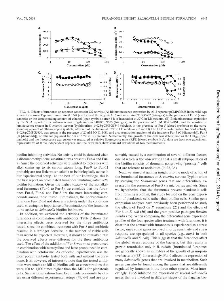
Brominated furanones do not inhibit QS-controlled geneexpression in Salmonella. Halogenated furanones have beenshown to inhibit AHL- and/or AI-2-mediated QS in severalgram-negative species, such as Chromobacterium violaceum(44), Erwinia carotovora (42), E. coli (56, 58), P. aeruginosa (24,25), Serratia liquefaciens (20, 54), Vibrio fischeri (20, 39, 41),and Vibrio harveyi (13, 58). It has therefore been generallyaccepted that halogenated furanones function in gram-nega-tive bacteria by interference with QS. However, none of theknown target genes of the two currently described QS systemsin Salmonella, the AI-2 system and the AHL receptor SdiA,were activated or repressed in our microarray study. In addi-tion, neither sdiA nor the genes that are necessary for thesynthesis of AI-2, luxS and pfs, were differentially regulated inthe presence of Fur-5 under the conditions tested.
To confirm these results, we studied the expression of theknown target genes srgE and lsrA of SdiA and AI-2, respec-tively, in the presence and absence of Fur-5 or Fur-8. Allexperiments were performed in TSB 1/20 at 16°C as well as inLB at 37°C. The latter conditions were included as controlssince the activities of the reporter systems used have beenpreviously described for these standard conditions (Fig. 6) (14,62) and because it has been reported that SdiA activation isspecifically dependent on the presence of AHLs at this tem-perature (62). However, all reporter systems were also active inTSB 1/20 at 16°C and similar results were obtained under bothconditions. None of the tested furanones inhibited the lumi-nescence reporter systems pJNS25 (PsrgE-luxCDABE), acti-vated by 5 nM 3O-C7-HSL or 3O-C7-HTL, and pCMPG5638(PlsrA-luxCDABE) at concentrations that did not inhibit theirbackground activities in the isogenic mutants BA612 andCMPG5602, as exemplified by the AI-2 reporter pCMPG5638in Fig. 6A. The same concentrations needed to inhibit the
reporter systems also inhibited the activity of pCMPG5849(PrpsM-luxCDABE), which constitutively expresses lumines-cence, as exemplified by the SdiA reporter pJNS25 in Fig. 6B.Fur-5 and Fur-8 also did not show activity at non-growth-inhibiting concentrations on GFP-reporter systems for srgEand lsrA activity, as exemplified by the SdiA reporterpCMPG5836 in Fig. 6C and D. In addition, the effect of thefuranones on biofilm formation could not be rescued by thesimultaneous addition of 3O-C7-HSL, 3O-C7-HTL, or syn-thetic DPD (data not shown). Therefore, we have found noevidence that furanones act on the QS systems that are cur-rently reported to be present in Salmonella.
DISCUSSION
Since the 1970s, microbiologists have realized that bacteriagrow predominantly as biofilms in a large diversity of environ-ments, rather than as free-living planktonic cells (22). Withinthese biofilms, the bacteria are better protected from externalstress factors like antibacterial agents and the immune systemof the host (9).
In this study, we have synthesized and screened a smallfocused library of brominated furanones for their activitiesagainst Salmonella biofilm formation. Since we envisaged themain application of compounds that inhibit biofilm formationin the environment outside the host, in order to limit thespread and the survival of this pathogen, we studied Salmonellabiofilm formation under nutrient-poor conditions at 16°C. Wefocused on differences in the alkyl chain lengths of the fura-nones to investigate whether this feature is important for theiractivity in Salmonella. The following structure-activity relation-ship can be derived from the results depicted in Table 1.Furanones without an alkyl chain (Fur-1 to Fur-3) were thestrongest biofilm inhibitors but were also more toxic for Sal-monella than the alkylated furanones, which might be corre-lated with the higher water solubility of the nonalkylated com-pounds. Of the 3-alkylated furanones, only molecules with onebromine atom on the ring structure and one on the methyli-dene side chain (Fur-5, Fur-6, and Fur-8) showed Salmonella
FIG. 5. Fur-5 retards swimming by S. enterica serovar Typhi-murium. TSB 1/20 swimming plates (0.25% agar) containing 50 �MFur-5 (open triangles) or the corresponding amount of solvent (etha-nol [closed squares]) were inoculated with S. enterica serovar Typhi-murium 14028 and incubated for 120 h at 16°C. The surface of theswimming colony was measured at six different time points. Means ofthe swimming area that were found to be significantly different fromthat of the control by the Tukey test (P 0.01) are indicated byasterisks.
FIG. 4. Fur-5 inhibits flagellar biosynthesis by S. enterica serovarTyphimurium. S. enterica serovar Typhimurium 14028 was grown for4 h in TSB 1/20 at 16°C in the presence of 50 �M Fur-5 (A) or thecorresponding amount of ethanol (B). Subsequently, the flagella werestained as described in Materials and Methods and observed usingphase-contrast microscopy. Flagellum orientation as well as the blackspots in the pictures may be an artifact of staining. The pictures arerepresentative of three independent repeats.
6644 JANSSENS ET AL. APPL. ENVIRON. MICROBIOL.
on April 21, 2014 by guest
http://aem.asm
.org/D
ownloaded from
biofilm-inhibiting activities. No activity could be detected whena dibromomethylidene substituent was present (Fur-4 and Fur-7). Since the observed activities were limited to molecules withalkyl chains up to six carbon atoms long, Fur-9 to Fur-11probably are too little water soluble to be biologically active inour experimental setup. To the best of our knowledge, this isthe first report on brominated furanones inhibiting Salmonellabiofilm formation. Given the higher toxicity of the nonalkyl-ated furanones (Fur-1 to Fur-3), we conclude that the furan-ones Fur-5, Fur-6, and Fur-8 are the most interesting com-pounds among those tested. Interestingly, the nonbrominatedfuranone Fur-12 did not show any activity under the conditionsused, stressing the importance of bromination of the furanonesto be active as Salmonella biofilm inhibitors.
In addition, we explored the activities of the brominatedfuranones in combination with antibiotics. Table 2 shows thatinteresting effects were observed for the three antibioticstested, since the combined treatment with Fur-8 and antibioticresulted in a stronger decrease in the number of viable cellsthan would be expected. However, it should be remarked thatthe observed effects were different for the three antibioticsused. The effect of the addition of Fur-8 was most pronouncedin combination with tetracycline and least pronounced in com-bination with cefotaxime. As expected, ciprofloxacin was themost potent antibiotic tested both with and without the fura-none. It is, however, of interest to note that the tested antibi-otics were unable to kill all biofilm cells at concentrations thatwere 100 to 1,000 times higher than the MICs for planktoniccells. Similar observations have been made previously by oth-ers using different experimental setups (49, 64) and are pre-
sumably caused by a combination of several different factors,one of which is the observation that a small subpopulation ofthe biofilm consists of dormant, nongrowing “persister” cellsthat are tolerant to antibiotics (9, 22, 36).
Next, we aimed at gaining insight into the mode of action ofthe brominated furanones on S. enterica serovar Typhimuriumby studying the Salmonella genes that are differentially ex-pressed in the presence of Fur-5 via microarray analysis. Sincewe hypothesize that the furanones prevent planktonic cellsfrom forming a biofilm, this study focused on the gene expres-sion of planktonic cells rather than biofilm cells. Similar geneexpression analyses have previously been performed to studythe effects of Fur-3 on P. aeruginosa (25) and the effects ofFur-6 on E. coli (56) and the gram-positive pathogen Bacillussubtilis (55). When comparing the differential gene expressionprofiles of the four species after treatment with furanones, it isclear that the contact with furanones is experienced as a stressfactor, since some genes involved in drug sensitivity and stressresponse are upregulated in all species (e.g., marA in bothSalmonella and E. coli). This suggests that the furanones affectthe global stress response of the bacteria, but this results ingrowth retardation only in B. subtilis (brominated furanonesare generally known as inhibitors of the growth of gram-posi-tive bacteria) (33). Interestingly, Fur-5 affects the expression ofmany Salmonella genes that are involved in metabolism. Suchgenes can also be found among the genes that are differentlyregulated by furanones in the three other species. Most inter-estingly, Fur-5 inhibited the expression of several Salmonellagenes that are involved in different stages of the flagellar bio-
FIG. 6. Effects of furanones on reporter systems for QS activity. (A) Bioluminescence expression by AI-2 reporter pCMPG5638 in the wild-typeS. enterica serovar Typhimurium strain SL1344 (circles) and the isogenic luxS mutant strain CMPG5602 (triangles) in the presence of Fur-5 (closedsymbols) or the corresponding amount of ethanol (open symbols) after 4 h of incubation at 37°C in LB medium. (B) Bioluminescence expressionby the SdiA reporter in S. enterica serovar Typhimurium 14028/pJNS25 (triangles), in the presence of 5 nM 3O-C7-HSL, and the constitutiveluminescence system in S. enterica serovar Typhimurium 14028/pCMPG5849 (circles), in the presence of Fur-5 (closed symbols) or the corre-sponding amount of ethanol (open symbols) after 6 h of incubation at 37°C in LB medium. (C and D) The GFP reporter system for SdiA activity,14028/pCMPG5836, was grown in the presence of 20 nM 3O-C7-HSL and a concentration gradient of the furanone Fur-5 (C [diamonds]), Fur-8(D [diamonds]), or ethanol (squares) for 6 h at 37°C in LB medium. Subsequently, the growth of the cells was determined as the OD600 (opensymbols) and the fluorescence expression was measured as relative fluorescence units (RFU [closed symbols]). All data are from one experiment,representative of three independent repeats, and the error bars show standard deviations of two measurements.
VOL. 74, 2008 FURANONES INHIBIT SALMONELLA BIOFILM FORMATION 6645
on April 21, 2014 by guest
http://aem.asm
.org/D
ownloaded from
synthesis (6). Similarly, it has been shown that furanones in-hibit the expression of flagellar biosynthesis genes in E. coli(56), although no phenotypic analysis was performed. We fo-cused on the flagellar biosynthesis to validate our microarraydata. Therefore, we studied the number of flagella formed inthe presence of the furanone and showed that almost no fla-gella were present after an incubation time of 4 h in thepresence of Fur-5, while several flagella were formed per cellin the absence of the furanone (Fig. 4). These results wereconfirmed by swimming experiments which showed that Fur-5retards swimming of Salmonella cells. Since it has been shownthat the presence of functional flagella is of importance for theformation of a normal biofilm by Salmonella (67), it is possiblethat interference with the flagellar assembly causes the ob-served biofilm defect. This would imply that furanones haveless influence on already established Salmonella biofilms,which has been confirmed in preliminary experiments (datanot shown). However, it still remains to be determined whetherthe furanones have a specific target in Salmonella. The inter-ference with the flagellar biosynthesis might be caused by aninteraction of the furanones with such a specific target or bythe more global metabolic effect that was observed. Furtherexperiments to unravel the mode of action of the furanones arecurrently ongoing in our laboratories.
Since brominated furanones are generally considered to in-terfere with QS systems in gram-negative bacteria (15), it wassurprising that none of the known target genes of the SdiA-and the AI-2-mediated QS systems of Salmonella were differ-entially regulated by Fur-5. Several experiments with genefusion reporter systems to measure the activity of these QSsystems corroborated this finding. All experiments were per-formed at both 16°C and 37°C, as it has been shown for SdiAthat this system is selectively activated by AHLs at 37°C but notat lower temperatures (62). However, similar results were ob-tained under both conditions. Whereas in a number of bacte-rial species brominated furanones have been reported to exerttheir effects by interfering with QS systems, we have found noevidence of a link between the effects of the furanones onSalmonella biofilm formation and the QS systems that are sofar identified in Salmonella. There are several possible expla-nations for our observations: (i) the furanones target anotheryet unknown Salmonella QS system, (ii) the target of the fura-nones is not part of a Salmonella QS system, or (iii) theobserved inhibition of biofilm formation results from a combi-nation of effects on several different targets.
In conclusion, we have shown that several brominated furan-ones have inhibitory effects on Salmonella biofilm formation.Additionally, pretreatment with furanones results in fewer bio-film cells surviving the treatment with several different antibi-otics. In an effort to unravel the working mechanism of thefuranones, we have determined the differential gene expres-sion of Salmonella in the presence of a furanone. This analysisled to the finding that the furanones interfere with flagellarbiosynthesis. Since our data suggest that the brominated fura-nones do not inhibit Salmonella biofilm formation by interfer-ence with the two putative QS systems of S. enterica serovarTyphimurium, we are currently investigating the specific tar-gets of the furanones.
ACKNOWLEDGMENTS
This work was supported by the Industrial Research Fund of Katho-lieke Universiteit Leuven (KP/06/014), the Research Council of Katho-lieke Universiteit Leuven (CoE EF/05/007 SymBioSys), and the Insti-tute for the Promotion of Innovation through Science and Technologyin Flanders (IWT-Vlaanderen) through scholarships to J.C.A.J. andH.S. S.C.J.D.K. is a Research Associate of the Belgian Fund for Sci-entific Research—Flanders (FWO-Vlaanderen). J.V. and D.E.D.V.are grateful for support in the frame of the IAP program FunctionalSupramolecular Systems.
We thank A. De Weerdt for technical assistance, S. Lebeer forinteresting suggestions concerning the biofilm assay, C. Varszegi forthe synthesis of DPD, and P. Van Hummelen and R. Maes at the VIBMicroarray Facility (Leuven, Belgium). We gratefully acknowledge B.Ahmer (Ohio State University) and R. Valdivia (Stanford UniversitySchool of Medicine, Stanford, CA) for kindly providing the S. entericaserovar Typhimurium strains 14028/pJNS25 and BA612/pJNS25 andthe plasmids pFPV25.1 and pFPV25, respectively.
REFERENCES
1. Ahmer, B. M. 2004. Cell-to-cell signalling in Escherichia coli and Salmonellaenterica. Mol. Microbiol. 52:933–945.
2. Ahmer, B. M. M., J. van Reeuwijk, C. D. Timmers, P. J. Valentine, and F.Heffron. 1998. Salmonella typhimurium encodes an SdiA homolog, a putativequorum sensor of the LuxR family, that regulates genes on the virulenceplasmid. J. Bacteriol. 180:1185–1193.
3. Andrews, J. M. 2001. Determination of minimum inhibitory concentrations.J. Antimicrob. Chemother. 48:5–16.
4. Cao, J. G., and E. A. Meighen. 1989. Purification and structural identificationof an autoinducer for the luminescence system of Vibrio harveyi. J. Biol.Chem. 264:21670–21676.
5. Chen, X., S. Schauder, N. Potier, A. Van Dorsselaer, I. Pelczer, B. L. Bassler,and F. M. Hughson. 2002. Structural identification of a bacterial quorum-sensing signal containing boron. Nature 415:545–549.
6. Chevance, F. F., and K. T. Hughes. 2008. Coordinating assembly of a bac-terial macromolecular machine. Nat. Rev. Microbiol. 6:455–465.
7. Cooke, F. J., E. J. Threlfall, and J. Wain. 2007. Current trends in the spreadand occurrence of human salmonellosis: molecular typing and emergingantibiotic resistance, p. 1–30. In M. Rhen, D. Maskell, P. Mastroeni, and J.Threlfall (ed.), Salmonella. Molecular biology and pathogenesis. HorizonBioscience, Wymondham, Norfolk, United Kingdom.
8. Daniels, R., J. Vanderleyden, and J. Michiels. 2004. Quorum sensing andswarming migration in bacteria. FEMS Microbiol. Rev. 28:261–289.
9. Davies, D. 2003. Understanding biofilm resistance to antibacterial agents.Nat. Rev. Drug Discov. 2:114–122.
10. Davies, D. G., M. R. Parsek, J. P. Pearson, B. H. Iglewski, J. W. Costerton,and E. P. Greenberg. 1998. The involvement of cell-to-cell signals in thedevelopment of a bacterial biofilm. Science 280:295–298.
11. Davies, J. 2007. Microbes have the last word. A drastic re-evaluation ofantimicrobial treatment is needed to overcome the threat of antibiotic-resistant bacteria. EMBO Rep. 8:616–621.
12. Defoirdt, T., R. Crab, T. K. Wood, P. Sorgeloos, W. Verstraete, and P.Bossier. 2006. Quorum sensing-disrupting brominated furanones protect thegnotobiotic brine shrimp Artemia franciscana from pathogenic Vibrio harveyi,Vibrio campbellii, and Vibrio parahaemolyticus isolates. Appl. Environ. Mi-crobiol. 72:6419–6423.
13. Defoirdt, T., C. M. Miyamoto, T. K. Wood, E. A. Meighen, P. Sorgeloos, W.Verstraete, and P. Bossier. 2007. The natural furanone (5Z)-4-bromo-5-(bromomethylene)-3-butyl-2(5H)-furanone disrupts quorum sensing-regu-lated gene expression in Vibrio harveyi by decreasing the DNA-binding ac-tivity of the transcriptional regulator protein luxR. Environ. Microbiol.9:2486–2495.
14. De Keersmaecker, S. C., C. Varszegi, N. van Boxel, L. W. Habel, K. Metzger,R. Daniels, K. Marchal, D. De Vos, and J. Vanderleyden. 2005. Chemicalsynthesis of (S)-4,5-dihydroxy-2,3-pentanedione, a bacterial signal moleculeprecursor, and validation of its activity in Salmonella typhimurium. J. Biol.Chem. 280:19563–19568.
15. de Nys, R., M. Givskov, N. Kumar, S. Kjelleberg, and P. D. Steinberg. 2006.Furanones. Prog. Mol. Subcell. Biol. 42:55–86.
16. de Nys, R., A. D. Wright, G. M. Konig, and O. Sticher. 1993. New haloge-nated furanones from the marine alga Delisea pulchra (cf. fimbriata). Tetra-hedron 49:11213–11220.
17. Fuqua, C., M. R. Parsek, and E. P. Greenberg. 2001. Regulation of geneexpression by cell-to-cell communication: acyl-homoserine lactone quorumsensing. Annu. Rev. Genet. 35:439–468.
18. Fuqua, W. C., S. C. Winans, and E. P. Greenberg. 1994. Quorum sensing inbacteria: the LuxR-LuxI family of cell density-responsive transcriptionalregulators. J. Bacteriol. 176:269–275.
19. Gabriele, B., G. Salerno, M. Costa, and G. P. Chiusoli. 1999. A new regio-
6646 JANSSENS ET AL. APPL. ENVIRON. MICROBIOL.
on April 21, 2014 by guest
http://aem.asm
.org/D
ownloaded from

selective synthesis of 3-substituted furan-2(5H)-ones by palladium-catalysedreductive carbonylation of alk-1-ynes. Tetrahedron Lett. 40:989–990.
20. Givskov, M., R. de Nys, M. Manefield, L. Gram, R. Maximilien, L. Eberl, S.Molin, P. D. Steinberg, and S. Kjelleberg. 1996. Eukaryotic interference withhomoserine lactone-mediated prokaryotic signalling. J. Bacteriol. 178:6618–6622.
21. Gram, L., R. de Nys, R. Maximilien, M. Givskov, P. Steinberg, and S.Kjelleberg. 1996. Inhibitory effects of secondary metabolites from the redalga Delisea pulchra on swarming motility of Proteus mirabilis. Appl. Environ.Microbiol. 62:4284–4287.
22. Hall-Stoodley, L., J. W. Costerton, and P. Stoodley. 2004. Bacterial biofilms:from the natural environment to infectious diseases. Nat. Rev. Microbiol.2:95–108.
23. Hentzer, M., and M. Givskov. 2003. Pharmacological inhibition of quorumsensing for the treatment of chronic bacterial infections. J. Clin. Investig.112:1300–1307.
24. Hentzer, M., K. Riedel, T. B. Rasmussen, A. Heydorn, J. B. Andersen, M. R.Parsek, S. A. Rice, L. Eberl, S. Molin, N. Hoiby, S. Kjelleberg, and M.Givskov. 2002. Inhibition of quorum sensing in Pseudomonas aeruginosabiofilm bacteria by a halogenated furanone compound. Microbiology 148:87–102.
25. Hentzer, M., H. Wu, J. B. Andersen, K. Riedel, T. B. Rasmussen, N. Bagge,N. Kumar, M. A. Schembri, Z. Song, P. Kristoffersen, M. Manefield, J. W.Costerton, S. Molin, L. Eberl, P. Steinberg, S. Kjelleberg, N. Hoiby, and M.Givskov. 2003. Attenuation of Pseudomonas aeruginosa virulence by quorumsensing inhibitors. EMBO J. 22:3803–3815.
26. Hoiseth, S. K., and B. A. Stocker. 1981. Aromatic-dependent Salmonellatyphimurium are non-virulent and effective as live vaccines. Nature 291:238–239.
27. Huber, B., K. Riedel, M. Hentzer, A. Heydorn, A. Gotschlich, M. Givskov, S.Molin, and L. Eberl. 2001. The cep quorum-sensing system of Burkholderiacepacia H111 controls biofilm formation and swarming motility. Microbiol-ogy 147:2517–2528.
28. Hume, E. B., J. Baveja, B. Muir, T. L. Schubert, N. Kumar, S. Kjelleberg,H. J. Griesser, H. Thissen, R. Read, L. A. Poole-Warren, K. Schindhelm, andM. D. Willcox. 2004. The control of Staphylococcus epidermidis biofilm for-mation and in vivo infection rates by covalently bound furanones. Biomate-rials 25:5023–5030.
29. Janssens, J. C. A., K. Metzger, R. Daniels, D. Ptacek, T. Verhoeven, L. W.Habel, J. Vanderleyden, D. E. De Vos, and S. C. De Keersmaecker. 2007.Synthesis of N-acyl homoserine lactone analogues reveals strong activatorsof SdiA, the Salmonella enterica serovar Typhimurium LuxR homologue.Appl. Environ. Microbiol. 73:535–544.
30. Jones, M. B., R. Jani, D. Ren, T. K. Wood, and M. J. Blaser. 2005. Inhibitionof Bacillus anthracis growth and virulence-gene expression by inhibitors ofquorum-sensing. J. Infect. Dis. 191:1881–1888.
31. Kearns, D. B., and R. Losick. 2003. Swarming motility in undomesticatedBacillus subtilis. Mol. Microbiol. 49:581–590.
32. Kim, W., and M. G. Surette. 2003. Swarming populations of Salmonellarepresent a unique physiological state coupled to multiple mechanisms ofantibiotic resistance. Biol. Proced. Online 5:189–196.
33. Kjelleberg, S., P. D. Steinberg, C. Holmstrom, and A. Back. April 2006.Inhibition of gram-positive bacteria. U.S. patent US7026353.
34. Kumar, N., and R. W. Read. September 2008. Synthesis of cyclic compounds.European patent EP1294705.
35. Labbate, M., S. Y. Queck, K. S. Koh, S. A. Rice, M. Givskov, and S.Kjelleberg. 2004. Quorum sensing-controlled biofilm development in Serratialiquefaciens MG1. J. Bacteriol. 186:692–698.
36. Lewis, K. 2007. Persister cells, dormancy and infectious disease. Nat. Rev.Microbiol. 5:48–56.
37. Lonn-Stensrud, J., F. C. Petersen, T. Benneche, and A. A. Scheie. 2007.Synthetic bromated furanone inhibits autoinducer-2-mediated communica-tion and biofilm formation in oral streptococci. Oral Microbiol. Immunol.22:340–346.
38. Lynch, M. J., S. Swift, D. F. Kirke, C. W. Keevil, C. E. Dodd, and P. Williams.2002. The regulation of biofilm development by quorum sensing in Aeromo-nas hydrophila. Environ. Microbiol. 4:18–28.
39. Manefield, M., R. de Nys, N. Kumar, R. Read, M. Givskov, P. Steinberg, andS. Kjelleberg. 1999. Evidence that halogenated furanones from Delisea pul-chra inhibit acylated homoserine lactone (AHL)-mediated gene expressionby displacing the AHL signal from its receptor protein. Microbiology 145:283–291.
40. Manefield, M., L. Harris, S. A. Rice, R. de Nys, and S. Kjelleberg. 2000.Inhibition of luminescence and virulence in the black tiger prawn (Penaeusmonodon) pathogen Vibrio harveyi by intercellular signal antagonists. Appl.Environ. Microbiol. 66:2079–2084.
41. Manefield, M., T. B. Rasmussen, M. Henzter, J. B. Andersen, P. Steinberg,S. Kjelleberg, and M. Givskov. 2002. Halogenated furanones inhibit quorumsensing through accelerated LuxR turnover. Microbiology 148:1119–1127.
42. Manefield, M., M. Welch, M. Givskov, G. P. Salmond, and S. Kjelleberg.2001. Halogenated furanones from the red alga, Delisea pulchra, inhibitcarbapenem antibiotic synthesis and exoenzyme virulence factor production
in the phytopathogen Erwinia carotovora. FEMS Microbiol. Lett. 205:131–138.
43. Manny, A. J., S. Kjelleberg, N. Kumar, R. de Nys, R. W. Read, and P.Steinberg. 1997. Reinvestigation of the sulfuric acid-catalysed cyclisation ofbrominated 2-alkyllevulinic acids to 3-alkyl-5-methylene-2(5H)-furanones.Tetrahedron 53:15813–15826.
44. Martinelli, D., G. Grossmann, U. Sequin, H. Brandl, and R. Bachofen. 2004.Effects of natural and chemically synthesized furanones on quorum sensingin Chromobacterium violaceum. BMC Microbiol. 4:25.
45. Michael, B., J. N. Smith, S. Swift, F. Heffron, and B. M. M. Ahmer. 2001.SdiA of Salmonella enterica is a LuxR homolog that detects mixed microbialcommunities. J. Bacteriol. 183:5733–5742.
46. Miller, S. T., K. B. Xavier, S. R. Campagna, M. E. Taga, M. F. Semmelhack,B. L. Bassler, and F. M. Hughson. 2004. Salmonella typhimurium recognizesa chemically distinct form of the bacterial quorum-sensing signal AI-2. Mol.Cell 15:677–687.
47. National Institutes of Health. 1997. Minutes of the National Advisory Den-tal and Craniofacial Research Council—153rd Meeting, Bethesda, MD.
48. Nealson, K. H. 1977. Autoinduction of bacterial luciferase. Occurrence,mechanism and significance. Arch. Microbiol. 112:73–79.
49. Olson, M. E., H. Ceri, D. W. Morck, A. G. Buret, and R. R. Read. 2002.Biofilm bacteria: formation and comparative susceptibility to antibiotics.Can. J. Vet. Res. 66:86–92.
50. Parsek, M. R., and E. P. Greenberg. 2005. Sociomicrobiology: the connec-tions between quorum sensing and biofilms. Trends Microbiol. 13:27–33.
51. Prouty, A. M., W. H. Schwesinger, and J. S. Gunn. 2002. Biofilm formationand interaction with the surfaces of gallstones by Salmonella spp. Infect.Immun. 70:2640–2649.
52. Provence, D. L., and R. Curtiss. 1994. Gene transfer in gram-negative bac-teria, p. 317–347. In P. Gerhardt, R. G. E. Murray, W. A. Wood, and N. R.Krieg (ed.), Methods for general and molecular bacteriology. AmericanSociety for Microbiology, Washington, DC.
53. Raffatellu, M., C. Tukel, D. Chessa, R. P. Wilson, and A. J. Baumler. 2007.The intestinal phase of Salmonella infections, p. 31–52. In M. Rhen,D. Maskell, P. Mastroeni, and J. Threlfall (ed.), Salmonella. Molecularbiology and pathogenesis. Horizon Bioscience, Wymondham, Norfolk,United Kingdom.
54. Rasmussen, T. B., M. Manefield, J. B. Andersen, L. Eberl, U. Anthoni, C.Christophersen, P. Steinberg, S. Kjelleberg, and M. Givskov. 2000. HowDelisea pulchra furanones affect quorum sensing and swarming motility inSerratia liquefaciens MG1. Microbiology 146:3237–3244.
55. Ren, D., L. A. Bedzyk, P. Setlow, D. F. England, S. Kjelleberg, S. M. Thomas,R. W. Ye, and T. K. Wood. 2004. Differential gene expression to investigatethe effect of (5Z)-4-bromo-5-(bromomethylene)-3-butyl-2(5H)-furanone onBacillus subtilis. Appl. Environ. Microbiol. 70:4941–4949.
56. Ren, D., L. A. Bedzyk, R. W. Ye, S. M. Thomas, and T. K. Wood. 2004.Differential gene expression shows natural brominated furanones interferewith the autoinducer-2 bacterial signaling system of Escherichia coli. Bio-technol. Bioeng. 88:630–642.
57. Ren, D., J. J. Sims, and T. K. Wood. 2002. Inhibition of biofilm formationand swarming of Bacillus subtilis by (5Z)-4-bromo-5-(bromomethylene)-3-butyl-2(5H)-furanone. Lett. Appl. Microbiol. 34:293–299.
58. Ren, D., J. J. Sims, and T. K. Wood. 2001. Inhibition of biofilm formationand swarming of Escherichia coli by (5Z)-4-bromo-5-(bromomethylene)-3-butyl-2(5H)-furanone. Environ. Microbiol. 3:731–736.
59. Rice, S. A., D. McDougald, N. Kumar, and S. Kjelleberg. 2005. The use ofquorum-sensing blockers as therapeutic agents for the control of biofilm-associated infections. Curr. Opin. Investig. Drugs 6:178–184.
60. Sambrook, J., E. F. Fritsch, and T. Maniatis. 1989. Molecular cloning: alaboratory manual, 2nd ed. Cold Spring Harbor Laboratory Press, ColdSpring Harbor, NY.
61. Smith, D., J. H. Wang, J. E. Swatton, P. Davenport, B. Price, H. Mikkelsen,H. Stickland, K. Nishikawa, N. Gardiol, D. R. Spring, and M. Welch. 2006.Variations on a theme: diverse N-acyl homoserine lactone-mediated quorumsensing mechanisms in gram-negative bacteria. Sci. Prog. 89:167–211.
62. Smith, J. N., and B. M. M. Ahmer. 2003. Detection of other microbial speciesby Salmonella: expression of the SdiA regulon. J. Bacteriol. 185:1357–1366.
63. Surette, M. G., M. B. Miller, and B. L. Bassler. 1999. Quorum sensing inEscherichia coli, Salmonella typhimurium, and Vibrio harveyi: a new family ofgenes responsible for autoinducer production. Proc. Natl. Acad. Sci. USA96:1639–1644.
64. Tabak, M., K. Scher, E. Hartog, U. Romling, K. R. Matthews, M. L. Chi-kindas, and S. Yaron. 2007. Effect of triclosan on Salmonella typhimurium atdifferent growth stages and in biofilms. FEMS Microbiol. Lett. 267:200–206.
65. Taga, M. E., S. T. Miller, and B. L. Bassler. 2003. Lsr-mediated transportand processing of AI-2 in Salmonella typhimurium. Mol. Microbiol. 50:1411–1427.
66. Taga, M. E., J. L. Semmelhack, and B. L. Bassler. 2001. The LuxS-depen-dent autoinducer AI-2 controls the expression of an ABC transporter thatfunctions in AI-2 uptake in Salmonella typhimurium. Mol. Microbiol. 42:777–793.
VOL. 74, 2008 FURANONES INHIBIT SALMONELLA BIOFILM FORMATION 6647
on April 21, 2014 by guest
http://aem.asm
.org/D
ownloaded from

67. Teplitski, M., A. Al-Agely, and B. M. Ahmer. 2006. Contribution of the SirAregulon to biofilm formation in Salmonella enterica serovar Typhimurium.Microbiology 152:3411–3424.
68. Thijs, I. M. V., S. C. De Keersmaecker, A. Fadda, K. Engelen, H. Zhao, M.McClelland, K. Marchal, and J. Vanderleyden. 2007. Delineation of theSalmonella enterica serovar Typhimurium HilA regulon through genome-wide location and transcript analysis. J. Bacteriol. 189:4587–4596.
69. Tusher, V. G., R. Tibshirani, and G. Chu. 2001. Significance analysis ofmicroarrays applied to the ionizing radiation response. Proc. Natl. Acad. Sci.USA 98:5116–5121.
70. Valdivia, R. H., and S. Falkow. 1996. Bacterial genetics by flow cytometry:
rapid isolation of Salmonella typhimurium acid-inducible promoters by dif-ferential fluorescence induction. Mol. Microbiol. 22:367–378.
71. Wang, Q., J. G. Frye, M. McClelland, and R. M. Harshey. 2004. Geneexpression patterns during swarming in Salmonella typhimurium: genes spe-cific to surface growth and putative new motility and pathogenicity genes.Mol. Microbiol. 52:169–187.
72. Waters, C. M., and B. L. Bassler. 2005. Quorum sensing: cell-to-cell com-munication in bacteria. Annu. Rev. Cell Dev. Biol. 21:319–346.
73. Williams, P., K. Winzer, W. C. Chan, and M. Camara. 2007. Look who’stalking: communication and quorum sensing in the bacterial world. Philos.Trans. R. Soc. Lond. B 362:1119–1134.
6648 JANSSENS ET AL. APPL. ENVIRON. MICROBIOL.
on April 21, 2014 by guest
http://aem.asm
.org/D
ownloaded from