Comparative Analysis and Serovar-Specific Identification of Multiple-Banded Antigen Genes of...
6
JOURNAL OF CLINICAL MICROBIOLOGY, 0095-1137/99/$04.0010 Mar. 1999, p. 538–543 Vol. 37, No. 3 Copyright © 1999, American Society for Microbiology. All Rights Reserved. Comparative Analysis and Serovar-Specific Identification of Multiple-Banded Antigen Genes of Ureaplasma urealyticum Biovar 1 FANRONG KONG, 1,2 XUEJUN ZHU, 1 WEIZHEN WANG, 1 XIANGZHAO ZHOU, 1 SUSANNA GORDON, 2 AND GWENDOLYN L. GILBERT 2 * Department of Dermatology, First Hospital of Beijing Medical University, Beijing 100034, People’s Republic of China, 1 and Centre for Infectious Diseases and Microbiology, Institute of Clinical Pathology and Medical Research, Westmead, New South Wales, Australia 2 Received 20 July 1998/Returned for modification 20 August 1998/Accepted 2 November 1998 Ureaplasma urealyticum is a causative agent of nongonococcal urethritis and is implicated in the pathogenesis of several other diseases. The species is divided into 14 serovars and two biovars, of which biovar 1 is most commonly isolated from clinical specimens. Reported associations between individual serovars and diseases have been difficult to confirm because of practical difficulties with serotyping. The multiple-banded antigen (MBA) is the predominant U. urealyticum antigen recognized during infections in humans and probably has a significant role in virulence. The 5* end of the MBA gene is relatively conserved but contains biovar, and possibly serovar, specificity. The 5* ends of the MBA genes of standard strains of U. urealyticum biovar 1, consisting of serovars 1, 3, 6, and 14, were amplified, cloned into pUC19, and sequenced to identify serovar- specific differences. The 5* end of the MBA gene sequence of serovar 3 was identical with the previously published sequence and differed by only three bases from that of serovar 14. Significant differences between the MBA gene sequences allowed biovar 1 to be divided into two subgroups, containing serovars 3/14 and serovars 1 and 6, respectively, using primers UMS-125–UMA269 and UMS-125–UMA269*. Serovars 1 and 6 were distinguished by restriction enzyme analysis of the amplicon and/or by PCR specific for serovar 6. These methods were used to identify and type U. urealyticum in 185 (46.3%) of 400 genital specimens from women. Biovar 1 was detected in 89.2% and biovar 2 in 18.3% of positive specimens. Of 165 specimens containing U. urealyticum biovar 1, 22.2% contained more than one serovar and 46.7, 46.1, and 25.5% contained serovars 1, 3/14, and 6, respectively. U. urealyticum was found in a significantly higher proportion of pregnant women than in sex workers and other women attending a sexually transmissible diseases clinic (P < 0.01). The methods described are relatively rapid, practicable, and specific for serotyping isolates and for direct detection and identification of individual serovars in clinical specimens containing U. urealyticum biovar 1. Ureaplasma urealyticum is a recognized cause of urethritis and disseminated infection in immunocompromised patients (8, 15–17). It has also been implicated in other genitourinary syndromes (10) and a number of complications of pregnancy, including chorioamnionitis, preterm birth, and chronic neona- tal lung disease (1–3). However, it is found commonly among the genital tract flora of healthy women, and its pathogenic role in a small proportion of individuals is difficult to prove (4, 23). There are two biovars and 14 serovars of U. urealyticum. The majority of human isolates of U. urealyticum belong to biovar 1 (1, 6, 7, 9, 23), which includes serovars 1, 3, 6, and 14. Some serovars have been associated with disease syndromes more often than they are found in normal flora (4, 9, 10). However, these findings are difficult to confirm because of problems with conventional serotyping methods (4, 9, 12). A rapid molecular method for identification of individual serovars would be of great value in studies of epidemiology and pathogenesis of infections with U. urealyticum (11, 13, 18, 23). The MBA (multiple-banded antigen) is the predominant antigen recognized during U. urealyticum infections and is probably an important virulence determinant. It is species spe- cific and contains both serovar-specific and cross-reactive epitopes (19, 21–23). The 59 end of the MBA gene is relatively conserved, but it also includes biovar- and serovar-specific re- gions (22). The aim of this study was to identify sequence differences among the 59 ends of the MBA genes of U. urea- lyticum serovars 1, 3, 6, and 14 of biovar 1 which could be used as the basis of a practical molecular-serotyping method. MATERIALS AND METHODS Bacterial strains and plasmids. The reference strains used were U. urealyticum serovar 1 (ATCC 27813), serovar 2 (ATCC 27814), serovar 3 (ATCC 27815), serovar 4 (ATCC 27816), serovar 5 (ATCC 27817), serovar 6 (ATCC 27818), serovar 7 (ATCC 27819), serovar 8 (ATCC 27618), serovar 9 (ATCC 33175), serovar 10 (ATCC 33699), serovar 11 (ATCC 33695), serovar 12 (ATCC 33696), serovar 13 (ATCC 33698), and serovar 14 (ATCC 33697). Clinical isolates of serovars 1 to 14, which had been serotyped by conventional methods, were kindly provided by H. L. Watson (Department of Microbiology, University of Alabama at Birmingham, Birmingham) Escherichia coli DH5a was the host for plasmid pUC19, which was obtained from the Institute of Genetics, Chinese Academy of Science. Clinical specimens. Cervical swabs from 200 female sex workers and 100 other female clients attending the Sexually Transmissible Diseases (STD) Clinic and 100 pregnant women attending the Antenatal Clinic of the First Hospital of Beijing Medical University were collected to test the practical usefulness of the direct detection, biotyping, and serotyping methods. DNA was prepared for PCR (see below) as soon as possible after receipt of specimens in the laboratory. Cultures were not performed for these specimens. Oligonucleotide primers. Oligonucleotide primers UMS-125, UMA226, UMS51, and UMA427, based on the previously published sequence of the serovar 3 MBA gene (19, 22), were used for amplification of the 59 ends of the * Corresponding author. Mailing address: Centre for Infectious Dis- eases and Microbiology, Institute of Clinical Pathology and Medical Research, Westmead Hospital, Darcy Rd., Westmead, New South Wales, 2145 Australia. Phone: (612) 9845 6255. Fax: (612) 9893 8659. E-mail: [email protected]. 538
-
Upload
independent -
Category
Documents
-
view
4 -
download
0
Transcript of Comparative Analysis and Serovar-Specific Identification of Multiple-Banded Antigen Genes of...
JOURNAL OF CLINICAL MICROBIOLOGY,0095-1137/99/$04.0010
Mar. 1999, p. 538–543 Vol. 37, No. 3
Copyright © 1999, American Society for Microbiology. All Rights Reserved.
Comparative Analysis and Serovar-Specific Identificationof Multiple-Banded Antigen Genes of
Ureaplasma urealyticum Biovar 1FANRONG KONG,1,2 XUEJUN ZHU,1 WEIZHEN WANG,1 XIANGZHAO ZHOU,1 SUSANNA GORDON,2
AND GWENDOLYN L. GILBERT2*
Department of Dermatology, First Hospital of Beijing Medical University, Beijing 100034, People’s Republic of China,1
and Centre for Infectious Diseases and Microbiology, Institute of Clinical Pathology andMedical Research, Westmead, New South Wales, Australia2
Received 20 July 1998/Returned for modification 20 August 1998/Accepted 2 November 1998
Ureaplasma urealyticum is a causative agent of nongonococcal urethritis and is implicated in the pathogenesisof several other diseases. The species is divided into 14 serovars and two biovars, of which biovar 1 is mostcommonly isolated from clinical specimens. Reported associations between individual serovars and diseaseshave been difficult to confirm because of practical difficulties with serotyping. The multiple-banded antigen(MBA) is the predominant U. urealyticum antigen recognized during infections in humans and probably has asignificant role in virulence. The 5* end of the MBA gene is relatively conserved but contains biovar, andpossibly serovar, specificity. The 5* ends of the MBA genes of standard strains of U. urealyticum biovar 1,consisting of serovars 1, 3, 6, and 14, were amplified, cloned into pUC19, and sequenced to identify serovar-specific differences. The 5* end of the MBA gene sequence of serovar 3 was identical with the previouslypublished sequence and differed by only three bases from that of serovar 14. Significant differences between theMBA gene sequences allowed biovar 1 to be divided into two subgroups, containing serovars 3/14 and serovars1 and 6, respectively, using primers UMS-125–UMA269 and UMS-125–UMA269*. Serovars 1 and 6 weredistinguished by restriction enzyme analysis of the amplicon and/or by PCR specific for serovar 6. Thesemethods were used to identify and type U. urealyticum in 185 (46.3%) of 400 genital specimens from women.Biovar 1 was detected in 89.2% and biovar 2 in 18.3% of positive specimens. Of 165 specimens containing U.urealyticum biovar 1, 22.2% contained more than one serovar and 46.7, 46.1, and 25.5% contained serovars 1,3/14, and 6, respectively. U. urealyticum was found in a significantly higher proportion of pregnant women thanin sex workers and other women attending a sexually transmissible diseases clinic (P < 0.01). The methodsdescribed are relatively rapid, practicable, and specific for serotyping isolates and for direct detection andidentification of individual serovars in clinical specimens containing U. urealyticum biovar 1.
Ureaplasma urealyticum is a recognized cause of urethritisand disseminated infection in immunocompromised patients(8, 15–17). It has also been implicated in other genitourinarysyndromes (10) and a number of complications of pregnancy,including chorioamnionitis, preterm birth, and chronic neona-tal lung disease (1–3). However, it is found commonly amongthe genital tract flora of healthy women, and its pathogenicrole in a small proportion of individuals is difficult to prove (4,23).
There are two biovars and 14 serovars of U. urealyticum. Themajority of human isolates of U. urealyticum belong to biovar 1(1, 6, 7, 9, 23), which includes serovars 1, 3, 6, and 14. Someserovars have been associated with disease syndromes moreoften than they are found in normal flora (4, 9, 10). However,these findings are difficult to confirm because of problems withconventional serotyping methods (4, 9, 12). A rapid molecularmethod for identification of individual serovars would be ofgreat value in studies of epidemiology and pathogenesis ofinfections with U. urealyticum (11, 13, 18, 23).
The MBA (multiple-banded antigen) is the predominantantigen recognized during U. urealyticum infections and is
probably an important virulence determinant. It is species spe-cific and contains both serovar-specific and cross-reactiveepitopes (19, 21–23). The 59 end of the MBA gene is relativelyconserved, but it also includes biovar- and serovar-specific re-gions (22). The aim of this study was to identify sequencedifferences among the 59 ends of the MBA genes of U. urea-lyticum serovars 1, 3, 6, and 14 of biovar 1 which could be usedas the basis of a practical molecular-serotyping method.
MATERIALS AND METHODS
Bacterial strains and plasmids. The reference strains used were U. urealyticumserovar 1 (ATCC 27813), serovar 2 (ATCC 27814), serovar 3 (ATCC 27815),serovar 4 (ATCC 27816), serovar 5 (ATCC 27817), serovar 6 (ATCC 27818),serovar 7 (ATCC 27819), serovar 8 (ATCC 27618), serovar 9 (ATCC 33175),serovar 10 (ATCC 33699), serovar 11 (ATCC 33695), serovar 12 (ATCC 33696),serovar 13 (ATCC 33698), and serovar 14 (ATCC 33697). Clinical isolates ofserovars 1 to 14, which had been serotyped by conventional methods, were kindlyprovided by H. L. Watson (Department of Microbiology, University of Alabamaat Birmingham, Birmingham) Escherichia coli DH5a was the host for plasmidpUC19, which was obtained from the Institute of Genetics, Chinese Academy ofScience.
Clinical specimens. Cervical swabs from 200 female sex workers and 100 otherfemale clients attending the Sexually Transmissible Diseases (STD) Clinic and100 pregnant women attending the Antenatal Clinic of the First Hospital ofBeijing Medical University were collected to test the practical usefulness of thedirect detection, biotyping, and serotyping methods. DNA was prepared for PCR(see below) as soon as possible after receipt of specimens in the laboratory.Cultures were not performed for these specimens.
Oligonucleotide primers. Oligonucleotide primers UMS-125, UMA226,UMS51, and UMA427, based on the previously published sequence of theserovar 3 MBA gene (19, 22), were used for amplification of the 59 ends of the
* Corresponding author. Mailing address: Centre for Infectious Dis-eases and Microbiology, Institute of Clinical Pathology and MedicalResearch, Westmead Hospital, Darcy Rd., Westmead, New SouthWales, 2145 Australia. Phone: (612) 9845 6255. Fax: (612) 9893 8659.E-mail: [email protected].
538

U. urealyticum serovar 1, 3, 6, and 14 MBA genes. UMS-125 (GTA TTT GCAATC TTT ATA TGT TTT CG) and UMA226 (CAG CTG ATG TAA GTGCAG CAT TAA ATT C) were used without modification. UMS51 and UMA427were modified as follows. (i) Primers UMS51 and UMA427 were modified byadding BamHI and EcoRI restriction enzyme sites, respectively (underlined inthe sequences below), to facilitate cloning of the coding DNA sequences of the59 ends of the serovar 1, 3, and 14 MBA genes. (ii) In ureaplasmas, the sequenceTGA encodes tryptophan (amino acid 13), but in E. coli it represents a stopcodon. Therefore, a point mutation was introduced into UMS51 (at base 38) tochange the sequence to TGG, which also encodes tryptophan. The modifiedprimer sequences used were UMS51, TGT GGA TCC TTC TGG GCT ATGACA TTA GGT GTT ACC (BamHI site underlined) and UMA427, CTC GAATTC ACC TGG TTG TGT AGT TTC AAA GTT CAC (EcoRI site underlined).
The additional primers UMA269 (serovar 3/14 specific), UMA2699 (serovar 1and 6 specific), and UMS-54 (serovar 6 specific) were designed, using the se-quence data generated by this study, and were used for PCR identification (seebelow). The sequences of these primers are as follows: UMA269 (serovar 3/14),CTA AAT GAC CTT TTT CAA GTG TAC; UMA2699 (serovars 1 and 6), CCAAAT GAC CTT TTG TAA CTA GAT; and UMS-54 (serovar 6), CTT AGTGTT CAT ATT TTT TAC TAG.
DNA preparations. Cells from 0.5 ml of ureaplasma broth (10B) cultures ofeach U. urealyticum serovar were harvested in late logarithmic growth phase bycentrifugation at 14,000 3 g for 20 min; clinical specimens were processeddirectly. DNA was isolated from both cultures and clinical specimens by treat-ment with 500 ml of digestion buffer (10 mM Tris-HCl, pH 8.0, 0.45% TritonX-100, and 0.45% Tween 20) and proteinase K (100 mg/ml) at 55°C for 1 h andthen extraction with phenol-chloroform-isoamyl alcohol (25:24:1) and chloro-form-isoamyl alcohol (24:1). DNA was precipitated with 1/10 volume of 3 Msodium acetate (pH 5.2) and 2 volumes of ethanol. The washed and dried pelletswere hydrated in 200 ml of ultrapure sterile water.
PCR. The 25-ml amplification reaction mixtures contained 2.5 ml of 103 PCRbuffer (13 PCR buffer is 10 mM Tris-HCl [pH 8.8 at 25°C], 1.5 mM MgCl2, 50mM KCl, and 0.1% Triton X-100), 0.5 U of Taq polymerase (Finnzymes OY,Espoo, Finland), 200 mM (each) deoxynucleoside triphosphate (dATP, dCTP,dGTP, and dTTP) (Boehringer, Mannheim, Germany), 10 pmol of each primer,5 ml of sample DNA, and ultrapure sterile water added to 25 ml. In each reaction,positive and negative controls were processed in parallel with the tested samplesto detect possible inhibition or contamination.
The denaturation, annealing, and elongation temperatures and times usedwere 95°C for 30 s, 58°C for 30 s, and 72°C for 1 min, respectively, for 40 cycleswith a Perkin-Elmer thermocycler. Eight microliters of PCR product was ana-
lyzed by electrophoresis on 2.0% agarose gels which were stained with 0.5 mg ofethidium bromide/ml. A visible band of the appropriate size on UV translumi-nation was considered a positive result.
Cloning and sequencing. The amplified fragments of the 59 ends of U. urea-lyticum MBA genes (amplified with UMS-125–UMA226 for serovar 6 andUMS51-UMA427 for serovars 1, 3, and 14) were ligated to plasmid vectorpUC19, which had been digested by SmaI. The ligation products were trans-formed into E. coli DH5a, and positive transformants were identified by PCR,using the primers UMS51-UMA427 for serovars 1, 3, and 14 and UMS-125–UMA226 for serovar 6. The positive clones were sequenced in two directionswith the ABI 373A DNA-sequencing system.
To confirm the results of cloning experiments, the PCR products of both setsof primers for ATCC reference strains and clinical isolates of all four serovars,
FIG. 1. Algorithm for identification of serovars of U. urealyticum biovar 1 inclinical specimens or cultures. (A) Biotyping of U. urealyticum; (B) serotyping ofU. urealyticum biovar 1.
FIG. 2. Multiple sequence alignment of the 59 ends of MBA gene sequencesof U. urealyticum serovars 1, 3, 6, and 14 (ATCC strains). Relevant primers(arrows) and restriction enzyme sites (bars) are shown.
VOL. 37, 1999 U. UREALYTICUM BIOVAR 1 SEROVAR IDENTIFICATION BY PCR 539

1, 3, 6, and 14, were directly sequenced in two directions with the ABI 373ADNA-sequencing system.
Identification of serovars by PCR with restriction enzyme analysis and sero-var-specific PCR. Primers UMS-125, UMA226, UMA269, and UMA2699 wereused to amplify DNA from reference (ATCC) strains, from clinical isolates thathad been serotyped previously by conventional methods, and directly from clin-ical specimens. Primers UMS-125 and UMA226 were used to detect and biotypeU. urealyticum. Primers UMS-125 and UMA269 were specific for serovars 3/14;primers UMS-125 and UMA2699 were specific for serovars 1 and 6. Two differ-ent methods were used to further identify U. urealyticum serovars 1 and 6 (Fig.1).
(i) Restriction enzyme analysis. Ten microliters of the PCR product of primersUMS-125 and UMA2699 was treated with the restriction enzyme HinfI (NewEngland Biolabs, Beverly, Mass.) and analyzed by electrophoresis. Those thatwere cut by the enzyme were identified as serovar 1, those not cut were identifiedas serovar 6, and those that were partially digested were assumed to be mixedserovars 1 and 6.
(ii) Confirmatory PCR for serovar 6. Primers UMS-54 (specific for serovar 6)and UMA2699 were used in a separate PCR to confirm the identity of serovar6-positive isolates or specimens.
Nucleotide sequence accession numbers. The sequence data obtained havebeen accepted by the GenBank nucleotide sequence database and have beenassigned accession no. AF056982 (serovar 14), AF056983 (serovar 1), andAF056984 (serovar 6). They will also appear in EMBL in Europe and the DNAdatabase Bank of Japan.
RESULTS
PCR. The amplified fragments of the 59 ends of the MBAgenes of all 14 U. urealyticum serovars obtained by PCR with
FIG. 3. Results of PCR amplification of MBA genes with primers UMS-125and UMA2699 followed by restriction enzyme analysis with HinfI. Lanes M,molecular weight markers FX174 DNA/HaeIII; lanes 1 and 2, U. urealyticumserovar 1 ATCC strain PCR products before and after digestion by HinfI; lanes3 and 4, U. urealyticum serovar 1 clinical isolate PCR products before and afterdigestion by HinfI; lanes 5 and 6, U. urealyticum serovar 1 positive clinicalspecimen PCR products after digestion by HinfI; lanes 7 and 8, U. urealyticumserovar 6 ATCC strain PCR product before and after digestion by HinfI; lane 9,U. urealyticum serovar 6 clinical isolate PCR product after digestion by HinfI;lanes 10 to 13, PCR products of four clinical specimens digested by HinfI. Lanes10 and 12 were identified as serovar 1, and lanes 11 and 13 were identified asserovar 6.
FIG. 4. Results of PCR amplification of MBA genes of all 14 serovars of U.urealyticum with primers UMS-125 and UMA269 (specific for serovars 3/14). Apositive result is shown by a 442-bp band. Lanes M, molecular weight markersFX174 DNA/HaeIII; lanes 1 and 2, U. urealyticum serovars 1 and 2 ATCCstrains; lanes 3 and 4, U. urealyticum serovar 3 ATCC strain and clinical isolate;lanes 5 to 14, U. urealyticum serovars 4 to 13 ATCC strains; lanes 15 and 16, U.urealyticum serovar 14 ATCC strain and clinical isolate.
FIG. 5. Results of PCR amplification of MBA genes of all 14 serovars of U.urealyticum with primers UMS-125 and UMA2699 (specific for serovars 1 and 6).A positive result is shown by a 442- or 443-bp band. Lanes M, molecular weightmarkers FX174 DNA/HaeIII; lanes 1 and 2, U. urealyticum serovar 1 ATCCstrain and clinical isolate; lanes 3 to 6, U. urealyticum serovars 2 to 5 ATCCstrains; lanes 7 and 8, U. urealyticum serovar 6 ATCC strain and clinical isolate;lanes 9 to 16, U. urealyticum serovars 7 to 14 ATCC strains.
TABLE 1. Comparative analysis of the 59 end sequences of MBAgenes among four serovars of U. urealyticum biovar 1
SiteBase
Serovar 3 Serovar 14 Serovar 1 Serovar 6
2103 G G G A2100 G G G A284 A A G A283 C A C A282 C T T T256 A A A T255 T T C A254 A A A G246 T241 A A G A26 T T T C29 G T G G45 T T A A81 A A A G102 T T C T114 G G A G122 A A A G131 C C C T144 C C C T146 G G A A162 G G G A196 A A G G199 G G A A218 G G A A220 A A G G251 C C T T269 G G A A271 A A C C272 C C T T274 C C G G277 G G A A278 A A C C291 A A G G359 G G A A370 T T C T387 T T C C390 G G G A
540 KONG ET AL. J. CLIN. MICROBIOL.

primers UMS-125 and UMA226 were 403 bp (404 bp forserovar 6) for biovar 1 and 448 bp for biovar 2; those obtainedwith primers UMS51 and UMA427 were 447 bp for biovar 1only. Both primer sets allowed a clear distinction betweenbiovar 1 (serovars 1, 3, 6, and 14) and biovar 2 (serovars 2, 4,5, 7, 8, 9, 10, 11, 12, and 13).
Cloning and sequencing. At least two positive clones wereidentified for each of the amplified fragments of the 59 ends ofthe MBA genes of all four serovars of U. urealyticum biovar 1,and at least one was chosen for sequencing. The other ampli-fied fragments were directly sequenced from two directions.The sequences of amplicons from clinical isolates of serovars 1,6, and 14 were identical to those of the ATCC referencestrains.
Comparison of the 5* end sequences of MBA genes anddifferentiation among serovars by PCR and restriction enzymeanalysis. The sequences of the MBA genes of U. urealyticumserovars 1, 3, 6, and 14 were compared with the publishedsequence for serovar 3 by using multiple sequence alignment,as shown in Fig. 2. The primer sequences and the restrictionsites for relevant enzymes are shown. The sequence of the 59end of the U. urealyticum serovar 3 MBA gene was identicalwith the previously published sequence (22). The sequence ofthe serovar 14 MBA gene was different from that of serovar 3at only three sites. Differences between the sequences of the 59ends of the serovar 3, 14, 1, and 6 MBA genes occurred at 37sites, as shown in Table 1.
Individual differences that could be used for restriction en-zyme analysis were identified. Serovar 6 differed from serovar1 and from serovars 3/14 in having a base change from T to Cat position 26, with loss of the HinfI restriction enzyme site.
This difference was used to distinguish serovars 6 and 1 afteramplification with primers UMS-125 and UMA2699 and treat-ment with HinfI. The PCR products of serovar 1, but not thoseof serovar 6, were cut by HinfI (Fig. 3).
The 59 ends of the MBA gene sequences of serovars 1 and 6differed from those of serovars 3/14 at two restriction endonu-clease sites. (i) At base 251, C in serovars 3/14 is replaced by Tin serovars 1 and 6, with loss of the PvuII restriction enzymesite. DNA from serovars 1, 3, 6, and 14 was amplified withprimers UMS51 and UMA427, and the products were com-pared before and after digestion with PvuII. The PCR productsof serovars 3 and 14, but not those of serovars 1 and 6, were cutwith PvuII. (ii) At bases 269 to 272, GTAC in serotypes 3/14 isreplaced by ATCT in serovars 1 and 6, with loss of the RsaIrestriction enzyme site. The presence of several differences inthe sequences between bases 269 and 282 allowed us to designnew primers, UMA269 and UMA2699; which were specific forserovars 3/14 and serovars 1 and 6, respectively, and whichcould be used with UMS-125 to distinguish these two serovarpairs by PCR. The sequence differences at 254, 255, and 256also allowed us to design primer UMS-54, which was specificfor serovar 6 and can be used with UMA2699 to differentiateserovar 6 from serovar 1.
The specificity of primers UMA269–UMA2699 and UMS-54were tested with all 14 serovars of U. urealyticum, and theresults are shown in Fig. 4, 5, and 6.
Detection, biotyping and serotyping of U. urealyticum biovar1 from clinical specimens. The results of direct PCR for de-tection and biotyping of U. urealyticum in clinical specimensare shown in Table 2. The U. urealyticum MBA gene wasidentified by PCR in 185 (46.3%) of 400 cervical swabs. Sero-vars belonging to biovar 1 only were detected in 151 swabs(37.8%), biovar 2 strains only were detected in 20 swabs(5.0%), and strains belonging to both biovars were detected in14 swabs (3.5%). U. urealyticum was detected significantlymore frequently in cervical swabs of pregnant women (58.0%)than in those of sex workers and other women attending theSTD clinic (42.3%) (P , 0.01; odds ratio, 1.9; 95% confidenceinterval, 1.2 to 3.0).
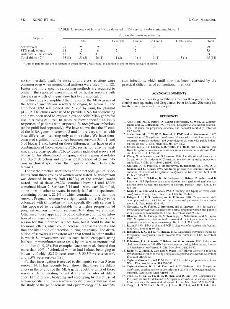
The serovars identified in 165 cervical swabs in which U.urealyticum biovar 1 was detected are shown in Table 3. Sero-var 1 and serovars 3/14 were found most commonly (and inapproximately equal numbers of specimens) in 77 (46.7%) and76 (46.1%) specimens, respectively; serovar 6 was found in 42(25.5%) positive specimens. A high proportion of specimens(22.2% overall) contained more than one serovar.
DISCUSSION
The two biovars of U. urealyticum can be distinguished byvarious molecular techniques (5, 13, 14, 18), while serotypingby conventional methods remains difficult. Currently, there are
FIG. 6. Results of PCR amplification of MBA genes of all 14 serovars of U.urealyticum with primers UMS-54 (specific for serovar 6) and UMA2699. Apositive result is shown by a 369-bp band. Lanes M, molecular weight markersFX174 DNA/HinfI; lanes 1 and 2, U. urealyticum serovar 1 ATCC strain andclinical isolate; lanes 3 to 6, U. urealyticum serovars 2 to 5 ATCC strains; lanes7 and 8, U. urealyticum serovar 6 ATCC strain and clinical isolate; lanes 9 to 16,U. urealyticum serovars 7 to 14 ATCC strains.
TABLE 2. Detection of U. urealyticum biovars 1 and 2 in cervical swabsa
SubjectsNo. (%) of swabs containing:
Total no. of swabs U. urealyticum Biovar 1 Biovar 2 Both biovars
Sex workers 200 87 (43.5) 68 (34.0) 8 (4.0) 11 (5.5)STD clinic clients 100 40 31 7 2Antenatal clinic clients 100 58 52 5 1Total 400 185 (46.3) 151 (37.8) 20 (5.0) 14 (3.5)
a Cervical swabs from women in three different groups were tested by PCR and restriction endonuclease analysis as outlined in Fig. 1. The proportion of womenattending an antenatal clinic in whom any U. urealyticum strain was detected (58.0%) was significantly higher than that in the two groups of women attending the STDclinic (40.0%) (P , 0.01; odds ratio, 1.88; 95% confidence interval, 1.19 to 2.98). The difference remained significant for biovar 1 only (52.0% vs 31.0%; P , 0.01).
VOL. 37, 1999 U. UREALYTICUM BIOVAR 1 SEROVAR IDENTIFICATION BY PCR 541
no commercially available antisera, and cross-reactions werecommon even when monoclonal antisera were used (4, 9, 12).Faster and more specific serotyping methods are required toconfirm the reported associations of particular serovars withdiseases in which U. urealyticum has been implicated.
In this study we amplified the 59 ends of the MBA genes ofthe four U. urealyticum serovars belonging to biovar 1. Theamplified DNA was cloned into E. coli by using the plasmidpUC19. The clones were used to provide DNA for sequencingand have been used to express biovar-specific MBA genes foruse in serological tests to measure biovar-specific antibodyresponses of patients with suspected U. urealyticum infections(to be published separately). We have shown that the 59 endsof the MBA genes in serovars 3 and 14 are very similar, withbase differences occurring only at three sites. We have dem-onstrated significant differences between serovars 3/14, 1, and6 of biovar 1 and, based on these differences, we have used acombination of biovar-specific PCR, restriction enzyme anal-ysis, and serovar-specific PCR to identify individual serovars ofbiovar 1. This allows rapid biotyping and serotyping of isolatesand direct detection and serovar identification of U. urealyti-cum in clinical specimens, the majority of which belong tobiovar 1.
To test the practical usefulness of our methods, genital spec-imens from three groups of women were tested. U. urealyticumwas detected in nearly half (46.3%) of the cervical swabstested, and of these, 89.2% contained biovar 1 and 18.3%contained biovar 2. Serovars 3/14 and 1 were each identified,alone or with other serovars, in nearly half of the specimenscontaining biovar 1, 22.2% of which contained more than oneserovar. Pregnant women were significantly more likely to becolonized with U. urealyticum, and specifically, with serovar 1.This appeared to be attributable to a higher proportion ofpregnant women in whom serovars 3/14 alone were found.Otherwise, there appeared to be no difference in the distribu-tion of serovars between the different groups of subjects. Thereason for this difference is uncertain, but it could be due tohormonal effects, which could increase ureaplasma counts, andthus the likelihood of detection, during pregnancy. The distri-bution of serovars is consistent with that found in other studiesin which U. urealyticum isolates have been serotyped, usingindirect-immunofluorescence tests, by antisera or monoclonalantibodies (4, 9, 23). For example, Naessens et al. showed thatmore than 90% of colonized women had isolates belonging tobiovar 1, of which 52.2% were serovar 3, 30.3% were serovar 6,and 9.5% were serovar 1 (9).
Further investigation is needed to distinguish serovar 3 fromserovar 14. It has recently been shown that there are differ-ences in the 39 ends of the MBA gene repetitive units of theseserovars, demonstrating potential alternative sites of differ-ence. In the future, biotyping and serotyping by direct use ofbiovar-specific and even serovar-specific primers will assist inthe study of the pathogenesis and epidemiology of U. urealyti-
cum infections, which until now has been restricted by thepractical difficulties of conventional methods.
ACKNOWLEDGMENTS
We thank Xueqian Gong and Shouyi Chen for their precious help incloning and sequencing and Greg James, Peter Jelfs, and Zhenfang Mafor their assistance with this project.
REFERENCES
1. Abele-Horn, M., J. Peters, O. Genzel-Boroviczeny, C. Wolff, A. Zimmer-mann, and W. Gottschling. 1997. Vaginal Ureaplasma urealyticum coloniza-tion: influence on pregnancy outcome and neonatal morbidity. Infection25:286–291.
2. Abele-Horn, M., C. Wolff, P. Dressel, F. Pfaff, and A. Zimmerman. 1997.Association of Ureaplasma urealyticum biovars with clinical outcome forneonates, obstetric patients, and gynecological patients with pelvic inflam-matory disease. J. Clin. Microbiol. 35:1199–1202.
3. Cassell, G. H., D. T. Crouse, K. B. Waites, P. T. Rudd, and J. K. Davis. 1988.Does Ureaplasma urealyticum cause respiratory disease in newborns? Pedi-atr. Infect. Dis. J. 7:535–541.
4. Cheng, X., A. Naessens, and S. Lauwers. 1994. Identification of serotype 1-,3-, and 6-specific antigens of Ureaplasma urealyticum by using monoclonalantibodies. J. Clin. Microbiol. 32:1060–1062.
5. Grattard, F., B. Pozzetto, B. de Barbeyrac, H. Renaudin, M. Clerc, O. G.Gaudin, and C. Bebear. 1995. Arbitrarily-primed PCR confirms the differ-entiation of strains of Ureaplasma urealyticum to two biovars. Mol. Cell.Probes 9:383–389.
6. Grattard, F., B. Soleihac, B. de Barbeyrac, C. Bebear, P. Seffert, and B.Pozzetto. 1995. Epidemiologic and molecular investigations of genital myco-plasmas from women and neonates at delivery. Pediatr. Infect. Dis. J. 14:853–858.
7. Kong, F., X. Zhu, and J. Zhou. 1996. Grouping and typing of Ureaplasmaurealyticum. Chung-Hua I Hsueh Tsa Chih 76:138–140.
8. Krieger, J. N., E. S. Boatman, and G. E. Kenny. 1989. Ureaplasma urealyti-cum upper urinary tract infection: persistence and pathogenicity in a caninemodel. J. Urol. 141:1437–1443.
9. Naessens, A., W. Foulon, J. Breynaert, and S. Lauwers. 1988. Serotype ofUreaplasma urealyticum isolated from normal pregnant women and patientswith pregnancy complications. J. Clin. Microbiol. 26:319–322.
10. Ohkawa, M., K. Yamaguchi, S. Tokunaga, T. Nakashima, and S. Fujita.1993. Ureaplasma urealyticum in the urogenital tract of patients with chronicprostatitis or related symptomatology. Br. J. Urol. 72:918–921.
11. Razin, S. 1994. DNA probes and PCR in diagnosis of mycoplasma infections.Mol. Cell. Probes 8:497–511.
12. Robertson, J. A., and G. W. Stemke. 1982. Expanded serotyping scheme forUreaplasma urealyticum strains isolated from humans. J. Clin. Microbiol.15:873–878.
13. Robertson, J. A., A. Vekris, C. Bebear, and G. W. Stemke. 1993. Polymerasechain reaction using 16S rRNA gene sequences distinguishes the two biovarsof Ureaplasma urealyticum. J. Clin. Microbiol. 31:824–830.
14. Ruifu, Y., Z. Minli, Z. Guo, and X. Wang. 1997. Biovar diversity is reflectedby variations of genes encoding urease of Ureaplasma urealyticum. Microbiol.Immunol. 41:625–627.
15. Taylor-Robinson, D., and P. M. Furr. 1997. Genital mycoplasma infections.Wien. Klin. Wochenschr. 109:578–583.
16. Taylor-Robinson, D., P. M. Furr, and A. D. Webster. 1985. Ureaplasmaurealyticum causing persistent urethritis in a patient with hypogammaglobu-linaemia. Genitourin. Med. 61:404–408.
17. Teng, K., M. Li, W. Yu, H. Li, D. Shen, and D. Liu. 1994. Comparison ofPCR with culture for detection of Ureaplasma urealyticum in clinical samplesfrom patients with urogenital infections. J. Clin. Microbiol. 32:2232–2234.
18. Teng, L. J., S. W. Ho, H. N. Ho, S. J. Liaw, H. C. Lai, and K. T. Luh. 1995.
TABLE 3. Serovars of U. urealyticum detected in 165 cervical swabs containing biovar 1
SubjectsNo. of swabs containing serovar(s):
1 3/14 6 1 and 3/14 1 and 6 3/14 and 6 1, 3/14, and 6 Total
Sex workers 29 25 9 6 5 2 3 79STD clinic clients 11 12 6 2 2 33Antenatal clinic clients 13 22 11 3 3 1 53Total (biovar 2)a 53 (5) 59 (3) 26 (1) 11 (2) 10 (1) 3 (1) 3 (1) 165 (14)
a Data in parentheses are specimens in which biovar 2 was found, in addition to one or more serovars of biovar 1.
542 KONG ET AL. J. CLIN. MICROBIOL.

Rapid detection and biovar differentiation of Ureaplasma urealyticum inclinical specimens by PCR. J. Formos. Med. Assoc. 94:396–400.
19. Teng, L. J., X. Zheng, J. I. Glass, H. L. Watson, J. Tsai, and G. H. Cassell.1994. Ureaplasma urealyticum biovar specificity and diversity are encoded inmultiple-banded antigen gene. J. Clin. Microbiol. 32:1464–1469.
20. Thirkell, D., A. D. Myles, and D. Taylor-Robinson. 1990. A comparison offour major antigens in five human and several animal strains of ureaplasmas.J. Med. Microbiol. 32:163–168.
21. Watson, H. L., D. K. Blalock, and G. H. Cassell. 1990. Variable antigens of
Ureaplasma urealyticum containing both serovar-specific and serovar-cross-reactive epitopes. Infect. Immun. 58:3679–3688.
22. Zheng, X., L. J. Teng, H. L. Watson, J. I. Glass, A. Blanchard, and G. H.Cassell. 1995. Small repeating units within the Ureaplasma urealyticum MBantigen gene encode serovar specificity and are associated with antigen sizevariation. Infect. Immun. 63:891–898.
23. Zheng, X., H. L. Watson, K. B. Waites, and G. H. Cassell. 1992. Serotypediversity and antigen variation among invasive isolates of Ureaplasma urea-lyticum from neonates. Infect. Immun. 60:3472–3474.
VOL. 37, 1999 U. UREALYTICUM BIOVAR 1 SEROVAR IDENTIFICATION BY PCR 543