Detection of influenza A matrix gene by real-time Taqman® RT
Upload
vlaanderenCategory
view
2download
0
Sequence Diversity in the Dickeya fliC Gene: Phylogenyof the Dickeya Genus and TaqManH PCR for ’D. solani’,New Biovar 3 Variant on Potato in EuropeJohan Van Vaerenbergh1*, Steve Baeyen1, Paul De Vos2,3, Martine Maes1
1Unit Plant Sciences-Crop Protection, Institute for Agricultural and Fisheries Research-ILVO, Merelbeke, Belgium, 2 Laboratory of Microbiology, Ghent University, Ghent,
Belgium, 3 BCCM/LMG Bacteria Collection, Laboratory of Microbiology Ghent University, Ghent, Belgium
Abstract
Worldwide, Dickeya (formerly Erwinia chrysanthemi) is causing soft rot diseases on a large diversity of crops and ornamentalplants. Strains affecting potato are mainly found in D. dadantii, D. dianthicola and D. zeae, which appear to have a markedgeographical distribution. Furthermore, a few Dickeya isolates from potato are attributed to D. chrysanthemi and D.dieffenbachiae. In Europe, isolates of Erwinia chrysanthemi biovar 1 and biovar 7 from potato are now classified in D.dianthicola. However, in the past few years, a new Dickeya biovar 3 variant, tentatively named ‘Dickeya solani’, has emergedas a common major threat, in particular in seed potatoes. Sequences of a fliC gene fragment were used to generatea phylogeny of Dickeya reference strains from culture collections and with this reference backbone, to classify pectinolyticisolates, i.e. Dickeya spp. from potato and ornamental plants. The reference strains of the currently recognized Dickeyaspecies and ‘D. solani’ were unambiguously delineated in the fliC phylogram. D. dadantii, D. dianthicola and ‘D. solani’displayed unbranched clades, while D. chrysanthemi, D. zeae and D. dieffenbachiae branched into subclades and lineages.Moreover, Dickeya isolates from diagnostic samples, in particular biovar 3 isolates from greenhouse ornamentals, formedseveral new lineages. Most of these isolates were positioned between the clade of ‘D. solani’ and D. dadantii as transitionvariants. New lineages also appeared in D. dieffenbachiae and in D. zeae. The strains and isolates of D. dianthicola and ‘D.solani’ were differentiated by a fliC sequence useful for barcode identification. A fliC TaqManHreal-time PCR was developedfor ‘D. solani’ and the assay was provisionally evaluated in direct analysis of diagnostic potato samples. This molecular toolcan support the efforts to control this particular phytopathogen in seed potato certification.
Citation: Van Vaerenbergh J, Baeyen S, De Vos P, Maes M (2012) Sequence Diversity in the Dickeya fliC Gene: Phylogeny of the Dickeya Genus and TaqManH PCRfor ’D. solani’, New Biovar 3 Variant on Potato in Europe. PLoS ONE 7(5): e35738. doi:10.1371/journal.pone.0035738
Editor: Sunghun Park, Kansas State University, United States of America
Received November 15, 2011; Accepted March 20, 2012; Published May 3, 2012
Copyright: � 2012 Van Vaerenbergh et al. This is an open-access article distributed under the terms of the Creative Commons Attribution License, which permitsunrestricted use, distribution, and reproduction in any medium, provided the original author and source are credited.
Funding: The research was performed with funds of the Government of Flanders. The funders had no role in study design, data collection and analysis, decisionto publish, or preparation of the manuscript.
Competing Interests: The authors have declared that no competing interests exist.
* E-mail: [email protected]
Introduction
The genus Dickeya was established by the reclassification of
Pectobacterium (Erwinia) chrysanthemi and Brenneria paradisiaca as D.
chrysanthemi and D. paradisiaca, respectively and for the accommo-
dation of four new species, i.e. D. dadantii, D. dianthicola, D.
dieffenbachiae and D. zeae; based on analysis of 16S rRNA gene
sequences, DNA:DNA reassociation kinetics and phenotypic
features including biochemical and serological reactions [1]. Multi
Locus Sequence Analysis underpinned that Dickeya constitutes
a distinct genetic clade in the soft rot Enterobacteriaceae [2]. Dickeya
species are broad host range phytopathogens which principal
disease symptom is maceration of plant tissues due to pectinolytic
activity [2,3]. Strains affecting potato are mainly found in three
Dickeya species, i.e. D. dadantii (biovar 3), D. dianthicola (biovars 1
and 7) and D. zeae (biovar 3). A few strains are assigned to D.
chrysanthemi (biovar 5 and 6) and to D. dieffenbachiae (biovar 2) [4].
Erwinia chrysanthemi is known in potato production in some
European countries for over 40 years and is associated with slow
wilt and internal stem necrosis. These strains are now assigned to
D. dianthicola [3,5]. However, in the past few years a new Dickeya
biovar 3 strain, tentatively named ‘Dickeya solani’, has emerged as
a common major threat, in particular on seed potatoes [5]. Across
wide environmental conditions it causes extensive maceration of
the seed tuber, rapid wilting and blackleg-like symptoms in the
stem with decomposition of the pith. Both D. dianthicola and ‘D.
solani’ are disseminated by infected or contaminated seed tubers.
Seed certification generally implements a zero tolerance for
blackleg in field inspections of high grade seed.
A phylogenetic analysis using concatenated atpD, carA and recA
loci resolved the relatedness in the plant pathogenic Enterobacter-
iaceae [6]. Although Dickeya formed a contiguous clade with
Pectobacterium, Brenneria and Samsonia, the gene amplicon sequences
were sufficiently distinct to support the generic status of the taxon.
Diagnostic identification of bacterial isolates as Dickeya is
principally done by testing the production of indigoidin on specific
culture medium [7], maceration of potato tuber tissue and the
production of the 420 bp amplicon in a PCR based on the pelADE
gene cluster [8]. However, robust tools for the differentiation of
Dickeya species were not available until fairly recently. In the past
few years, several gene loci have been found to reliably
differentiate species in several taxa of plant pathogenic bacteria,
e.g. Ralstonia solanacearum [9], Pseudomonas syringae [10] and
PLoS ONE | www.plosone.org 1 May 2012 | Volume 7 | Issue 5 | e35738
Xanthomonas [11]. The recA locus was used for the first phylogenetic
analysis of all species within the genus Dickeya, extending previous
studies based on 16S rDNA [3]. It displayed new genomic clades
and, in particular, a clonal delineation of an emerging biovar 3
variant isolated from potato in the past decade in Europe [12].
This new genetic clade was later on validated in a polyphasic
analysis using dnaX sequence data and genomic fingerprinting [5].
These studies suggest that this new variant may represent a new
species for which the name ‘Dickeya solani’ is provisionally used, but
it is not formally accepted yet [4]. Further evidence for the
taxonomic discrimination of this separate biological unit may be
derived from sequence information of genes involved in patho-
genesis and virulence.
Many phytopathogenic bacteria are motile by means of flagella
and flagellar genes contribute to virulence [13,14,15] and to host-
pathogen interactions, i.e. for pectinolytic Enterobacteriaceae
[16,17,18]. The flagellar filament is composed of a single protein,
flagellin, which is encoded by the fliC gene. The flagellin proteins
contribute to antigenic variation [19] that is also displayed in
Dickeya [20,21,22]. More than 10 different serogroups have been
identified [23] and differences were found among Dickeya isolates
from potato [24]. Sequence variability of the fliC gene has been
used to differentiate among isolates of several clinical bacterial
species [25,26,27] and the bacterial phytopathogen R. solanacearum
[28], for molecular typing and phylogenetic analysis [29], for
taxonomic applications [30] and it showed potential as a biomarker
for phylogenetic and epidemiological studies [31].
This paper reports on the application of fliC sequences to
differentiate Dickeya strains at the species and infraspecific level and
to specifically diagnose ‘D. solani’ with a fliC barcode or TaqManHreal-time PCR.
Results
FliC phylogeny of the reference strainsThe fliC-1/fliC-2 primers were used to produce the reference
PCR amplicon of the fliC gene. The results for the individual
strains are presented in Table S1. A single amplicon of
approximately 650 bp was produced for all strains tested of D.
chrysanthemi, D. dadantii, D. dianthicola, ‘D. solani’ and D. zeae. The
amplicon sequences correspond to the fliC ORF region of D.
dadantii 3937 strain (GenBank accession CP002038.1). A single
amplicon of approximately 900 bp was obtained for the strains of
D. paradisiaca. It shows homology with flagellin gene sequences of
Dickeya strain Ech703 (complete genome sequence in GenBank
accession CP001654). It did not reveal, however, a significant
homology with fliC sequences in other available Dickeya genomes,
i.e. D. dadantii 3937, Dickeya strain Ech586 and Dickeya strain
Ech1591. D. paradisiaca has a limited biological and geographical
distribution. Apparently strains have not been isolated over the
past 30 years. Furthermore, the strains tested did not exhibit
indigoidin production on NGM nor a genuine maceration activity
in potato. Multiple amplicons were produced for the strains of D.
dieffenbachiae. These did not share a valid homology with the fliC
sequence of D. dadantii 3937. Ultimately, global sequence
alignment was performed with 621 bp consensus sequences for
all Dickeya reference strains, except for those of D. dieffenbachiae and
D. paradisiaca. The customised phylogenetic relatedness is displayed
in Figure 1. A separate clade with a clonal structure and a single
sequevar is formed by the strains of ‘D. solani’, which are isolates
from potato in Europe and Israel and one isolate from hyacinth in
The Netherlands. The D. dianthicola reference strains also form
a single clade with a single sequevar, regardless their different
biological and geographical origin. D. dianthicola shows a very close
relationship with ‘D. solani’. Both are most related to the strains of
D. dadantii which form a third clade. Although biologically and
geographically quite diverse, D. dadantii strains also constituted
a single sequevar. The D. zeae reference strains are attributed to
two sub-clades and to a separate branch. The first sub-clade,
phylotype 1 (P1), represents a single sequevar and contains strains
isolated on the American and European continent. The second
sub-clade, phylotype 2 (P2), also represents a single sequevar and
consists of strains isolated on the Asian and Australian continent.
Strain LMG 2497 isolated from sweet corn in the USA is
attributed to a detached lineage of D. zeae and is considered
a separate sequevar. The D. chrysanthemi reference strains form an
aggregate clade with the type strain, other strains from
chrysanthemum and strains from euphorbia, sunflower and carrot
in a large sub-clade containing two sequevars. A biovar 6 strain
from Parthenium and a strain from potato, both isolated in the USA,
form a dichotomous branch in the D. chrysanthemi clade. An
aggregate clade of Pectobacterium was formed containing P.
betavasculorum, P. atrosepticum, P. carotovorum ssp. odoriferum and P.
carotovorum ssp. brasiliensis. A fliC amplicon was not produced for
the type strain of Pectobacterium carotovorum ssp. carotovorum and
Pectobacterium wasabiae, nor for the potato associated bacteria tested.
FliC-based identification of Dickeya isolatesFrom the fifty isolates obtained from diagnostic samples, thirty-
eight were attributed to Dickeya and twelve to Pectobacterium on the
basis of indigoidin production on NGM, maceration of potato
tuber tissue and a PCR amplicon produced with pelADE or pelY
primers respectively. Subsequently, fliC amplicons were obtained
and sequenced. The fliC phylogeny of the reference strains was
used as backbone to position the isolates. All isolates preliminary
identified as Dickeya with the above mentioned methods, were
validated by their position in the phylogenetic fliC tree. The results
are displayed in Figure 2 and in Table S2. The Dickeya isolates
from potato were either classified in the D. dianthicola clade or in
the ‘D. solani’ clade. Furthermore, the fliC sequences were identical
for all strains tested of D. dianthicola and ‘D. solani’. The consensus
sequences exhibit twenty-five different signature positions which
provide reliable barcodes to allocate isolates to one of these clades.
Dickeya strain LMG 2918, isolated from Phalaenopsis orchids, is
attributed to a separate branch, which is considered as an
unassigned Dickeya lineage (UDL-1). Thirteen Dickeya biovar 3
isolates from greenhouse ornamentals exhibited substantial
sequence variation. Six of those isolates are classified in two
additional unassigned Dickeya lineages (UDL-2 and UDL-3). Four
isolates are classified in the D. dadantii clade and one isolate is
assigned to the D. zeae phylotype 1 sub-clade. Furthermore,
another Dickeya biovar 3 isolate from Phalaenopsis orchids
constitutes a fourth unassigned lineage (UDL-4) and one from
Freesia clusters in the separate lineage with strain LMG 2497 from
sweet corn which is now specified as UDL-5. A Dickeya biovar 3
isolate from corn in Belgium is assigned to the D. zeae phylotype 2
sub-clade and, finally, a Dickeya biovar 3 isolate from Belgian
lettuce is placed in yet another unassigned lineage (UDL-6). These
fifteen Dickeya biovar 3 isolates represent seven additional
sequevars. The eighteen Dickeya fliC sequevars determined in this
study are listed with their associated GenBank accession numbers
in Table S3. Eight Pectobacterium isolates are assigned to the
aggregate cluster containing P. carotovorum ssp. odoriferum and P.
carotovorum ssp. brasiliensis. The fliC amplicon was not produced for
four Pectobacterium isolates from potato.
Sequence Diversity in the Dickeya fliC Gene
PLoS ONE | www.plosone.org 2 May 2012 | Volume 7 | Issue 5 | e35738

FliC-based identification of Dickeya dieffenbachiaeA second primer set (fliC-for/fliC-rev) was used for D.
dieffenbachiae to produce a single fliC amplicon (,370 bp) located
inside the ,650 bp fliC amplicon. Comparative sequence analysis
was done on 353 bp consensus sequences which were used to
position the D. dieffenbachiae strains and isolates in the background
of the eighteen Dickeya sequevars identified for the reference fliC
fragment. The phylogram of fliC sequences trimmed at the shorter
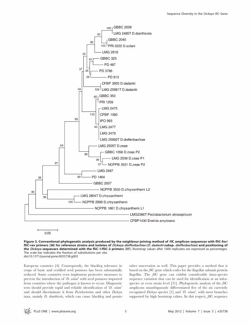
fragment is displayed in Figure 3. D. dieffenbachiae displayed an
aggregate clade with two sub-clades. One contains the strains
isolated from Dieffenbachia and a Dutch isolate from potato and
exhibits an almost clonal structure. Another isolate from potato
was attributed to a second sub-clade together with a Belgian isolate
from Dieffenbachia sp. The D. dieffenbachiae clade showed a high
degree of relatedness to D. dadantii.
Figure 1. Conventional phylogenetic analysis produced by the neighbour-joining method of fliC amplicon sequences with fliC-1/fliC-2 primers [37] for reference strains of the recognized Dickeya taxa, except D. dieffenbachiae (D. dadantii subsp. dieffenbachiae) andD. paradisiaca, of ‘D. solani’, and taxa of Pectobacterium. Bootstrap values after 1000 replicates are expressed as percentages. The scale barindicates the fraction of substitutions per site.doi:10.1371/journal.pone.0035738.g001
Sequence Diversity in the Dickeya fliC Gene
PLoS ONE | www.plosone.org 3 May 2012 | Volume 7 | Issue 5 | e35738

FliC TaqManH PCR for identification of ‘Dickeya solani’The TaqManH real-time PCR for ‘D. solani’ was designed to
amplify a 112 bp stretch of the fliC amplicon (Table 1). The assay
was applied on the fifty–six reference strains and the fifty
diagnostic isolates. Positive reactions, attested by Ct-values #25,
were only obtained for the nine reference strains and the seven
diagnostic isolates of ‘D. solani’. Negative results, demonstrated by
the absence of a Ct -value after 40 PCR cycles, were obtained for
all other bacterial cultures tested. The qualitative results of the fliC
TaqManH PCR are given in Tables S1 and S2.
Fast fliC TaqMan PCR diagnosis of ‘Dickeya solani’ insymptomatic potato tissueThirty diagnostic samples from seed potato production in
Flanders were analysed in this study. They consisted of either
wilting potato stems with blackleg symptoms, necrosis and
maceration of the stem pith or wilting stems without blackleg
but with a macerated mother tuber. A fast diagnostic procedure
for ‘Dickeya solani’ was performed by applying the fliC TaqManHPCR without prior enrichment of the sample extracts. The test
results were compared with the conventional diagnostic protocol
in which the bacteria were cultured from the sample extract by
dilution plating and further characterization of the isolates in
phenotypic and molecular tests. The results are summarized in
Table 2. Definite positive results in the fliC TaqManH PCR,
attested by 18.1, Ct ,28.1, were obtained for twelve out of the
thirty sample extracts, diagnosing the presence of ‘D. solani’. In the
conventional diagnostic protocol, ‘D. solani’ was indeed isolated as
a distinct colony morphotype from these samples and sequence
analysis of the fliC amplicon produced with primers fliC-1/fliC-2
confirmed the identity. The ‘D. solani’ morphotype was not isolated
from the other eighteen sample extracts, which underpinned the
negative reactions in the fliC TaqManH PCR on the extracts.
Furthermore, seven of these samples were found to contain
a different Dickeya variant producing indigoidin on NGM medium,
maceration of potato tuber tissue and the pelADE amplicon in
PCR. These isolates were identified as D. dianthicola by sequence
analysis of the fliC amplicon. Pectobacterium spp. was diagnosed in
the remaining eleven samples, as confirmed by maceration of
potato tuber tissue, the absence of indigoidin production on NGM
medium, a positive reaction in the pelY PCR and a negative
reaction in the pelADE PCR. More detail of the diagnostic tests are
presented in Table S4.
Discussion
In less than a few years, a biovar 3 of Dickeya, provisionally
named ‘Dickeya solani’, has become the predominate cause of
wilting, blackleg and tuber maceration of potato in several
European countries. The new form has been described as more
aggressive, more likely to infect at lower cell densities and to
spread from plant to plant along and even across plant rows, and
causing damage in a wider range of conditions than observed for
the ‘traditional’ blackleg (Pectobacterium atrosepticum) or for Dickeya
dianthicola which is known for over 40 years in potato in some
Figure 2. Conventional phylogenetic analysis produced by theneighbour-joining method of fliC amplicon sequences with fliC-1/fliC-2 primers [37] for reference strains of the recognizedDickeya taxa, except D. dieffenbachiae (D. dadantii subsp.dieffenbachiae) and D. paradisiaca, of ‘D. solani’ and Dickeya andPectobacterium isolates from diagnostic samples. Bootstrapvalues after 1000 replicates are expressed as percentages. The scalebar indicates the fraction of substitutions per site.doi:10.1371/journal.pone.0035738.g002
Sequence Diversity in the Dickeya fliC Gene
PLoS ONE | www.plosone.org 4 May 2012 | Volume 7 | Issue 5 | e35738
European countries [4]. Consequently, the blackleg tolerance in
crops of basic and certified seed potatoes has been substantially
reduced. Some countries even implement protective measures to
prevent the introduction of ‘D. solani’ with seed potatoes imported
from countries where the pathogen is known to occur. Diagnostic
tests should provide rapid and reliable identification of ‘D. solani’
and should discriminate it from Pectobacterium and other Dickeya
taxa, mainly D. dianthicola, which can cause blackleg and potato
tuber maceration as well. This paper provides a method that is
based on the fliC gene which codes for the flagellar subunit protein
flagellin. The fliC gene can exhibit considerable intra-species
sequence variation that can be used for identification at an infra-
species or even strain level [31]. Phylogenetic analysis of the fliC
amplicons unambiguously differentiated five of the six currently
recognized Dickeya species [1] and ‘D. solani’, with most branches
supported by high bootstrap values. In this respect, fliC sequence
Figure 3. Conventional phylogenetic analysis produced by the neighbour-joining method of fliC amplicon sequences with fliC-for/fliC-rev primers [38] for reference strains and isolates of Dickeya dieffenbachiae (D. dadantii subsp. dieffenbachiae) and positioning ofthe Dickeya sequevars determined with the fliC-1/fliC-2 primers [37]. Bootstrap values after 1000 replicates are expressed as percentages.The scale bar indicates the fraction of substitutions per site.doi:10.1371/journal.pone.0035738.g003
Sequence Diversity in the Dickeya fliC Gene
PLoS ONE | www.plosone.org 5 May 2012 | Volume 7 | Issue 5 | e35738

comparison confirmed that ‘D. solani’ is clearly a new, separate and
clonal clade within the genus. The short phylogenetic branches
indicate the relatively close relatedness of D. dianthicola, D. dadantii
and ‘D. solani’, suggesting that taxonomically these taxa may well
be delineated at subspecies level. The fliC phylogeny does not
extensively support that ‘D. solani’ deserves a separate species status
according to the current bacterial species concept [32]. Larger fliC
sequence variation exist within D. zeae with two phylotypes and
within D. chrysanthemi as demonstrated in the recA phylogeny [3]. A
strain isolated from sweet corn is assigned to a lineage detached of
D. zeae. This strain was classified as D. zeae phylotype 1 in the recA
phylogram [3]. All other corn strains of D. zeae in the fliC clade
were isolated from maize varieties used for livestock fodder or
processing and thus the contrasting position in the fliC phylogram
may reflect the association of fliC to host designation. The D.
chrysanthemi strains clustered in an aggregate clade with a large sub-
clade identified as D. chrysanthemi biovar chrysanthemi and a detached
D. chrysanthemi biovar parthenii lineage according to the species
description [1]. The D. chrysanthemi strain isolated from potato in
the USA is more related to the ‘parthenii’ biovar than to the
‘chrysanthemi’ biovar. The assignment of D. dieffenbachiae in the fliC
phylogeny was performed with an amplicon internal to the regular
650 bp amplicon. The grouping of Dickeya taxa and sequevars
remained stable. The strains and isolates of D. dieffenbachiae are
closely related to D. dadantii which underpins the recent re-
classification of D. dieffenbachiae as D. dadantii ssp. dieffenbachiae [33].
The robustness of the fliC phylogeny is further underpinned by
analogies in phylogenies based on recA [3] and dnaX [5] loci,
presented in Table S5.
The isolates from diagnostic samples were introduced the fliC
phylogram allowing their assignment to a Dickeya taxon. All isolates
from potato were assigned to D. dianthicola or ‘D. solani’ and
accentuated the clonal structure of these two clades. Dickeya biovar
3 isolates from various ornamentals exhibited significant sequence
variability which resulted in six unassigned lineages and, typically,
strains isolated from the same plant family (the Araceae, with
Philodendron and Aglaonema) constituted one of these lineages (UDL-
2). So, again not totally unexpected, fliC-based bacterial typing
could be informative on different plant hosts of strains.
Furthermore, three of these lineages are positioned in between
‘D. solani’ and D. dadantii, the latter being biologically and
geographically the most diverse Dickeya taxon [3]. Although the
phylogenetic significance of these up to now not formally classified
lineages remains to be clarified, it is our opinion that the presented
data allude to the origin of ‘D. solani’ as being from one of the
variants existing on ornamentals which then spread clonally in
potato. Alternatively, fliC sequence drift when residing on different
plant hosts could be responsible for the existence of the unassigned
lineages. The apparent pectinolytic activity of Dickeya spp. make
them broad host range pathogens which increases the potential for
genetic exchange as a result of adaptation to a different
environment, i.e. a new plant host [34]. Furthermore, the short
branch lengths in the fliC phylogeny tend to reveal that D.
dianthicola, ‘D. solani’, D. dadantii and the unassigned lineages UDL-
1, UDL-2, UDL-3 and UDL-4 are a species complex. Further
analysis should clarify the taxonomic position of these taxa, i.e. the
classification at subspecies level as already done for the D. dadantii –
D. dieffenbachiae aggregate [33].
The apparent clonal structure of the ‘D. solani’ clade enabled the
development of a TaqManH PCR to specifically identify this
variant and for its direct diagnosis in symptomatic potato stems
and tubers. The primers are positioned in the more variable
stretches of the fliC gene amplicon and the TaqManH probe is
situated in a region with two single nucleotide polymorphisms.
Table 1. Primers for conventional fliC PCR and primers and TaqMan probe for identification and diagnosis of ‘D. solani’.
primer orprobe sequence (59 39) reference derived from amplicon use
fliC-1 TATCAACAGCGCCAAAGACAACGC 37 D. dadantii CFBP3855
,650 bp PCR & sequencing
fliC-2 ACGGCTCATGTTGGATACTTCGTT
fliC-for GACCGTACTGCAATCCAGC 38 D. dadantii CFBP3855
,370 bp PCR & sequencing
fliC-rev CTGGAAGCGGTTCAGAGT
ds-f GCGAACTTCAACGGTAAA this study ‘D. solani’ GBBC2040
112 bp TaqManH real-timePCR
ds-r CAGAGCTACCAACAGAGA
ds-p* CTCTGCTGGACGGTTC
*probe.doi:10.1371/journal.pone.0035738.t001
Table 2. Analysis of diagnostic samples of seed potato plants showing wilting, blackleg or tuber maceration symptoms.
# samplesmorphotype onPDA
potatomaceration
indigoidin opNGM pelADE pelY
fliC qPCRextract
fliC qPCRculture result
7 A + + + 2 2 2 D. dianthicola1
12 B + + + 2 + + ‘D. solani’
11 C/D + 2 2 + 2 2 Pectobacterium sp.
1The isolates of D. dianthicola were identified by sequencing of the fliC PCR amplicon.doi:10.1371/journal.pone.0035738.t002
Sequence Diversity in the Dickeya fliC Gene
PLoS ONE | www.plosone.org 6 May 2012 | Volume 7 | Issue 5 | e35738
Furthermore, the 39 MGB probe is more appropriate for single
base mismatches, thus increasing the specificity of the assay. ‘D.
solani’ is differentiated from all other reference strains and isolates,
i.e. from D. dianthicola isolates from potato and from the Dickeya
biovar 3 isolates from greenhouse ornamentals which were
attributed to unassigned lineages. However, it did not differentiate
the ‘D. solani’ isolate from a hyacinthus bulb which sets focus on
the relation of this variant with the cultivation of bulb-producing
ornamentals. The specificity of the molecular assay for ‘D. solani’
was also demonstrated in direct analysis of potato samples. Such
diagnostics are essential if legislation that imposes a zero tolerance
in seed potatoes is to be effective. The assay has not yet been
validated for detection of latent infections and is, pending the
outcome of these tests, proposed here as a diagnostic tool.
ConclusionsThe sequence diversity of the Dickeya fliC gene produced
a phylogeny of the currently recognized Dickeya taxa and the new
Dickeya biovar 3 variant from potato in Europe, tentatively named
‘D. solani’. Dickeya isolates from diagnostic samples were introduced
into this phylogenetic backbone displaying new, unassigned
lineages in the fliC phylogeny, in particular of certain Dickeya
biovar 3 isolates from ornamentals which were positioned as ‘D.
solani’ – D. dadantii transition variants. These may have spread into
potato and become clonally established as ‘D. solani’ by seed potato
propagation. A TaqManH real-time PCR was developed on the
unique fliC sequence of ‘D.solani’ and provisionally evaluated.
This diagnostic tool was effective for diagnosis of ‘D. solani’ in
potato plants and tubers.
Materials and Methods
Bacterial strainsThe strains and isolates used are listed in Tables S1 and S2. The
reference set (Table S1) consisted of strains from the six currently
recognized Dickeya species, typed Dickeya isolates from the new clade
of biovar 3 strains from potato (‘D. solani’), strains from the
Pectobacterium taxa and strains of Clavibacter michiganensis subsp.
sepedonicus, Ralstonia solanacearum and Paenibacillus macerans. Most
reference strains were acquired from public and certified culture
collections, while the isolates of ‘D. solani’ were obtained in the
framework of the European Dickeya consortium (Dickeya Research
Network hosted by the James Hutton Institute, Dundee, Scotland,
UK). The second set (Table S2) consisted of Dickeya and
Pectobacterium isolates from diagnostic samples that were obtained
from the Diagnostic Centre for Plants of ILVO (GBBC numbers)
and from diagnostic culture collections in The Netherlands (PD
and IPO/PRI numbers). These isolates were mainly recovered
from symptomatic potato plants and tubers and from greenhouse
ornamentals. All strains and isolates were archived in cryopreser-
vation.
The reference strains were first cultured on Difco LB agar
(Miller’s modification) and then subcultured on nutrient sucrose
agar (NSA = Difco Nutrient Agar supplemented with 5%
sucrose). Dickeya strains were verified with the Dickeya genus
specific pelADE primers [8], used in colony PCR. A single colony
from a 48 hr culture on NSA was suspended in 1 ml of sterile
10 mM phosphate buffer (PB) pH 7.2 and DNA was obtained by
alkaline lysis [35]. After pulse centrifugation to sediment cell
debris, two microliters of the supernatant were used in the PCR
reactions.
The isolates from diagnostic samples were cultured on nutrient
glycerol agar supplemented with manganese chloride (NGM) for
production of indigoidin pigment, which is characteristic for
Dickeya spp. [7]. The macerating properties of the isolates were
determined on potato tubers ‘Spunta’ derived from minitubers.
Cell suspensions with density of approximately 107 colony forming
units per ml were prepared in sterile 10 mM PB. A conical tissue
core was removed at the heel end of the tubers and 100 ml of thecell suspension was pipetted onto the cut surface. The tissue cone
was reinstalled after the applied volume was absorbed and the
cone was then tightened by parafilm tape. Three tubers were used
for each isolate in one unreplicated assay. They were placed in
moist sterilised white sand in an appropriate receptacle that was
closed with a lid and aerobically incubated for 48 hours at 28uC.Macerative isolates producing the indigoidin pigment were further
identified as Dickeya spp. with the pelADE PCR. Macerative
isolates not producing the indigoidin pigment were further tested
with the pelY PCR to identify Pectobacterium strains [36]. PCRs were
performed on bacterial DNA prepared as described above for the
collection strains.
Conventional fliC PCR and amplicon sequencingSingle bacterial colonies were transferred in 3 ml LB broth and
grown in a shaking incubator (200 rpm) at 28uC. DNA was
isolated from overnight broth cultures using the Qiagen DNeasy
Blood & Tissue kit as described by the manufacturer, including the
pre-treatment for gram-negative bacteria. DNA concentration and
quality (according to A260/280 and A260/230 ratios) were assessed
using a Nanodrop ND-1000 spectrophotometer. Isolated DNA
was adjusted to approximately 50 ng/ml. The fliC gene fragment
was amplified with PCR primers (Table 1) designed for the Dickeya
dadantii 3937 strain. PCR with the fliC-1 and fliC-2 primers [37]
was performed with 5 ml DNA template in 1 x Faststart High
Fidelity reaction buffer (Roche Applied Science) with 2 mM
MgCl2, 0.2 mM of each dNTP, 0.2 mM of each primer, 1 unit of
FastStart Taq DNA polymerase (Roche Applied Science) and
sterile molecular-grade water for a total volume of 50 ml. PCR was
performed in a Bio-Rad Laboratories C1000 thermal cycler with
initial denaturation at 95uC for 4 minutes, followed by 35 cycles of
95uC for 30 seconds, 55uC for 1 minute and 72uC for 45 seconds,
and a terminal extension step of 7 minutes at 72uC and
subsequent cooling to 12uC. PCR with the fliC-for and fliC-rev
primers [38] was performed with 5 ml DNA template in 1 x
OneTaq standard reaction buffer (New England Biolabs) with
2 mM MgCl2, 0.2 mM of each dNTP, 0.2 mM of each primer,
1,25 unit of OneTaq Hotstart polymerase (New England Biolabs)
and sterile molecular-grade water for a total volume of 50 ml. PCRwas performed in a Bio-Rad Laboratories C1000 thermal cycler
with initial denaturation at 94uC for 30 seconds, followed by
30 cycles of 94uC for 30 seconds, 53uC for 1 minute and 68uC for
30 seconds, and a terminal extension step of 5 minutes at 68uCand subsequent cooling to 12uC.PCR amplicons were resolved by electrophoresis in a 1.5%
agarose gel stained with ethidium bromide. PCR amplicons were
extracted from gel with the Nucleospin Extract II kit (Macherey-
Nagel). DNA concentration and quality were assessed in
a Nanodrop ND-1000 spectrophotometer. Purified PCR ampli-
cons were sequenced in both directions by a commercial
sequencing service (Macrogen Ltd, Korea), using the same primer
set as for PCR amplification.
Sequence alignment and phylogenetic analysisThe fliC consensus sequences were delineated by clipping the
PCR amplicon sequences to a standard start position [A(A/
G)TC(A/G)GC(A/G)T at 59 end] and finish position [TG(A/G/
C)G(C/A) (A/G)G(T/A)(C/T)AT(A/G) at 39 end]. Phylogenetic
and molecular evolutionary analysis were conducted using MEGA
Sequence Diversity in the Dickeya fliC Gene
PLoS ONE | www.plosone.org 7 May 2012 | Volume 7 | Issue 5 | e35738
version 5 software [39]. Sequence alignment of the trimmed
sequences was done using the clustalW algorithm [40] in MEGA 5
and phylogenetic trees were generated using the neighbour-
joining, maximum parsimony and maximum likelihood algorithm
[41]. Distance estimation was calculated using the p-distance
substitution model [42] with 1000 bootstrapping replications.
Based on the sequence distances, fliC clades were differentiated by
monophyletic clustering [43] with type strains and reference
strains. Sequevars were designated within these clades on the basis
of at least 1% sequence difference [9]. Sequevar sequences were
submitted to GenBank (Table S3). The fliC sequence of Erwinia
amylovora CFBP 1430 (GenBank accession AY743588) was used to
root the phylogenies.
FliC TaqManH real-time PCR for ‘Dickeya solani’Primers and TaqManH MGB (59-FAM/39-BHQ1) probe (Life
Technologies) specific for the fliC amplicon of ‘D. solani’ (Table 1)
were designed with Premier Biosoft’s Allele ID version 7 software.
The real-time PCR was performed in a 25 ml volume in
a MicroAmp Optical 96 well reaction plate with Optical Caps
(Life Technologies). Briefly, 2 ml DNA template was added to
12,5 ml Taqman Gene Expression master mix 2x, 0,5 ml of
primers Dsf and Dsr (15 mM), 0,5 ml of probe Dsp (10 mM) and
molecular-grade water up to a final volume of 25 ml. Amplification
and signal detection was done in an ABI Prism 7900HT Sequence
Detection System (Life Technologies). The cycling profile is
consisted of 2 minutes at 50uC for UNG-activation, 10 minutes at
95uC followed by 40 cycles of 15 seconds at 95uC and 1 minute at
63uC. The specificity of the TaqMan assay was tested by colony
PCR with all reference strains and diagnostic isolates. Finally,
suspensions of about 106 colony forming units per ml in sterile
10 mM PB were tested as described for pelADE and pelY PCR
using 2 ml of target per TaqMan PCR reaction.
Molecular diagnosis of ‘Dickeya solani’ in symptomaticpotato samplesThirty diagnostic samples were analysed. For classical diagnosis,
pathogen identification was done by isolation and further
characterisation of the dominant bacterial type cultured upon
plating of serial decimal dilutions of the extract from the
symptomatic tissue. Therefore, minute quantities of affected tissue
were aseptically removed at the margin of disease development in
the stem or in the tuber and transferred in 1 ml of sterile 10 mM
PB in a microvial. After vortexing of the preparation, dilution
plating was performed on potato dextrose agar (Oxoid PDA)
supplemented with cycloheximide. Isolated bacterial colonies
representative at the higher extract dilutions were cultured on
NGM and macerative properties were assessed as described
before. Presumptive identification of macerative isolates was done
by conventional pelADE or pelY PCR for Dickeya spp. or
Pectobacterium spp. respectively. Suspensions of single colonies were
prepared in 1 ml of sterile 10 mM PB and bacterial cells were
subjected to alkaline lysis as explained above. D. dianthicola isolates
were identified by sequencing of the PCR amplicon with the fliC-1
and fliC-2 primers and ‘D. solani’ isolates were identified in fliC
TaqManH real-time PCR as described.
The fliC TaqManH real-time PCR was also performed directly
on the potato sample extracts. The extract was allowed to settle for
15 minutes and then 100 ml was transferred in a 1.5 ml microvial
and centrifuged for 10 minutes at 13000 g. The supernatant was
removed and the pellet was resuspended in 100 ml 1 mM Tris-
HCl pH=8. DNA isolation was performed with the QuickPickTM
Plant DNA kit (Bio-Nobile) using the Pickpen-8M magnetic tool
according to the manufacterer’s protocol for 100 mg starting
material. The TaqMan PCR was performed using the protocol
described above using 2 ml of eluted DNA as template.
Supporting Information
Table S1 Reference strains of the recognized Dickeya taxa and
‘D. solani’, of Pectobacterium taxa and taxa of other phytopathogenic
bacteria from potato, investigated in fliC phylogeny and fliC
TaqmanH real-time PCR. 1as identified in [1], [3] or [5] 2the
strain in bold was used when different strain designations are
displayed 3amplicon of the fliC locus with primers fliC-1 & fliC-2
[37]: ,650 bp (+), multiple amplicons (+a), a larger amplicon of
,900 bp (+b) or no amplicon (2) 4Dickeya clades identified on the
basis of global alignment of 621 bp fliC amplicon [37], except for
D.dieffenbachiae which is identified on the basis of global alignment
of 353 bp fliC amplicon [38] 5fliC sequevar or sequence variant
[9]: strains/isolates with .1% sequence variation in the 621 bp
fragment. D. dieffenbachiae and D. paradisiaca are not considered in
this classification 6negative result = no Ct LMG = Laboratory of
Microbiology, Ghent University, Belgium NCPPB = National
Collection of Plant Pathogenic Bacteria, York, UK CFBP =
Collection Francaise des Bacteries Phytopathogenes, Angers
France IPO/PRI = Plant Research International, Wageningen,
The Netherlands
(XLS)
Table S2 Isolates of Dickeya and Pectobacterium from diagnostic
samples investigated in fliC phylogeny and fliC TaqmanH real-
time PCR.1the strain in bold was used when different strain
designations are displayed 2amplicon of the fliC locus with primers
fliC-1 & fliC-2 [37]: ,650 bp (+), multiple amplicons (+a), a largeramplicon of ,900 bp (+b) or no amplicon (2) 3Dickeya clades
identified on the basis of global alignment of 621 bp fliC amplicon
[37], except for D. dieffenbachiae which is identified on the basis of
global alignment of 353 bp fliC amplicon [38] 4fliC sequevar or
sequence variant [9]: strains/isolates with .1% sequence
variation in the 621 bp fragment. D. dieffenbachiae and D. paradisiaca
are not considered in this classification 5negative result = no Ct
GBBC = Culture collection of ILVO Diagnostic Centre for Plants
(DCP) LMG = Laboratory of Microbiology, Ghent University,
Belgium NCPPB = National Collection of Plant Pathogenic
Bacteria, York, UK IPO = Plant Research International,
Wageningen, The Netherlands
(XLS)
Table S3 Dickeya fliC sequevars and their associated Genbank
accession numbers. UDL = Unassigned Dickeya Lineage.
(XLS)
Table S4 Diagnostic analysis of samples of seed potato plants
showing wilting, blackleg or tuber maceration symptoms. 1Ct
value 2The isolates attributed to D. dianthicola were identified by
sequencing of the fliC amplicon [37].
(XLS)
Table S5 Agreement of recA/dnaX and fliC classification for 52
Dickeya strains and isolates. The recA attribution was obtained from
[11]. The dnaX attribution was obtained from [5]. The fliC
attribution is obtained in this study.
(XLS)
Author Contributions
Conceived and designed the experiments: JVV MM SB PDV. Performed
the experiments: SB JVV. Analyzed the data: JVV SB MM PDV. Wrote
the paper: JVV MM SB PDV.
Sequence Diversity in the Dickeya fliC Gene
PLoS ONE | www.plosone.org 8 May 2012 | Volume 7 | Issue 5 | e35738

References
1. Samson R, Legendre J, Christen R, Fischer-Le Saux M, Achouak W, et al.(2005) Transfer of Pectobacterium chrysanthemi (Burkholder et al. 1953) Brenner et
al. 1973 and Brenneria paradisiaca to the genus Dickeya gen. nov. as Dickeya
chrysanthemi comb. nov. and Dickeya paradisiaca comb. nov. and delineation of four
novel species, Dickeya dadantii sp. nov., Dickeya dianthicola sp. nov., Dickeya
dieffenbachiae sp. nov. and Dickeya zeae sp. nov. International Journal of Systematic
and Evolutionary Microbiology 55: 1415–1427.
2. Ma B, Hibbing ME, Kim H-S, Reedy RM, Yedidia I, et al. (2007) Host range
and molecular phylogenies of the soft rot enterobacterial genera Pectobacterium
and Dickeya. Phytopathology 97: 1150–1163.
3. Parkinson N, Stead D, Bew J, Heeney J, Tsror L, et al. (2009) Dickeya species
relatedness and clade structure determined by comparison of recA sequences.International Journal of Systematic and Evolutionary Microbiology 59:
2388–2393.
4. Toth IK, van der Wolf JM, Saddler GS, Lojkowska E, Helias V, et al. (2011)
Dickeya species: an emerging problem for potato production in Europe. PlantPathology Doi: 10.1111/j.1365-3059.2011.02427.x.
5. Slawiak M, van Beckhoven JRCM, Speksnijder AGCL, Czajkowski R, Grabe G,et al. (2009) Biochemical and genetical analysis reveal a new clade of biovar 3
Dickeya spp. strains isolated from potato in Europe. European Journal of Plant
Pathology 125: 245–261.
6. Young J, Park D (2007) Relationships of plant pathogenic enterobacteria based
on partial atpD, carA and recA as individual and concatenated nucleotide andpeptide sequences. Systematic and Applied Microbiology 30, 343–354.
7. Lee Y-A, Yu C-P (2006) A differential medium for the isolation and rapididentification of a soft rot pathogen, Erwinia chrysanthemi. Journal of Microbio-
logical Methods 64: 200–206.
8. Nassar A, Darasse A, Lemattre M, Kotoujansky A, Dervin C, et al. (1996)
Characterization of Erwinia chrysanthemi by pectinolytic isozyme polymorphismand restriction fragment length polymorphism analysis of PCR-amplified
fragments of pel genes. Applied and Environmental Microbiology 62:
2228–2235.
9. Fegan M, Prior P (2005) How complex is the ‘Ralstonia solanaceaum species
complex’? 449-461 in Allen A, Prior P, Hayward AC (ed.), Bacterial wilt diseaseand the Ralstonia solanacearum species complex. APS Press, St. Paul, USA.
10. Sarkar SF, Guttman DS (2004) The evolution of the core genome of Pseudomonassyringae, a highly clonal, endemic plant pathogen. Applied and Environmental
Microbiology 70: 1999–2012.
11. Parkinson N, Aritua V, Heeney J, Cowie C, Bew J, et al. (2007) Phylogenetic
analysis of Xanthomonas species by comparison of partial gyrase B gene sequences.International Journal of Systematic and Evolutionary Microbiology 57:
2881–2887.
12. Waleron M, Waleron K, Podhajska A, Lojkowka E (2002) Genotyping of
bacteria belonging to the former Erwinia genus by PCR-RFLP analyisis of a recA
gene fragment. Microbiology 148, 583–595.
13. Young GM, Schmiel DH, Miller VL (1999) A new pathway for the secretion of
virulence factors by bacteria: the flagellar export apparatus functions as a proteinsecretion system. Proceedings of the National Academy of Science (PNAS) USA
96: 6556–6461.
14. Wang Q, Suzuki A, Mariconda S, Porwollik, Harshey RM (2005) Sensing
wetness: a new role for the bacterial flagellum. The EMBO Journal 24:2034–2042.
15. Antunez-Lamas M, Cabrera-Ordonez E, Lopez-Solanilla E, Raposo R, Trelles-Salazar O, et al. (2009) Role of motility and chemotaxis in the pathogenesis of
Dickeya dadantii 3937 (ex Erwinia chrysanthemi 3937). Microbiology 155: 434–442.
16. Jahn CE, Willis DK, Charkowski AO (2008) The flagellar Sigma factor FliA is
required for Dickeya dadantii virulence. Molecular Plant Microbe Interactions 21:
14314–1442.
17. Hossein MM, Shibata S, Aizawa SI, Tsuyumu S (2005) Motility is an important
determinant for pathogenesis of Erwinia carotovora subsp. carotovora. Physiologicaland Molecular Plant Pathology 17: 943–950.
18. Mulholland V, Hinton JCD, Sidebotham J, Toth IK, et al. (1993) A pleiotrophicreduced virulence (Rvi-) mutant of Erwinia carotovora subspecies atroseptica is
defective in flagella assembly proteins that are conserved in plant and animalbacterial pathogens. Molecular Microbiology 9: 343–356.
19. Parish CR, Wistar R, Ada GL (1969) Cleavage of bacterial flagellin withcyanogen bromide: antigenic properties of the protein fragments. Biochemistry
Journal 113: 501–506.
20. Yakrus M, Schaad NW (1978) Serological relationships among strains of Erwinia
chrysanthemi. Phytopathology 69: 517–522.
21. Dickey RS, Zumoff CH, Uyemoto JK (1984) Erwinia chrysanthemi: serological
relationships among strains from several hosts. Phytopathology 74: 1388–1394.
22. Janse JD, Ruissen MA (1988) Characterization and classification of Erwinia
chrysanthemi strains from several hosts in the Netherlands. Phytopathology 78:800–808.
23. Samson R, Nassan-Agha N (1978) Biovars and serovars among 129 strains ofErwinia chrysanthemi. In: Ride M, ed. Proceedings of the 4th International
Conference on Plant Pathogenic Bacteria: 547–553. INRA Angers, France.
24. Samson R, Poutier F, Sailly M, Jouan B (1987) Caracterisation des Erwinia
chrysanthemi isolees de Solanum tuberosum et d’autres plantes-hotes selon les biovars
et serogroupes. EPPO Bulletin 17: 11–16.25. Wang L, Rothemund D, Curd H, Reeves PR (2003) Species-wide variation in
the Escherichia coli flagellin (H-antigen) gene. Journal of Bacteriology 185:
2936–2943.26. Paiva JB, Cavallini JS, Silva MD, Almeida MA, Angela HL, et al. (2009)
Molecular differentiation of Salmonella Gallinarum and Salmonella Pullorum byRFLP of fliC gene from Brazilian isolates. Brazilian Journal of Poultry Science
11: 271–276.27. Winstanley C, Hales BA, Morgan AW, Gallagher MJ, Puthucheary SD, et al.
(1999) Analysis of fliC variation among clinical isolates of Burkholderia cepacia.
Journal of Medical Microbiology 48: 657–662.28. Schonfeld J, Heuer H, van Elsas JD, Smalla K (2003) Specific and sensitive
detection of Ralstonia solanacearum in soil on the basis of PCR amplification of fliCfragments. Applied and Environmental Microbiology 69: 7248–7256.
29. Amhaz JMK, Andrade A, Bando SY, Tanaka TL, Moreira-Filho C, et al. (2004)
Molecular typing and phylogenetic analysis of enteroinvasive Escherichia coli usingthe fliC gene sequence. FEMS Microbiology Letters 235: 259–264.
30. Bellingham NF, Morgan JA, Saunders JR, Winstanley C (2001) Flagellin genesequence variation in the genus Pseudomonas. Systematic and Applied
Microbiology 24: 157–165.31. Winstanley C, Morgan JAW (1997) The bacterial flagellin gene as a biomarker
for detection, population genetics and epidemiological analysis. Microbiology
143: 3071–3084.32. Stackebrandt E, Frederiksen W, Garrity GM, Grimont PAD, Kampfer P, et al.
(2002) Report of the ad hoc committee for the re-evaluation of the speciesdefinition in bacteriology. International Journal of Systematic and Evolutionary
Microbiology 52: 1043–1047.
33. Brady CL, Cleenwerk I, Denman S, Venter SN, Rodriguez-Palenzuela P, et al.(2011) Proposal to reclassify Brenneria quercina (Hildebrand & Schroth 1967)
Hauben et al. 1999 into a novel genus, Lonsdalea gen. nov., as Lonsdalea quercina
comb. nov., descriptions of Lonsdalea quercina subsp. quercina comb. nov., Lonsdalea
quercina subsp. iberica subsp. nov. and Lonsdalea quercina subsp. britannica subsp.nov., emendation of the description of the genus Brenneria, reclassification of
Dickeya dieffenbachiae as Dickeya dadantii subsp. dieffenbachiae comb. nov., and
emendation of the description of Dickeya dadantii. International Journal ofSystematic and Evolutionary Microbiology DOI:10.1099/ijs.0.035055-0.
34. Juhas M, van der Meer JR, Gaillard M, Harding RM, Hood DW, et al. (2009)Genomic islands: tools of bacterial horizontal gene transfer and evolution.
FEMS Microbiology Reviews 33: 376–393.
35. Zhang S, Goodwin P H (1997) Rapid and sensitive detection of Xanthomonasfragarie by simple alkaline DNA extraction and the Polymerase Chain Reaction.
Journal of Phytopathogy 145: 267–270.36. Darasse A, Priou S, Kotoujansky A, Bertheau Y (1994) PCR and Restriction
Fragment Length Polymorphism of a pel gene as a tool to identify Erwinia
carotovora in relation to potato diseases. Applied and Environmental Microbiol-
ogy 60: 1437–1443.
37. Venkatesh B, Babujee L, Liu H, Hedley P, Fujikawa T, et al. (2006) The Erwiniachrysanthemi 3937 PhoQ sensor kinase regulates several virulence determinants.
Journal of Bacteriology 188: 3088–3098.38. Haque M, Nahar K, Rahim M, Gomes I, Tsuyumu S (2006) PhoP-PhoQ two-
component system required for colonization leading to virulence of Dickeya
dadantii 3937 in planta. Bangladesh Journal of Microbiology 25: 36–40.39. Tamura K, Peterson D, Peterson N, Stecher G, Nei M, et al. (2011) MEGA5:
Molecular Evolutionary Genetics Analysis using Maximum Likelihood,Evolutionary Distance, and Maximum Parsimony Methods. Molecular Biology
and Evolution 28: 2731–2739.
40. Higgins D, Thompson J (1994) CLUSTAL W: improving the sensitivity ofprogressive multiple sequence alignment through sequence weighting, position-
specific gap penalties and weight matrix choice. Nucleic Acids research 22:4673–4680.
41. Felsenstein J (2004) Inferring Phylogenies. Sunderland, MA: Sinauer Associates.42. Saitou N, Nei M (1987) The neighbour-joining method: a new method of
constructing phylogenetic trees. Molecular Biology and Evolution 4: 1406–1425.
43. Nei M, Kumar S (2000) Molecular Evolution and Phylogenetics. OxfordUniversity Press, New York.
Sequence Diversity in the Dickeya fliC Gene
PLoS ONE | www.plosone.org 9 May 2012 | Volume 7 | Issue 5 | e35738
Copyright © 2022 FDOKUMEN