
Detection of influenza A matrix gene by real-time Taqman® RT
18
OIE/FAO international reference laboratory for AI Page 1 of 18 SOP: VI.493 Edition 21 (September 2020) Detection of influenza A matrix gene by real-time Taqman® RT- PCR This protocol is a copy of the standard operating procedure used by the OIE/FAO International Reference laboratory for AI at the Animal and Plant Health Agency. If you have any technical queries please contact [email protected]
-
Upload
khangminh22 -
Category
Documents
-
view
1 -
download
0
Transcript of Detection of influenza A matrix gene by real-time Taqman® RT
OIE/FAO international reference laboratory for AI
Page 1 of 18 SOP: VI.493 Edition 21 (September 2020)
Detection of influenza A matrix gene by real-time Taqman® RT-
PCR
This protocol is a copy of the standard operating procedure used by the OIE/FAO
International Reference laboratory for AI at the Animal and Plant Health Agency. If
you have any technical queries please contact [email protected]
OIE/FAO international reference laboratory for AI Detection of influenza A matrix gene by real time TaqMan® RT-PCR
2 SOP: VI.493 Edition 21 (September 2020)
Contents
1 INTRODUCTION .......................................................................................................................... 3
1.1 PURPOSE/SCOPE OF THIS SOP .............................................................................................. 3 1.2 BACKGROUND INFORMATION ................................................................................................... 3
2 SAFETY ........................................................................................................................................ 4
3 MATERIALS ................................................................................................................................. 4
3.1 DOCUMENTATION AND SOFTWARE ........................................................................................... 4 3.2 CHEMICALS AND REAGENTS .................................................................................................... 4 3.3 EQUIPMENT ............................................................................................................................ 7
4 PROCEDURE/METHOD .............................................................................................................. 7
4.1 TEST RELIABILITY ................................................................................................................... 7 4.2 PREPARATION OF PCR MASTER MIX AND LOADING ................................................................... 7 4.3 PREPARATION OF AIV RNA STANDARDS ................................................................................. 9 4.4 MANUAL ADDITION OF SAMPLES AND STANDARDS ................................................................... 10 4.5 REVERSE TRANSCRIPTASE AND PCR .................................................................................... 11
5 RESULTS ................................................................................................................................... 12
5.1 ANALYSIS AND DISPLAY OF RESULTS ..................................................................................... 12 5.2 INTERPRETATION OF RESULTS ............................................................................................... 13
6 REFERENCES............................................................................................................................ 14
APPENDIX 1 ........................................................................................................................................ 16
APPENDIX 2 ........................................................................................................................................ 17
OIE/FAO international reference laboratory for AI Detection of influenza A matrix gene by real time TaqMan® RT-PCR
3 SOP: VI.493 Edition 21 (September 2020)
1 INTRODUCTION
1.1 Purpose/Scope of this SOP
1.1.1 Three assays (Nagy v1, Nagy v2 and Spackman et al M-gene assays, respectively) are described which rapidly detect influenza A virus (IAV) RNA originating from avian (avian influenza virus (AIV)); but also in the
case of the Nagy v2 M-gene assay, swine (swine influenza virus (SwIV)), human IAV and IAVs from other mammalian hosts. AIV and SwIV RNA is extracted from clinical specimens, IAV cultures amplified in avian (usually chicken) embryonated eggs and cell cultures (avian and / or mammalian). The test results from all three assays provide an early warning for the presence of IAV (any subtype) in a sample.
1.2 Background information
1.2.1 Real-time reverse transcription (RRT)-PCR technology combines DNA amplification with detection of the products in a single tube. In the case of AIVs, an initial reverse transcriptase step is required to convert the viral RNA into cDNA. This one-step format is highly beneficial as it removes the significant contamination risk caused by opening tubes for pre- and post-PCR manipulation. It is also less time consuming than gel based analysis and can supply a quantitative result.
1.2.2 Current detection methods are based on changes in fluorescence proportional to the increase in product. Fluorescence is monitored during each PCR cycle to provide an amplification plot, allowing the user to follow the reaction.
1.2.3 Three RRT-PCR protocols are described which amplify different regions within the matrix (M) gene which is well-conserved among AIVs of all subtypes derived from all geographic regions. However, it is important to note differences in the three M-gene RRT-PCRs which can guide their selection for a particular AIV investigation:
(i) The method originally described by Spackman et al (2002) has proven its value for the generic detection of all AIV subtypes from all geographic regions. This was founded on primer and probe design within the conserved M-gene of all AIV subtypes. Additional validation (Lee & Suarez, 2004) was followed by a blind Ring Trial assessment of this method (Slomka et al 2007) which led to its inclusion in the EU AI Diagnostic Manual (EU 2006). The Spackman et al (2002) M-gene RRT-PCR is presently retained in this SOP as some laboratories may still be using this assay, and as a general back-up to both or either of the Nagy v1 and Nagy v2 assays. The Spackman assay may also provide an additional option for valuable confirmatory testing in statutory diagnostic / surveillance activities.
(ii) However, diagnostic investigations of field outbreaks caused by H5N1 “goose/Guangdong (GsGd) lineage” highly pathogenic (HP) AIVs

OIE/FAO international reference laboratory for AI Detection of influenza A matrix gene by real time TaqMan® RT-PCR
4 SOP: VI.493 Edition 21 (September 2020)
during 2009 showed that emerging clades were detected less-sensitively by the Spackman et al (2002) M-gene RRT-PCR. This was shown to be due to mismatches in the reverse primer of this M-gene RRT-PCR for viruses belonging to these clades (Slomka et al 2012). Consequently a
third M-gene RRT-PCR, namely the Nagy v2 assay, which includes primers originally described by Nagy et al. (2010; Nagy v1 assay), has been validated at APHA to include avian, swine and human IAV RNA detection. The Nagy v2 assay (Nagy et al., 2020) uses a new MGB probe which replaces the proprietary commercial LNA probe UPL104 (locked nucleotides not disclosed by the manufacturer) which is used in the Nagy v1 M-gene RRT-PCR (Nagy et al., 2010) which is retained in this SOP.
The Nagy v2 assay is a single RRT-PCR for universal detection of all IAVs of all subtypes (haemagglutinnin (HA): H1-H16; neuraminidase (NA): N1-N9), species origins and global distribution. In summary, the Nagy v2 M-gene RRT-PCR retains the v1 primers (Nagy et al., 2010) but with the probe redesigned as a longer oligonucleotide which now includes an MGB moiety at its 3’ end (Nagy et al., 2020).
2 SAFETY
2.1 It is the responsibility of the individual laboratory to ensure all work described in this protocol is conducted to a high safety standard. This includes an awareness of risks relating to e.g. dangerous or toxic chemicals, potentially hazardous procedures etc….Here local safety rules in your laboratory should be understood by all relevant members of staff.
3 MATERIALS
3.1 Documentation and software
3.1.1 This procedure refers to the Mx3000 series and AriaMx real time machines. Use relevant guidance for the machines to be used in the individual laboratory if different from above.
3.2 Chemicals and reagents
3.2.1 Either:- Nagy v1 M-gene assay Primers and probe;
SVIP-MP-F : 5’ - GGC CCC CTC AAA GCC GA – 3’ SVIP-MP-R: 5’- CGT CTA CGY TGC AGT CC-3’
OIE/FAO international reference laboratory for AI Detection of influenza A matrix gene by real time TaqMan® RT-PCR
5 SOP: VI.493 Edition 21 (September 2020)
LNA probe: 5’-FAM-CTG GGC AC-BHQ1-3’ UPL probe library probe no: 104 Cat. no: 04 692 225 001 (Roche). Real-time PCR master mix; This utilises the Quantifast Probe RT-PCR +ROX Vial Kit (400) (Cat.No. 204554)
3.2.2 Or:- Nagy v2 M-gene RRT-PCR
Primers for Nagy v2 assay are unchanged from the Nagy v1 RRT-PCR, which amplifies within the same 182 base pair region in the M-gene:
SVIP-MP-F : 5’ - GGC CCC CTC AAA GCC GA – 3’ SVIP-MP-R: 5’- CGT CTA CGY TGC AGT CC-3’
New MGB probe for amplicon detection in the Nagy v2 assay; SVIP-MP-P2-MGB: 5’-FAM-TCA CTK GGC ACG GTG AGC GT-3’ MGB
NB: The MGB moiety is associated with an appropriate quencher which is supplier-dependent. The above new MGB probe for the Nagy v2 RRT-PCR replaces the proprietary commercial LNA probe UPL104 (Roche) which is used in the Nagy v1 M-gene RRT-PCR.
RRT-PCR master mix: This utilises the Quantifast Probe RT-PCR +ROX Vial Kit (400) (Cat.No. 204554) (X2) QuantiFast Probe RT-PCR Master Mix (without ROX) QuantFast RT mix (x50) ROX Dye Solution (supplied in kit) DEPC-treated (i.e. RNAse-free) water, Ambion or similar
Or:- 3.2.3 Spackman M-gene RRT-PCR
Primers and probe, amplifying and detecting a 101 base pair amplicon:
Sep 1 5’-AGA TGA GTC TTC TAA CCG AGG TCG-3’
Sep2 5’-TGC AAA AAC ATC TTC AAG TCT CTG-3’
OIE/FAO international reference laboratory for AI Detection of influenza A matrix gene by real time TaqMan® RT-PCR
6 SOP: VI.493 Edition 21 (September 2020)
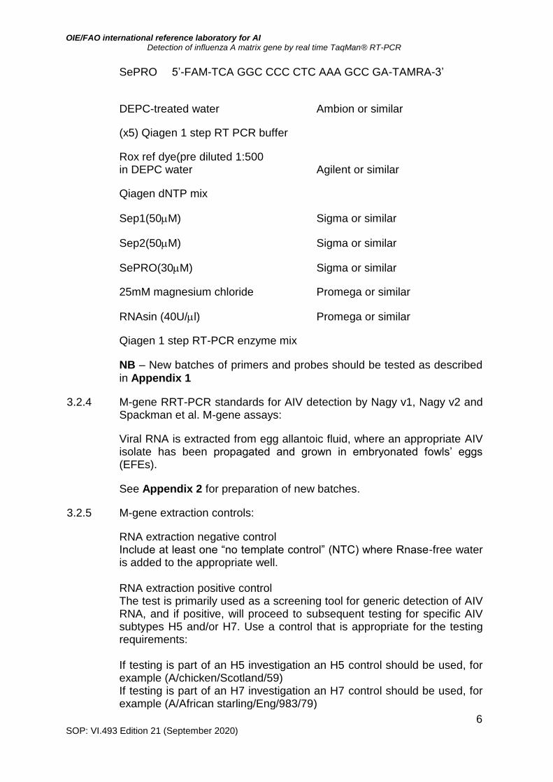
SePRO 5’-FAM-TCA GGC CCC CTC AAA GCC GA-TAMRA-3’
DEPC-treated water Ambion or similar
(x5) Qiagen 1 step RT PCR buffer
Rox ref dye(pre diluted 1:500 in DEPC water Agilent or similar
Qiagen dNTP mix
Sep1(50M) Sigma or similar
Sep2(50M) Sigma or similar
SePRO(30M) Sigma or similar
25mM magnesium chloride Promega or similar
RNAsin (40U/l) Promega or similar
Qiagen 1 step RT-PCR enzyme mix
NB – New batches of primers and probes should be tested as described
in Appendix 1
3.2.4 M-gene RRT-PCR standards for AIV detection by Nagy v1, Nagy v2 and Spackman et al. M-gene assays:
Viral RNA is extracted from egg allantoic fluid, where an appropriate AIV isolate has been propagated and grown in embryonated fowls’ eggs (EFEs).
See Appendix 2 for preparation of new batches.
3.2.5 M-gene extraction controls:
RNA extraction negative control Include at least one “no template control” (NTC) where Rnase-free water is added to the appropriate well. RNA extraction positive control The test is primarily used as a screening tool for generic detection of AIV RNA, and if positive, will proceed to subsequent testing for specific AIV subtypes H5 and/or H7. Use a control that is appropriate for the testing requirements: If testing is part of an H5 investigation an H5 control should be used, for example (A/chicken/Scotland/59) If testing is part of an H7 investigation an H7 control should be used, for example (A/African starling/Eng/983/79)
OIE/FAO international reference laboratory for AI Detection of influenza A matrix gene by real time TaqMan® RT-PCR
7 SOP: VI.493 Edition 21 (September 2020)
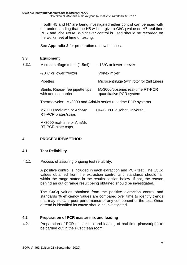
If both H5 and H7 are being investigated either control can be used with the understanding that the H5 will not give a Ct/Cq value on H7 real-time PCR and vice versa. Whichever control is used should be recorded on the worksheet at time of testing.
See Appendix 2 for preparation of new batches.
3.3 Equipment
3.3.1 Microcentrifuge tubes (1.5ml) -18C or lower freezer
-70C or lower freezer Vortex mixer
Pipettes Microcentrifuge (with rotor for 2ml tubes)
Sterile, Rnase-free pipette tips Mx3000/5pseries real-time RT-PCR with aerosol barrier quantitative PCR system
Thermocycler: Mx3000 and AriaMx series real-time PCR systems
Mx3000 real-time or AriaMx QIAGEN BioRobot Universal RT-PCR plates/strips
Mx3000 real-time or AriaMx RT-PCR plate caps
4 PROCEDURE/METHOD
4.1 Test Reliability
4.1.1 Process of assuring ongoing test reliability:
A positive control is included in each extraction and PCR test. The Ct/Cq values obtained from the extraction control and standards should fall within the range stated in the results section below. If not, the reason behind an out of range result being obtained should be investigated. The Ct/Cq values obtained from the positive extraction control and standards % efficiency values are compared over time to identify trends that may indicate poor performance of any component of the test. Once a trend is identified its cause should be investigated.
4.2 Preparation of PCR master mix and loading
4.2.1 Preparation of PCR master mix and loading of real-time plate/strip(s) to be carried out in the PCR clean room.
OIE/FAO international reference laboratory for AI Detection of influenza A matrix gene by real time TaqMan® RT-PCR
8 SOP: VI.493 Edition 21 (September 2020)
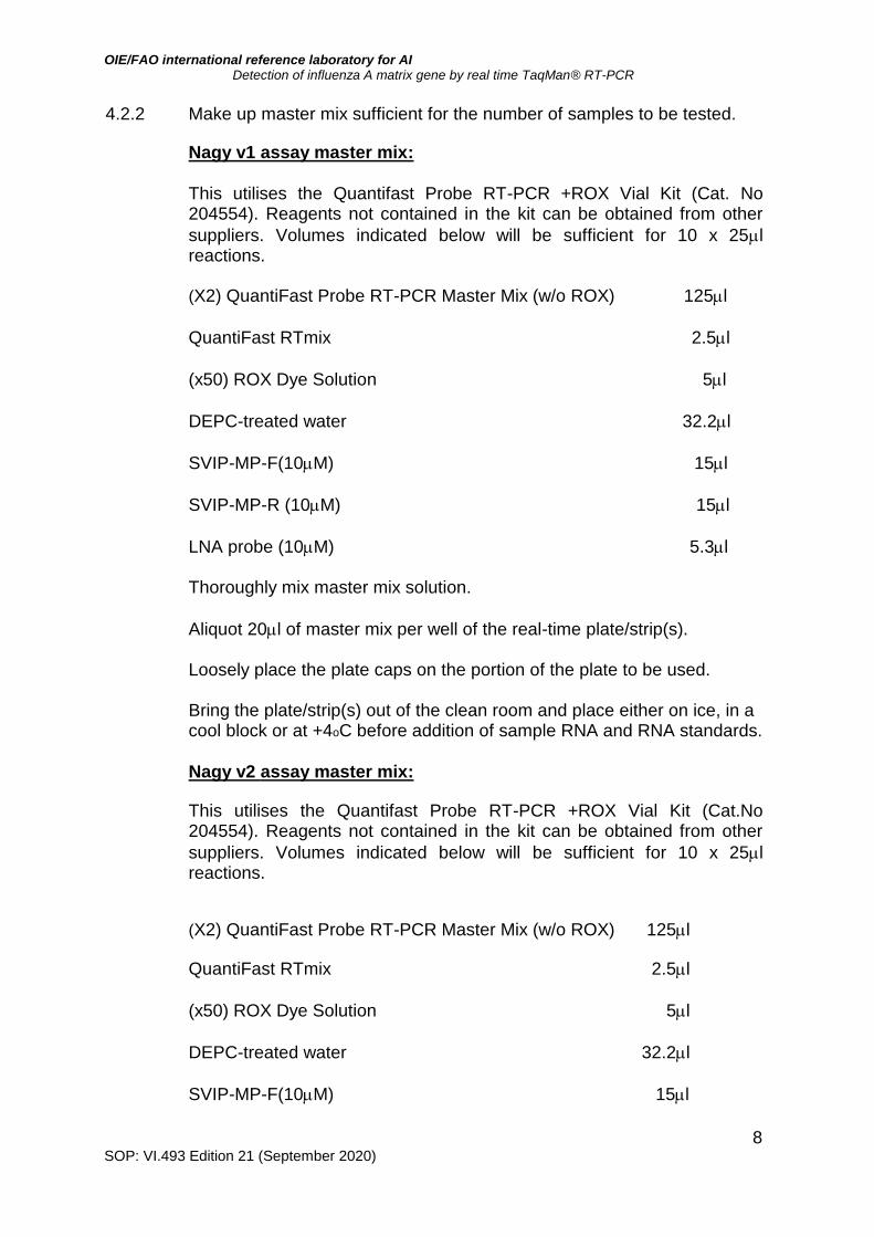
4.2.2 Make up master mix sufficient for the number of samples to be tested.
Nagy v1 assay master mix:
This utilises the Quantifast Probe RT-PCR +ROX Vial Kit (Cat. No 204554). Reagents not contained in the kit can be obtained from other
suppliers. Volumes indicated below will be sufficient for 10 x 25l reactions.
(X2) QuantiFast Probe RT-PCR Master Mix (w/o ROX) 125l
QuantiFast RTmix 2.5l
(x50) ROX Dye Solution 5l
DEPC-treated water 32.2l
SVIP-MP-F(10M) 15l
SVIP-MP-R (10M) 15l
LNA probe (10M) 5.3l Thoroughly mix master mix solution.
Aliquot 20l of master mix per well of the real-time plate/strip(s). Loosely place the plate caps on the portion of the plate to be used. Bring the plate/strip(s) out of the clean room and place either on ice, in a cool block or at +4oC before addition of sample RNA and RNA standards.
Nagy v2 assay master mix:
This utilises the Quantifast Probe RT-PCR +ROX Vial Kit (Cat.No 204554). Reagents not contained in the kit can be obtained from other
suppliers. Volumes indicated below will be sufficient for 10 x 25l reactions.
(X2) QuantiFast Probe RT-PCR Master Mix (w/o ROX) 125l
QuantiFast RTmix 2.5l
(x50) ROX Dye Solution 5l
DEPC-treated water 32.2l
SVIP-MP-F(10M) 15l
OIE/FAO international reference laboratory for AI Detection of influenza A matrix gene by real time TaqMan® RT-PCR
9 SOP: VI.493 Edition 21 (September 2020)
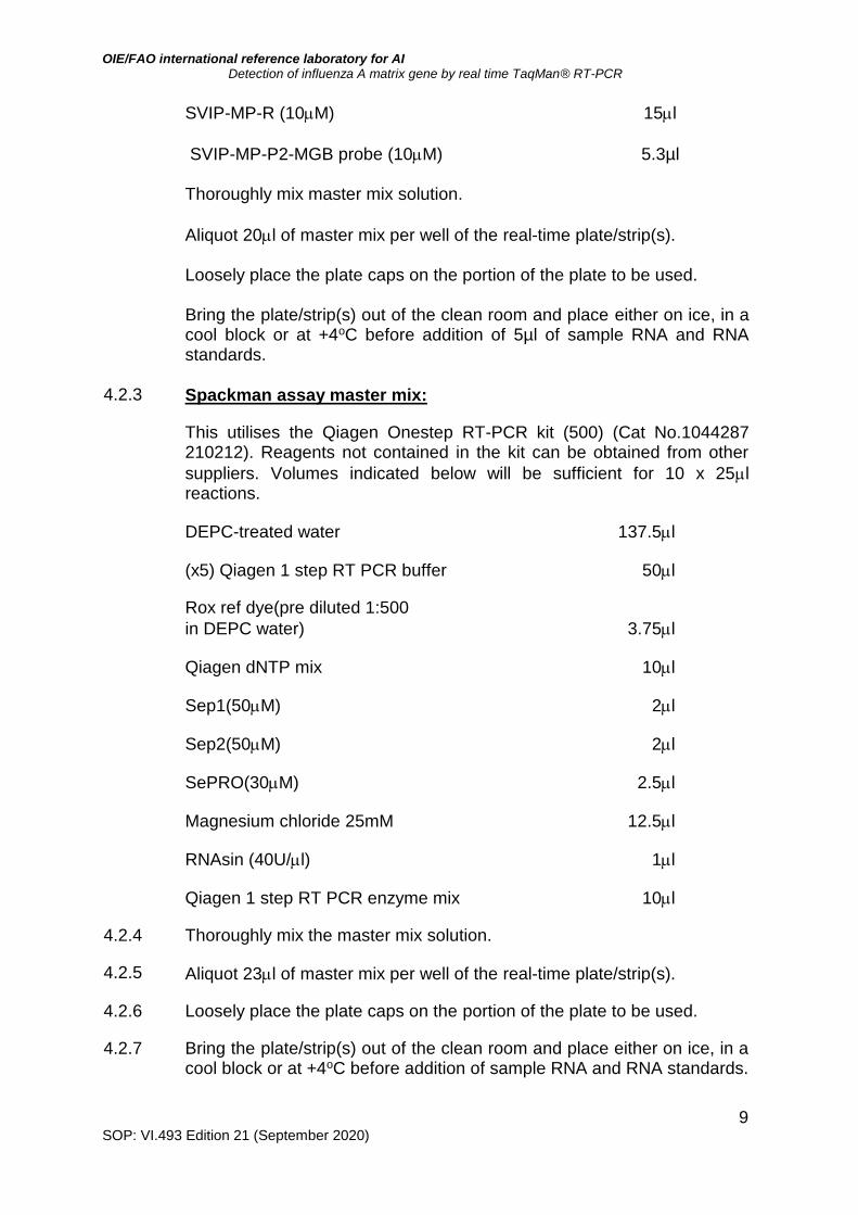
SVIP-MP-R (10M) 15l
SVIP-MP-P2-MGB probe (10M) 5.3µl 5.3l Thoroughly mix master mix solution.
Aliquot 20l of master mix per well of the real-time plate/strip(s). Loosely place the plate caps on the portion of the plate to be used. Bring the plate/strip(s) out of the clean room and place either on ice, in a cool block or at +4oC before addition of 5µl of sample RNA and RNA standards.
4.2.3 Spackman assay master mix:
This utilises the Qiagen Onestep RT-PCR kit (500) (Cat No.1044287 210212). Reagents not contained in the kit can be obtained from other
suppliers. Volumes indicated below will be sufficient for 10 x 25l reactions.
DEPC-treated water 137.5l
(x5) Qiagen 1 step RT PCR buffer 50l
Rox ref dye(pre diluted 1:500
in DEPC water) 3.75l
Qiagen dNTP mix 10l
Sep1(50M) 2l
Sep2(50M) 2l
SePRO(30M) 2.5l
Magnesium chloride 25mM 12.5l
RNAsin (40U/l) 1l
Qiagen 1 step RT PCR enzyme mix 10l
4.2.4 Thoroughly mix the master mix solution.
4.2.5 Aliquot 23l of master mix per well of the real-time plate/strip(s).
4.2.6 Loosely place the plate caps on the portion of the plate to be used.
4.2.7 Bring the plate/strip(s) out of the clean room and place either on ice, in a cool block or at +4oC before addition of sample RNA and RNA standards.
OIE/FAO international reference laboratory for AI Detection of influenza A matrix gene by real time TaqMan® RT-PCR
10 SOP: VI.493 Edition 21 (September 2020)
4.3 Preparation of AIV RNA standards
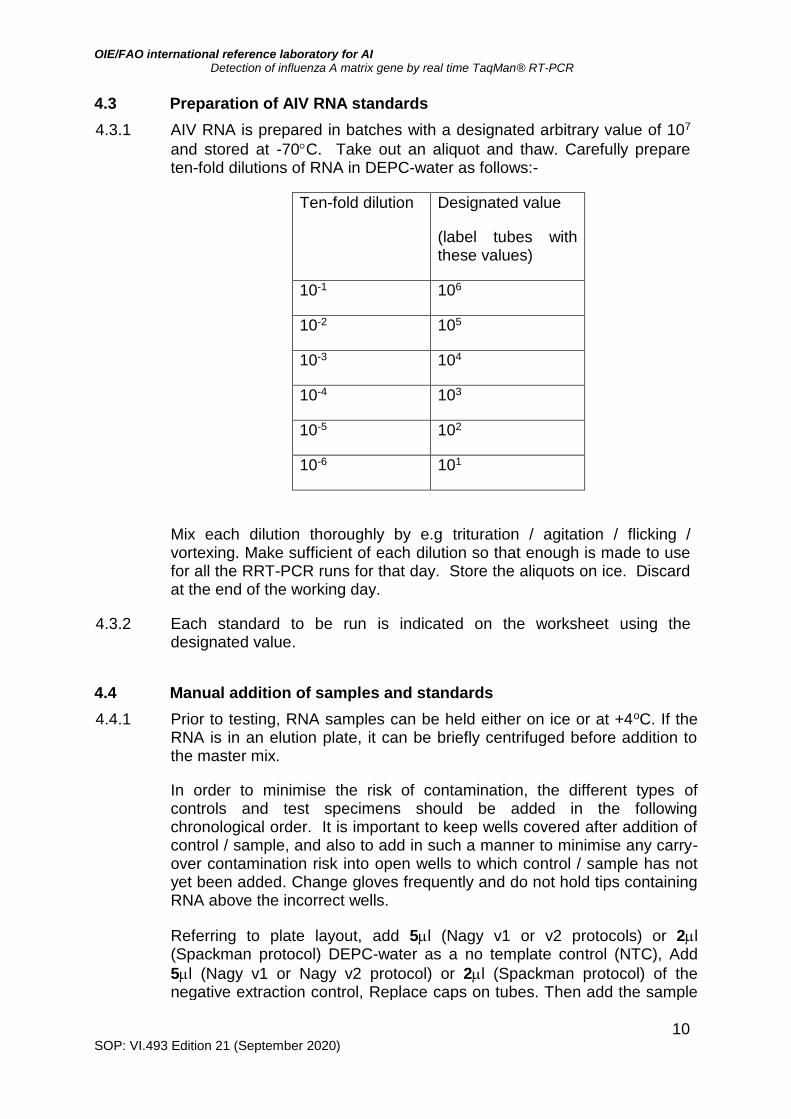
4.3.1 AIV RNA is prepared in batches with a designated arbitrary value of 107
and stored at -70C. Take out an aliquot and thaw. Carefully prepare ten-fold dilutions of RNA in DEPC-water as follows:-
Ten-fold dilution Designated value
(label tubes with these values)
10-1 106
10-2 105
10-3 104
10-4 103
10-5 102
10-6 101
Mix each dilution thoroughly by e.g trituration / agitation / flicking / vortexing. Make sufficient of each dilution so that enough is made to use for all the RRT-PCR runs for that day. Store the aliquots on ice. Discard at the end of the working day.
4.3.2 Each standard to be run is indicated on the worksheet using the designated value.
4.4 Manual addition of samples and standards
4.4.1 Prior to testing, RNA samples can be held either on ice or at +4oC. If the RNA is in an elution plate, it can be briefly centrifuged before addition to the master mix.
In order to minimise the risk of contamination, the different types of controls and test specimens should be added in the following chronological order. It is important to keep wells covered after addition of control / sample, and also to add in such a manner to minimise any carry-over contamination risk into open wells to which control / sample has not yet been added. Change gloves frequently and do not hold tips containing RNA above the incorrect wells.
Referring to plate layout, add 5l (Nagy v1 or v2 protocols) or 2l (Spackman protocol) DEPC-water as a no template control (NTC), Add
5l (Nagy v1 or Nagy v2 protocol) or 2l (Spackman protocol) of the negative extraction control, Replace caps on tubes. Then add the sample
OIE/FAO international reference laboratory for AI Detection of influenza A matrix gene by real time TaqMan® RT-PCR
11 SOP: VI.493 Edition 21 (September 2020)
RNA to the master mix. Replace caps on tubes.
4.4.2 Add the real time PCR RNA standards (from weakest to strongest) and positive RNA extraction control to appropriate wells based on your worksheet layout.
4.4.3 Once RNA is added, fit caps to all wells.
It is important that the caps are fitted firmly and correctly onto the wells before the plate is inserted into the real time machine.
4.4.4 Where necessary, briefly spin down plate/strip(s) contents in a centrifuge.
If the real time plate/strip(s) are not to be loaded onto the real-time machine straight away, keep the plate/strip(s) on ice until ready to test.
4.5 Reverse transcriptase and PCR
4.5.1 Place the real time plate/strip(s) in the appropriate real time machine. Following the plate layout, assign well type to each well. Unknown samples are designated as such. AIV RNA standards (10-fold dilution series) are designated as standards and the value entered corresponds to its designated value (see paragraph 4.3.1). The positive, negative and the no template controls are assigned FAM POS, FAM NEG and NTC, respectively.
4.5.2 Select the correct filters – ROX and FAM.
Set ROX as the reference dye
4.5.3 Select the correct thermal profile. The thermocycling profile and plate setup can be re-entered for each experiment or the thermal profile template imported from previous experiments if desired.
Nagy v1 protocol
RT step: 50C for 15 mins,
95C for 5 mins
PCR step (x 45 cycles): 95C for 10 secs
60C for 20 secs
72C for 10 secs
Collect fluorescence data during the 60C annealing step using the ROX and FAM filters and the endpoint read option with 1 read.
OIE/FAO international reference laboratory for AI Detection of influenza A matrix gene by real time TaqMan® RT-PCR
12 SOP: VI.493 Edition 21 (September 2020)
Nagy v2 protocol
RT step: 45C for 10 mins,
95C for 5 mins
PCR step (x 40 cycles): 95C for 10 secs
64C for 30 secs
72C for 10 secs
Collect fluorescence data during the 64C annealing step using the ROX and FAM filters and the endpoint read option with 1 read.
Spackman protocol
RT step: 50C for 30 mins,
95C for 15 mins
PCR step (x 40 cycles): 95C for 10 secs
60C for 20 secs
Collect fluorescence data during the 60C annealing step using the ROX and FAM filters and the endpoint read option with 1 read.
4.5.4 When the plate setup and thermoprofile windows have been entered, select run to start thermocycling. Then click ‘start run’ in the box in the bottom right-hand corner of the screen.
A storage window will automatically open. Save the file in an appropriate folder on the shared drive of the computer.
5 RESULTS
5.1 Analysis and display of results
5.1.1 The fluorescence data during the PCR reaction by using the raw data plots tab in the run section.
5.1.2 The filter set gain setting multiplies up the amount of raw fluorescence
seen by the machine. If the raw fluorescence is too high then the
accuracy of the readings is reduced. Raw fluorescence (“R”, see 5.1.6
below) above 35,000 relative fluorescence units (RFU) in the exponential
phase is too high. The baseline signal for all the amplification plots must
also be above 3000 RFU.
5.1.3 To analyse the data select the Analysis section button and select the
OIE/FAO international reference laboratory for AI Detection of influenza A matrix gene by real time TaqMan® RT-PCR
13 SOP: VI.493 Edition 21 (September 2020)
wells to be examined in the Analysis/Setup window.
5.1.4 To view the results click on the Results tab in the Analysis section and view the amplification plots. There are four options for analysing the fluorescence:
R (Multicomponent view) displays the raw fluorescence in arbitrary units.
dR displays the baseline-corrected fluorescence. As all reactions and wells will start with a slightly different fluorescence reading this option sets a baseline value of 0 to all plots. This correction is determined by the fluorescence values obtained during the initial rounds of the PCR. The adaptive baseline algorithm calculates the best baseline for each plot individually.
Rn displays the fluorescence normalised to the passive reference dye (ROX). This allows for fluctuations in fluorescence, which are not due to cleavage of the Taqman probe.
dRn displays the baseline-corrected normalised fluorescence.
The dRn option with the ROX channel switched on is the most appropriate option for analysis of the data.
5.2 Interpretation of results
5.2.1 Analyse the data by comparing the results obtained for the positive AIV RNA standards and NTC.
5.2.2 Construct a standard curve with the positive standards Cq values, click on the corresponding radio button at the bottom of the page (above figure). This should include a minimum of three standards on the straight line with respect to providing qualitative data e.g. flock level diagnostic results, namely infected / uninfected. If individual bird level results are required the minimum should be four standards on the straight line.
The curve should comply with the following criteria:
Efficiency: >80% and <120%
Slope between –3.1 and –3.8
R2 value of the straight line: >0.980
5.2.3 Check the Ct/Cq values for any RNA extraction controls. In order for the test run to be valid, the extraction controls must amplify at 30 +/- 3 Ct/Cq values. Greater deviations from the predetermined Ct/Cq value suggest
OIE/FAO international reference laboratory for AI Detection of influenza A matrix gene by real time TaqMan® RT-PCR
14 SOP: VI.493 Edition 21 (September 2020)
that:
The M-gene positive control may have degraded (if Ct/Cq value greater than predetermined value).
Software settings on the instrument are inaccurate / inappropriate where the fluorescence threshold cut-off (horizontal blue line) may have been set by the software to an excessively high / low level & is affecting the Ct/Cq value of the predetermined M gene positive control. It may be possible to reset the software after the experiment to restore the expected Ct value provided the other controls are in order. Such a reset can accept deviations of up to + / - 3 Ct/Cqs.
5.2.4 If all controls are within acceptable limits analyse the data for the test samples. Record the standard efficiency and positive control values in the relevant log book.
5.2.5 An increase in fluorescence will be observed at an early stage for positive samples. The NTC and negative samples should not result in an increase in fluorescence above the baseline.
5.2.6 Criteria for assessing whether a sample is positive or negative for either
assay are as follows:
Ct/Cq value <30 - samples are M-gene strong positive. Ct/Cq value 30-36 with sigmoidal / logarithmic appearance – samples are weak positive. Ct/Cq value >36 samples are negative if the amplification plot has a linear shape. These plots may represent spurious probe degradation or non-specific fluorescence.
5.2.7 Any diagnostic samples yielding a Ct/Cq value of 35-37 are re-extracted from the original sample and re-tested on the M-gene RRT-PCR using either assay together with the original extract of RNA from the sample. Any subsequent positives are further tested by H5 and H7 RRT-PCR, and the original sample subjected to virus isolation.
6 REFERENCES
EU (European Union) (2006). Diagnostic manual for avian influenza [Internet]. Official Journal of European Union L237/1 (31Aug). Available at:
https://eur-lex.europa.eu/legal-content/EN/TXT/?uri=CELEX%3A32006D0437
OIE/FAO international reference laboratory for AI Detection of influenza A matrix gene by real time TaqMan® RT-PCR
15 SOP: VI.493 Edition 21 (September 2020)
Lee, C.-W. and Suarez, D.L. (2004). Application of real time RT-PCR for the quantification and competitive replication study of H5 and H7 subtype avian influenza virus. J. Viro. Methods. 119, 151-158.
Nagy, A., Vostinakova, V., Pirchanova, Z., Cernikova,L., Dirbakova,
Z., Mojzis, M., Jirinkova, H., Havlikova, M., Dan, A., Ursu, K., Vilcek,
S. and Hornickova, J. (2010). Development and evaluation of a one-step real-time RT-PCR assay for universal detection of influenza A viruses from avian and mammal species. Arch. Virol. 155: 665-673.
Nagy, A., Černíková, L., Kunteová, K., Dirbáková, Z., Thomas, S.S.,
Slomka, M.J., Dán, A., Varga, T., Máté, M., Jiřincová, H. and Brown,
I.H. (2020). A universal RT-qPCR assay for “One Health” detection of influenza A viruses. Submitted to PLoS One (29/6/20), also available at bioRxiv: https://www.biorxiv.org/content/10.1101/2020.06.29.171306v1
Slomka M.J., Coward V.J., Banks J., Löndt B.Z., Brown I.H.,
Voermans J., Koch G., Handberg K.J., Jørgensen P.H., Cherbonnel-
Pansart M., Jestin V., Cattoli G., Capua I., Ejdersund A., Thorén P.
and Czifra G. (2007). Identification of sensitive and specific avian influenza polymerase chain reaction methods through blind ring trials organized in the European Union. Avian Diseases, 51: 227-234.
Slomka M.J., To T.L., Tong H.H., Coward V.J., Hanna A., Shell W.,
Pavlidis T., Densham A.L.E., Kargiolakis G., Arnold M.E., Banks J.
and Brown I.H. (2012). Challenges for accurate and prompt molecular diagnosis of clades of highly pathogenic avian influenza H5N1 viruses emerging in Vietnam. Avian Pathology, 41: 177-193
Spackman, E., Senne, D.A., Myers, T.J., Bulaga, L.L., Garber, L.P.,
Perdue, M.L., Lohman, K., Daum, L.T. and Suarez, D.L. (2002). Development of a real time reverse transcriptase PCR assay for type A influenza virus and the avian H5 and H7 haemagglutination subtypes. J. Clin. Microbiol. 40, 3256-3260.

OIE/FAO international reference laboratory for AI Detection of influenza A matrix gene by real time TaqMan® RT-PCR
16 SOP: VI.493 Edition 21 (September 2020)
Appendix 1
Testing new batches of reagents New batches of probe and primers should be tested when they are first re-suspended. Probe and primers should be aliquoted once re-suspended to avoid repeated freeze thawing. Test new reagents in the same real-time PCR as the current reagents using the positive standards and NTC according to this SOP. If the new reagents are fit for purpose both sets of reagents should give equivalent results.

OIE/FAO international reference laboratory for AI Detection of influenza A matrix gene by real time TaqMan® RT-PCR
17 SOP: VI.493 Edition 21 (September 2020)
Appendix 2
Preparation of TaqMan® real-time RT-PCR positive extraction controls and
RNA standards These can be prepared from either live or inactivated egg-grown antigen and can be supplied either as an aliquot of freeze-dried material or an aliquot from a wet ‘antigen stock.
NB - For live antigen follow safety procedures in place for the virus type being
produced. Work at the appropriate containment level for the virus being used
– refer to the virus categorisation risk document
To prepare positive extraction controls
1. If the antigen is supplied freeze-dried, reconstitute the vial with 1ml molecular grade water. Ensure the solution is mixed thoroughly by gentle vortexing.
2. Prepare a 10-fold dilution series of the antigen in PBS e.g. from 10-1 - 10-6
dilutions inclusive.
3. Freeze 200 µl aliquots of each dilution at -70 ºC.
4. Thaw-out an aliquot of each dilution and extract RNA from each aliquot either manually or on the BioRobot according to procedure RNA extraction from biological samples or extraction of nucleic acids from swabs using the Qiagen BioRobot Universal.
5. Test the RNA from each member of the dilution series by the appropriate real-time PCR assay. Choose the optimal 10-fold dilution from the resulting Ct/Cq values. The acceptable Ct/Cq value range is documented in the relevant SOP for the real-time PCR assay used.
6. Prepare the chosen dilution of the antigen in bulk using PBS, assign a batch number, dispense into 200µl aliquots, label and freeze at -70 ºC.
7. Remove an aliquot from the freezer, extract the RNA and test by the appropriate RRT-PCR to confirm the Ct values from the dilution fall within the required range.
To prepare RNA PCR standards
1. If the antigen is supplied freeze–dried, reconstitute the vial with 1ml molecular grade water. Ensure the solution is mixed thoroughly by gentle vortexing.
2. Manually extract the RNA from the whole reconstituted antigen from 1ml of wet stock according to the SOP VIR.0165, RNA extraction from biological samples.
3. Pool together the RNA extractions. 4. Freeze a 5µl aliquot at-70 ºC 5. Thaw this aliquot and prepare a 10-fold dilution series in molecular grade
water from as described above from 10-1 - 10-7 6. Test the dilution series by the appropriate RRT-PCR. Test the current ‘in-use’
RNA standard batch in parallel on the same PCR test run.
OIE/FAO international reference laboratory for AI Detection of influenza A matrix gene by real time TaqMan® RT-PCR
18 SOP: VI.493 Edition 21 (September 2020)
7. Directly compare the results of the two batches. The Ct/Cq values and test efficiencies should be similar i.e. Ct/Cq values +/- 2.0 per corresponding dilution. If there is an apparent significant difference, adjust the dilution of the new batch of RNA (eg. make a 1/10 dilution) and repeat stages 3-7 above.
8. Once the required dilution is achieved, prepare 5µl aliquots, assign a batch number, label the tubes and store at -70 ºC.