
23 Spleen - TaiLieu.VN
585
23 Spleen Timothy M. Nywening Maria B. Doyle A. Anatomy. The spleen is derived from the mesoderm and resides in the left upper quadrant of the abdomen, where it is protected by the ninth to eleventh ribs. The average adult spleen is 12 cm long × 7 cm wide × 4 cm thick and weighs between 1,000 and 1,500 g. The spleen is highly vascularized, receiving up to 5% of cardiac output. The splenic artery, a branch of the celiac axis, runs posterior to the pancreas and most commonly arborizes into multiple small arteries to enter the hilum of the spleen. The inferior mesenteric vein drains into the splenic vein, which ultimately joins with the superior mesenteric vein to form the portal vein. Accessory spleens are found in 10% to 20% of the population and can be located anywhere in the abdomen but are most commonly found in the splenic hilum (Fig. 23-1). B. Function. Histology of the spleen reveals highly vascularized red pulp interspersed with areas of white pulp. Red pulp consists of branching, thin walled sinuses and splenic cords filled with red blood cells (erythrocytes) and phagocytic cells. White pulp consists of T-cell rich periarteriolar sheaths, B-cell containing lymphoid nodules, and the marginal zone that serves as an interface between the lymphoid-dominant white pulp and erythrocyte-rich red pulp. These two histologies constitute the two major functions of the spleen: 1. Reticuloendothelial system: The red pulp serves to cull senescent erythrocytes and remodel healthy red cells. The spleen also serves as a reservoir for platelets. While extramedullary hematopoiesis uncommon in adults, the spleen may be a site of erythrocyte production in some disease states (i.e., myelofibrosis). 2. Immune system: The spleen is involved in both the innate (opsonization) and adaptive (antigen presentation) immune system. Opsonization of pathogens by the complement system results in enhanced phagocytosis and clearance in the spleen. The white pulp also acts as a site of antigen presentation to lymphocytes that, along with an appropriate cytokine milieu, leads to effective T-cell mediated cytotoxic activity and B-cell antibody responses. C. Indications for Splenectomy (Table 23-1) 1. Hematologic conditions a. Thrombocytopenias (1) Idiopathic Immune Thrombocytopenic Purpura (ITP) is the most common indication > Table of Contents > 23 - Spleen
-
Upload
khangminh22 -
Category
Documents
-
view
3 -
download
0
Transcript of 23 Spleen - TaiLieu.VN
23Spleen
Timothy M. Nywening
Maria B. Doyle
A. Anatomy. The spleen is derived from the mesoderm and resides in the left upper quadrant ofthe abdomen, where it is protected by the ninth to eleventh ribs. The average adult spleen is 12cm long × 7 cm wide × 4 cm thick and weighs between 1,000 and 1,500 g. The spleen is highlyvascularized, receiving up to 5% of cardiac output. The splenic artery, a branch of the celiac axis,runs posterior to the pancreas and most commonly arborizes into multiple small arteries to enterthe hilum of the spleen. The inferior mesenteric vein drains into the splenic vein, which ultimatelyjoins with the superior mesenteric vein to form the portal vein. Accessory spleens are found in10% to 20% of the population and can be located anywhere in the abdomen but are mostcommonly found in the splenic hilum (Fig. 23-1).
B. Function. Histology of the spleen reveals highly vascularized red pulp interspersed with areasof white pulp. Red pulp consists of branching, thin walled sinuses and splenic cords filled with redblood cells (erythrocytes) and phagocytic cells. White pulp consists of T-cell rich periarteriolarsheaths, B-cell containing lymphoid nodules, and the marginal zone that serves as an interfacebetween the lymphoid-dominant white pulp and erythrocyte-rich red pulp. These two histologiesconstitute the two major functions of the spleen:
1. Reticuloendothelial system: The red pulp serves to cull senescent erythrocytes andremodel healthy red cells. The spleen also serves as a reservoir for platelets. Whileextramedullary hematopoiesis uncommon in adults, the spleen may be a site of erythrocyteproduction in some disease states (i.e., myelofibrosis).
2. Immune system: The spleen is involved in both the innate (opsonization) and adaptive(antigen presentation) immune system. Opsonization of pathogens by the complement systemresults in enhanced phagocytosis and clearance in the spleen. The white pulp also acts as a siteof antigen presentation to lymphocytes that, along with an appropriate cytokine milieu, leads toeffective T-cell mediated cytotoxic activity and B-cell antibody responses.
C. Indications for Splenectomy (Table 23-1)
1. Hematologic conditions
a. Thrombocytopenias
(1) Idiopathic Immune Thrombocytopenic Purpura (ITP) is the most common indication
> Table of Contents > 23 - Spleen
P.408
for elective splenectomy. It is an acquired disease that results from autoantibodies to plateletglycoprotein and results in immune mediated thrombocytopenia. The spleen
is both the major site of production of these antibodies as well as the principal site of plateletdestruction.
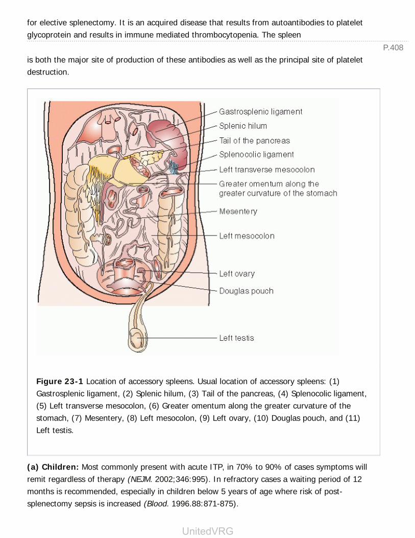
Figure 23-1 Location of accessory spleens. Usual location of accessory spleens: (1)Gastrosplenic ligament, (2) Splenic hilum, (3) Tail of the pancreas, (4) Splenocolic ligament,(5) Left transverse mesocolon, (6) Greater omentum along the greater curvature of thestomach, (7) Mesentery, (8) Left mesocolon, (9) Left ovary, (10) Douglas pouch, and (11)Left testis.
(a) Children: Most commonly present with acute ITP, in 70% to 90% of cases symptoms willremit regardless of therapy (NEJM. 2002;346:995). In refractory cases a waiting period of 12months is recommended, especially in children below 5 years of age where risk of post-splenectomy sepsis is increased (Blood. 1996.88:871-875).
UnitedVRG
P.409
(b) Adults: Usually present with chronic ITP. First-line treatment with steroids results in a 50%to 75% response rate and may be combined with other modalities such as intravenous immuneglobulin (IVIG) and/or anti-Rh(D) infusions. However, 80% will have recurrence after cessation oftherapy. Splenectomy results in 65% long-term remission (>5 years) and remains the treatment
of choice in patients with platelets less the 30,000/mm3 or with a high risk of bleeding. Mostpatients will achieve a response to splenectomy within
10 days postoperatively (Am J Surg. 2004;187:720-723). Alternatives to splenectomy includeRituximab (anti-CD20 monoclonal antibody) and thrombopoietin receptor antagonists which haveshown efficacy as second-line agents (Blood. 2012;120:960-969). Rituximab has also been shownto have some efficacy in patient failing to respond to splenectomy (Am J Hematology.2005;78:275-280) (Fig. 23-2).
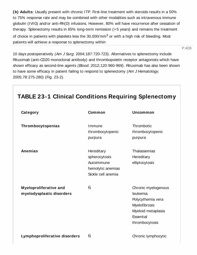
TABLE 23-1 Clinical Conditions Requiring Splenectomy
Category Common Uncommon
Thrombocytopenias Immunethrombocytopenicpurpura
Thromboticthrombocytopenicpurpura
Anemias HereditaryspherocytosisAutoimmunehemolytic anemiasSickle cell anemia
ThalassemiasHereditaryelliptocytosis
Myeloproliferative andmyelodysplastic disorders
Ñ Chronic myelogenousleukemiaPolycythemia veraMyelofibrosisMyeloid metaplasiaEssentialthrombocytosis
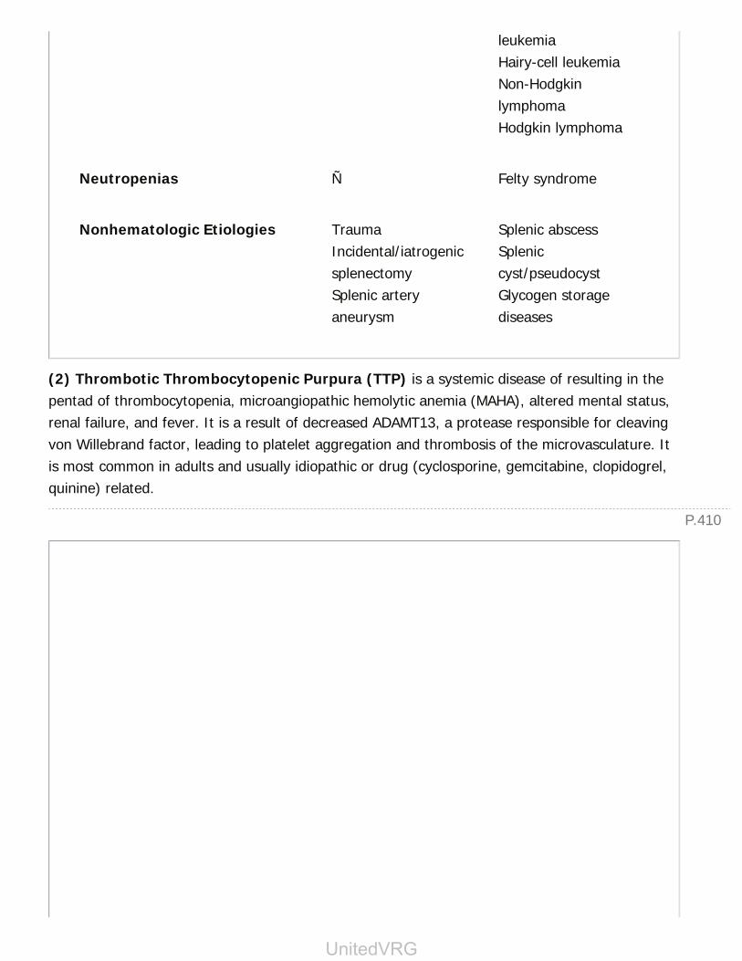
Lymphoproliferative disorders Ñ Chronic lymphocytic
P.410
leukemiaHairy-cell leukemiaNon-HodgkinlymphomaHodgkin lymphoma
Neutropenias Ñ Felty syndrome
Nonhematologic Etiologies TraumaIncidental/iatrogenicsplenectomySplenic arteryaneurysm
Splenic abscessSpleniccyst/pseudocystGlycogen storagediseases
(2) Thrombotic Thrombocytopenic Purpura (TTP) is a systemic disease of resulting in thepentad of thrombocytopenia, microangiopathic hemolytic anemia (MAHA), altered mental status,renal failure, and fever. It is a result of decreased ADAMT13, a protease responsible for cleavingvon Willebrand factor, leading to platelet aggregation and thrombosis of the microvasculature. Itis most common in adults and usually idiopathic or drug (cyclosporine, gemcitabine, clopidogrel,quinine) related.
UnitedVRG
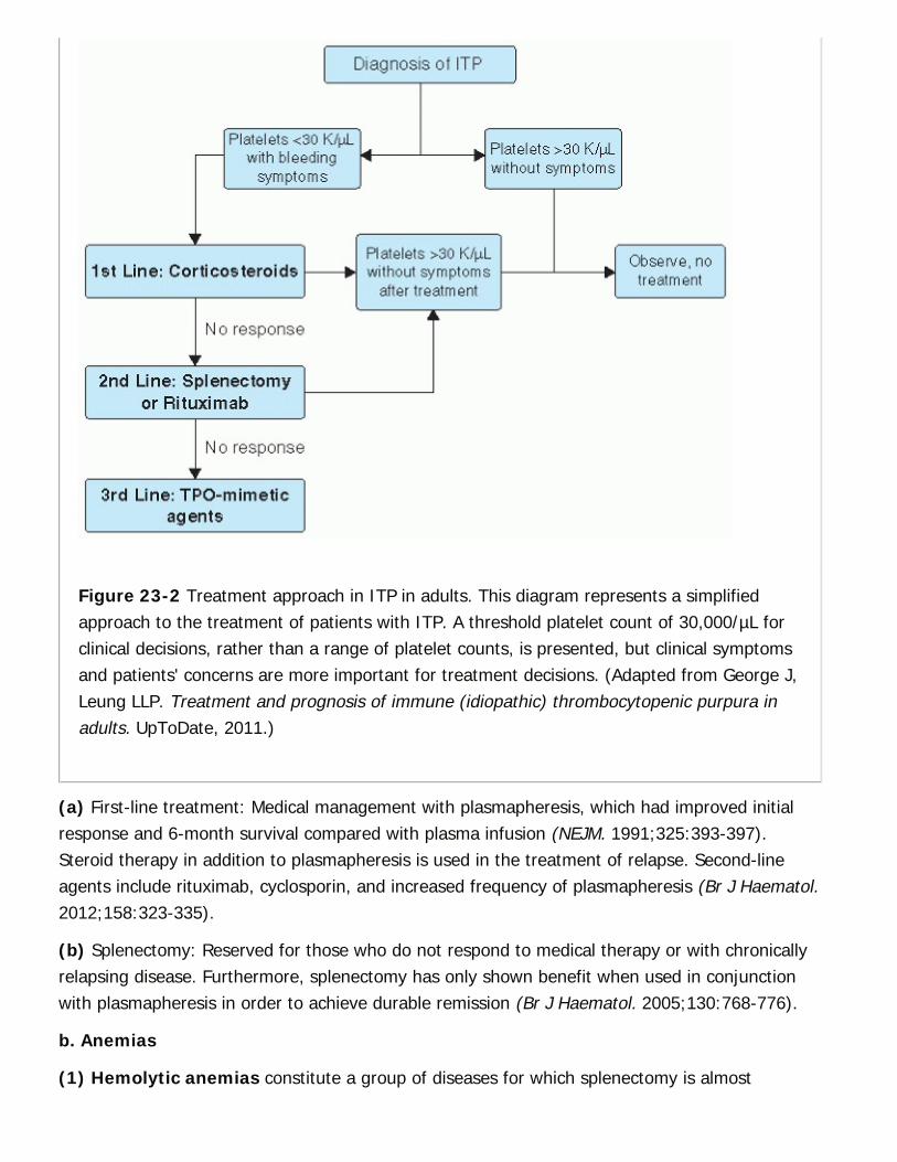
Figure 23-2 Treatment approach in ITP in adults. This diagram represents a simplifiedapproach to the treatment of patients with ITP. A threshold platelet count of 30,000/µL forclinical decisions, rather than a range of platelet counts, is presented, but clinical symptomsand patients' concerns are more important for treatment decisions. (Adapted from George J,Leung LLP. Treatment and prognosis of immune (idiopathic) thrombocytopenic purpura inadults. UpToDate, 2011.)
(a) First-line treatment: Medical management with plasmapheresis, which had improved initialresponse and 6-month survival compared with plasma infusion (NEJM. 1991;325:393-397).Steroid therapy in addition to plasmapheresis is used in the treatment of relapse. Second-lineagents include rituximab, cyclosporin, and increased frequency of plasmapheresis (Br J Haematol.2012;158:323-335).
(b) Splenectomy: Reserved for those who do not respond to medical therapy or with chronicallyrelapsing disease. Furthermore, splenectomy has only shown benefit when used in conjunctionwith plasmapheresis in order to achieve durable remission (Br J Haematol. 2005;130:768-776).
b. Anemias
(1) Hemolytic anemias constitute a group of diseases for which splenectomy is almost

P.411
universally curative.
(a) Hereditary spherocytosis is an autosomal dominant disorder characterized by a defect inan RBC membrane protein. The most common mutation is in the protein spectrin, but
other mutations in ankyrin, band 3, and palladin have been found. This defect results in small,spherical, rigid erythrocytes that fail to deform adequately to transverse the splenicmicrocirculation. This ultimately leads to the sequestration and destruction of erythrocytes in thespleen. Symptoms include anemia, jaundice (indirect bilirubinemia), and pigmented gallstones.Diagnosis is confirmed by the presence of spherocytes on peripheral blood smear, + osmoticfragility test, and decreased eosin-5-maleimide (EMA) binding (Blood Rev. 2013;27:167-178).Treatment includes folate supplementation and splenectomy for moderate to severe cases.
(b) Hereditary elliptocytosis is an autosomal dominant disorder in which an RBC cytoskeletalprotein defect results in elliptical shaped erythrocytes. Most patients are asymptomatic with amild anemia and do not require additional treatment. For select patients with symptomaticanemia splenectomy is usually curative.
(2) Acquired autoimmune hemolytic anemias
(a) Warm autoimmune hemolytic anemia occurs when IgG autoantibodies interact optimallywith antigens at 37¡C. Diagnosis is confirmed with a positive direct Coombs test (incubation withanti-IgG serum results in RBC agglutination). Etiology is most often idiopathic but may alsoinclude chronic lymphocytic leukemia (CLL), non-Hodgkin lymphoma, collagen vascular disease,and drugs. Splenectomy is reserved for nonresponders or those requiring high steroid doses andis 60% to 70% effective in achieving remission. Rituximab has also shown efficacy and is suitablesecond-line treatment for those patients who do not desire to undergo splenectomy (Blood.2010;116:1831-1838).
(b) Cold autoimmune hemolytic anemias are mediated by C3 complement fixation to IgMautoantibodies resulting in hemolysis at temperatures approaching 0¡C. Features include Reynaudlike symptoms along with anemia. Most cases respond to protective clothing; however severeepisodes may require cyclophosphamide, rituximab, or interferon. Splenectomy does not play arole in the treatment of cold autoimmune hemolytic anemias.
c. Congenital hemoglobinopathies
(1) Sickle cell anemia is a result of homozygous inheritance of the S variant of the hemoglobinbeta chain. Autosplenectomy usually occurs secondary to repeated vaso-occlusive events andsplenectomy is rarely required. However, splenectomy may be reasonable for selected patientswith splenic abscess, symptomatic splenomegaly, hypersplenism, or acute splenic sequestrationcrisis.
(2) Thalassemias are hereditary anemias that result from a defect in hemoglobin synthesis. β-thalassemia major is typically treated
UnitedVRG
P.412
P.413
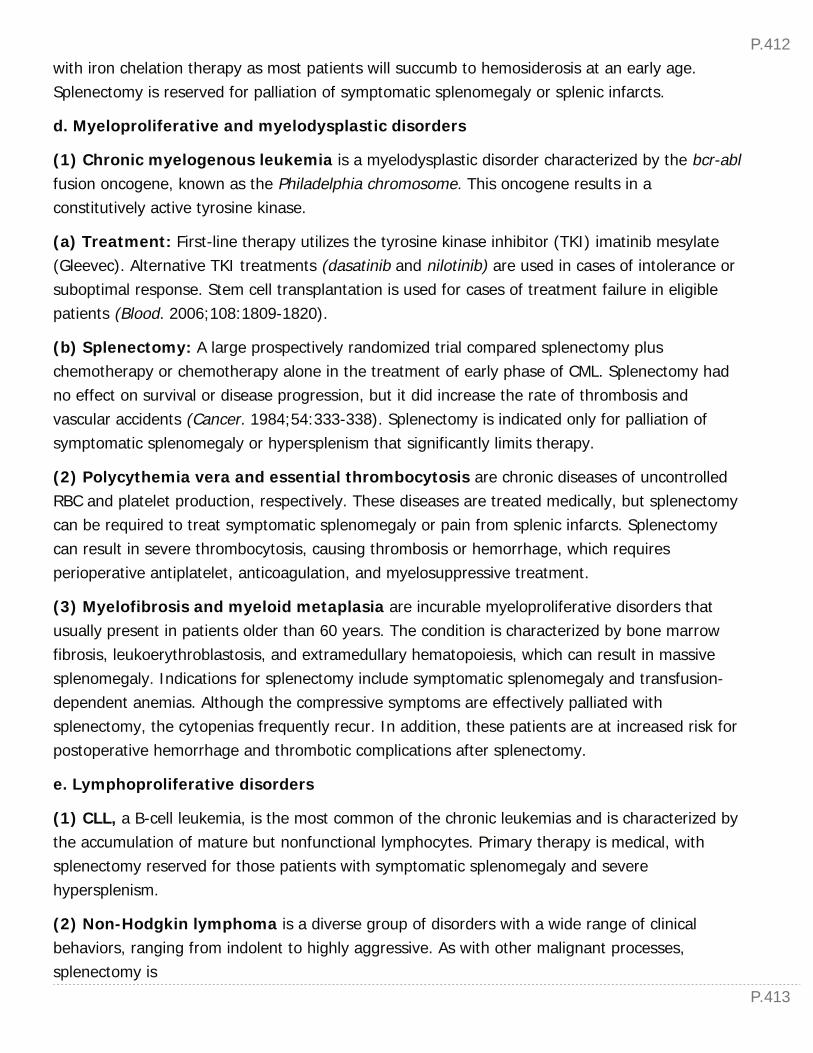
with iron chelation therapy as most patients will succumb to hemosiderosis at an early age.Splenectomy is reserved for palliation of symptomatic splenomegaly or splenic infarcts.
d. Myeloproliferative and myelodysplastic disorders
(1) Chronic myelogenous leukemia is a myelodysplastic disorder characterized by the bcr-ablfusion oncogene, known as the Philadelphia chromosome. This oncogene results in aconstitutively active tyrosine kinase.
(a) Treatment: First-line therapy utilizes the tyrosine kinase inhibitor (TKI) imatinib mesylate(Gleevec). Alternative TKI treatments (dasatinib and nilotinib) are used in cases of intolerance orsuboptimal response. Stem cell transplantation is used for cases of treatment failure in eligiblepatients (Blood. 2006;108:1809-1820).
(b) Splenectomy: A large prospectively randomized trial compared splenectomy pluschemotherapy or chemotherapy alone in the treatment of early phase of CML. Splenectomy hadno effect on survival or disease progression, but it did increase the rate of thrombosis andvascular accidents (Cancer. 1984;54:333-338). Splenectomy is indicated only for palliation ofsymptomatic splenomegaly or hypersplenism that significantly limits therapy.
(2) Polycythemia vera and essential thrombocytosis are chronic diseases of uncontrolledRBC and platelet production, respectively. These diseases are treated medically, but splenectomycan be required to treat symptomatic splenomegaly or pain from splenic infarcts. Splenectomycan result in severe thrombocytosis, causing thrombosis or hemorrhage, which requiresperioperative antiplatelet, anticoagulation, and myelosuppressive treatment.
(3) Myelofibrosis and myeloid metaplasia are incurable myeloproliferative disorders thatusually present in patients older than 60 years. The condition is characterized by bone marrowfibrosis, leukoerythroblastosis, and extramedullary hematopoiesis, which can result in massivesplenomegaly. Indications for splenectomy include symptomatic splenomegaly and transfusion-dependent anemias. Although the compressive symptoms are effectively palliated withsplenectomy, the cytopenias frequently recur. In addition, these patients are at increased risk forpostoperative hemorrhage and thrombotic complications after splenectomy.
e. Lymphoproliferative disorders
(1) CLL, a B-cell leukemia, is the most common of the chronic leukemias and is characterized bythe accumulation of mature but nonfunctional lymphocytes. Primary therapy is medical, withsplenectomy reserved for those patients with symptomatic splenomegaly and severehypersplenism.
(2) Non-Hodgkin lymphoma is a diverse group of disorders with a wide range of clinicalbehaviors, ranging from indolent to highly aggressive. As with other malignant processes,splenectomy is
P.414
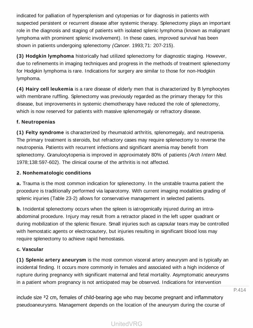
indicated for palliation of hypersplenism and cytopenias or for diagnosis in patients withsuspected persistent or recurrent disease after systemic therapy. Splenectomy plays an importantrole in the diagnosis and staging of patients with isolated splenic lymphoma (known as malignantlymphoma with prominent splenic involvement). In these cases, improved survival has beenshown in patients undergoing splenectomy (Cancer. 1993;71: 207-215).
(3) Hodgkin lymphoma historically had utilized splenectomy for diagnostic staging. However,due to refinements in imaging techniques and progress in the methods of treatment splenectomyfor Hodgkin lymphoma is rare. Indications for surgery are similar to those for non-Hodgkinlymphoma.
(4) Hairy cell leukemia is a rare disease of elderly men that is characterized by B lymphocyteswith membrane ruffling. Splenectomy was previously regarded as the primary therapy for thisdisease, but improvements in systemic chemotherapy have reduced the role of splenectomy,which is now reserved for patients with massive splenomegaly or refractory disease.
f. Neutropenias
(1) Felty syndrome is characterized by rheumatoid arthritis, splenomegaly, and neutropenia.The primary treatment is steroids, but refractory cases may require splenectomy to reverse theneutropenia. Patients with recurrent infections and significant anemia may benefit fromsplenectomy. Granulocytopenia is improved in approximately 80% of patients (Arch Intern Med.1978;138:597-602). The clinical course of the arthritis is not affected.
2. Nonhematologic conditions
a. Trauma is the most common indication for splenectomy. In the unstable trauma patient theprocedure is traditionally performed via laparotomy. With current imaging modalities grading ofsplenic injuries (Table 23-2) allows for conservative management in selected patients.
b. Incidental splenectomy occurs when the spleen is iatrogenically injured during an intra-abdominal procedure. Injury may result from a retractor placed in the left upper quadrant orduring mobilization of the splenic flexure. Small injuries such as capsular tears may be controlledwith hemostatic agents or electrocautery, but injuries resulting in significant blood loss mayrequire splenectomy to achieve rapid hemostasis.
c. Vascular
(1) Splenic artery aneurysm is the most common visceral artery aneurysm and is typically anincidental finding. It occurs more commonly in females and associated with a high incidence ofrupture during pregnancy with significant maternal and fetal mortality. Asymptomatic aneurysmsin a patient whom pregnancy is not anticipated may be observed. Indications for intervention
include size ≥2 cm, females of child-bearing age who may become pregnant and inflammatorypseudoaneurysms. Management depends on the location of the aneurysm during the course of
UnitedVRG

the splenic artery. Proximal and middle third aneurysms may be excluded by proximal and distalligation of the artery. Splenic perfusion persists via collateralization from the short gastric vessels.For more distal lesions proximal ligation with splenectomy is required. Alternatives treatmentsinclude endovascular approaches with transcatheter embolization.
TABLE 23-2 The American Association for the Surgeryof Trauma (AAST) Spleen Injury Scale (2008 Edition)
Grade InjuryType
Injury Description
I HematomaLaceration
Subcapsular: <10% surface areaCapsular: <1 cm parenchymal depth
II Hematoma Subcapsular: 10-50% surface areaIntraparenchymal: <5 cm in diameter
Laceration Parenchymal depth: 1-3 cmandNo trabecular vessel involvement
III Hematoma Subcapsular: >50% surface area or expanding/ruptured
Intraparenchymal hematoma: >5 cm or expanding/ruptured
Laceration Parenchymal depth: >3 cmorInvolving trabecular vessel
IV Laceration Laceration involving segmental or hilar vessels producingmajor devascularization (>25% of spleen)
V LacerationVascular
Shattered spleenHilar vascular injury

P.415
aAdvance one grade for multiple injuries up to grade III.Adapted from Tinkoff G, Esposito TJ, Reed J, et al. American Association for the Surgeryof Trauma Organ Injury Scale 1: Spleen, liver and kidney. J Trauma 2008;207(5):646-655.
d. Infectious
(1) Parasitic infections account for more than two-thirds of splenic cysts worldwide but arerare in the United States. The majority are hydatid cysts caused by Echinococcus species. Theyare typically asymptomatic but may rupture or cause symptoms due
to splenomegaly. The primary treatment is splenectomy, with careful attention not to spill thecyst contents. The cyst may be aspirated and injected with hypertonic saline prior to mobilizationif concern about rupture exists.
(2) Splenic abscesses are rare, but potentially lethal if not accurately diagnosed and timelytreatment instituted. Two-thirds arise from seeding of the spleen by a distant site, mostcommonly endocarditis and urinary tract infections. Abdominal CT and/or ultrasound imaging arethe diagnostic modalities of choice. CT images reveal a low intensity lesion that does not enhancewith contrast. Staphylococcus and streptococcus account for the most commonly identifiedorganisms, accounting for >50% of cases. Fungal infections are rare, and may resolve with anti-fungal treatment alone. Percutaneous drainage may be used in select cases; however,splenectomy and appropriate antibiotic therapy is definitive treatment.
e. Cystic lesions of the spleen may be either true cysts or pseudocysts, but this differentiation isdifficult to make preoperatively.
(1) True cysts (or primary cysts) have an epithelial lining and are most often congenital. Otherrare true cysts include epidermoid and dermoid cysts.
(2) Pseudocysts (or secondary cysts) lack an epithelial lining and make up more than two-thirdsof nonparasitic cysts. They typically result from traumatic hematoma formation and subsequentlyresorb.
(3) Treatment of splenic cysts depends on the size of the lesion and associated symptoms. Mostare typically asymptomatic, but they may present with left upper abdominal or shoulder pain.Those smaller than 5 cm can be followed with ultrasonography and often resolve spontaneously.Larger cysts risk rupture and require cyst unroofing or splenectomy. Percutaneous aspiration isassociated with infection and reaccumulation and is not indicated. Laparoscopic management ofsplenic cysts yields shorter hospital length of stay and fewer complications with no adverseeffects (Surg Endosc. 2007;21:206-208).
D. Preoperative Preparation
UnitedVRG
P.416
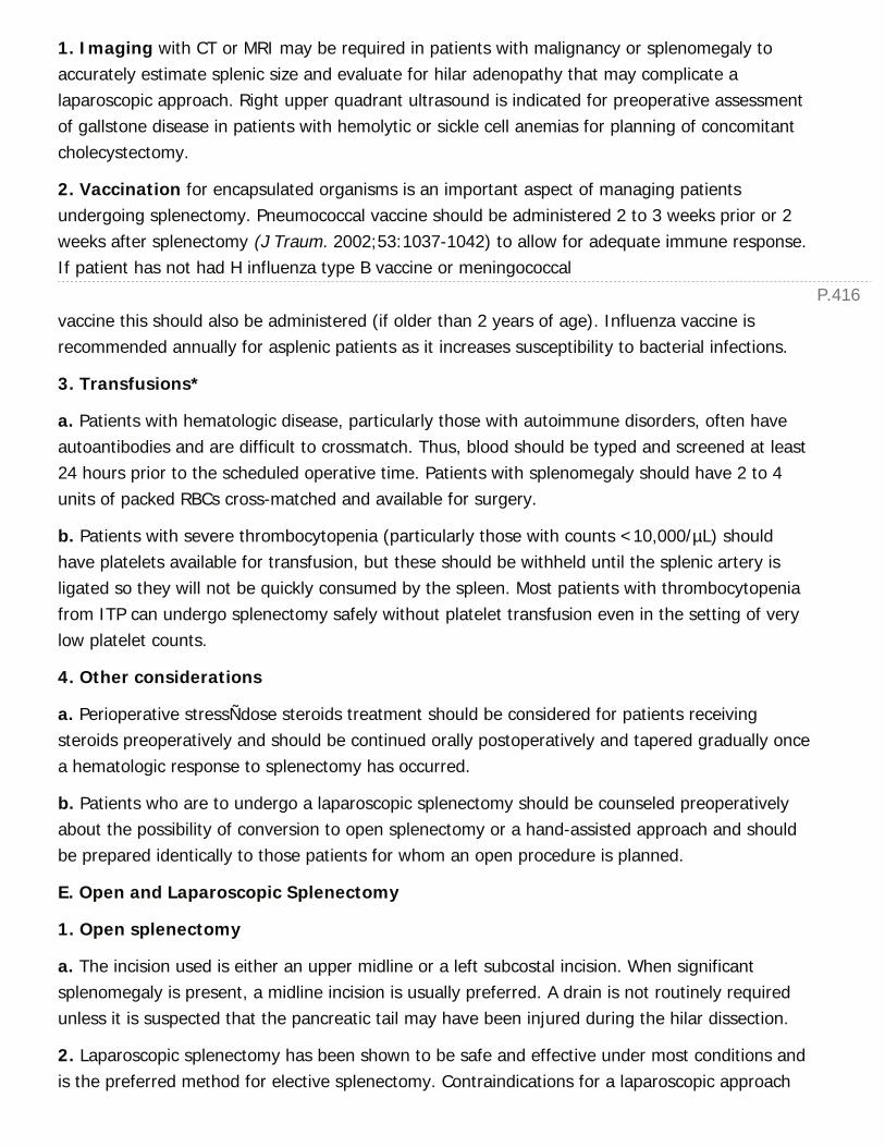
1. Imaging with CT or MRI may be required in patients with malignancy or splenomegaly toaccurately estimate splenic size and evaluate for hilar adenopathy that may complicate alaparoscopic approach. Right upper quadrant ultrasound is indicated for preoperative assessmentof gallstone disease in patients with hemolytic or sickle cell anemias for planning of concomitantcholecystectomy.
2. Vaccination for encapsulated organisms is an important aspect of managing patientsundergoing splenectomy. Pneumococcal vaccine should be administered 2 to 3 weeks prior or 2weeks after splenectomy (J Traum. 2002;53:1037-1042) to allow for adequate immune response.If patient has not had H influenza type B vaccine or meningococcal
vaccine this should also be administered (if older than 2 years of age). Influenza vaccine isrecommended annually for asplenic patients as it increases susceptibility to bacterial infections.
3. Transfusions*
a. Patients with hematologic disease, particularly those with autoimmune disorders, often haveautoantibodies and are difficult to crossmatch. Thus, blood should be typed and screened at least24 hours prior to the scheduled operative time. Patients with splenomegaly should have 2 to 4units of packed RBCs cross-matched and available for surgery.
b. Patients with severe thrombocytopenia (particularly those with counts <10,000/µL) shouldhave platelets available for transfusion, but these should be withheld until the splenic artery isligated so they will not be quickly consumed by the spleen. Most patients with thrombocytopeniafrom ITP can undergo splenectomy safely without platelet transfusion even in the setting of verylow platelet counts.
4. Other considerations
a. Perioperative stressÑdose steroids treatment should be considered for patients receivingsteroids preoperatively and should be continued orally postoperatively and tapered gradually oncea hematologic response to splenectomy has occurred.
b. Patients who are to undergo a laparoscopic splenectomy should be counseled preoperativelyabout the possibility of conversion to open splenectomy or a hand-assisted approach and shouldbe prepared identically to those patients for whom an open procedure is planned.
E. Open and Laparoscopic Splenectomy
1. Open splenectomy
a. The incision used is either an upper midline or a left subcostal incision. When significantsplenomegaly is present, a midline incision is usually preferred. A drain is not routinely requiredunless it is suspected that the pancreatic tail may have been injured during the hilar dissection.
2. Laparoscopic splenectomy has been shown to be safe and effective under most conditions andis the preferred method for elective splenectomy. Contraindications for a laparoscopic approach

P.417
are listed in Table 23-3.
a. Splenomegaly increases the complexity of the laparoscopic approach because of the difficultyof manipulating the organ atraumatically and achieving adequate exposure of the ligaments andhilum. Large spleens are also more difficult to place in an entrapment bag using a strictlylaparoscopic approach. Although the size limits for attempting laparoscopic or laparoscopic-assisted splenectomy are evolving, most moderately enlarged spleens (<1,000 g weight or 15 to20 cm in length) can be removed in a minimally invasive fashion, often without a hand-portdevice. For spleens larger than 20 cm in longitudinal length or those that weigh between 1,000and 3,000 g, the use of a hand port should be considered. The use of a hand port
in this setting has been associated with reduced operative times, less blood loss, and lower ratesof conversion to open operation (Arch Surg. 2006;141:755-761). In general, massivesplenomegaly (spleens greater than 30 cm in craniocaudal length and weighing >3,000 g) shouldbe approached in an open fashion because of the reduced working space and increased difficultlyin manipulating the spleen. A search for accessory splenic tissue should always be conducted,particularly if the patient has a hematologic indication for splenectomy.
TABLE 23-3 Contraindications for LaparoscopicSplenectomy
Absolute Contraindications Relative Contraindication
Massive splenomegaly (>30 cm) Moderate splenomegaly (20-25 cm)
Portal hypertension Severe, uncorrectable cytopenia
Splenic trauma (unstable patient) Splenic vein thrombosis
Ñ Splenic trauma (stable patient)
Ñ Bulky hilar adenopathy
Ñ Morbid obesity
b. Outcomes of laparoscopic splenectomy. Several large series of laparoscopic splenectomy have
UnitedVRG
P.418
been published with excellent results. In a meta-analysis of 51 reports including 2,940 patients,laparoscopic splenectomy was associated with significantly fewer complications overall, primarilyas a result of fewer wound and pulmonary complications (Surgery. 2003;134:647-653).
F. Complications
1. Intraoperative
a. Hemorrhage is the most common intraoperative complication of splenectomy, which can occurduring the hilar dissection or from a capsular tear during retraction. The incidence of thiscomplication is 2% to 3% during open splenectomy but is nearly 5% using the laparoscopicapproach. Bleeding during laparoscopic splenectomy may necessitate conversion to a hand-assisted or open procedure.
b. Pancreatic injury occurs in 0% to 6% of splenectomies, whether done open or laparoscopically.A retrospective review of one center's experience with laparoscopic splenectomy found pancreaticinjury in 16% of patients; half of these were isolated instances of hyperamylasemia (J Surg.1996;172(5):596-599). If one suspects that the pancreatic parenchyma has been violated duringlaparoscopic splenectomy, a closed suction drain should be placed adjacent to the
pancreas, and a drain amylase obtained prior to removal after the patient is eating a regular diet.
c. Bowel injury
(1) Colonic injuries are rare but because of the close proximity of the splenic flexure to the lowerpole of the spleen, it is possible to injure the colon during mobilization. Mechanical bowelpreparation is not indicated preoperatively.
(2) Gastric injuries can occur by direct trauma or can result from thermal injury during division ofthe short gastric vessels. Use of energy devices too close to the greater curvature of the stomachcan result in a delayed gastric necrosis and perforation.
(3) Diaphragmatic injury has been described during the mobilization of the superior pole,especially with perisplenitis, and is of no consequence if recognized and repaired. In laparoscopicsplenectomies, it may be more difficult to recognize the injury given the pneumoperitoneum, butcareful dissection of the splenophrenic ligament can minimize its occurrence. The pleural spaceshould be evacuated under positive-pressure ventilation prior to closure to minimize thepneumothorax.
2. Postoperative complications
a. Early
(1) Pulmonary complications develop in nearly 10% of patients after open splenectomy, andthese range from atelectasis to pneumonia and pleural effusion. Pulmonary complications aresignificantly less common with the laparoscopic approach (Surgery. 2003;134:647-653).
P.419
(2) Subphrenic abscess occurs in 2% to 3% of patients after open splenectomy but is uncommonafter laparoscopic splenectomy (0.7%). Treatment usually consists of percutaneous drainage andthe intravenous antibiotics.
(3) Wound problems such as hematomas, seromas, and wound infections are common afteropen splenectomy (4% to 5%). Splenectomy utilizing minimally invasive techniques is associatedwith wound complications that are usually minor (hematoma, seroma) and less frequent (1% to2%).
(4) Thrombocytosis and thrombotic complications can occur after either open or laparoscopicsplenectomy. The presumed causes of thrombosis after splenectomy may relate to the occurrenceof thrombocytosis, alterations in platelet function, and a low-flow stasis phenomenon in theligated splenic vein. As a result, splenomegaly is a major risk factor for splenic/portal veinthrombosis. Symptomatic portal vein thrombosis occurs more commonly than expected (8% to12.5%) and can result in extensive mesenteric thrombosis if not recognized promptly and treatedexpeditiously (Surg Endosc. 2004;18:1140-1143). Symptoms of portal vein thrombosis may besubtle and include abdominal pain and low-grade fever. Massive splenomegaly and myelofibrosisare the two main risk factors for portal vein thrombosis (Ann Surg.
2005;5:745-746). All patients undergoing splenectomy should be considered for thrombolyticprophylaxis with low-molecularweight heparin or suitable alternative.
(5) Ileus can occur after open splenectomy, but a prolonged postoperative ileus should promptthe surgeon to search for concomitant problems such as a subphrenic abscess or portal veinthrombosis.
b. Late
(1) Overwhelming postsplenectomy infection (OPSI) is an uncommon complication ofsplenectomy that may occur at any point in an asplenic or hyposplenic patient's lifetime. The riskof overwhelming infection is very small with an estimated mortality of 0.73 per 1,000 patientyears (Ann Intern Med. 1995;122:187-188). Patients present with nonspecific flu-like symptomsrapidly progressing to fulminant sepsis, consumptive coagulopathy, bacteremia, and ultimatelydeath within 12 to 48 hours. Encapsulated bacteria, especially Streptococcus pneumoniae,Haemophilus influenzae type B, and Neisseria meningitidis, are the most commonly involvedorganisms. Successful treatment of OPSI requires early supportive care and high-dose third-generation cephalosporins. OPSI appears to have a higher incidence in children, particularlybelow the age of 5. Daily prophylactic antibiotics (oral penicillin) have been recommended afteroperation in all children younger than 5 years and in immunocompromised patients because thesepatients are unlikely to produce adequate antibody in response to pneumococcal vaccination. Allpatients who have had splenectomy should be educated about the risk of OPSI, and the need forearly physician consultation in the event that fever or other prodromal symptoms should occur.
(2) Splenosis is the presence of disseminated intraabdominal splenic tissue, which usually occurs
UnitedVRG

P.420
after splenic rupture. Splenosis does not appear to be more common after laparoscopicsplenectomy, but care should be taken during splenic morcellation to avoid bag rupture andspillage of splenic tissue.
CHAPTER 23: SPLEENMultiple Choice Questions1. Which of the following concerning thrombotic thrombocytopenicpurpura (TTP) is true?
a. Rituximab is standard first-line treatment.
b. Splenectomy is limited to patients who do not respond to medicalmanagement.
c. Plasmapheresis improves survival compared with plasma infusions.
d. It is associated with severe deficiency of ADAMTS-13.
e. Results in a hemolytic anemia with a positive Coombs test.
View Answer
2. Splenic abscesses:
a. Abdominal CT reveals a hyperechoic lesion that intensifies with contrast
b. Fungal abscesses mandate operative intervention
c. Percutaneous drainage is contraindicated
d. Are most commonly due to seeding from distant site of infection
e. Are predominately caused by Gram-negative rods
View Answer
3. Which of the following is true regarding overwhelmingpostsplenectomy sepsis?
a. It is highest in patients who have undergone splenectomy for trauma.
b. It is most commonly due to H. influenzae.
c. Treatment should include the empiric use of an anti-fungal agent.
d. May be prevented with the use of prophylactic antibiotics in selectedpatients.
e. Most commonly occurs several years after splenectomy.
View Answer
4. The most common cause of elective splenectomy is:
a. Hodgkin lymphoma

P.421
b. Thrombotic thrombocytopenic purpura
c. Sickle cell anemia
d. Idiopathic thrombocytopenic purpura
e. Hereditary spherocytosis
View Answer
5. A 25-year-old female presents with incidental finding of a proximal 2cm splenic artery aneurysm. Which of the following therapies would bemost appropriate?
a. Conservative management with routine surveillance
b. Aneurysm exclusion and in situ reconstruction with vein graft
c. Aneurysm exclusion and in situ reconstruction with PTFE
d. Resection with splenectomy
e. Proximal and distal ligation of the splenic artery
View Answer
6. A 55-year-old female who underwent splenectomy 7 days ago formyelofibrosis and massive splenomegaly presents with abdominal pain,fever, and WBC of 17,000. CT of the abdomen reveals a small amount ofpneumatosis in the small bowel and ascites.
The most likely etiology is:
a. Nonocclusive mesenteric ischemia
b. Portal vein thrombus
c. Perforated viscus
d. SMA occlusion
e. Clostridium difficile colitis
View Answer
7. The most common site of an accessory spleen is:
a. Splenorenal ligament
b. Mesentery of the small bowel
c. Bifurcation of the aorta
d. Gastrohepatic ligament
e. Splenic hilum
View Answer
UnitedVRG
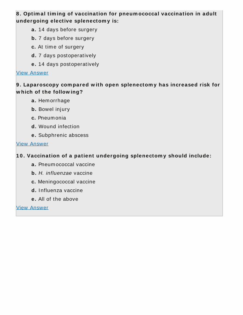
8. Optimal timing of vaccination for pneumococcal vaccination in adultundergoing elective splenectomy is:
a. 14 days before surgery
b. 7 days before surgery
c. At time of surgery
d. 7 days postoperatively
e. 14 days postoperatively
View Answer
9. Laparoscopy compared with open splenectomy has increased risk forwhich of the following?
a. Hemorrhage
b. Bowel injury
c. Pneumonia
d. Wound infection
e. Subphrenic abscess
View Answer
10. Vaccination of a patient undergoing splenectomy should include:
a. Pneumococcal vaccine
b. H. influenzae vaccine
c. Meningococcal vaccine
d. Influenza vaccine
e. All of the above
View Answer
P.423
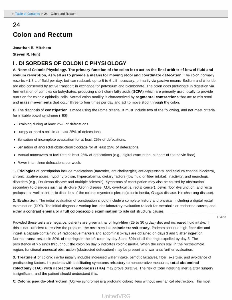
24Colon and Rectum
Jonathan B. Mitchem
Steven R. Hunt
I. DISORDERS OF COLONIC PHYSIOLOGYA. Normal Colonic Physiology. The primary function of the colon is to act as the final arbiter of bowel fluid andsodium resorption, as well as to provide a means for moving stool and coordinate defecation. The colon normallyresorbs ÷1.5 L of fluid per day, but can reabsorb up to 5 to 6 L if necessary, primarily via passive means. Sodium and chlorideare also conserved by active transport in exchange for potassium and bicarbonate. The colon does participate in digestion viafermentation of complex carbohydrates, producing short chain fatty acids (SCFA) which are primarily used locally to providenutrition for colonic epithelial cells. Normal colon motility is characterized by segmental contractions that act to mix stooland mass movements that occur three to four times per day and act to move stool through the colon.
B. The diagnosis of constipation is made using the Rome criteria. It must include two of the following, and not meet criteriafor irritable bowel syndrome (IBS):
Straining during at least 25% of defecations.
Lumpy or hard stools in at least 25% of defecations.
Sensation of incomplete evacuation for at least 25% of defecations.
Sensation of anorectal obstruction/blockage for at least 25% of defecations.
Manual maneuvers to facilitate at least 25% of defecations (e.g., digital evacuation, support of the pelvic floor).
Fewer than three defecations per week.
1. Etiologies of constipation include medications (narcotics, anticholinergics, antidepressants, and calcium channel blockers),chronic laxative abuse, hypothyroidism, hypercalcemia, dietary factors (low fluid or fiber intake), inactivity, and neurologicdisorders (e.g., Parkinson disease and multiple sclerosis). Symptoms of constipation may also be caused by obstructionsecondary to disorders such as stricture (Crohn disease [CD], diverticulitis, rectal cancer), pelvic floor dysfunction, and rectalprolapse, as well as intrinsic disorders of the colonic myenteric plexus (colonic inertia, Chagas disease, Hirschsprung disease).
2. Evaluation. The initial evaluation of constipation should include a complete history and physical, including a digital rectalexamination (DRE). The initial diagnostic workup includes laboratory evaluation to look for metabolic or endocrine causes, andeither a contrast enema or a full colonoscopic examination to rule out structural causes.
Provided these tests are negative, patients are given a trial of high-fiber (25 to 30 g/day) diet and increased fluid intake; ifthis is not sufficient to resolve the problem, the next step is a colonic transit study. Patients continue high-fiber diet andingest a capsule containing 24 radiopaque markers and abdominal x-rays are obtained on days 3 and 5 after ingestion.Normal transit results in 80% of the rings in the left colon by day 3 and 80% of all the rings expelled by day 5. Thepersistence of >5 rings throughout the colon on day 5 indicates colonic inertia. When the rings stall in the rectosigmoidregion, functional anorectal obstruction (obstructed defecation) may be present and warrants further evaluation.
3. Treatment of colonic inertia initially includes increased water intake, osmotic laxatives, fiber, exercise, and avoidance ofpredisposing factors. In patients with debilitating symptoms refractory to nonoperative measures, total abdominalcolectomy (TAC) with ileorectal anastomosis (IRA) may prove curative. The risk of total intestinal inertia after surgeryis significant, and the patient should understand this.
C. Colonic pseudo-obstruction (Ogilvie syndrome) is a profound colonic ileus without mechanical obstruction. This most
> Table of Contents > 24 - Colon and Rectum
UnitedVRG
P.424
commonly occurs in critically ill or institutionalized patients, and lack of mechanical obstruction must be confirmed via imagingstudies or colonoscopy. Initial management in patients without evidence of peritonitis or perforation consists of nasogastricdecompression, bowel rest, correction of systemic contributing factors (i.e., shock, heart failure, metabolic derangements),and discontinuation of medications that decrease colonic motility (including narcotics). If these conservative measures are notsufficient after 24 to 48 hours, neostigmine should be considered. Neostigmine is not a benign medication and should only begiven in a monitored setting as it may cause significant bradyarrhythmia. If patients are not candidates for or have failedneostigmine, colonoscopic decompression should be considered. Patients with evidence of perforation, peritonitis, orprolonged distension unresponsive to therapy should undergo total colectomy with end ileostomy (EI) unless thepatient's comorbid conditions preclude operative intervention.
D. Volvulus accounts for nearly 10% to 15% of colonic obstruction in the United States.
1. Sigmoid volvulus accounts for ÷60% of all cases and is most common in the elderly or institutionalized, as well aspatients with neurologic disorders. It is an acquired condition resulting from sigmoid redundancy with narrowing of themesenteric pedicle.
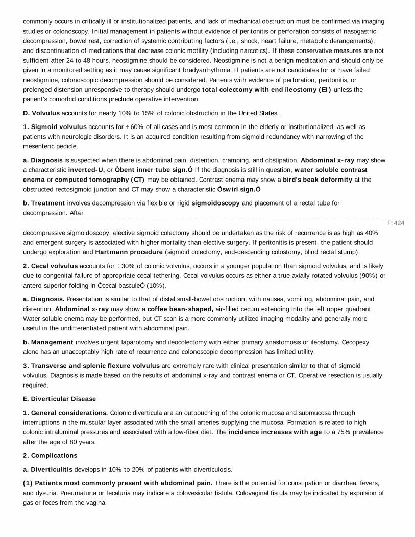
a. Diagnosis is suspected when there is abdominal pain, distention, cramping, and obstipation. Abdominal x-ray may showa characteristic inverted-U, or Òbent inner tube sign.Ó If the diagnosis is still in question, water soluble contrastenema or computed tomography (CT) may be obtained. Contrast enema may show a bird's beak deformity at theobstructed rectosigmoid junction and CT may show a characteristic Òswirl sign.Ó
b. Treatment involves decompression via flexible or rigid sigmoidoscopy and placement of a rectal tube fordecompression. After
decompressive sigmoidoscopy, elective sigmoid colectomy should be undertaken as the risk of recurrence is as high as 40%and emergent surgery is associated with higher mortality than elective surgery. If peritonitis is present, the patient shouldundergo exploration and Hartmann procedure (sigmoid colectomy, end-descending colostomy, blind rectal stump).
2. Cecal volvulus accounts for ÷30% of colonic volvulus, occurs in a younger population than sigmoid volvulus, and is likelydue to congenital failure of appropriate cecal tethering. Cecal volvulus occurs as either a true axially rotated volvulus (90%) orantero-superior folding in Òcecal basculeÓ (10%).
a. Diagnosis. Presentation is similar to that of distal small-bowel obstruction, with nausea, vomiting, abdominal pain, anddistention. Abdominal x-ray may show a coffee bean-shaped, air-filled cecum extending into the left upper quadrant.Water soluble enema may be performed, but CT scan is a more commonly utilized imaging modality and generally moreuseful in the undifferentiated patient with abdominal pain.
b. Management involves urgent laparotomy and ileocolectomy with either primary anastomosis or ileostomy. Cecopexyalone has an unacceptably high rate of recurrence and colonoscopic decompression has limited utility.
3. Transverse and splenic flexure volvulus are extremely rare with clinical presentation similar to that of sigmoidvolvulus. Diagnosis is made based on the results of abdominal x-ray and contrast enema or CT. Operative resection is usuallyrequired.
E. Diverticular Disease
1. General considerations. Colonic diverticula are an outpouching of the colonic mucosa and submucosa throughinterruptions in the muscular layer associated with the small arteries supplying the mucosa. Formation is related to highcolonic intraluminal pressures and associated with a low-fiber diet. The incidence increases with age to a 75% prevalenceafter the age of 80 years.
2. Complications
a. Diverticulitis develops in 10% to 20% of patients with diverticulosis.
(1) Patients most commonly present with abdominal pain. There is the potential for constipation or diarrhea, fevers,and dysuria. Pneumaturia or fecaluria may indicate a colovesicular fistula. Colovaginal fistula may be indicated by expulsion ofgas or feces from the vagina.
P.425
P.426
(2) Evaluation and staging in the acute setting is done using CT scan. Colonoscopy and barium or water-soluble enemas arenot recommended in the acute setting.
(3) Treatment is tailored to severity.
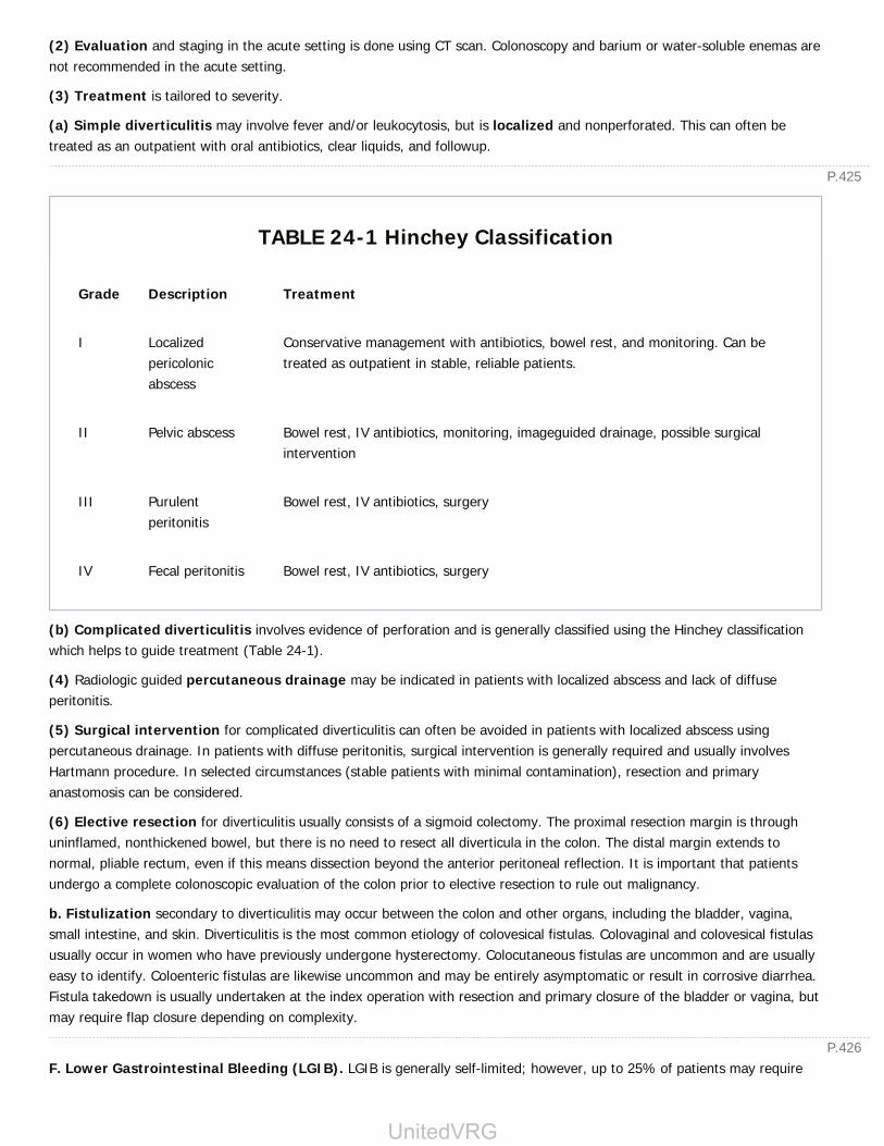
(a) Simple diverticulitis may involve fever and/or leukocytosis, but is localized and nonperforated. This can often betreated as an outpatient with oral antibiotics, clear liquids, and followup.
TABLE 24-1 Hinchey Classification
Grade Description Treatment
I Localizedpericolonicabscess
Conservative management with antibiotics, bowel rest, and monitoring. Can betreated as outpatient in stable, reliable patients.
II Pelvic abscess Bowel rest, IV antibiotics, monitoring, imageguided drainage, possible surgicalintervention
III Purulentperitonitis
Bowel rest, IV antibiotics, surgery
IV Fecal peritonitis Bowel rest, IV antibiotics, surgery
(b) Complicated diverticulitis involves evidence of perforation and is generally classified using the Hinchey classificationwhich helps to guide treatment (Table 24-1).
(4) Radiologic guided percutaneous drainage may be indicated in patients with localized abscess and lack of diffuseperitonitis.
(5) Surgical intervention for complicated diverticulitis can often be avoided in patients with localized abscess usingpercutaneous drainage. In patients with diffuse peritonitis, surgical intervention is generally required and usually involvesHartmann procedure. In selected circumstances (stable patients with minimal contamination), resection and primaryanastomosis can be considered.
(6) Elective resection for diverticulitis usually consists of a sigmoid colectomy. The proximal resection margin is throughuninflamed, nonthickened bowel, but there is no need to resect all diverticula in the colon. The distal margin extends tonormal, pliable rectum, even if this means dissection beyond the anterior peritoneal reflection. It is important that patientsundergo a complete colonoscopic evaluation of the colon prior to elective resection to rule out malignancy.
b. Fistulization secondary to diverticulitis may occur between the colon and other organs, including the bladder, vagina,small intestine, and skin. Diverticulitis is the most common etiology of colovesical fistulas. Colovaginal and colovesical fistulasusually occur in women who have previously undergone hysterectomy. Colocutaneous fistulas are uncommon and are usuallyeasy to identify. Coloenteric fistulas are likewise uncommon and may be entirely asymptomatic or result in corrosive diarrhea.Fistula takedown is usually undertaken at the index operation with resection and primary closure of the bladder or vagina, butmay require flap closure depending on complexity.
F. Lower Gastrointestinal Bleeding (LGIB). LGIB is generally self-limited; however, up to 25% of patients may require
UnitedVRG

P.427
surgical intervention. The most common causes of LGIB are diverticulosis (30% to 35%), hemorrhoids (20%), colorectalpolyps (13%), colorectal cancer (9%), intestinal ischemia (6.6%), and angiodysplasia (6%).
1. The management of LGIB in the acute setting varies by the volume of bleeding. Patients with a small amount of bleedingcan be worked up as an outpatient. Patients may, however, present with hemodynamic instability to the emergencydepartment. Massive LGIB is defined as any patient who requires >2 U of red blood cells in a 24-hour period. In theunstable patient, principles of resuscitation should be followed including the ABCs and ensuring the patient has adequateaccess for resuscitation (see Chapter 7, Critical Care).
2. Once hemodynamic stability has been assured or resuscitation has been initiated, it is important to discern the cause ofbleeding. The workup of LGIB involves the use of multiple different imaging and diagnostic modalities.
a. As always, the history and physical is key to discerning the source of bleeding. Hematochezia is more likely to come froman LGI source whereas melena may originate from an UGI or small bowel (SB) source. Recent weight loss or history ofanemia may point to a chronic process, such as cancer or inflammatory bowel disease (IBD). Stigmata of cirrhosis may beevident. Rectal examination should be performed in all patients as this may point out an obvious source such as hemorrhoids,rectal mass, or fissure. An NGT should be placed to determine an obvious UGI source.
b. Laboratory studies include a coagulation profile, basic metabolic profile, hepatic function panel, and complete bloodcount. This will indicate the degree of anemia and coagulopathy. Hepatic function may point toward liver dysfunction and theserum creatinine whether the patient has renal failure.
c. Diagnosing the source of hemorrhage is key, as this will help to tailor therapy and is important in the event the patientmay require surgical intervention.
(1) Endoscopy: EGD should be considered in any patient with massive LGIB or melena if an UGI source has not alreadybeen ruled out. Colonoscopy can be both diagnostic and therapeutic. Actively bleeding lesions may be injected with diluteepinephrine solution for vasoconstriction, cauterized or clipped. In stable patients who have no evidence of bleeding on EGDor colonoscopy with persistent transfusion requirement, capsule endoscopy or SB ÒpushÓ enteroscopy should beconsidered.
(2) Nuclear scan using technetium-99m sulfur colloid or tagged RBCs can identify bleeding sources with rates as low as 0.1to 0.5 mL/minute. Tagged RBC scan can identify bleeding up to 24 hours after isotope injection, but does not definitivelyidentify the anatomic source of bleeding.
(3) Mesenteric angiography should be performed in the patient with a positive nuclear medicine bleeding scan to identifythe anatomic source of bleeding. This may be diagnostic and therapeutic. Angiography can localize bleeding exceeding 1mL/minute and allows therapeutic vasopressin infusion (0.2 unit/minute) or embolization, which together are successful in85% of cases.
(4) In the rare patient who continues to bleed with an unidentifiable source, diagnostic laparoscopy or laparotomywith intraoperative endoscopy can be considered.
II. COLITIDESA. IBD is an umbrella term that traditionally covers ulcerative colitis (UC), CD, and Òindeterminate colitis.Ó The exact etiologyof IBD is as yet unclear, but there is clearly both an environmental and genetic component. Extraintestinal manifestations canbe associated with both UC and CD and include primary sclerosing cholangitis (÷3%), pyoderma gangrenosum, erythemanodosum, iritis/uveitis (2% to 8%), and stomatitis. In addition, patients with IBD have an increased risk of thrombosisincluding portal and mesenteric venous thrombosis, as well as deep venous thrombosis (DVT) and pulmonary embolus (PE).
1. UC is an inflammatory process of the colonic mucosa. There is a slight male predominance. The disease always involvesthe rectum and extends continuously for a variable distance proximally. Patients can present with bloody diarrhea,tenesmus, abdominal pain, fever, and weight loss. As the duration of the inflammation increases, pathologic changesprogress. Initially, mucosal ulcers and crypt abscesses are seen. Later, mucosal edema and pseudopolyps (islands of normalmucosa surrounded by deep ulcers) develop, and the end-stage pathologic changes show a flattened, dysplastic mucosa.
P.428
P.429
Cancer must be considered in any colonic stricture in a patient with UC. The risk of colon cancer is increased in patients withUC, but is related to length of disease with risk increasing significantly after 20 years, approaching almost 10% after thatduration.
a. Diagnosis is made primarily by colonoscopy with biopsy and by the constellation of symptoms. Imaging studies can helpto determine if the patient has SB disease or fistulae indicative of CD.
b. Medical management revolves around therapy which decreases colonic inflammation. Patients with distal disease(proctitis) often respond to topical 5-aminosalicylic acid derivatives (5-ASA) in the form of enemas or suppositories. For thosewith more proximal disease, oral 5-ASA or sulfasalazine (SSZ) will induce remission in the majority of patients with mild ormoderate disease. Patients unresponsive to topical and/or oral 5-ASA and SSZ can then be treated with oral corticosteroidsand transitioned back to 5-ASA or SSZ. Intravenous corticosteroids are given to those that are unresponsive oralcorticosteroids or are systemically ill with severe
colitis. Azathioprine (AZA) and 6-mercaptopurine (6-MP) have been shown to help wean patients off steroids and can be usedas maintenance therapy. Biologic therapy with TNF-α inhibitors has been shown to decrease colectomy rates in studies withshort-term followup. Long-term data will be forthcoming as experience with these medications increases.
c. Surgery is indicated in patients who have a high risk of malignancy; disease refractory to medical therapy; and cannot beweaned from steroids, toxic colitis, or intractable bleeding. In the acutely ill patient the operation of choice is TAC with EI.These patients, once stabilized and healthy, can be considered for restorative proctocolectomy with ileal pouch analanastomosis (IPAA) and diverting loop ileostomy (DLI). This is considered a three-stage approach. In patients who aresubacutely ill or stable, a two-stage approach can be considered, consisting of total proctocolectomy (TPC) and IPAA withDLI at the index operation followed by takedown of DLI at a later date. Anticipated function after restorative proctocolectomywith IPAA is approximately six to eight bowel movements a day often with the aid of bulking agents (>50%). Additionalcomplications to consider are impaired continence, sexual dysfunction/infertility, pouchitis, and bowel obstruction. Despite therisks, 95% of patients are satisfied with the procedure and have a good quality of life after IPAA (Dis Colon Rectum.2003;46(11):1489-1491). The S-pouch or W-pouch are other options for restoration of continuity that have utility in specificsituations and are done in some specialized centers. Restoration is contraindicated in patients with poor precolectomycontinence. In addition, older patients and the obese have worse outcomes with restoration. IPAA should be approached withcaution in patients where CD is a concern.
2. CD is a transmural inflammatory process that can affect any area of the GI tract, from the mouth to the anus. It has afemale predominance. The disease has a segmental distribution, with normal mucosa interspersed between areas ofdiseased bowel. Common symptoms include diarrhea, abdominal pain, nausea and vomiting, weight loss, and fever. Therecan be an abdominal mass or perianal fistulas on physical examination. The terminal ileum is involved in up to 45% ofpatients at presentation. Common pathologic changes include fissures, fistulae, transmural inflammation, and granulomas.Grossly, the mucosa shows aphthoid ulcers that often deepen over time and are associated with fat wrapping and bowel wallthickening. As the disease progresses, the bowel lumen narrows, and obstruction or perforation may result. SB CD isdiscussed in Chapter 19 and perianal CD is discussed in Chapter 25.
a. Diagnosis is made using colonoscopy, imaging, and the clinical picture. Unfortunately, patients with Crohn colitis (CC) willoften present similarly to patients with UC and up to one-third of patients with CC or UC will be diagnosed incorrectly prior tooperative intervention. Based on the clinical picture, CC can be discerned from UC by the presence of perianal disease, Òskiplesions,Ó ileal
inflammation on colonoscopy, and the presence of SB involvement on imaging (SBFT, CT, or MRI/MRE).
b. Medical management revolves around the use of immune suppression. In the acute setting, sepsis should becontrolled by drainage of abscesses and immune suppression. After the initial control of patients with oral or IVsteroids, patients are weaned using immunomodulators as listed above for UC. In addition, budesonide, a topicalcorticosteroid administered orally without systemic absorption, can be administered. Biologic therapy using TNF-α inhibitorsinfliximab, certolizumab, and adalimumab has been shown to decrease steroids and prolong surgical interventionin CD.
UnitedVRG
P.430
c. Surgical intervention is indicated in patients with medically refractory disease, acute systemic sepsis/perforation,uncontrolled hemorrhage, failure to thrive/malnutrition, and dysplasia/malignancy. Patients with CD with segmental diseaseshould be considered for limited resection. Colectomy with IRA can be entertained for patients with colitis and rectal sparingwith limited perianal disease. In the setting of total proctocolitis, patients will likely require TPC with EI. In some centers, TPCwith IPAA is considered for isolated CC with no perianal disease. While stricturoplasty has a role in the treatment of SB CD,stricturoplasty plays no role in the treatment of CC, as there is a 7% risk of malignancy over 20 years.
3. ÒIndeterminate colitisÓ is a term used for cases in which the pathologic pattern does not fall clearly into one or theother of the aforementioned patterns (10% to 15% of patients with IBD). The indeterminacy can be due either to inadequatetissue biopsy or to a truly indeterminate form of disease. Typically, surgical therapy for these patients is approached similarlyto UC, although they may have a slightly higher rate of pouch complications than patients with UC.
B. Ischemic colitis may result from many low-flow states, including venous or arterial thrombosis, embolization, iatrogenicinferior mesenteric artery (IMA) ligation after abdominal aortic aneurysm repair, and vasculopathy. It is idiopathic in themajority of patients. Patients are usually elderly and present with lower abdominal pain localizing to the left and melena orhematochezia. Contrast enema may show thumbprinting that corresponds to submucosal hemorrhage and edema.Diagnosis depends on the appearance of the mucosa on colonoscopy. This disease is present most frequently at thewatershed areas of the splenic flexure and sigmoid colon. In the presence of full-thickness necrosis or peritonitis, emergentresection with diversion is recommended. Patients without peritonitis or free air but with fever or an elevated white blood cell(WBC) count may be treated with bowel rest, close observation, and intravenous antibiotics. Up to 50% of patients developfocal colonic strictures eventually.
C. Radiation proctocolitis results from pelvic irradiation for the treatment of various malignancies. Risk factors include adose of greater than 6,000 cGy, vascular disease, diabetes mellitus, hypertension, prior low anterior resection, and advancedage. The early phase occurs within days to weeks.
Mucosal injury, edema, and ulceration develop, with associated nausea, vomiting, diarrhea, and tenesmus. The late phaseoccurs within weeks to years, and is associated with tenesmus and hematochezia with bowel thickening and fibrosis.Ulceration with bleeding, stricture, and fistula formation may occur. Medical treatment may be successful in mild cases, withthe use of stool softeners, steroid enemas, and topical 5-aminosalicylic acid products. If these measures fail, transanalapplication of formalin 4% to affected mucosa may be efficacious in patients with transfusion-dependent rectal bleeding.Patients with stricture or fistula require proctoscopy and biopsy to rule out locally recurrent disease or primary neoplasm.Strictures may be treated by endoscopic dilation, but often recur. Surgical treatment consists of a diverting colostomy and isreserved for medical failures, recurrent strictures, and fistulae.
D. Infectious Colitis
1. Pseudomembranous colitis is an acute diarrheal illness resulting from toxins produced by overgrowth of Clostridiumdifficile after antibiotic treatment (especially the use of clindamycin, ampicillin, or cephalosporins). Antibiotics already havebeen discontinued in one-fourth of cases, and symptoms can occur up to 6 weeks after even a single dose. Diagnosis ismade by detection of toxin A in one of at least three stool samples or stool culture if toxin A is not found but symptoms arepresent. Proctoscopy demonstrates sloughing colonic mucosa or pseudomembranes, and CT often shows transmural colonicthickening. Treatment begins with stopping unnecessary antibiotics and starting oral or intravenous metronidazole. Oral(not intravenous) vancomycin is an alternative expensive therapy. For severe cases in patients unable to take oralmedications, vancomycin enemas (500 mg in 250 mL saline) may be useful. Rarely, pseudomembranous colitis presents withsevere sepsis and colonic distention with toxic megacolon or perforation. Emergency laparotomy with total colectomy andend-ileostomy is required.
2. Other causes of colitis include bacteria (E. coli, Shigella), amoebic colitis, CMV colitis, and actinomycosis;however, these conditions are rarely encountered. Typically they are diagnosed by fecal testing or culture and treatment isdictated based on these results. Actinomycosis is treated with appropriate antibiotic therapy, CMV colitis is treated withganciclovir, and amoebic colitis is treated with oral flagyl.
3. Neutropenic enterocolitis after chemotherapy occurs most commonly in the setting of acute myelogenous leukemiaafter cytosine arabinoside therapy. Patients present with abdominal pain, fever, bloody diarrhea, distention, and sepsis.
P.431
P.432
Initial treatment includes bowel rest, total parenteral nutrition, granulocyte colony-stimulating factor (G-CSF), and broad-spectrum intravenous antibiotics. Laparotomy with total colectomy and ileostomy is required only if peritonitis develops.
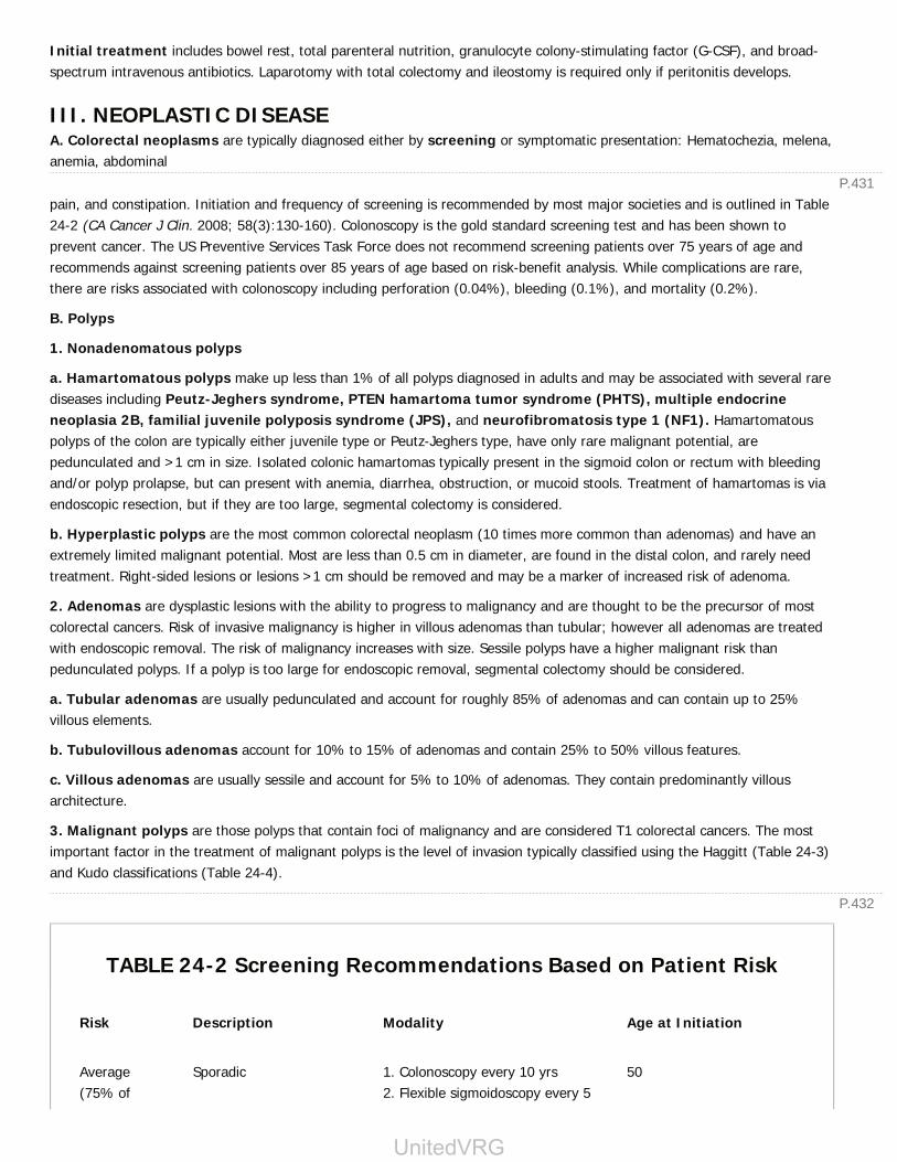
III. NEOPLASTIC DISEASEA. Colorectal neoplasms are typically diagnosed either by screening or symptomatic presentation: Hematochezia, melena,anemia, abdominal
pain, and constipation. Initiation and frequency of screening is recommended by most major societies and is outlined in Table24-2 (CA Cancer J Clin. 2008; 58(3):130-160). Colonoscopy is the gold standard screening test and has been shown toprevent cancer. The US Preventive Services Task Force does not recommend screening patients over 75 years of age andrecommends against screening patients over 85 years of age based on risk-benefit analysis. While complications are rare,there are risks associated with colonoscopy including perforation (0.04%), bleeding (0.1%), and mortality (0.2%).
B. Polyps
1. Nonadenomatous polyps
a. Hamartomatous polyps make up less than 1% of all polyps diagnosed in adults and may be associated with several rarediseases including Peutz-Jeghers syndrome, PTEN hamartoma tumor syndrome (PHTS), multiple endocrineneoplasia 2B, familial juvenile polyposis syndrome (JPS), and neurofibromatosis type 1 (NF1). Hamartomatouspolyps of the colon are typically either juvenile type or Peutz-Jeghers type, have only rare malignant potential, arepedunculated and >1 cm in size. Isolated colonic hamartomas typically present in the sigmoid colon or rectum with bleedingand/or polyp prolapse, but can present with anemia, diarrhea, obstruction, or mucoid stools. Treatment of hamartomas is viaendoscopic resection, but if they are too large, segmental colectomy is considered.
b. Hyperplastic polyps are the most common colorectal neoplasm (10 times more common than adenomas) and have anextremely limited malignant potential. Most are less than 0.5 cm in diameter, are found in the distal colon, and rarely needtreatment. Right-sided lesions or lesions >1 cm should be removed and may be a marker of increased risk of adenoma.
2. Adenomas are dysplastic lesions with the ability to progress to malignancy and are thought to be the precursor of mostcolorectal cancers. Risk of invasive malignancy is higher in villous adenomas than tubular; however all adenomas are treatedwith endoscopic removal. The risk of malignancy increases with size. Sessile polyps have a higher malignant risk thanpedunculated polyps. If a polyp is too large for endoscopic removal, segmental colectomy should be considered.
a. Tubular adenomas are usually pedunculated and account for roughly 85% of adenomas and can contain up to 25%villous elements.
b. Tubulovillous adenomas account for 10% to 15% of adenomas and contain 25% to 50% villous features.
c. Villous adenomas are usually sessile and account for 5% to 10% of adenomas. They contain predominantly villousarchitecture.
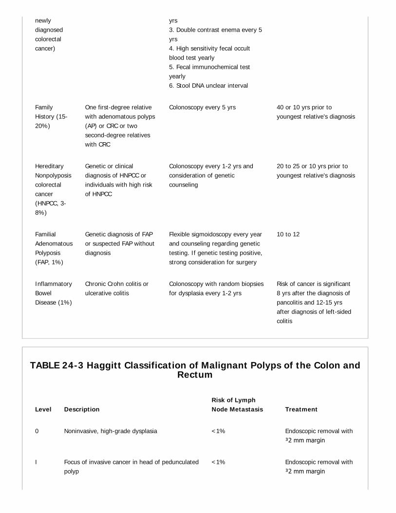
3. Malignant polyps are those polyps that contain foci of malignancy and are considered T1 colorectal cancers. The mostimportant factor in the treatment of malignant polyps is the level of invasion typically classified using the Haggitt (Table 24-3)and Kudo classifications (Table 24-4).
TABLE 24-2 Screening Recommendations Based on Patient Risk
Risk Description Modality Age at Initiation
Average(75% of
Sporadic 1. Colonoscopy every 10 yrs2. Flexible sigmoidoscopy every 5
50
UnitedVRG
newlydiagnosedcolorectalcancer)
yrs3. Double contrast enema every 5yrs4. High sensitivity fecal occultblood test yearly5. Fecal immunochemical testyearly6. Stool DNA unclear interval
FamilyHistory (15-20%)
One first-degree relativewith adenomatous polyps(AP) or CRC or twosecond-degree relativeswith CRC
Colonoscopy every 5 yrs 40 or 10 yrs prior toyoungest relative's diagnosis
HereditaryNonpolyposiscolorectalcancer(HNPCC, 3-8%)
Genetic or clinicaldiagnosis of HNPCC orindividuals with high riskof HNPCC
Colonoscopy every 1-2 yrs andconsideration of geneticcounseling
20 to 25 or 10 yrs prior toyoungest relative's diagnosis
FamilialAdenomatousPolyposis(FAP, 1%)
Genetic diagnosis of FAPor suspected FAP withoutdiagnosis
Flexible sigmoidoscopy every yearand counseling regarding genetictesting. If genetic testing positive,strong consideration for surgery
10 to 12
InflammatoryBowelDisease (1%)
Chronic Crohn colitis orulcerative colitis
Colonoscopy with random biopsiesfor dysplasia every 1-2 yrs
Risk of cancer is significant8 yrs after the diagnosis ofpancolitis and 12-15 yrsafter diagnosis of left-sidedcolitis
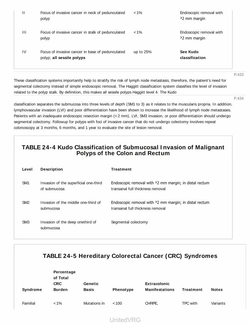
TABLE 24-3 Haggitt Classification of Malignant Polyps of the Colon andRectum
Level DescriptionRisk of LymphNode Metastasis Treatment
0 Noninvasive, high-grade dysplasia <1% Endoscopic removal with≥2 mm margin
I Focus of invasive cancer in head of pedunculatedpolyp
<1% Endoscopic removal with≥2 mm margin
P.434
P.433
II Focus of invasive cancer in neck of pedunculatedpolyp
<1% Endoscopic removal with≥2 mm margin
III Focus of invasive cancer in stalk of pedunculatedpolyp
<1% Endoscopic removal with≥2 mm margin
IV Focus of invasive cancer in base of pedunculatedpolyp; all sessile polyps
up to 25% See Kudoclassification
These classification systems importantly help to stratify the risk of lymph node metastasis, therefore, the patient's need forsegmental colectomy instead of simple endoscopic removal. The Haggitt classification system classifies the level of invasionrelated to the polyp stalk. By definition, this makes all sessile polyps Haggitt level 4. The Kudo
classification separates the submucosa into three levels of depth (SM1 to 3) as it relates to the muscularis propria. In addition,lymphovascular invasion (LVI) and poor differentiation have been shown to increase the likelihood of lymph node metastases.Patients with an inadequate endoscopic resection margin (<2 mm), LVI, SM3 invasion, or poor differentiation should undergosegmental colectomy. Followup for polyps with foci of invasive cancer that do not undergo colectomy involves repeatcolonoscopy at 3 months, 6 months, and 1 year to evaluate the site of lesion removal.
TABLE 24-4 Kudo Classification of Submucosal Invasion of MalignantPolyps of the Colon and Rectum
Level Description Treatment
SM1 Invasion of the superficial one-thirdof submucosa
Endoscopic removal with ≥2 mm margin; in distal rectumtransanal full thickness removal
SM2 Invasion of the middle one-third ofsubmucosa
Endoscopic removal with ≥2 mm margin; in distal rectumtransanal full thickness removal
SM3 Invasion of the deep onethird ofsubmucosa
Segmental colectomy
TABLE 24-5 Hereditary Colorectal Cancer (CRC) Syndromes
Syndrome
Percentageof TotalCRCBurden
GeneticBasis Phenotype
ExtracolonicManifestations Treatment Notes
Familial <1% Mutations in <100 CHRPE, TPC with Variants
UnitedVRG

P.435
adenomatouspolyposis(FAP)
tumorsuppressorgene APC(5q21)
adenomatouspolyps; near100% withCRC by age40 yrs
osteomas,epidermal cysts,periampullaryneoplasms
end-ileostomy orIPAA or TACwith IRAand lifelongsurveillance
include Turcot(CNS tumors)and Gardener(desmoids)syndromes
Hereditarynonpolyposiscolorectalcancer(HNPCC)
5-7% Defectivemismatchrepair:MSH2 andMLH1(90%),MSH6(10%)
Few polyps,predominantlyrightsidedCRC, 80%lifetime risk ofCRC
At risk foruterine, ovarian,small intestinal,pancreaticmalignancies
Geneticcounseling;considerprophylacticresections,includingTAH/BSO
Highmicrosatelliteinstability(MSI-H)tumors, betterprognosis thansporadic CRC
Peutz-Jeghers (PJS)
<1% Loss oftumorsuppressorgeneLKB1/STK11(19p13)
Hamartomasthroughout GItract
Mucocutaneouspigmentation,risk forpancreaticcancer
SurveillanceEGD andcolonoscopyq3yr; resectpolyps >1.5cm
Majoritypresent withSBO due tointussusceptingpolyp
Familialjuvenilepolyposis(FJP)
<1% MutatedSMAD4/DPC(18q21)
Hamartomasthroughout GItract; >3juvenilepolyps; 15%with CRC byage 35 yrs
Gastric,duodenal, andpancreaticneoplasms;pulmonary AVMs
Geneticcounseling;considerprophylacticTAC withIRA fordiffusedisease
Presents withrectal bleedingor diarrhea
AVM, arteriovenous malformation; CHRPE, congenital hypertrophy of retinal pigmented epithelium; CNS, central nervoussystem; EGD, esophagogastroduodenoscopy; GI, gastrointestinal; IPAA, ileal pouch-anal anastomosis; IRA, ileal-rectalanastomosis; TAC, total abdominal colectomy; TAH/BSO, total abdominal hysterectomy and bilateral salpingo-oophorectomy; TPC, total proctocolectomy.
a. Malignant polyps of the proximal two-thirds of the rectum can be treated as colon polyps; however, there issome controversy regarding the treatment of malignant polyps of the distal one-third of the rectum as these lesions may havean increased risk of lymph node metastasis. All T1 lesions of the distal rectum should be approached with at
least transanal full thickness excision using traditional transanal excision, Transanal Endoscopic Microsurgery (TEM) orTransanal Minimally Invasive Surgery (TAMIS) techniques.
C. Colon Cancer
1. There are approximately 150,000 new diagnoses of colorectal cancer each year, of which 70% to 75% are colon cancer.Colorectal cancer is the fourth leading cause of cancer death worldwide and about one-third of patients diagnosed withcolorectal cancer will eventually die of their disease. See Table 24-5 for hereditary colorectal cancer syndromes.
P.436
P.437
P.438
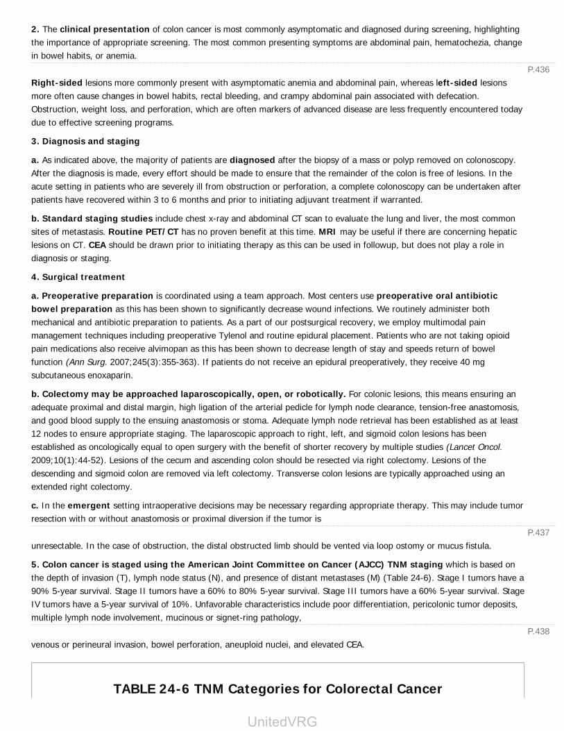
2. The clinical presentation of colon cancer is most commonly asymptomatic and diagnosed during screening, highlightingthe importance of appropriate screening. The most common presenting symptoms are abdominal pain, hematochezia, changein bowel habits, or anemia.
Right-sided lesions more commonly present with asymptomatic anemia and abdominal pain, whereas left-sided lesionsmore often cause changes in bowel habits, rectal bleeding, and crampy abdominal pain associated with defecation.Obstruction, weight loss, and perforation, which are often markers of advanced disease are less frequently encountered todaydue to effective screening programs.
3. Diagnosis and staging
a. As indicated above, the majority of patients are diagnosed after the biopsy of a mass or polyp removed on colonoscopy.After the diagnosis is made, every effort should be made to ensure that the remainder of the colon is free of lesions. In theacute setting in patients who are severely ill from obstruction or perforation, a complete colonoscopy can be undertaken afterpatients have recovered within 3 to 6 months and prior to initiating adjuvant treatment if warranted.
b. Standard staging studies include chest x-ray and abdominal CT scan to evaluate the lung and liver, the most commonsites of metastasis. Routine PET/CT has no proven benefit at this time. MRI may be useful if there are concerning hepaticlesions on CT. CEA should be drawn prior to initiating therapy as this can be used in followup, but does not play a role indiagnosis or staging.
4. Surgical treatment
a. Preoperative preparation is coordinated using a team approach. Most centers use preoperative oral antibioticbowel preparation as this has been shown to significantly decrease wound infections. We routinely administer bothmechanical and antibiotic preparation to patients. As a part of our postsurgical recovery, we employ multimodal painmanagement techniques including preoperative Tylenol and routine epidural placement. Patients who are not taking opioidpain medications also receive alvimopan as this has been shown to decrease length of stay and speeds return of bowelfunction (Ann Surg. 2007;245(3):355-363). If patients do not receive an epidural preoperatively, they receive 40 mgsubcutaneous enoxaparin.
b. Colectomy may be approached laparoscopically, open, or robotically. For colonic lesions, this means ensuring anadequate proximal and distal margin, high ligation of the arterial pedicle for lymph node clearance, tension-free anastomosis,and good blood supply to the ensuing anastomosis or stoma. Adequate lymph node retrieval has been established as at least12 nodes to ensure appropriate staging. The laparoscopic approach to right, left, and sigmoid colon lesions has beenestablished as oncologically equal to open surgery with the benefit of shorter recovery by multiple studies (Lancet Oncol.2009;10(1):44-52). Lesions of the cecum and ascending colon should be resected via right colectomy. Lesions of thedescending and sigmoid colon are removed via left colectomy. Transverse colon lesions are typically approached using anextended right colectomy.
c. In the emergent setting intraoperative decisions may be necessary regarding appropriate therapy. This may include tumorresection with or without anastomosis or proximal diversion if the tumor is
unresectable. In the case of obstruction, the distal obstructed limb should be vented via loop ostomy or mucus fistula.
5. Colon cancer is staged using the American Joint Committee on Cancer (AJCC) TNM staging which is based onthe depth of invasion (T), lymph node status (N), and presence of distant metastases (M) (Table 24-6). Stage I tumors have a90% 5-year survival. Stage II tumors have a 60% to 80% 5-year survival. Stage III tumors have a 60% 5-year survival. StageIV tumors have a 5-year survival of 10%. Unfavorable characteristics include poor differentiation, pericolonic tumor deposits,multiple lymph node involvement, mucinous or signet-ring pathology,
venous or perineural invasion, bowel perforation, aneuploid nuclei, and elevated CEA.
TABLE 24-6 TNM Categories for Colorectal Cancer
UnitedVRG
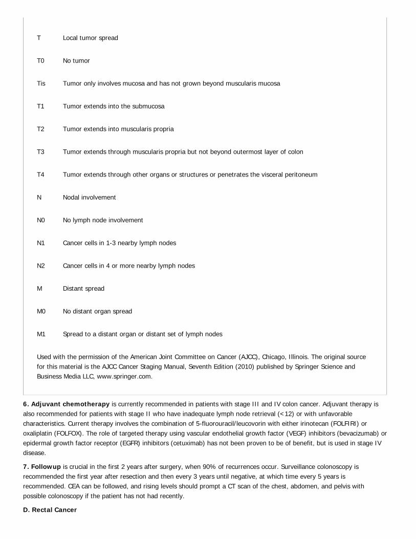
T Local tumor spread
T0 No tumor
Tis Tumor only involves mucosa and has not grown beyond muscularis mucosa
T1 Tumor extends into the submucosa
T2 Tumor extends into muscularis propria
T3 Tumor extends through muscularis propria but not beyond outermost layer of colon
T4 Tumor extends through other organs or structures or penetrates the visceral peritoneum
N Nodal involvement
N0 No lymph node involvement
N1 Cancer cells in 1-3 nearby lymph nodes
N2 Cancer cells in 4 or more nearby lymph nodes
M Distant spread
M0 No distant organ spread
M1 Spread to a distant organ or distant set of lymph nodes
Used with the permission of the American Joint Committee on Cancer (AJCC), Chicago, Illinois. The original sourcefor this material is the AJCC Cancer Staging Manual, Seventh Edition (2010) published by Springer Science andBusiness Media LLC, www.springer.com.
6. Adjuvant chemotherapy is currently recommended in patients with stage III and IV colon cancer. Adjuvant therapy isalso recommended for patients with stage II who have inadequate lymph node retrieval (<12) or with unfavorablecharacteristics. Current therapy involves the combination of 5-fluorouracil/leucovorin with either irinotecan (FOLFIRI) oroxaliplatin (FOLFOX). The role of targeted therapy using vascular endothelial growth factor (VEGF) inhibitors (bevacizumab) orepidermal growth factor receptor (EGFR) inhibitors (cetuximab) has not been proven to be of benefit, but is used in stage IVdisease.
7. Followup is crucial in the first 2 years after surgery, when 90% of recurrences occur. Surveillance colonoscopy isrecommended the first year after resection and then every 3 years until negative, at which time every 5 years isrecommended. CEA can be followed, and rising levels should prompt a CT scan of the chest, abdomen, and pelvis withpossible colonoscopy if the patient has not had recently.
D. Rectal Cancer
P.439P.440
1. The pathophysiology of rectal cancer differs from that of colon cancer because of several anatomic factors: (1)Confinement of pelvis and sphincters; (2) proximity to urogenital structures and nerves; (3) dual blood supply and lymphaticdrainage; and (4) transanal accessibility. The rectum is defined by the NCI as 12 cm above the anal verge on rigidproctoscopy.
2. Diagnosis and staging of the rectum is done using the AJCC staging as outlined above for colon cancer with additionalconsiderations regarding local staging. DRE can give information on the size, height, fixation, ulceration, local invasion, andlymph node status. Rigid sigmoidoscopy and biopsy are important for precisely measuring the distance to the anal verge anddentate line. Transrectal ultrasonography or rectal protocol magnetic resonance imaging (MRI) is an integral partof staging rectal tumors to evaluate depth of invasion, the circumferential resection margin (CRM), and lymph node status asthis will help determine the need for preoperative chemoradiation therapy.
Distant spread is evaluated (as with colon cancer) with abdominal CT and chest x-ray or CT. It is helpful to have apreoperative CEA for patient followup.
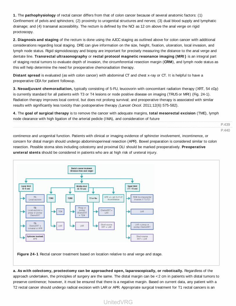
3. Neoadjuvant chemoradiation, typically consisting of 5-FU, leucovorin with concomitant radiation therapy (XRT, 54 cGy)is currently standard for all patients with T3 or T4 lesions or node positive disease on imaging (TRUS or MRI) (Fig. 24-1).Radiation therapy improves local control, but does not prolong survival; and preoperative therapy is associated with similarresults with significantly less toxicity than postoperative therapy (Lancet Oncol. 2011;12(6):575-582).
4. The goal of surgical therapy is to remove the cancer with adequate margins, total mesorectal excision (TME), lymphnode clearance with high ligation of the arterial pedicle (IMA), and consideration of future
continence and urogenital function. Patients with clinical or imaging evidence of sphincter involvement, incontinence, orconcern for distal margin should undergo abdominoperineal resection (APR). Bowel preparation is considered similar to colonresection. Possible stoma sites including colostomy and proximal DLI should be marked preoperatively. Preoperativeureteral stents should be considered in patients who are at high risk of ureteral injury.
Figure 24-1 Rectal cancer treatment based on location relative to anal verge and stage.
a. As with colectomy, proctectomy can be approached open, laparoscopically, or robotically. Regardless of theapproach undertaken, the principles of surgery are the same. The distal margin can be <2 cm in patients with distal tumors topreserve continence; however, it must be ensured that there is a negative margin. Based on current data, any patient with aT2 rectal cancer should undergo radical excision with LAR or APR. Appropriate surgical treatment for T1 rectal cancers is an
UnitedVRG

P.441
area of considerable study at present and includes transanal excision techniques. Rectal cancer with extension into thebladder, sacrum, vagina, or other local pelvic structures can be reliably resected for cure. It is important to remember theanatomic confines and anatomy of the pelvis as infertility, sexual dysfunction, and continence are affected by theparasympathetic and sympathetic nerves and are common complications. In addition, leak is more common forcoloproctostomy than colocolostomy, and leak testing in the operating room is recommended. Proximal diversion isrecommended for any low or tenuous anastomosis, as leaks can have devastating consequences.
5. Obstructing rectal cancers should be evaluated by hypaque enema and/or colonoscopy in patients without clear signsof peritonitis. Endoluminal stents can be used as a short-term bridge to operative therapy, but should not be used to getpatients through preoperative chemoradiation therapy as they have been shown to have a high risk or perforation andcomplications in this setting. In addition, stents should not be used in patients with mid to low rectal cancers as this can leadto considerable pain and urgency issues.
6. Rectal cancer recurrence typically presents with pain, rectal bleeding, or on followup testing. Diagnosis is confirmed byexamination and biopsy. Patients should then be worked up for systemic recurrence including CT and PET-CT. If there is noevidence of systemic recurrence, resection can be considered if patients are fit. Pelvic MRI is useful to evaluate therelationship to other pelvic structures. Preoperative therapy can be considered if patients have not received XRT previously.Curative resection of recurrent rectal cancer can lead to significant long-term survival (Ann Surg. 1994;220(4):586-595).
E. Other Colorectal Tumors
1. Lymphoma is most often metastatic to the colorectum, but primary non-Hodgkin colonic lymphoma accounts for 10% ofall GI lymphomas. The GI tract is also a common site of non-Hodgkin lymphoma associated with human immunodeficiencyvirus. The most common
presenting symptoms include abdominal pain, altered bowel habits, weight loss, and hematochezia. Biopsies are often notdiagnostic because the lesion is submucosal. Treatment is resection with postoperative chemotherapy. Intestinal bypass,biopsy, and postoperative chemotherapy should be considered for locally advanced tumors.
2. Retrorectal tumors usually present with postural pain and a posterior rectal mass on physical examination and CT scan.
a. The differential diagnosis includes congenital, neurogenic, osseous, and inflammatory masses. Chordomas are the mostcommon malignant retrorectal tumor; they typically are slow growing but difficult to resect for cure.
b. Diagnosis is based on CT scan and physical findings. Biopsy should not be performed. Formal resection should beundertaken if there is significant concern for malignancy or symptoms.
3. Carcinoid tumor
a. Colonic carcinoids account for 2% of GI carcinoids. Lesions less than 2 cm in diameter rarely metastasize, but 80% oflesions greater than 2 cm in diameter have local or distant metastases, with a median length of survival of less than 12months. These lesions are treated with local excision if small and with formal resection if greater than 2 cm.
b. Rectal carcinoid accounts for 15% of GI carcinoids. As with colonic carcinoids, lesions less than 2 cm in diameter havelow malignant potential and can be treated with transanal or endoscopic resection. Rectal carcinoids greater than 2 cm indiameter are malignant in 90% of cases. Treatment of large rectal carcinoids is controversial, but low anterior resection orAPR is probably warranted.
IV. INTESTINAL STOMASA. Ileostomy creation and care was revolutionized with the description of the eversion technique by Brooke in 1952. Thesmall intestine adapts to ileostomy formation within 10 days postoperatively. Average output is 500 mL/day, but may be up to1,500 mL/day. Volumes above this may be pathologic and/or cause dehydration and electrolyte abnormalities. Stomaconstruction of either a loop ileostomy or end-ileostomy should be ÒBrookedÓ or everted 2 to 2.5 cm to create an easierstoma to pouch. Stoma creation should be within the rectus abdominis to decrease the risk of peristomal herniation.Preoperative marking of the planned site prevents improper placement near bony prominences, belt/pant lines, abdominalcreases, and scars. Reversal of a loop ileostomy is relatively straightforward and rarely requires laparotomy. A side-to-side,
P.442
P.443
functional end-to-end technique with a GIA stapler is often utilized.
B. Colostomy construction is typically associated with fewer electrolyte and physiologic derangements than ileostomy. Left-sided or sigmoid colostomies are preferred to right-sided or transverse colostomies. Colostomies can be created in either aloop or end-loop configuration. Common
colostomy complications include obstipation, prolapse, and parastomal hernia. Obstipation/constipation can be treated withstoma irrigation and/or hypaque enema, which is diagnostic and therapeutic. Parastomal hernia repair is indicated for thesame reasons as other abdominal wall hernias. Colostomy prolapse does not require revision unless there is an inability toreduce the mucosa or obstruction results. End colostomy takedown can be difficult and all patients should undergo fullcolonoscopic evaluation including the distal defunctionalized colon/rectum to rule out stricture or mass prior to takedown.
CHAPTER 24: COLON AND RECTUMMultiple Choice Questions1. Which of the following is part of the Rome criteria for the diagnosis of constipation?
a. Three or fewer bowel movements per week
b. Manual maneuvers to assist with 50% of bowel movement
c. Fulfilling criteria of irritable bowel syndrome
d. Sensation of incomplete evacuation with 100% of bowel movements
View Answer
2. When administering neostigmine to a patient with Ogilvie syndrome, why is it importantto ensure the patient is in a monitored setting?
a. Often there is a rapid response causing a large evacuation which can be difficult to manage
b. There is a high risk of hypotension due to vasovagal stimulation related to having a largebowel movement
c. Neostigmine can cause significant bradyarrhythmias potentially requiring cardioversion
d. There is a significant risk of perforation with the administration of neostigmine
View Answer
3. The most important aspect in the care of a patient with LGIB is which of the following?
a. Obtaining early tagged red blood cell scan
b. Ensuring appropriate resuscitation and stabilizing patient
c. Using fecal occult blood test to test for bleeding
d. Placing an NGT to rule out an upper GI source
View Answer
4. Definitive treatment of sigmoid volvulus is accomplished by:
a. Endoscopic decompression
b. Endoscopic decompression and placement of a long rectal tube
c. Sigmoidopexy
d. Sigmoidectomy
View Answer
5. The most common cause of LGIB is:
a. Diverticulosis
UnitedVRG

P.444
b. Colorectal cancer
c. Ischemic colitis
d. Ulcerative colitis
View Answer
6. Hinchey IV diverticulitis demands operative therapy and is characterized by:
a. Pericolonic abscess
b. Fecal peritonitis
c. Generalized peritonitis
d. Pelvic abscess
View Answer
7. An important distinguishing feature of Crohn disease when compared to ulcerative colitisis:
a. The lack of “skip” lesions
b. Response to biologic therapy
c. Perianal disease
d. The presence of pyoderma gangrenosum
View Answer
8. Surgical treatment of medically refractory ulcerative colitis includes:
a. Abdominoperineal resection with end colostomy
b. Total proctocolectomy with ileal-anal anastomosis
c. Segmental colectomy involving the diseased area and colo- or ileo-colostomy
d. Total abdominal colectomy with end ileostomy
View Answer
9. A patient presents to the ED with abdominal pain and hematochezia after endovascularaortic aneurysm repair (EVAAR), how would you confirm your clinical suspicion?
a. Flexible sigmoidoscopy
b. CT scan of the abdomen and pelvis
c. Barium enema
d. Acute abdominal series
View Answer
10. The most common type of colonic polyps diagnosed on endoscopy are:
a. Adenomatous polyps
b. Malignant polyps
c. Hamartomatous polyps
d. Hyperplastic polyps
View Answer
11. The Kudo classification of polyp invasion is important to the treatment of malignantcolon and rectal polyps because:
a. The Kudo classification is more sensitive than the Haggitt classification for the diagnosis of

P.445
malignancy
b. The Kudo classification accurately predicts who needs adjuvant therapy after resection
c. The Kudo classification predicts the risk of lymph node metastasis and the need for surgicalresection
d. The Kudo classification accurately predicts which polyps are technically amenable toendoscopic retrieval
View Answer
12. An asymptomatic patient presents to your office for consultation regarding screeningcolonoscopy due to the fact that the patient's father was diagnosed with colon cancer. Whatis the most important factor when considering initiating screening colonoscopy?
a. Recent weight loss
b. Smoking history
c. The age of the patient's father at diagnosis
d. The patient's mother had breast cancer
View Answer
13. On pathologic examination after right colectomy a patient is diagnosed with a T3 tumorwith 0 or 9 lymph nodes negative. What do you tell this patient about his or her need foradjuvant therapy?
a. The patient does not need adjuvant therapy because there is only marginal benefit in patientswith stage II disease.
b. Adjuvant therapy should be considered because although the patient is stage II, there wasinadequate lymph node harvest.
c. The patient should consider not receiving adjuvant therapy because although the patient hasstage III disease, they have low-risk stage III disease.
d. The patient should receive adjuvant therapy because there is clearly a benefit for patients withstage III disease.
View Answer
14. To appropriately stage rectal cancer, patients need what imaging studies?
a. Chest x-ray, abdomen CT, pelvic MRI
b. Chest x-ray, abdomen and pelvis CT, PET/CT
c. Abdomen CT, pelvic MRI, PET/CT
d. Chest x-ray, abdomen and pelvis CT, pelvic MRI, PET/CT
View Answer
15. The principles of surgical resection for the treatment of rectal cancer include which ofthe following?
a. Resection of Denonvilliers fascia to ensure an adequate anterior margin
b. Ensuring an intact and complete total mesorectal excision
c. Resection of the hypogastric nerves along the pelvic sidewall as this is a common site ofrecurrence
d. Performing an abdominoperineal resection for any patient with a tumor <5 cm from thedentate line due to the dual blood supply of the distal rectum
View Answer
UnitedVRG

25Anorectal Disease
Ashley M. Holder
Matthew G. Mutch
I. ANORECTAL PHYSIOLOGYA. Normal Anorectal Function
1. The rectum functions as a capacitance organ, with a reservoir of 650 to 1,200 mLcompared to an average daily stool output of 250 to 750 mL.
2. The anal sphincter mechanism allows defecation and maintains continence. The internalsphincter (involuntary) accounts for 80% of resting pressure, whereas the external sphincter(voluntary) accounts for 20% of resting pressure and 100% of squeeze pressure. The externalanal sphincter contracts in response to sensed rectal contents and relaxes during defecation.
3. Defecation has four components: (1) Mass movement of feces into the rectal vault; (2) rectalÑanal inhibitory reflex, by which distal rectal distention causes involuntary relaxation of theinternal sphincter and the external sphincter contracts (this process is known as sampling andallows for determination of contents as gas, liquid, or solid); (3) voluntary relaxation of theexternal sphincter mechanism and puborectalis muscle; and (4) increased intraabdominalpressure.
4. Continence requires normal capacitance, normal sensation at the anorectal transition zone,puborectalis function for solid stool, external sphincter function for fine control, and internalsphincter function and hemorrhoidal pillars for resting pressure.
B. Incontinence is the inability to prevent elimination of rectal contents.
1. Etiologies include (1) mechanical defects, such as sphincter damage from obstetrictrauma, fistulotomy, and scleroderma affecting the external sphincter; (2) neurogenic defects,including spinal cord injuries, pudendal nerve injury due to birth trauma or lifelong straining, andsystemic neuropathies such as multiple sclerosis; and (3) stool content-related causes, suchas diarrhea and radiation proctitis.
2. Evaluation includes visual and digital examination observing for gross tone or squeezeabnormalities and determining muscle bulk. Anal manometry quantitatively measuresparameters of anal function, including resting and squeeze pressure (normal mean >40 and >80mm Hg, respectively), sphincter length (4 cm in men, 3 cm in women), and minimal sensoryvolume of the rectum. Pudendal nerve terminal motor latency (PNTML) testing and endoanal
> Table of Contents > 25 - Anorectal Disease
UnitedVRG

P.447
ultrasound provide neural and anatomic information.
3. Treatment depends upon the underlying cause. Neurogenic and minor mechanical analsphincter defects are initially treated using dietary fiber
to increase stool bulk and biofeedback to strengthen muscle and improve early sensation. Majordefects require anal sphincter reconstruction, in which the anatomic sphincter defect isrepaired. Sacral nerve stimulation, used in patients with an intact sphincter complex or even ifthere is less than a 30-degree defect, is emerging as the most durable treatment for fecalincontinence: (1) Patients maintain a journal of their bowel and continence function for 2 weeks;(2) temporary leads are then imbedded in the S2 to S4 nerve roots and the journal maintained foranother 2 weeks; and (3) if there is >50% improvement in incontinence episodes, patients areeligible for implantation of the permanent device. Artificial anal sphincters may be used inpatients without a reconstructible native anal sphincter or with neurogenic incontinence.However, long-term success rate is complicated by a 60% explantation rate. A palliative divertingcolostomy is indicated when all other treatment modalities fail.
C. Obstructed defecation (pelvic floor outlet obstruction) presents with symptoms of chronicconstipation, straining with bowel movements, incomplete evacuation of the rectum, pelvicpressure, and the need for perineal pressure to evacuate. Evaluation includes: (1) Videodefecography to evaluate fixation of the posterior rectum to the sacrum and relaxation of thepuborectalis; (2) anal manometry and surface EMG testing to assess rectal sensation, abilityto expel a balloon, and paradoxical contraction of the external sphincter with straining; and (3)colonic transit study to assess colonic motility. Problems associated with obstructive defecationmay include fecal impaction and stercoral ulcer (mucosal ulceration due to pressure necrosisfrom impacted stool); both are treated with enemas, increased dietary fiber, and stool softeners.Attempts at surgical correction of any of the following conditions without addressing theunderlying pathology are doomed to failure.
1. Anal stenosis is a rare cause of obstructed defecation and presents with frequent thin stoolsand bloating. The most common etiologies include scarring after anorectal surgery (rare), chroniclaxative abuse, radiation, recurrent anal ulcer, inflammation, and trauma. Initial treatment is analdilation, although advanced cases are treated with advancement flaps of normal perianal skin.
2. Nonrelaxation of puborectalis results in straining and incomplete evacuation. Colonictransit time reveals outlet obstruction. Persistent puborectalis distortion is seen on defecography.Biofeedback is the treatment of choice.
3. Descending perineum syndrome occurs when chronic straining causes pudendal nervestretch and subsequent neurogenic defect. Rectocele results from a weak, distorted rectovaginalseptum that allows the anterior rectal wall to bulge into the vagina due to failure of the pelvicfloor to relax during defecation. Treatment includes bowel regimens with high fiber, suppositories,enemas, and biofeedback.
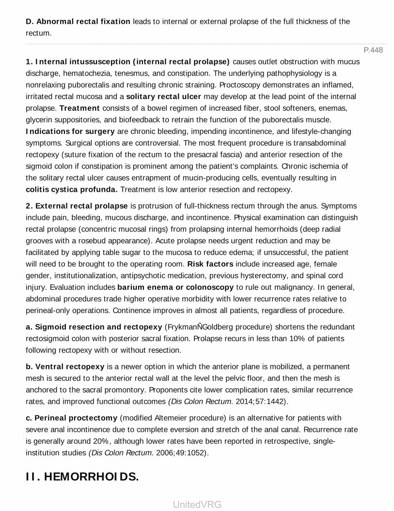
P.448
D. Abnormal rectal fixation leads to internal or external prolapse of the full thickness of therectum.
1. Internal intussusception (internal rectal prolapse) causes outlet obstruction with mucusdischarge, hematochezia, tenesmus, and constipation. The underlying pathophysiology is anonrelaxing puborectalis and resulting chronic straining. Proctoscopy demonstrates an inflamed,irritated rectal mucosa and a solitary rectal ulcer may develop at the lead point of the internalprolapse. Treatment consists of a bowel regimen of increased fiber, stool softeners, enemas,glycerin suppositories, and biofeedback to retrain the function of the puborectalis muscle.Indications for surgery are chronic bleeding, impending incontinence, and lifestyle-changingsymptoms. Surgical options are controversial. The most frequent procedure is transabdominalrectopexy (suture fixation of the rectum to the presacral fascia) and anterior resection of thesigmoid colon if constipation is prominent among the patient's complaints. Chronic ischemia ofthe solitary rectal ulcer causes entrapment of mucin-producing cells, eventually resulting incolitis cystica profunda. Treatment is low anterior resection and rectopexy.
2. External rectal prolapse is protrusion of full-thickness rectum through the anus. Symptomsinclude pain, bleeding, mucous discharge, and incontinence. Physical examination can distinguishrectal prolapse (concentric mucosal rings) from prolapsing internal hemorrhoids (deep radialgrooves with a rosebud appearance). Acute prolapse needs urgent reduction and may befacilitated by applying table sugar to the mucosa to reduce edema; if unsuccessful, the patientwill need to be brought to the operating room. Risk factors include increased age, femalegender, institutionalization, antipsychotic medication, previous hysterectomy, and spinal cordinjury. Evaluation includes barium enema or colonoscopy to rule out malignancy. In general,abdominal procedures trade higher operative morbidity with lower recurrence rates relative toperineal-only operations. Continence improves in almost all patients, regardless of procedure.
a. Sigmoid resection and rectopexy (FrykmanÑGoldberg procedure) shortens the redundantrectosigmoid colon with posterior sacral fixation. Prolapse recurs in less than 10% of patientsfollowing rectopexy with or without resection.
b. Ventral rectopexy is a newer option in which the anterior plane is mobilized, a permanentmesh is secured to the anterior rectal wall at the level the pelvic floor, and then the mesh isanchored to the sacral promontory. Proponents cite lower complication rates, similar recurrencerates, and improved functional outcomes (Dis Colon Rectum. 2014;57:1442).
c. Perineal proctectomy (modified Altemeier procedure) is an alternative for patients withsevere anal incontinence due to complete eversion and stretch of the anal canal. Recurrence rateis generally around 20%, although lower rates have been reported in retrospective, single-institution studies (Dis Colon Rectum. 2006;49:1052).
II. HEMORRHOIDS.
UnitedVRG
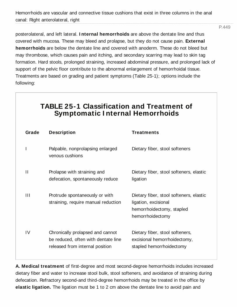
P.449
Hemorrhoids are vascular and connective tissue cushions that exist in three columns in the analcanal: Right anterolateral, right
posterolateral, and left lateral. Internal hemorrhoids are above the dentate line and thuscovered with mucosa. These may bleed and prolapse, but they do not cause pain. Externalhemorrhoids are below the dentate line and covered with anoderm. These do not bleed butmay thrombose, which causes pain and itching, and secondary scarring may lead to skin tagformation. Hard stools, prolonged straining, increased abdominal pressure, and prolonged lack ofsupport of the pelvic floor contribute to the abnormal enlargement of hemorrhoidal tissue.Treatments are based on grading and patient symptoms (Table 25-1); options include thefollowing:
TABLE 25-1 Classification and Treatment ofSymptomatic Internal Hemorrhoids
Grade Description Treatments
I Palpable, nonprolapsing enlargedvenous cushions
Dietary fiber, stool softeners
II Prolapse with straining anddefecation, spontaneously reduce
Dietary fiber, stool softeners, elasticligation
III Protrude spontaneously or withstraining, require manual reduction
Dietary fiber, stool softeners, elasticligation, excisionalhemorrhoidectomy, stapledhemorrhoidectomy
IV Chronically prolapsed and cannotbe reduced, often with dentate linereleased from internal position
Dietary fiber, stool softeners,excisional hemorrhoidectomy,stapled hemorrhoidectomy
A. Medical treatment of first-degree and most second-degree hemorrhoids includes increaseddietary fiber and water to increase stool bulk, stool softeners, and avoidance of straining duringdefecation. Refractory second-and third-degree hemorrhoids may be treated in the office byelastic ligation. The ligation must be 1 to 2 cm above the dentate line to avoid pain and

P.450
infection. One quadrant is ligated every 2 weeks in the office, and the patient is warned that thenecrotic hemorrhoid may slough in 7 to 10 days with bleeding occurring at that time. Patients onanticoagulation should have their anticoagulation stopped for a full 7 to 10 days after banding.Severe sepsis may occur after banding in immunocompromised patients or those who have hadfull-thickness rectal prolapse ligated by mistake. Patients present with severe pain, fever, andurinary retention within 12 hours of ligation. Patients with this life-threatening disorder shouldundergo examination under anesthesia, immediate removal of rubber bands, and debridement ofany necrotic tissue, accompanied by broad-spectrum intravenous antibiotics. Patients whoundergo banding still have a 30% recurrence rate (Dis Colon Rectum. 2004;47:1364).
B. Excisional hemorrhoidectomy is reserved for large third- and fourth-degree hemorrhoids,mixed internal and external hemorrhoids, and thrombosed, incarcerated hemorrhoids withimpending gangrene. The procedure is performed with the patient in the prone flexed position,often with monitored anesthesia care/sedation and local anesthetic or spinalanesthesia, and the resulting elliptical defects are completely closed with chromic suture(Ferguson hemorrhoidectomy). Complications include a 10% to 50% incidence of urinaryretention, bleeding, infection, sphincter injury, and anal stenosis from taking too much anoderm.Urinary retention, the most common complication, can be minimized by the judicious use ofintravenous fluids perioperatively.
C. Stapled hemorrhoidectomy is an alternative to traditional excisional hemorrhoidectomy forlarge prolapsing, bleeding third-degree hemorrhoids with minimal external disease. Thisprocedure is performed by a circumferential excision of redundant rectal mucosa approximately 5cm superior to the dentate line using a specially designed circular stapler, ensuring avoidance ofvaginal tissue in female patients (Dis Colon Rectum. 2004;47:1824). Stapled hemorrhoidectomyresults in significantly less perioperative discomfort, but there is a higher recurrence rate followingstapled hemorrhoidectomy (Cochrane Database Syst Rev. 2006;4:5393).
D. Acutely thrombosed external hemorrhoids are treated by excision of the thrombosedvein outside the mucocutaneous junction, which can be done in the office or emergency roomwith the wound left open. If the thrombosis is more than 48 hours old, the patient is treated withnonsurgical management. The recurrence rate of thrombosed external hemorrhoids wassignificantly higher with expectant management (25%) than excision (6%) (Dis Colon Rectum.2004;47:1493).
III. ANAL FISSURE.Anal fissure is a split in the anoderm. Ninety percent of anal fissures occur posteriorly and 10%occur anteriorly; location elsewhere should prompt examination under anesthesia and biopsy.Symptoms include tearing pain with defecation and severe anal spasm that lasts for hoursafterward and blood (usually on the toilet paper). Manometry and digital rectal examinationdemonstrate increased sphincter tone, muscular hypertrophy in the distal one-third of the internal
UnitedVRG

P.451
sphincter, and exaggerated constriction of the internal sphincter muscle associated with theanorectal inhibitory reflex. An external skin tag or Òsentinel pileÓ may also be present. Atypicalfissures are those that occur laterally and the differential diagnosis includes Crohn disease,tuberculosis, anal cancer, abscess or fistula, cytomegalovirus, herpes simplex virus, chlamydia,and syphilis. Ninety percent of patients heal with medical treatment that includesincreased fiber, sitz baths, and topical nifedipine ointment (0.2%) TID. If surgery is required,options include BOTOX therapy or lateral internal sphincterotomy. Botox provides atemporary chemical sphincterotomy that lasts for 3 months and offers 60% success versus lateralinternal sphincterotomy which is 95% successful. Recurrence and minor incontinence occur infewer than 10% of patients.
IV. INFECTION OF THE ANORECTUMA. Anorectal Abscess
1. Cryptoglandular abscess results from infection of the anal glands in the crypts at thedentate line. The initial abscess occurs in the intersphincteric space. Infection then can spread (1)superficial to the external sphincter into the perianal space; (2) cephalad in theintersphincteric plane; (3) through the external sphincter into the ischiorectal space (whichin turn may connect posteriorly via the deep postanal space, resulting in a horseshoe abscess); or(4) deep to the external sphincter into the supralevator space.
a. Diagnosis usually is obvious, with severe anal pain and a palpable, tender, fluctuant mass. Anintersphincteric abscess yields only a painful bulge in the rectal wall and no externalmanifestations.
b. Treatment is surgical drainage, with the skin incision kept close to the anal verge to avoid thepossible creation of a long fistula tract. Intersphincteric abscesses are drained by an internalsphincterotomy over the entire length of the abscess. Draining an intersphincteric abscessexternally will result in a supralevator fistula, which are technically challenging to repair. Perianaland ischiorectal abscesses are drained through the perianal skin with a small mushroom-shapedcatheter placed to keep the abscess unroofed. Supralevator abscesses, originating fromintersphincteric abscesses, should be drained into the rectum. Antibiotic therapy is not necessaryunless the patient (1) is immunocompromised, (2) is diabetic, (3) has extensive cellulitis, or (4)has valvular heart disease. Immunocompromised patients may present with anal pain withoutfluctuance because of the paucity of leukocytes. The painful indurated region must still bedrained, and the underlying tissue must undergo biopsy and culture.
c. Outcome from drainage alone shows that 40% of patients develop a chronic fistula. We donot advocate fistulotomy at the initial operation because the abscess will not recur in 60% ofcases, the internal opening may not be evident, and a complicated fistulotomy may result insphincter injury.
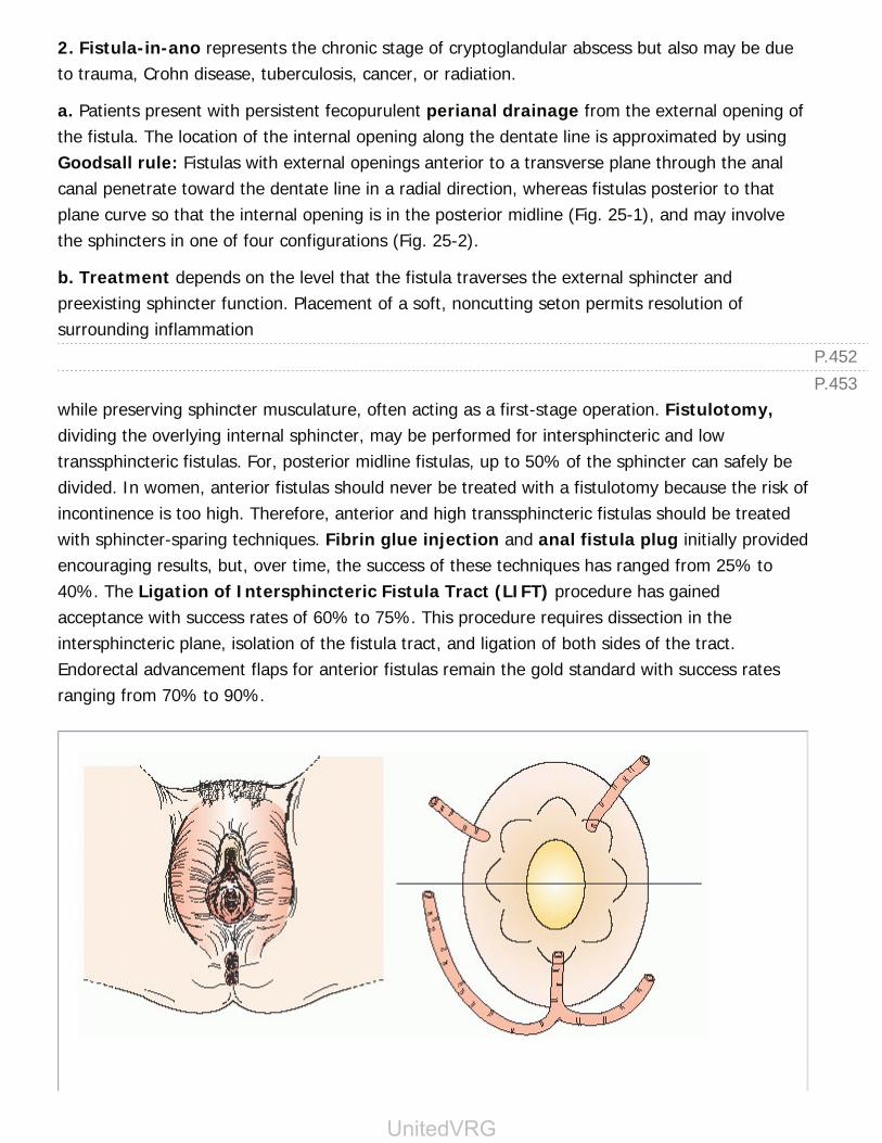
P.452P.453
2. Fistula-in-ano represents the chronic stage of cryptoglandular abscess but also may be dueto trauma, Crohn disease, tuberculosis, cancer, or radiation.
a. Patients present with persistent fecopurulent perianal drainage from the external opening ofthe fistula. The location of the internal opening along the dentate line is approximated by usingGoodsall rule: Fistulas with external openings anterior to a transverse plane through the analcanal penetrate toward the dentate line in a radial direction, whereas fistulas posterior to thatplane curve so that the internal opening is in the posterior midline (Fig. 25-1), and may involvethe sphincters in one of four configurations (Fig. 25-2).
b. Treatment depends on the level that the fistula traverses the external sphincter andpreexisting sphincter function. Placement of a soft, noncutting seton permits resolution ofsurrounding inflammation
while preserving sphincter musculature, often acting as a first-stage operation. Fistulotomy,dividing the overlying internal sphincter, may be performed for intersphincteric and lowtranssphincteric fistulas. For, posterior midline fistulas, up to 50% of the sphincter can safely bedivided. In women, anterior fistulas should never be treated with a fistulotomy because the risk ofincontinence is too high. Therefore, anterior and high transsphincteric fistulas should be treatedwith sphincter-sparing techniques. Fibrin glue injection and anal fistula plug initially providedencouraging results, but, over time, the success of these techniques has ranged from 25% to40%. The Ligation of Intersphincteric Fistula Tract (LIFT) procedure has gainedacceptance with success rates of 60% to 75%. This procedure requires dissection in theintersphincteric plane, isolation of the fistula tract, and ligation of both sides of the tract.Endorectal advancement flaps for anterior fistulas remain the gold standard with success ratesranging from 70% to 90%.
UnitedVRG

Figure 25-1 Goodsall rule. The anteriorÑposterior location of the external opening of thefistula helps to identify the internal opening of the fistula.
Figure 25-2 The four main anatomic types of fistula. (From Mulholland MW, Lillemoe KD,Doherty GM, et al. Greenfield's surgery: scientific principles and practice, 4th ed. New York,NY: Lippincott Williams and Wilkins, 2005, with permission.)
B. Necrotizing anorectal infection (Fournier gangrene) can result in massive, life-threatening tissue destruction. Patients present with systemic toxicity and perianal pain. Theremay be crepitance and extensive necrosis under relatively normal skin. Synergistic flora (includingclostridial and streptococcal species) of anorectal and urogenital origin may be involved.Immediate wide surgical debridement of all nonviable tissue and intravenous antibiotics aremandatory. Early treatment is critical, but mortality still approximates 50%. Fecal diversion is

P.454
rarely needed as most patients can be managed with a medical colostomy (constipation) andTPN.
C. Pilonidal disease occurs secondary to infection of a hair-containing sinus in the postsacralintergluteal fold, 5 cm superior to the anus. Patients present with pain, swelling, and drainagewhen the sinuses become infected. The disease is most prevalent in men in the second and thirddecades of life. Symptoms are distinguished from perianal abscess by the lack of anal pain, themore superior location of the fluctuant mass, and the presence of midline cutaneous pits.Treatment is incision, drainage, and curettage, with allowance for secondary closure when thesinus is acutely inflamed. The disease tends to recur, however, and once the active inflammationhas resolved, the sinus can be excised electively, with primary closure and a higher chance ofcure.
D. Hidradenitis suppurativa is an infection of the apocrine sweat glands and mimics fistula-in-ano except that involvement is external to the anal verge and has superficial tracts. Thetreatment of choice is wide incision of the involved skin.
E. Pruritus ani is a common symptom of hemorrhoids, fissure, rectal prolapse, rectal polyp, analwarts, and intraepithelial dysplasia of squamous or apocrine gland origin. Treatment is directedtoward resolution of the underlying cause. Failure to find an underlying cause should promptinvestigation of dietary factors (e.g., coffee and alcohol). Children should be
evaluated for pinworms, which, if found, are treated with piperazine citrate. However, as themost common cause is over cleaning, therapy includes a high-fiber diet, minimizing wiping, andavoiding the use of soaps and alcohol-containing products.
F. Condyloma acuminatum is an anorectal and urogenital wart caused by infection withhuman papilloma virus (HPV). The virus is sexually transmitted and presents with visible perianalgrowth, often accompanied by pruritus, anal discharge, bleeding, and pain. Common treatmentsinclude topical trichloroacetic acid, Aldara (imiquimod), or excision with electrocoagulation underlocal anesthesia. The role of HPV vaccination is as yet unknown, but vaccination is not effective ifadministered after infection. Smoke generated by coagulation contains viable organisms and mustbe completely evacuated. Anal canal warts must be destroyed at the same time as external warts.Biopsies should be obtained looking for highgrade squamous intraepithelial lesions as malignanttransformation can occur resulting in squamous carcinoma, requiring treatment according toNigro protocol.
V. ANAL NEOPLASMSA. Tumors of the Anal Margin. Any atypical anal lesions should be biopsied to attempt todiagnose these lesions at the earliest stage possible.
1. Squamous cell carcinoma behaves like cutaneous squamous cell carcinoma, is welldifferentiated and keratinizing, and is treated with wide local excision and chemoradiation if large
UnitedVRG
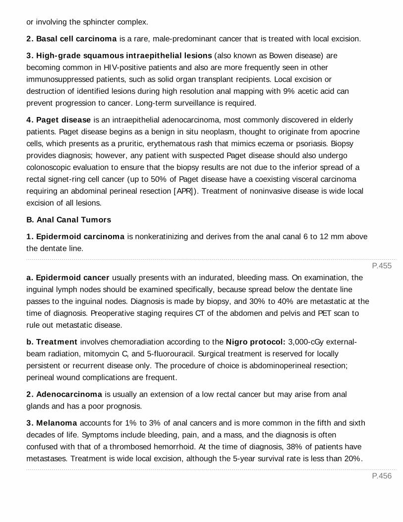
P.455
P.456
or involving the sphincter complex.
2. Basal cell carcinoma is a rare, male-predominant cancer that is treated with local excision.
3. High-grade squamous intraepithelial lesions (also known as Bowen disease) arebecoming common in HIV-positive patients and also are more frequently seen in otherimmunosuppressed patients, such as solid organ transplant recipients. Local excision ordestruction of identified lesions during high resolution anal mapping with 9% acetic acid canprevent progression to cancer. Long-term surveillance is required.
4. Paget disease is an intraepithelial adenocarcinoma, most commonly discovered in elderlypatients. Paget disease begins as a benign in situ neoplasm, thought to originate from apocrinecells, which presents as a pruritic, erythematous rash that mimics eczema or psoriasis. Biopsyprovides diagnosis; however, any patient with suspected Paget disease should also undergocolonoscopic evaluation to ensure that the biopsy results are not due to the inferior spread of arectal signet-ring cell cancer (up to 50% of Paget disease have a coexisting visceral carcinomarequiring an abdominal perineal resection [APR]). Treatment of noninvasive disease is wide localexcision of all lesions.
B. Anal Canal Tumors
1. Epidermoid carcinoma is nonkeratinizing and derives from the anal canal 6 to 12 mm abovethe dentate line.
a. Epidermoid cancer usually presents with an indurated, bleeding mass. On examination, theinguinal lymph nodes should be examined specifically, because spread below the dentate linepasses to the inguinal nodes. Diagnosis is made by biopsy, and 30% to 40% are metastatic at thetime of diagnosis. Preoperative staging requires CT of the abdomen and pelvis and PET scan torule out metastatic disease.
b. Treatment involves chemoradiation according to the Nigro protocol: 3,000-cGy external-beam radiation, mitomycin C, and 5-fluorouracil. Surgical treatment is reserved for locallypersistent or recurrent disease only. The procedure of choice is abdominoperineal resection;perineal wound complications are frequent.
2. Adenocarcinoma is usually an extension of a low rectal cancer but may arise from analglands and has a poor prognosis.
3. Melanoma accounts for 1% to 3% of anal cancers and is more common in the fifth and sixthdecades of life. Symptoms include bleeding, pain, and a mass, and the diagnosis is oftenconfused with that of a thrombosed hemorrhoid. At the time of diagnosis, 38% of patients havemetastases. Treatment is wide local excision, although the 5-year survival rate is less than 20%.
CHAPTER 25: ANORECTAL DISEASEMultiple Choice Questions1. A 41-year-old woman has a feeling of fullness after defecation andbright red blood on the toilet paper, especially after straining. Physicalexamination reveals two large, pink nonreducible columns of mucosaprotruding from her anal canal. Which is the most likely complicationfollowing surgical treatment of her anorectal disease?
a. Sphincter injury
b. Bleeding
c. Infection
d. Urinary retention
e. Anal stenosis
View Answer
2. A 68-year-old man has perianal mucus and pain. Physicalexamination reveals a fistula. On examination under anesthesia, youdiscover the fistula crosses the internal and external anal sphincters.Which is the most appropriate treatment at this time?
a. Fistulotomy using electrocautery over entire fistula tract
b. Division of the internal sphincter using electrocautery and placement ofseton encircling the external sphincter
c. Diverting colostomy
d. Antibiotics only
e. Anal advancement flap
View Answer
3. The goal of distal transection beyond gross tumor in rectal cancer is:
a. 2 cm because there is no correlation between local recurrence and theextent of distal margin when it is greater than 2 cm.
b. 5 cm because 30% of patients have microscopic evidence of intramuralspread beyond 3 cm from the palpable tumor.
c. 4 cm as local recurrence rates correlate with distal resection marginsless than 4 cm.
d. Just beyond gross tumor (<5 mm).
e. 3 cm because only 30% of patients have microscopic evidence ofintramural spread beyond that.
View Answer
UnitedVRG

P.457
4. Which of the following is correct regarding the anal sphinctermechanism?
a. The internal sphincter is innervated by the autonomic nervous system;thus it is subject to voluntary control.
b. The increase in pressure during voluntary contraction (squeezepressure) is due to the external sphincter.
c. The external sphincter accounts for the majority of resting pressure.
d. Distal rectal distention causes involuntary relaxation of the externalsphincter.
e. The rectal-anal inhibitory reflex refers to the external sphincter.
View Answer
5. A 54-year-old woman has anal pain and blood on the toilet tissueafter defecation. Physical examination reveals a 2 × 2 cm ulcer withinthe anal canal. Biopsy of the ulcer returns as squamous cell carcinoma.Which of the following is the appropriate treatment?
a. Low anterior resection with diverting ileostomy
b. Abdominoperineal resection
c. Primary chemoradiation therapy
d. Wide local excision, skin grafting, permanent colostomy
e. Wide local excision and primary closure
View Answer
6. A 75-year-old man has a perianal pruritic, erythematous rash. Biopsyreveals Paget disease. Which of the following is correct regarding Pagetdisease?
a. Regardless of level of invasion, all patients require abdominoperinealresection.
b. Since Paget disease is an intraepithelial neoplasm, patients do notrequire colonoscopy.
c. Extramammary Paget disease is most common in 20- to 30-year-oldwomen.
d. Fulguration with laser or electrocautery is the most effective treatment.
e. Wide local excision is the most appropriate treatment in noninvasivedisease.
View Answer
7. A 26-year-old man has severe anal pain during and after bowelmovements for the past 6 weeks. Physical examination reveals a split inthe anoderm in the posterior midline. Which of the following is correctregarding anal fissures?
a. Lateral sphincterotomy is only effective in 30% to 40% of cases.
b. Fissurectomy is the mainstay of treatment.
c. Ninety percent of patients heal with medical treatment, including fiber,sitz baths, and topical nifedipine ointment.
d. An equal number of fissures occur posteriorly as anteriorly.
e. The anal fissure triad consists of internal hemorrhoid, sentinel skin tag,and fissure.
View Answer
UnitedVRG

26Cerebrovascular Disease
Babafemi (Wande) B. Pratt
Jeffrey Jim
Gregorio A. Sicard
CEREBROVASCULAR DISEASEAtherosclerotic occlusive disease of the extracranial carotid artery is a major risk factor for stroke,the primary cause of disability, and the fifth most common cause of death in the United States.Nearly 800,000 new strokes occur annually, with an estimated total cost of more than $40 billion.The initial mortality from stroke is approximately 30%. Among those who survive the initial strokeevent, 60% suffer long-term disability, and 40% recover with mild or no deficits.
PRESENTATIONThe clinical presentation of patients with symptomatic occlusive disease is a neurologic deficit.However, many patients have asymptomatic stenosis that is identified by a healthcare providerbased on auscultation of carotid bruits or screening duplex ultrasound. Lateralizing ischemicevents can result in aphasia (expressive or receptive), combined sensory and motor deficits, andvarious visual disturbances. Deficits such as these are usually associated with the anteriorcerebral circulation (i.e., the internal carotid artery [ICA] and its branches).
Transient ischemic attacks (TIAs) are transient hemispheric neurologic deficits that may lastfrom several seconds to hours, but no longer than 24 hours. TIAs that occur in rapid succession,interspersed with complete recovery, but with progressively smaller intervals between attacks, aretermed crescendo TIAs, and carry a high risk of progression to a permanent neurologic deficit;emergent evaluation is mandatory. Amaurosis fugax (temporary monocular blindness)Ñoftendescribed as a shade coming down over one eyeÑresults from atheroemboli lodging in theophthalmic artery. Fundoscopic examination demonstrates Hollenhorst plaques.
If the neurologic deficit persists beyond 24 hours, it is considered a stroke. In addition, somepatients may present with a neurologic deficit that fluctuates, gradually worsening over a periodof hours or days, while the patient is under observation. This situation is considered a stroke inevolution, and similar to crescendo TIAs, demands immediate attention. Unlike crescendo TIAs,symptoms of stroke in evolution do not return to baseline. Global ischemic events aremanifested by symptoms such as vertigo, dizziness, perioral numbness, ataxia, or drop attacks.These are usually associated with interruption of the posterior circulation supplying the brain stem
> Table of Contents > 26 - Cerebrovascular Disease
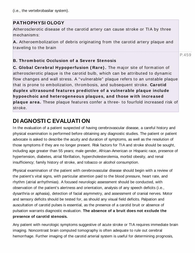
P.459
(i.e., the vertebrobasilar system).
PATHOPHYSIOLOGYAtherosclerotic disease of the carotid artery can cause stroke or TIA by threemechanisms:
A. Atheroembolization of debris originating from the carotid artery plaque andtraveling to the brain
B. Thrombotic Occlusion of a Severe Stenosis
C. Global Cerebral Hypoperfusion (Rare). The major site of formation ofatherosclerotic plaque is the carotid bulb, which can be attributed to dynamicflow changes and wall stress. A “vulnerable” plaque refers to an unstable plaquethat is prone to embolization, thrombosis, and subsequent stroke. Carotidduplex ultrasound features predictive of a vulnerable plaque includehypoechoic and heterogeneous plaques, and those with increasedplaque area. These plaque features confer a three- to fourfold increased risk ofstroke.
DIAGNOSTIC EVALUATIONIn the evaluation of a patient suspected of having cerebrovascular disease, a careful history andphysical examination is performed before obtaining any diagnostic studies. The patient or patientadvocate is asked to describe the acuity and duration of symptoms, as well as the resolution ofthose symptoms if they are no longer present. Risk factors for TIA and stroke should be sought,including age greater than 55 years; male gender, African-American or Hispanic race, presence ofhypertension, diabetes, atrial fibrillation, hypercholesterolemia, morbid obesity, and renalinsufficiency; family history of stroke, and tobacco or alcohol consumption.
Physical examination of the patient with cerebrovascular disease should begin with a review ofthe patient's vital signs, with particular attention paid to the blood pressure, heart rate, andrhythm (atrial arrhythmias). A focused neurologic assessment should be conducted, withobservation of the patient's alertness and orientation, analysis of any speech deficits (i.e.,dysarthria or aphasia), detection of facial asymmetry, and assessment of cranial nerves. Motorand sensory deficits should be tested for, as should any visual field deficits. Palpation andauscultation of carotid pulses is essential, as the presence of a carotid bruit or absence ofpulsation warrants diagnostic evaluation. The absence of a bruit does not exclude thepresence of carotid stenosis.
Any patient with neurologic symptoms suggestive of acute stroke or TIA requires immediate brainimaging. Noncontrast brain computed tomography is often adequate to rule out cerebralhemorrhage. Further imaging of the carotid arterial system is useful for determining prognosis,

P.460
and can be employed to classify the degree of stenosis. Because of methodologic differences incalculating the percentage of stenosis encountered in different studies, there is somedisagreement about exact cutoff percentages. However, four levels of stenosis are typicallydescribed: Mild (<50%), moderate (50% to 79%), severe (80% to 99%), andoccluded (100%). For consideration of optimal treatment strategies, a variety of noninvasiveand invasive diagnostic studies are available:
A. Color-flow duplex scanning uses real-time B-mode ultrasound and color-enhanced pulsedDoppler flow measurements to determine the extent of the carotid stenosis. This is the initialscreening test for carotid disease. When using an accredited vascular laboratory, carotidendarterectomy (CEA) can be undertaken on the basis of ultrasound duplex scanning alone in upto 95% of cases. However, the vascular laboratory must have clear and validated criteria formeasuring carotid stenosis, well-trained technologists, and must provide information about plaquemorphology and the extent of distal ICA involvement.
B. Conventional catheter angiography is the gold standard for imaging the extracranial andintracranial circulation in cerebrovascular disease. Unlike duplex scanning, angiography is aninvasive procedure with inherent risks, such as contrast allergy, renal toxicity, and stroke (lessthan 1% of patients). Because of these risks, and improvements in duplex ultrasonography,carotid angiography is generally limited to patients with technically inadequate duplexultrasonography, patients felt to be candidates for carotid artery stenting (CAS), patients in whomcarotid occlusion must be verified, or patients with discordant noninvasive study findings.
C. Magnetic resonance angiography (MRA) and computed tomographic angiography(CTA) are reliable means of supplementary imaging, but remain inferior to conventionalangiography. Both have advantages of being noninvasive, with the former allowing for theavoidance of radiation exposure. Limitations of MRA include poor sensitivity and specificity fordiagnosing 50% to 69% stenoses, the presence of flow-related artifacts, and a tendency tooverestimate stenosis severity. However, image quality can be improved after administration ofgadolinium. CTA can be performed with excellent three-dimensional reconstructions, but carriesthe risk of contrast-induced nephropathy. For patients with complete occlusion and contralateralhigh-grade occlusion, results of duplex ultrasound are often confirmed by CTA, or occasionally byangiography. CTA has the advantage of being fast, inexpensive compared to MRA, and offeringexceptional spatial resolution and visualization of soft tissues, bone, and vessels. It also is usefulto define the extracranial and intracranial vascular anatomy, and the extent of vessel calcification,particularly in the aortic arch.
MANAGEMENTA. Best Medical Therapy. It is important to make every effort to modify risk factors to preventprogression of carotid occlusive disease. Control of hypertension and hyperglycemia, cessation ofsmoking, management of lipid disorders, attainment of ideal body weight, and regular exercise

should be undertaken, and are the cornerstones of medical therapy. The components of bestmedical therapy are summarized in Table 26-1. No drug therapy has been shown to reduce therisk of stroke in patients with asymptomatic carotid disease. Medical management in symptomaticpatients is focused primarily on the use of antiplatelet agents, specifically aspirin. Aspirin iseffective in reducing stroke and stroke-related deaths. In a meta-analysis of more than 8,000patients, the risk of major vascular events was reduced by 22% in patients receiving aspirin (BrMed J. 1988;296:320). Low doses (81 mg/day) are as efficacious as higher doses (325 mg/day).Clopidogrel (Plavix) is a potent antiplatelet agent, but it has not been evaluated as part of medicaltherapy compared to CEA. Anticoagulation with heparin is beneficial in patients who have cardiacemboli. In addition, heparin may be useful in preventing progression of thrombus in evolvingnonhemorrhagic strokes. The major contraindication to heparinization is a recent hemorrhagicbrain infarct; therefore, a CT scan of the brain should be obtained before heparin is given.
TABLE 26-1 Summary of Effect of Best Medical Therapy
Best Medical Therapy for Prevention of Stroke and Cardiovascular Events
Treatment Target Evidence
Antiplatelettherapy
1. Either single ordual antiplatelettherapyacceptable
2. Aspirin 81 to 325mg/day isrecommended
3. Plavix 75 mgequivalent toaspirin
4. Ticlopidine 250mg twice dailysimilar to aspirinin effectiveness
Reduces both stroke risk and overallcardiovascular morbidity
Antihypertensivetherapy
1. Decrease systolicblood pressureby 10 mm Hg
Reduces stroke recurrenceReduce blood pressure 24 hrs after acutestroke
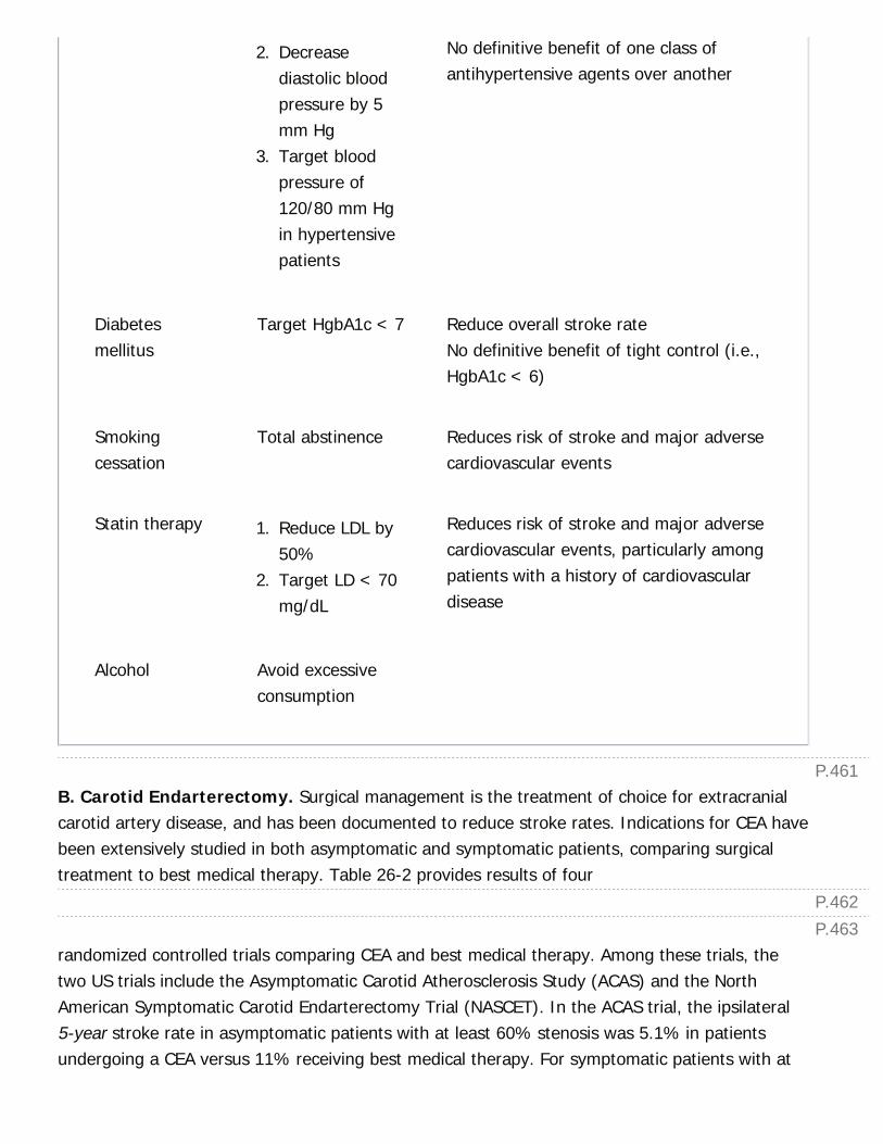
P.462P.463
P.461
2. Decreasediastolic bloodpressure by 5mm Hg
3. Target bloodpressure of120/80 mm Hgin hypertensivepatients
No definitive benefit of one class ofantihypertensive agents over another
Diabetesmellitus
Target HgbA1c < 7 Reduce overall stroke rateNo definitive benefit of tight control (i.e.,HgbA1c < 6)
Smokingcessation
Total abstinence Reduces risk of stroke and major adversecardiovascular events
Statin therapy 1. Reduce LDL by50%
2. Target LD < 70mg/dL
Reduces risk of stroke and major adversecardiovascular events, particularly amongpatients with a history of cardiovasculardisease
Alcohol Avoid excessiveconsumption
B. Carotid Endarterectomy. Surgical management is the treatment of choice for extracranialcarotid artery disease, and has been documented to reduce stroke rates. Indications for CEA havebeen extensively studied in both asymptomatic and symptomatic patients, comparing surgicaltreatment to best medical therapy. Table 26-2 provides results of four
randomized controlled trials comparing CEA and best medical therapy. Among these trials, thetwo US trials include the Asymptomatic Carotid Atherosclerosis Study (ACAS) and the NorthAmerican Symptomatic Carotid Endarterectomy Trial (NASCET). In the ACAS trial, the ipsilateral5-year stroke rate in asymptomatic patients with at least 60% stenosis was 5.1% in patientsundergoing a CEA versus 11% receiving best medical therapy. For symptomatic patients with at
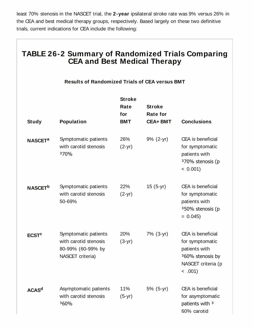
least 70% stenosis in the NASCET trial, the 2-year ipsilateral stroke rate was 9% versus 26% inthe CEA and best medical therapy groups, respectively. Based largely on these two definitivetrials, current indications for CEA include the following:
TABLE 26-2 Summary of Randomized Trials ComparingCEA and Best Medical Therapy
Results of Randomized Trials of CEA versus BMT
Study Population
StrokeRateforBMT
StrokeRate forCEA+BMT Conclusions
NASCETa Symptomatic patientswith carotid stenosis≥70%
26%(2-yr)
9% (2-yr) CEA is beneficialfor symptomaticpatients with≥70% stenosis (p< 0.001)
NASCETb Symptomatic patientswith carotid stenosis50-69%
22%(2-yr)
15 (5-yr) CEA is beneficialfor symptomaticpatients with≥50% stenosis (p= 0.045)
ECSTc Symptomatic patientswith carotid stenosis80-99% (60-99% byNASCET criteria)
20%(3-yr)
7% (3-yr) CEA is beneficialfor symptomaticpatients with≥60% stenosis byNASCET criteria (p< .001)
ACASd Asymptomatic patientswith carotid stenosis≥60%
11%(5-yr)
5% (5-yr) CEA is beneficialfor asymptomaticpatients with ≥60% carotid
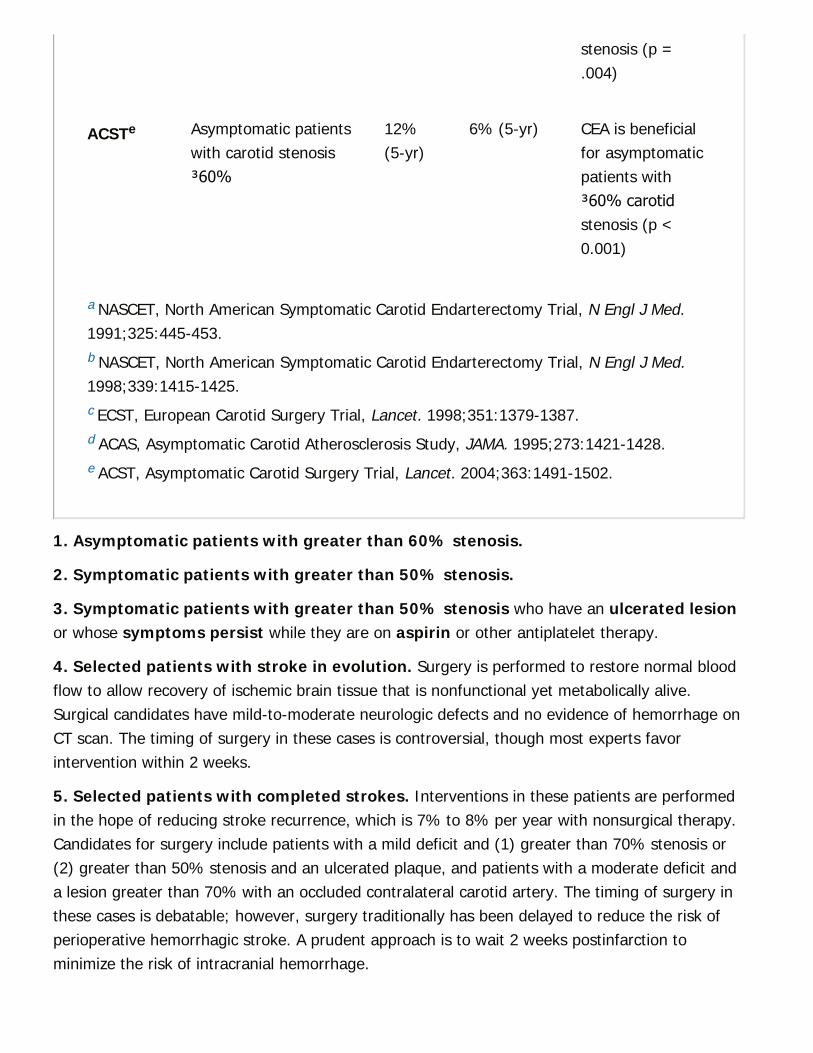
stenosis (p =.004)
ACSTe Asymptomatic patientswith carotid stenosis≥60%
12%(5-yr)
6% (5-yr) CEA is beneficialfor asymptomaticpatients with≥60% carotidstenosis (p <0.001)
a NASCET, North American Symptomatic Carotid Endarterectomy Trial, N Engl J Med.1991;325:445-453.b NASCET, North American Symptomatic Carotid Endarterectomy Trial, N Engl J Med.1998;339:1415-1425.c ECST, European Carotid Surgery Trial, Lancet. 1998;351:1379-1387.d ACAS, Asymptomatic Carotid Atherosclerosis Study, JAMA. 1995;273:1421-1428.e ACST, Asymptomatic Carotid Surgery Trial, Lancet. 2004;363:1491-1502.
1. Asymptomatic patients with greater than 60% stenosis.
2. Symptomatic patients with greater than 50% stenosis.
3. Symptomatic patients with greater than 50% stenosis who have an ulcerated lesionor whose symptoms persist while they are on aspirin or other antiplatelet therapy.
4. Selected patients with stroke in evolution. Surgery is performed to restore normal bloodflow to allow recovery of ischemic brain tissue that is nonfunctional yet metabolically alive.Surgical candidates have mild-to-moderate neurologic defects and no evidence of hemorrhage onCT scan. The timing of surgery in these cases is controversial, though most experts favorintervention within 2 weeks.
5. Selected patients with completed strokes. Interventions in these patients are performedin the hope of reducing stroke recurrence, which is 7% to 8% per year with nonsurgical therapy.Candidates for surgery include patients with a mild deficit and (1) greater than 70% stenosis or(2) greater than 50% stenosis and an ulcerated plaque, and patients with a moderate deficit anda lesion greater than 70% with an occluded contralateral carotid artery. The timing of surgery inthese cases is debatable; however, surgery traditionally has been delayed to reduce the risk ofperioperative hemorrhagic stroke. A prudent approach is to wait 2 weeks postinfarction tominimize the risk of intracranial hemorrhage.
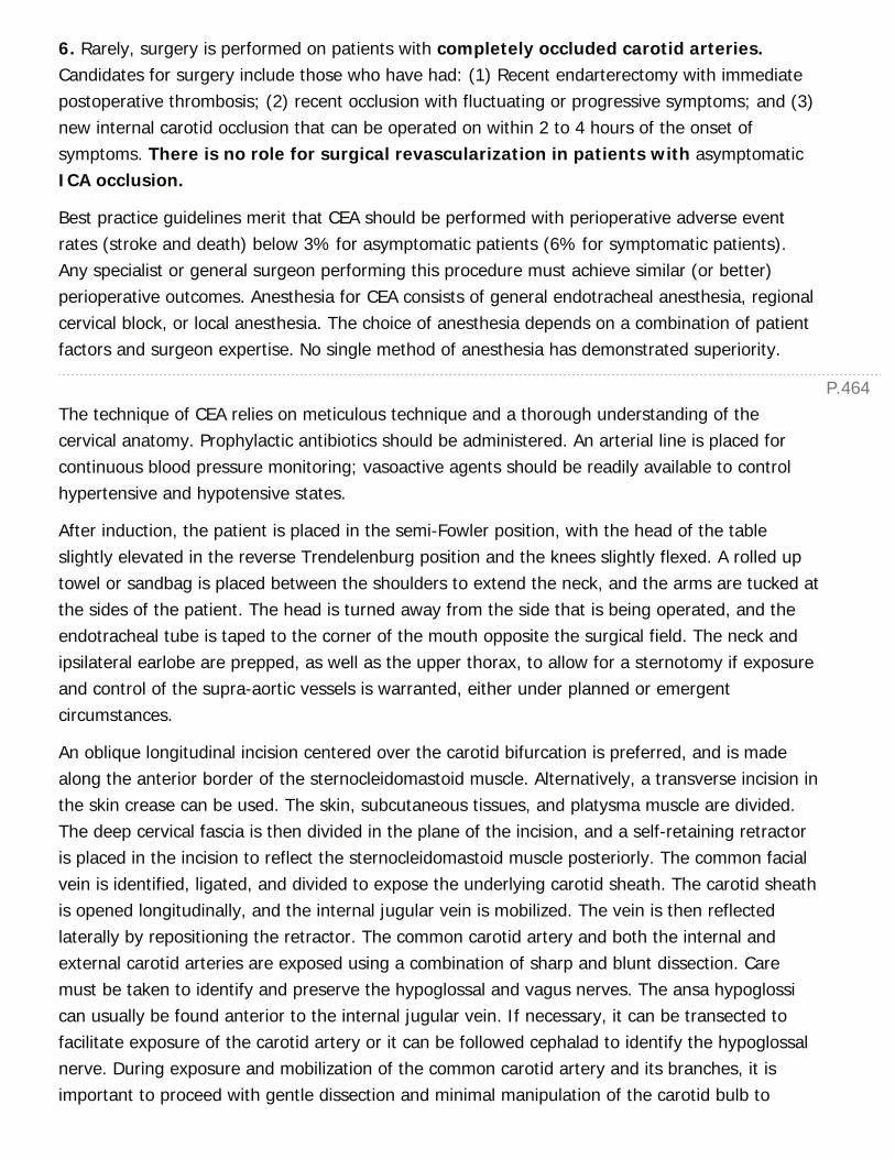
P.464
6. Rarely, surgery is performed on patients with completely occluded carotid arteries.Candidates for surgery include those who have had: (1) Recent endarterectomy with immediatepostoperative thrombosis; (2) recent occlusion with fluctuating or progressive symptoms; and (3)new internal carotid occlusion that can be operated on within 2 to 4 hours of the onset ofsymptoms. There is no role for surgical revascularization in patients with asymptomaticICA occlusion.
Best practice guidelines merit that CEA should be performed with perioperative adverse eventrates (stroke and death) below 3% for asymptomatic patients (6% for symptomatic patients).Any specialist or general surgeon performing this procedure must achieve similar (or better)perioperative outcomes. Anesthesia for CEA consists of general endotracheal anesthesia, regionalcervical block, or local anesthesia. The choice of anesthesia depends on a combination of patientfactors and surgeon expertise. No single method of anesthesia has demonstrated superiority.
The technique of CEA relies on meticulous technique and a thorough understanding of thecervical anatomy. Prophylactic antibiotics should be administered. An arterial line is placed forcontinuous blood pressure monitoring; vasoactive agents should be readily available to controlhypertensive and hypotensive states.
After induction, the patient is placed in the semi-Fowler position, with the head of the tableslightly elevated in the reverse Trendelenburg position and the knees slightly flexed. A rolled uptowel or sandbag is placed between the shoulders to extend the neck, and the arms are tucked atthe sides of the patient. The head is turned away from the side that is being operated, and theendotracheal tube is taped to the corner of the mouth opposite the surgical field. The neck andipsilateral earlobe are prepped, as well as the upper thorax, to allow for a sternotomy if exposureand control of the supra-aortic vessels is warranted, either under planned or emergentcircumstances.
An oblique longitudinal incision centered over the carotid bifurcation is preferred, and is madealong the anterior border of the sternocleidomastoid muscle. Alternatively, a transverse incision inthe skin crease can be used. The skin, subcutaneous tissues, and platysma muscle are divided.The deep cervical fascia is then divided in the plane of the incision, and a self-retaining retractoris placed in the incision to reflect the sternocleidomastoid muscle posteriorly. The common facialvein is identified, ligated, and divided to expose the underlying carotid sheath. The carotid sheathis opened longitudinally, and the internal jugular vein is mobilized. The vein is then reflectedlaterally by repositioning the retractor. The common carotid artery and both the internal andexternal carotid arteries are exposed using a combination of sharp and blunt dissection. Caremust be taken to identify and preserve the hypoglossal and vagus nerves. The ansa hypoglossican usually be found anterior to the internal jugular vein. If necessary, it can be transected tofacilitate exposure of the carotid artery or it can be followed cephalad to identify the hypoglossalnerve. During exposure and mobilization of the common carotid artery and its branches, it isimportant to proceed with gentle dissection and minimal manipulation of the carotid bulb to
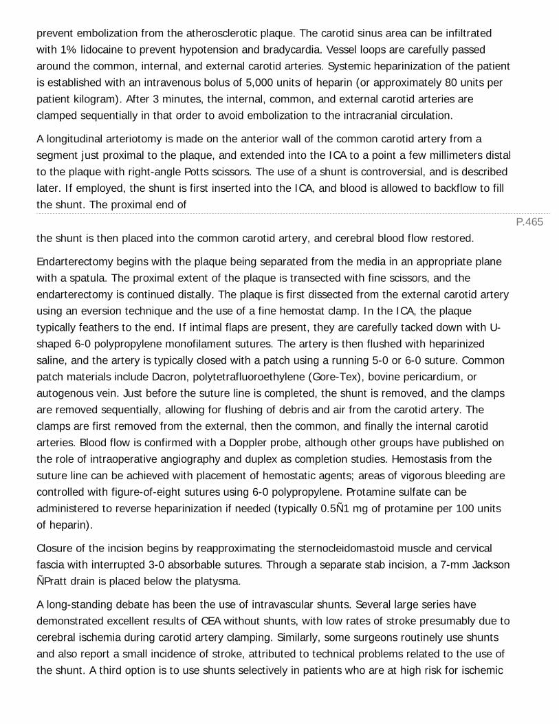
P.465
prevent embolization from the atherosclerotic plaque. The carotid sinus area can be infiltratedwith 1% lidocaine to prevent hypotension and bradycardia. Vessel loops are carefully passedaround the common, internal, and external carotid arteries. Systemic heparinization of the patientis established with an intravenous bolus of 5,000 units of heparin (or approximately 80 units perpatient kilogram). After 3 minutes, the internal, common, and external carotid arteries areclamped sequentially in that order to avoid embolization to the intracranial circulation.
A longitudinal arteriotomy is made on the anterior wall of the common carotid artery from asegment just proximal to the plaque, and extended into the ICA to a point a few millimeters distalto the plaque with right-angle Potts scissors. The use of a shunt is controversial, and is describedlater. If employed, the shunt is first inserted into the ICA, and blood is allowed to backflow to fillthe shunt. The proximal end of
the shunt is then placed into the common carotid artery, and cerebral blood flow restored.
Endarterectomy begins with the plaque being separated from the media in an appropriate planewith a spatula. The proximal extent of the plaque is transected with fine scissors, and theendarterectomy is continued distally. The plaque is first dissected from the external carotid arteryusing an eversion technique and the use of a fine hemostat clamp. In the ICA, the plaquetypically feathers to the end. If intimal flaps are present, they are carefully tacked down with U-shaped 6-0 polypropylene monofilament sutures. The artery is then flushed with heparinizedsaline, and the artery is typically closed with a patch using a running 5-0 or 6-0 suture. Commonpatch materials include Dacron, polytetrafluoroethylene (Gore-Tex), bovine pericardium, orautogenous vein. Just before the suture line is completed, the shunt is removed, and the clampsare removed sequentially, allowing for flushing of debris and air from the carotid artery. Theclamps are first removed from the external, then the common, and finally the internal carotidarteries. Blood flow is confirmed with a Doppler probe, although other groups have published onthe role of intraoperative angiography and duplex as completion studies. Hemostasis from thesuture line can be achieved with placement of hemostatic agents; areas of vigorous bleeding arecontrolled with figure-of-eight sutures using 6-0 polypropylene. Protamine sulfate can beadministered to reverse heparinization if needed (typically 0.5Ñ1 mg of protamine per 100 unitsof heparin).
Closure of the incision begins by reapproximating the sternocleidomastoid muscle and cervicalfascia with interrupted 3-0 absorbable sutures. Through a separate stab incision, a 7-mm JacksonÑPratt drain is placed below the platysma.
A long-standing debate has been the use of intravascular shunts. Several large series havedemonstrated excellent results of CEA without shunts, with low rates of stroke presumably due tocerebral ischemia during carotid artery clamping. Similarly, some surgeons routinely use shuntsand also report a small incidence of stroke, attributed to technical problems related to the use ofthe shunt. A third option is to use shunts selectively in patients who are at high risk for ischemic
P.466
stroke. Intraoperative techniques to identify patients who need a shunt include intraoperativemeasurement of carotid stump pressure after the common and external carotid arteries havebeen clamped, intraoperative neurologic monitoring with EEG or somatosensory evokedpotentials, measurement of middle cerebral artery flow by transcranial Doppler ultrasound, andmonitoring with cerebral oximetry.
Placement of an intravascular shunt from the common carotid artery to the ICA duringendarterectomy is a controversial practice. Adequate cerebral perfusion without shunting occursin 85% to 90% of patients. Awake assessment is the most sensitive and specific method ofdetermining the need for shunt placement. Intraoperative neurologic assessment of the awakepatient under local anesthesia can be as simple as having the patient squeeze a noise toy in thecontralateral hand and
answering a few simple questions after carotid occlusion. Patients who develop weakness orchanges in mental status should be shunted. For patients under general anesthesia, stumppressures lower than 50 mm Hg have been found to correlate with cerebral ischemia by EEGcriteria, and thus should be shunted.
C. Postoperative Care. Immediately after endarterectomy, neurologic function and bloodpressure alterations should be monitored. Hypertension and hypotension are common afterendarterectomy and may cause neurologic complications. The extremes of blood pressure shouldbe treated with either sodium nitroprusside or phenylephrine to keep the systolic blood pressurewithin 20 mm Hg of preoperative levels (slightly higher in chronically hypertensive patients). Thewound should be examined for hematoma formation. Aspirin is resumed in the immediatepostoperative period. Some advocate the use of dextran-40 (up to 20 mL/kg/day for up to 72hours) as an additional antithrombotic agent, which can be started intraoperatively and continuedinto the early postoperative period. Dextran-40 is an intravascular volume expander and shouldtherefore be used with caution in patients with preexisting congestive heart failure.
D. Patient Followup. A baseline duplex scan is obtained 1 month after the procedure, andagain at 12 months. Patients can then be followed annually. Patients who can tolerate aspirin aregiven 81 or 325 mg/day.
E. Complications
1. Stroke rates must be low (3% for asymptomatic patients; 6% for symptomatic) to makeoperative management of cerebrovascular disease reasonable, especially in asymptomaticpatients.
2. Myocardial infarction remains the most common cause of death in the early postoperativeperiod. As many as 25% of patients, who undergo endarterectomy have severe, correctablecoronary artery lesions. The timing of coronary intervention relative to CEA is under debate.
3. Cranial nerve injuries, the majority of which are transient, occur in 5% to 10% of patients who

P.467
undergo CEA. The most commonly injured nerve is the hypoglossal nerve, followed by therecurrent laryngeal, superior laryngeal, marginal mandibular, and glossopharyngeal nerves.
4. Recurrent carotid stenosis has been reported to occur in 5% to 10% of cases, althoughsymptoms are present in fewer than 3%. Recurrent stenoses are more common among women,patients who continue to smoke, and in those who have hyperlipidemia, diabetes, orhypertension. Early recurrent stenosis typically develops within 2 years of CEA, and results fromneointimal hyperplasia. Lesions that develop more than 2 to 3 years after CEA generally resultfrom progressive or new atherosclerotic disease. The presence of symptoms is an indication fortreatment of a recurrent lesion. Frequently, these lesions do not lend themselves toendarterectomy and are best treated by CAS.
5. Cerebral hyperperfusion syndrome is a feared complication that usually occurs severaldays after CEA. The incidence is 0.5% to 5% in published reports. It is often associated withsevere hypertension,
and patients initially complain of a severe headache. Intracranial hemorrhage is the mostdevastating consequence of this phenomenon. Patients suspected of having cerebralhyperperfusion syndrome should undergo noncontrast brain computed tomography to rule outcerebral hemorrhage, and should be admitted, preferably to an intensive care unit. Strict controlof systemic blood pressure and administration of antiseizure medications is paramount.
F. Carotid Artery Stenting. The indications for CAS are the same as those for a CEA; however,this technique remains under investigation. Several studies have been completed or are underway to examine the efficacy of CAS compared to CEA, particularly in high-risk patients. Outcomeshave varied, especially as device technology and operator experience have improved. The CarotidRevascularization Endarterectomy versus Stenting Trial (CREST) study randomly assigned patientswith symptomatic or asymptomatic carotid stenosis to undergo CAS or CEA. This trial showed thatthe risk of composite stroke, MI, or death did not differ significantly in the group undergoing CASand the group undergoing CEA. During the periprocedural period, there was a higher risk ofstroke with stenting and a higher risk of MI, defined as an elevation in troponin, withendarterectomy. However, the patients who suffered a stroke were more affected than those whohad an MI. Because CEA is well tolerated and has a very low risk of complications, CAS iscommonly reserved for high-risk patients, including (but not limited to) patients with the followingconditions:
1. Severe cardiac disease, including NYHA class III or IV congestive heart failure, left ventricularejection fraction <30%, recent unstable angina or myocardial infarction
2. Prior ipsilateral neck surgery
3. Prior neck radiation
4. Contralateral vocal cord paralysis or recurrent laryngeal nerve injury
P.468
5. Surgically inaccessible lesion (i.e., C2 or higher based on appropriate imaging)
Relative contraindications to CAS include severe tortuosity of the common and internal carotidarteries; complex aortic arch anatomy (increasing difficulty as great vessels arise from ascendingrather than transverse aortic arch); severe calcification or extensive thrombus formation; near-complete or complete ICA occlusion.
Carotid stenting has evolved since its introduction. Meticulous technique is critical for reducing theincidence of stroke. Special attention must be taken to avoid catheter and wire manipulation ofthe lesion prior to cerebral protection device deployment, and to ensure removal of all air bubbleswithin the angiography tubing. Retrograde femoral access, followed by a 6F introducer accesssheath, is the most commonly used technique. Other approaches include transcervical (via a 2- to3-cm neck incision and surgical exposure of the common carotid artery) and transbrachial (usinga 6F introducer access sheath) access. When the common carotid artery has been reached, anarteriogram is obtained with anteroposterior and lateral projections to obtain minimal overlap
of the internal and external carotid arteries. After angiography, the stenosis is crossed and anembolic protection device is deployed into the ICA. The typical device used today is a filter-likedevice that is advanced across the lesion and then opened in the distal ICA prior to angioplastyand stent deployment. After placement of the embolic protection device, predilatation of thestenosed artery is performed if necessary. Self-expanding stents are deployed across the carotidbifurcation and dilated as necessary. These come in a variety of diameters (6 to 10 mm), lengths(3 to 4 cm), and shapes (cylindrical or tapered). The diameter is typically sized to the diameter ofthe distal common carotid artery.
G. Patient Followup. Followup using duplex ultrasound is important to identify patients withrestenosis and is usually performed at baseline following CAS, then at 1 month, and at least everyyear thereafter. Significant elevation of peak systolic velocity from baseline should prompt furtherevaluation with angiography. Patients should receive antiplatelet therapy with Clopidogrel for aminimum of 3 months.
H. Complications
1. Embolic stroke is the most common complication of CAS. Risk factors include lack of a cerebralprotection device, long or multiple lesions, and age above 80 years. Thrombolysis may be asuccessful treatment option, especially if the source of emboli is an acute thrombus. When anembolus is composed of atheroma or chronic thrombus, however, mechanical removal of embolimay become necessary to restore flow.
2. Hemodynamic instability may occur during manipulation and angioplasty of the carotidbifurcation. Bradycardia should be anticipated and pretreated with atropine or glycopyrrolate priorto dilation of the carotid bifurcation. Postoperatively, as with CEA, patients should be monitoredto avoid extremes of blood pressure.
P.469
3. Restenosis occurs in approximately 5% of patients at 12 to 24 months and is typicallysecondary to intimal hyperplasia. These lesions are often amenable to repeat endovascularintervention.
The ROADSTER trial is a multicenter clinical study intended to support premarket approval of theSilk Road Medical Carotid Stent System and Neuroprotection System. The Silk Road procedure isa hybrid approach to carotid intervention, pairing the advantages of a transcervical approach withthe benefits of reverse flow embolic protection and stenting technology. Direct carotid access isaccomplished via a small 2- to 3-cm incision above the clavicle, and an arterial access sheath isplaced. A second access sheath is placed in the femoral vein, and the two sheaths are connectedby a flow controller to divert embolic debris from the ICA after clamping of the proximal commoncarotid artery. The debris is trapped in an inline filter and never enters the venous circulation.The results of the trial showed a 30-day composite stroke, death, and MI rate of 3.5%. Thestroke rate of 1.4% to date represents the lowest rate for stenting in Òhigh-riskÓ patients, and iscomparable to results of Òstandard-riskÓ patients in the CEA arm of CREST.
CHAPTER 26: CEREBROVASCULAR DISEASEMultiple Choice Questions1. Which one of the following is not considered a risk factor for strokeand TIA?
a. Age greater than 55 years
b. African-American or Hispanic race
c. Female gender
d. Hypertension
e. Smoking
View Answer
2. A 67-year-old male is referred to your clinic by his primary carephysician after a routine check-up revealed a right carotid bruit. Asubsequent duplex ultrasound revealed 50% stenosis of his rightcarotid artery. According to the ACAS trial, carotid endarterectomy forasymptomatic patients is indicated for stenoses defined by which of thefollowing?
a. ³90%
b. ³80%
c. ³70%
d. ³60%
P.470
e. ³50%
View Answer
3. According to the NASCET trial, what is the approximate reduction instroke rate over 2 years if CEA is completed for a symptomatic patientwith 70% stenosis of her left carotid artery?
a. 6% absolute risk reduction (i.e., 11% control vs. 5% CEA)
b. 7% absolute risk reduction (i.e., 22% control vs. 15% CEA)
c. 13% absolute risk reduction (i.e., 20% control vs. 7% CEA)
d. 17% absolute risk reduction (i.e., 26% control vs. 9% CEA)
e. 20% absolute risk reduction (i.e., 25% control vs. 5% CEA)
View Answer
4. Which of the following represents the major cranial nerve mostcommonly injured during carotid endarterectomy?
a. The vagus nerve
b. The hypoglossal nerve
c. The glossopharyngeal nerve
d. The facial nerve
e. The recurrent laryngeal nerve
View Answer
5. Which of the following veins is commonly ligated during carotidendarterectomy?
a. The facial vein
b. The internal jugular vein
c. The superior thyroid vein
d. The anterior jugular vein
e. The subclavian vein
View Answer
6. The first intracranial branch of the internal carotid artery is:
a. The superior thyroid artery
b. The inferior thyroid artery
c. The lingual artery
d. The ophthalmic artery
e. None of the above (there are no intracranial branches of the internalcarotid artery)
View Answer
7. The first branch of the external carotid artery is:
a. The superior thyroid artery
b. The inferior thyroid artery
c. The lingual artery
d. The ophthalmic artery
e. None of the above
View Answer
8. A 72-year-old female with a history of TIAs is found to have 80%stenosis of the left carotid artery on duplex ultrasound. The mostappropriate next step in management is:
a. Repeat ultrasound in 6 months
b. Start Aspirin and Clopidogrel
c. Start Aspirin only
d. Recommend carotid stenting
e. Recommend carotid endarterectomy
View Answer
9. The following, by themselves, are all acceptable indications forcarotid artery stenting, except:
a. Surgically inaccessible lesion
b. Contralateral internal carotid artery occlusion
c. Contralateral vocal cord paralysis
d. Prior ipsilateral neck surgery
e. Prior neck radiation
View Answer
10. While undergoing carotid stenting, a patient develops bradycardiaand hypotension, during predilatation of a stenotic region. This is mostlikely due to which process?
a. Global cerebral hypoperfusion
b. Myocardial infarction
c. Stretching of the carotid body

d. Stimulation of the hypoglossal nerve
e. Formation of atheroembolism
View Answer
27Thoracoabdominal Vascular Disease
J. Westley Ohman
Chandu Vemuri
Luis A. Sanchez
The majority of vascular diseases are secondary to atherosclerotic changes of the arterial wallwhich are multifactorial and are influenced by genetic predisposition, age, and environmentalfactorsÑparticularly diet, smoking, and exercise. The arterial wall is comprised of the intimal,medial, and adventitial layers. Endothelium lines the intima. The media contains layers of smoothmuscle cells and extracellular matrix (ECM) proteins. The adventitia is composed of looseconnective tissue and fibroblasts. A true aneurysm is a permanent localized dilation of all threelayers (intima, media, adventitia) greater than 50% of the normal or adjacent vessel diameter.Degenerative disorders of the thoracoabdominal aorta affect the media most, whereas occlusivedisorders result in luminal narrowing from atherosclerosis.
I. ABDOMINAL AORTIC ANEURYSMS.This is the most common type of aortic aneurysm, occurring in 3% to 9% of people above 50years of age in the Western world (Br J Surg. 1998;85:155. PMID: 9501808). In the UnitedStates, ruptured abdominal aortic aneurysms (AAAs) are the 13th leading cause of death overalland the 10th leading cause of death in men above 55 years, a rate that has held steady for thepast two decades despite improvements in operative technique and perioperative management.
A. Pathophysiology. Ninety percent of AAAs are degenerative in origin, 5% are inflammatory,and the remainder are idiopathic (J Vasc Surg. 2003;38:584). The most accepted predictor ofrupture is maximal diameter, and the major risk factors for aortic dilation. Several trials (UKSAT;Lancet. 1998;352(9141):1649-1655; and ADAM; Ann Intern Med. 1997;126(6):441-449) haveestablished a risk of rupture of 0.6% to 1%/year, with the risk rising to 10% at 6 cm, 20% at 6.5cm, and 30% at 7.5 cm. Eighty-five percent of AAAs are infrarenal; 25% involve the iliac arteries,and 2% involve the renal or other visceral arteries (J Cardiovasc Surg. 1991;32:636). Fourteenpercent are associated with peripheral (e.g., femoral or popliteal) aneurysms (J Vasc Surg.2000;31:863). However, 62% of patients with popliteal aneurysms and 82% with femoralaneurysms have an associated AAA, mandating screening in these patients.
B. Diagnosis
1. Clinical manifestations. Seventy-five percent of AAAs are asymptomatic and are found
> Table of Contents > 27 - Thoracoabdominal Vascular Disease
P.472
incidentally. Aneurysm expansion or rupture may cause severe back, flank, or abdominal pain andvarying degrees of shock. Distal embolization, thrombosis, and duodenal or ureteral compressioncan produce symptoms. Fifty percent of AAAs are identifiable
on physical examination as a pulsatile mass at or above the umbilicus. AAA rupture may mimicrenal colic, peritonitis, duodenal perforation, pancreatitis, degenerative spine disease, acute diskherniation, or myocardial infarction.
2. Radiologic evaluation
a. Ultrasonography and computed tomography (CT) scanning. Ultrasound is an acceptedscreening study for the presence of AAAs with sensitivity of 87% and specificity nearing 100%;however for patients nearing size criteria for repair the study of choice is CT angiography (J VascSurg. 2009;50(4 Suppl):S2-S49).
b. Magnetic resonance (MR) scan is comparable to CT but avoids radiation exposure and isuseful in patients with intravenous contrast contraindications.
c. Aortography is not sensitive for the diagnosis of AAA because the study by definition is aluminogram and as such will underestimate the total aortic size due to the presence of muralthrombus.
C. Elective Management of AAA. The risk of aneurysm rupture correlates best with aneurysmsize (Ann Surg. 1966;164:678). However, even small aneurysms can rupture.
1. Medical management. Patients with small aneurysms (<4.5 cm in diameter) without riskfactors for rupture can be followed using ultrasound or CT scan yearly, with larger ones beingfollowed more frequently. Smoking cessation, exercise, control of hypertension, and treatment ofchronic pulmonary obstructive disease are critical (J Vasc Surg. 2002;35:72).
2. Elective surgical treatment. Operative mortality ranges from less than 5% foruncomplicated AAA to greater than 50% for ruptured AAA (Br J Surg. 1998;85:1624). Five-yearsurvival after elective repair is no different from that for age-matched patients without AAA.Associated cardiovascular disease, hypertension, decreased renal function, chronic obstructivelung disease, and morbid obesity increase operative risk (J Vasc Surg 2009;50(4 Suppl):S2-S49.PMID: 19786250). Indications for surgical management include (J Vasc Surg. 2003;37:1106):
a. Symptomatic aneurysms of any size.
b. Aneurysms exceeding 5.5 cm diameter (5 cm for females).
c. Increase in diameter by more than 0.5 cm/6 months or 1 cm/year.
d. Saccular aneurysms.
e. Relative contraindications to elective repair include recent myocardial infarction, intractablecongestive heart failure, unreconstructable coronary artery disease, life expectancy of less than 2
P.473
years, and incapacitating neurologic deficits after a stroke.
3. Open operative technique. The two open surgical approaches are the transabdominal andretroperitoneal approaches. In the transabdominal repair, the aneurysm is approached through amidline abdominal incision and exposed by eviscerating the small bowel, and mobilizing the leftcolon before incising the retroperitoneum. Next, the duodenum and left renal vein are dissectedoff the aorta. After systemic heparinization,
the aorta and iliac arteries are cross-clamped. An aortotomy is made and extended longitudinallyto the aneurysm Òneck,Ó where the aorta is either transected or cut in a T fashion. Thethrombus is removed, and any bleeding lumbar arteries are oversewn. The proximal anastomosisis performed to nonaneurysmal aorta using a graft. The distal anastomosis is completed at theaortic bifurcation (tube graft) or at the iliac or femoral arteries (bifurcated graft), as the extent ofthe aneurysmal disease dictates. The aneurysm wall is then closed over the graft. An alternativeapproach is through a left retroperitoneal incision. This approach is advantageous in obesepatients, those with chronic pulmonary obstructive disease, patients with previous intraabdominalsurgery, and is the preferred approach at Washington University in St. Louis. In addition, proximalsuprarenal or supraceliac control of the aorta is more easily achieved via this latter approach.
D. Management of Ruptured AAA
1. Preoperative management. Unstable patients with a presumed diagnosis of a rupturedaneurysm (hypotension, abdominal or back pain, and a pulsatile abdominal mass or history ofaneurysmal disease) are gently resuscitated with fluids (crystalloid, colloid, or blood) to maintainorgan perfusion while avoiding over-resuscitation with the associated diluting of clotting factors,destabilization of thrombus, and hemorrhage (so called Òpermissive hypotensionÓ). Unstablepatients are transferred immediately to the operating room for exploration, whereas those whoare stable should undergo emergent CT scanning to confirm the diagnosis.
2. Operative management is aimed at rapidly controlling the aorta. Anesthetic induction isdelayed until incision. Through a midline incision, the supraceliac aorta is controlled at thediaphragmatic hiatus by dividing the pars flaccida, mobilizing the left lateral segment of the livertoward the patient's right, and isolating the aorta from the adjacent esophagus and attainingvascular control with an aortic clamp, or direct pressure against the spine. The retroperitonealhematoma is opened, and the proximal neck of the aneurysm is identified and cross-clamped.Distal vessel dissection continues, and management is similar to repair of an elective AAA. Theuse of bifurcated grafts should be avoided in favor of the more expeditious tube graftreconstruction. Heparin should also be avoided in these patients who are likely to already becoagulopathic due to the high risk of intraoperative and postoperative bleeding. However, incenters with EVAR capabilities, the endovascular approach has become the preference for initialapproach to a ruptured AAA.
3. Complications from open AAA repair
P.474
a. Arrhythmia, myocardial ischemia, or infarction.
b. Intraoperative hemorrhage can be reduced by clamping the aorta proximal to theaneurysm and the iliac arteries distally. Once the aneurysm is opened, retrograde bleeding fromlumbar arteries must be controlled rapidly with transfixing ligatures. Blood should be salvaged inthe operating room and autotransfused to the patient.
c. Management of resuscitation, electrolytes, and pressors is critically important during aorticunclamping due to sudden hypotension from a decrease in systemic vascular resistance andintroduction of previously sequestered metabolites.
d. Renal insufficiency may be related to the use of intravenous contrast, inadequate hydration,hypotension, renal ischemia from a period of aortic clamping above the renal arteries, orembolization to the renal arteries.
e. Lower-extremity ischemia may result from embolism or thrombosis, especially inemergency operations for which heparin might not be used. This can be prevented by minimizingmanipulation of the aneurysm prior to clamping. Use of Fogarty balloon catheters to removedistal emboli from lower-extremity vessels are indicated when leg ischemia is identified in theoperating room.
f. Microemboli arising from atherosclerotic debris can cause cutaneous ischemia (trash foot),which is usually treated expectantly as long as the major vessels are patent. Amputation may berequired if significant necrosis results.
g. Gastrointestinal complications consist of prolonged paralytic ileus, anorexia, periodicconstipation, or diarrhea, which is diminished by using the left retroperitoneal approach.Ischemic colitis of the sigmoid colon is related to ligation of the inferior mesenteric artery(IMA) in the absence of adequate collateral circulation. Symptoms include leukocytosis, significantfluid requirement in the first 8 to 12 hours postoperatively, fever, and peritonitis. Diagnosis isconfirmed by flexible sigmoidoscopy to 20 cm above the anal verge. Necrosis that is limited to themucosa may be treated expectantly with intravenous antibiotics and bowel rest. Necrosis of themuscularis causes segmental strictures, which may require delayed segmental resection.Transmural necrosis requires immediate resection of necrotic colon and construction of an endcolostomy.
h. Paraplegia, a rare complication of infrarenal aneurysm surgery, may occur after repair of aruptured AAA due to spinal cord ischemia. Supraceliac cross-clamping, prolonged hypotension,and obliteration or embolization of important collateral flow to the spinal artery (internal iliacarteries or an abnormally low origin of the accessory spinal artery [artery of Adamkiewicz])increases the risk.
i. Sexual dysfunction and retrograde ejaculation result from damage to the sympathetic plexus(ÒNervi erigentesÓ) during dissection near the proximal left common iliac artery.

P.475
E. Endovascular management of AAAs has dramatically decreased the acute morbidity ofaneurysm surgery. Indications are no different than those for traditional open repair.
1. The most important selection criterion for endovascular treatment of an AAA is appropriateaortoiliac anatomy. Further advances in fenestrated and branched devices are allowing treatmentof pararenal and
other more complex AAAs. At this time, preoperative CT assessment includes the followingfactors:
a. Current devices require at least 15 mm of healthy infrarenal aorta.
b. Angle between the neck and aneurysm.
c. Intraluminal thrombus in the proximal neck represents a relative contraindication forendovascular treatment.
d. A cone-shaped neck or reverse taper (i.e., widens more distally) may preclude adequateapposition of the endograft to the aortic wall.
e. Iliac artery tortuosity, calcification, and luminal narrowing are critical factors for endograftdelivery and deployment.
f. Patent aortic branches may influence the decision as to whether to proceed with endovascularrepair. Large accessory renal artery or the presence of a horseshoe kidney with multiple renalarteries is often a contraindication for endograft placement. Patent lumbar arteries arising fromthe aneurysm do not preclude endograft placement. A large patent IMA suggests abnormalmesenteric blood supply and risk of large-bowel ischemia with endograft coverage of the IMAorifice.
2. Technique. Endovascular devices are introduced retrograde through open or percutaneousfemoral arterial access. Most commonly utilized devices are bifurcated and modular in design.Appropriate oversizing of stent grafts (by 20%) is based on preoperative assessment of arterialdiameter to ensure adequate graft apposition to the aortic wall. Length can be tailored byoverlapping extension segments. The distal end of the iliac limbs is typically positioned proximalto the hypogastric orifices to maintain pelvic perfusion.
3. Complications of endograft repairs of AAAs
a. Branch occlusion, distal embolization, graft thrombosis, and arterial injury(especially iliac artery avulsion at the iliac bifurcation).
b. Arterial dissection may occur. Additional stents can be used to treat the dissected vessel ifnecessary.
c. Bowel ischemia may occur postoperatively secondary to embolization or hypoperfusion, butthis is rare compared to open surgical repair. Renal dysfunction may occur because of the
P.476
nephrotoxicity of the contrast agent used for intraoperative angiography or because of directinjury due to embolization or renal artery occlusion.
d. Graft migration occurs in 1% to 6% of patients, and is associated with challenging arterialanatomy and poor graft placement. This complication can typically be treated with secondaryendovascular procedures.
e. Endoleak is defined as failure to fully exclude the aneurysmal sac from arterial blood flow,potentially predisposing to rupture (J Endovasc Surg. 1997;4:152. PMID: 9185003). Managementstrategies for endoleaks discovered on followup imaging studies are evolving. For any endoleakthat is associated with aneurysm sac enlargement, intervention is required. Endoleaks are usuallycorrected by endovascular means but may require conversion to open surgical repair.
1. In general, endoleaks from the proximal or distal attachment sites (type I) warrantintervention because of continued direct flow into the AAA. Options include: conversion to open,or angioplasty with proximal stent placement.
2. Type II endoleaks are due to collateral flow (IMA, lumbar arteries) and may be closelyobserved in the absence of aneurysm expansion. Type II endoleaks may be treated withembolization through collateral vessels or directly through injection of the aneurysm sac via atranslumbar approach, although the longterm outcomes are equivocal (J Vasc Surg.2012;55(5):1263Ñ1267. PMID: 22322122).
3. Type III endoleaks are caused by inadequate seal between graft components. They should becorrected as soon as they are diagnosed as they too result in direct flow in the AAA.
4. Type IV endoleaks are due to porosity of the graft material. This is rare with newergenerations of endografts but these must be treated if found.
4. Results. Relative to open surgical repair, endovascular treatment of AAA is associated with areduction in perioperative morbidity, shorter duration of hospitalization (J Vasc Surg.2003;37:262), and reduction in perioperative mortality (EVAR 1; Lancet. 2004;364:843-848;DREAM; NEJM. 2005;352:2398-2405). However, studies of long-term outcome comparing openversus endovascular repair have demonstrated similar rates of survival after 4 years (DREAM;NEJM. 2010;362:1881-1889; UK EVAR; NEJM. 2010;362:1863-1871). Close followup with CTscanning every 6 months for 1 year, and then yearly, is essential to maintaining long-term clinicalsuccess using this technique.
II. THORACIC AORTIC ANEURYSMS.Thoracic aortic aneurysms (TAAs) are primarily a disease of the elderly, with an estimatedincidence of 10.4 per 100,000 person-years (JAMA. 1998;280(22):1926). Ascending aorticaneurysms are most common (÷60%) followed by aneurysms of the descending aorta (÷35%)and of the transverse aortic arch (<10%). Most descending TAAs begin just distal to the orifice of

P.477
left subclavian artery.
A. Pathophysiology. TAAs are divided into five main types: Ascending, transverse, descending,thoracoabdominal, and traumatic. Ascending aortic aneurysms are usually caused by medialdegeneration. Transverse, descending, and thoracoabdominal aortic aneurysms are related toatherosclerosis with hypertension contributing to their expansion. Traumatic aneurysms areusually due to blunt injury to the chest.
B. Diagnosis
1. Clinical manifestations are usually absent; most are detected as incidental findings on chestimaging obtained for other purposes. A minority of patients may present with chest discomfort orpain that intensifies with aneurysm expansion or rupture, aortic valvular regurgitation, congestiveheart failure, compression of adjacent structures (recurrent laryngeal nerve, left main-stembronchus, esophagus,
superior vena cava), erosion into adjacent structures (esophagus, lung, airway), or distalembolization.
2. Radiologic evaluation.
a. Chest x-ray may reveal a widened mediastinum or an enlarged calcific aortic shadow.Traumatic aneurysms may be associated with skeletal fractures.
b. MR or CT imaging with intravenous contrast provides precise estimation of the size andextent of aneurysms and facilitates surgical planning.
c. Echocardiography may be useful in evaluating aneurysms involving the aortic arch.
d. Aortography demonstrates the proximal and distal extent of the aneurysm and itsrelationship with aortic branch vessels arising from it.
C. Surgical management varies by type and location of the TAA. Repair of proximal archaneurysms requires cardiopulmonary bypass and circulatory arrest. Preclotted wovenpolyethylene terephthalate (Dacron) is the graft of choice. Ascending and transverse arches arerepaired through a median sternotomy incision. Descending and thoracoabdominal aneurysms areapproached through a left posterolateral thoracotomy or thoracoabdominal incision.Intraoperative management of patients undergoing thoracotomy is facilitated by selectiveventilation of the right lung using a double-lumen endobronchial tube. Several adjuncts forlimiting postoperative paraplegia following surgery for descending and thoracoabdominalaneurysm are employed, including cerebrospinal fluid drainage and retrograde perfusion.
1. Ascending aortic arch aneurysms
a. Size criteria for TAA repair are not as clearly defined as for infrarenal AAAs. Indications forsurgical repair include symptomatic or rapidly expanding aneurysms, aneurysms greater than orequal to 6 cm in diameter, ascending (type A) aortic dissections, mycotic aneurysms, and

P.478
asymptomatic aneurysms greater than or equal to 5.5 cm in diameter in patients with Marfansyndrome (Coron Artery Dis. 2002;13:85).
b. Operative management. An aneurysm arising distal to the coronary ostia is replaced withan interposition graft. An aneurysm resulting in aortic valve incompetence is replaced with acomposite valved conduit (Bentall procedure) or a supracoronary graft with separate aorticvalve replacement. All ascending arch aneurysms due to connective tissue disorders are repairedwith aortic valve replacement owing to the high incidence of valvular incompetence associatedwith aneurysmal dilation of the native aortic root. When a composite graft is used, the coronaryarteries are anastomosed directly to the conduit.
2. Transverse aortic arch aneurysms
a. Indications for repair include aneurysms greater than or equal to 6 cm in diameter, aorticarch dissections, and ascending arch aneurysms that extend into the transverse arch.
b. Operative management. After opening the aorta under hypothermic circulatory arrest, thedistal anastomosis is performed using a
beveled graft, followed by anastomosis of a patch that incorporates the orifices of thebrachiocephalic vessels to the superior aspect of the graft. The proximal anastomosis isconstructed to the supracoronary aorta (if the aortic valve is not involved) or to a segment of thecomposite valved conduit interposed to complete the arch reconstruction. Involvement of thetransverse arch and its branch vessels requires interposition grafting to the involved vessels.
3. Descending TAAs
a. Indications for repair include asymptomatic aneurysms greater than or equal to 6 cm indiameter and any symptomatic aneurysms.
b. Operative management. After the distal clamp is applied, a proximal clamp is placed justdistal to the left subclavian artery or between the left common carotid and left subclavianarteries. Selected intercostal branches are reattached to the aortic interposition graft. Left heart(atriofemoral) bypass is often used, both to protect the heart from overdistention and to providedistal blood flow while the aorta is clamped. Cerebrospinal fluid drainage is used as an adjunct todecrease the incidence of postoperative paraplegia.
4. Thoracoabdominal aneurysms
a. Indications for repair include aneurysms greater than or equal to 6 cm in diameter and anysymptomatic aneurysms.
b. Operative management consists of tube graft replacement along with anastomosis of themajor visceral branches to the graft. Aneurysms involving the thoracic and proximal abdominalaortic segments may be approached through a left posterolateral thoracotomy extended to theumbilicus. Use of left heart (atriofemoral) bypass is a valuable adjunct to allow for both cerebral
P.479
perfusion and visceral perfusion off the bypass circuit via a femoral cannula. The thoracic aorta isclamped and opened to perform the proximal anastomosis. The aorta is clamped distally openingthe remaining aneurysm. The orifices of all major aortic branches are occluded with ballooncatheters or vascular clamps. Temporary perfusion can be maintained to those branches duringaneurysm repair by using balloon catheters connected to the atriofemoral bypass. Theanastomoses of significant aortic branches to the graft are performed as a patch or with separatebypasses. The clamp is moved to the graft below the renal arteries to reperfuse all visceralvessels in an antegrade fashion. The distal anastomosis is made either to the uninvolved aorta orto the iliac arteries.
5. Traumatic aortic aneurysms
a. Indications. Urgent repair is indicated, except when precluded by more compelling life-threatening injuries or major central nervous system trauma.
b. Classification. Traumatic aortic injuries have the following categories:
(1) Grade I: Intimal tear
(2) Grade II: Intramural hematoma
(3) Grade III: Pseudoaneurysm
(4) Grade IV: Rupture
c. Management. Historically, these aneurysms were repaired by primary aortorrhaphy,aneurysmectomy, and end-to-end reanastomosis or by interposition grafting. In current practice,Grade I injuries may be observed on serial imaging and Grade IIÑIV injuries should undergoprompt endovascular repair, if amenable. Age should not disqualify from endovascular repair (JVasc Surg. 2011;53(1):187Ñ192).
D. Possible complications of thoracic aortic surgery are similar to those for abdominal aorticsurgery. The incidence of paraplegia may be as high as 30% with some types of TAAs (AnnThorac Surg. 2007;83:S856). This risk can has been reduced to less than 2% by multimodaltherapies used to minimize spinal cord ischemia: Distal aortic perfusion, intercostal and lumbarartery reimplantation, pre- or intraoperative localization of spinal cord blood supply, hypothermia,cerebrospinal fluid drainage, and pharmacotherapy (World J Surg. 2008;32(3):355-360).
E. Endovascular Management of TAA
1. Indications and technique. Because of the considerable morbidity and mortality associatedwith surgical repair of descending thoracic aneurysms, the endovascular approach to aneurysmexclusion is particularly attractive. Treatment with endovascular stent graft placement requiresspecific anatomic criteria: Adequate length (2 cm) and diameter (20 to 45 mm) of the proximaland distal aneurysm necks, absence of significant mural thrombus within the sealing zones, andaortic and iliofemoral anatomy amenable to device introduction. In situations in which the
P.480
proximal neck length is too short, coverage of the left subclavian artery can be performed with orwithout an adjunctive left carotidÑleft subclavian transposition or bypass.
2. Results and complications. Reported results for the use of endovascular devices areencouraging, with low morbidity and mortality with high rates of aneurysm exclusion. Fenestratedand branched grafts along with hybrid techniques using extra-anatomic bypass procedures arebeing used to approach more complex aneurysm anatomy.
III. OTHER ARTERIAL ANEURYSMSA. Infected aneurysms have risen in incidence with the increased prevalence ofimmunocompromised patients and invasive transarterial procedures.
1. Pathophysiology. Infected aneurysms can be divided into four types: Mycotic aneurysm,microbial arteritis with aneurysm, infection of preexisting aneurysm, and posttraumatic infectedfalse aneurysm. Staphylococcus aureus is the most common pathogen, although Salmonellaspecies (arteritis), Streptococcus species, and Staphylococcus epidermidis (preexistinganeurysms) also may occur. Gram-negative infections have the highest risk of rupture.
2. Diagnosis
a. Clinical manifestations may be absent or include fever, tenderness, or sepsis. Physicalexamination may demonstrate a tender, warm, palpable mass in an infected peripheralaneurysm. Laboratory tests may
reveal leukocytosis. Aerobic and anaerobic blood cultures should be obtained, but are positive inonly 50% of patients.
b. MRI or CTA can demonstrate an aneurysm and verify its rupture. Angiography delineatesthe characteristics of the aneurysm. Aneurysms that are saccular, multilobed, or eccentric with anarrow neck are more likely a result of infection.
B. Management
1. Preoperative. Broad-spectrum antibiotics should be administered intravenously after aerobicand anaerobic blood cultures have been obtained.
2. Intraoperative. Goals of surgery include (1) controlling hemorrhage; (2) obtaining arterialspecimens for Gram stain, aerobic and anaerobic cultures, and drug sensitivities; (3) resecting theaneurysm with wide debridement and drainage; and (4) reconstructing major arteries throughuninfected tissue planes. Extra-anatomic bypass may be necessary to avoid contamination of thegraft. Inline reconstructions with antibiotic-impregnated grafts, cryopreserved homografts, ornative veins are alternatives that can be used for arterial reconstructions depending on thelocation of the aneurysm and the extent of the infection.
3. Postoperative. Adequate drainage of the aneurysm cavity and longterm antibiotic therapy for

P.481
at least 6 weeks typically are required.
IV. ACUTE AORTIC SYNDROMESA. Aortic dissection is a tear in the intima allowing blood to travel between the intima and themedia resulting in the creation of two flow channels: The true lumen and the Òfalse lumen.Ó Theincidence is ÷30 cases per million individuals per year (J Cardiovasc Surg (Torino). 2010;51:601-608), and the natural history is marked by a high mortality rate, as high as 1% per hour over thefirst 24 hours (Circulation. 1950;1:360-387). Dissections are classified according to the StanfordClassification system based on involvement of the ascending arch, which simplified the previouslyused DeBakey classification.
1. Diagnosis
a. Clinical manifestations include the hallmark sudden onset, ripping chest/abdominal pain,radiating to the back. There may be an associated blood pressure discrepancy between the upperextremities, new onset heart murmur, and, less commonly, paraplegia or paresthesias.
b. Risk factors include long-standing uncontrolled hypertension, family history, and collagenvascular diseases such as Marfan syndrome.
c. Radiographic tests
(1) CT Angiography remains the gold standard for diagnosis of dissections in the acute settingwith modern techniques having a sensitivity and specificity of 100% (Radiology. 1996;199:347-352), and gives accurate information regarding the location of the intimal injury, and anyassociated involvement of branch vessels.
(2) MR Angiography has been supplanted by CT Angiography in the acute setting, but is usedin long-term followup.
2. Type A dissections are those involving the proximal ascending aorta, and are treated assurgical emergencies. These are the most common type of dissection, accounting forapproximately 60% of all dissections. The high mortality rate is due to the feared progression ofretrograde dissection, hemopericardium, and cardiac tamponade.
a. Management of Type A dissection nearly universally involve emergent surgery withreplacement of the proximal ascending arch with or without valve replacement.
3. Type B dissections are those isolated to the descending thoracic aorta distal to the leftsubclavian. The mortality rates approach 10% at 30 days, 25% at 3 years, and 50% at 5 years.These are subdivided in ÒacuteÓ (<14 days) and ÒchronicÓ (>14 days) and are furthersubdivided into ÒuncomplicatedÓ or ÒcomplicatedÓ depending on evidence of malperfusion ofend organs.
a. Management of complicated Type B dissections is aimed at revascularization of the affected
P.482
segment through whatever means is most expedient, utilizing both endovascular and opensurgical options.
b. Management of uncomplicated Type B dissections has recently been treated with medicalmanagement including β-blockade and long-term antihypertensive regimens. Despite this,uncomplicated Type B dissections may progress to become complicated, and up to 25% requireintervention within 4 years (ADSORB trial. Eur J Vasc Endovasc Surg. 2014;48:285-291).
c. Surgical management either via direct graft replacement of the affected segment, removalof the flap via aortotomy, or endovascular fenestration and stenting have all been used astechniques to restore perfusion to malperfused end organs.
d. Endovascular coverage of the intimal tear in uncomplicated Type B dissections has becomethe standard of treatment. In the chronic setting, this has been shown to promote favorableaortic remodeling with improved mortality at 5 years (INSTEAD-XL, Circ Cardiovasc Interv.2013;6:407-416). Trials are ongoing in the acute setting, and also demonstrate favorable aorticremodeling (ADSORB trial. Eur J Vasc Endovasc Surg. 2014;48:285-291).
B. Intramural hematomas are a focal hemorrhage in the wall of the aorta between the intimaand media layer due to the rupture of the vasa vasorum without evidence of an intimal tear ordissection flap due to iatrogenic injuries, trauma, or hypertension. These are classified similar todissections, with those involving the ascending aorta being corrected surgically, and theremainder being medically managed unless the hematoma is >1 cm think (ninefold higherprogression), aortic diameter greater than 40 mm (30-fold higher risk of progression), orprogression on serial imaging (J Vasc Surg. 2002;35:1179-1183).
C. Penetrating aortic ulcer is a projection into an intramural hematoma that is associated withhigher rates of disease progression. These are associated with smoking and atheroscleroticdisease. These are classified and treated similar to intramural hematomas.
V. RENOVASCULAR DISEASE.Stenosis or occlusion of the renal arteries may result in hypertension, ischemic nephropathy, orboth. Renovascular hypertension is the most common form of surgically correctablesecondary hypertension.
A. There are several clinical features that may be used to identify patients with potentialrenovascular hypertension:
1. The onset of hypertension before the age of 18 or after the age of 55.
2. Accelerated, resistant, or malignant hypertension.
3. Unexplained impairment of renal function.
4. Refractory to appropriate multidrug therapy.

P.483
5. Hypertension in a patient with extensive coronary disease, cerebral vascular disease, orperipheral vascular disease.
B. The majority have a normal physical examination; however, an epigastric, subcostal, orflank bruit or findings of a unilateral small kidney on any imaging study are possible indicators.
C. Pathophysiology
1. Renal arterial stenosis (RAS) leads to activation of the reninÑangiotensin-aldosteronesystem by the ipsilateral kidney and results in volume expansion and peripheral vasoconstriction.Even in the absence of hypertension, RAS may lead to renal failure.
a. In acute renal failure, RAS should be considered in the differential diagnosis if the workup isunrevealing or the patient was recently started on an angiotensin-converting enzyme (ACE)inhibitor or another antihypertensive or diuretic.
b. RAS may account for up to 20% of unexplained chronic renal failure in patients older than50 years of age.
c. Isolated unilateral RAS generally do not cause rise in serum creatinine.
2. Atherosclerosis accounts for nearly 90% of cases of renovascular HTN and usually affectsthe ostia and proximal renal artery.
3. The second-most common renovascular lesion is fibromuscular dysplasia, most commonlymedial fibroplasia. These lesions are multifocal, with a string-of-beads appearance onangiography, and typically occur in young women.
D. Diagnosis. Testing for clinically significant renal artery disease must evaluate the anatomicand physiologic changes.
1. Arteriography remains the Ògold standardÓ for the diagnosis of anatomic RAS. However, theusual risks of arteriography, especially the nephrotoxic effects of the contrast agent, areimportant caveats to consider.
2. Duplex scanning is the preferred method for screening in patients with indicators ofrenovascular hypertension.
3. MR angiography is useful for evaluating kidney and main renal artery morphology withoutthe use of nephrotoxic agents.
4. Two rarely used tests to determine the functional significance of a renal artery lesion arecaptopril renal scintigraphy and selective renal vein renin measurement when otherworkup is unrevealing.
E. Management of Fibromuscular Disease
1. Rarely causes renal failure.
2. Endovascular techniques (balloon angioplasty) have a high success rate for thetreatment of this arterial pathology with 75% free from hypertension and 95% free fromreduction in GFR at 5 years (J Vasc Surg. 2012;55(2):421Ñ427). For failure of endovasculartreatment (see Section F.3), surgical therapy may be employed.
F. Management of atherosclerotic disease aims to control target organ damage fromhypertension and avoid progressive ischemic renal failure. Response to therapy is difficult topredict because a patient's hypertension may be primarily essential and the renal failure due tohypertensive glomerulosclerosis.
1. Medical therapy is often successful in the management of patients with renovascularhypertension and remains the cornerstone of treatment. A combination of β-blockers and acalcium channel blocker, an ACE inhibitor, or an angiotensin II-receptor inhibitor is commonlyused as first-line therapy.
2. Surgical therapy
a. Surgical revascularization is a durable option for patients with long segment disease anddeclining renal function (Ann Surg. 1999;230(4):524). In patients undergoing aortic surgery foraneurysmal or occlusive disease with concomitant renal stenoses, consideration should be givento renal revascularization.
b. Procedures
(1) Aortorenal bypass is the classic treatment of renal revascularization using saphenous vein,autologous hypogastric artery (in children), or prosthetic graft.
(2) Renal endarterectomy is another option and is often used for bilateral orificial lesions. Atransverse arteriotomy is made over the both orifices.
(3) Alternative bypass procedures are used for unfavorable anatomy. Grafts can be takenfrom the supraceliac aorta or the superior mesenteric, common hepatic, gastroduodenal, splenic,or iliac arteries. Results are comparable to direct aortic reconstruction with less morbidity andmortality.
(4) Nephrectomy may be required in patients who have unreconstructable disease or a normalcontralateral kidney and who are high-risk surgical candidates.
c. Postoperative care
(1) Immediately after operation, patients should be hydrated to maintain adequate urineoutput. Concern about the patency of the reconstruction may be addressed by a renal or duplexscan.
(2) Patient followup should consist of blood pressure monitoring, a renal scan, and creatininedetermination at 3 months, 12 months, and then yearly. Any recurrence of hypertension ordeterioration in renal function should prompt diagnostic imaging.

P.484d. Complications of surgery include persistent hypertension, acute renal failure, renal arteryrestenosis, thrombosis, aneurysm formation, and distal embolization.
3. Endovascular management of renal artery stenosis
a. Indications for angioplasty of RAS include failure of medical management of renovascularhypertension. Renal artery stents are used for restenosis after previous angioplasty, proceduralcomplications (e.g., dissection), and atherosclerotic ostial lesions.
b. Results. Technical success for renal artery angioplasty is defined as a less than 30% residualstenosis and a pressure gradient less than 10 mm Hg. For patients with atherosclerotic renalartery stenosis large studies (ASTRAL; NEJM. 2009;361:1953-1962 and CORAL; NEJM.2014;370:13-22) have shown no impact on renal function or major adverse renal andcardiovascular events compared to medical management.
VI. MESENTERIC ISCHEMIA.Mesenteric ischemia can be a difficult diagnosis to make because most patients are asymptomaticuntil late in the disease process. Although considerable advances have been made in theperioperative care as well as the diagnosis and treatment of intestinal ischemia, mortality remains60% to 80% (Langenbecks Arch Surg. 2008;393:163).
A. Acute Mesenteric Ischemia
1. Pathophysiology. The most common cause of acute mesenteric ischemia (AMI) isembolization to the SMA; other causes include thrombosis of the SMA or portomesenteric venousthrombosis. Patients with AMI often have multiple risk factors, including significant cardiacdisease (frequently atrial fibrillation) and severe atherosclerotic disease of nonmesenteric vessels,and may have a history consistent with chronic intestinal ischemia.
a. Abdominal pain is sudden in onset and intermittent at first, progressing to continuous severepain. It is often described as pain out-of-proportion to examination. These patients mayalso have bloody diarrhea.
b. Mesenteric venous thrombosis presents with varying manifestations, ranging from anasymptomatic state to catastrophic illness. Patients usually complain of prolonged, generalizedabdominal pain that develops somewhat less rapidly than with acute mesenteric arterial occlusion.These patients may have occult gastrointestinal bleeding but no frank hemorrhage.
2. Diagnosis
a. Angiography of the mesenteric circulation, including lateral views of the celiac axis and SMA,remains the gold standard. However, most centers use CT angiography especially for thediagnosis of AMI. Abdominal plain radiographs are of limited utility.
P.485
b. Other laboratory findings can include elevated white blood cell count with a left shift,persistent metabolic acidosis, and lactic acidosis in more advanced cases, but are insensitive andnonspecific
for the diagnosis of mesenteric ischemia (Langenbeck's Arch of Surg. 2011;396:3-11).
3. Surgical therapy
a. Patients with AMI frequently require intestinal resection; therefore, laparotomy with openrevascularization is the preferred method of treatment.
(1) Assessment of bowel viability at laparotomy is made based on the gross characteristics ofthe bowel. The bowel is likely viable if it appears pink and if arterial pulsations are present in theadjacent vascular arcades. Other techniques have been described, including the use of fluoresceindye, Doppler studies, and tissue oximetry, but these are not substitutes for experienced clinicaljudgment.
(2) Second-look procedures are prudent when bowel viability is questionable. This isespecially important in patients who have extensive bowel involvement and in whom resection ofall questionable areas could result in short-bowel syndrome.
b. For venous occlusion, surgical intervention or lytic therapy rarely is helpful. Systemicanticoagulation should begin as soon as the diagnosis is made to limit progression of thethrombotic process. Frequently, the diagnosis is made at laparotomy. If the diagnosis is madebefore exploration, however, operation should be reserved until evidence of bowel infarctionexists.
c. Nonocclusive mesenteric ischemia (NOMI) is intestinal ischemia in the absence ofthromboembolic occlusion. It occurs in patients with a low-cardiac-output state and chronicintestinal angina. Mortality associated with NOMI is high and treatment is directed towardimproving circulatory support and increasing cardiac output. In cases in which cardiac recovery isexpected, intraarterial infusions of vasodilators (e.g., Papaverine, Prostaglandin E1, nitroglycerin)
have been attempted, without clear benefit.
4. Perioperative care usually requires maximal medical support; are frequentlyhemodynamically unstable and develop multiple organ system failure. Admission to the intensivecare unit, prolonged endotracheal intubation, parenteral nutrition, and broad-spectrum antibiotictherapy are typically required.
B. Chronic Mesenteric Ischemia (CMI)
1. Patients with CMI present with intestinal angina, which is pain usually beginning within anhour after eating and abating within 4 hours (postprandial pain). Such patients experiencesignificant weight loss related to the decreased intake secondary to recurrent pain (food fear).The diagnosis usually is made from obtaining a thorough history alone because physical findings
P.486
P.487
are usually lacking.
2. Surgical therapy should be reserved for symptomatic patients. Surgical revascularization viabypass or endarterectomy remains the treatment of choice. Studies comparing surgery andendovascular approaches show higher patency in the surgical group, with no significant differencein
2-year mortality, recurrence of symptoms or imaging findings, or reintervention (J Vasc Surg.2007;45(6):1162-1171).
3. Perioperative care. These patients often are malnourished. Some advocate parenteralnutrition for 1 to 2 weeks before surgery, and continued postoperatively. Some patients develop arevascularization syndrome consisting of abdominal pain, tachycardia, leukocytosis, and intestinaledema. Concern about the adequacy of revascularization should prompt diagnostic imaging.
4. Complications include intestinal infarction, perforation, prolonged multisystem organ failure,and need for dialysis.
CHAPTER 27: THORACOABDOMINAL VASCULAR DISEASEMultiple Choice Questions1. Which of the following is not associated with development of anabdominal aortic aneurysm (AAA)?
a. DM
b. HTN
c. HLD
d. Smoking
e. Positive family history
View Answer
2. Which of the following is not a useful diagnostic modality for AAA?
a. CT
b. Ultrasound
c. MRI
d. Aortography
View Answer
3. Which of the following patients with AAA can be medically managedat this point in time?
a. A 75-year-old male with a 6.3-cm AAA

P.488
b. A 68-year-old female with an AAA that has grown from 4.9 to 5.3 cm inthe last year
c. A 72-year-old male with a known 4.8-cm AAA and intractable pain
d. A 70-year-old male with an AAA that has grown from 4.8 to 5.1 cm inthe last year
View Answer
4. 24 hours after undergoing elective AAA repair, the ICU nurse noticespurple-blackish discoloration to the toes on both feet of the patient. Hehas palpable pedal pulses, what is the next step in management?
a. CT angiogram
b. Operative reexploration
c. Angioplasty
d. Expectant management
e. Guillotine amputation
View Answer
5. 10 hours after undergoing elective AAA, the patient becomestachycardic, febrile to 39°C, and has required 5 L of fluid to maintain hisblood pressure goals, and develops diarrhea. His abdomen is diffuselytender and sigmoidoscopy reveals transmural necrosis of the mucosa.What is the next step?
a. IV hydration, antibiotics, and bowel rest
b. Delayed segmental resection
c. Emergent resection of the involved segment
d. Endoscopic placement of a rectal tube
e. Repeat sigmoidoscopy in 24 hours
View Answer
6. During elective EVAR, both hypogastric arteries are inadvertentlycovered by the graft. All of the following are potential consequences ofacute hypogastric artery occlusion EXCEPT:
a. Buttock claudication
b. Paraplegia
c. Small bowel ischemia
d. Colonic ischemia
e. Perineal skin necrosis
View Answer
7. Following endovascular repair of a ruptured AAA in which the patientreceived 12 units of blood, 2 units of platelets, and 10 units of plasma,he remains anuric, with peak airway pressures of 50 mm Hg leading todifficulty with adequate oxygenation, and his abdomen is distended.What is the next step?
a. Emergent laparotomy and decompression
b. Observation in ICU
c. Place the patient in prone positioning for oxygenation
d. Flush, and if necessary replace, Foley catheter
View Answer
8. Which of the following is true regarding the structure of the aorta?
a. The intima is composed of smooth muscle and extracellular matrixproteins.
b. The media is the layer most involved with atherosclerotic changes.
c. The adventia is composed of loose connective tissue and fibroblasts.
d. An aneurysm is dilation of the intima and the media, but not theadventia.
e. The majority of degeneration in an aneurysm develops in the adventia.
View Answer
9. All of the following are contraindications to endovascular repair ofAAA (EVAR) EXCEPT:
a. A patient with 4 mm of healthy tissue between the renal arteries andthe aneurysm
b. A patient with significant thrombus at the proximal landing zone
c. A patient with an occluded left hypogastric artery
d. A patient with a large IMA with a meandering mesenteric artery
e. A patient with a horseshoe kidney
View Answer
10. Which of the following is true with regard to endoleaks?
a. Type I endoleaks are via collateral circulation, and must be treated.
b. Type II endoleaks are due to leaks at proximal or distal components,
P.489
and may be observed.
c. Type III endoleaks are due to inadequate seal of the graft components.
d. Type IV endoleaks are due to neovascularization of the AAA sac.
e. Type V endoleaks are due to porosity of the graft material.
View Answer
11. Large-scale studies comparing EVAR to open AAA repair have shownreduction in all of the following with EVAR EXCEPT:
a. Perioperative morbidity
b. Duration of hospitalization
c. Perioperative mortality
d. 4-year survival rates
View Answer
12. A 78-year-old male is found to have a widened mediastinum onroutine CXR. A CT scan shows a 5 cm aortic aneurysm beginning distalto the left subclavian artery. Which of the following is true?
a. This aneurysm should be repaired immediately
b. Open repair is accomplished via a median sternotomy
c. Repair requires hypothermic circulatory arrest
d. In the absence of symptoms, this can safely be watched until 6 cm
e. There is no role for endovascular treatment of this aneurysm
View Answer
13. A 68-year-old male presents to the ED with sudden onset rippingchest pain radiating to his back with a systolic blood pressure of 210.He has no EKG changes, but CT angiogram shows an intimal flapbeginning in the ascending aortic segment. Which is the next step inmanagement?
a. Transesophageal echocardiography to confirm location of flap
b. Emergent open repair with conduit replacement
c. Admission to ICU with anti-impulse control
d. Endovascular coverage of intimal flap
e. Observation, as this is a uniformly fatal diagnosis
View Answer
P.490
14. A 56-year-old male presents to the ED with a worsening of rippingchest pain radiating to his back. He reports the pain began 3 weeksago, but has progressed this morning. His systolic blood pressure is205. A CT angiogram shows an intimal flap beginning distal to the leftsubclavian artery. He is admitted to the ICU and his pain improves withblood pressure control. Which of the following is true?
a. This patient should be evaluated for endovascular coverage of theintimal tear
b. This patient has an acute Type B dissection
c. No further intervention is warranted, as this has a low long-termmortality rate
d. This is a complicated Type B dissection
e. β-blockers should never be used in the care of this disease
View Answer
15. All of the following are clinical features of patients that may be usedto identify potential renovascular hypertension EXCEPT:
a. Onset in a young adult
b. Refractory to multidrug therapy
c. Accelerated onset
d. Unexplained renal impairment
e. Onset at the age of 45
View Answer
16. You are called to see a patient in the MICU with “abdominal painout of proportion to examination.” A 72-year-old female was withmarked heart failure due to viral myocarditis, requiring significantinotropic and vasopressor agents. She has begun passing bloody stoolswhile in the MICU. Due to acute kidney injury, a noncontrast CT isobtained, that shows minimal calcification of the aorta or any of thevisceral branches. Which of the following is true?
a. This condition has a low mortality rate
b. These patients are best managed with laparotomy and resection ofviable bowel
c. Treatment is directed at increasing cardiac output and circulatorysupport

d. Open revascularization is the preferred therapy
e. Enteral nutrition is indicated
View Answer
28Peripheral Arterial Disease
Sashi K. Inkollu
Patrick J. Geraghty
The predominant etiology of occlusive disease of the lower extremities is atherosclerotic change ofthe arterial intima and media. Major risk factors for developing atherosclerosis include cigarettesmoking, diabetes, dyslipidemia, hypertension, and hyperhomocysteinemia. Atheroscleroticdisease is a systemic illness, and although symptomatic disease may predominate in one organ,subclinical disease, particularly of the coronary arteries, is generally present. In fact, 50% of themortality associated with peripheral arterial reconstructions for atherosclerotic disease is cardiac innature. Other, less common causes of occlusive disease include fibromuscular dysplasia, radiation-induced vascular injury, and the vasculitides (e.g., Takayasu arteritis and Buerger disease).
ACUTE ARTERIAL OCCLUSION OF THE EXTREMITYSymptoms of acute arterial insufficiency occur abruptly. The presentation generally includes thesix Ps of acute ischemia: Pain, pallor, pulselessness, paresthesias, poikilothermy (coldextremity), and paralysis. The level of occlusion may be localized by the absence of pulses andthe proximal extent of coolness and sensorimotor changes. If adequate collateral circulation is notpresent, irreversible changes may appear as early as 4 to 6 hours after onset. Therefore, prioritymust be given to prompt restoration of blood flow.
I. ETIOLOGYA. The Most Common Cause of Acute Arterial Insufficiency is Embolization.
1. Cardiac sources account for more than 70% of emboli and are usually the result of muralthrombi that develop due to cardiac aneurysms following myocardial infarction or arrhythmiassuch as atrial fibrillation. Other cardiac sources of emboli include valvular heart disease, prostheticheart valves, bacterial endocarditis, and atrial myxoma.
2. ArterialÑarterial emboli can result from ulcerated atheroma or aneurysms, althoughembolization from abdominal aortic aneurysms is distinctly rare. The blue toe syndrome occurs inpatients with microemboli from unstable proximal arterial plaques and is characterized by intactpulses and painful ischemic lesions in the distal extremity. Atheroemboli in the lower extremity canalso occur secondary to plaque disruption by catheters. A severely diseased distal aorta in some ofthese patients is evident on computed tomography (CT) scan and arteriography and has beentermed shaggy aorta. Upper-extremity ischemia/gangrene can occur due to emboli arising from
> Table of Contents > 28 - Peripheral Arterial Disease
P.492
subclavian stenosis and/or poststenotic aneurysmal dilation in arterial
thoracic outlet syndrome. In these patients, first rib/anomalous cervical rib or band causescompression of subclavian artery with subsequent poststenotic dilation and mural thrombusformation.
3. VenousÑarterial emboli (paradoxical emboli) can result from an intracardiac shunt (e.g.,patent foramen ovale) or intrapulmonary arteriovenous malformations (e.g., OslerÑWeberÑRendusyndrome).
4. Occasionally, it is difficult to discern whether a person with advanced atherosclerotic diseasehas had an embolus or whether an already compromised vessel has undergone acute thrombosis.This is particularly true in patients without arrhythmias or prior myocardial infarction. In thisclinical scenario, the presence of contralateral pulses and the absence of a history of claudicationdirect suspicion toward an embolic source.
B. Direct arterial trauma is frequently obvious but may initially be occult. Arterial stenosis orocclusion may occur in a delayed fashion, after progression of an intimal flap or arterial wallhematoma. Arterial compromise can also occur in the setting of compression by joint dislocations(e.g., knee), bone fragments (e.g., tibial plateau fracture), or compartment syndrome.
C. Other causes of acute ischemia include arterial thrombosis, aortic dissection, venous outflowocclusion, and low-flow state.
II. DIAGNOSIS AND EVALUATIONA. See Table 28-1 for Rutherford classification of clinical categories of acute limb ischemia. Ifhistory and physical examination demonstrate clear evidence and location of embolization,definitive therapy should not be delayed. If there is a concern that the occlusive process maybe thrombotic, however, arteriography may be indicated. Computed tomographyangiography (CTA) provides a wealth of anatomic detail, and delayed sequences are needed tovisualize arteries beyond the level of occlusion. Radiographically, embolic occlusions can bedistinguished from thrombotic occlusions by their occurrence at vascular bifurcations and by theconcave shadow formed at the interface with the contrast. In general, patients with acuteischemia unrelated to trauma should be considered to have coexistent cardiac disease. All patientsshould have an electrocardiogram and chest x-ray performed. After limb revascularization, atransesophageal echocardiogram can be useful in diagnosing a cardiac source. In patients whopresent with embolism, systemic anticoagulation and postoperative hypercoagulable workup arerecommended.
B. Patients who present with penetrating trauma, long-bone fractures, or joint dislocations mayhave vascular injuries. Duplex scan of the injured area can be useful in the diagnosis of intimalflap, pseudoaneurysm, or arterial or venous thrombi. Patients with penetrating injuries whodisplay ÒhardÓ signs of arterial injury need urgent surgical intervention without preoperative
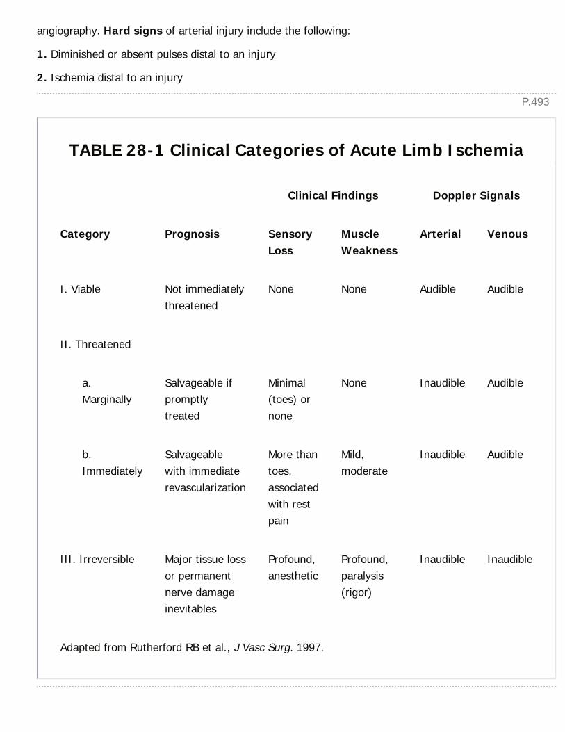
P.493
angiography. Hard signs of arterial injury include the following:
1. Diminished or absent pulses distal to an injury
2. Ischemia distal to an injury
TABLE 28-1 Clinical Categories of Acute Limb Ischemia
Clinical Findings Doppler Signals
Category Prognosis SensoryLoss
MuscleWeakness
Arterial Venous
I. Viable Not immediatelythreatened
None None Audible Audible
II. Threatened
a.Marginally
Salvageable ifpromptlytreated
Minimal(toes) ornone
None Inaudible Audible
b.Immediately
Salvageablewith immediaterevascularization
More thantoes,associatedwith restpain
Mild,moderate
Inaudible Audible
III. Irreversible Major tissue lossor permanentnerve damageinevitables
Profound,anesthetic
Profound,paralysis(rigor)
Inaudible Inaudible
Adapted from Rutherford RB et al., J Vasc Surg. 1997.
P.4943. Visible arterial bleeding from a wound
4. A bruit at or distal to the site of injury
5. Large, expanding, or pulsatile hematomas
Soft signs of injury include the anatomic proximity of a wound to a major vessel, injury to ananatomically related nerve, unexplained hemorrhagic shock, or a moderately sized hematoma. Inthose with only soft signs, a careful documentation of pulses by Doppler pressure distal to theinjury should be undertaken, along with comparison with the contralateral (uninjured) limb. Adifference of greater than 10% to 20% in the ankle-brachial indices (ABIs) suggeststhe need for arteriography or exploration.
III. MANAGEMENTA. Once a diagnosis of acute arterial ischemia due to emboli or thrombi is made, heparin shouldbe administered immediately. An intravenous bolus of 80 units/kg followed by an intravenousinfusion of 18 units/kg/hour is usually satisfactory. Partial thromboplastin time (PTT) should bemaintained between 60 and 80 seconds.
1. Surgical therapy, such as embolectomy, should be performed urgently in patients with anobvious embolus and acute ischemia. Once the artery is isolated, a Fogarty catheter is passedproximally and distally to extract the embolus and associated thrombus. In some cases,intraoperative thrombolysis may be necessary because distal vessels may be thrombosed beyondthe reach of the Fogarty catheter. Distal patency can be documented with an intraoperativearteriogram, depending on the status of distal vessels and pulses after embolectomy. If adequatedistal perfusion is not established and an angiogram demonstrates distal thrombus, the distalpopliteal artery and tibial arteries may be explored via angiographic or surgical approach. Inaddition, it is not uncommon for vasospasm to occur following embolectomy and direct intra-arterial infusion of 50 to 100 µg of nitroglycerin may resolve it. In conjunction with steerableguidewires, Fogarty catheters can be used to select the tibial arteries to retrieve distal thrombus.When angiographic approaches fail, popliteal artery cutdown can allow direct access to thesevessels. Patch angioplasty or bypass grafting may be required if significant preexisting arterialdisease in the affected segment is discovered.
2. Thrombolytic therapy may be useful in patients with clearly viable extremities in whomthrombosis is the likely underlying cause of their acute ischemia. Thrombolytic agents work beston fresh clot. Thrombolysis and followup angiography frequently identify an underlying stenosisthat may be treated by balloon angioplasty/stent or by surgical intervention.
Lytic agents, such as alteplase or reteplase, are instilled through an intra-arterial catheterpositioned within the thrombosed vessel. These agents are also commonly used in conjunctionwith percutaneous mechanical thrombectomy (PMT) for large clot burdens.
P.495
3. There are several PMT devices available that work based on different principles. Rotationaldevices use a high-velocity rotating helix to break
the thrombus and include the Trerotola device (Arrow International, PA). The Trellis system(Medtronic, MN) uses an oscillating nitinol wire to agitate a lytic solution that is isolated betweenproximal and distal occlusion balloons to reduce systemic spread of the lytic agent. The AngioJetthrombectomy system (Boston Scientific, MA) uses a high pressure saline jet flow with aspirationof softened thrombus into the catheter. The EkoSonic catheter (EKOS Corporation, WA) delivershigh frequency, low-energy ultrasound in a radial fashion to enhance the penetration of lyticagents by exposing plasminogen receptor sites. This mechanism probably has less hemolytic effectthan with saline pressure thrombectomy and less endothelial damage than with rotationalthrombectomy; however it requires multiple sessions.
B. In the setting of trauma, operative exploration should be performed in any limb that isischemic or if arteriography demonstrates a significant intimal flap or other pathology. In thepresence of coexistent neurologic or orthopedic injuries, it is prudent to reestablish arterial flowfirst, whether by direct repair, bypass grafting, or temporary shunting. If the decision is made totemporarily shunt, shunt patency should be assessed by handheld Doppler examinationthroughout the case. At the conclusion of the orthopedic repair, the arterial repair should bereexamined to ensure that it has not been disrupted and has been correctly fashioned to the finalbone length. In cases of joint dislocation, reduction of the dislocation should be accomplished firstbecause this may alleviate the need for arterial reconstruction.
1. It is essential to obtain proximal and distal control of the injured artery before exploringthe hematoma or wound. When feasible, an end-to-end anastomosis is preferable. A fewcentimeters of the artery can usually be mobilized proximally and distally to accomplishreapproximation. However, the uninjured leg or other potential vein harvest site should beprepared in case a conduit is required. It is preferable to use autologous tissue in this setting. Acompletion angiogram can help to document distal flow. This is especially important if significantspasm is present and distal pulses are not readily palpable, which can be treated with an intra-arterial infusion of 50 to 100 µg of nitroglycerin.
2. In general, injuries to the subclavian, axillary, brachial, femoral, superficial femoral, profundafemoral, and popliteal arteries should be repaired. The radial or ulnar artery may be ligated if theother vessel is intact and the hand is well perfused. Similarly, isolated injuries to a solitary tibialartery may be ligated if one or more of the tibial arteries remain intact and the foot is wellperfused.
IV. COMPLICATIONSA. Reperfusion injury results from the formation of oxygen-free radicals thatdirectly damage the tissue and cause white blood cell accumulation andsequestration in the microcirculation. This process prolongs the ischemic intervalbecause it impairs adequate nutrient flow to the tissue, despite the restoration of

P.496
axial blood flow.
B. Rhabdomyolysis following reperfusion releases the by-products of ischemicmuscle, including potassium, lactic acid, myoglobin, and creatine phosphokinase.The electrolyte and pH changes that occur can trigger dangerous arrhythmias,and precipitation of myoglobin in the renal tubules can cause pigmentnephropathy and acute renal failure. The likelihood that a patient will developthese complications relates to the duration of ischemia and the muscle mass atrisk. Aggressive hydration, diuresis with mannitol, and intravenousinfusion of bicarbonate to alkalinize the urine are accepted methods ofmitigating renal impairment secondary to rhabdomyolysis.
C. Compartment syndrome results when prolonged ischemia and delayedreperfusion cause cell membrane damage and leakage of fluid into theinterstitium. Additional muscle and nerve necrosis occurs when theintracompartmental pressures exceed capillary perfusion pressure (generally>30 mm Hg). A four-compartment fasciotomy should be performed when thereis concern about the possible development of lower leg compartment syndrome;less commonly, thigh compartment fasciotomies may be indicated. In the upperextremity, fasciotomies of the forearm and hand may be needed to preventdevelopment of compartment syndrome after emergent revascularization.
Fasciotomy should be routinely considered in any patient with more than 6hours of acute extremity ischemia, or in the presence of combined arterial andvenous injuries.
D. Followup care is usually directed at treating the underlying cause of theobstruction. Patients with mural thrombi or arrhythmias require long-termanticoagulation. The in-hospital mortality rate associated with embolectomy is ashigh as 30%.
CHRONIC ARTERIAL OCCLUSIVE DISEASEThe lower extremities are most frequently affected by chronic occlusive disease, although upper-extremity disease can occur. The principal early symptom of arterial occlusive disease isclaudication, usually described as a cramping pain or heaviness in the affected extremity thatoccurs after physical exertion. Claudication is relieved by rest but recurs predictably with exercise.Lower-extremity occlusive disease may be subdivided into three anatomic sections on the basis ofsymptoms and treatment options. Aortoiliac occlusive disease, or Òinflow disease,Ó affects theinfrarenal aorta and the common and external iliac arteries. FemoralÑpopliteal occlusive disease,or Òoutflow disease,Ó affects the common femoral, superficial femoral, and popliteal arteries.Finally, tibialÑperoneal disease, or Òrunoff disease,Ó affects the vessels distal to the poplitealartery. An algorithm for the clinical approach to claudication is shown in Figure 28-1.
P.497P.498
I. CLINICAL PRESENTATIONA. Aortoiliac disease presents with symptoms of lower-extremity claudication, usually ofthe hip, thigh, or buttock. It may coexist with
femoralÑpopliteal disease, contributing to more distal symptoms as well. The symptoms usuallydevelop gradually, although sudden worsening suggests acute thrombosis of a diseased vessel.Patients ultimately develop incapacitating claudication but not rest pain unless distal disease isalso present. Leriche syndrome (sexual impotence, buttock and leg claudication, leg musculatureatrophy, trophic changes of the feet, and leg pallor) is a constellation of symptoms that resultsfrom the gradual occlusion of the terminal aorta.

Figure 28-1 Clinical approach to claudication.
P.499
B. Patients with femoralÑpopliteal and tibialÑperoneal disease present with claudication ofthe lower extremity, usually most prominent in the calves. More severe impairment of arterialflow can present as rest pain. Rest pain is a burning pain in the distal foot, exacerbated by limbelevation, and often relieved by placing the leg in a dependent position. Examination findings ofthe chronically ischemic extremity include the following:
1. Decreased or absent distal pulses
2. Dependent rubor
3. Trophic changes that include thickening of the nails, loss of leg hair, shiny skin, and ulcerationat the tips of the toes
C. Symptomatic arterial occlusive disease of the upper extremity is relatively rare.
1. The proximal subclavian artery is most commonly affected by atherosclerotic disease,followed by axillary and brachial arteries. These patients typically present with arm claudication orfingerÑhand ischemia or necrosis. Occasionally, ulcerated plaques of the innominate or subclavianarteries can be a source of embolization to the hand.
2. Although many patients with proximal subclavian lesions are asymptomatic, subclavian stealcan result when an occlusive subclavian artery lesion is located proximal to the origin of thevertebral artery. With exercise of the affected limb, the arm's demand for blood is supplied byretrograde flow in the ipsilateral vertebral artery, shunting blood from the posterior cerebralcirculation and resulting in drop attacks, ataxia, sensory loss, or diplopia.
II. DIAGNOSIS.Diagnosis of chronic arterial occlusive disease is concerned with determining the presence ofsignificant flow-limiting lesions and distinguishing the disease from other conditions that maymimic it.
A. For patients presenting with lower-extremity symptoms, it is essential to examine thefemoral and distal pulses at rest and after exercise. The absence of femoral pulses is indicative ofaortoiliac disease, although some patients with aortoiliac stenoses have palpable pulses at restthat are lost after exercise. Bruits may also be appreciated over the lower abdomen or femoralvessels. It is also important to differentiate ulcers that arise from arterial insufficiency versus thosegenerated by venous insufficiency and neuropathy.
1. Arterial insufficiency ulcers are usually painful and have an irregular appearance.
2. Neuropathic ulcers are painless and usually occur over bony prominences, particularly theplantar aspect of the metatarsophalangeal joints.
3. Venous stasis ulcers are located on the malleolar surface (ÒgaiterÓ distribution) and are darkand irregular in shape.
P.500
B. Segmental arterial Doppler readings with waveforms should be performed in all patientswith suspected symptomatic arterial disease. The ankle-brachial index (ABI), the ratio of thesystolic blood pressure in the leg to that in the arm, allows one to quantify the degree of ischemia.In general, patients without vascular disease have an ABI of 1.0, patients with claudication havean ABI of less than 0.8, and patients with rest pain and severe ischemia have an ABI of less than0.4. Waveform changes help to localize the level of disease, and the severity of obstruction.Patients with history of claudication and normal resting waveforms require postexercise ABImeasurements. In patients with diabetes and renal insufficiency, calcified vessels can result in afalsely elevated ABI measurement. Digit pressures are less affected by calcification and oftenprovide a more accurate representation of arterial perfusion.
C. Digital subtraction arteriography is the gold standard for evaluating the arterial tree beforeplanned revascularization. Typical digital subtraction arteriography of the lower extremitiesextends from the proximal abdomen to the toes. Noninvasive angiography using imagingmodalities such as CTA and magnetic resonance angiography (MRA) has also gained widespreaduse. CTA produces high-resolution images of the vascular tree. However, diffuse calcificationsmay make interpretation of CT angiography images difficult. In addition, CT angiography doesrequire iodinated contrast, which may adversely affect patients with renal insufficiency. MRA is anexcellent imaging modality for assessing PAD and is useful for selecting patients who areendoluminal candidates. However, MRA may overestimate the degree of stenosis and may beinaccurate in stented arteries. Following the linkage of nephrogenic systemic fibrosis (NSF) togadolinium administration in renal failure patients, alternative noncontrast MRA techniques haveemerged (e.g., time-of-flight).
III. MANAGEMENTA. The recently issued Society for Vascular Surgery Practice Guidelines for AtheroscleroticOcclusive Disease of the Lower Extremities (Conte et al., J Vasc Surg. 2015) offer detailedrecommendations for management of patients with peripheral arterial disease. With adequatecontrol of risk factors, intermittent claudication follows a benign course in most patients. Inpatients presenting with claudication alone, 70% to 80% remain stable or improve and 10% to20% worsen over the ensuing 5-year period. Only 5% to 10% of patients develop gangrene andare at risk for limb loss. Therefore, first-line treatment for patients with claudicationshould emphasize risk factor modification and structured exercise therapy.
Despite this appropriate focus on medical optimization of the claudicant, incapacitatingclaudication that jeopardizes a patient's livelihood or
severely influences his or her quality of life may be considered for revascularization after failure ofrisk factor modification and exercise therapy.
Critical limb ischemia (CLI) is characterized by severely diminished arterial flow (ABI <0.4, andtoe pressures <50 mm Hg), ischemic rest pain, and the development of ischemic ulceration or

P.501
pedal gangrene. The Society for Vascular Surgery Lower Extremity Threatened Limb ClassificationSystem (Mills et al., J Vasc Surg. 2013) uses the severity of limb ischemia, wound complexity, andfoot infection to estimate the likelihood of limb salvage in CLI patients. In addition to maximalmedical therapy, revascularization is indicated for symptom relief and limb preservation.
B. Medical Therapy. Society for Vascular Surgery Guidelines for Atherosclerotic Occlusivedisease of the Lower extremities (Conte et al., J Vasc Surg. 2015) describe medical managementand exercise therapy in detail.
1. Risk factor modification is the most important intervention for reducing the impact ofadvanced atherosclerotic disease. Control of hypertension and serum glucose, cessation ofsmoking, management of lipid disorders, attainment of ideal body weight, and regular exerciseshould be the goals.
2. Lipid reduction is imperative in patients with PAD because the majority of the morbidityassociated with PAD is related to cardiac events. On the basis of the Heart Protection Studyinvolving statins, it is recommended to keep the low-density-lipoprotein level of patients with PADless than 100 mg/dL, and <70 mg/dL for patients with disease in two vascular beds, to reduce thelikelihood of morbidity associated with cardiac events (J Vasc Surg. 2007;45:645).
3. Antihypertensives should be administered to normalize blood pressure. The Intersocietyconsensus for the management of peripheral arterial disease (TASC II, Eur J Vasc Endovasc Surg.2007) recommends blood pressure control to <140/90 mm Hg in all patients or <130/80 mm Hg ifthey also have diabetes or renal insufficiency.
4. Because many of these patients have concomitant coronary artery or cerebrovascular disease,daily aspirin therapy (81 or 325 mg) is indicated to reduce the risk of myocardial infarction orstroke.
5. Clopidogrel is an antiplatelet agent that has been shown to reduce cardiac and cerebralevents in patients with systemic atherosclerosis. Although rigorous proof of its utility followingperipheral arterial interventions is lacking, clopidogrel is frequently prescribed following theseprocedures.
6. Cilostazol is a type III phosphodiesterase inhibitor and the newest agent available fortreatment of claudication. Cilostazol inhibits platelet aggregation and causes vasodilation. Given at50 mg or 100 mg twice daily, it increases walking distances when compared with placebo andpentoxifylline. Early studies suggest that the drug is safe in most patients, although its use iscontraindicated in those with class III or IV heart failure due to the toxicity ofphosphodiesterase inhibitors in these patients.
C. Preoperative care of patients with PAD includes complete arterial imaging and procedural riskstratification. Myocardial complications account for the majority of early and late deaths. TheAmerican College of Cardiology/American Heart Association Guidelines on Perioperative
P.502
Cardiovascular Evaluation and Management (Fleisher et al., Circulation. 2014) offer a structuredapproach to the assessment and subsequent treatment of concomitant coronary artery disease.
D. Open Surgical Therapy
1. Aortoiliac occlusive disease
a. Aortobifemoral grafting is the treatment of choice in low-risk patients with diffuse aortoiliacstenoses and occlusions. Aortobifemoral bypass may be performed via transperitoneal orretroperitoneal approach. Distal endarterectomy and/or profundaplasty may be performed inconjunction with a bypass to improve outflow. Results are excellent, with reported patency ratesof up to 95% at 5 years.
b. Femorofemoral bypass is an alternative in high-risk patients with unilateral iliac disease. Thepatency rates are lower than those achieved with aortobifemoral grafts.
c. Axillobifemoral bypass provides a less-invasive option for high-risk patients who needrevascularization. This bypass avoids an intraabdominal procedure and the need for aortic cross-clamping. Patency rates are poorer than those achieved with aortobifemoral bypass.
d. Aortoiliac endarterectomy may be considered for patients who have disease localized to thedistal aorta and common iliac vessels, although its use is now uncommon. Advantages include theavoidance of prosthetic material and preservation of antegrade flow into the hypogastric arteries.
2. Femoral, popliteal, and tibial occlusive disease
a. In patients with SFA occlusion, a femoralÑabove-knee popliteal bypass may be constructed. Inpatients who have disease below the knee, a distal bypass may be performed to the below-kneepopliteal, posterior tibial, anterior tibial, or peroneal arteries. If all tibial vessels are occluded,pedal vessels may serve as suitable outflow vessels. These grafts usually originate from thecommon femoral artery, although a more distal vessel may be used if the inflow into that vessel isunobstructed.
b. The best results are obtained with the use of autologous vein. Single-segment greatersaphenous vein is the conduit of choice, but the lesser saphenous vein or the arm veins providesuitable alternatives. These autologous grafts can be used either in situ or reversed orientation.The advantages of the in situ bypass are that (1) the vein's nutrient supply is left intact and (2)the vein orientation allows for a better size match (the large end of the vein is sewn to the largecommon femoral artery, and the small end is sewn to the distal vessel). The advantage of thereversed-vein bypass is that endothelial trauma is minimized because valve lysis is not necessary.
c. When autologous vein is not available, polytetrafluoroethylene (PTFE) grafts and cryopreservedvein grafts can be used. Patency rates for PTFE above-knee grafts approach those achieved withvenous conduit, but use of PTFE for more distal bypass procedures is associated with substantiallylower patency and is reserved for patients with CLI who lack venous conduit. An alternative
P.503
technique when performing PTFE bypass is the use of a small cuff of vein (Miller cuff) or patchangioplasty (Taylor patch) at the distal anastomosis. These modifications appear to improveprosthetic graft patency by improving compliance match at the distal anastomosis. Cryopreservedvein graft patency also fares poorly in comparison to autologous conduit, but may prove usefulwhen bypass is required in an infected field.
d. Endarterectomy is most commonly used to address severe stenosis or occlusion of the commonfemoral and profunda femoris arteries.
e. Amputation is reserved for patients with gangrene or persistent painful ischemia not amenableto vascular reconstruction. These patients often have severe coexistent vascular andcardiovascular disease, and the survival rate for patients undergoing major amputations isapproximately 50% at 3 years and 30% at 5 years.
(1) The level of amputation is determined clinically. Important factors include the necessity ofremoving all the infected tissue and the adequacy of the blood supply to heal the amputation. Ageneral principle is to preserve as much length of the extremity as safely possible, as thisimproves the patient's opportunity for rehabilitation.
(2) Digital amputations are performed for isolated gangrene and/or recalcitrant osteomyelitis.
(3) Transmetatarsal amputations are usually performed when several toes are involved in theischemic process or after previous single-digit amputations.
(4) Below-knee amputation (BKA) is the most common type of amputation performed for patientswith severe occlusive disease.
(5) Above-knee amputation (AKA) heals more easily than BKA and is useful in older patients whodo not ambulate.
(6) Hip disarticulation is rarely performed for PVD.
3. Upper-extremity occlusive disease
a. For proximal subclavian disease, the choice of bypass procedure depends primarily on thepatency of the ipsilateral common carotid artery.
b. If the ipsilateral common carotid artery is patent, carotidÑsubclavian bypass is performedthrough a supraclavicular approach using a prosthetic graft (vein grafts are to be avoided).Subclavian artery transposition to ipsilateral carotid artery is an excellent alternative ifanatomically feasible.
c. If the ipsilateral carotid artery is occluded, subclavianÑsubclavian bypass may be performed.This extraanatomic approach uses a longer segment prosthetic graft, with reduced patency.
4. Intraoperative anticoagulation is employed during most vascular reconstructions. Generally,unfractionated heparin (100 units/kg) is administered intravenously shortly before cross-clamping

P.504
and supplemented as necessary until the cross-clamps are removed. Anticoagulation can bemonitored intraoperatively by following activated clotting time (ACT) levels, with a goal of greaterthan 250 seconds. The anticoagulant effect of heparin can be reversed with protamineadministration. For patients with heparin-induced thrombocytopenia, direct thrombin inhibitorssuch as bivalirudin are preferred.
E. Postoperative Care
1. Open aortic procedures are initially managed in the intensive care unit, due to the need forcontinuous monitoring and rapid intervention. Assessment of distal pulses should be doneregularly. Early ambulation is encouraged.
2. For distal bypass grafts, pulses should be assessed frequently. Antibiotics are continuedfor 24 hours, or longer if infected ulcers warrant additional treatment. Early ambulation isencouraged in patients without tissue necrosis. In patients who are unable to ambulateimmediately, physical therapy can help to increase strength in the limb and prevent contracture.Sitting with the hips flexed to 90 degrees is discouraged in any patient with a femoralanastomosis. Patients should be instructed to elevate their legs while resting because this willmitigate the edema that develops in the revascularized extremity.
3. Perioperative antithrombotic therapy should include aspirin (81 to 325 mg/day) for allinfrainguinal reconstructions. In patients sensitive to aspirin, clopidogrel (75 mg/day) may besubstituted.
4. Postoperative oral anticoagulation has a more limited role. Owing in part to the increased riskof hemorrhage, anticoagulation with warfarin (INR 2 to 3) is generally limited to grafts consideredto be at a high risk for thrombosis.
5. Following major amputations, weightbearing is delayed for 4 to 6 weeks. Some advocate theuse of compressive wraps to aid in the maturation of the stump. In all cases, early consultationwith a physical therapist and prosthetist is recommended.
6. Surveillance of distal bypass grafts consists of serial evaluations of vein graft patency byclinical examination and duplex ultrasound. Less frequent followup is necessary for aortoiliacbypasses. Detection of severe stenosis predicts impending graft failure, and such grafts shouldundergo arteriography and correction. Repair or revision of stenosed grafts results in higher long-term patency than repairing or replacing occluded grafts.
F. Complications
1. Early complications occur in approximately 5% to 10% of patients after aortic surgery andfrequently relate to preoperative comorbid disease. Myocardial infarction, congestive heart failure,pulmonary insufficiency, and renal insufficiency are most common. Complications related directlyto the aortic reconstruction include hemorrhage, embolization
or thrombosis of the distal arterial tree, microembolization, ischemic colitis, ureteral injuries,
UnitedVRG

P.505
impotence, paraplegia, and wound infection. Late complications include anastomoticpseudoaneurysm or graft dilation, graft limb occlusion, aortoenteric erosion or fistula, and graftinfections.
2. In distal revascularizations, most of the early complications are also related to comorbidconditions. Early graft thrombosis (within 30 days of surgery) most often results from technicalerrors, hypercoagulability, inadequate distal runoff, and postoperative hypotension. Technicalerrors include graft kinks, retained valve leaflets, valvulotome trauma, intimal flaps, significantresidual arteriovenous fistulas, and the use of a poor quality conduit.
G. Endovascular Options
1. Aortoiliac occlusive disease
a. Indications. Balloon angioplasty and intravascular stent placement for aortoiliac occlusivelesions produce excellent results. These procedures are indicated for symptomatic stenotic orocclusive lesions. Short-segment stenoses (less than 3 cm in length) of the common and externaliliac arteries display excellent long-term patency rates when treated with angioplasty alone, orwith stent placement. Angioplasty failure (defined as residual stenosis of ≥30%, residual meantranslesional pressure gradient of ≥10 mm Hg, or flow-limiting dissection) is an indication for stentdeployment.
b. Technique. Access for iliac artery angioplasty and stenting is generally via a femoral arterialapproach. When the occlusive lesion is in the distal aorta or ostial common iliac artery, angioplastyshould be performed using two balloons, one in each iliac artery and both partially projecting intothe distal aorta (Òkissing balloonsÓ). The rationale for this technique is that lesions in proximity tothe aortic bifurcation typically involve the distal aorta and both common iliac arteries. Unilateralballoon dilation may cause plaque shifting with compromise of the contralateral iliac artery lumen.Stenting may produce a more favorable result if postangioplasty dissection or lesion recoil isnoted. Balloon-expandable and self-expanding stents are generally oversized 10% to 15% relativeto the adjacent normal artery to ensure satisfactory stent apposition to the vessel wall. If stentdeployment is required in proximity to the aortic bifurcation, Òkissing stentsÓ are utilized in afashion similar to that described above.
c. Complications. Procedural complications of iliac angioplasty and stenting include arterialdissection, vessel occlusion, arterial rupture, and distal embolization, which may result in the needfor surgical intervention or amputation.
d. Results. Immediate balloon angioplasty failure can result from elastic recoil of atheroscleroticplaque or arterial wall dissection. These complications are potentially amenable to stentplacement. Early failure is usually due to intimal hyperplasia, whereas late failure may
also be caused by progressive atherosclerosis. Iliac artery balloon angioplasty 2-year patency ratesbetween 60% and 70% have been reported. Reports of iliac artery stenting demonstrate 4-yearpatency rates as high as 85%. In general, the results of angioplasty and stenting are better for
P.506
common iliac artery lesions than for external iliac artery lesions, and are better for short-segmentdisease than for long-segment disease.
2. Infrainguinal occlusive disease
a. Indications. Balloon angioplasty and stenting of infrainguinal occlusive lesions has been widelyapplied for the treatment of claudication and CLI. See Figures 28-2, 28-3 and 28-4 for an exampleof angiographic intervention for superficial femoral artery occlusion. Aggressive modification of riskfactors, institution of antiplatelet and statin medications, and a trial of exercise therapy arerecommended prior to intervention, particularly in the setting of claudication. The Trans-AtlanticInter-Society Consensus (TASC II) group has provided recommendations regarding thecharacteristics of femoropopliteal and infrapopliteal lesions that are best addressed by eitherendovascular
or surgical therapy (Table 28-2). Short, focal stenoses (TASC A) are felt to be amenable toendovascular therapy, whereas long-segment occlusions (TASC D) are best addressed by surgicalbypass.
UnitedVRG
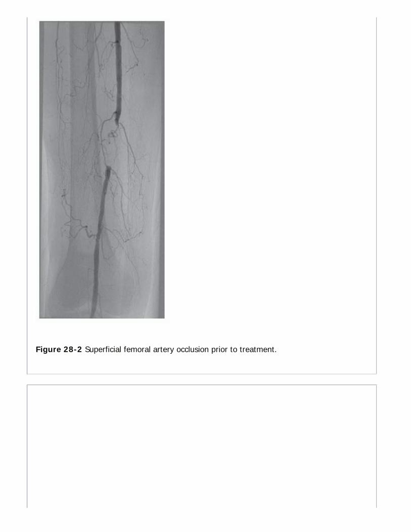
Figure 28-2 Superficial femoral artery occlusion prior to treatment.

Figure 28-3 Recoil following balloon angioplasty.
b. Technique. Arterial access for infrainguinal intervention is usually accomplished via retrogradecontralateral femoral artery approach or ipsilateral antegrade femoral artery approach. Retrogradetibial/pedal access is also being studied as an alternate access option. The most frequent cause oftreatment failure is the inability to negotiate across the stenosis or occlusion and into the distaloutflow target vessel. In general, once guidewire access to the distal target vessel has beenestablished, technical success rates are excellent. Hydrophilic guidewires and catheters, occlusioncrossing devices, lumen reentry devices, and specialized sheaths have been developed to facilitatethis process.
c. Complications. Procedural complications of infrainguinal endovascular intervention include
UnitedVRG
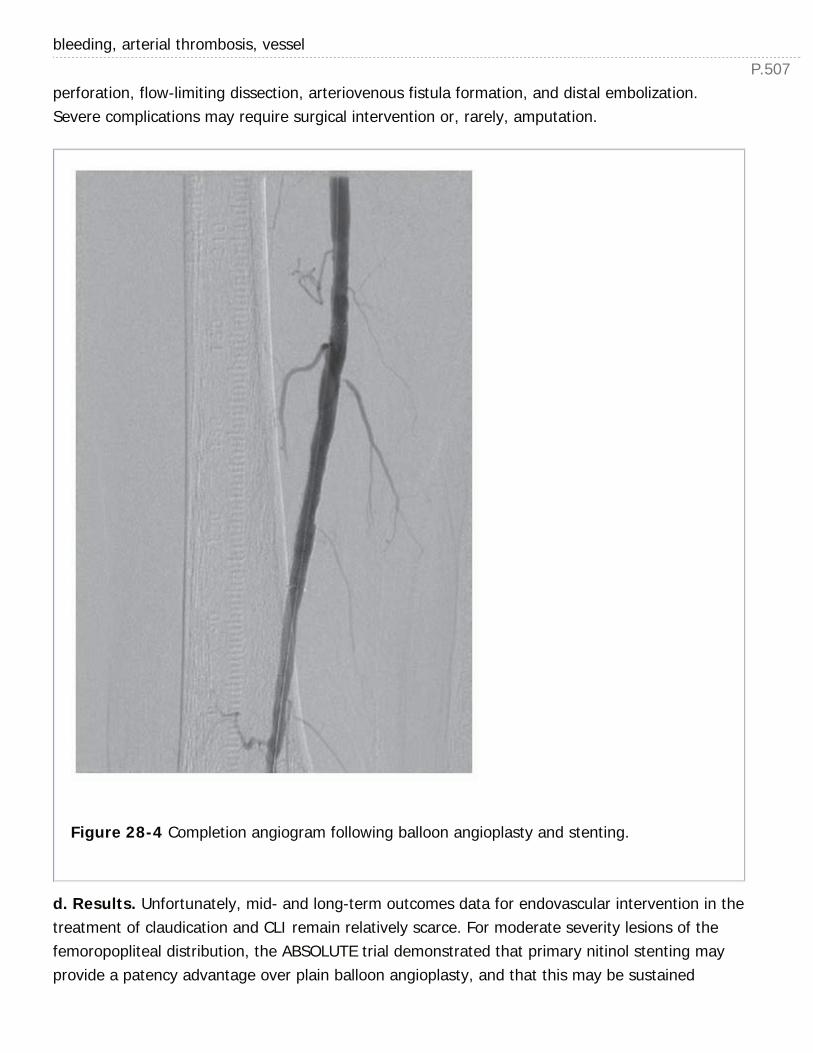
P.507
bleeding, arterial thrombosis, vessel
perforation, flow-limiting dissection, arteriovenous fistula formation, and distal embolization.Severe complications may require surgical intervention or, rarely, amputation.
Figure 28-4 Completion angiogram following balloon angioplasty and stenting.
d. Results. Unfortunately, mid- and long-term outcomes data for endovascular intervention in thetreatment of claudication and CLI remain relatively scarce. For moderate severity lesions of thefemoropopliteal distribution, the ABSOLUTE trial demonstrated that primary nitinol stenting mayprovide a patency advantage over plain balloon angioplasty, and that this may be sustained
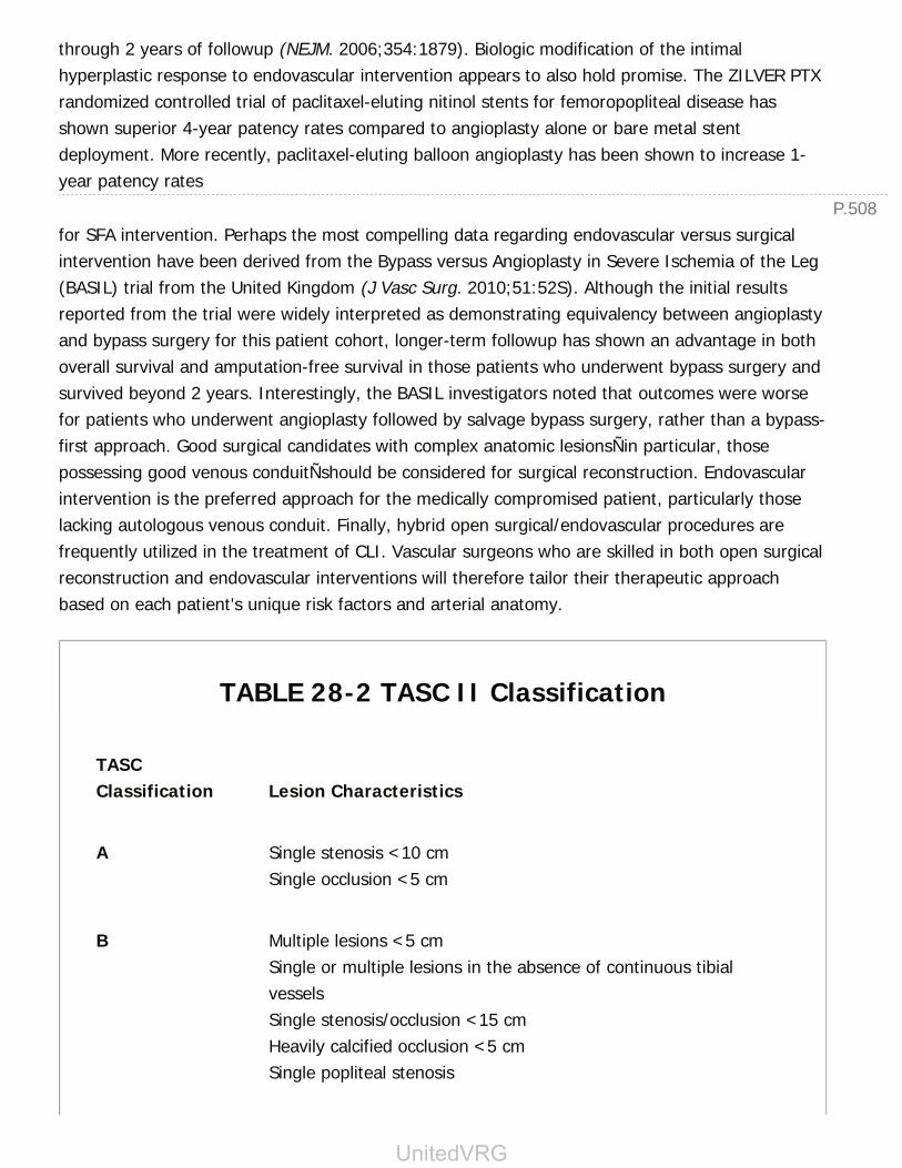
P.508
through 2 years of followup (NEJM. 2006;354:1879). Biologic modification of the intimalhyperplastic response to endovascular intervention appears to also hold promise. The ZILVER PTXrandomized controlled trial of paclitaxel-eluting nitinol stents for femoropopliteal disease hasshown superior 4-year patency rates compared to angioplasty alone or bare metal stentdeployment. More recently, paclitaxel-eluting balloon angioplasty has been shown to increase 1-year patency rates
for SFA intervention. Perhaps the most compelling data regarding endovascular versus surgicalintervention have been derived from the Bypass versus Angioplasty in Severe Ischemia of the Leg(BASIL) trial from the United Kingdom (J Vasc Surg. 2010;51:52S). Although the initial resultsreported from the trial were widely interpreted as demonstrating equivalency between angioplastyand bypass surgery for this patient cohort, longer-term followup has shown an advantage in bothoverall survival and amputation-free survival in those patients who underwent bypass surgery andsurvived beyond 2 years. Interestingly, the BASIL investigators noted that outcomes were worsefor patients who underwent angioplasty followed by salvage bypass surgery, rather than a bypass-first approach. Good surgical candidates with complex anatomic lesionsÑin particular, thosepossessing good venous conduitÑshould be considered for surgical reconstruction. Endovascularintervention is the preferred approach for the medically compromised patient, particularly thoselacking autologous venous conduit. Finally, hybrid open surgical/endovascular procedures arefrequently utilized in the treatment of CLI. Vascular surgeons who are skilled in both open surgicalreconstruction and endovascular interventions will therefore tailor their therapeutic approachbased on each patient's unique risk factors and arterial anatomy.
TABLE 28-2 TASC II Classification
TASCClassification Lesion Characteristics
A Single stenosis <10 cmSingle occlusion <5 cm
B Multiple lesions <5 cmSingle or multiple lesions in the absence of continuous tibialvesselsSingle stenosis/occlusion <15 cmHeavily calcified occlusion <5 cmSingle popliteal stenosis
UnitedVRG
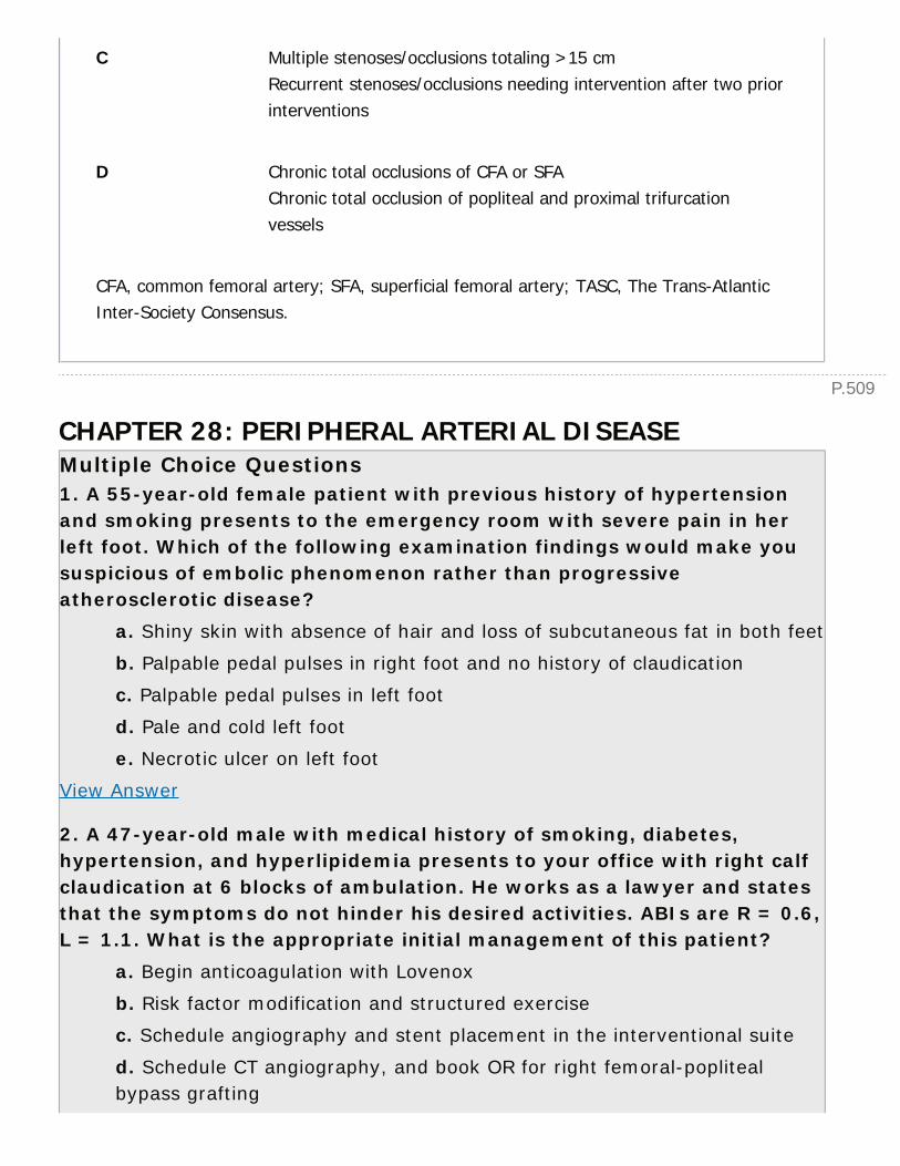
P.509
C Multiple stenoses/occlusions totaling >15 cmRecurrent stenoses/occlusions needing intervention after two priorinterventions
D Chronic total occlusions of CFA or SFAChronic total occlusion of popliteal and proximal trifurcationvessels
CFA, common femoral artery; SFA, superficial femoral artery; TASC, The Trans-AtlanticInter-Society Consensus.
CHAPTER 28: PERIPHERAL ARTERIAL DISEASEMultiple Choice Questions1. A 55-year-old female patient with previous history of hypertensionand smoking presents to the emergency room with severe pain in herleft foot. Which of the following examination findings would make yoususpicious of embolic phenomenon rather than progressiveatherosclerotic disease?
a. Shiny skin with absence of hair and loss of subcutaneous fat in both feet
b. Palpable pedal pulses in right foot and no history of claudication
c. Palpable pedal pulses in left foot
d. Pale and cold left foot
e. Necrotic ulcer on left foot
View Answer
2. A 47-year-old male with medical history of smoking, diabetes,hypertension, and hyperlipidemia presents to your office with right calfclaudication at 6 blocks of ambulation. He works as a lawyer and statesthat the symptoms do not hinder his desired activities. ABIs are R = 0.6,L = 1.1. What is the appropriate initial management of this patient?
a. Begin anticoagulation with Lovenox
b. Risk factor modification and structured exercise
c. Schedule angiography and stent placement in the interventional suite
d. Schedule CT angiography, and book OR for right femoral-poplitealbypass grafting
P.510
e. Prescribe 20 to 30 mm Hg graded compression stockings
View Answer
3. You have completed a femoral embolectomy on a patient in atrialfibrillation who presented with an 18-hour history of acute onset left legpain accompanied by calf muscle weakness and loss of sensation. Thepedal pulses are now palpable, but the calf muscle is tense. You should:
a. Apply nitropaste to the foot
b. Administer systemic thrombolytics
c. Perform four-compartment calf fasciotomies
d. Recommend early ambulation
e. Begin cilostazol
View Answer
4. A 51-year-old male smoker with past medical history of type 1diabetes mellitus, hypertension, congestive heart failure, COPD presentsto you with intermittent bilateral calf claudication for 6 months andupon further evaluation his ABIs are consistent with his symptoms (0.7bilaterally). You counsel him regarding smoking cessation, bloodpressure control, and glucose control. Which of the followingmedications should be added to his management first?
a. Statin
b. Cilostazol
c. Pentoxifylline
d. Coumadin
e. Clopidogrel
View Answer
5. A 19-year-old male restrained front seat passenger is brought by EMSfollowing a head on motor vehicle collision. During an initial primary andsecondary survey, no pulse can be felt in his left foot and there appearsto be a deformity of his left knee. He is complaining of severe pain in hisleft knee and down to his foot. There is no obvious hematoma in thevicinity of left popliteal fossa. X-rays of the left knee are performed andreveal a posterior dislocation. What is the next step in management?
a. Perform a CT angiogram of left lower extremity
b. Perform an angiogram of left lower extremity in the operating room
UnitedVRG

c. Perform a reduction of left knee dislocation with sedation in the ER
d. Exploration of left popliteal fossa in the operating room
e. Perform formal ABIs in the vascular laboratory
View Answer
29Venous and Lymphatic Disease
Joshua A. F. Balderman
Kathleen G. Raman
VENOUS ANATOMYVenous anatomy is divided into three compartments: Superficial, perforating veins, deep. Ingeneral, blood flows from the superficial to the deep veins through the perforating system. In thelower extremity, the major superficial veins are the greater saphenous vein, formed from theunion of the dorsal vein of the great toe and the dorsal venous arch; the small saphenous vein,formed from the joining of the dorsal vein of the fifth toe and the dorsal venous arch; and theposterior arch vein, also called Leonardo vein, beginning in the medial ankle and joining thegreater saphenous vein below the knee. The deep veins in the leg are named according to theirpaired arteries. The deep veins of the calf typically are duplicated as venae comitantes withnumerous communicating branches. The posterior tibial and peroneal veins also communicatewith the soleal sinusoids. In the thigh, the deep venous system includes the femoral and deepfemoral veins that join approximately 4 cm below the inguinal ligament. Perforating veins connectthe superficial and deep systems through both direct and indirect mechanisms. Venous returnfrom the lower extremities depends largely on compression of the deep veins by the muscles ofthe calf (gastrocnemius, soleus) during walking. Flow is unidirectional due to a series of one-wayvalves, which prevent reflux during this cycle of compression. Failure of these valves to closeleads to pooling, stasis, and congestion of veins in the lower extremities, and subsequent dilationof the superficial veins.
CHRONIC VENOUS INSUFFICIENCYChronic venous disease includes telangiectasias, varicose veins, venous ulceration, andclaudication.
I. PATHOPHYSIOLOGYA. Etiology
1. Congenital
2. Primary (cause undetermined)
3. Secondary (postthrombotic, posttraumatic, or other)
B. Risk Factors
1. Obesity
> Table of Contents > 29 - Venous and Lymphatic Disease
UnitedVRG
P.512
2. Tobacco use
3. Multiparity
4. Hormone therapy
5. Obstruction (e.g., from adenopathy, arterial compression, or pregnancy)
6. History of deep venous thrombosis (DVT). DVT accounts for mostsecondary cases and may be responsible for a significant number of other casesbecause many deep vein thrombi are asymptomatic.
C. Reflux disease from venous valvular incompetence accounts for most(>80%) chronic venous disease.
1. Valve malfunction can be inherited or acquired through sclerosis orelongation of valve cusps or dilation of the valve annulus despite normal valvecusps.
2. Varicose veins may represent superficial venous insufficiency in the presenceof competent deep and perforator systems, or they may be a manifestation ofperforator or deep venous disease.
3. Valvular disease below the knee appears to be more critical in thepathophysiology of severe venous disease than disease above the knee.
4. The perforator veins are frequently implicated when venous ulcers exist, butany component of the venous system, either alone or in combination, may beincompetent.
5. All of the above components need evaluation in the workup of chronic venousinsufficiency (CVI) (Am Surg. 2010;76:125).
D. Obstructive physiology is a less common cause of venous pathology, withreflux often being present simultaneously.
II. DIFFERENTIAL DIAGNOSISA. Arterial Disease
1. Ulcers with discrete edges and pale bases; more painful than venous ulcers,generally at the tips of the toes
2. Poor or absent pulses on examination
3. Dependent rubor
4. Pallor with elevation
5. Claudication
B. Lymphedema
1. Pitting edema without pigmentation and ulceration

P.513
2. Less responsive to elevation, usually requires several days to improve
C. Squamous Cell Carcinoma—can occur in patients with chronic wounds orde novo. A biopsy is required for diagnosis.
D. Trauma
E. Arteriovenous Malformation
F. Orthostatic Edema
III. NOMENCLATUREA. CEAP Classification
1. Standardized nomenclature of chronic venous disease (J Vasc Surg. 2004;40:1248; Eur J VascEndovasc Surg. 1996;12:487).
2. CEAP: Clinical signs, Etiology, Anatomic distribution, and Pathophysiology (Table 29-1).
TABLE 29-1 Classification of Chronic Lower-extremityVenous Disease
Classification Definition
C Clinical classification
C0: No visible or palpable signs of venous disease
C1: Telangiectasias or reticular veins
C2: Varicose veins; distinguished from reticular veins by a diameter
of 3 mm or more
C3: Edema
C4: Changes in skin and subcutaneous tissue secondary to CVD
C : Pigmentation or eczema
UnitedVRG
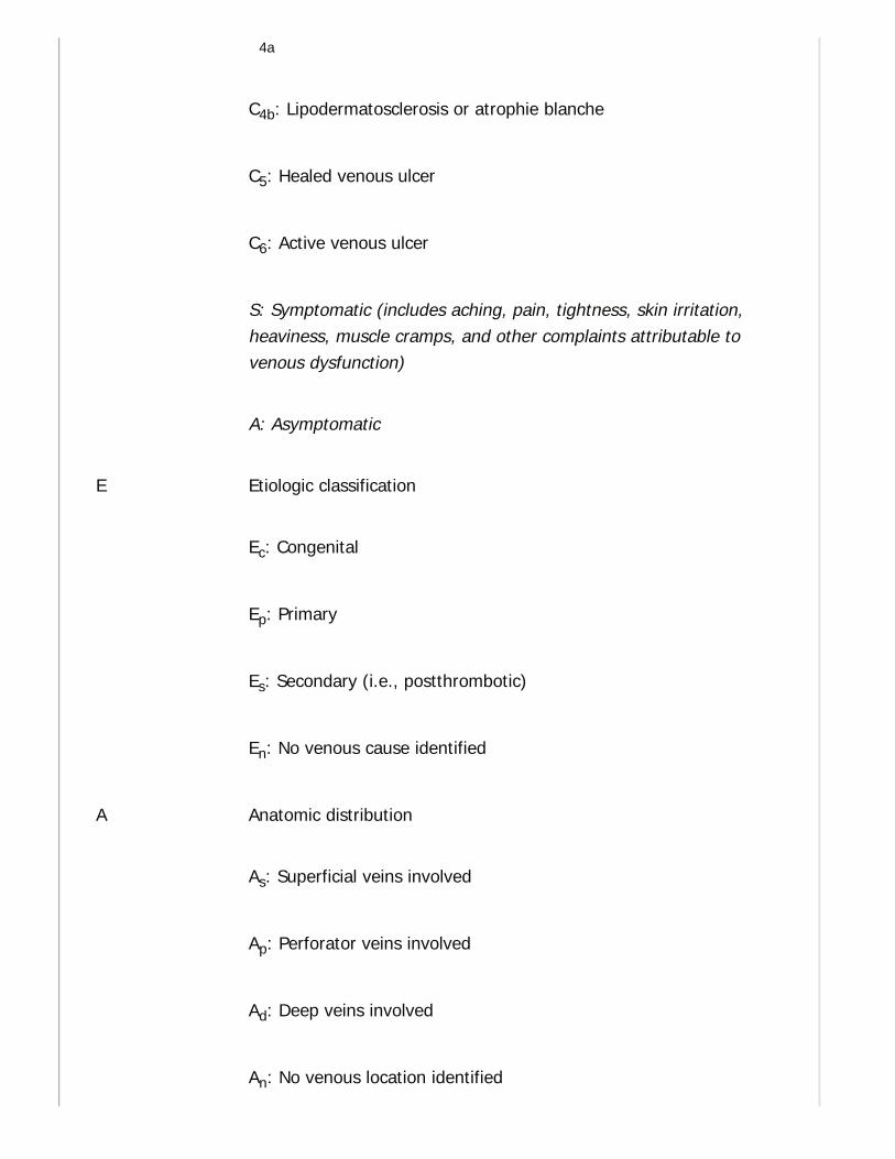
4a
C4b: Lipodermatosclerosis or atrophie blanche
C5: Healed venous ulcer
C6: Active venous ulcer
S: Symptomatic (includes aching, pain, tightness, skin irritation,heaviness, muscle cramps, and other complaints attributable tovenous dysfunction)
A: Asymptomatic
E Etiologic classification
Ec: Congenital
Ep: Primary
Es: Secondary (i.e., postthrombotic)
En: No venous cause identified
A Anatomic distribution
As: Superficial veins involved
Ap: Perforator veins involved
Ad: Deep veins involved
An: No venous location identified
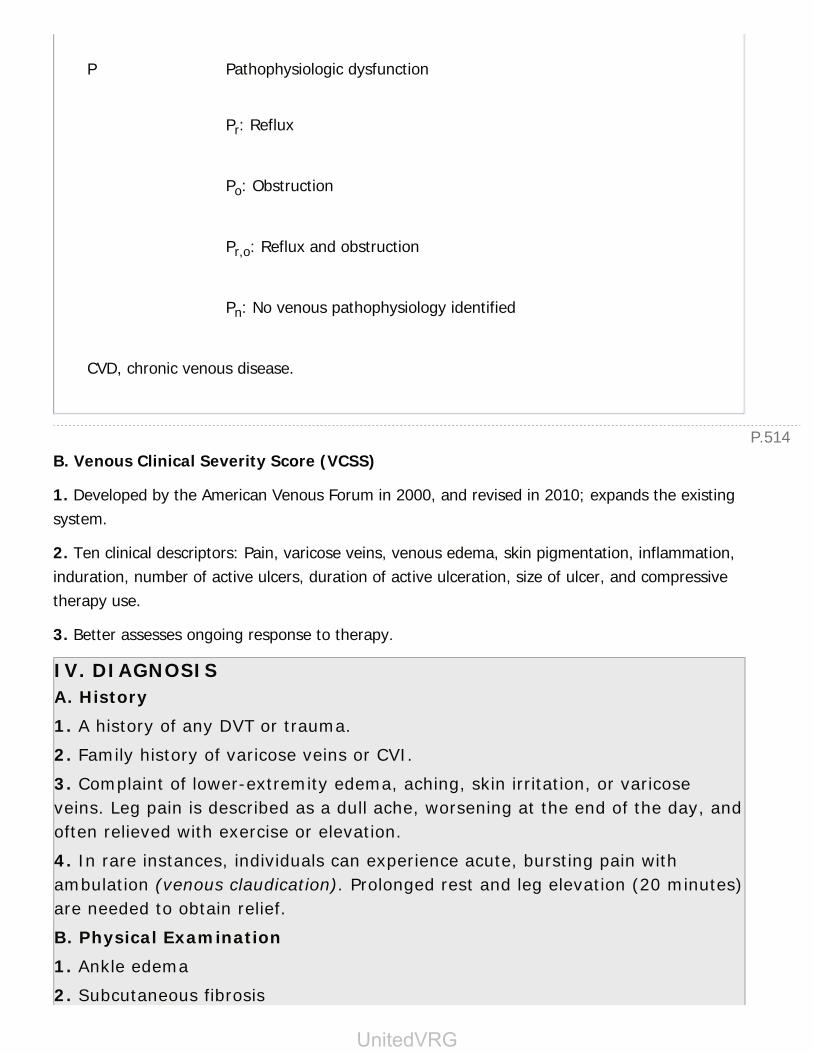
P.514
P Pathophysiologic dysfunction
Pr: Reflux
Po: Obstruction
Pr,o: Reflux and obstruction
Pn: No venous pathophysiology identified
CVD, chronic venous disease.
B. Venous Clinical Severity Score (VCSS)
1. Developed by the American Venous Forum in 2000, and revised in 2010; expands the existingsystem.
2. Ten clinical descriptors: Pain, varicose veins, venous edema, skin pigmentation, inflammation,induration, number of active ulcers, duration of active ulceration, size of ulcer, and compressivetherapy use.
3. Better assesses ongoing response to therapy.
IV. DIAGNOSISA. History
1. A history of any DVT or trauma.
2. Family history of varicose veins or CVI.
3. Complaint of lower-extremity edema, aching, skin irritation, or varicoseveins. Leg pain is described as a dull ache, worsening at the end of the day, andoften relieved with exercise or elevation.
4. In rare instances, individuals can experience acute, bursting pain withambulation (venous claudication). Prolonged rest and leg elevation (20 minutes)are needed to obtain relief.
B. Physical Examination
1. Ankle edema
2. Subcutaneous fibrosis
UnitedVRG
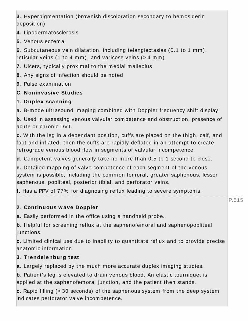
P.515
3. Hyperpigmentation (brownish discoloration secondary to hemosiderindeposition)
4. Lipodermatosclerosis
5. Venous eczema
6. Subcutaneous vein dilatation, including telangiectasias (0.1 to 1 mm),reticular veins (1 to 4 mm), and varicose veins (>4 mm)
7. Ulcers, typically proximal to the medial malleolus
8. Any signs of infection should be noted
9. Pulse examination
C. Noninvasive Studies
1. Duplex scanning
a. B-mode ultrasound imaging combined with Doppler frequency shift display.
b. Used in assessing venous valvular competence and obstruction, presence ofacute or chronic DVT.
c. With the leg in a dependant position, cuffs are placed on the thigh, calf, andfoot and inflated; then the cuffs are rapidly deflated in an attempt to createretrograde venous blood flow in segments of valvular incompetence.
d. Competent valves generally take no more than 0.5 to 1 second to close.
e. Detailed mapping of valve competence of each segment of the venoussystem is possible, including the common femoral, greater saphenous, lessersaphenous, popliteal, posterior tibial, and perforator veins.
f. Has a PPV of 77% for diagnosing reflux leading to severe symptoms.
2. Continuous wave Doppler
a. Easily performed in the office using a handheld probe.
b. Helpful for screening reflux at the saphenofemoral and saphenopoplitealjunctions.
c. Limited clinical use due to inability to quantitate reflux and to provide preciseanatomic information.
3. Trendelenburg test
a. Largely replaced by the much more accurate duplex imaging studies.
b. Patient's leg is elevated to drain venous blood. An elastic tourniquet isapplied at the saphenofemoral junction, and the patient then stands.
c. Rapid filling (<30 seconds) of the saphenous system from the deep systemindicates perforator valve incompetence.

P.516
d. When tourniquet is released, additional filling of the saphenous systemoccurs if the saphenofemoral valve is also incompetent.
4. Descending phlebography has a positive predictive value of 44%, and islimited by its inability to study valves distal to a competent proximal valve.
V. NONSURGICAL TREATMENTA. Infected Ulcers
1. Necessitates treatment of the infection first.
2. Staphylococcus aureus, Streptococcus pyogenes, and Pseudomonas speciesare responsible for most infections.
3. Usually treated with local wound care, wet-to-dry dressings, and oralantibiotics.
4. Topical antiseptics should be avoided.
5. Severe infections require intravenous antibiotics.
B. Leg elevation can temporarily decrease edema and should be institutedwhen swelling occurs. This should be done before a patient is fitted for stockingsor boots.
C. Compression therapy is an adjunctive treatment for CVI.
1. Elastic compression stockings
a. Fitted to provide a compression gradient from 30 to 40 mm Hg, with thegreatest compression at the ankle.
b. Should be worn from awakening and removed at bedtime.
c. Effective in healing ulcers, but can take months to obtain good results.
d. Study of 113 patients treated with initial bed rest, local wound care, andelastic compression stockings demonstrated a 93% ulcer healing rate in a meanof 5.3 months (Surgery. 1991;109:575).
e. Stockings do not correct the abnormal venous hemodynamics and must beworn after the ulcer has healed to prevent recurrence.
f. Patient compliance is principal limiting factor.
g. Recurrence for compliant patients in the same study was 16% at a meanfollowup of 30 months.
2. Unna boots
a. Employed when there is actual skin ulceration, they combine compressiontherapy with a zinc oxide paste that assists in wound healing. Used to helpprevent further skin breakdown.
UnitedVRG
b. Provide nonelastic compression therapy.
c. Changed once or twice a week.
3. Pneumatic compression devices
a. Provide dynamic sequential compression.
b. Used primarily in the prevention of deep vein thrombi in hospitalizedpatients.
c. Also used successfully to treat venous insufficiency.
D. Topical Medications
1. Largely ineffective as a stand-alone therapy for venous stasis ulcers.
2. Topical therapy is directed at absorbing wound drainage and avoidingdesiccation of the wound.
3. Antiseptics can be counterproductive. Hydrogen peroxide, povidone-iodine,acetic acid, and sodium hypochlorite are toxic to cultured fibroblasts and shouldbe used for the shortest duration necessary to control ulcer infection.
VI. SURGICAL THERAPY.Surgical therapy is indicated for severe disease refractory to medical treatment and for patientswho cannot comply with the lifelong regimen of compression therapy. Surgical therapy includessclerotherapy, saphenous vein stripping, endovenous ablation of the saphenous vein, SubfascialEndoscopic Perforating Vein Surgery (SEPS), and varicose vein stab avulsion.
A. Sclerotherapy
1. Effective in treating telangiectasias, reticular varicosities, and small varicose veins.
2. If saphenous reflux is present, it should be corrected first.
3. Contraindications include arterial occlusive disease, immobility, acute thrombophlebitis, andhypersensitivity to sclerosing agent.
4. Sclerosing agents
a. Sodium tetradecyl sulfate
b. Sodium morrhuate
c. Hypertonic saline
d. Polidocanol
5. Varices are marked while the patient is standing. A 25-gauge needle is used to inject 0.25 to0.50 mL of sclerosant slowly into the lumen of larger veins. A 30-gauge needle is used forsclerosing reticular veins and telangiectasias in supine patients.
P.517
6. Compression stockings are applied at the end of the procedure and are worn for several daysto 6 weeks. Patients should walk for 30 minutes after the procedure.
7. Complications include cutaneous necrosis, hyperpigmentation, telangiectatic matting (new,fine, red telangiectasias), thrombophlebitis, anaphylaxis, allergic reaction, visual disturbances,venous thromboembolism
(VTE), and even death (J Vasc Surg. 2010;52:939; Dermatol Surg. 1995;21:19).
B. Saphenous vein stripping, once considered the gold standard for superficial venoussurgery, has since been replaced by the use of minimally invasive techniques.
C. Endovenous Ablation of the Saphenous Vein
1. This was shown to effectively treat saphenous reflux and associated varicose veins with lessmorbidity than saphenectomy (J Vasc Surg. 2003;38:207).
2. A probe is inserted into the greater saphenous vein under ultrasound guidance. The probeemits either laser or radiofrequency energy, which coagulates and coapts the vein walls, causingcomplete obliteration of the lumen.
3. Potential complications
a. Skin burns
b. DVT
c. Pulmonary thromboembolism
d. Vein perforation and hematoma
e. Paresthesias
f. Phlebitis
4. Reported outcomes achieved with endovenous radiofrequency and laser obliteration arecomparable to those resulting from saphenectomy (Ann Vasc Surg. 2010;24:360; J Vasc IntervRadiol. 2009;20:752; J Vasc Surg. 2008;47:151). Incomplete obliteration and recanalization occurin a small percentage of patients.
5. A contraindication to endovenous obliteration is saphenous vein thrombosis.
D. Subfascial Endoscopic Perforating Vein Surgery (SEPS)
1. This is associated with decreased morbidity as compared to vein stripping and has gainedrecognition as an alternative treatment option.
2. Performed by making small port incisions in unaffected skin in the calf and fascia of theposterior superficial compartment. Various types of endoscopes (laparoscopic, arthroplastic, orbronchoscopic) can be used for visualization. Carbon dioxide insufflation in the subfascial space
UnitedVRG
P.518
may or may not be used. A balloon expander can expand the subfascial space to improvevisualization. Typically, 3 to 14 perforators are identified and ligated.
3. Most patients are discharged within 24 hours of surgery.
E. Varicose Vein Stab Avulsion
1. Preoperatively, the patient's varicose veins are carefully marked with indelible ink while thepatient is standing.
2. Small incisions (2 to 3 mm) are made next to the markings. The vein is elevated from theincision with a small vein hook, divided, and avulsed from the subcutaneous tissue. This processcan be repeated many times to remove large clusters of veins. The small incisions can be closedwith Steri-Strips. The patient's leg is wrapped with a compression stocking for several days toweeks. This technique is often used in conjunction with other modalities to provide optimalresults.
VENOUS THROMBOEMBOLISM
I. EPIDEMIOLOGY.Venous thromboembolic disease represents a significant problem, with 250,000 hospitalizationsfor deep vein thrombosis/pulmonary embolus (DVT/PE) annually. Approximately 50% to 60% ofDVT episodes are asymptomatic. Of those patients with DVTs, 30% will have a symptomatic PEwith a mortality of 17.5% if untreated. DVT and PE can occur in approximately 10% to 40% ofgeneral surgical patients without perioperative prophylaxis, and 40% to 60% following majororthopedic surgery (Chest. 2008;133:381S-453S).
II. PATHOPHYSIOLOGY.DVT starts as a platelet nidus, usually on the venous valves of the calf. The thrombogenic natureof the nidus activates the clotting cascade, leading to platelet and fibrin accumulation. Thefibrinolytic system is subsequently activated, with thrombus propagation if thrombogenesispredominates over thrombolysis. A thrombus can detach from the endothelium and migrate intothe pulmonary system, becoming a PE; alternatively, it can also organize and grow into theendothelium, resulting in venous incompetency and phlebitis. Thrombi localized to the calf haveless tendency to embolize than thrombi that extend to the thigh veins (Am Rev Respir Dis.1990;141:1). Approximately 20% of cases of calf DVT propagate to the thigh, and 50% of casesof thigh or proximal DVT embolize.
III. RISK FACTORS FOR VENOUS THROMBOEMBOLISMA. Malignancy
B. Endothelial Injury
P.519
C. Venous Stasis
D. Oral Contraceptives (OCPs)
1. These have been linked to increased risk of venous thrombus formation. Many studies havefound an odds ratio of 3 to 5 for risk of DVT in patients taking OCPs compared to nonÑOCP-usingpatients. Smoking and increased age increase the risk of DVT formation for patient taking OCPs.
E. Hypercoagulable States
1. Primary hypercoagulable states are inherited conditions that can lead to abnormal endothelialcell thromboregulation.
2. Secondary hypercoagulable states are states in which endothelial activation by cytokines leadsto an inflammatory, thrombogenic vessel wall.
IV. DIAGNOSISA. Initial Evaluation
1. Approximately 75% of patients with suspected DVT or PE do not have ahypercoagulable condition.
2. Assessment of risk factors (see Section III).
3. Clinical presentation
a. Extremity pain
b. Increased calf circumference with respect to contralateral extremity
c. Dilation of superficial veins of the suspected extremity only
d. Calf pain on dorsiflexion of the ankle
e. Phlegmasia cerulea dolens represents a more severe manifestation of DVTin which the deep venous channels of the extremity are affected while sparingcollateral veins and therefore maintaining some degree of venous return.Patients present with blanching of the extremity, edema, and discomfort.
f. Phlegmasia alba dolens occurs with extension of thrombus into thecollateral venous system, resulting in limb pain and swelling, accompanied bycyanosis, a sign of arterial ischemia.
B. Suspected DVT
1. Duplex ultrasonography of the femoral, popliteal, and calf trifurcation veins ishighly sensitive (>90%) in detecting thrombosis of the proximal veins (femoraland popliteal) but less sensitive (50%) in detecting calf vein thrombosis.
2. It represents the preferred diagnostic modality because it is less invasivethan the reference standard of venography and is more sensitive than
UnitedVRG
P.520
impedance plethysmography.
3. Approximately 2% of patients with initial normal ultrasound results havepositive results on repeat tests performed 7 days later. Delayed detection rateis attributed to extension of calf vein thrombi or small, nonocclusive proximalvein thrombi.
C. Assessment of PE
1. Contrast-enhanced spiral chest computed tomography (CT) hassensitivity (70% to 90%) comparable to that of pulmonary angiography. ChestCT is preferable to pulmonary angiography (less invasive and less expensive).
2. Chest CT can be combined with CT angiography of pelvic and deep thighveins to detect DVT as well as PE.
3. Radionucleotide ventilation and perfusion lung imaging (V/Q scan) has beenreplaced by chest CT as the initial imaging test for suspected PE. V/Q scanningis used in situations in which CT is deemed not feasible. A V/Q scan result of“high probability” strongly suggests the presence of PE. However, more than50% of patients have “intermediate probability” results. Because approximately25% of these patients have PE, further evaluation or initiation of empirictreatment must be considered.
4. Pulmonary angiography, the reference test, is reserved for patients inwhom diagnosis is still uncertain.
V. PREVENTION AND TREATMENT OF VENOUSTHROMBOEMBOLISM.For anticoagulation treatments following specific procedures, please see the recent guidelinespublished by the American College of Chest Physicians (Chest. 2012;141(2 Suppl):7S-47S).
A. Low-Dose Unfractionated Heparin (LDUH)
1. Administered subcutaneously at 5,000 units 1 to 2 hours preoperatively, and every 8 or 12hours postoperatively (N Engl J Med. 1988;318:18).
2. LDUH reduces the risk of VTE by 50% to 70% (N Engl J Med. 1988;318:18) and does notrequire laboratory monitoring. Because of the potential for minor bleeding, it should not be usedfor patients undergoing cerebral, ocular, or spinal surgery.
B. Graduated Compression Stockings
1. Effective in preventing DVT formation by reducing venous stasis.
2. In surgery patients, the use of graduated compression stockings appears to augment theprotective benefit of low-dose heparin by nearly 75%. The combination of graduated compression

P.521
stockings and LDUH is significantly more effective than LDUH alone, with DVT rates of 4% and15%, respectively (Cochrane Database Syst Rev. 2000;1:CD001484; Br J Surg. 1985;72:7).
3. Graduated compression stockings are relatively inexpensive and should be considered for allhigh-risk patients, even when other forms of prophylaxis are used. Furthermore, the early use ofeither over-thecounter or custom-fit stockings following diagnosis of DVT results in a reduction inthe incidence of postthrombotic syndromes (Lancet. 1997;349:759; Ann Intern Med.2004;141:249).
C. Intermittent Pneumatic Compression of the Extremities
1. Enhances blood flow in the deep veins, and increases blood fibrinolytic activity throughupregulation of thrombomodulin, fibrinolysin, t-PA, and endothelial nitric oxide synthaseexpression (Acta Anaesthesiologica Scandinavica. 2005;49:660).
2. For patients with significant bleeding risk with anticoagulation, pneumatic compression is aneffective alternative.
3. Compression devices should not be placed on an extremity with known DVT.
4. In the case of known bilateral lower-extremity DVT, compression devices can be placed on theupper extremity, as the upregulated agents have systemic effects.
D. Low-Molecular-Weight Heparins (LMWHs)
1. Several advantages over unfractionated heparin, including longer half-lives, a more predictabledoseÑresponse curve, a lower risk of heparin-induced thrombocytopenia (HIT), and it allow forambulatory therapy.
2. In large randomized trials of patients with DVT, outpatient treatment with an LMWH was assafe and effective as inpatient treatment with intravenous unfractionated heparin (J ThrombThrombolysis. 2005;19(3):173-181). Per CHEST Guidelines published in 2012, enoxaparin isrecommended over unfractionated heparin.
E. Other medications such as the direct thrombin inhibitors (DTIs) and fondaparinux representa possible alternative to the unfractionated and LMWHs in the prevention of thromboembolicdisease.
F. Caval Interruption with Intracaval Filters. The American College of Chest Physiciansrecommends inferior vena cava (IVC) filter placement only in those patients with proven VTE witha contraindication for anticoagulation, a complication of anticoagulation, or recurrent VTE despiteadequate anticoagulation. No randomized trials have examined the prophylactic use of IVC filtersin any patient population. In fact, several meta-analyses found no difference in the rates of PEamong patients with and without prophylactic IVC filters (J Trauma. 2000;49:140; J Am Coll Surg.1999;189:314). Absolute and relative indications for caval interruption are listed in Table 29-2(Chest. 2012;141(2 Suppl):7S-47S; Am J Med. 2007;120:S13; Prog Cardiovasc Dis. 2006;49:98; J
UnitedVRG

P.522
Am Coll Surg. 2005;201:957; Chest. 2004;126:401S; J Vasc Interv Radiol. 2003;14:425; Blood.2000;95:3669). Complications related to filter insertion occur in 4% to 11% of patients. The mostcommon complications are thrombotic in nature: Insertion site thrombosis (2% to 28%); IVCthrombosis (3% to 11%); and recurrent DVT (6% to 35%). Other complications include filtermigration, penetration of the IVC, filter fracture, vena caval obstruction, and guidewireentrapment. The specific types of retrievable and permanent filters are beyond the scope of thischapter, but the use of retrievable filters can reduce
the incidence of thrombotic complications (Am J Med. 2007;120:S13). A simplified algorithm forIVC filter placement is found in Figure 29-1.
TABLE 29-2 Use of Inferior Vena Cava Filters
Absolute indications (strongly recommended according to evidence-basedguidelines)
Proven VTE with contraindication for anticoagulation.
Proven VTE with complication of anticoagulation treatment.
Recurrent VTE despite anticoagulation treatment (Òfailure of anticoagulationÓ).
Relative indications (expanded use; not guideline recommended)
Recurrent PE complicated by pulmonary hypertension.
Patients with DVT and limited cardiopulmonary reserve or chronic obstructive pulmonarydisease.
Patients with large, free-floating ileofemoral thrombus.
Following thrombectomy, embolectomy, or thrombolysis of DVT.
High-risk trauma patients (head and spinal cord injury, pelvic or lower-extremityfractures) with a contraindication for anticoagulation.
Patients with DVT who have cancer or burns, or are pregnant.
Contraindications for filter placement
Chronically thrombosed IVC.
Anatomical abnormalities preventing access to the IVC for filter placement.
VTE, venous thromboembolism; PE, pulmonary embolism; DVT, deep venousthrombosis; IVC, inferior vena cava.
UnitedVRG
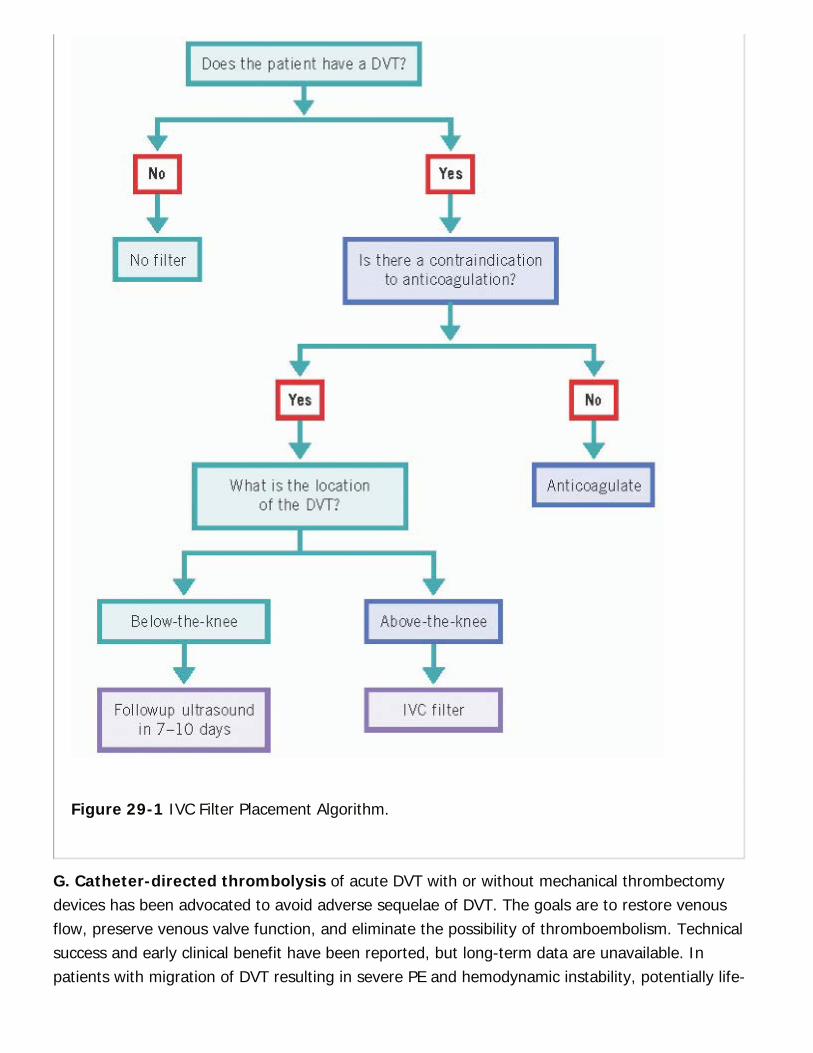
Figure 29-1 IVC Filter Placement Algorithm.
G. Catheter-directed thrombolysis of acute DVT with or without mechanical thrombectomydevices has been advocated to avoid adverse sequelae of DVT. The goals are to restore venousflow, preserve venous valve function, and eliminate the possibility of thromboembolism. Technicalsuccess and early clinical benefit have been reported, but long-term data are unavailable. Inpatients with migration of DVT resulting in severe PE and hemodynamic instability, potentially life-
P.523
saving thrombolytics should be considered (Curr Opin Anaes. 2006;19:52; CHEST guidelines.2012).
VI. UPPER-EXTREMITY DVTA. In patients with upper-extremity DVT that involves axillary or more proximal veins, the ChestGuidelines suggest anticoagulation (CHEST. 2012;141(2)).
B. If the upper-extremity DVT is associated with a catheter, it does not need to be removed if thecatheter is functional and necessary. Rather, anticoagulation should be started and continued for3 months after.
LYMPHEDEMAI. PATHOPHYSIOLOGYA. Primary lymphedema is the result of congenital aplasia, hypoplasia, orhyperplasia of lymphatic vessels and nodes that causes the accumulation of aprotein-rich fluid in the interstitial space. Swelling of the patient's leg initiallyproduces pitting edema, which progresses to a nonpitting form and may lead todermal fibrosis and disfigurement.
1. Primary lymphedema is classified according to age at presentation.
a. Congenital primary lymphedema (present at birth) represents 10% to 15% ofall cases, which can be hereditary (Milroy disease) or nonhereditary.
b. Praecox (early in life) or Meige disease represents 70% to 80% of cases.
(1) 80% to 90% of patients are female.
(2) Presents during the second and third decades of life, typically with localizedswelling of the foot and ankle. Such swelling is worsened by prolonged standing.
(3) A single lower extremity is affected in 70% of patients.
c. Tarda (late in life) primary lymphedema, representing 10% to 15% of cases,is seen equally in men and women and presents after the third or fourth decadeof life.
B. Secondary lymphedema results from impaired lymphatic drainagesecondary to a known cause and is the most common cause of lymphedema inthe United States. Surgical or traumatic interruption of lymphatic vessels (oftenfrom an axillary or groin lymph node dissection), carcinoma, infection, venousthrombosis, and radiation are causes of secondary lymphedema. Secondarylymphedema in the context of filariasis, caused by the parasite Wuchereriabancrofti, represents the most common worldwide etiology of the disease.
II. DIAGNOSIS
UnitedVRG
P.524
A. Clinical Presentation
1. Symptoms
a. Early lymphedema is characterized by unilateral or bilateral arm or pedalswelling that resolves overnight. With disease progression, the swellingincreases and extends up the extremity, producing discomfort and thickenedskin. With more advanced disease, swelling is not relieved with elevation.Significant pain is unusual.
b. Patients with secondary lymphedema commonly present with repeatedepisodes of cellulitis secondary to high interstitial protein content.
2. Physical examination
a. When a lower extremity is involved, the toes are often spared.
b. With advanced disease, the extremity becomes tense, with nonpittingedema.
c. Dermal fibrosis results in skin thickening, hair loss, and generalized keratosis.
B. Imaging Studies
1. Lymphoscintigraphy is the injection of radiolabeled (technetium-99m)colloid into the web space between the patient's second and third toes orfingers. The patient's limb is exercised periodically, and images are taken of theinvolved extremity and the whole body. Lymphedema is seen as an abnormalaccumulation of tracer or as slow tracer clearance along with the presence oflymphatic collaterals. The study has a sensitivity and specificity of 92% and100%, respectively in the diagnosis of lymphedema (J Vasc Surg. 1989;9:683).
2. CT and magnetic resonance (MR) scan are able to exclude any massobstructing the lymphatic system. MR scan has been able to differentiatelymphedema from chronic venous edema and lipedema (excessivesubcutaneous fat and fluid).
3. Lymphangiography involves catheter placement and injection ofradiopaque dye directly into lymphatic channels; it has largely been replaced bylymphoscintigraphy and CT.
III. DIFFERENTIAL DIAGNOSIS.Differential diagnosis includes all other causes of a swollen extremity.
A. Trauma
B. Infection
C. Arterial Disease
P.525
D. CVI
E. Lipedema
F. Neoplasm
G. Radiation Effects
H. Systemic diseases, such as right ventricular failure, myxedema, nephrosis, nephritis, andprotein deficiency. These causes must be excluded before invasive study.
IV. TREATMENTA. Medical management is limited by the physiologic and anatomic nature ofthe disease. The use of diuretics to remove fluid is not effective because of thehigh interstitial protein concentration. Development of fibrosis and irreversiblechanges in the subcutaneous tissue further limit options. The objectives ofconservative treatment are to control edema, maintain healthy skin, and avoidcellulitis and lymphangitis.
1. Combination of physical therapies (CPT) is the primary approachrecommended in a consensus document by the International Society ofLymphology Executive Committee (Lymphology. 2009;42:51). CPT consists of atwo-stage treatment program, beginning with skin care, followed by theapplication of compression bandages.
2. Sequential pneumatic compression has been shown to improvelymphedema.
3. Skin care and good hygiene are important. Topical hydrocortisone creammay be needed for eczema.
4. Benzopyrones (such as warfarin) have been effective in reducinglymphedema due to filariasis. Their action is believed to derive from enhancedmacrophage activity and extralymphatic absorption of interstitial proteins.
5. Cellulitis and lymphangitis should be suspected when sudden onset ofpain, swelling, or erythema of the leg occurs. Intravenous antibiotics should beinitiated to cover staphylococci and β-hemolytic streptococci. Limb elevation andimmobilization should be initiated, and warm compresses can be used forsymptomatic relief. Topical antifungal cream may be needed for chronicinfections.
B. Surgical Options. Surgical intervention is an alternative approach forpatients whose lymphedema has been refractory to nonoperative therapies.Only 10% of patients with lymphedema are surgical candidates, and surgery isdirected at reducing limb size. Indications for operation are related to functionbecause cosmetic deformities persist postoperatively. Results are best when
UnitedVRG
P.526
surgery is performed for severely impaired movement and recurrent cellulitis.
1. Total subcutaneous excision is performed for extensive swelling and skinchanges. Circumferential excision of the skin and subcutaneous tissue from thetibial tuberosity to the malleoli is performed. The defect is closed with a split- orfull-thickness skin graft from the resected specimen or a split-thickness skingraft from an uninvolved site.
2. Closure of disrupted lymphatic channels.
3. Omental transposition.
4. Lymphatic transposition includes direct (lymphovenous bypass, lymphaticgrafting) and indirect (mesenteric bridge, omental flap) procedures. Lymphaticgrafting is performed for upper-extremity or unilateral lower-extremitylymphedema. Good results have been reported in 80% of patients (PlastReconstr Surg. 1990;85:64).
5. Microsurgical lymphovenous anastomoses bypass the obstructed lymphaticsystem in patients with chronic lymphedema. With improved microvasculartechniques, patency rates of 50% to 70% can be expected many months aftersurgery (J Vasc Surg. 1986;4:148).
CHAPTER 29: VENOUS AND LYMPHATIC DISEASEMultiple Choice Questions1. A 72-year-old man presents to your clinic with 3 days of right legswelling. After completing a thorough history and physical examinationyou determine that a venous duplex is indicated. The results confirmyour suspicion of proximal DVT. Which of the following statements istrue regarding DVT?
a. The diagnosis of DVT is easily made by clinical examination.
b. Only half of patients with DVT have even one identifiable risk factor.
c. IVC filters are indicated in all patients with iliofemoral DVT.
d. Male gender is an independent risk factor for DVT.
e. DVT is a rare complication of orthopedic surgery.
View Answer
2. A 65-year-old female returns to your wound clinic for routinefollowup of her venous ulcer. She had a venous duplex showing refluxat her saphenofemoral junction and you performed a radiofrequencyablation of her left GSV 1 year ago. Today you observe that her ulcer iswell healed. What is the CEAP classification of her venous disease ?
P.527
a. C5 Es As Pr
b. C5 Es Ad Pr
c. C45 Es Ad Pr
d. C5 Ep As Pr
e. C5 Ep As Pp
View Answer
3. You are part of a hospital task-force dedicated to reducing theincidence of DVT. As part of your presentation you recommend givingall patients low-dose subcutaneous heparin TID. A hospitaladministrator asks you how effective this treatment is for preventingDVT as this proposal is projected to cost the hospital a milliondollars/year. Low-dose unfractionated heparin reduces the risk of DVTby how much?
a. 10% to 30%
b. 30% to 50%
c. 50% to 70%
d. 70% to 90%
e. 90% to 100%
View Answer
4. You perform radiofrequency ablation of the right greater saphenousvein for symptomatic varicose veins. A venous duplex obtained 7 daysafter the procedure reveals a thrombosed GSV but there is propagationof clot into the common femoral vein. What is the appropriatetreatment?
a. Prescribe Warfarin and refer to anticoagulation clinic for furthermanagement
b. No medications, this clot is a known risk of RFA and will resolve on itsown
c. No medications, repeat duplex in 1 week to monitor clot
d. Enoxaparin with bridge to Coumadin therapy for 3 months for provokedDVT
e. Repeat radiofrequency ablation, this time on the femoral vein
View Answer
UnitedVRG
5. You are serving as the vascular surgery consult resident. You arecalled by the neurosurgery service to place an IVC filter on a patientprior to spine surgery, there is no evidence of DVT on the duplex butthe patient will be nonambulatory for 7 days postop. Which of thefollowing are complications of IVC filter placement?
a. Filter fracture
b. Filter embolus
c. DVT
d. Hematoma
e. All of the above
View Answer
6. A 43-year-old female presents to your Vascular Surgery clinic for anewly diagnosed peroneal vein DVT. Patient has no symptomsassociated with this DVT. What is the correct recommendation fortreatment?
a. IVC filter placement
b. Lovenox for 2 weeks with a repeat ultrasound
c. Repeat ultrasound in 1 to 2 weeks
d. Lovenox with bridge to Coumadin for 3 months
e. Lovenox with bridge to Coumadin for 6 months
View Answer
30Hemodialysis Access
Michael J. Beckman
Surendra Shenoy
Hemodialysis (HD) is the most common renal replacement therapy used to prolong survival inpatients with end-stage renal disease (ESRD). In December 2012, there were 636,905 ESRDpatients in the United States. Approximately 408,711 (91% of all dialysis dependent patients)were on HD, 40,631 on peritoneal dialysis (PD), and 186,303 had a working renal transplant(USRDS. 2014). HD, based on body mass and the type of dialysis prescription (intermittent, daily,nocturnal etc.), requires 200 to 500 mL/minute of patient's blood circulation through the dialyzer.ÒVascular AccessÓ (VA) is a port or site in the body capable of supporting the blood flow requiredfor dialysis. Adequacy of HD, responsible for the longevity of the ESRD patient, is directlydependent on the function of the dialysis access.
I. INDICATIONS FOR DIALYSISA. Short-term HD (temporary) is often urgently indicated in patients with acute renal failure inclinical situations such as (i) severe fluid overload, (ii) refractory hypertension, (iii) symptomatichyperkalemia, (iv) uremiainduced GI symptoms or bleeding, encephalopathy, seizures, orpericarditis, (v) intractable metabolic acidosis, and (vi) occasionally with medication overdose orintoxication.
B. Long-term HD (permanent) is initiated in patients with stage 5 chronic kidney disease (CKD)presenting with intractable (i) fluid overload, (ii) electrolyte imbalance, (iii) hypertension, and/or(iv) uremia.
II. TYPES OF ACCESSA. VA. Short-term or urgent HD needs are met by placing HD catheters temporarily in the centralveins. Long-term dialysis requires surgical creation of subcutaneous conduits with high volumeblood flow that can be accessed with needles to circulate blood through the dialyzer and back tothe body.
B. PD Access. PD uses a silicone (Tenckhoff) catheter or polyurethane catheter, inserted with itstip placed in the dependent part of the peritoneal cavity (pelvis), that allows free in- and outflowof dialysate. The peritoneal membrane acts as a blood filter to exchange electrolytes and uremictoxins retained from renal failure.
> Table of Contents > 30 - Hemodialysis Access
UnitedVRG
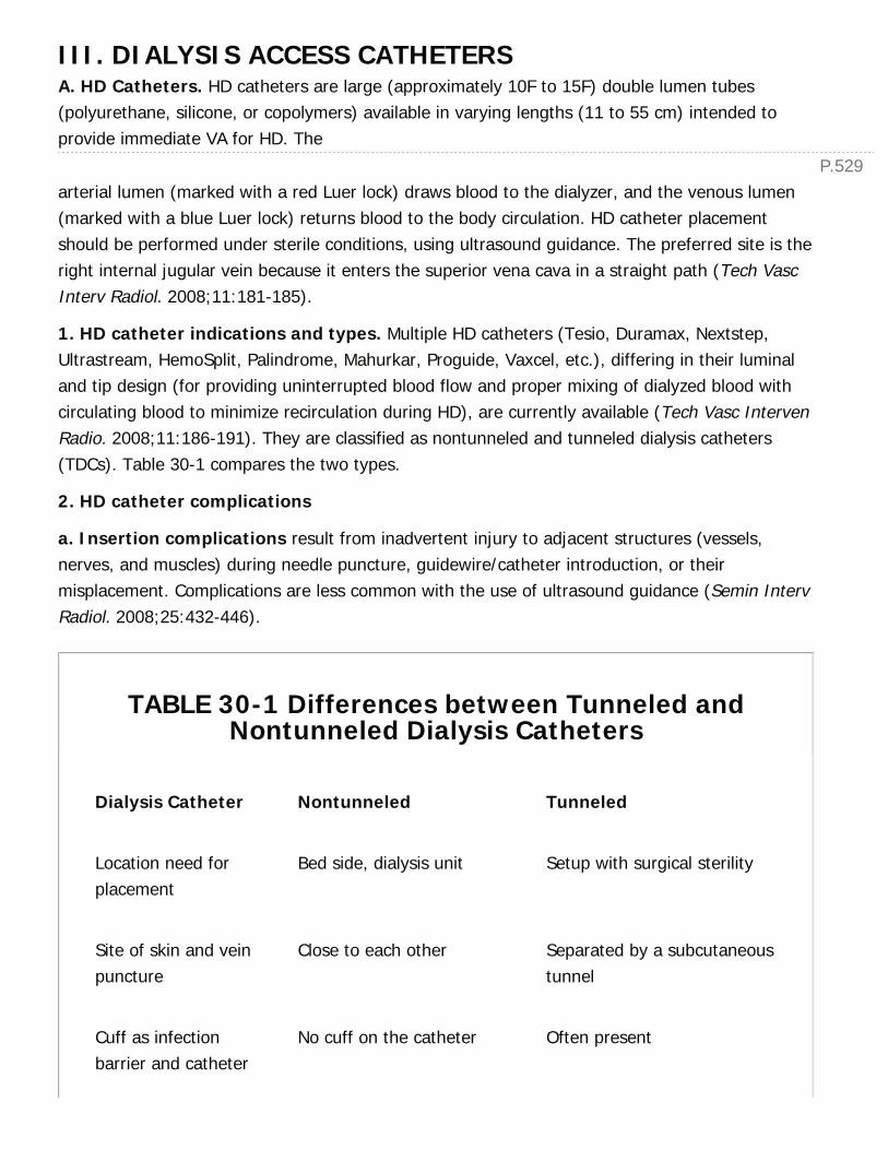
P.529
III. DIALYSIS ACCESS CATHETERSA. HD Catheters. HD catheters are large (approximately 10F to 15F) double lumen tubes(polyurethane, silicone, or copolymers) available in varying lengths (11 to 55 cm) intended toprovide immediate VA for HD. The
arterial lumen (marked with a red Luer lock) draws blood to the dialyzer, and the venous lumen(marked with a blue Luer lock) returns blood to the body circulation. HD catheter placementshould be performed under sterile conditions, using ultrasound guidance. The preferred site is theright internal jugular vein because it enters the superior vena cava in a straight path (Tech VascInterv Radiol. 2008;11:181-185).
1. HD catheter indications and types. Multiple HD catheters (Tesio, Duramax, Nextstep,Ultrastream, HemoSplit, Palindrome, Mahurkar, Proguide, Vaxcel, etc.), differing in their luminaland tip design (for providing uninterrupted blood flow and proper mixing of dialyzed blood withcirculating blood to minimize recirculation during HD), are currently available (Tech Vasc IntervenRadio. 2008;11:186-191). They are classified as nontunneled and tunneled dialysis catheters(TDCs). Table 30-1 compares the two types.
2. HD catheter complications
a. Insertion complications result from inadvertent injury to adjacent structures (vessels,nerves, and muscles) during needle puncture, guidewire/catheter introduction, or theirmisplacement. Complications are less common with the use of ultrasound guidance (Semin IntervRadiol. 2008;25:432-446).
TABLE 30-1 Differences between Tunneled andNontunneled Dialysis Catheters
Dialysis Catheter Nontunneled Tunneled
Location need forplacement
Bed side, dialysis unit Setup with surgical sterility
Site of skin and veinpuncture
Close to each other Separated by a subcutaneoustunnel
Cuff as infectionbarrier and catheter
No cuff on the catheter Often present
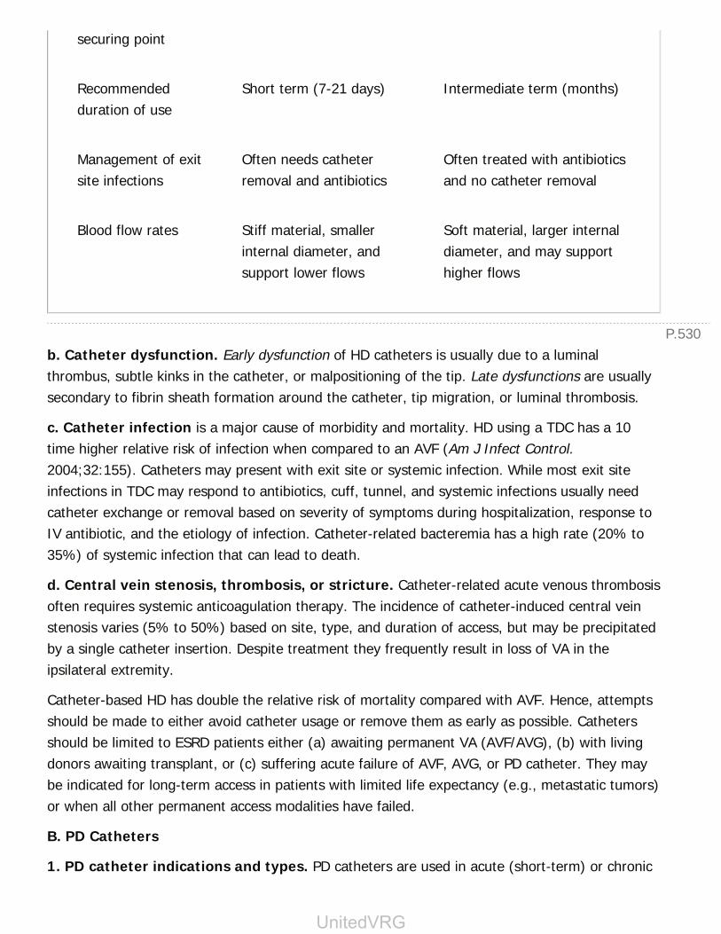
P.530
securing point
Recommendedduration of use
Short term (7-21 days) Intermediate term (months)
Management of exitsite infections
Often needs catheterremoval and antibiotics
Often treated with antibioticsand no catheter removal
Blood flow rates Stiff material, smallerinternal diameter, andsupport lower flows
Soft material, larger internaldiameter, and may supporthigher flows
b. Catheter dysfunction. Early dysfunction of HD catheters is usually due to a luminalthrombus, subtle kinks in the catheter, or malpositioning of the tip. Late dysfunctions are usuallysecondary to fibrin sheath formation around the catheter, tip migration, or luminal thrombosis.
c. Catheter infection is a major cause of morbidity and mortality. HD using a TDC has a 10time higher relative risk of infection when compared to an AVF (Am J Infect Control.2004;32:155). Catheters may present with exit site or systemic infection. While most exit siteinfections in TDC may respond to antibiotics, cuff, tunnel, and systemic infections usually needcatheter exchange or removal based on severity of symptoms during hospitalization, response toIV antibiotic, and the etiology of infection. Catheter-related bacteremia has a high rate (20% to35%) of systemic infection that can lead to death.
d. Central vein stenosis, thrombosis, or stricture. Catheter-related acute venous thrombosisoften requires systemic anticoagulation therapy. The incidence of catheter-induced central veinstenosis varies (5% to 50%) based on site, type, and duration of access, but may be precipitatedby a single catheter insertion. Despite treatment they frequently result in loss of VA in theipsilateral extremity.
Catheter-based HD has double the relative risk of mortality compared with AVF. Hence, attemptsshould be made to either avoid catheter usage or remove them as early as possible. Cathetersshould be limited to ESRD patients either (a) awaiting permanent VA (AVF/AVG), (b) with livingdonors awaiting transplant, or (c) suffering acute failure of AVF, AVG, or PD catheter. They maybe indicated for long-term access in patients with limited life expectancy (e.g., metastatic tumors)or when all other permanent access modalities have failed.
B. PD Catheters
1. PD catheter indications and types. PD catheters are used in acute (short-term) or chronic
UnitedVRG

P.531
renal failure (long-term). PD catheter designs vary in cuff, body, and tip configuration (straightwith disc, weight balloon and fluting, or coiled with similar variations, Swan-neck presternalcatheters etc.). None have shown convincing superiority in function or complication rates (PeritDial Int. 2007;27:S119-S125). Surgeons, nephrologists, or interventional radiologists (based onexpertise available) can place single cuff Tenckhoff catheters for short-term immediate use at thebed side. Long-term PD catheters (usually two cuffs) are placed surgically with or without the useof radiologic imaging, peritoneoscopy, or laparoscopy. PD is the preferred technique for children.Some surgical contraindications for PD catheter placement include recent abdominal surgery,peritoneal infections, adhesions, large abdominal wall hernia, significant gastroesophageal reflux,and diaphragmatic hernia.
2. PD catheter complications
a. Early complications
(1) Injury to intraperitoneal[u3] structures resulting in intestinal perforation, peritonitis, andbleeding.
(2) Dialysate infusion resulting in hydrothorax, exacerbation of hernia (scrotal swelling), andabdominal wall edema.
(3) Catheter dysfunction due to malposition or kink, fibrin sheath or coagulum, or constipation.
b. Late complications
(1) Catheter related: Leakage[u4] from fractures, intra-abdominal bleeding from tugging,intestinal obstruction, and intestinal perforations from erosions.
(2) Dialysate-related problems (listed above) can occasionally manifest late.
c. Infections
(1) Exit site infection[u5] or cuff extrusion can be treated with antibiotics and cuff removal indouble cuff catheters.
(2) Peritonitis is a major cause of hospitalization, PD failure, and mortality (35 deaths per 1,000years at risk). Infections can include Gram-positive bacteria, Gram-negative bacteria includingPseudomonas, fungal infections, or tuberculous infections. First cultures should be obtained, thenempiric intraperitoneal therapy should be initiated with vancomycin or a cephalosporin to coverGram-positive bacteria and third-generation cephalosporin or an aminoglycoside to cover Gram-negative bacteria. Once cultures and sensitivities are reported, the regimen should be targeted tothe specific microbe (Perit Dial Int. 2010;30:393). Severe, recurrent, or resistant infectionsrequire catheter removal.
IV. ARTERIOVENOUS ACCESS.
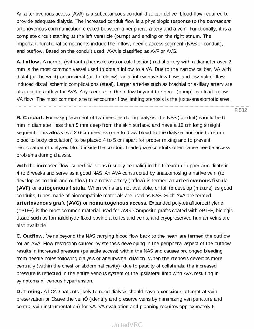
P.532
An arteriovenous access (AVA) is a subcutaneous conduit that can deliver blood flow required toprovide adequate dialysis. The increased conduit flow is a physiologic response to the permanentarteriovenous communication created between a peripheral artery and a vein. Functionally, it is acomplete circuit starting at the left ventricle (pump) and ending on the right atrium. Theimportant functional components include the inflow, needle access segment (NAS or conduit),and outflow. Based on the conduit used, AVA is classified as AVF or AVG.
A. Inflow. A normal (without atherosclerosis or calcification) radial artery with a diameter over 2mm is the most common vessel used to obtain inflow to a VA. Due to the narrow caliber, VA withdistal (at the wrist) or proximal (at the elbow) radial inflow have low flows and low risk of flow-induced distal ischemic complications (steal). Larger arteries such as brachial or axillary artery arealso used as inflow for AVA. Any stenosis in the inflow beyond the heart (pump) can lead to lowVA flow. The most common site to encounter flow limiting stenosis is the juxta-anastomotic area.
B. Conduit. For easy placement of two needles during dialysis, the NAS (conduit) should be 6mm in diameter, less than 5 mm deep from the skin surface, and have a 10 cm long straightsegment. This allows two 2.6-cm needles (one to draw blood to the dialyzer and one to returnblood to body circulation) to be placed 4 to 5 cm apart for proper mixing and to preventrecirculation of dialyzed blood inside the conduit. Inadequate conduits often cause needle accessproblems during dialysis.
With the increased flow, superficial veins (usually cephalic) in the forearm or upper arm dilate in4 to 6 weeks and serve as a good NAS. An AVA constructed by anastomosing a native vein (todevelop as conduit and outflow) to a native artery (inflow) is termed an arteriovenous fistula(AVF) or autogenous fistula. When veins are not available, or fail to develop (mature) as goodconduits, tubes made of biocompatible materials are used as NAS. Such AVA are termedarteriovenous graft (AVG) or nonautogenous access. Expanded polytetrafluoroethylene(ePTFE) is the most common material used for AVG. Composite grafts coated with ePTFE, biologictissue such as formaldehyde fixed bovine arteries and veins, and cryopreserved human veins arealso available.
C. Outflow. Veins beyond the NAS carrying blood flow back to the heart are termed the outflowfor an AVA. Flow restriction caused by stenosis developing in the peripheral aspect of the outflowresults in increased pressure (pulsatile access) within the NAS and causes prolonged bleedingfrom needle holes following dialysis or aneurysmal dilation. When the stenosis develops morecentrally (within the chest or abdominal cavity), due to paucity of collaterals, the increasedpressure is reflected in the entire venous system of the ipsilateral limb with AVA resulting insymptoms of venous hypertension.
D. Timing. All CKD patients likely to need dialysis should have a conscious attempt at veinpreservation or Òsave the veinÓ (identify and preserve veins by minimizing venipuncture andcentral vein instrumentation) for VA. VA evaluation and planning requires approximately 6
UnitedVRG

P.533
months. Planning renal replacement when the patient reaches CKD stage 4 (GFR < 30mL/minute) should provide ample opportunity for VA referral and placement.
V. PREOPERATIVE EVALUATIONA. History. Obtain demographics, previous VA attempts, and details related to arterial disease(claudication, rest pain, limb/digit loss, neuropathy etc.) and venous problems (venipunctures,catheter placements, percutaneously introduced central catheters [PICC], cardiac implantableelectronic device [CIED], etc.). Evaluate comorbidities such as smoking, diabetes, cardiacproblems, peripheral neuropathy, and/or thrombophilia that may contribute to VA failure.
B. Physical Examination
1. Arterial (inflow) evaluation. Palpate and compare strength of pulse and quality of arterialwall (radial, brachial, and axillary) bilaterally.
When indicated, the femoral, popliteal, and pedal arteries should be evaluated. Bilateral bloodpressures should be measured (difference >20 mm Hg is significant for proximal stenosis). Therole of the Allen test, intended to assess the integrity of palmar circulation, is not certain for endto side VA placement. Scars from previous surgery and catheters should be noted.
2. Venous (outflow) evaluation. Using vein dilation techniques (tourniquets, warmth, tapping,use of gravity etc.) is critical for peripheral vein evaluation. Presence of edema, unequalextremities, and/or chest and shoulder collaterals may suggest central venous stenosis. Patencyand continuity of the vein can be assessed by eliciting fluid thrill.
C. Doppler Duplex ultrasound scanning (DDUS) should always complement clinicalexamination. Best results from ultrasound vessel mapping are obtained when the operatingsurgeon evaluates the scan in real time. DDUS provides both functional (distensibility, flowpattern, and volumes) and structural evaluation of the peripheral arteries and veins (superficialand deep). It is not very reliable in evaluation of central veins.
D. Diagnostic Imaging
1. Contrast venography is the gold standard for determining the patency (luminal dimension)and adequacy of central venous anatomy. Peripheral vein visualization is limited to downstreamtributaries receiving contrast from the distal peripheral vein injection site.
2. Arteriography remains the gold standard for the evaluation of a suspected arterial inflowstenosis or occlusion. MRA (noncontrast) or CTA may be used to evaluate larger veins andarterial run off.
E. Laboratory Studies. Hyperkalemia and acidosis are the most common electrolyteabnormalities seen in ESRD patients. Testing (day of surgery) should include hemoglobin, serumpotassium, and glucose levels to avoid possible procedural or anesthesia-related complications.
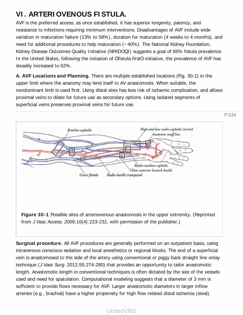
P.534
VI. ARTERIOVENOUS FISTULA.AVF is the preferred access, as once established, it has superior longevity, patency, andresistance to infections requiring minimum interventions. Disadvantages of AVF include widevariation in maturation failure (13% to 58%), duration for maturation (4 weeks to 4 months), andneed for additional procedures to help maturation (~40%). The National Kidney Foundation,Kidney Disease Outcomes Quality Initiative (NFKDOQI) suggests a goal of 65% fistula prevalence.In the United States, following the initiation of Òfistula firstÓ initiative, the prevalence of AVF hassteadily increased to 62%.
A. AVF Locations and Planning. There are multiple established locations (Fig. 30-1) in theupper limb where the anatomy may lend itself to AV anastomosis. When suitable, thenondominant limb is used first. Using distal sites has less risk of ischemic complication, and allowsproximal veins to dilate for future use as secondary options. Using isolated segments ofsuperficial veins preserves proximal veins for future use.
Figure 30-1 Possible sites of arteriovenous anastomosis in the upper extremity. (Reprintedfrom J Vasc Access. 2009;10(4):223-232, with permission of the publisher.)
Surgical procedure. All AVF procedures are generally performed on an outpatient basis, usingintravenous conscious sedation and local anesthetics or regional blocks. The end of a superficialvein is anastomosed to the side of the artery using conventional or piggy back straight line onlaytechnique (J Vasc Surg. 2012;55:274-280) that provides an opportunity to tailor anastomoticlength. Anastomotic length in conventional techniques is often dictated by the size of the vesselsused and need for spatulation. Computational modeling suggests that a diameter of 3 mm issufficient to provide flows necessary for AVF. Larger anastomotic diameters in larger inflowarteries (e.g., brachial) have a higher propensity for high flow related distal ischemia (steal).
UnitedVRG
P.535
B. Postoperative Evaluation. Early assessment (7 to 10 days) is necessary to evaluate thesurgical wound, fistula patency, and detect any distal vascular or neurologic problems. It alsoprovides an opportunity to further evaluate early postoperative problems that may interfere withmaturation. A maturation evaluation (assess inflow, conduit, and outflow) should beperformed between 4 and 6 weeks postoperatively using clinical evaluation and DDUS. Anyproblems detected should be further evaluated. AVF meeting maturation criteria can then beaccessed. Fistulae that have good inflow but are deep and not accessible need superficialization(VA: Principle and Practice. Williams and Wilkins. 2010:1996-205). Occasionally, fistulae mayneed a few additional weeks to mature.
VII. AV GRAFT.AV grafts are conduits (anastomosed to an artery to obtain inflow and a vein for outflow) placedin a subdermal location to provide NAS for dialysis. Based on the configuration of the conduit theyare termed loop or straight grafts. The graft limb receiving arterial inflow, termed arterial limb,provides blood to the dialysis circuit. The graft limb delivering dialyzed blood into the outflow veinis termed the venous limb.
A. AVG Locations and Planning. The proximal radial, brachial, and axillary arteries usuallyprovide graft inflow. Since patients requiring AVG often lack sufficient superficial venous anatomyfor AVF, the venae comitantes or the deep veins of the upper arm (basilic, brachial, or axillary)provide the outflow for AVG. They can be placed in the thigh using femoral artery
and saphenous or femoral veins. Distal ischemia becomes more of an issue when larger vesselsare used for inflow in AVG. Lower-extremity grafts are at a higher risk for infection.
B. Surgical Procedure. AV grafts are placed under local anesthesia with conscious sedation.Prophylactic antibiotics (e.g., second-generation cephalosporins) are commonly administeredimmediately prior to the surgery.
C. Postoperative Evaluation. While conventional grafts need good tissue incorporation prior tocannulation (3 to 6 weeks), newer composite graft materials permit early cannulation whenindicated. A standard 6 mm caliber graft, placed in a subdermal location, and with rigid wallsmake graft cannulation easier than many AVF. Once placed, grafts are more predictably availablefor dialysis; however, they are prone to repeated thrombosis requiring a significantly highernumber of interventions and have poor longevity compared to a well-matured AVF.
VIII. AVA PLANNING.The goal of clinical evaluation is to plan a sequence of options to provide VA throughout the lifespan of the ESRD patient. Access planning should take into consideration the life expectancy ofthe ESRD patient, renal function at the time of referral (CKD 4 or 5 or already on dialysis), andthe available access options based on clinical and ultrasound evaluation. Based on these factors,the surgeon should plan to create a VA that has the longest durability, needing the least

P.536
interventions, in a timely fashion, without jeopardizing future access options (J Vasc Access.14;15:S1-S5). When the anatomy is suitable, AVF is the preferred access. AVG should beconsidered when AVF is not an option or is not able to provide a timely access.
IX. COMPLICATIONS OF AVA.VA dysfunction is a major cause of morbidity and mortality in ESRD. Stenosis (responsible forover 90% of VA dysfunctions) can present as thrombosis, infection, and aneurysm. NKF-KDOQIguidelines recommend prospective monitoring and surveillance using physical examination,ultrasound evaluation of flow, and assessment of dialysis adequacy to help detect accessdysfunction. Interventions guided by DDUS/angiography imaging or surgical techniques are usedto prolong access patency.
A. Stenosis. The functional impact of stenoses depend on their location, diameter relative to theinflow volume, and pressure (J Vasc Acc. 2014;15:409-414). Development of venous neointimalhyperplasia (VNH), the cause for luminal narrowing, is common in areas of outflow (e.g., graftvein anastomosis of AVG and JAS of AVF) experiencing hemodynamic stress resulting from bloodflow increase. Balloon angioplasty, using DDUS or contrast angiography, prior to development ofthrombosis helps improve access patency and delay failure. Surgical venoplasty and vascularstents are also used based on indications or when angioplasty fails.
B. Thrombosis. Thrombosis seen early after AVA placement is attributed to flow interruption dueto technical factors, inflammatory narrowing of outflow veins, preexisting vein occlusions, orkinked grafts. Occasionally
patient-related factors such as thrombophilia and hypotension may be responsible. Earlythrombosis of AVF often results in loss of the access site. AVG with early thrombosis may besalvaged by surgical or radiologic image-guided thrombectomy. Late thrombosis both in AVG andAVF are mostly due to development of stenosis in the circuit. They are salvaged by thrombectomyand dilation of stenotic lesions using surgical or interventional means.
C. Infection. Infections in AVA are caused by cutaneous flora (commonly Staphylococcusspecies) contamination, mostly through the needle access sites, presenting as skin ulceration,infections in chronic thrombi within aneurysms, or seeding of graft material. Management ofinfection depends on severity and presentation. Most graft infections result in removal of the graftand loss of the access site. Focal infections may be salvaged with resection of the infected portionand reconstruction with a bypass graft. Most AVF infections are treated with antibiotics, with orwithout surgical intervention.
D. Aneurysms and Pseudoaneurysms. These develop secondary to loss of AVG integrity,repeated needle puncture, and extravasation of blood which is walled off by surrounding tissue(pseudoaneurysms). AVF aneurysms are often true dilations of the vein developing over time dueto stretch on the healing puncture site from the pressure within. Aneurysms can develop chronic
UnitedVRG

P.537
thrombi resulting in access difficulty. They may also be at risk of rupture and bleeding due tothinning of the overlying skin. In such situations both AVG and AVF aneurysms can be electivelyrepaired.
E. Arterial ÒStealÓ Syndrome. All AV accesses divert or ÒstealÓ a fraction of blood from thedistal circulation which is usually well tolerated by the distal extremity. In approximately 1% to4% of patients (diabetics, underlying neuropathy, and vascular disease) this may precipitateischemic pain, worsening neuropathy, ulceration, or gangrene (Ann Vasc Surg. 2000;14(2):138-144). Mild symptoms such as subjective coolness and paresthesias without sensory or motor lossmay be managed expectantly with increasing exercise tolerance. Failure to improve orincreasingly severe symptoms require further evaluation. Reduction of AVA flow may improvesymptoms due to high VA flows. Symptomatic patients with low AVA flows may benefit from distalflow enhancement techniques. Severe ischemia requires immediate evaluation and managementincluding access ligation to avoid irreversible nerve injury.
F. Venous Hypertension. Venous hypertension caused by central vein stenosis manifests asedema, skin discoloration, and/or hyperpigmentation in the affected limb. Management optionsinclude venoplasty of the stenosis, stent graft repair, or surgical provision of improved outflowdrainage.
G. Congestive Heart Failure (CHF). High volume flow, often resulting from large artery basedaccess AVA can occasionally precipitate CHF. This is more common in patients with underlyingheart problems or preexisting fluid overload. Based on the situation, this may necessitate flowreduction or access ligation.
CHAPTER 30: HEMODIALYSIS ACCESSMultiple Choice Questions1. What is the appropriate flow rate for adequate hemodialysis?
a. 100 to 200 mL/minute
b. 500 to 700 mL/minute
c. 800 to 1,000 mL/minute
d. 200 to 500 mL/minute
View Answer
2. What is the most common cause of AV graft dysfunction?
a. Infection
b. Thrombosis
c. Stenosis
d. Maturation failure
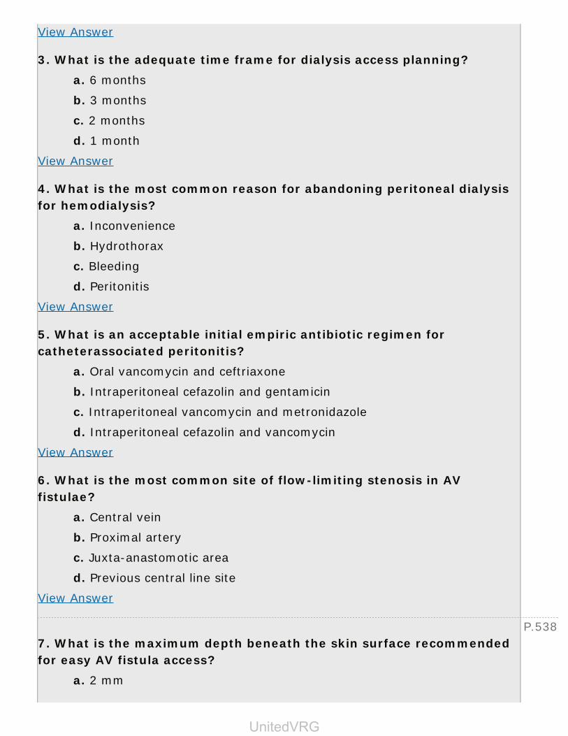
P.538
View Answer
3. What is the adequate time frame for dialysis access planning?
a. 6 months
b. 3 months
c. 2 months
d. 1 month
View Answer
4. What is the most common reason for abandoning peritoneal dialysisfor hemodialysis?
a. Inconvenience
b. Hydrothorax
c. Bleeding
d. Peritonitis
View Answer
5. What is an acceptable initial empiric antibiotic regimen forcatheterassociated peritonitis?
a. Oral vancomycin and ceftriaxone
b. Intraperitoneal cefazolin and gentamicin
c. Intraperitoneal vancomycin and metronidazole
d. Intraperitoneal cefazolin and vancomycin
View Answer
6. What is the most common site of flow-limiting stenosis in AVfistulae?
a. Central vein
b. Proximal artery
c. Juxta-anastomotic area
d. Previous central line site
View Answer
7. What is the maximum depth beneath the skin surface recommendedfor easy AV fistula access?
a. 2 mm
UnitedVRG
b. 10 mm
c. 7 mm
d. 5 mm
View Answer
8. What is the correct terminology for the implanted synthetic tubeplaced during creation of an AV graft?
a. Autogenous fistula
b. Nonautogenous conduit
c. Autogenous outflow
d. Nonautogenous inflow
View Answer
9. Salvage procedures used for vascular steal in a high-flow AVF accessinclude:
a. Distal revascularization with interval ligation (DRIL)
b. Banding the access
c. Proximalization of arterial inflow (PAI)
d. Fistula ligation
View Answer
10. Which vascular access poses the highest risk of infection?
a. AV fistula
b. Nontunneled dialysis catheter
c. Tunneled dialysis catheter
d. AV graft
View Answer

31Transplantation
Bernard J. DuBray Jr.
Jason R. Wellen
PHYSIOLOGIC IMMUNITYOur immune system did not evolve to stymie the efforts of transplant surgeons, but rather toevade microbes that aim to overrun us. To understand why tissues from one individual arerejected by another, one has to appreciate the components of our immune system in itsphysiologic state.
I. THE IMMUNE SYSTEM ENCOMPASSES 2 COMPLEMENTARYARMSA. Innate immune system recognizes general distress as well as conserved moieties fromubiquitous pathogens, such as lipopolysaccharides of gramnegative bacteria. The response isdirect, nonspecific, and lacks memory.
1. Mediators
a. The complement cascade is a soluble group of proteins whose activation promotesformation of the membrane attack complex (MAC). This embeds itself within cell membranes ofpathogens causing disruption and cell lysis. In addition, byproducts of the complement cascadeopsonize pathogens, which promotes phagocytosis by antigen presenting cells (APCs).
b. Natural Killer cells recognize cells that lack self-Major Histocompatibility Complex (MHC) andare part of the body's immunosurveillance for cancer.
B. The adaptive immune system recognizes specific, pathogenic antigens in the context ofMHC, which help the immune system distinguish self from non-self. Foreign antigens presented inthe context of the MHC are targets of the adaptive immune system. In humans, these complexesare referred to as Human Leukocyte Antigens (HLA) and are located on chromosome 6.
1. Classes of HLA
a. Class I (A, B, C) are present on all nucleated cells and are targets for cytotoxic (CD8) T cells
b. Class II (DR, DP, and DQ) are present on APCs and are targets for helper (CD4) T cells.They trigger an antibody (humoral) mediated immune response.
The most important HLA in solid organ transplantation are A, B, and DR. Since each
> Table of Contents > 31 - Transplantation
UnitedVRG

P.540
person has 2 MHC complexes, one on each copy of chromosome 6, everyone has a total of 6 HLAantigens that are relevant to organ transplantation.
2. Adaptive immune responses
a. Cell-mediated. Antigens in the peripheral tissues are presented to T-cells located in lymphnodes and the spleen. The T-cell receptor (TCR)
recognizes a specific antigen in the context of MHC. Formation of the TCR is the result of DNArearrangement that occurs within the thymus during fetal development. Following rearrangement,T-cells are selected based on their ability to bind self MHC without activating a response. MHCencountered in the tissues not involved in thymic education activates an immune response. Thisis the basis of alloreactivity.
(1) Helper T cells (CD4) recognize exogenous antigens presented in the context of MHC class IIon the surface of APCs (B cells, dendrites, and macrophages). Activation releases IL-2, whichcauses B cell maturation into Plasma cells and IL-4, which causes maturation of cytotoxic T cells.
(2) Cytotoxic T cells (CD8) recognize endogenous (i.e., TB, viruses) pathogens presented in thecontext of MHC class I.
b. Antibody-mediated (humoral). B cells (Bone) activate antibody-mediated (humoral)immunity. IL-4 from helper T cells transforms B cells into plasma cells, which secrete antibodiesspecific to the offending pathogen.
TRANSPLANT IMMUNOLOGYA. Isografts. Tissue Transfer from Genetically Identical Individuals (i.e., Twins)
B. Xenografts. Tissue Transfer between Species
C. Allografts. Tissue Transfer among Members of the same Species
1. Alloreactivity/Histocompatibility
a. ABO blood compatibility is necessary for all transplants, except liver.
b. HLA – A, -B, -DR are the most important for compatibility; -DR is most important overall.
(1) Cross-matching detects preformed antibodies against donor HLA. It involves mixingrecipient serum with donor lymphocytes.
(2) Panel reactive antibodies (PRA) help to predict the likelihood of a positive cross-match. Itis determined by testing the potential recipient's serum against a panel of cells of various HLAspecificities in a manner similar to the cross-match. The percentage of specificities in the panelwith which the patient's sera react is the PRA. Patients who have been exposed to other HLAs viablood transfusion, pregnancy, or prior transplantation will have higher PRAs.
P.541
TRANSPLANT REJECTION
I. TYPES OF REJECTIONA. Hyperacute rejection is the result of preformed anti-HLA antibodies that bind the allograftendothelium to initiate a cascade of events culminating in vascular thrombosis and ischemicnecrosis. The only therapeutic option is to remove the allograft immediately. This isextraordinarily uncommon in the modern era of cross-matching.
B. Accelerated rejection is caused by sensitized T cells that produce a secondary immuneresponse. This generally occurs within 1 week of transplantation. Treatment includes pulsesteroids and OKT3.
C. Acute rejection is cell-mediated and involves T lymphocytes (cytotoxic and helper). Thistypically occurs 1 week to 1 month after transplantation. There are two basic treatmentmodalities: High-dose methylprednisolone and an antilymphocyte preparation.
D. Chronic rejection is a poorly understood phenomenon that can occur weeks to years aftertransplantation. Emerging evidence suggests that the humoral immune response is an importantcontributor. Plasmapheresis, intravenous immunoglobulin, and rituximab have been used to treatantibody-mediated rejection.
IMMUNOSUPPRESSION
I. CLASSES OF IMMUNOSUPPRESSIVE DRUGSA. Corticosteroids (Prednisone or methylprednisolone/Solu-Medrol) play the broadest role inimmunomodulation and are used for induction, maintenance, and treatment of rejection. Steroidsinhibit transcription of cytokines that promote lymphocyte proliferation, which dampens theinflammatory response and migration of neutrophils. Toxicities include poor wound healing,hyperglycemia, infections, cataracts, hypertension, weight gain, and bone disease.
B. Antiproliferative Agents/Antimetabolites
1. Azathioprine (Imuran) is an antimetabolite that is a thioguanine derivative of mercaptopurine.This purine analog alters the function or synthesis of DNA and RNA, inhibiting T- and B-lymphocyte proliferation. Myelosuppression manifested as leukopenia and thrombocytopenia areits main toxicities.
2. Mycophenolic acid (CellCept, Myfortic) inhibits inosine monophosphate dehydrogenase (rate-limiting enzyme in guanine monophosphate synthesis) inhibiting RNA synthesis. In doing so, thisdrug selectively inhibits T- and B-cell proliferation, cytotoxic T-cell generation, and antibodyformation. Toxicities include GI disturbance and leukopenia.
C. Calcineurin Inhibitors
UnitedVRG

P.542
1. Cyclosporine (Sandimmune, Neoral, Gengraf) binds cyclophilin protein and inhibits IL-2production. This blocks T cell activation and proliferation. Nephrotoxicity, hypertension, tremors,seizures, hyperkalemia, hyperuricemia, hypercholesterolemia, gingival hyperplasia, and hirsutismare the main toxicities.
2. Tacrolimus (FK506, Prograf) binds FK binding protein; action similar to CSA but is 10 to 100times more potent. Toxicities are similar to CSA, but include more GI and neurologic changes.
3. Sirolimus (Rapamune) is an anti-T-cell agent that inhibits the mTOR molecule, blocking T-cellsignal transduction. Toxicity includes
thrombocytopenia, hyperlipidemia, oral ulcers, anemia, proteinuria, and impairment of woundhealing.
D. Co-Stimulation Blockade. Belatacept (Nulojix) is a fusion protein that is homologous toCD27 and binds to the B7 complex to irreversibly block the Signal 2 pathway associated theMHC/APC binding process.
E. Antithymocyte Antibodies. Polyclonal antibodies are immunologic products with antibodiesto a wide variety of T-cell antigens, adhesion molecules, costimulatory molecules, cytokines, theT-cell receptor, and class I and II MHC molecules. These agents are used as induction therapy inthe perioperative period or as rescue therapy following acute rejection. The most commonly usedantithymocyte immunoglobulin in the United States is thymoglobulin, which is derived fromrabbit serum.
II. COMPLICATIONS OF IMMUNOSUPPRESSIONA. Bacterial Infections. Pneumonia and urinary tract infections (UTIs) occur fairly commonlyafter transplantation. Infectious complications from opportunistic organisms are now uncommonbecause of appropriate prophylactic strategies.
B. Viral Infections
1. CMV infection can occur at any time but is most common 1 to 4 months posttransplant, in theabsence of prophylaxis. CMV may infect the recipient's liver, lungs, or GI tract. Signs andsymptoms include fever, chills, malaise, anorexia, nausea, vomiting, cough, abdominal pain,hypoxia, leukopenia, and elevation in liver transaminases. CMV peripheral blood PCR or serologicassays are the most common tools for diagnosis. Prophylaxis may be useful in any patient whoreceives a CMV-positive allograft because many of these patients develop a significant CMVinfection if left untreated. Treatment consists of decreasing immunosuppression andadministering ganciclovir, which inhibits DNA synthesis.
2. EBV can infect B cells at any time after transplantation and may be associated with thedevelopment of a type of lymphoma, termed posttransplant lymphoproliferative disorder (PTLD),usually of monoclonal B-cell origin. Infiltration of the hematopoietic system, central nervous
P.543
system (CNS), lungs, or other solid organs may occur. The patient usually presents with fever,chills, sweats, enlarged lymph nodes, and elevated uric acid. Diagnosis is made by physicalexamination; EBV serology; computed tomography (CT) scan of the head, chest, and abdomen(to evaluate lymph nodes or masses); and biopsy of potential sites or lesions. Treatment consistsof reducing or withdrawing immunosuppression.
3. HSV causes characteristic ulcers on the oral mucosa, in the genital region, and in theesophagus. Renal transplant patients, if not on ganciclovir, are given prophylactic acyclovir. ActiveHSV infections are treated by decreasing the patient's immunosuppression and institutingacyclovir therapy.
4. BK virus is a member of the polyoma virus family. Approximately 90% of individuals areseropositive. BK viruria develops in 30% of kidney transplant recipients and progresses to viremiain 15% of recipients within the first year. Persistent viremia leads to BK nephropathy, whichoccurs in up to 10% of kidney transplant recipients during the first year. There is no knowneffective treatment.
C. Fungal infections can range from asymptomatic colonization to lethal invasive infections.Oral candidiasis can be prevented and treated with oral nystatin or fluconazole. Esophagealcandidiasis can be treated with a short course of intravenous amphotericin B or fluconazole.Serious fungal infections are treated with intravenous amphotericin B, although use of lessnephrotoxic agents such as caspofungin and anidulafungin is increasing.
D. Malignancies. Cancers that occur at a higher frequency in transplant recipients includesquamous cell carcinoma, basal cell carcinoma, Kaposi sarcoma, lymphomas, hepatobiliarycarcinoma, and cervical carcinoma.
ORGAN ALLOCATIONOver 120,000 individuals in the United States are currently waiting for the life-saving gift from anorgan donor. The gap between supply and demand grows daily, so a system exists that allocatessolid organs to individuals based on two overarching themes (unos.org):
A. Justice. Each candidate is given fair consideration based on individual circumstances andmedical need.
B. Utility. The system tries to maximize the number of transplants performed and the survival ofboth patients and allografts.
The United Network of Organ Sharing is the organization contracted by the federal government tooversee organ allocation in the United States. Fifty-eight local Organ Procurement Organizations(OPOs) serve 11 UNOS regions.
Livers are allocated based on the MELD scoring system. The MELD score is derived from alogarithmic formula that incorporates the values for bilirubin, serum creatinine, and the
UnitedVRG
P.544
international normalized ratio (INR) and ranges from 6 to 40 (unos.org). Livers are allocated toappropriate patients with the highest MELD scores. Special exception points may be granted, suchas in cases of hepatocellular carcinoma (HCC), hilar cholangiocarcinoma, hepatopulmonarysyndrome, or hepatorenal syndrome. Children receive a Pediatric End-Stage Liver Disease (PELD)score.
Given the effectiveness of renal replacement therapy in the form of hemodialysis, kidneytransplantation is never a medical emergency and kidney allocation is largely based on waitingtimes, the degree of sensitization (PRA score), and histocompatibility.
ORGAN DONATION
I. TYPES OF ORGAN DONATIONA. Living Donation. Given the stability in the number of deceased donors, living donation is animportant means for increasing the donor pool and
has become an integral part of renal transplant practice. Advantages of living-donortransplantation include improved short- and long-term graft survival (1-year survival >95%),improved immediate allograft function, planned operative timing to allow for medical optimization(and, often, avoidance of dialysis). The evaluation of potential living donors includes assessmentof their overall health, co-morbid conditions and psychosocial influences. Compatibility with theirintended recipient is determined though ABO blood typing and HLA histocompatibility. Donorswho are not compatible with their intended recipient may still donate through paired exchangeand ABO-incompatible protocols.
B. Deceased Donation
1. Heart beating (brain dead). Strict criteria for establishing brain death include irreversiblecoma and the absence of brain stem reflexes (i.e., pupillary, corneal, vestibulo-ocular, and gagreflexes). Other useful diagnostic tests include blood flow scan, arteriography, and an apnea test.
2. Nonheart beating (Donation after cardiac death DCD) refers to those potential organ donorswho do not meet strict brain death criteria but who are considered to have nonrecoverabledevastating neurologic insults. Life support is discontinued in the operating room, and organprocurement is initiated after a specified interval following cardiac asystole. While effectivelyincreasing the donor pool, 16% to 28% of DCD livers have biliary complications, includingischemic cholangiopathy (World of Gastroenterol. 2014;20(20):6159-6169).
II. SUITABILITY FOR TRANSPLANTATION**
A. Contraindications
1. Active infection. While evidence of HIV or TB are absolute contraindications to organdonation, localized infections such as UTIs and pneumonia, in absence of dissemination, are

P.545
routinely given consideration. Even in the presence of bacteremia, appropriate initiation ofantibiotic therapy ensures a small risk of transmission.
2. Cancer. With the exception of primary CNS tumors, active cancer, whether treated or not, isan absolute contraindication to organ donation. While the blood-brain barrier protects CNS tumorcells from widespread dissemination in the heavily immunosuppressed patient, this is not the casefor other malignancies. Depending on the type of cancer, patients may be listed after a cancerfree wait time ranging from 2 to 5 years.
B. General Considerations
1. Age. As experience with less than ideal donors has grown, it has become apparent thatarbitrary limits on donor age are unnecessary. Good allograft function has been achieved withkidney and liver donors with advanced age.
2. Overall health. As the donor pool ages, systemic diseases that can have an effect on specificorgan function must be taken into consideration. Hypertension and atherosclerosis can hinder thesuitability of kidney
allografts, while obesity with hepatic steatosis limits the suitability of liver allografts.
3. Social behaviors. While all donors are tested for HIV, hepatitis, and other viral infections,donors who engage in high-risk behaviors may still transmit an infection if donation were to occurwithin the window period prior to seroconversion. Potential recipients are counseled regardingthese socially high-risk donors and given the option whether to consider organs allocated fromthis group.
ORGAN PROCUREMENT AND PRESERVATIONInitial dissection aims to control the abdominal aorta and IMV for the placement of cannulae.Identification of hepatic hilar structures aids later dissection in the cold. After cross-clamping thesupraceliac aorta, the abdominal viscera are then flushed and cooled with University of Wisconsin(UW) preservation solution or HTK (histidine-tryptophan-ketoglutarate). The organs are packedwith ice, while the solution infuses. Ventilation of blood is into the chest via the IVC. The donorliver is removed with its diaphragmatic attachments, a cuff of aorta surrounding the celiac axisand the SMA, and a portion of the supra- and infrahepatic vena cava. The liver is packaged inpreservation solution and surrounded by iced saline during transportation.
The donor kidneys are removed separately or en bloc. The ureters are dissected widely tominimize devascularization and are divided near the bladder.
The pancreas may also be removed for transplantation, with the pancreas, duodenum, and spleenremoved en bloc. The blood supply for the pancreas allograft comes from the donor splenic andsuperior mesenteric arteries, and outflow is via the portal vein (Fig. 31-1).
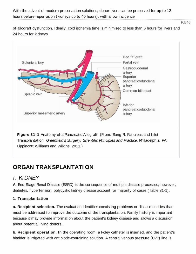
P.546
With the advent of modern preservation solutions, donor livers can be preserved for up to 12hours before reperfusion (kidneys up to 40 hours), with a low incidence
of allograft dysfunction. Ideally, cold ischemia time is minimized to less than 6 hours for livers and24 hours for kidneys.
Figure 31-1 Anatomy of a Pancreatic Allograft. (From: Sung R. Pancreas and IsletTransplantation. Greenfield's Surgery: Scientific Principles and Practice. Philadelphia, PA:Lippincott Williams and Wilkins, 2011.)
ORGAN TRANSPLANTATION
I. KIDNEYA. End-Stage Renal Disease (ESRD) is the consequence of multiple disease processes; however,diabetes, hypertension, polycystic kidney disease account for majority of cases (Table 31-1).
1. Transplantation
a. Recipient selection. The evaluation identifies coexisting problems or disease entities thatmust be addressed to improve the outcome of the transplantation. Family history is importantbecause it may provide information about the patient's kidney disease and allows a discussionabout potential living donors.
b. Recipient operation. In the operating room, a Foley catheter is inserted, and the patient'sbladder is irrigated with antibiotic-containing solution. A central venous pressure (CVP) line is

P.547
inserted, and a first-generation cephalosporin is administered. The transplant renal vein andartery typically are anastomosed to the external iliac vein and artery, respectively. A heparinbolus of 3,000 units may be administered before clamping the iliac vessels. Before reperfusion ofthe kidney, mannitol (25 g) and furosemide (100 mg) are administered intravenously, and thepatient's systolic blood pressure (BP) is maintained above 120 mm Hg, with a CVP of at least 10mm Hg to ensure optimal perfusion of the transplanted kidney. The ureter can be anastomosedto either the recipient bladder or the ipsilateral ureter, although the bladder is preferred.Establishing
an antireflux mechanism is essential for preventing posttransplantation reflux pyelonephritis. Thisis accomplished by performing an extravesical ureteroneocystostomy (Litch). A double-J ureteralstent is commonly used.
TABLE 31-1 Causes of Renal Failure RequiringTransplantation
Type Characteristics
Congenital Aplasia, obstructive uropathy
Hereditary Alport syndrome (hereditary nephritis), polycystic kidney disease,tuberous sclerosis
Neoplastic Renal cell carcinoma, Wilms tumor
Progressive Diabetic neuropathy, chronic pyelonephritis, Goodpasture syndrome(antiglomerular basement membrane disease), hypertension, chronicglomerulonephritis, lupus nephritis, nephrotic syndrome, obstructiveuropathy, scleroderma, amyloidosis
Traumatic Vascular occlusion, parenchymal destruction
c. Postoperative considerations
(1) Intravenous fluid replacement. In general, the patient should be kept euvolemic or mildlyhypervolemic in the early posttransplantation period with a goal of 130 mm Hg systolic BP to
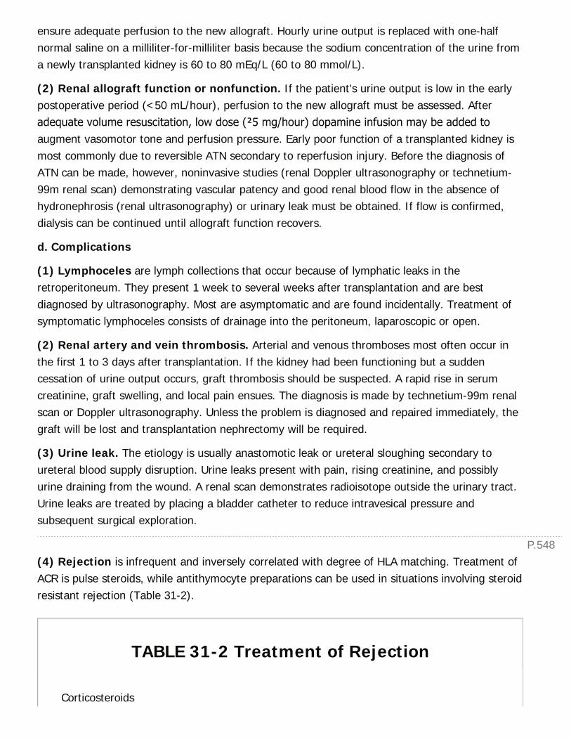
P.548
ensure adequate perfusion to the new allograft. Hourly urine output is replaced with one-halfnormal saline on a milliliter-for-milliliter basis because the sodium concentration of the urine froma newly transplanted kidney is 60 to 80 mEq/L (60 to 80 mmol/L).
(2) Renal allograft function or nonfunction. If the patient's urine output is low in the earlypostoperative period (<50 mL/hour), perfusion to the new allograft must be assessed. Afteradequate volume resuscitation, low dose (≤5 mg/hour) dopamine infusion may be added toaugment vasomotor tone and perfusion pressure. Early poor function of a transplanted kidney ismost commonly due to reversible ATN secondary to reperfusion injury. Before the diagnosis ofATN can be made, however, noninvasive studies (renal Doppler ultrasonography or technetium-99m renal scan) demonstrating vascular patency and good renal blood flow in the absence ofhydronephrosis (renal ultrasonography) or urinary leak must be obtained. If flow is confirmed,dialysis can be continued until allograft function recovers.
d. Complications
(1) Lymphoceles are lymph collections that occur because of lymphatic leaks in theretroperitoneum. They present 1 week to several weeks after transplantation and are bestdiagnosed by ultrasonography. Most are asymptomatic and are found incidentally. Treatment ofsymptomatic lymphoceles consists of drainage into the peritoneum, laparoscopic or open.
(2) Renal artery and vein thrombosis. Arterial and venous thromboses most often occur inthe first 1 to 3 days after transplantation. If the kidney had been functioning but a suddencessation of urine output occurs, graft thrombosis should be suspected. A rapid rise in serumcreatinine, graft swelling, and local pain ensues. The diagnosis is made by technetium-99m renalscan or Doppler ultrasonography. Unless the problem is diagnosed and repaired immediately, thegraft will be lost and transplantation nephrectomy will be required.
(3) Urine leak. The etiology is usually anastomotic leak or ureteral sloughing secondary toureteral blood supply disruption. Urine leaks present with pain, rising creatinine, and possiblyurine draining from the wound. A renal scan demonstrates radioisotope outside the urinary tract.Urine leaks are treated by placing a bladder catheter to reduce intravesical pressure andsubsequent surgical exploration.
(4) Rejection is infrequent and inversely correlated with degree of HLA matching. Treatment ofACR is pulse steroids, while antithymocyte preparations can be used in situations involving steroidresistant rejection (Table 31-2).
TABLE 31-2 Treatment of Rejection
Corticosteroids
Intravenous pulse, methylprednisolone
7 mg/kg QD for 3 days
Consider if rejection is early (<3 mo) or mild
Oral pulse, prednisone
3 mg/kg QD in 2-4 divided doses for 3-5 days
After pulse, restart steroids at previous dose
Use if patient is reliable and rejection is early or mild
Tacrolimus
Target 12-hr trough level 5-15 ng/mL
Antilymphocyte preparations
Thymoglobulin
2-3 mg/kg IV for 3-4 days
Myfortic
720 mg PO BID
Rapamycin
4 mg PO QD, target level 8-20 ng/mL
Plasmapheresis
P.549
Consider for antibody-mediated rejection
BID, twice a day; IV, intravenously; PO, orally; QD, daily.
TABLE 31-3 Long-term MaintenanceImmunosuppression for Renal Transplantation
Myfortic 720 mg PO BID
Reduce to 360 mg PO BID when used with tacrolimus and for WBC <5,000/mm3,diarrhea, first week posttransplant
Prednisone
1 mg/kg QD for days 1-3
20 mg QD for days 4-14
15 mg QD for week 3
10 mg QD for week 4
5 mg QD for week 5 and onward
Tacrolimus
Dose titrated to achieve trough level 5-7 ng/mL (FPIA)a
Levels >15 ng/mL are considered toxic
a aFPIA, fluorescence polarization immunoassay.
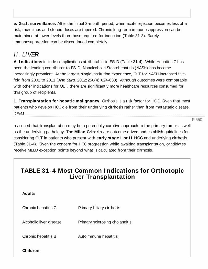
P.550
e. Graft surveillance. After the initial 3-month period, when acute rejection becomes less of arisk, tacrolimus and steroid doses are tapered. Chronic long-term immunosuppression can bemaintained at lower levels than those required for induction (Table 31-3). Rarelyimmunosuppression can be discontinued completely.
II. LIVERA. Indications include complications attributable to ESLD (Table 31-4). While Hepatitis C hasbeen the leading contributor to ESLD, Nonalcoholic Steatohepatitis (NASH) has becomeincreasingly prevalent. At the largest single institution experience, OLT for NASH increased five-fold from 2002 to 2011 (Ann Surg. 2012;256(4):624-633). Although outcomes were comparablewith other indications for OLT, there are significantly more healthcare resources consumed forthis group of recipients.
1. Transplantation for hepatic malignancy. Cirrhosis is a risk factor for HCC. Given that mostpatients who develop HCC die from their underlying cirrhosis rather than from metastatic disease,it was
reasoned that transplantation may be a potentially curative approach to the primary tumor as wellas the underlying pathology. The Milan Criteria are outcome driven and establish guidelines forconsidering OLT in patients who present with early stage I or II HCC and underlying cirrhosis(Table 31-4). Given the concern for HCC progression while awaiting transplantation, candidatesreceive MELD exception points beyond what is calculated from their cirrhosis.
TABLE 31-4 Most Common Indications for OrthotopicLiver Transplantation
Adults
Chronic hepatitis C Primary biliary cirrhosis
Alcoholic liver disease Primary sclerosing cholangitis
Chronic hepatitis B Autoimmune hepatitis
Children

P.551
Extrahepatic biliary atresia Primary hepatic tumors
α1-Antitrypsin deficiency Metabolic liver disease
Cystic fibrosis
B. Recipient Operation. OLT comprises three distinct sequential phases. The first phaseinvolves the dissection and removal of the recipient's diseased liver. The second phase, known asthe anhepatic phase, refers to the period starting with devascularization of the recipient's liverand ending with revascularization of the newly implanted liver. During the anhepatic phase,venovenous bypass (VVB) may be used (Fig. 31-2). VVB shunts blood from the portal vein andinfrahepatic inferior vena cava (IVC) to the axillary, subclavian, or jugular veins. Alternatively,many transplant surgeons will create a temporary portocaval shunt, which has the advantages ofVVB with much less risk and cost. Maintenance of venous return from the kidneys and lowerextremities results in a smoother hemodynamic course, allows time for a more deliberateapproach to hemostasis, reduces visceral edema and splanchnic venous pooling, and lowers theincidence of postoperative renal dysfunction. The liver allograft is implanted by anastomosing firstthe suprahepatic vena cava and then the infrahepatic IVC. The portal vein anastomosis isperformed, and blood flow to the liver is reestablished. Finally, the hepatic arterial anastomosis isperformed. If the recipient hepatic artery is not suitable for anastomosis, a donor iliac arterialgraft can be used as a conduit from the infra- or suprarenal aorta. The third phase includes biliaryreconstruction and abdominal closure. Biliary continuity is established via a duct-to-ductanastomosis or a choledochojejunostomy. A duct-to-duct anastomosis is preferable, but may notbe possible when there is a donor-recipient bile duct size discrepancy or a diseased recipient bileduct (e.g., with primary sclerosing cholangitis, biliary atresia, or secondary biliary cirrhosis).
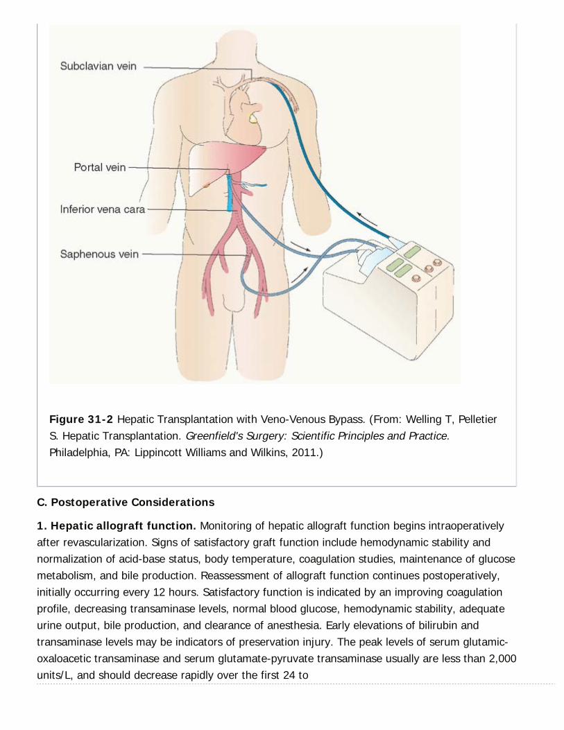
Figure 31-2 Hepatic Transplantation with Veno-Venous Bypass. (From: Welling T, PelletierS. Hepatic Transplantation. Greenfield's Surgery: Scientific Principles and Practice.Philadelphia, PA: Lippincott Williams and Wilkins, 2011.)
C. Postoperative Considerations
1. Hepatic allograft function. Monitoring of hepatic allograft function begins intraoperativelyafter revascularization. Signs of satisfactory graft function include hemodynamic stability andnormalization of acid-base status, body temperature, coagulation studies, maintenance of glucosemetabolism, and bile production. Reassessment of allograft function continues postoperatively,initially occurring every 12 hours. Satisfactory function is indicated by an improving coagulationprofile, decreasing transaminase levels, normal blood glucose, hemodynamic stability, adequateurine output, bile production, and clearance of anesthesia. Early elevations of bilirubin andtransaminase levels may be indicators of preservation injury. The peak levels of serum glutamic-oxaloacetic transaminase and serum glutamate-pyruvate transaminase usually are less than 2,000units/L, and should decrease rapidly over the first 24 to

P.55248 hours postoperatively. Persistent transaminitis should prompt a liver ultrasound to assessvessel patency and flow.
D. Complications
1. Primary nonfunction is characterized by hemodynamic instability, poor quantity and qualityof bile, renal dysfunction, failure to regain consciousness, increasing coagulopathy, persistenthypothermia, and lactic acidosis in the face of patent vascular anastomosis (as demonstrated byDoppler ultrasonography). The incidence is approximately 1% and without retransplantation,death ensues.
2. Hepatic artery thrombosis in the early posttransplantation period may lead to fever,hemodynamic instability, and rapid deterioration, with a marked elevation of the transaminases.An associated bile leak may be noted soon after liver transplantation due to the loss of the bileducts' main vascular supply. Acute thrombosis may be treated by attempted thrombectomy;however, this is usually unsuccessful and retransplantation is needed.
3. Portal vein stenosis or thrombosis is rare. When it occurs, the patient's condition maydeteriorate rapidly, with profound hepatic dysfunction, massive ascites, renal failure, andhemodynamic instability. Although surgical thrombectomy may be successful, urgentretransplantation is often necessary.
4. Biliary stricture is diagnosed by cholangiography. A single short bile duct stricture may betreated by either percutaneous or retrograde balloon dilation. A long stricture, ampullarydysfunction, or failed dilation necessitates revision of the biliary anastomosis.
III. PANCREASA. Indications. Most patients are type I diabetics with concomitant nephropathy who areevaluated for a pancreas transplant in conjunction with kidney transplantation. Ninety-fivepercent of all pancreas transplants are performed in conjunction with a kidney transplant. Wholeorgan pancreas transplantation represents the only therapeutic option for long-term insulinindependence.
B. Graft Selection
1. Simultaneous kidney-pancreas transplantation (SPK) may be considered in insulin-dependent diabetic patients who are dialysis dependent (or imminent) and have a creatinineclearance of less than 30 mL/minute. Since alloreactivity among donor organs is concordant, anadvantage of combined transplantation includes the ability to monitor pancreas rejection bymonitoring renal rejection.
2. Pancreas after kidney transplantation (PAK). Patients with living donors can be listedseparately for deceased pancreas transplantation; however, since the organs are immunologicallydistinct, pancreas allograft monitoring is more difficult and outcomes are worse.
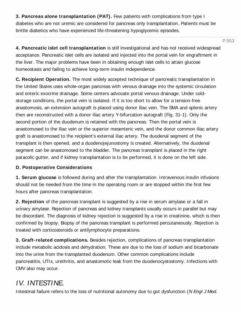
P.553
3. Pancreas alone transplantation (PAT). Few patients with complications from type Idiabetes who are not uremic are considered for pancreas only transplantation. Patients must bebrittle diabetics who have experienced life-threatening hypoglycemic episodes.
4. Pancreatic islet cell transplantation is still investigational and has not received widespreadacceptance. Pancreatic islet cells are isolated and injected into the portal vein for engraftment inthe liver. The major problems have been in obtaining enough islet cells to attain glucosehomeostasis and failing to achieve long-term insulin independence.
C. Recipient Operation. The most widely accepted technique of pancreatic transplantation inthe United States uses whole-organ pancreas with venous drainage into the systemic circulationand enteric exocrine drainage. Some centers advocate portal venous drainage. Under cold-storage conditions, the portal vein is isolated. If it is too short to allow for a tension-freeanastomosis, an extension autograft is placed using donor iliac vein. The SMA and splenic arterythen are reconstructed with a donor iliac artery Y-bifurcation autograft (Fig. 31-1). Only thesecond portion of the duodenum is retained with the pancreas. Then the portal vein isanastomosed to the iliac vein or the superior mesenteric vein, and the donor common iliac arterygraft is anastomosed to the recipient's external iliac artery. The duodenal segment of thetransplant is then opened, and a duodenojejunostomy is created. Alternatively, the duodenalsegment can be anastomosed to the bladder. The pancreas transplant is placed in the rightparacolic gutter, and if kidney transplantation is to be performed, it is done on the left side.
D. Postoperative Considerations
1. Serum glucose is followed during and after the transplantation. Intravenous insulin infusionsshould not be needed from the time in the operating room or are stopped within the first fewhours after pancreas transplantation.
2. Rejection of the pancreas transplant is suggested by a rise in serum amylase or a fall inurinary amylase. Rejection of pancreas and kidney transplants usually occurs in parallel but maybe discordant. The diagnosis of kidney rejection is suggested by a rise in creatinine, which is thenconfirmed by biopsy. Biopsy of the pancreas transplant is performed percutaneously. Rejection istreated with corticosteroids or antilymphocyte preparations.
3. Graft-related complications. Besides rejection, complications of pancreas transplantationinclude metabolic acidosis and dehydration. These are due to the loss of sodium and bicarbonateinto the urine from the transplanted duodenum. Other common complications includepancreatitis, UTIs, urethritis, and anastomotic leak from the duodenocystostomy. Infections withCMV also may occur.
IV. INTESTINE.Intestinal failure refers to the loss of nutritional autonomy due to gut dysfunction (N Engl J Med.

P.554
2009;361:10). Both anatomic and functional disease states can result in intestinal failure;however, loss of intestinal length resulting in malabsorption is the leading cause.
A. Total parenteral nutrition (TPN) is the mainstay for intestinal replacement, although long-term therapy can be associated with significant morbidity and mortality. While the 1-year survivalrate of patients on TPN is
approximately 90%, the 3- and 5-year rates drop to 70% and 63%, respectively (N Engl J Med.2009;361:10).
1. Complications of long-term TPN
a. Cholestatic liver disease develops in approximately one-half of adults and children whoremain on continuous therapy. Recent lipid-sparing PN strategies have shown promise inimproving their hepatic, biochemical profile associated with cholestasis (J Ped Surg. 2013;48:228-232). If left untreated, PN-associated liver disease can result in liver failure.
b. Line infections are costly complications that are the result of chronic indwelling catheters.This can lead to multiple hospital readmissions for IV antibiotics and significant morbidity if sepsisensues.
c. Central vein stenosis/thrombosis can occur and result in the loss of vascular access for PNadministration.
B. Intestinal Transplantation. The Centers for Medicare and Medicaid Services (CMS)recognize intestinal transplantation as the standard of care for patients who have failed TPN.Approved indications for intestinal transplant include the life-threatening complications associatedwith TPN therapy (Table 31-5). Timing of referral to an intestinal center can be difficult and isaided by a patient-centered, clinical algorithm (Nat Rev Gastroenterol Hepatol. doi:10.1038/nrgastro.2014.216; Fig. 31-3)
1. Types of grafts
a. Isolated intestinal allograft. Transplantation of the jejunoileum
b. Composite liver and intestinal graft. In cases of severe hepatic dysfunction, compositegrafts are necessary. Inclusion of the pancreas and duodenum facilitates en bloc procurementand engraftment (N Engl J Med. 2009;361:10).
TABLE 31-5 Failure of Parenteral Nutrition, as Definedby the Centers for Medicare and Medicaid Servicesa
Impending or overt liver failure due to TPN-induced liver injury
P.555
Thrombosis of two or more central veins
Two or more episodes per year of catheter-related systemic sepsis that requireshospitalization
A single episode of line-related fungemia, septic shock, or acute respiratory distresssyndrome
Frequent episodes of severe dehydration despite intravenous fluid supplementation inaddition to TPN
aaTPN denotes total parenteral nutrition.From: Fishbein TM. Intestinal transplantation. N Engl J Med. 2009;361(10):998-1008,with permission.

Figure 31-3 Decision making algorithm in the management of intestinal failure. (Abu-Elmagd K. The concept of gut rehabilitation and the future of visceral transplantation. NatRev Gastroenterol Hepatol 2015;12:108(2)-120, with permission.)
c. Multivisceral graft. Exenteration of the native foregut allows en bloc engraftment of donorstomach, duodenum, pancreas, small intestine and liver. Inclusion of the colon is sometimesperformed and is currently under investigation.
(1) Recipient operation. Patients who receive isolated intestinal allografts have vascularanastomoses created between the donor superior mesenteric vein and the recipient portal vein,and between the donor SMA and the recipient aorta. Vascular reconstruction for patients whoreceive combined liver-intestinal grafts parallels that for patients undergoing a standard OLT.Supra- and infrahepatic vena caval anastomoses are completed, and arterial inflow is

P.556
P.557
accomplished after the portal vein anastomosis by using a patch of aorta that contains the SMAand celiac.
(2) Complications
d. Graft rejection. The introduction of antithymocyte antibodies into induction protocols hascorrelated with reduction in early acute cellular rejection (ACR). Previously reported in 70% to90% of intestinal transplant recipients, now only one-third to one-half of patients experience ACR(N Engl J Med. 2009;361:10). Given the lack of a reliable noninvasive marker of rejection,intestinal stomas created during transplantation give an access to serial endoscopic biopsies forgraft surveillance.
e. Infection. The foremost clinical conundrum that faces intestinal transplant surgeons isdistinguishing infectious enteritis from allograft rejection. Both present similarly and havemasquerading features on biopsy specimens. Adenovirus, calicivirus, Clostridium difficile, and CMVmust all be distinguished from rejection.
f. Renal dysfunction. Intestinal transplant recipient are at a higher risk of nephrotoxicity fromcalcineurin inhibitors because of the higher dose used for immunosuppression. Ojo et al. reported21.3% of intestinal transplant recipients at 5 years experienced renal dysfunction (N Engl J Med.2003;349:931-940). Recent use of antithymocyte antibody induction therapy associated withdecreased target levels of tacrolimus is anticipated to preserve renal function of future recipients(AJT. 2014;14:1976-1984).
2. Outcomes
a. Survival. Experienced centers report 1-year patient survival between 86% to 93%, which ison par with other solid organs (AJT. 2014;14:1976-1984). Unfortunately, these results do notpersist as 3- and 5-year survival rates are a modest 61% and 47%, respectively (N Engl J Med.2009;361:10).
b. Nutritional autonomy. Most recipients wean from TPN and achieve nutritional autonomyafter transplantation (OPTN/SRTR 2011 Annual Data Report). Oral aversion can be an issue inchildren who have never developed feeding skills.
c. Cost. Similar to renal transplantation, intestinal transplantation becomes cost effective overcontinuous PN after 2 years (Gastroenterology. 2006;130:s158-s162).
d. Quality of Life (QOL). Patients who receive an intestinal transplant seem to enjoy a betterQOL than those who remain on continuous PN (AJT. 2014;14:1976-1984). Factors that diminishQOL after transplantation include persistent g-tube or ostomies and repeat hospitalizations.
CHAPTER 31: TRANSPLANTATION
Multiple Choice Questions1. During preoperative evaluation of a 33-year-old male with ESRD frompolycystic kidney disease, his mother comes forward and wishes todonate one of her kidneys to her son. What factors are taken intoconsideration when evaluating potential living donors?
a. Blood type
b. HLA type
c. Co-morbid conditions
d. Psychosocial influences
e. All of the above
View Answer
2. A 65-year-old male is 3 days status postorthotopic livertransplantation. On rounds you note new bilious output from hissurgical drain. This finding along with a persistent transaminitis shouldprompt an evaluation for:
a. Portal vein thrombosis
b. Enterocutaneous fistula
c. Hepatic arterial thrombosis
d. Rejection
e. All of the above
View Answer
3. A 55-year-old male with cirrhosis from HCV visits your office todiscuss his recent surveillance imaging. “They told me I have a fewspots on my liver.” What findings would preclude this patient frombeing considered for transplantation for HCC?
a. Single 4.5 cm lesion
b. 3 lesions: 2.5 cm, 2 cm, 3.5 cm
c. 2 lesions: 2.5 cm, 2 cm
d. Single 3 cm lesion
e. All of the above are contraindications for transplantation
View Answer
4. A 76-year-old female s/p left-sided brachiobasilic AVF placementover 1 year ago presents to the emergency department complaining ofleft upper arm swelling and pain. On examination, she has good distal
P.558
perfusion, but you are not able to palpate a thrill, only a dopplerablesignal. What is the next appropriate step in management of venoushypertension with outflow stenosis?
a. Percutaneous fistulogram with therapeutic dilation of stenotic outflowsegment
b. Open thromboembolectomy
c. Anticoagulation
d. Arm elevation and physical therapy
e. Watchful waiting
View Answer
5. A 45-year-old female with type I diabetes complicated bynephropathy and ESRD is referred to your office for kidney transplantevaluation. She asks you whether she would benefit from a combinedkidney-pancreas transplant. Which of the following statementsregarding whole-organ pancreas transplantation are true?
a. It is usually performed alone without concomitant kidneytransplantation
b. It is the only therapeutic modality that can achieve long term insulinindependence
c. Compared to islet cell transplantation, there is a greater chance ofrequiring insulin after whole-organ pancreas transplantation
d. Sequela of diabetes, such as neuropathy and retinopathy areunaffected after pancreas transplantation
e. All of the above
View Answer
6. A 55-year-old male is 2 days status postliver transplantation. Despitenormalization of his coagulation profile and bilirubin level, histransaminases are persistently elevated. Which noninvasive test wouldbe most appropriate in the workup of this patient?
a. MRCP
b. CT angiography
c. KUB
d. Liver duplex ultrasound
e. No further testing is indicated at this time

P.559
View Answer
7. Three days following deceased-donor renal transplantation, yourpatient begins to complain of swelling and tenderness over her graft.Despite having adequate urine output over the last 12 hours, you noticea moderate rise in her serum creatinine. Which study would be mosthelpful in making the diagnosis?
a. Ultrasound of the graft
b. Nuclear medicine scan
c. Retrograde urethrogram
d. CT scan
e. MRI
View Answer
8. A 45-year-old female with intestinal failure secondary to short gutsyndrome is hospitalized with her third serious line infection in the last12 months. Which of the following are indications for intestinaltransplantation?
a. Frequent episodes of dehydration despite IV fluid supplementation withTPN
b. Two or more serious line-related infection per year, 1 line-relatedfungal infection or episode of shock due to line sepsis
c. Loss of two or more central venous access sites
d. Peripheral nutrition associated liver disease (PNALD)
e. All of the above
View Answer
9. A 55-year-old female with ESRD secondary to Alport syndrome isundergoing evaluation for kidney transplantation. Which of thesefactors should not raise her PRA?
a. Previous pregnancy
b. History of blood transfusion
c. Previous transplant
d. Prior blood donation
e. All of the above will raise one's PRA
View Answer

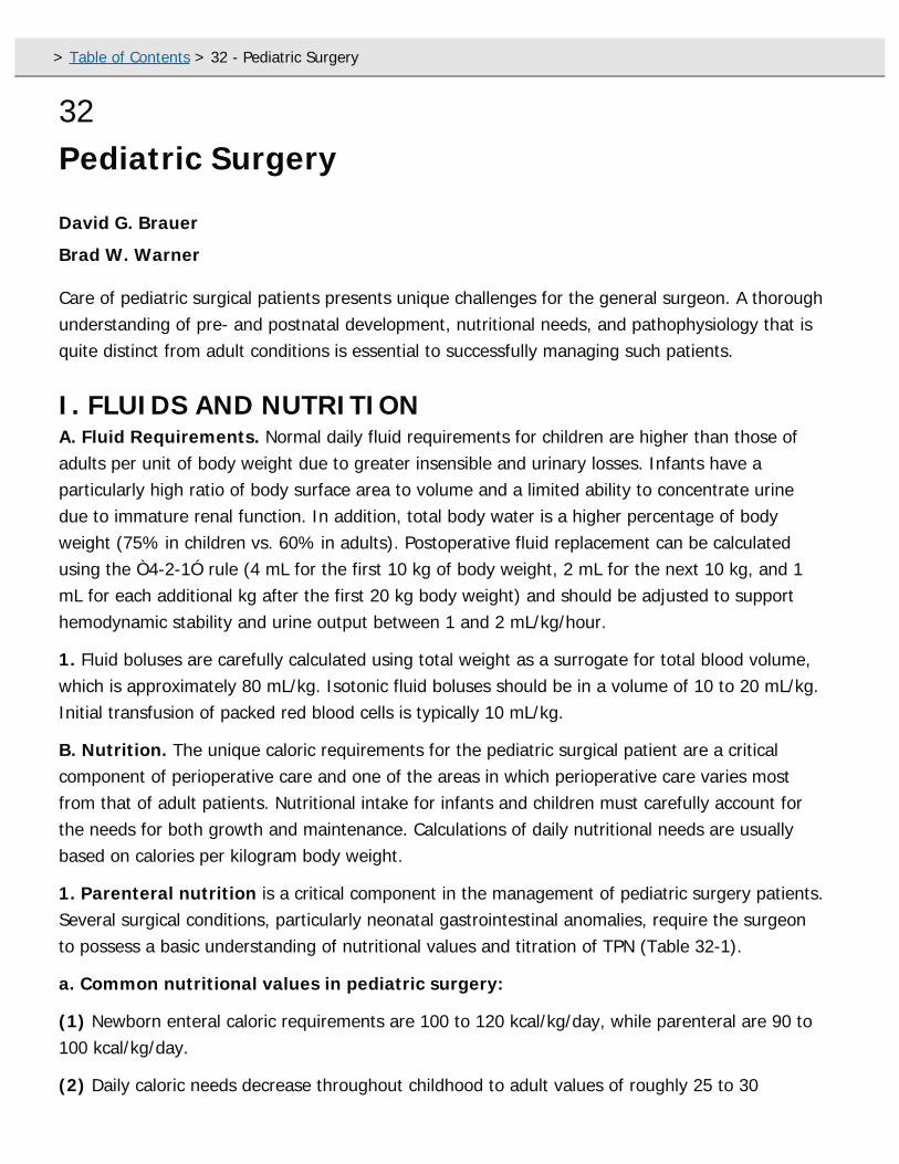
32Pediatric Surgery
David G. Brauer
Brad W. Warner
Care of pediatric surgical patients presents unique challenges for the general surgeon. A thoroughunderstanding of pre- and postnatal development, nutritional needs, and pathophysiology that isquite distinct from adult conditions is essential to successfully managing such patients.
I. FLUIDS AND NUTRITIONA. Fluid Requirements. Normal daily fluid requirements for children are higher than those ofadults per unit of body weight due to greater insensible and urinary losses. Infants have aparticularly high ratio of body surface area to volume and a limited ability to concentrate urinedue to immature renal function. In addition, total body water is a higher percentage of bodyweight (75% in children vs. 60% in adults). Postoperative fluid replacement can be calculatedusing the Ò4-2-1Ó rule (4 mL for the first 10 kg of body weight, 2 mL for the next 10 kg, and 1mL for each additional kg after the first 20 kg body weight) and should be adjusted to supporthemodynamic stability and urine output between 1 and 2 mL/kg/hour.
1. Fluid boluses are carefully calculated using total weight as a surrogate for total blood volume,which is approximately 80 mL/kg. Isotonic fluid boluses should be in a volume of 10 to 20 mL/kg.Initial transfusion of packed red blood cells is typically 10 mL/kg.
B. Nutrition. The unique caloric requirements for the pediatric surgical patient are a criticalcomponent of perioperative care and one of the areas in which perioperative care varies mostfrom that of adult patients. Nutritional intake for infants and children must carefully account forthe needs for both growth and maintenance. Calculations of daily nutritional needs are usuallybased on calories per kilogram body weight.
1. Parenteral nutrition is a critical component in the management of pediatric surgery patients.Several surgical conditions, particularly neonatal gastrointestinal anomalies, require the surgeonto possess a basic understanding of nutritional values and titration of TPN (Table 32-1).
a. Common nutritional values in pediatric surgery:
(1) Newborn enteral caloric requirements are 100 to 120 kcal/kg/day, while parenteral are 90 to100 kcal/kg/day.
(2) Daily caloric needs decrease throughout childhood to adult values of roughly 25 to 30
> Table of Contents > 32 - Pediatric Surgery
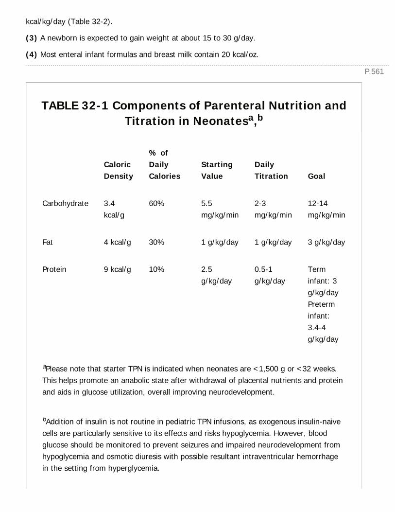
P.561
kcal/kg/day (Table 32-2).
(3) A newborn is expected to gain weight at about 15 to 30 g/day.
(4) Most enteral infant formulas and breast milk contain 20 kcal/oz.
TABLE 32-1 Components of Parenteral Nutrition andTitration in Neonatesa,b
CaloricDensity
% ofDailyCalories
StartingValue
DailyTitration Goal
Carbohydrate 3.4kcal/g
60% 5.5mg/kg/min
2-3mg/kg/min
12-14mg/kg/min
Fat 4 kcal/g 30% 1 g/kg/day 1 g/kg/day 3 g/kg/day
Protein 9 kcal/g 10% 2.5g/kg/day
0.5-1g/kg/day
Terminfant: 3g/kg/dayPreterminfant:3.4-4g/kg/day
aPlease note that starter TPN is indicated when neonates are <1,500 g or <32 weeks.This helps promote an anabolic state after withdrawal of placental nutrients and proteinand aids in glucose utilization, overall improving neurodevelopment.
bAddition of insulin is not routine in pediatric TPN infusions, as exogenous insulin-naivecells are particularly sensitive to its effects and risks hypoglycemia. However, bloodglucose should be monitored to prevent seizures and impaired neurodevelopment fromhypoglycemia and osmotic diuresis with possible resultant intraventricular hemorrhagein the setting from hyperglycemia.
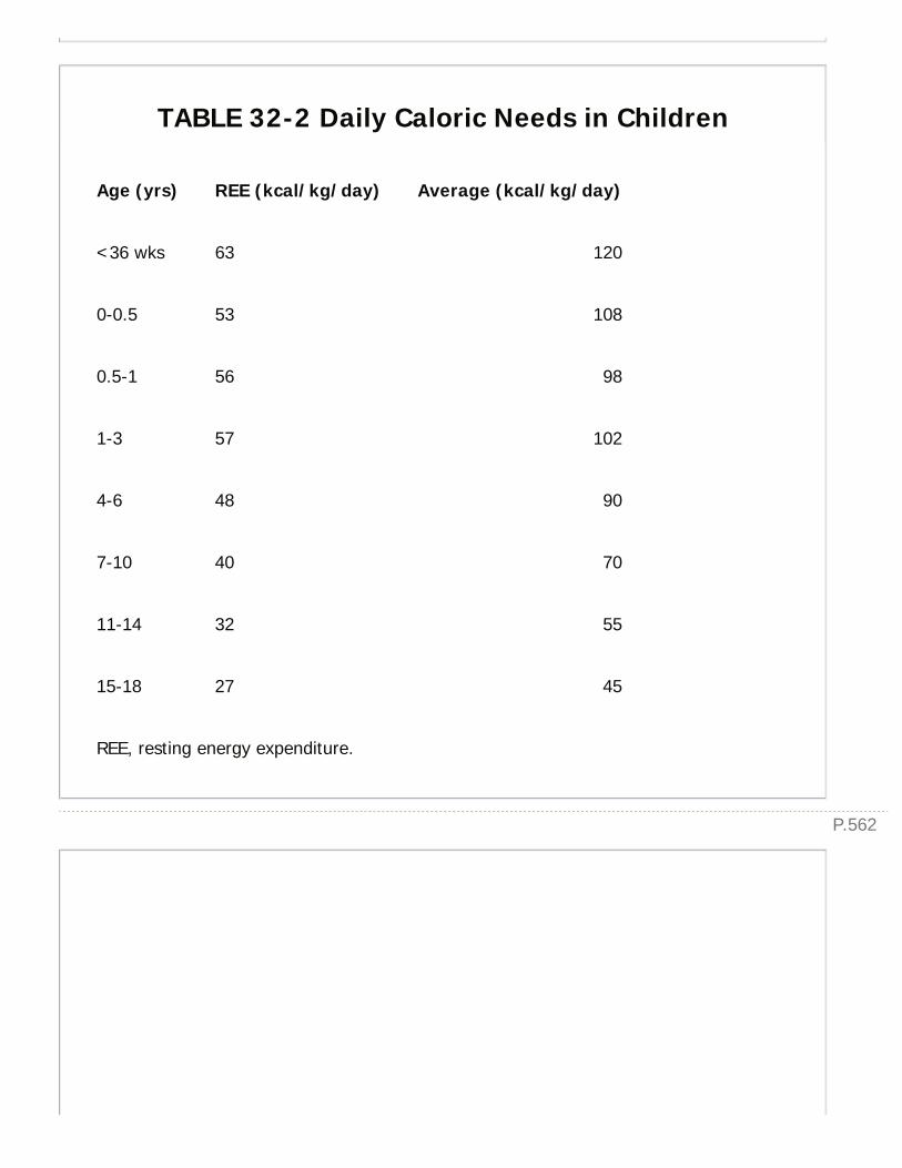
P.562
TABLE 32-2 Daily Caloric Needs in Children
Age (yrs) REE (kcal/kg/day) Average (kcal/kg/day)
<36 wks 63 120
0-0.5 53 108
0.5-1 56 98
1-3 57 102
4-6 48 90
7-10 40 70
11-14 32 55
15-18 27 45
REE, resting energy expenditure.
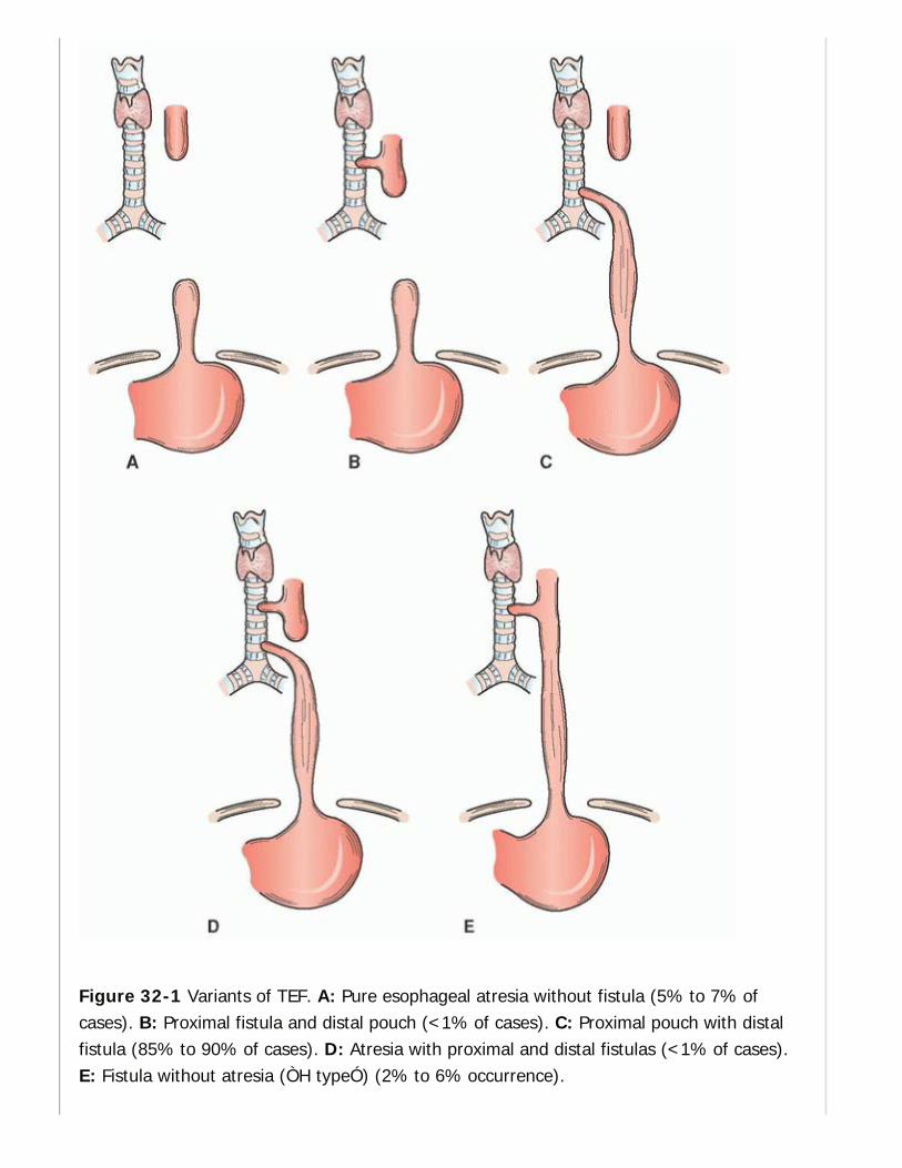
Figure 32-1 Variants of TEF. A: Pure esophageal atresia without fistula (5% to 7% ofcases). B: Proximal fistula and distal pouch (<1% of cases). C: Proximal pouch with distalfistula (85% to 90% of cases). D: Atresia with proximal and distal fistulas (<1% of cases).E: Fistula without atresia (ÒH typeÓ) (2% to 6% occurrence).
P.564
P.563
II. NEONATAL SURGICAL CONDITIONSA. Tracheoesophageal Malformations. Tracheoesophageal malformations describe aspectrum of anomalies including esophageal atresia (EA) alone or as a component of atracheoesophageal fistula (TEF) (Fig. 32-1). The incidence of these disorders is 3 in 10,000 births,with a slight male predominance.
1. Diagnosis
a. Clinical examination. Suspicion should be raised for EA/TEF in the neonatal patient withdifficulty clearing secretions, regurgitation of saliva, or respiratory difficulty during feeds. Theclassic clinical finding in EA/TEF is inability to pass an oro- or nasogastric tube.
b. Imaging. Visualization of a coiled orogastric tube in the upper chest on plain radiographimplies EA (Fig. 32-2), and associated presence
of gas in the GI tract confirms a distal communication between the respiratory and GI systems(TEF). Imaging should also be reviewed with concern for aspiration of feeds and secretions.These imaging modalities are often sufficient. Contrast studies are rarely necessary to visualizethe level of EA and/or TEF, and are associated with risk for aspiration.

Figure 32-2 Radiographic findings of esophageal atresia. Note the orogastric tube ending inupper chest and the absence of bowel gas, suggesting pure esophageal atresia without anycommunication between the airway and gastrointestinal tract.
P.565
2. Workup. Up to two-thirds of EA/TEF patients have associated anomalies. Physical examinationand imaging should be used to search for VACTERL anomalies (Vertebral, Anorectal, Cardiac,Tracheal, Esophageal, Renal, and Limb).
3. Management
a. Preoperative management should include decompression of the proximal esophageal pouchalong with elevation of the head of the bed to 30 degrees to reduce the risk of aspiration fromreflux of gastric contents through the TEF. Ventilation may prove challenging in the presence of aTEF, as positive-pressure can be directed through the TEF into the GI system, resulting inincreased abdominal distention and pressure. Passage of an endotracheal tube beyond the fistula,right mainstem intubation, or high frequency oscillatory ventilation may be necessary toappropriately ventilate the patient.
b. Operative approach is typically determined by the side of the aortic arch. A TEF with anormal left-sided aortic arch is approached through a right fourth intercostal space, retropleuralthoracotomy. The fistula is ligated, and the proximal esophageal stump is mobilized in an attemptto create a tension-free primary anastomosis. Pure EA without TEF is typically associated with along-gap, and a gastrostomy is the usual first procedure. Later, long-gap EA can be reconstructedwith a variety of techniques including lengthening myotomy, colonic interposition, gastric pull-upor delayed repair after a period of growth or traction (the Foker process). An isolated TEF withoutEA is often more proximal and can be approached via a cervical incision.
B. Congenital Diaphragmatic Hernia (CDH). CDH has an incidence of 1 in every 2,000 to5,000 births and an equal male to female distribution. CDH develops as a result of incompletediaphragm development at 8 weeks of gestation. Abdominal contents herniate into the chest,resulting in lung compression and subsequent hypoplasia that is worse on the affected side.Moreover, the pulmonary vasculature develops increased tone in the muscular arterioles, whichpredisposes to pulmonary hypertension and vasospasm. The majority (80% to 90%) of herniasare posterolateral (Bochdalek), with the remainder presenting anteriorly (Morgagni).Approximately 90% of hernias occur on the left.
1. Diagnosis of CDH is frequently made during routine prenatal screening ultrasounds. Prenataldiagnosis should set in motion a team of critical care neonatologists and surgeons available forrespiratory management at the time of birth. For patients not diagnosed prenatally, significantrespiratory distress including tachypnea, cyanosis, and retractions should
raise immediate concern. Asymmetric chest wall diameter and a scaphoid abdomen as a result ofabdominal contents herniating into the chest are additional clinical clues. A second set of patientswill present after an initial ÒhoneymoonÓ phase of several hours that can occur before signs ofpulmonary hypertension and hypoxemia present. Finally, less than 20% of cases present after thefirst 24 hours of life with respiratory distress, pneumonia, and feeding intolerance or intestinal
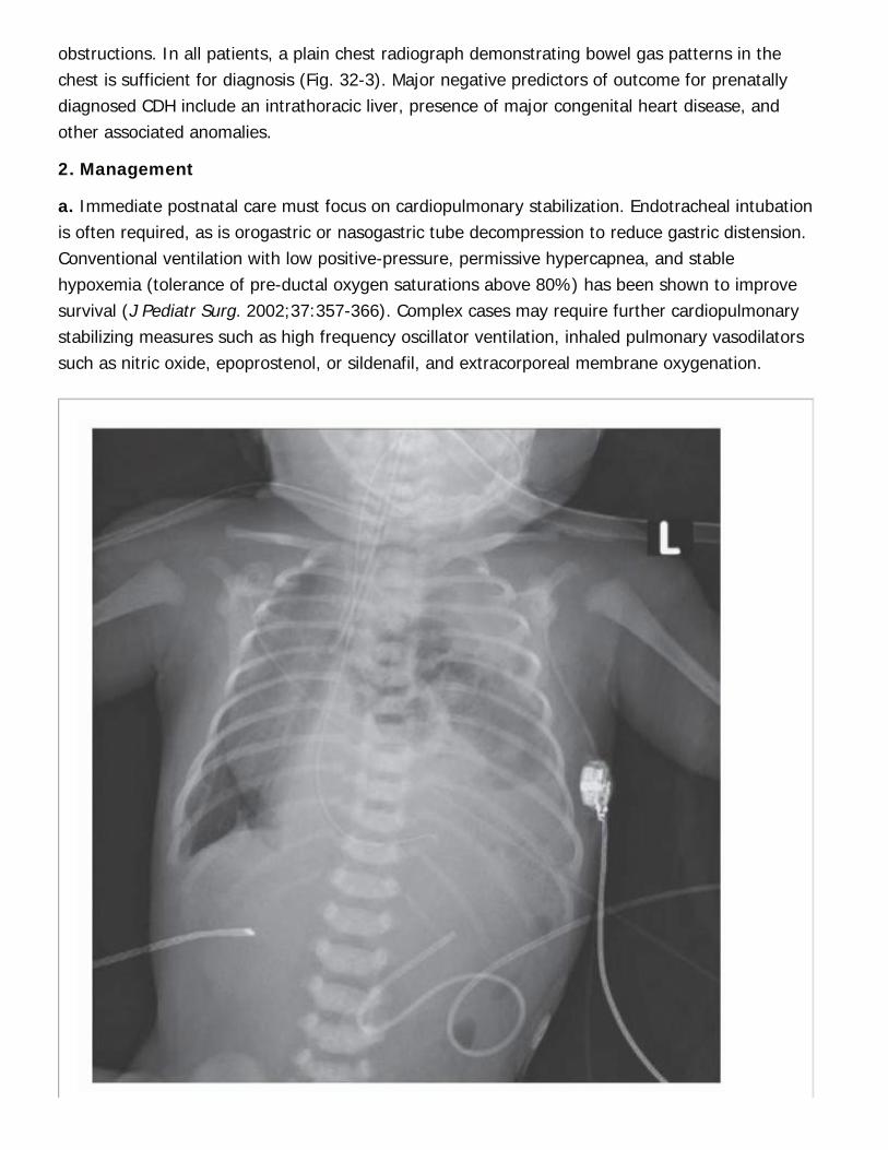
obstructions. In all patients, a plain chest radiograph demonstrating bowel gas patterns in thechest is sufficient for diagnosis (Fig. 32-3). Major negative predictors of outcome for prenatallydiagnosed CDH include an intrathoracic liver, presence of major congenital heart disease, andother associated anomalies.
2. Management
a. Immediate postnatal care must focus on cardiopulmonary stabilization. Endotracheal intubationis often required, as is orogastric or nasogastric tube decompression to reduce gastric distension.Conventional ventilation with low positive-pressure, permissive hypercapnea, and stablehypoxemia (tolerance of pre-ductal oxygen saturations above 80%) has been shown to improvesurvival (J Pediatr Surg. 2002;37:357-366). Complex cases may require further cardiopulmonarystabilizing measures such as high frequency oscillator ventilation, inhaled pulmonary vasodilatorssuch as nitric oxide, epoprostenol, or sildenafil, and extracorporeal membrane oxygenation.
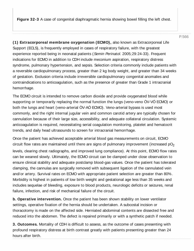
P.566
Figure 32-3 A case of congenital diaphragmatic hernia showing bowel filling the left chest.
(1) Extracorporeal membrane oxygenation (ECMO), also known as Extracorporeal LifeSupport (ECLS), is frequently employed in cases of respiratory failure, with the greatestexperience reported being in neonatal patients (Semin Perinatol. 2005;29:24-33). Frequentindications for ECMO in addition to CDH include meconium aspiration, respiratory distresssyndrome, pulmonary hypertension, and sepsis. Selection criteria commonly include patients witha reversible cardiopulmonary process, greater than 2 kg body weight, and greater than 34 weeksof gestation. Exclusion criteria include irreversible cardiopulmonary congenital anomalies andcontraindications to anticoagulation, such as the presence of greater than Grade 1 intracranialhemorrhage.
The ECMO circuit is intended to remove carbon dioxide and provide oxygenated blood whilesupporting or temporarily replacing the normal function the lungs (veno-veno ÒV-VÓ ECMO) orboth the lungs and heart (veno-arterial ÒV-AÓ ECMO). Veno-arterial bypass is used mostcommonly, and the right internal jugular vein and common carotid artery are typically chosen forcannulation because of their large size, accessibility, and adequate collateral circulation. Systemicanticoagulation is required, necessitating serial coagulation monitoring, platelet and hematocrittrends, and daily head ultrasounds to screen for intracranial hemorrhage.
Once the patient has achieved acceptable arterial blood gas measurements on circuit, ECMOcircuit flow rates are maintained until there are signs of pulmonary improvement (increased pO2
levels, clearing chest radiographs, and improved lung compliance). At this point, ECMO flow ratescan be weaned slowly. Ultimately, the ECMO circuit can be clamped under close observation toensure clinical stability and adequate postclamp blood-gas values. Once the patient has toleratedclamping, the cannulas are surgically removed with subsequent ligation of the cannulated veinand/or artery. Survival rates on ECMO with appropriate patient selection are greater than 80%.Morbidity is highest in patients of low birth weight and gestational age less than 35 weeks andincludes sequelae of bleeding, exposure to blood products, neurologic deficits or seizures, renalfailure, infection, and risk of mechanical failure of the circuit.
b. Operative intervention. Once the patient has been shown stability on lower ventilatorsettings, operative fixation of the hernia should be undertaken. A subcostal incision orthoracotomy is made on the affected side. Herniated abdominal contents are dissected free andreduced into the abdomen. The defect is repaired primarily or with a synthetic patch if needed.
3. Outcomes. Mortality of CDH is difficult to assess, as the outcome of cases presenting withprofound respiratory distress at birth contrast greatly with patients presenting greater than 24hours after birth.

P.567Overall survival rates greater than 70% are frequently reported (J Pediatr Surg. 2004;39:657-660). Significant respiratory disease at birth can result in neurologic deficits, includingdevelopmental delay and seizures. In addition, gastroesophageal reflux is common. Patients mustbe followed to observe for respiratory symptoms, management of chronic lung disease,surveillance for hernia recurrence, and the possibility of reoperation if a patch was used in theinitial operation.
C. Abdominal Wall Defects. During normal development of the human embryo, the midgutherniates outward through the umbilical ring and continues to grow. By the 11th week ofgestation, the midgut returns back into the abdominal cavity and undergoes counterclockwiserotation and fixation, along with closure of the umbilical ring. Omphalocele is the failure of theabdominal contents to reduce back into the abdomen, resulting in a large hernia covered by aperitoneal sac. In contrast, gastroschisis is believed to be the result of an isolated intrauterinevascular insult resulting in an abdominal wall defect to the right of the umbilical cord. The bowelherniates through the defect but is not covered by a sac. Gastroschisis defects tend to be smallerthan those of an omphalocele. Although there is a small incidence of associated anomalies withgastroschisis (10% rate of associated intestinal atresias), omphalocele is more typically associatedwith congenital anomalies, with 50% of cases having an associated genetic and/or cardiacanomaly that often has a major impact on the prognosis of the infant.
1. Diagnosis is often made at the time of prenatal ultrasound after 13 weeks of gestation orsimply by clinical examination at the time of birth.
2. Management. Naso- or orogastric tube decompression and broad-spectrum prophylacticantibiotics should be initiated. Heat and fluid losses from exposed viscera should be correctedwith intravenous fluids and warming while covering the exposed organs and lower body in a clearplastic bag. Covering the bowel with gauze and pouring warm saline should be avoided as thegauze prevents visualization of the bowel prior to surgery, and the warm saline ultimately dropsto room temperature, thereby cooling the infant. In cases of gastroschisis, the bowel may beplaced within a silastic silo at the bedside, with gradual reduction into the abdomen whilecarefully monitoring for abdominal compartment syndrome (J Pediatr Surg. 2009;44(11):2126-2129). In cases of omphalocele, the sac serves as sufficient coverage until operative closure isundertaken.
a. Operative repair. Once the herniated abdominal contents can be reduced into the abdomen,the abdominal wall defect may be closed primarily. In omphalocele, the sac should be excisedwith care to dissect out and ligate the umbilical vessels. Repair of gastroschisis should involvecareful examination of the length of the bowel for atresias, which may be managed primarily orwith diversion and subsequent repair. In the case of large abdominal wall defects that cannot beclosed primarily, the skin can be closed over the bowel or, if this is not possible, the exposedbowel can be allowed to granulate. Either management method requires operative repair of thefascial defect at a later date.

P.569
P.568D. Necrotizing Enterocolitis (NEC). NEC is the most common neonatal gastrointestinalemergency. It is characterized by an acute inflammatory disease of the intestine associated withulceration and necrosis of the gastrointestinal tract most frequently affecting the small bowel. Thepathogenesis is believed to be multifactorial involving prematurity, an immature gut barrierdefense, bacteria, enteral feeding, and hypoxia/ischemia or low-flow states. The incidence of NECis 1 to 3 per 1,000 live births, with prematurity being the single most prominent risk factor.
1. Diagnosis requires a high clinical suspicion. NEC is unusual within the first few days of life butapproximately 80% of cases occur within the first month of life. Clinical examination maydemonstrate a lethargic or irritable patient with abdominal distention, feeding intolerance, orpassage of bloody stools. Advanced cases may show signs of peritonitis including erythema of theabdominal wall. Temperature instability is common, as well as increasing incidence of apneic orbradycardic episodes. Laboratory studies may reveal increasing leukocytosis or leukopenia as wellas metabolic acidosis and thrombocytopenia. Plain radiographs often reveal dilated loops of bowelwith evidence of ischemia including pneumatosis intestinalis (Fig. 32-4), portal venous gas, orpneumoperitoneum. Based on clinical examination and laboratory and imaging data, patients andtheir expectant management can be guided by Bell staging (Ann Surg. 1978;187(1):1-7).
2. Initial management of the hemodynamically stable patient should focus on bowel rest,nasogastric decompression, parenteral nutrition, and broad-spectrum antibiotics. Antibioticregimen historically was a
combination of ampicillin, gentamicin, and clindamycin (J Pediatr Surg. 1980;15(4):569-573) butstudies have failed to show any single superior regimen (Cochrane Database Syst Rev.2012;8:CD007448). Serial abdominal examinations, laboratory studies including CBC and bloodgas, and plain radiographs are valuable studies to determine the success of nonoperativemanagement (Fig. 32-5). Fifty percent of patients will experience improvement and resolution ofsigns of NEC with nonoperative management.
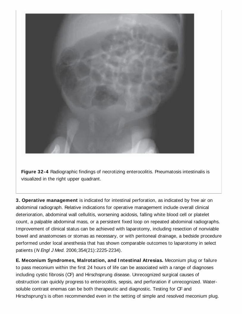
Figure 32-4 Radiographic findings of necrotizing enterocolitis. Pneumatosis intestinalis isvisualized in the right upper quadrant.
3. Operative management is indicated for intestinal perforation, as indicated by free air onabdominal radiograph. Relative indications for operative management include overall clinicaldeterioration, abdominal wall cellulitis, worsening acidosis, falling white blood cell or plateletcount, a palpable abdominal mass, or a persistent fixed loop on repeated abdominal radiographs.Improvement of clinical status can be achieved with laparotomy, including resection of nonviablebowel and anastomoses or stomas as necessary, or with peritoneal drainage, a bedside procedureperformed under local anesthesia that has shown comparable outcomes to laparotomy in selectpatients (N Engl J Med. 2006;354(21):2225-2234).
E. Meconium Syndromes, Malrotation, and Intestinal Atresias. Meconium plug or failureto pass meconium within the first 24 hours of life can be associated with a range of diagnosesincluding cystic fibrosis (CF) and Hirschsprung disease. Unrecognized surgical causes ofobstruction can quickly progress to enterocolitis, sepsis, and perforation if unrecognized. Water-soluble contrast enemas can be both therapeutic and diagnostic. Testing for CF andHirschsprung's is often recommended even in the setting of simple and resolved meconium plug.
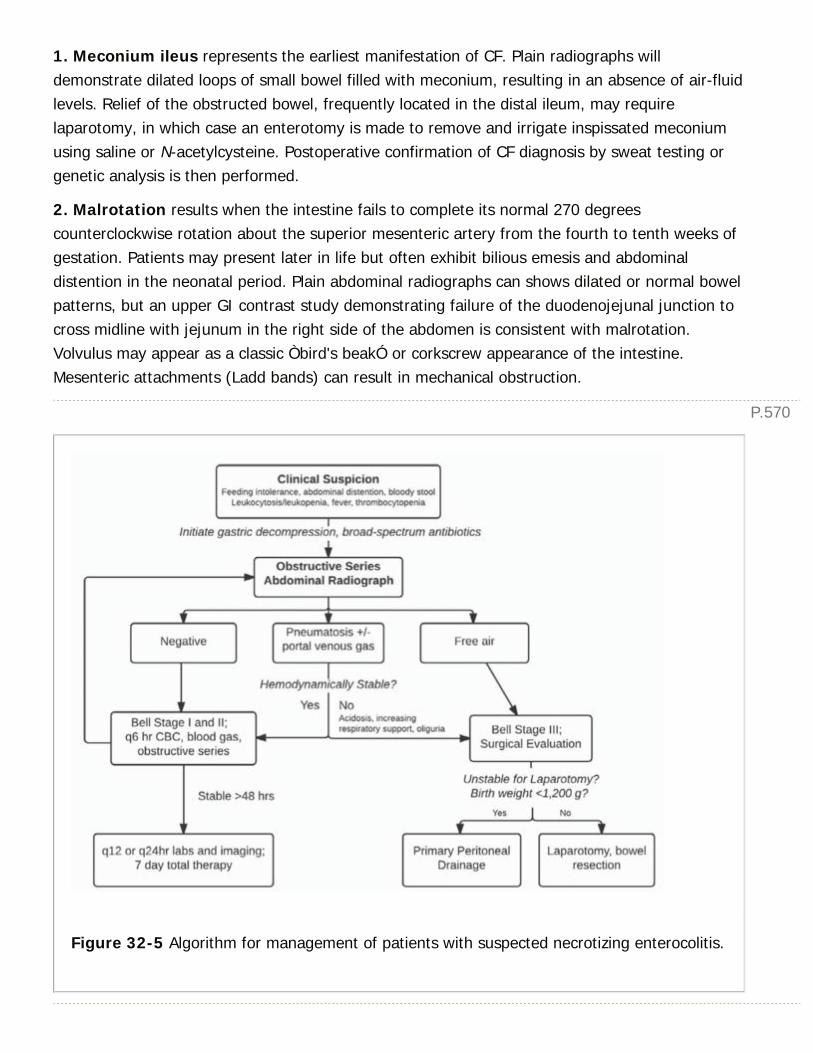
P.570
1. Meconium ileus represents the earliest manifestation of CF. Plain radiographs willdemonstrate dilated loops of small bowel filled with meconium, resulting in an absence of air-fluidlevels. Relief of the obstructed bowel, frequently located in the distal ileum, may requirelaparotomy, in which case an enterotomy is made to remove and irrigate inspissated meconiumusing saline or N-acetylcysteine. Postoperative confirmation of CF diagnosis by sweat testing orgenetic analysis is then performed.
2. Malrotation results when the intestine fails to complete its normal 270 degreescounterclockwise rotation about the superior mesenteric artery from the fourth to tenth weeks ofgestation. Patients may present later in life but often exhibit bilious emesis and abdominaldistention in the neonatal period. Plain abdominal radiographs can shows dilated or normal bowelpatterns, but an upper GI contrast study demonstrating failure of the duodenojejunal junction tocross midline with jejunum in the right side of the abdomen is consistent with malrotation.Volvulus may appear as a classic Òbird's beakÓ or corkscrew appearance of the intestine.Mesenteric attachments (Ladd bands) can result in mechanical obstruction.
Figure 32-5 Algorithm for management of patients with suspected necrotizing enterocolitis.
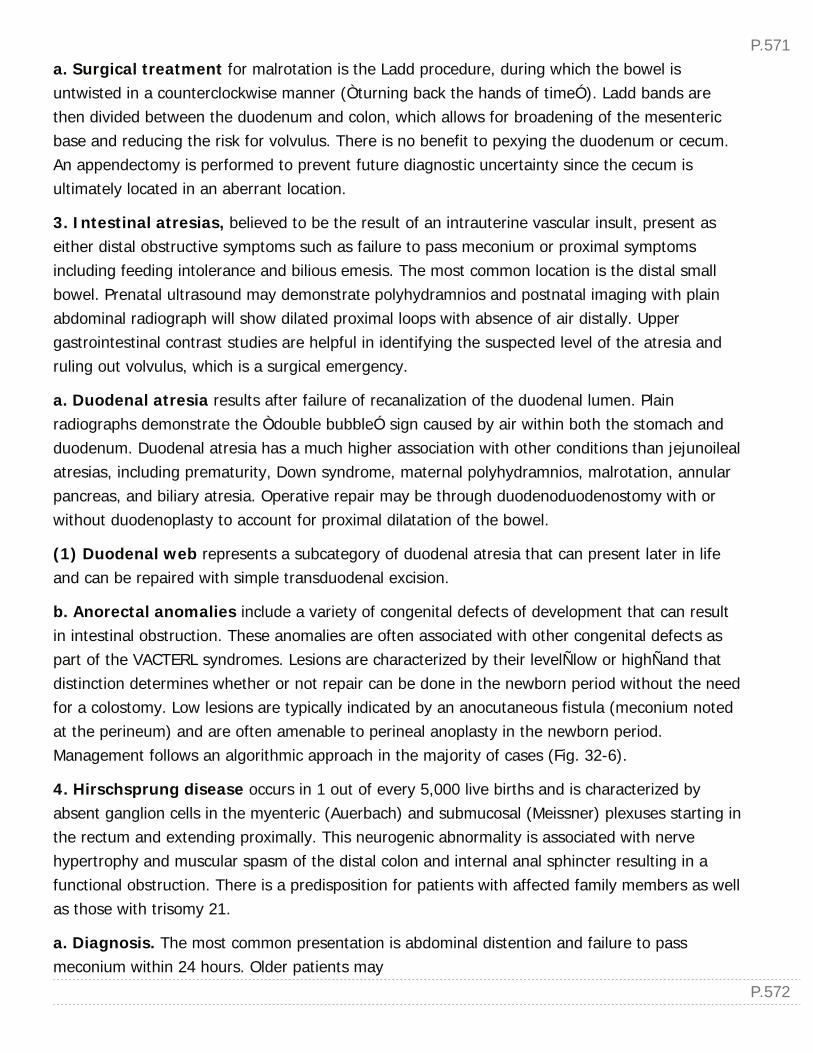
P.572
P.571a. Surgical treatment for malrotation is the Ladd procedure, during which the bowel isuntwisted in a counterclockwise manner (Òturning back the hands of timeÓ). Ladd bands arethen divided between the duodenum and colon, which allows for broadening of the mesentericbase and reducing the risk for volvulus. There is no benefit to pexying the duodenum or cecum.An appendectomy is performed to prevent future diagnostic uncertainty since the cecum isultimately located in an aberrant location.
3. Intestinal atresias, believed to be the result of an intrauterine vascular insult, present aseither distal obstructive symptoms such as failure to pass meconium or proximal symptomsincluding feeding intolerance and bilious emesis. The most common location is the distal smallbowel. Prenatal ultrasound may demonstrate polyhydramnios and postnatal imaging with plainabdominal radiograph will show dilated proximal loops with absence of air distally. Uppergastrointestinal contrast studies are helpful in identifying the suspected level of the atresia andruling out volvulus, which is a surgical emergency.
a. Duodenal atresia results after failure of recanalization of the duodenal lumen. Plainradiographs demonstrate the Òdouble bubbleÓ sign caused by air within both the stomach andduodenum. Duodenal atresia has a much higher association with other conditions than jejunoilealatresias, including prematurity, Down syndrome, maternal polyhydramnios, malrotation, annularpancreas, and biliary atresia. Operative repair may be through duodenoduodenostomy with orwithout duodenoplasty to account for proximal dilatation of the bowel.
(1) Duodenal web represents a subcategory of duodenal atresia that can present later in lifeand can be repaired with simple transduodenal excision.
b. Anorectal anomalies include a variety of congenital defects of development that can resultin intestinal obstruction. These anomalies are often associated with other congenital defects aspart of the VACTERL syndromes. Lesions are characterized by their levelÑlow or highÑand thatdistinction determines whether or not repair can be done in the newborn period without the needfor a colostomy. Low lesions are typically indicated by an anocutaneous fistula (meconium notedat the perineum) and are often amenable to perineal anoplasty in the newborn period.Management follows an algorithmic approach in the majority of cases (Fig. 32-6).
4. Hirschsprung disease occurs in 1 out of every 5,000 live births and is characterized byabsent ganglion cells in the myenteric (Auerbach) and submucosal (Meissner) plexuses starting inthe rectum and extending proximally. This neurogenic abnormality is associated with nervehypertrophy and muscular spasm of the distal colon and internal anal sphincter resulting in afunctional obstruction. There is a predisposition for patients with affected family members as wellas those with trisomy 21.
a. Diagnosis. The most common presentation is abdominal distention and failure to passmeconium within 24 hours. Older patients may

P.573present with chronic constipation. Plain radiographs demonstrate absence or paucity of air distalto the obstruction and contrast enema reveals a transition zone between proximally dilated anddistally decompressed pathologically abnormal bowel. Rectal biopsy, which can be performedtransanally with a suction device or on the seromuscular surface of the bowel via an abdominalapproach is required for pathologic confirmation of the diagnosis.
Figure 32-6 Algorithm for management of neonatal anorectal anomalies.
b. Management. Preoperative management includes decompression and saline colonicirrigations to evacuate impacted stool. Operative technique is aimed at identifying the transitionzone (ÒlevelingÓ) with serial biopsies, then bringing ganglionated bowel down to the anus whilepreserving sphincter function. The definitive management of Hirschsprung disease involvesvariations among three main procedures. In the Swenson procedure, the aganglionic bowel isremoved down to the level of the internal sphincters and a coloanal anastomosis is performed onthe perineum. In the Duhamel procedure, the aganglionic rectal stump is left in place and theganglionated, normal colon is pulled behind and anastomosed to this stump. Finally, the Soaveprocedure involves an endorectal mucosal dissection within the aganglionic distal rectum. Thenormally ganglionated colon is then pulled through the remnant muscular cuff and a coloanalanastomosis performed. Transanal procedures involving an endorectal pull-through are associatedwith fewer complications and fewer episodes of enterocolitis without higher rates of incontinencewhen compared to transabdominal approaches (J Pediatr Surg. 2010;45(6):1213-1220).
c. Surgical results are quite favorable with minimal risk of mortality, but morbidity involvingconstipation and/or incontinence as well as enterocolitis remains.
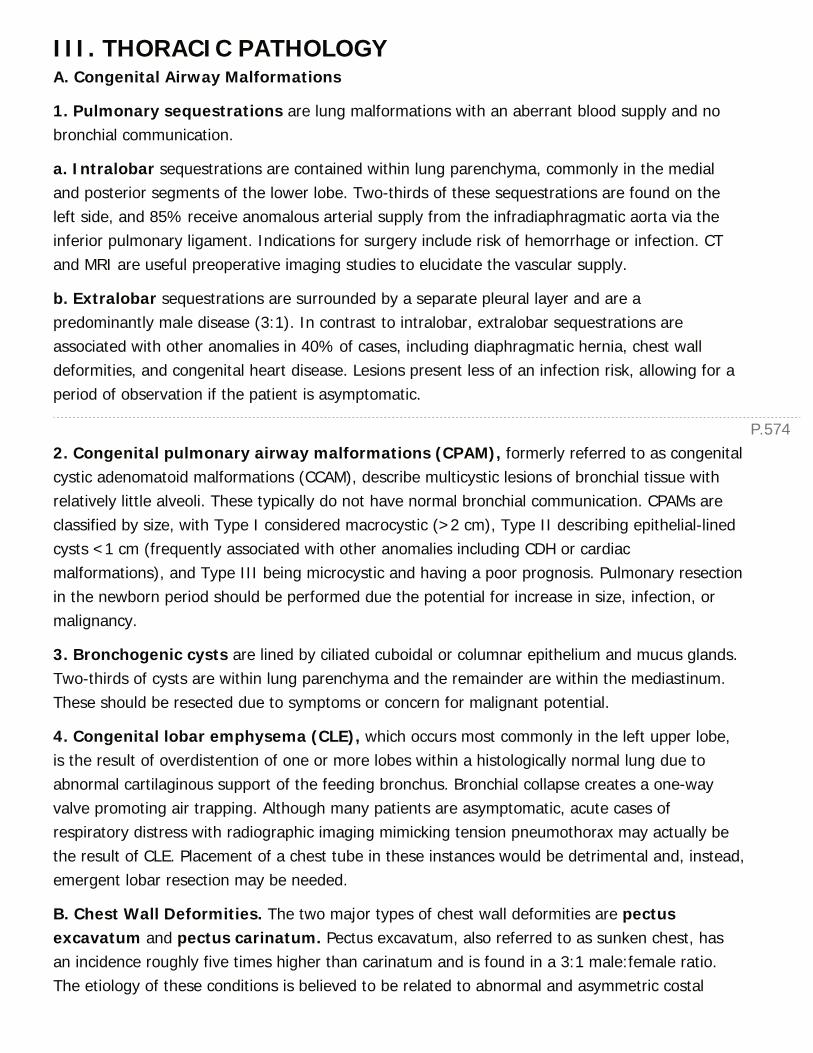
P.574
III. THORACIC PATHOLOGYA. Congenital Airway Malformations
1. Pulmonary sequestrations are lung malformations with an aberrant blood supply and nobronchial communication.
a. Intralobar sequestrations are contained within lung parenchyma, commonly in the medialand posterior segments of the lower lobe. Two-thirds of these sequestrations are found on theleft side, and 85% receive anomalous arterial supply from the infradiaphragmatic aorta via theinferior pulmonary ligament. Indications for surgery include risk of hemorrhage or infection. CTand MRI are useful preoperative imaging studies to elucidate the vascular supply.
b. Extralobar sequestrations are surrounded by a separate pleural layer and are apredominantly male disease (3:1). In contrast to intralobar, extralobar sequestrations areassociated with other anomalies in 40% of cases, including diaphragmatic hernia, chest walldeformities, and congenital heart disease. Lesions present less of an infection risk, allowing for aperiod of observation if the patient is asymptomatic.
2. Congenital pulmonary airway malformations (CPAM), formerly referred to as congenitalcystic adenomatoid malformations (CCAM), describe multicystic lesions of bronchial tissue withrelatively little alveoli. These typically do not have normal bronchial communication. CPAMs areclassified by size, with Type I considered macrocystic (>2 cm), Type II describing epithelial-linedcysts <1 cm (frequently associated with other anomalies including CDH or cardiacmalformations), and Type III being microcystic and having a poor prognosis. Pulmonary resectionin the newborn period should be performed due the potential for increase in size, infection, ormalignancy.
3. Bronchogenic cysts are lined by ciliated cuboidal or columnar epithelium and mucus glands.Two-thirds of cysts are within lung parenchyma and the remainder are within the mediastinum.These should be resected due to symptoms or concern for malignant potential.
4. Congenital lobar emphysema (CLE), which occurs most commonly in the left upper lobe,is the result of overdistention of one or more lobes within a histologically normal lung due toabnormal cartilaginous support of the feeding bronchus. Bronchial collapse creates a one-wayvalve promoting air trapping. Although many patients are asymptomatic, acute cases ofrespiratory distress with radiographic imaging mimicking tension pneumothorax may actually bethe result of CLE. Placement of a chest tube in these instances would be detrimental and, instead,emergent lobar resection may be needed.
B. Chest Wall Deformities. The two major types of chest wall deformities are pectusexcavatum and pectus carinatum. Pectus excavatum, also referred to as sunken chest, hasan incidence roughly five times higher than carinatum and is found in a 3:1 male:female ratio.The etiology of these conditions is believed to be related to abnormal and asymmetric costal

P.575
cartilage development.
1. Preoperative evaluation must include examination for scoliosis, which is present in 15% ofcases, as well as any cardiorespiratory abnormalities. Chest radiograph is considered a standardcomponent of the preoperative assessment. Additional workup may require an echocardiogramand an ophthalmologic examination if concern for cardiac abnormalities or Marfan syndromeexists. Pulmonary function tests should be obtained if there is concern for the condition causingpulmonary impairment. CT scan allows evaluation of the Haller Index, the transverse chestdiameter divided by the anterior-posterior diameter, to document the severity of the defect, but ithas little bearing on operative consideration.
2. Indications for surgery are largely cosmetic, as the condition becomes exaggerated bothphysically and psychosocially in the pubescent years. Correction is generally performed after 8years of age in order to prevent restrictive chest wall deformities. Two standard techniques, theRavitch and Nuss procedures, offer effective approaches, with both requiring subsequentsurgery to remove hardware. Carinatum defects can be similarly repaired with cartilage resectionand fixation or more frequently with external chest compression (J Pediatr Surg. 2006;41:40-45).
IV. ABDOMINAL PATHOLOGYA. Acquired Alimentary Obstruction
1. Hypertrophic pyloric stenosis (HPS) is the most common surgical cause of nonbiliousvomiting, occurring in 1 of every 400 births, commonly between 2 and 8 weeks of age and with amale to female ratio of 4:1. Infants of Northern European descent are most commonly affectedand there appears to be an increased incidence for children of parents with HPS. Pyloric stenosishas no known causative factor and is characterized by hypertrophy of the circular muscle of thepylorus resulting in obstruction of the gastric outlet leading to nonbilious projectile emesis.
Diagnosis is the result of suspicion based on a history of an appropriately aged infant presentingwith projectile nonbilious emesis associated with feeding. Persistent fussiness or presumedhunger are all characteristic. Occasionally, the emesis may be coffee-ground. Parents may initiallypresent to a pediatrician for evaluation of their child and can be given more common diagnosessuch as reflux or intolerance of feeds requiring a change in formula. Patients must be evaluatedfor dehydration by sufficient urine production and electrolyte abnormalities. Hypokalemic,hypochloremic metabolic alkalosis is the most common electrolyte abnormality due to a loss ofhydrochloric acid from gastric secretions and potassium from the kidney in attempt tocompensate for hypovolemia. Physical examination may reveal the Òolive sign,Ó the palpablepylorus to the right of and superior to the umbilicus.
a. Abdominal ultrasound is used for diagnosis, with criteria including a pyloric channel lengthgreater than 14 mm and single-wall muscular thickness of 4 mm or greater. These criteria have99.5% sensitivity and 100% specificity for identifying HPS (J Pediatr Surg. 2007;16(1):27-33).

P.576
b. Management should initially include evaluation and treatment of dehydration and electrolyteabnormalities. Patients are made NPO to prevent further episodes of emesis and are administeredintravenous fluids titrated to urine output greater than 1 mL/kg/hour. Addition of potassium intofluids should be withheld until urine output has been restored. Surgical timing is largely decidedby the severity of electrolyte abnormalities, particularly the HCO3, which, if significantly elevated,
results in a compensatory respiratory acidosis that may result in apnea, a devastatingconsequence particularly exaggerated by postanesthetic respiratory depression.
c. Pyloromyotomy, separation of the external muscular layers down to the level of mucosa, isperformed either through right upper quadrant or periumbilical incision, or with laparoscopy.
d. Postoperative feeding protocols typically start with an electrolyte solution with gradualadvancement over the first 24 hours postoperatively to formula or breast milk volumes that areappropriate for age. Vomiting is a common postoperative issue, but, rarely, may be an indicatorof incomplete myotomy. Additional complications include
perforation of the mucosa, which is treated by mucosal repair and nasogastric drainage.
2. Intussusception is another common acquired cause of intestinal obstruction, resulting frominvagination or telescoping of the proximal intestine into the distal bowel. This is most common ininfants 3 months to 3 years of age and most frequently originates at the ileocecal junction.Although a lead-point (pathologic or anatomic source) is most commonly not present, definitiveidentification of a contributing structure may occur in up to 12% of patients, most frequentlyeither a Meckel diverticulum (remnant of the omphalomesenteric duct), intestinal polyp, or tumor.Affected infants present with periods of intense crying during which they retract their legs up inpain; these ÒattacksÓ subside within a few minutes. Recent gastrointestinal or upper respiratoryillness may also be part of the patient's history. Obstruction may result in ischemia, whichproduces stool with a mix of blood and mucous (i.e., the pathognomonic Òcurrant jellyÓ stools).
a. Diagnosis. Plain abdominal radiographs may be sufficient for diagnosis in as many as 50% ofcases, revealing abnormal gas patterns concerning for a mass with subsequent paucity of gas inthe right lower abdomen. Ultrasound may identify a ÒtargetÓ lesion representing a transverseview of the intussuscepted layers of bowel.
b. Nonoperative management of active intussusception is performed by contrast or airenema. This is successful in 80% of cases, with recurrence rates of approximately 10% within thefirst 24 hours. Management of recurrence is usually again attempted nonoperatively, but failureor a second recurrence are indications for surgery, as are peritonitis or concern for bowelischemia at any stage of presentation.
c. Operative management can be approached via either open or laparoscopic techniques. Withopen surgery, reduction is achieved with retrograde (distal to proximal) squeezing or milking ofthe intussusceptum until reduced. During laparoscopy, the bowel is pulled apart. The affected
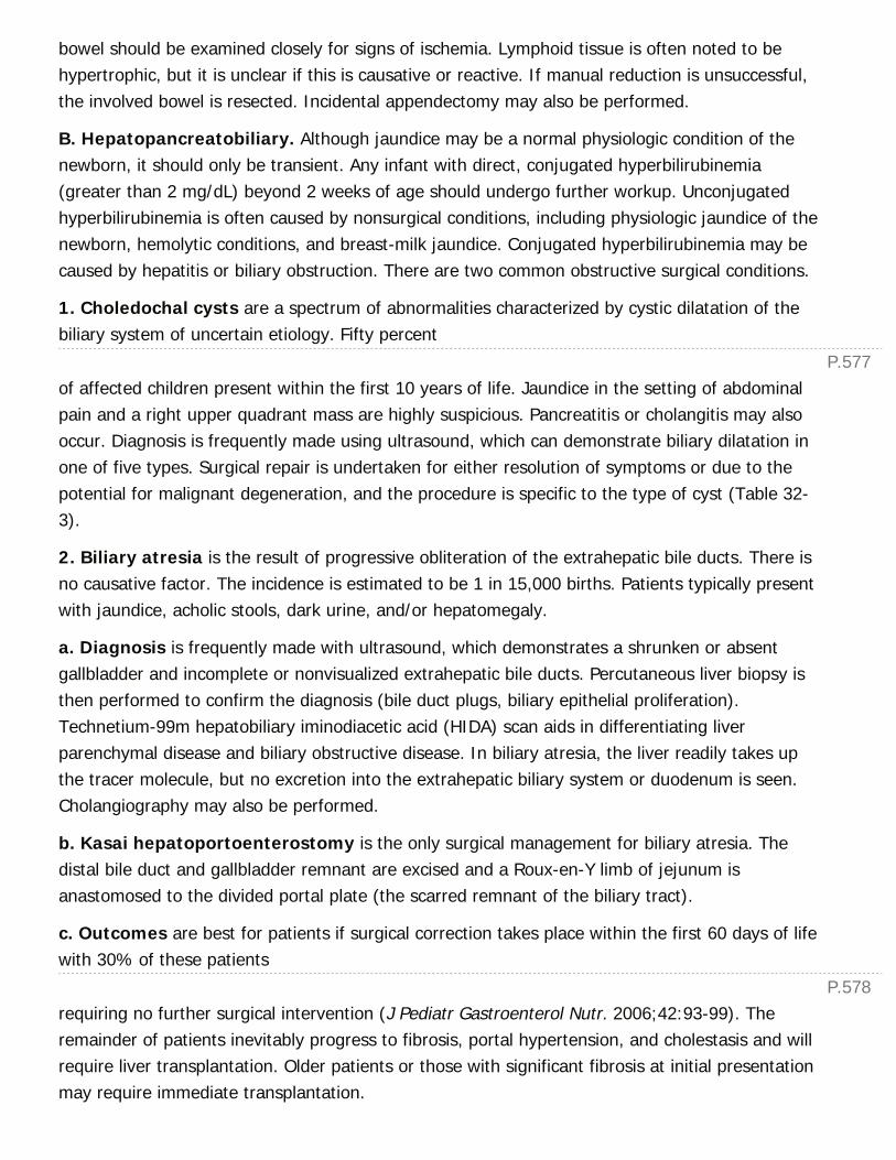
P.577
P.578
bowel should be examined closely for signs of ischemia. Lymphoid tissue is often noted to behypertrophic, but it is unclear if this is causative or reactive. If manual reduction is unsuccessful,the involved bowel is resected. Incidental appendectomy may also be performed.
B. Hepatopancreatobiliary. Although jaundice may be a normal physiologic condition of thenewborn, it should only be transient. Any infant with direct, conjugated hyperbilirubinemia(greater than 2 mg/dL) beyond 2 weeks of age should undergo further workup. Unconjugatedhyperbilirubinemia is often caused by nonsurgical conditions, including physiologic jaundice of thenewborn, hemolytic conditions, and breast-milk jaundice. Conjugated hyperbilirubinemia may becaused by hepatitis or biliary obstruction. There are two common obstructive surgical conditions.
1. Choledochal cysts are a spectrum of abnormalities characterized by cystic dilatation of thebiliary system of uncertain etiology. Fifty percent
of affected children present within the first 10 years of life. Jaundice in the setting of abdominalpain and a right upper quadrant mass are highly suspicious. Pancreatitis or cholangitis may alsooccur. Diagnosis is frequently made using ultrasound, which can demonstrate biliary dilatation inone of five types. Surgical repair is undertaken for either resolution of symptoms or due to thepotential for malignant degeneration, and the procedure is specific to the type of cyst (Table 32-3).
2. Biliary atresia is the result of progressive obliteration of the extrahepatic bile ducts. There isno causative factor. The incidence is estimated to be 1 in 15,000 births. Patients typically presentwith jaundice, acholic stools, dark urine, and/or hepatomegaly.
a. Diagnosis is frequently made with ultrasound, which demonstrates a shrunken or absentgallbladder and incomplete or nonvisualized extrahepatic bile ducts. Percutaneous liver biopsy isthen performed to confirm the diagnosis (bile duct plugs, biliary epithelial proliferation).Technetium-99m hepatobiliary iminodiacetic acid (HIDA) scan aids in differentiating liverparenchymal disease and biliary obstructive disease. In biliary atresia, the liver readily takes upthe tracer molecule, but no excretion into the extrahepatic biliary system or duodenum is seen.Cholangiography may also be performed.
b. Kasai hepatoportoenterostomy is the only surgical management for biliary atresia. Thedistal bile duct and gallbladder remnant are excised and a Roux-en-Y limb of jejunum isanastomosed to the divided portal plate (the scarred remnant of the biliary tract).
c. Outcomes are best for patients if surgical correction takes place within the first 60 days of lifewith 30% of these patients
requiring no further surgical intervention (J Pediatr Gastroenterol Nutr. 2006;42:93-99). Theremainder of patients inevitably progress to fibrosis, portal hypertension, and cholestasis and willrequire liver transplantation. Older patients or those with significant fibrosis at initial presentationmay require immediate transplantation.
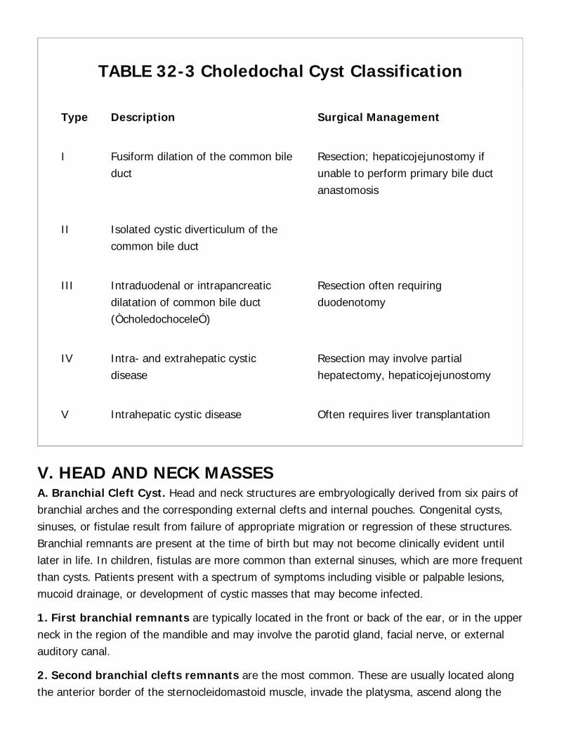
TABLE 32-3 Choledochal Cyst Classification
Type Description Surgical Management
I Fusiform dilation of the common bileduct
Resection; hepaticojejunostomy ifunable to perform primary bile ductanastomosis
II Isolated cystic diverticulum of thecommon bile duct
III Intraduodenal or intrapancreaticdilatation of common bile duct(ÒcholedochoceleÓ)
Resection often requiringduodenotomy
IV Intra- and extrahepatic cysticdisease
Resection may involve partialhepatectomy, hepaticojejunostomy
V Intrahepatic cystic disease Often requires liver transplantation
V. HEAD AND NECK MASSESA. Branchial Cleft Cyst. Head and neck structures are embryologically derived from six pairs ofbranchial arches and the corresponding external clefts and internal pouches. Congenital cysts,sinuses, or fistulae result from failure of appropriate migration or regression of these structures.Branchial remnants are present at the time of birth but may not become clinically evident untillater in life. In children, fistulas are more common than external sinuses, which are more frequentthan cysts. Patients present with a spectrum of symptoms including visible or palpable lesions,mucoid drainage, or development of cystic masses that may become infected.
1. First branchial remnants are typically located in the front or back of the ear, or in the upperneck in the region of the mandible and may involve the parotid gland, facial nerve, or externalauditory canal.
2. Second branchial clefts remnants are the most common. These are usually located alongthe anterior border of the sternocleidomastoid muscle, invade the platysma, ascend along the

P.579
carotid sheath to the level of the hyoid bone, and extend medially between the carotid arterybifurcation. The fistula then courses behind the posterior belly of the digastric and stylohyoidmuscles to end in the tonsillar fossa.
3. Third branchial cleft remnants usually do not have associated sinuses or fistulae. Thesemost often contain cartilage, are located in the suprasternal notch or clavicular region, andpresent as a firm mass or as a subcutaneous abscess.
B. Thyroglossal duct cysts are common lesions in the midline of the neck and frequentlypresent in preschool-age children. These cysts represent incomplete thyroid gland formation and,as such, are located along the normal course of thyroid migration: From the foramen cecum tothe anatomically normal site of the pyramidal lobe of the thyroid gland. Frequently the tongue orhyoid bone are involved.
1. Indications for surgery include increasing size, infection or risk for infection, and risk forcarcinoma (1% to 2%). Preoperative ultrasound can aid significantly in identifying involvedstructures including the site of normal thyroid tissue, which should be preserved if possible.Standard surgery, the Sistrunk procedure, involves excision of the central portion of the hyoidbone in addition to the cyst in continuity with its tract.
C. Cystic hygroma is a lymphatic malformation resulting from abnormal development of alymphatic network that fails to drain into the venous system. Seventy-five percent of these lesionsinvolve the lymphatic jugular
sacks, presenting as a posterior neck mass. The majority of cystic hygromas present at birth(50% to 65%), with most becoming apparent by the second year of life.
1. Indications for resection are largely cosmetic, although expansion that threatenscompression of the airway; infection; or pain, possibly due to hemorrhage, are also acceptableindications for treatment. Complete surgical excision is preferred. Preoperative magneticresonance imaging can assist in determining the extent of the lesion and any neurovascularinvolvement. Postoperative morbidity includes recurrence, lymphatic leak, infection, andneurovascular injury. Additional therapeutic modalities include injection of sclerosing agents suchas bleomycin.
D. Cervical lymphadenopathy is a frequent diagnosis for referral to a pediatric surgeon.Infectious etiologies are the overwhelming majority of diagnoses, including cat-scratch fever as aresult of Bartonella, which contributes to approximately 3% of lymphadenopathy cases. Lymphnodes that are unilateral, firm, fixed, or greater than 2 cm should prompt additional workup,including a chest radiograph to evaluate for mediastinal lymphadenopathy or masses.
VI. TUMORS AND NEOPLASMSA. Neuroblastoma is the most common neoplasm of childhood, accounting for 6% to 10% of allchildhood cancers with an incidence of 1 in 10,000 cases annually and a median age of diagnosis

P.580
of 2 years old. Only 25% of patients present with isolated disease, often found incidentally onexamination or imaging, for which surgical therapy is standard of care. The remainder presentwith metastatic disease and have a poor prognosis. These neoplasms are of neural crest originand are thus found along the sympathetic nervous system, with 75% in the abdomen or pelvisand half of these within the adrenal medulla.
1. Preoperative evaluation should include routine laboratory work such as CBC and CMP.Blood pressure should be checked to investigate the potential for hypertension, and urine shouldbe tested for catecholamine metabolites. CT and MRI assist in preoperative planning, particularlyas neuroblastoma is capable of invading into adjacent vascular structures. They are also usefulfor metastatic workup, with frequent sites of metastases being bone and lung. Radiolabeledmetaiodobenzyl guanidine (MIBG) is a high-yield study to document the presence of metastaticdisease. Bone marrow aspirate and biopsy complete the staging evaluation. Staging and survivalare determined by burden of disease as well as genetic markers including N-myc overexpressionand chromosomal deletions, which portend a worse prognosis. Young patients with early diseaseand favorable pathology have >90% survival, while older patients with unfavorable or metastaticdisease can have survival as low as 10%.
2. Surgical management is reserved for anatomically resectable cases of early nonmetastaticdisease or can be undertaken after neoadjuvant therapies.
B. Wilms tumor accounts for 6% of all malignancies in children and is the most common renalmalignancy in children. Approximately 500 cases are diagnosed annually in the United States,with an average age of diagnosis of 3 or 4 years old. There is no gender predominance, and 5%of cases are bilateral. Although there are heritable forms of Wilms tumor including an associationwith Beckwith-Wiedemann syndrome, most cases are sporadic and present as incidentalabdominal masses. Patients may have hypertension or hematuria.
1. Preoperative evaluation includes ultrasound to confirm renal origin of the mass and toevaluate for any potential intravascular extension. CT or MRI may further assist in differentiatingWilms tumor from neuroblastoma, as will urine catecholamines. Chest imaging via CT scan isneeded for complete staging.
2. Surgical intervention requires radial nephroureterectomy and lymph node sampling.Intraoperative spillage of tumor contents must be carefully avoided, as it upstages the patient'sdisease (Table 32-4). For resectable disease, surgery and chemotherapy together result in agreater than 90% cure rate.
C. Hepatic tumors make up fewer than 5% of all intraabdominal pediatric malignancies but aremalignant in up to 70% of cases.
1. Hepatoblastoma typically presents before 3 years of age and is often unifocal.Chemotherapy is somewhat effective and overall survival is dictated primarily as to whether the
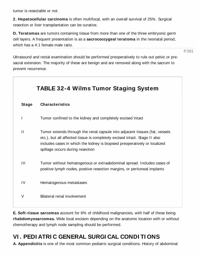
P.581
tumor is resectable or not.
2. Hepatocellular carcinoma is often multifocal, with an overall survival of 25%. Surgicalresection or liver transplantation can be curative.
D. Teratomas are tumors containing tissue from more than one of the three embryonic germcell layers. A frequent presentation is as a sacrococcygeal teratoma in the neonatal period,which has a 4:1 female:male ratio.
Ultrasound and rectal examination should be performed preoperatively to rule out pelvic or pre-sacral extension. The majority of these are benign and are removed along with the sacrum toprevent recurrence.
TABLE 32-4 Wilms Tumor Staging System
Stage Characteristics
I Tumor confined to the kidney and completely excised intact
II Tumor extends through the renal capsule into adjacent tissues (fat, vesselsetc.), but all affected tissue is completely excised intact. Stage II alsoincludes cases in which the kidney is biopsied preoperatively or localizedspillage occurs during resection
III Tumor without hematogenous or extraabdominal spread. Includes cases ofpositive lymph nodes, positive resection margins, or peritoneal implants
IV Hematogenous metastases
V Bilateral renal involvement
E. Soft-tissue sarcomas account for 6% of childhood malignancies, with half of these beingrhabdomyosarcomas. Wide local excision depending on the anatomic location with or withoutchemotherapy and lymph node sampling should be performed.
VI. PEDIATRIC GENERAL SURGICAL CONDITIONSA. Appendicitis is one of the most common pediatric surgical conditions. History of abdominal

P.582
pain followed by nausea along with physical examination findings of tenderness at McBurneypoint and obturator, iliopsoas, and Rovsing signs can be helpful clinical cues. Scoring systemsincorporating history, physical, and laboratory values such as the Alvarado score (Ann EmergMed. 1986;15(5):557-564) can aid in objectively risk-stratifying patients.
Ultrasound is routinely the first imaging modality of choice in the pediatric population. A positivestudy should result in surgical management, whereas a negative study or one in which theappendix was nonvisualized should prompt either observation, evaluation of other diagnoses, orconsideration for obtaining a CT scan, particularly in patients showing signs of advanced illness.
B. Indirect inguinal hernias affect approximately 1% to 5% of children, with a predominance inmales (8:1) and an increased rate in premature infants (7% to 30%). Bilateral hernias arepresent in 10% to 40% and occur more frequently in premature infants and girls. Due to anincreased risk of recurrence and post-anesthesia apnea in premature patients, neonatal inguinalhernias are frequently repaired prior to the patient leaving the hospital or at 50 weekspostconception (J Pediatr Surg. 1996;31:1166-1169).
C. Hydroceles are fluid collections within the processus vaginalis that envelop the testicles. Theyoccur in approximately 6% of full-term male newborns.
1. Communicating hydroceles allow the free flow of peritoneal fluid down to the scrotumthrough a patent processus vaginalis. This must be regarded as a hernia and repaired as such.
2. Noncommunicating hydroceles contain fluid confined to the scrotum due to an obliteratedprocessus vaginalis. This is usually a self-limiting process that resolves in 6 to 12 months.
D. Umbilical hernia is common in children due to a persistence of the umbilical ring. In the vastmajority of cases, these close spontaneously by age 4 to 5 but can be repaired if they areexceptionally large (>2 cm, due to low likelihood of spontaneous closure) or if the patient has hadincarceration of bowel within the hernia.
E. Trauma
1. Imaging. Limiting radiation exposure and unnecessary testing is important in children.However, the inability to obtain reliable examinations
in this population, particularly in trauma settings, complicates the decision to obtain imaging.Diffuse abdominal pain, a seatbelt sign, or distracting injuries can be indications for computedtomography scan. In addition, elevated serum glutamic-oxaloacetic transaminase (SGOT) orserum glutamic-pyruvic transaminase (SGPT) levels higher than 200 or 100 IU/L, respectively, areaccepted thresholds for obtaining a CT scan in the setting of blunt trauma.
TABLE 32-5 Nonoperative Management of Pediatric
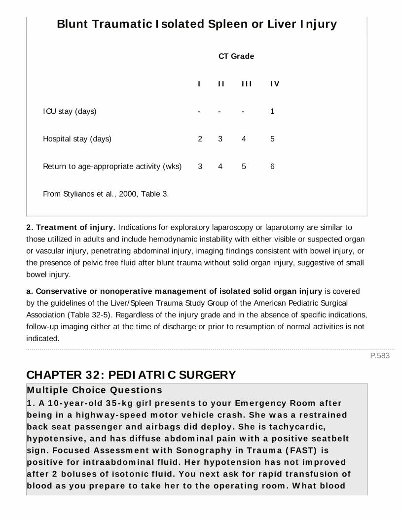
P.583
Blunt Traumatic Isolated Spleen or Liver Injury
CT Grade
I II III IV
ICU stay (days) - - - 1
Hospital stay (days) 2 3 4 5
Return to age-appropriate activity (wks) 3 4 5 6
From Stylianos et al., 2000, Table 3.
2. Treatment of injury. Indications for exploratory laparoscopy or laparotomy are similar tothose utilized in adults and include hemodynamic instability with either visible or suspected organor vascular injury, penetrating abdominal injury, imaging findings consistent with bowel injury, orthe presence of pelvic free fluid after blunt trauma without solid organ injury, suggestive of smallbowel injury.
a. Conservative or nonoperative management of isolated solid organ injury is coveredby the guidelines of the Liver/Spleen Trauma Study Group of the American Pediatric SurgicalAssociation (Table 32-5). Regardless of the injury grade and in the absence of specific indications,follow-up imaging either at the time of discharge or prior to resumption of normal activities is notindicated.
CHAPTER 32: PEDIATRIC SURGERYMultiple Choice Questions1. A 10-year-old 35-kg girl presents to your Emergency Room afterbeing in a highway-speed motor vehicle crash. She was a restrainedback seat passenger and airbags did deploy. She is tachycardic,hypotensive, and has diffuse abdominal pain with a positive seatbeltsign. Focused Assessment with Sonography in Trauma (FAST) ispositive for intraabdominal fluid. Her hypotension has not improvedafter 2 boluses of isotonic fluid. You next ask for rapid transfusion ofblood as you prepare to take her to the operating room. What blood

volume should you initially transfuse?
a. 100 mL
b. 175 mL
c. 250 mL
d. 350 mL
e. 475 mL
View Answer
2. You receive a call from a primary care physician who is working up a4-year old female for chronic abdominal pain, distention, and jaundice.The physician describes a CT scan finding of a cystic structure arisingfrom the common bile duct that is separate from the gallbladder. Yoususpect choledochal cyst as a possible diagnosis. This imaging isconsistent with what type of choledochal cyst?
a. Type I
b. Type II
c. Type III
d. Type IV
e. Type V
View Answer
3. You are caring for a 14-year-old boy who was presented to theEmergency Room after injuring himself in an ATV accident. He had beencomplaining of abdominal pain and eventually received a CT scanshowing a blunt splenic injury with a subcapsular hematoma occupying<50% of the surface and associated with a 3-cm splenic laceration. Hehas been hemodynamically stable. You inform the family that the boyshould be admitted. His family asks how long he will need to beadmitted and how long he will have to engage in only light activity afterdischarge. You tell them:
a. 24 hour overnight hospitalization, return to activity as tolerated
b. 2 day hospitalization, 3 weeks light activity
c. 3 day hospitalization, 4 weeks light activity
d. 4 day hospitalization, 5 weeks light activity
e. 5 day hospitalization including 24 hours in the ICU, 6 weeks lightactivity
View Answer
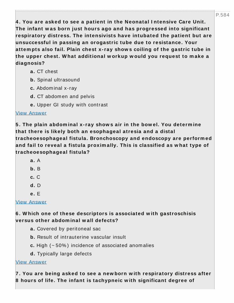
P.5844. You are asked to see a patient in the Neonatal Intensive Care Unit.The infant was born just hours ago and has progressed into significantrespiratory distress. The intensivists have intubated the patient but areunsuccessful in passing an orogastric tube due to resistance. Yourattempts also fail. Plain chest x-ray shows coiling of the gastric tube inthe upper chest. What additional workup would you request to make adiagnosis?
a. CT chest
b. Spinal ultrasound
c. Abdominal x-ray
d. CT abdomen and pelvis
e. Upper GI study with contrast
View Answer
5. The plain abdominal x-ray shows air in the bowel. You determinethat there is likely both an esophageal atresia and a distaltracheoesophageal fistula. Bronchoscopy and endoscopy are performedand fail to reveal a fistula proximally. This is classified as what type oftracheoesophageal fistula?
a. A
b. B
c. C
d. D
e. E
View Answer
6. Which one of these descriptors is associated with gastroschisisversus other abdominal wall defects?
a. Covered by peritoneal sac
b. Result of intrauterine vascular insult
c. High (~50%) incidence of associated anomalies
d. Typically large defects
View Answer
7. You are being asked to see a newborn with respiratory distress after8 hours of life. The infant is tachypneic with significant degree of

P.585
retractions. Oxygen saturations are below 80%. Chest x-ray showsherniated abdominal contents in the left chest. What treatment optionis the next best choice?
a. Emergent thoracotomy
b. ECMO cannulation
c. Intubation
d. Bedside echocardiogram
View Answer
8. Hirschsprung disease results from a failure of neural crest celldevelopment in the Meissner and Auerbach plexi. In what layers of thebowel wall are these plexi contained?
Choice Meissner Auerbach
A Mucosal Mucosal
B Submucosal Mucosal
C Muscular Submucosal
D Submucosal Muscular
E Serosal Muscular
View Answer
9. Twenty-four hours after a pyloromyotomy and tolerating a normalvolume of formula every 3 to 4 hrs, your patient develops feedingintolerance, mild tachypnea, and abdominal distention. The infant isotherwise stable. What is your next step?
a. Continue feeding
b. Change formula
c. Make NPO, start nasogastric drainage
d. Laparotomy
View Answer
10. A young patient presents to you with <12 hours of severeabdominal pain, diffuse tenderness on examination, and bilious emesis.You are concerned about malrotation and, while awaiting imaging, plan

your operative approach. Which step is not a common component ofLadd procedure?
a. Reduction of volvulus by counterclockwise rotation
b. Division of Ladd bands
c. Appendectomy
d. Pexying the cecum to the peritoneal sidewall
View Answer
33Cardiac Surgery
Matthew C. Henn
Jennifer S. Lawton
This chapter focuses on the preoperative evaluation, surgical indications, procedures, andpostoperative management of adult cardiac surgery patients.
I. ANATOMYA. Coronary Arteries. The coronary arteries are visible and accessible on the epicardial surfaceof the heart or may be found intramyocardially. The left and right coronary arteries arise fromwithin the sinuses of Valsalva just above the right and left coronary cusps of the aortic valve. Theleft main coronary artery travels posterior toward the pulmonary artery, then divides into itsmain branches, the left anterior descending artery (LAD) and the left circumflex artery(LCx). In 10% to 15% of patients, the LCx gives off the posterior descending artery (PDA),termed a left dominant coronary circulation. The right coronary artery (RCA) begins onthe anterior surface of the aorta and descends in the anterior AV groove, where, in rightdominant coronary circulation (80% to 85% of cases), it gives off the PDA (Fig. 33-1).
Clinical correlation: Dominance is important in selecting targets to graft duringcoronary artery bypass grafting (CABG) surgery and in predicting location (anterior orposterior) of culprit vessel in the diagnosis of post myocardial infarction (MI)ventricular septal defect (VSD).
Clinical correlation: Because of its anterior location, intracardiac air preferentiallyenters the orifice of the RCA when weaning from cardiopulmonary bypass (CPB),resulting in right ventricular dysfunction.
B. Coronary Veins. The coronary sinus is located in the posterior AV groove and receivesvenous drainage mainly from the left ventricular system. Its main tributaries are the great,middle, and small cardiac veins. The anterior cardiac veins drain the right coronary system,and ultimately into the right atrium.
Clinical correlation: The coronary sinus is an important structure in the myocardialprotection strategy for most cardiac surgery. Infusion of hyperkalemic solution intothis venous structure (retrograde cardioplegia) provides protection to all territories(except the right heart as the venous drainage is into the right atrium) in a moreuniform fashion versus the antegrade direction in patients with diffuse coronary
> Table of Contents > 33 - Cardiac Surgery

P.587P.588
disease.
C. Valves
1. AV valves. These valves are continuous with the annuli fibrosi at the base of the heart andsecured by chordae tendineae, which attach the free leaflets to the intraventricular papillarymuscles. The tricuspid valve separates the right chambers and consists of a large
anterior leaflet, a posterior leaflet, and a septal leaflet attached to the interventricular septum.The mitral (bicuspid) valve separates the left chambers and consists of a large anterior(aortic) leaflet and a posterior (mural) leaflet.
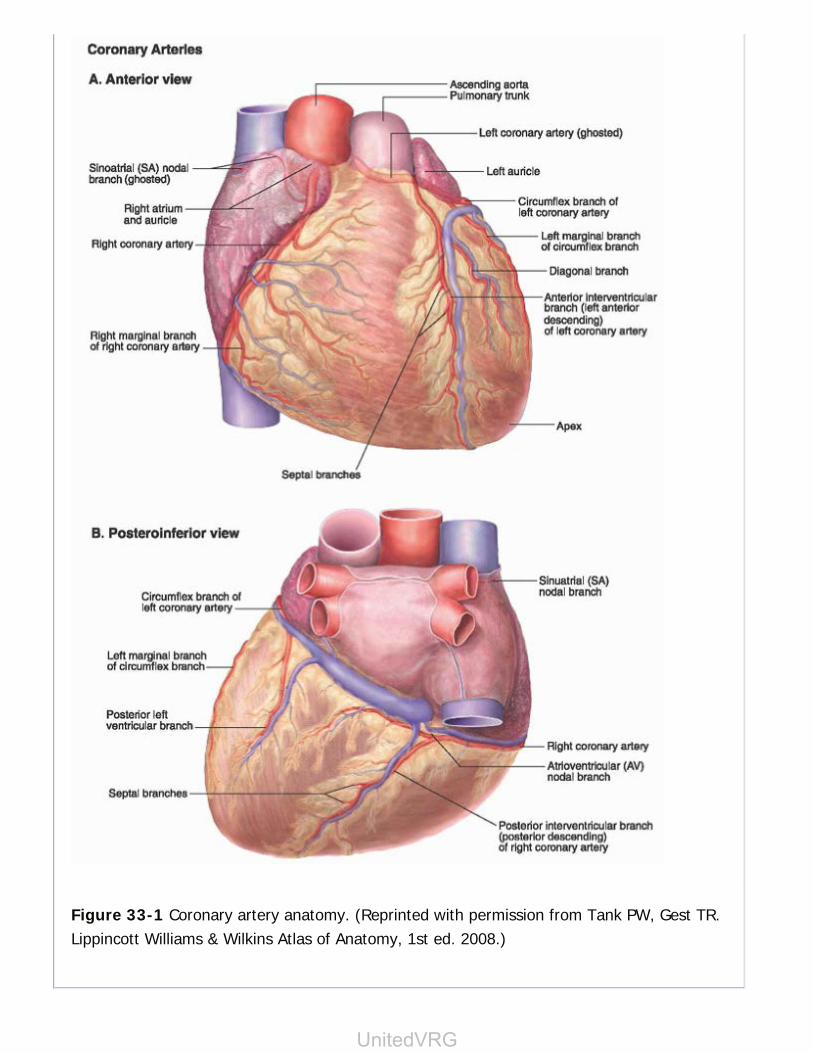
Figure 33-1 Coronary artery anatomy. (Reprinted with permission from Tank PW, Gest TR.Lippincott Williams & Wilkins Atlas of Anatomy, 1st ed. 2008.)
UnitedVRG
P.589
2. Semilunar valves. The pulmonary and aortic valves are essentially trileaflet and thecoronary arteries arise just distal to the aortic valve. Just distal to the valves are gentle dilationsof the ascending aorta, known as sinuses of Valsalva.
D. Electrophysiology. Electrical activation of the heart begins in the sinoatrial (SA) node(located at the junction of the anteromedial aspect of the superior vena cava and the rightatrium), travels to the AV node (located in the triangle of Koch; Fig. 33-2) defined by thecoronary sinus, tendon of Todaro, and the septal leaflet of the tricuspid valve. The AV nodeprotects the ventricle from atrial tachyarrhythmias. From the AV node, conduction travels to theright and left bundles of His.
Clinical correlation: The AV node is of particular importance as it may be injuredduring cardiac surgery. Specifically, during tricuspid valve repair or replacement,during aortic valve replacementÑin the commissure between the noncoronary andright coronary cusps, and in mitral valve repair or replacementÑin the annulus nearthe A1 portion of the anterior leaflet just under the aortic valve.
E. Myocardium. The working chambers of the heart (ventricles) provide cardiac output to thelungs and body. Cardiac output is the volume of blood
ejected by the heart per minute and is mainly influenced by heart rate and stroke volume(volume of blood ejected from one ventricle per heart beat). Cardiac output may be manipulatedby altering heart rate, preload, afterload, and contractility (Fig. 33-3).
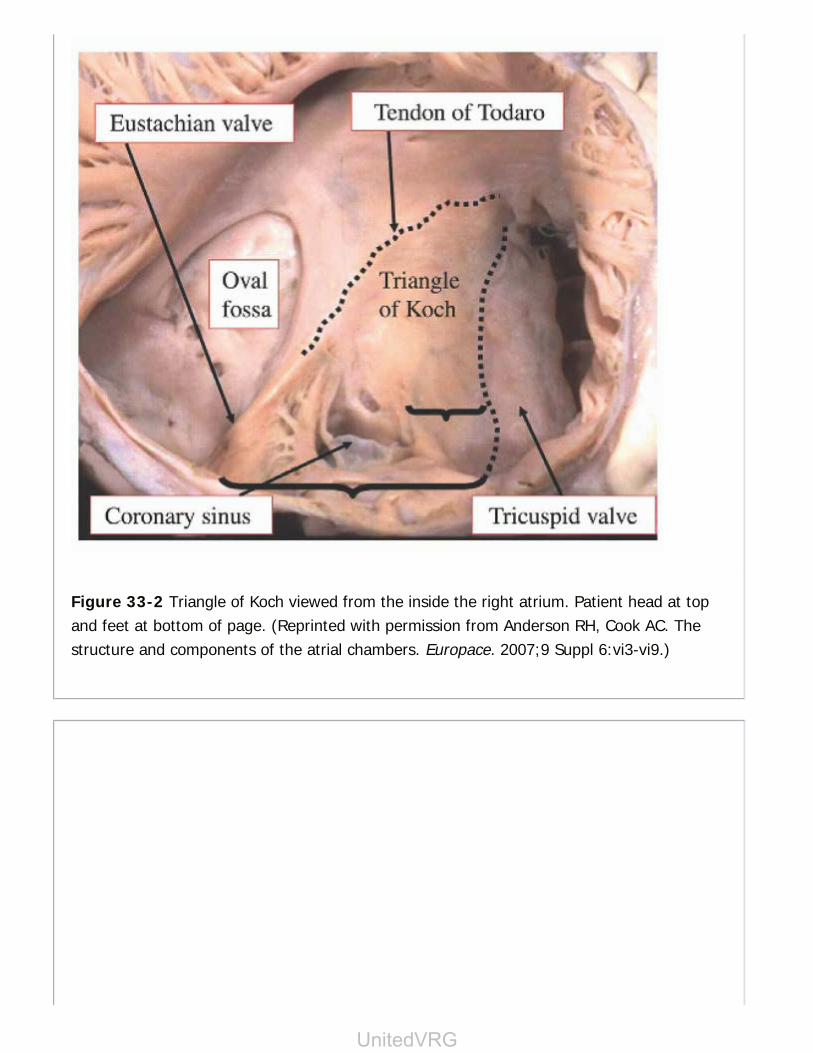
Figure 33-2 Triangle of Koch viewed from the inside the right atrium. Patient head at topand feet at bottom of page. (Reprinted with permission from Anderson RH, Cook AC. Thestructure and components of the atrial chambers. Europace. 2007;9 Suppl 6:vi3-vi9.)
UnitedVRG
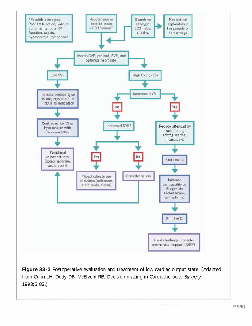
P.590
Figure 33-3 Postoperative evaluation and treatment of low cardiac output state. (Adaptedfrom Cohn LH, Dody DB, McElvein RB. Decision making in Cardiothoracic. Surgery.1993;2:83.)
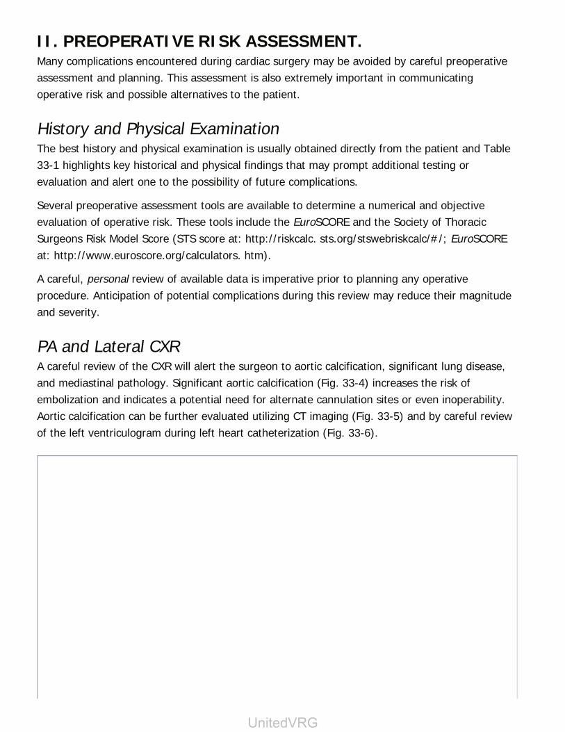
II. PREOPERATIVE RISK ASSESSMENT.Many complications encountered during cardiac surgery may be avoided by careful preoperativeassessment and planning. This assessment is also extremely important in communicatingoperative risk and possible alternatives to the patient.
History and Physical ExaminationThe best history and physical examination is usually obtained directly from the patient and Table33-1 highlights key historical and physical findings that may prompt additional testing orevaluation and alert one to the possibility of future complications.
Several preoperative assessment tools are available to determine a numerical and objectiveevaluation of operative risk. These tools include the EuroSCORE and the Society of ThoracicSurgeons Risk Model Score (STS score at: http://riskcalc. sts.org/stswebriskcalc/#/; EuroSCOREat: http://www.euroscore.org/calculators. htm).
A careful, personal review of available data is imperative prior to planning any operativeprocedure. Anticipation of potential complications during this review may reduce their magnitudeand severity.
PA and Lateral CXRA careful review of the CXR will alert the surgeon to aortic calcification, significant lung disease,and mediastinal pathology. Significant aortic calcification (Fig. 33-4) increases the risk ofembolization and indicates a potential need for alternate cannulation sites or even inoperability.Aortic calcification can be further evaluated utilizing CT imaging (Fig. 33-5) and by careful reviewof the left ventriculogram during left heart catheterization (Fig. 33-6).
UnitedVRG
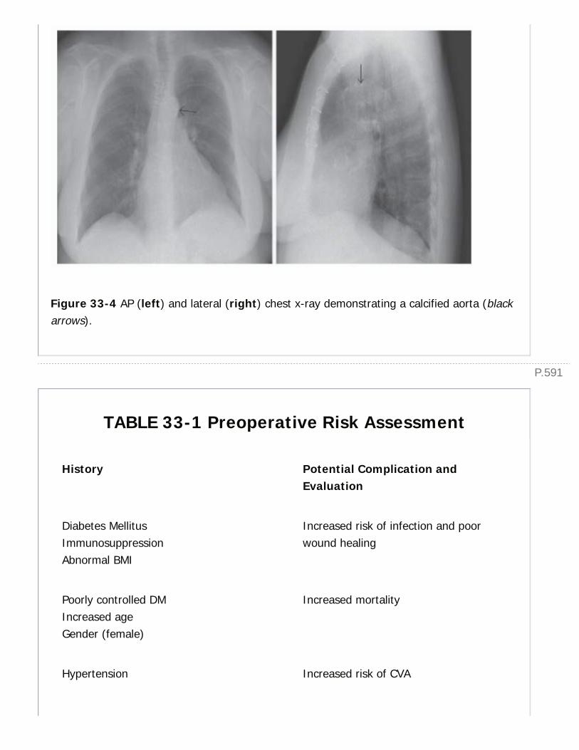
P.591
Figure 33-4 AP (left) and lateral (right) chest x-ray demonstrating a calcified aorta (blackarrows).
TABLE 33-1 Preoperative Risk Assessment
History Potential Complication andEvaluation
Diabetes MellitusImmunosuppressionAbnormal BMI
Increased risk of infection and poorwound healing
Poorly controlled DMIncreased ageGender (female)
Increased mortality
Hypertension Increased risk of CVA

Recent cath Increased risk of renal failure
Home oxygen useLung diseaseTobacco usePrevious tracheostomy
Increased risk of prolonged ventilation;consider PFTs
Liver diseaseRecent antiplatelet agent use (Plavix, IIb-IIIa inhibitors, thrombin inhibitors)Thrombocytopenia
Increased risk of bleeding, considerhematology or hepatology consults
Previous sternotomyChest wall XRTPericarditis
Anticipate difficult dissection
Poor social supportNeurologic dysfunction
Difficult rehabilitation
Exercise tolerance Good indicator of outcome
Pulsatile abdominal mass Abdominal ultrasound
Carotid bruitHistory of carotid endarterectomyTIA symptomsHistory of stroke
Carotid Doppler
Poor dental hygiene Panorex prior to valve procedure
BMI, body mass index; DM, diabetes mellitus; XRT, radiation therapy; CVA,cerebrovascular accident; PFT, pulmonary function test; TIA, transient ischemic attack.Adapted from Lawton JS and Gay WA. On Pump Coronary Artery Bypass Grafting inComplications in Cardiothoracic Surgery Avoidance and Treatment, 2010 BlackwellPublishing, 334-335.
UnitedVRG
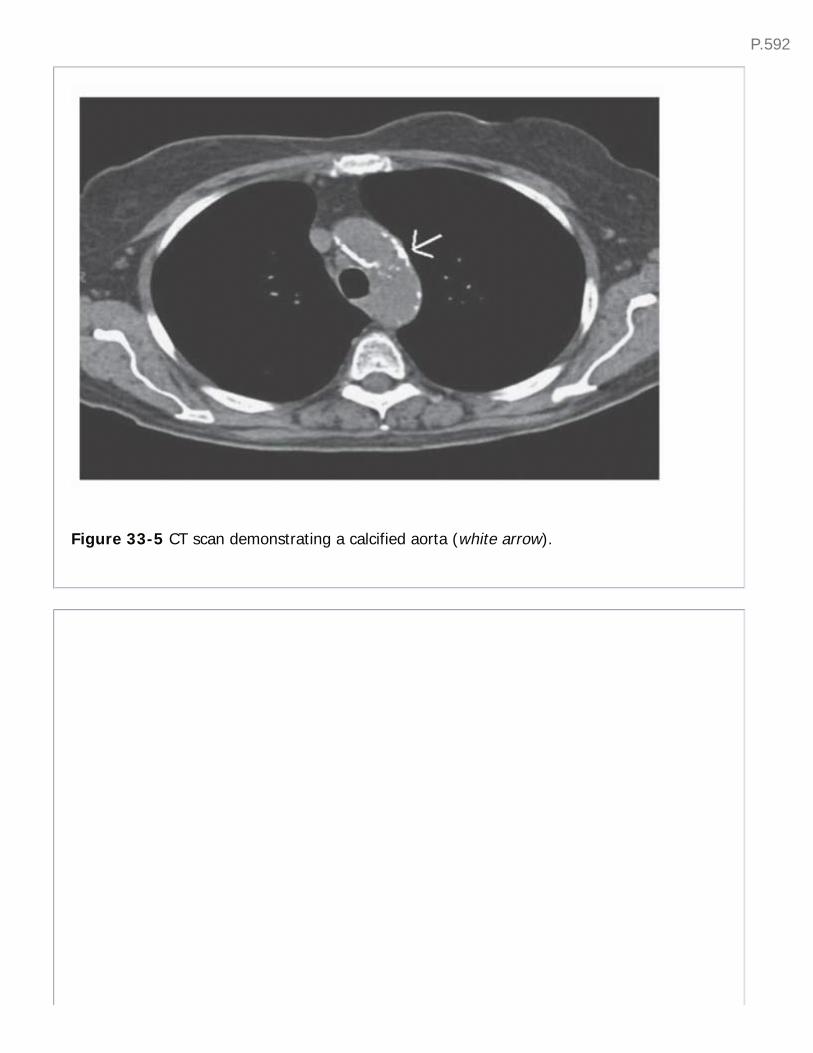
P.592
Figure 33-5 CT scan demonstrating a calcified aorta (white arrow).
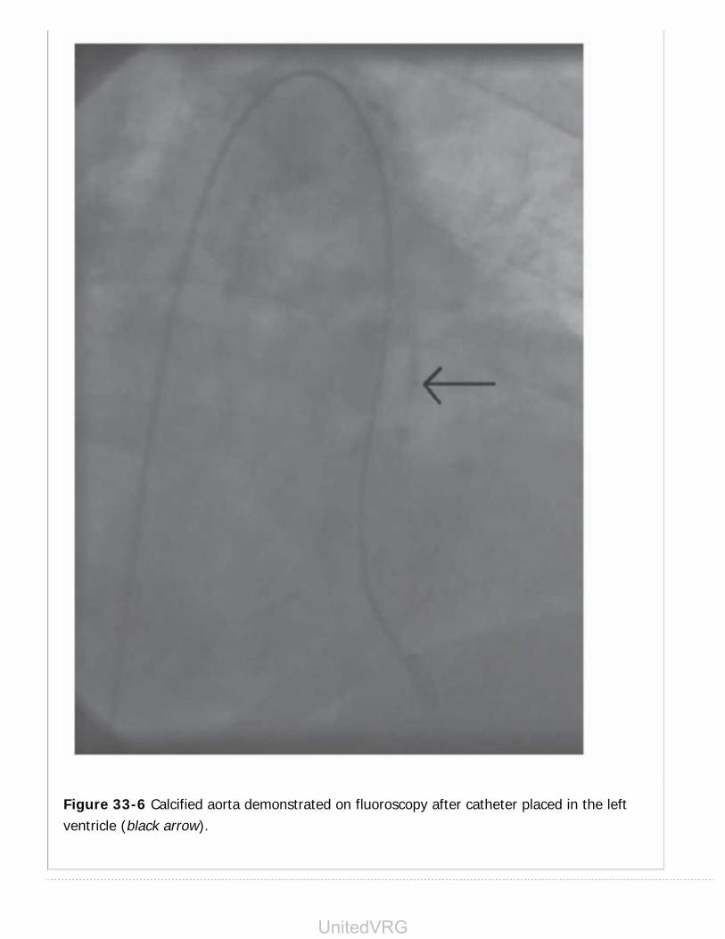
Figure 33-6 Calcified aorta demonstrated on fluoroscopy after catheter placed in the leftventricle (black arrow).
UnitedVRG

P.593
Cardiac CatheterizationNearly all patients undergoing cardiac surgery undergo cardiac catheterization to determine thepresence of coronary disease and to delineate coronary anatomy for bypass planning. Prior to theinjection of dye, calcified valve annuli and coronary arteries may be visualized. Significantlystenotic coronary arteries must be of sufficient size and have a patent, anatomically accessiblelocation in order to accept a bypass graft. Knowledge of the size of the injection catheter utilizedcan indicate the size of the vessel opacified, particularly if magnification has been utilized toproduce the images. Proposed target sites for bypass grafts must be free of significant calciumand stent material. Intramyocardial vessels may also be anticipated by viewing the injectionthrough the cine cycle. The left ventricular injection is particularly valuable as it provides anestimation of wall motion and ejection fraction (EF) as well as the size of the ascending aorta anda visual estimation of mitral valve competence. In addition, injections of the descending aorta,renal arteries, or iliac vessels provide information regarding significant peripheral vascular or renalartery occlusive disease.
Transthoracic or Transesophageal EchocardiogramTransthoracic or transesophageal echocardiogram (TEE) will provide information in theassessment of EF and valvular pathology. TEE is vital to the planning of mitral valve repair versusreplacement.
Clinical correlation: Preop TEE is considered mandatory by many surgeons prior tomitral valve surgery to determine the extent of regurgitation. The extent ofregurgitation may be underestimated intraoperatively during assessment undergeneral anesthesia.
Viability StudyIf a significantly reduced EF is noted, then an assessment of viability should be considered.Pharmacologic as well as radiologic examinations may be obtained to determine myocardialviability. Large areas of nonviable or infarcted myocardium should alert the surgeon to heavilyweigh the risk of surgery.
A computerized tomography scan of the chest or abdomen may provide valuable informationregarding calcification of the aorta, the presence or lack of a tissue plane between the heart andthe sternum in the case of redo surgery, significant lung pathology, and unexpected findings inother organs.
Conduit Selection for CABG or Concomitant Bypass during OtherCardiac SurgeryPlans for conduit use for coronary artery bypass are typically determined during the initial patient
P.594
evaluation. Considerations include the patient's age (with consideration of graft patency rates),comorbidities, and quantity and quality of vein available. Obesity, poorly controlled diabetesmellitus, emergency surgery, significant subclavian artery stenosis, and history of radiation to thechest wall may be factors indicating caution when planning the use of one or two internalmammary arteries. Factors involved in the decision to utilize the radial artery include degree ofcoronary artery stenosis (patency is reduced in noncritical lesions), the result of an Allen test(abnormal result indicates poor flow to the hand and the radial should not be harvested),
perceived patient life span, emergent nature of surgery, patient's occupation (upper extremityfine motor skills and manual dexterity may be vital and may be reduced in rare patients followingradial harvest), presence of severe diabetes or peripheral vascular disease, patient preference,and the patient's desire to avoid a visible arm scar. Preoperative venous mapping may beobtained to locate and determine the presence and size of bilateral greater and lesser saphenousveins (particularly in redo CABG cases). Preoperative lower-extremity arterial Doppler examinationmay guide vein harvest, as the risk of wound infection and poor wound healing is increased in alower extremity with peripheral vascular disease.
Left internal mammary artery patency is 94% at 10 years and its use has been shown to prolongpatient survival (J Thorac Cardiovasc Surg. 1999;117:855). Use of the left internal mammaryartery is now a Medicare quality measure. The use of two internal mammary arterieshas been documented to prolong survival compared to that of one internal mammaryartery (Ann Thorac Surg. 2004;78:2005). Patency of a free (attached to the aorta) leftinternal mammary artery graft offers the same patency of an in situ graft (stillattached at the subclavian artery). Patency of radial artery grafts is approximately 80% at 10years and that of saphenous vein grafts is approximately 50% to 60% at 10 years (Ann ThorSurg. 2004;77:93; J Thorac Cardiovasc Surg. 2010;140:73). The use of a radial artery hasalso been documented to prolong survival in propensity matched studies (Circulation.2003;108:1350-1354).
Timing of SurgeryPreoperative hemodynamic instability and/or unrelenting unstable angina often prompt urgent oremergent surgery. In stable patients, factors for consideration include the time since cardiaccatheterization, the administration of nephrotoxic medications or agents, and the baselinecreatinine due to the risk of postoperative renal dysfunction. Caution should be taken in thestable patient with an acute MI in the setting of poor left ventricular function.
Systemic illnesses and preoperative antithrombotic therapy may also indicate a need for furtherworkup or delay in elective surgery. The Report from the STS Workforce on Evidence BasedSurgery states that it is reasonable to discontinue clopidogrel for 5 to 7 days before CABG due tothe increased risk of bleeding and that patients on glycoprotein IIb/IIIa inhibitors should beconsidered high risk for bleeding depending on the half-life of the agent administered (Ann
UnitedVRG

P.595
Thorac Surg. 2012;94:1761-1781).
Operative PlanCareful planning of the procedure in advance will equip the surgeon with the opportunity toprovide a calm and systematic resolution to unexpected operative findings. Prior to surgery, aÒback up planÓ should be considered in the event that the procedure does not proceed asplanned. For example, the discovery of a porcelain aorta or a significantly dilated ascending aortawould quickly warrant a change in the operative plan.
III. CARDIOPULMONARY BYPASS.Extracorporeal circulation, in the form of CPB, allows for the performance of cardiothoracicsurgery in a quiet, bloodless field. Technologic advances have made the use of CPB readilyavailable and
technically feasible, thereby reducing the morbidity and mortality of cardiac surgery in the currentera.
Figure 33-7 Cardiopulmonary bypass circuit. (Reprinted with permission from Punjabi PP

P.596
and Taylor KM. The science and practice of cardiopulmonary bypass: From cross circulationto ECMO and SIRS. Glob Cardiol Sci Pract. 2013;2013(3):249-260.)
The CPB circuit consists of a venous cannula, a venous line (tubing), a reservoir, a heatexchanger, a membrane oxygenator, an air vent, an arterial line (tubing), and the arterialcannula. The venous reservoir stores the blood volume that drains by gravity and allows for theescape of air via the reservoir. All pump suction devices also drain to this reservoir. A membraneoxygenator performs gas exchange. A heat exchanger regulates patient temperature. Thearterial pump is usually a roller pump and requires calibration to ensure accurate flow (Fig. 33-7). This serves to pump the blood volume back to the patient via the arterial cannula.
The use of CPB introduces the blood to nonendothelial surfaces that result in detrimental changesmanifested throughout the body. The mechanisms responsible include a generalized low flowstate, relative ischemia and reperfusion injury, exposure to anticoagulation and its reversal,hemodilution and the destruction of the blood constituents, the embolization of gas or particulatematter, the placement of intravascular cannulae, and the activation of a systemic inflammatoryresponse. Complications directly related to the CPB machine include power interruption, airintroduction via physical jarring of the machine or circuit manipulation, oxygenator failure,dislodgement of a connection, pump head reversal, tubing rupture, improper occlusion, andstopcock dislodgement
or turning. It may be difficult to ascertain what portion of observed complications are attributedto CPB alone; however, it is clear that prolonged CPB time is a significant risk factor for allpostoperative complications.
IV. SURGICAL PROCEDURES AND INDICATIONS FORSURGERYA. Coronary Artery Bypass Grafting (CABG). Coronary artery disease (CAD) is the No. 1cause of death in men and women in the United States and in most developed countries. Riskfactors for CAD include tobacco abuse (including passive exposure to smoke), hypertension,diabetes mellitus, hyperlipidemia, obesity, sedentary lifestyle, advanced age, rheumatoid arthritisor lupus, and a family history of premature CAD in a first-degree relative. Risk factors unique towomen in include preeclampsia, gestational diabetes, and pregnancy-induced hypertension (J AmColl Cardiol. 2012;59:1663). Patients may present with angina pectoris, MI, or chronic ischemiccardiomyopathy.
Angina pectoris, or chest pain, occurs when reversible myocardial ischemia is present withoutcellular necrosis. Angina may manifest as a pain or pressure that often radiates to the leftshoulder and down the left arm or into the neck. An anginal equivalent may manifest asepigastric discomfort or shortness of breath. Angina occurs during times of increased myocardial
UnitedVRG
P.597
oxygen demand (exercise) and typically resolves with rest or the administration of nitrates.Unstable angina refers to chest pain that occurs at rest or with increasing frequency, duration, orseverity. MI results when there is a lack of myocardial oxygen supply with irreversible muscleinjury and cell death. Increases in cardiac specific enzymes and ECG changes (ST-segmentelevation, T-wave inversions, and new Q waves) are observed. Early and late sequelae of MI caninclude arrhythmias, congestive heart failure (CHF), VSD, papillary muscle rupture, left ventricularaneurysm, and ischemic cardiomyopathy.
Clinical correlation: Continued angina following MI warrants consideration for urgentor emergent CABG. Medical optimization includes nitrates, intravenous heparin, andplacement of an IABP prior to revascularization.
Arrhythmias (potentially fatal ventricular arrhythmias, atrial fibrillation [AF], atrial flutter, heartblock, or junctional rhythm) are common during the first 24 hours after acute MI.
CHF may result when a large portion of the left ventricle is infarcted. The extent to which thepatient's activity is limited can be graded according to the New York Heart Association (NYHA)classification: Class I, no symptoms; class II, symptoms with heavy exertion; class III, symptomswith mild exertion; class IV, symptoms at rest.
VSD occurs in approximately 2% of patients after MI (anterior wall in 60%, inferior wall in 40%).An acute VSD occurs in the anterior or posterior septum depending upon the location of thecoronary artery occlusion and coronary artery dominance (right or left) between 5 days and 2weeks or more after an acute MI.
Clinical correlation: The onset of a new postinfarction VSD is noted when a newholosystolic murmur is found on physical examination with hemodynamicdeterioration (differential diagnosis also includes
papillary muscle rupture as noted below). Diagnosis is made by cardiac cath (oxygen stepup in the right heart and left to right shunt noted on left ventriculogram) and by TEE. Treatmentis intra-aortic balloon pump (IABP) placement and emergent surgery.
Papillary muscle rupture with severe mitral regurgitation (MR) results when MI involves a papillarymuscle and often the adjacent myocardial wall.
Clinical correlation: Acute papillary muscle rupture is noted when a new holosystolicmurmur is found on physical examination with hemodynamic deterioration(differential diagnosis also includes VSD as noted above). Diagnosis is made byechocardiography and treatment includes IABP and emergent surgery.
Left ventricular aneurysm occurs late following MI. A well-defined fibrous scar that is dyskineticdevelops in 5% to 10% of patients in the infarcted territory. Large dyskinetic left ventricularaneurysms reduce left ventricular EF, result in symptoms of CHF, serve as the substrate forischemic reentrant ventricular arrhythmias, and create an area of stagnant blood that may lead to

P.598
thrombus and peripheral emboli.
Clinical correlation: Akinetic left ventricular walls (which are related to ischemicnontransmural infarcted or hibernating myocardium) must be differentiated fromdyskinetic (where blood moves in the opposite direction of normal blood flow) in trueleft ventricular aneurysms. Left ventricular aneurysmectomy (DOR procedure) may beindicated in appropriate patients who may benefit from the removal of the dyskineticwalls.
Ischemic cardiomyopathy may develop after multiple MIs and is associated with signs andsymptoms of heart failure. Treatment is optimal medical management, placement of an automaticimplantable cardioverter defibrillator for prevention of sudden cardiac death in patients withsignificantly reduced EF, and consideration of other support (see section on heart failure).
Indications for CABGMyocardial revascularization may be accomplished via percutaneous coronary intervention (PCI)(percutaneous transluminal coronary angioplasty, stent, atherectomy) or CABG. Goals are torelieve angina, prevent repeat revascularization and MI, and to prolong survival. The AmericanHeart Association (AHA) and American College of Cardiology (ACC) have established guidelinesfor CABG and PCI (Circulation. 2011;124:e652). Current guidelines recommend that CABG beperformed to improve survival in patients with significant (≥50% stenosis) left main coronaryartery stenosis, in patients with significant (≥70% stenosis) in three major coronary arteries or inthe proximal LAD plus one other major artery, in patients with significant stenosis in two majorcoronary arteries with severe or extensive myocardial ischemia or target vessels supplying a largearea of viable myocardium, in patients with LV dysfunction (EF, 35% to 50%) and significantstenosis when viable myocardium in the area of intended revascularization, in patients withsignificant stenosis in the proximal LAD and evidence of extensive ischemia, in patients withcomplex three-vessel CAD with or without involvement of the LAD who are good candidates forsurgery, and in patients with multivessel CAD with diabetes mellitus.
CABG versus Medical TherapyCABG offers symptom relief, reduced risk of MI, less repeat revascularization, survival advantageand improvement in functional status over medical therapy in patients with LMCA disease, multi-vessel disease, and those with left ventricular dysfunction (Circulation. 1979;60:888; Lancet.1982;2:1173; Circulation. 1983;68:939; N Engl J Med. 1984;311:1333; N Engl J Med.1988;319:332; Circulation. 1990;82:1629; J Am Coll Cardiol. 2004;43:1743).
CABG versus PCIThe SYNTAX trial, the largest, prospective randomized controlled trial, compared CABG to PCI(with drug-eluting stents) in patients with untreated left main or three-vessel CAD. At 1 year, PCI
UnitedVRG

P.599
was associated with higher rates of major adverse cardiac or cerebrovascular events (MACCE)(17.8% PCI vs. 12.4% CABG, p = 0.002) and higher rates of repeat revascularization (13.5% PCIvs. 5.9% CABG, p<0.001) but CABG was associated with higher rates of stroke. By 3 years, therewere no statistically significant differences in stroke rates between groups (2.9% CABG vs. 2.6%PCI, p = 0.64). The 5-year results demonstrated persistent divergence in all event rates exceptstroke in patients with three-vessel CAD (MACCE 37.5% PCI vs. 24.2% CABG, p<001; all causedeath 14.6% PCI vs. 9.2% CABG, p = 0.006; MI 10.6% PCI vs. 3.3% CABG, p<0.001; repeatrevascularization 25.4% PCI vs. 12.6% CABG, p<0.001; and stroke 3.0% PCI vs. 3.4% CABG, p= 0.66) (Eur Heart J. 2014;35:2821).
Contraindications for CABGCABG results in initial elimination of angina in more than 90% of patients. Perioperative mortalityranges from 1% to 2% in low-risk patients to more than 10% to 15% in high-risk patients. Therisks and benefits of the procedure must be individualized for each patient before proceeding withsurgery.
Clinical correlation: Questions to consider when evaluating a patient for CABGinclude:
1. Does the patient have suitable conduit and targets for the procedure?
2. Are there any particular anatomic issues that that may either prevent the procedure or increasethe complexity to an unacceptably high level (i.e., porcelain aorta, mediastinal radiation)?
3. Do the expected perioperative risks outweigh the potential benefit of the procedure? Considerfactors that may be prohibitive (preexisting stroke, frailty, dementia, severe COPD, bleedingdyscrasias, malnutrition, or liver insufficiency).
Minimally invasive CABGMinimally invasive approaches for CABG surgery include off pump (OPCAB), robotic or endoscopicCABG, hybrid CABG, (includes CABG to LAD and DES placed to other vessels), and CABG withonly a left anterior thoracotomy for bypass of the LAD.
OPCABG is technically more challenging and is performed on the beating heart with the use ofsuction stabilizers or other techniques. OPCAB has been shown to decrease morbidity (J ThoracCardiovasc Surg. 2003;125:797; BMJ. 2006;332:1365), while others have reported reduced long-term survival (J Thorac
Cardiovasc Surg. 2014;148:5). Graft patency rates following OPCAB vary between studies (JAMA.2004;291:1841-1849; Br Med J. 2006;332:1365; J Thorac Cardiovasc Surg. 2009;137:295; N EnglJ Med. 2009;361:1827-1837). Fewer blood transfusions, shorter hospital length of stay, shorterduration of intubation, reduced pulmonary complications, and reduced cost have beenconsistently associated with off pump CABG in randomized trials (Chest. 2005;127:892; B Med J.
P.600
2006;332:1365; J Card Surg. 2006;21:35). Guidelines from the International Society of MinimallyInvasive Cardiac Surgery (ISMICS) have been published and will be updated in 2015(Innovations. 2012;7:229). Patients at high risk (severe atheromatous aortic plaque, renal failure,or the elderly) may derive the greatest benefit from off pump CABG.
Clinical correlation: Appropriate patient selection is important. Considerations includesurgeon experience and learning curve, size, and disease of target vessels, location ofcoronary stenoses, and need for Òno-touchÓ aortic technique in the case of aporcelain aorta (Fig. 33-5).
B. Valvular Heart Disease
1. Aortic valve disease
a. Aortic stenosis (AS) may result from senile degeneration and calcification of a normal valve,a congenitally bicuspid aortic valve, or an abnormal valve (rheumatic disease). The degree ofvalvular stenosis is graded using echocardiography from mild to severe. AS places a pressureoverload on the left ventricle, resulting in left ventricular hypertrophy. Symptoms include chestpain or angina, shortness of breath or CHF, and syncope.
Clinical correlation: Physical examination findings include a loud systolic murmuracross the precordium and a murmur audible in the carotid arteries.
Aortic valve replacement provides relief of symptoms as well as a survival benefit in patients with
symptomatic severe (AVA <1 cm2) AS compared to medical management alone (Circulation.1982;66:1105; J Thorac Cardiovasc Surg. 2014;64:1763). Aortic valve replacement is alsoindicated in asymptomatic patients with severe AS undergoing CABG or other cardiac surgery andin patients with severe AS and left ventricular systolic dysfunction (EF < 0.50) (J ThoracCardiovasc Surg. 2014;64:1763).
Clinical correlation: Patients who undergo surgical aortic valve replacement oftenhave significant left ventricular hypertrophy. Because of this hypertrophy, initialconsideration of treatment of hypotension should include volume resuscitation ratherthan vasoconstrictor agents.
b. Aortic insufficiency (AI) is often the result of valve leaflet pathology resulting in thickening,calcification and fixation (rheumatic heart disease or combined with AS), leaflet redundancy ordestruction (myxomatous degeneration), maladaptation (aortic root dilatation, aortic dissection),inflammatory disease (ankylosing spondylitis), trauma (blunt chest injury, balloon dilatation, ortransaortic valve implantation), or destruction (endocarditis). Chronic AI
results in volume overload of the left ventricle, causing chamber enlargement and wall thickeningand pulmonary congestion. Severity of insufficiency is graded from mild to severe byechocardiography. Acute AI is not well tolerated and often results in fulminant pulmonary edema,myocardial ischemia, and cardiovascular collapse.
UnitedVRG
P.601
Clinical correlation: IABP is contraindicated with severe AI as the balloon inflatesduring diastole, resulting in increased AI.
Indications for surgery in symptomatic patients include severe AI, chronic moderate to severe AIand left ventricular dysfunction (EF < 50%), and patients with chronic severe AI who areundergoing other cardiac surgery. Surgery is reasonable in patients without symptoms andnormal left ventricular function but who have severe left ventricular dilatation (left ventricular end
systolic dimension >50 mm or indexed left ventricular end systolic dimension >25 mm/m2 (JThorac Cardiovasc Surg. 2014;64:1763).
Surgical treatment may include open valve repair or replacement or alternative access approachesdepending upon the pathology, leaflet quality, and patient comorbidities. One alternative accessapproach is transcatheter aortic valve implantation (TAVI). TAVI is the replacement of the aorticvalve through a transfemoral, transaortic, or transapical approach. The first-generation devicesÑthe balloon-expandable SAPIEN valve (Edwards Lifesciences, Irvine, CA) and the self-expandable CoreValve (Medtronic, Minneapolis, MN)Ñhave been thoroughly tested in the UnitedStates by large, multicenter trials demonstrating their safety and efficacy in high-risk patients.Second-generation devices have been developed to reduce the shortcomings of the first-generation devices and are currently being tested in large, multicenter trials in the United States.
2. Mitral valve disease
a. Mitral stenosis (MS) is caused by valve leaflet thickening and calcification due to rheumaticfever or senile calcification, collagen vascular diseases, amyloidosis, congenital stenosis, or mitralinflow obstruction (tumors or masses). MS places a pressure overload on the left atrium, withrelative sparing of ventricular function. Left atrial dilation to more than 45 mm is associated witha high incidence of AF and subsequent thromboembolism. Critical MS occurs when the valve area
is 1.5 cm2 or less.
Clinical correlation: Physical examination findings include an apical diastolic murmurand a loud S1.
Symptoms usually develop late and reflect pulmonary congestion (dyspnea), reduced leftventricular preload (low-cardiac-output syndrome), or AF (thromboembolism). Surgery isindicated in patients with severe MS and severe symptoms, patients with moderate MSundergoing cardiac surgery for other indications, or in patients who have had recurrent embolicevents while receiving adequate anticoagulation and excision of the left atrial appendage may be
considered. Percutaneous mitral balloon valvuloplasty is indicated in symptomatic patients withsevere mitral rheumatic stenosis who have favorable valve morphology in the absence ofcontraindications (J Thorac Cardiovasc Surg. 2014;64:1763). b. MR may be categorized asprimary (degenerative) or chronic secondary (functional). Primary causes include those thatinvolve the leaflets (mitral valve prolapse, fibroelastic deficiency, connective tissue disorders,

P.602
rheumatic heart disease, cleft mitral valve, radiation injury, trauma following mitral valvuloplasty,or endocarditis), the chordae tendineae (rupture [endocarditis or MI], fusion, or elongation), orthe papillary muscles (ischemic papillary muscle dysfunction or rupture secondary to MI [notedunder MI]) (J Thorac Cardiovasc Surg. 2014;64:1763). Chronic secondary MR is due to eitherchronic ischemic cardiomyopathy or idiopathic myocardial disease causing secondary dilation ofthe annulus itself.
Clinical correlation: Physical examination findings include loud systolic murmurloudest at the apex.
MR places a volume overload on the left ventricle and atrium, causing chamber enlargement andwall thickening and pulmonary congestion. AF often develops due to left atrial dilation. Acutesevere MR results in pulmonary congestion and low cardiac output.
Clinical correlation: In acute, severe MR, IABP is beneficial as it decreases afterload,thereby increasing forward flow and decreasing regurgitant volume.
Mitral valve repair or replacement is recommended for symptomatic patients with chronic severeMR and left ventricular EF greater than 30%, and is recommended in asymptomatic patients withchronic severe primary MR and left ventricular dysfunction (EF 30% to 60%) (J Thorac CardiovascSurg. 2014;64:1763). Specific indications and recommendations for repair of the mitral valvecontinue to evolve (J Thorac Cardiovasc Surg. 2014;64:1763). Mitral surgery in patients withchronic ischemic MR is challenging and is associated with increased operative risk. In a recentrandomized trial, mitral valve replacement provided a more durable freedom from MR comparedto mitral repair in patients with severe ischemic MR (N Eng J Med. 2014;370:1).
3. Tricuspid valve disease
a. Tricuspid stenosis (TS) is most commonly secondary to rheumatic disease. TS may beassociated with regurgitation and may not be detectable on bedside examination (J ThoracCardiovasc Surg. 2014;64:1763). Tricuspid valve surgery is recommended in patients with severeTS at the time of operation for left-sided valve disease and in isolated symptomatic severe TS (JThorac Cardiovasc Surg. 2014;64:1763). Tricuspid balloon commissurotomy might be consideredin patients with isolated severe TS without TR (J Thorac Cardiovasc Surg. 2014;64:1763).
b. Tricuspid insufficiency (TI) most often results from a secondary (functional) dilation of thevalve annulus caused by pulmonary
hypertension, caused by intrinsic mitral or aortic valve disease. Causes of primary TI includerheumatic heart disease, bacterial endocarditis (often in IV drug users), carcinoid tumors, Ebsteinanomaly, and blunt trauma. Mild-to-moderate TI usually is well tolerated. Tricuspid repair isindicated in the case of moderate to severe TI at the time of surgery for other cardiac anomalies.The majority of tricuspid valves can be repaired with annuloplasty techniques rather thanreplacement.
UnitedVRG

P.603
Clinical correlation: TI may be associated with a systolic murmur, a prominent jugularvenous pulse, and a pulsatile liver.
Clinical correlation: The AV node is of particular importance during tricuspid valvesurgery because it may be injured resulting in heart block. Tricuspid valve repairtechniques involve incomplete rings to avoid injury to the conduction system andsutures in the annulus at the time of valve replacement are preferentially placed inthe leaflet tissue only (Fig. 33-2).
4. Pulmonic valve disease
a. Pulmonic insufficiency (PI) that is mild to moderate does not require intervention ifasymptomatic and if associated with normal right ventricular function and size (J ThoracCardiovasc Surg. 2014;64:1763). PI in adults more commonly requires treatment in patientsfollowing childhood repair of Tetralogy of Fallot. The pulmonic valve may be affected byendocarditis, tumors (carcinoid, papillary fibroelastoma), or radiation injury.
b. Pulmonic stenosis (PS) is mostly seen as a congenital disorder and is rarely seen in adults.
5. Infective endocarditis. Endocarditis may involve infection of native valves (typicallyabnormal: Elderly sclerotic disease or rheumatic valvular disease) or prosthetic valves.Endocarditis may be related to bacteremia (poor dental hygiene, intravenous drug abuse [IVDA],nosocomial infection, indwelling intravenous catheters or dialysis). Surgical treatment isassociated with increased survival compared to antibiotic therapy alone and should be aggressivein order to eradicate all infected tissues (Heart. 2001;89:269; Heart. 2001;88:61). The risk ofembolization is reduced significantly following the initiation of appropriate antibiotics. Indicationsfor surgery due to endocarditis may include hemodynamic instability, CHF, large foci of disease(>1 cm), aggressive bacterial organisms or fungal organisms, prosthetic valve endocarditis, aorticroot abscess, and persistent evidence of infection despite appropriate antibiotic therapy(persistent fever, recurrent septic emboli, persistent positive blood cultures). The risk forreoperation for recurrent infective endocarditis is about 17% for IV drug users and 5% for non-IVdrug users (Ann Thorac Surg. 2007;83:30).
Clinical correlation: Aortic root abscess is a surgical emergency. Daily ECGs should bemonitored for signs of heart block that may occur with destruction of the AV nodebetween the right and noncoronary cusps of the aortic annulus.
6. Prosthetic valve selection. Tissue bioprostheses include porcine aortic valves or valvesconstructed with bovine pericardium. These prostheses are associated with a low rate ofthromboembolism, even without longterm anticoagulation (a daily aspirin is recommended). Theyare less durable than mechanical valves (mean time to failure is approximately 10 to 15 years)and this can potentially be prolonged in newer valves due to modern preservation methods.Bioprostheses are often the preferred valves for older patients (>65 years) or patients with a

P.604
contraindication to anticoagulation.
Mechanical valves have excellent long-term durability; however, the rate of thromboemboliccomplications (0.5% to 3% per year) is cumulative and these valves require lifelonganticoagulation. Newer valve options (On-X) with alternative construction allow for a lower targetINR (J Thorac Cardiovasc Surg. 2014;147:1202).
Homograft/allograft and autografts are useful for replacement of the aortic valve, particularly inthe setting of endocarditis or for the Ross procedure (J Heart Valve Dis. 1994;3:377). Thesetissues have reasonable durability and a low incidence of thromboembolism. Their use is limiteddue to supply and technical expertise needed for implantation and to the drawbacks of long-termfunction noted in autografts in the aortic position.
7. New technology. Following the introduction of TAVI, the treatment of valvular diseasecontinues to rapidly evolve. New transcatheter therapies for the mitral valve have recently beenintroduced that allow for mitral valve repair via a transfemoral approach by clipping the twoleaflets of the mitral valve (MitraClip, Abbott Laboratories, Abbott Park, Il). Transcatheter mitralvalve replacement is also on the horizon and first-in-man trials are underway. Suture-less valveshave been designed for rapid surgical deployment. The trend toward minimally invasiveprocedures and the combination of transcatheter and surgical techniques have mandated amultidisciplinary approach to the treatment of valvular disease.
C. Hypertrophic Cardiomyopathy (HCM). HCM characterized by asymmetric hypertrophy andfibrosis of the myocardium, causing obstruction of the left ventricular outflow tract. Medicaltherapy with β-blockade or calcium channel blockade is the preferred first-line treatment.Nifedipine, nitroglycerin, angiotensin-converting enzyme inhibitors, and angiotensin II blockersare all generally contraindicated due to their vasodilatory properties, which can exacerbate theoutflow tract obstruction. Surgical treatment of HCM is myectomy, with a postoperative mortalityof 1% or less (Ann Thorac Surg. 2000;69:1732). By convention, surgery is recommended forsymptomatic patients who have failed medical therapy or septal ablation with a documented at-rest outflow tract gradient of at least 30 mm Hg.
D. Atrial Fibrillation. AF affects 1% to 2% of the general population and it affects nearly 10%of individuals >80 years of age. Morbidity includes patient discomfort, hemodynamic compromise,and thromboembolism.
Nonsurgical management of AF includes antiarrhythmic drugs, cardioversion, and catheterablation. Indications for surgery include symptomatic AF in patients undergoing other cardiacprocedures, selected asymptomatic AF patients undergoing cardiac surgery in whom ablation canbe performed with minimal risk, and for symptomatic AF patients who prefer a surgical approach,have failed one or more attempts at catheter ablation, or are not candidates for catheter ablation(Heart Rhythm. 2007;4:816). The Coxmaze procedure is the gold standard for surgical AFablation and its current iteration, the Cox-maze IV, utilizes a combination of cryoablation and
UnitedVRG
P.605
bipolar radiofrequency ablation. Postoperative freedom from AF at 93%, 90%, and 90% at 6, 12,and 24 months, respectively, has been demonstrated (J Interv Card Electrophysiol. 2011;31:47).Results have been variable when less complete surgical ablations are performed.
E. Surgical Treatment of Heart Failure. It is estimated that approximately 250,000 peoplesuffer from advanced heart failure in the United States (Curr Heart Fail Rep. 2010;7:140). Themanagement of heart failure involves aggressive medical and surgical care.
The IABP (see myocardial infarction) is used as the first-line device to provide circulatory supportin acute heart failure (Ann Thorac Surg. 1992;54:11). Indications may include preoperative low-cardiac-output states, preoperative unstable angina refractory to medical therapy, intraoperativeweaning from CPB after inotropic agents are maximized, and postoperative low cardiac outputstates. The ECG and the femoral (or aortic) pressure waveform are monitored continuously on abedside console. The device may be triggered using either the ECG or the pressure tracing forevery heartbeat (1:1) or less frequently (1:2, 1:3). With clinical improvement the IABP isgradually weaned by decreasing the augmentation frequency from 1:1 to 1:3 in steps. Placementof an IABP is not without risk and potential complications include incorrect placement resulting inabdominal organ malperfusion, perforation of the aorta, injury to the femoral artery, and ischemiaof the lower extremity. Rupture of the balloon is an indication for immediate removal due to riskof thrombosis.
Clinical correlation: Contraindications for IABP include AI, aortic dissection, andsevere peripheral vascular disease.
Percutaneous assist devices are devices that can be deployed via catheter-based approach andfunction as ventricular assist devices (VADs) without the need for open surgery. They are typicallyused only for short-term support. Examples of these include the TandemHeart, the Impella, andthe CentriMag (Expert Rev Cardiovasc Ther. 2010;8:1247).
Cardiac resynchronization therapy (biventricular pacing) has emerged as a valid treatmentmodality for patients with heart failure and concomitant intraventricular conduction delaymanifested by QRS complex greater than 120 milliseconds.
Extracorporeal membrane oxygenation (ECMO) is a temporary circulatory support that isnonphysiologic and usually nonpulsatile. It consists of a centrifugal pump and an oxygenator;thus, anticoagulation is necessary. Most commonly, it is used to allow patients to recover fromreversible myocardial
dysfunction (stunning), adult respiratory distress syndrome, or pulmonary insufficiency of variousetiologies.
VADs may be used to support the left (LVAD) or the right ventricle (RVAD). When both an LVADand an RVAD are used, the combination is termed a biventricular assist device (BiVAD). Theyprovide mechanical decompression of the ventricle and restoration of cardiac output, resulting in
P.606
decreased myocardial oxygen consumption. Indications include three separate broad categories:Inability to separate from CPB despite inotropic and IABP support (Òbridge to recoveryÓ),intermediate-term cardiac support (Òbridge to transplantÓ), and permanent replacement therapyin nontransplant candidates (Òdestination therapyÓ) (J Heart Lung Transplant. 2013;32:1147-1162).
VAD subtypes include nonpulsatile devices (centrifugal or axial flow pumps) and pulsatile devices(external and long-term implantable devices). Device selection depends on surgeon familiarityand preference, practical advantages and disadvantages of each device, and unique patientcharacteristics and body habitus. Devices currently in use include the HeartMate II (ThoratecCorp., Pleasanton, CA), which is a second-generation nonpulsatile axial flow continuous flow VAD,and the HVAD (Heartware, Inc., Framingham, MA), which is a third-generation nonpulsatilecentrifugal continuous flow VAD.
Following VAD placement, the activated clotting time should be maintained at approximately 200seconds once postoperative bleeding is acceptable. Factors that affect a low-flow-state statuspost LVAD include right ventricular dysfunction, pulmonary hypertension, hypovolemia, andtamponade. Pulmonary vasodilators (nitric oxide or inhaled prostacyclin) are frequently used tolower pulmonary vascular resistance and load on the right ventricle. Complications includebleeding, thrombus formation, embolization, hemolysis, infection at the drive line site, and devicefailure.
Cardiac transplantation can provide relief from symptoms in patients with end-stagecardiomyopathy who are functionally incapacitated despite optimal medical therapy and who arenot candidates for other procedures. Heart transplantation is typically utilized in patients withischemic, idiopathic, postpartum, or chemotherapy-induced cardiomyopathy and is associatedwith a 1-year survival of 84.5% and a 5-year survival of 72.5% (J Heart Lung Transplant.2014;33:996-1008). Relative indications include refractory cardiogenic shock, instability in fluidbalance or renal function despite optimal medical therapy, severe persistent angina not amenableto revascularization, markedly reduced exercise capacity (peak VO2 <10 to 14 mL/kg/minute),
and recurrent refractory ventricular arrhythmias (Mayo Clin Proc. 2014;89:662).
Relative contraindications to transplantation include age older than 65 years, irreversiblepulmonary hypertension, active infection or malignancy, recent pulmonary embolus, andexcessive comorbidity (renal dysfunction, hepatic dysfunction, systemic disease such asamyloidosis, significant peripheral vascular disease, active peptic ulcer disease, uncontrolleddiabetes mellitus, morbid obesity, mental illness, active substance abuse,
inadequate social support, or psychosocial instability) (Mayo Clin Proc. 2014;89:662).
Complications of heart transplant include acute and chronic rejection that may be diagnosed byendomyocardial biopsy or echocardiography. Coronary artery vasculopathy (CAV), thought torepresent chronic vascular rejection, occurs in a significant percentage of cardiac transplant
UnitedVRG
recipients and is a major limitation on the long-term success of cardiac transplantation, beingresponsible for 30% of deaths in transplanted patients after 5 years (J Heart Lung Transplant.2007;26:769). CAV is usually not amenable to conventional revascularization owing to small-vessel, nonfocal disease and often requires retransplantation.
V. POSTOPERATIVE MANAGEMENT AND COMPLICATIONSA. Monitoring and Management. Early extubation following cardiac surgery ispreferred if the patient is hemodynamically stable and bleeding is minor.Neurologic assessment is obtained as soon as possible. Normal hemodynamicvalues obtained from a Swan-Ganz catheter are listed in Table 33-2. A cardiac
index of 2 L/minute/m2 is generally a minimum acceptable value. A mixed-venous oxygen saturation of less than 60% suggests inadequate peripheraltissue perfusion and increased peripheral oxygen extraction. Etiologies of lowcardiac output must be aggressively corrected by manipulation of factorsaffecting cardiac output (heart rate, preload, afterload, and contractility).Myocardial stunning is common following heart surgery and requires additionalsupport (pharmacologic and mechanical). Stunning is defined as a transientpostischemic myocardial dysfunction that persists despite adequate reperfusionand in the absence of irreversible damage (Circulation. 1982;66:1146). One ofthe easiest methods to increase cardiac output is to increase the heart rate.This may be accomplished by using the temporary epicardial pacing electrodes(placed at the time of operation) at 80 to 100 beats/minute. Optimal pacingalways involves maintaining AV synchrony.
B. Complications. Complications related to CPB were detailed in thecardiopulmonary bypass section.
Arrhythmias are common following cardiac surgery. Supraventriculararrhythmias (AF, atrial flutter, atrial tachycardia) are most common and areassociated with an increased risk of transient or permanent neurologic deficits (JCard Surg. 2005;20:425). Postoperative AF occurs in approximately 30% ofpatients and has a peak incidence on postoperative day 2 (J Thorac CardiovascSurg. 2011;141:559). Supraventricular arrhythmias with hemodynamiccompromise can rapidly be treated with electrical cardioversion (with 50 to 100J). For patients with hemodynamic stability and atrial flutter, overdrive pacingmay be used to terminate the arrhythmia. For AF and flutter, ventricular ratecontrol may be facilitated with β-blockade and amiodarone. AF or flutter thatpersists beyond 12 hours or that is recurrent despite amiodarone,anticoagulation (IV heparin as a bridge to oral Coumadin with a goal INR of 2.0to 3.0) should be considered to avoid the complication of stroke. Sustainedventricular arrhythmias other than

P.607premature ventricular contractions suggest underlying ischemic pathology andetiology should be investigated.
TABLE 33-2 Normal Hemodynamic Parameters
Parameter Normal Value Unit
Central venous pressure 2-8 mm Hg
Right ventricular pressure (syst/diast) 15-30/2-8 mm Hg
Pulmonary artery pressure (syst/diast) 15-30/4-12 mm Hg
Pulmonary capillary wedge pressure 2-15 mm Hg
Left ventricular pressure (syst/diast) 100-140/3-12 mm Hg
Cardiac output 3.5-5.5 L/min
Cardiac index 2-4 L/min/m2 BSA
Pulmonary vascular resistance 20-130 dynes sec/cm5
Systemic vascular resistance 700-1,600 dynes sec/cm5
Mixed-venous oxygen saturation 65-75 Percent
BSA, body-surface area; diast, diastolic; syst, systolic
Respiratory failure may occur following cardiac surgery. A prompt postoperativetrial of extubation should be attempted along with the limitation of sedation,tracheostomy, and early nutritional support. Appropriate antibiotics are
UnitedVRG
P.608P.609
administered for ventilator-associated pneumonia.
Postoperative bleeding is relatively common after cardiac surgery andnecessitates reexploration in up to 5% of patients. A treatment algorithm forthe management of postoperative bleeding is represented in Figure 33-8.Cardiac tamponade is a potentially lethal cause of low cardiac output withassociated narrowed pulse pressure, increased jugular venous distention, risingCV, muffled heart sounds, pulsus paradoxus, widened mediastinal silhouette onchest radiograph, and decreased urine output. Diagnosis is clinical and may beconfirmed by echocardiography. Treatment is emergent drainage in theoperating room. Delayed tamponade occurs after the acute postoperative periodand may be extremely difficult to diagnose. A high index of suspicion for thisdiagnosis must be maintained. A decline in renal or liver function, poor urineoutput, or an elevated coagulation profile may be the initial clue. Anechocardiogram is helpful in making the diagnosis.
This complication must be rapidly treated with reexploration of the chest andevacuation of mediastinal fluid.
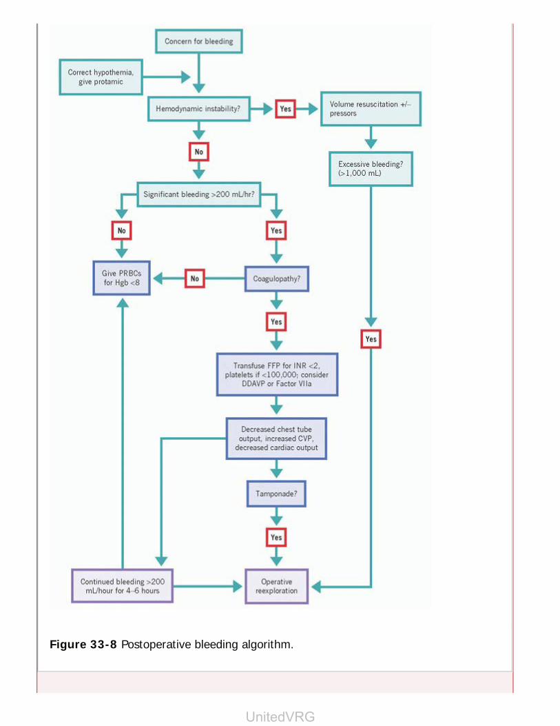
Figure 33-8 Postoperative bleeding algorithm.
UnitedVRG
Perioperative MI occurs in approximately 1% to 2% of patients and can bediagnosed by chest pain, ventricular arrhythmias, ECG changes, elevatedcardiac specific enzymes, or new changes on echocardiography. Newlyconstructed bypass grafts can be occluded due to thrombosis or spasm. Cardiaccatheterization can delineate appropriate treatment, which may include returnto the operating room or stenting for myocardial revascularization.
Renal dysfunction in the postoperative period significantly increases mortality.To limit renal injury, an acceptable cardiac index and mean arterial pressuremust be maintained. Nephrotoxic agents should be avoided or limited, and anyrenally excreted medication should have dose adjustment.
Cerebrovascular accident (CVA) is the most devastating of neurologiccomplications following cardiac surgery. Neurologic complications may bemanifested by a wide range of signs and symptoms from delirium and confusionto permanent stroke and may be related to intraoperative (aortic atheroscleroticor air emboli) or perioperative events. An acceptable cardiac output and meanarterial pressure must be maintained. Patients with postoperative AF ordocumented poor EF with intracardiac clot should be anticoagulated to preventcerebral embolization.
Postoperative infection may be clinically significant in the sternal wound, urinarytract, conduit harvest sites, or lungs. Perioperative intravenous antibiotics areadministered prior to incision and continued for only 48 hours postoperatively.Prophylaxis with a second-generation cephalosporin has been associated with afivefold decrease in wound infection rates compared to placebo in a meta-analysis (J Thorac Cardiovasc Surg. 1992;104:590). Additional antibiotic useshould be guided by specific culture and sensitivity results. In addition,aggressive blood sugar management (often with insulin infusion) is utilized tolimit the occurrence of sternal wound infection. Central lines and chest tubesare promptly discontinued when no longer necessary or useful. In addition,other sites (conduit harvest sites, urinary tract, and lungs) are closelymonitored and promptly treated should an infection occur.
Serous drainage from the sternal incision is worrisome and should be treated byapplication of a sterile dressing twice daily and the administration of IV or oralantibiotics. Purulent wound drainage, a sternal click, gross movement of thesternal edges, or substernal air on the chest x-ray may indicate a deep sternalinfection. A CT scan of the chest can confirm this diagnosis. Deep sternalinfections require operative debridement of devitalized sternal and soft tissue,administration of broad-spectrum IV antibiotics, and muscle flap closure of thesoft tissue defects.
Gastrointestinal (GI) complications are uncommon after cardiac surgical

P.610
P.611
procedures. Prompt diagnosis and treatment requires a high index of suspicion.GI ischemia may occur following periods of hypoperfusion due to low cardiacoutput, hypotension from blood loss, or as a result of vascular
emboli. GI bleeding may be exacerbated by the use of antiplatelet agents andanticoagulation. Adequate gastric mucosal protection should be provided withproton pump inhibitors to prevent stress ulceration. In addition, early enteralfeeding should be initiated in the case of prolonged ventilation. Acutecholecystitis, usually acalculous, is associated with a high mortality. Antibioticsshould be discontinued promptly when they are no longer clinically indicated toprevent Clostridium difficile colitis.
CHAPTER 33: CARDIAC SURGERYMultiple Choice Questions1. A 75-year-old female underwent coronary bypass grafting with fourgrafts and returns to the ICU in stable condition on no pressors. Uponarrival to the ICU, the patient is noted to have a cardiac index of 1.7 butis normotensive and there is no evidence of bleeding. The admissionECG is unchanged, an echo shows no effusion, the CVP is found to be 20mm Hg, and the admission laboratory values are unremarkable. What isthe next best step in management?
a. Transfuse 1 unit of blood
b. Start Dobutamine
c. Bolus 1 L of crystalloid
d. Place an intra-aortic balloon pump
e. Return to the operating room for exploration
View Answer
2. A 60-year-old male returns to the ICU after a redo aortic valvereplacement. In the first 10 minutes after the arrival the ICU, thepatient has 150 mL of sanguineous output from his mediastinal chesttubes, but is hemodynamically stable. What is the best next step inmanagement?
a. Return to the operating room for exploration
b. Re-open the chest in the ICU
c. Transfuse platelets
d. Transfuse FFP
UnitedVRG

P.612
e. Place a warmer on the patient and give protamine
View Answer
3. A 55-year-old male comes to the emergency room with severe chestpain and is diagnosed with an ST-elevation myocardial infarction. Onthe way to the cath laboratory, he becomes hypoxic and has to beintubated. He undergoes left heart cath and he is found to have 95%left main and 99% RCA lesions, as well as an EF of 10% onventriculogram. The patient is then referred to a cardiac surgeon for aCABG, and the surgeon asks the interventionalist to place an IABP.Which of the following findings on echo would be a contraindication toplacement of IABP?
a. Aortic stenosis
b. Aortic insufficiency
c. Mitral stenosis
d. Mitral insufficiency
e. Tricuspid insufficiency
View Answer
4. Which of the following patients should be referred for CABG?
a. A 48-year-old male with an NSTEMI and 70% mid left anteriordescending artery lesion and otherwise clean coronaries.
b. A 60-year-old female with unstable angina and a failed dobutaminestress test.
c. A 55-year-old male with chronic CAD s/p multiple PCI who presentswith angina and on a cath has a chronically occluded right coronary, 40%in-stent stenosis of the LAD.
d. A 52-year-old male with a STEMI and 90% proximal LAD and 90%proximal RCA lesions.
e. A 78-year-old s/p cardiac arrest and 30 minutes of CPR in the field whohad a cath on arrival that showed 90% LAD, 95% left main coronary, and95% right main coronary lesions, and a porcelain aorta.
View Answer
5. A 59-year-old male with a history of CAD s/p CABGx4, 10 years agocomes to the hospital complaining of shortness of breath. He isadmitted to the ICU where an echo shows an EF of 10% and a left heart
cath shows patent grafts to RCA and LAD, but otherwise diffuse diseasethroughout the left and right coronary systems. A balloon pump isplaced, he is placed on a lasix drip, and high doses of inotropes, but hedoes not improve. Regarding cardiac transplantation, which of thefollowing is true?
a. His age of 59 years precludes him from being listed for transplant.
b. Acute renal failure would be a relative contraindication for transplant.
c. If he were to get an LVAD, he no longer would be a transplantcandidate.
d. If he were to get pneumonia, he would no longer be a transplantcandidate until the infection is treated.
e. Ventricular tachycardia would preclude him from transplant.
View Answer
UnitedVRG

P.614
34Lung and Mediastinal Diseases
Stephanie H. Chang
Varun Puri
I. PNEUMOTHORAXA. Pneumothorax is the presence of air in the pleural cavity, leading to separation of thevisceral and parietal pleura. This results in abnormal pulmonary mechanics that can progress totension pneumothorax, which causes cardiac compromise and is a true emergency.Pneumothoraces may be spontaneous, iatrogenic, or due to trauma.
1. Spontaneous pneumothoraces are usually caused by the rupture of an apical bleb. Thetypical patients are tall young males who present with acute shortness of breath and chest pain.Older patients usually have significant parenchymal disease, such as emphysema. These patientspresent with a ruptured bulla and can have a more dramatic presentation, including tachypnea,cyanosis, and hypoxia. Other etiologies of spontaneous pneumothorax include cystic fibrosis (CF)and, rarely, lung cancer. The risk of ipsilateral recurrence of a spontaneous pneumothorax is50%, 62%, and 80% after the first, second, and third episodes, respectively, if managedconservatively without surgery.
2. Iatrogenic pneumothoraces are usually the result of pleural injury during central venousaccess attempts, pacemaker placement, or transthoracic or transbronchial lung biopsy.
3. Traumatic pneumothoraces will be discussed in Chapter 11 (Chest Trauma).
B. Physical examination demonstrates decreased breath sounds on the involved side.Examination for signs of tension pneumothorax, including deviation of the trachea to the oppositeside, respiratory distress, and hypotension, must be performed. An upright chest x-ray (CXR) canestablish the diagnosis of pneumothorax (Fig. 34-1); however, confirming the diagnosis of tensionpneumothorax is unnecessary and leads to dangerous delays. Smaller pneumothoraces may onlybe evident on expiratory CXRs or CT scan.
C. Management options include observation, aspiration, needle thoracostomy, chest tubeplacement with or without pleurodesis, and surgery.
1. Observation is an option in a healthy, asymptomatic patient with a small pneumothorax.Supplemental oxygen may help to reabsorb the pneumothorax by affecting the gradient ofnitrogen in the pneumothorax.
> Table of Contents > 34 - Lung and Mediastinal Diseases
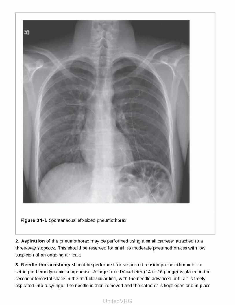
Figure 34-1 Spontaneous left-sided pneumothorax.
2. Aspiration of the pneumothorax may be performed using a small catheter attached to athree-way stopcock. This should be reserved for small to moderate pneumothoraces with lowsuspicion of an ongoing air leak.
3. Needle thoracostomy should be performed for suspected tension pneumothorax in thesetting of hemodynamic compromise. A large-bore IV catheter (14 to 16 gauge) is placed in thesecond intercostal space in the mid-clavicular line, with the needle advanced until air is freelyaspirated into a syringe. The needle is then removed and the catheter is kept open and in place
UnitedVRG

P.615
until a formal chest tube is expeditiously placed.
4. Percutaneous catheters are essentially mini chest tubes that may be placed using Seldingertechnique. Multiple commercial kits exist and allow for the catheter to be placed to a water sealor suction. The catheters are of small caliber, so use is limited to situations of simplepneumothorax. If there is concern for lung adhesions, bedside percutaneous catheters should beavoided.
5. Tube thoracostomy remains the gold standard for large pneumothoraces, associatedeffusion, or when there is an expected need for pleurodesis. Chest tubes may be connected to aHeimlich one-way valve, a
simple underwater-seal system, or to vacuum suction (typically -20 cm). If the water-sealchamber bubbles with expiration or with coughing, it is evident that an air leak persists.
6. Bedside pleurodesis. Sclerosing agents may be administered through the chest tube toinduce fusion of the parietal and visceral pleural surfaces. Doxycycline, bleomycin, and talc haveall been described. Bedside pleurodesis can be associated with an inflammatory pneumonitis inthe lung on the treated side. In patients with limited pulmonary reserve, this may present asclinically significant hypoxia.
7. Surgery is performed using a video-assisted thoracoscopic surgery (VATS) or rarely viathoracotomy.
a. Indications for operation for pneumothorax include
(1) Recurrent spontaneous ipsilateral pneumothoraces
(2) Bilateral pneumothoraces
(3) Persistent air leaks on chest tube suction (usually >3 to 5 days)
(4) First spontaneous pneumothorax in patients with high-risk occupations (e.g., pilots anddivers) or live at a great distance from medical facilities
b. Operative management consists of stapled wedge resection of blebs or bullae, usuallyfound in the apex of the upper lobe or superior segment of the lower lobe. Pleural abrasion(pleurodesis) should be performed to promote formation of adhesions between visceral andparietal pleurae. In older patients, intraoperative talc insufflation in the pleural space providesreliable pleurodesis.
II. PLEURAL EFFUSIONA. Pleural effusions are buildup of fluid in the pleural space and may result from a widespectrum of benign, malignant, and inflammatory conditions. They are broadly categorized aseither transudative (protein-poor fluid from increased intravascular pressure) or exudative(protein or cell rich fluid resulting from increased vascular permeability).
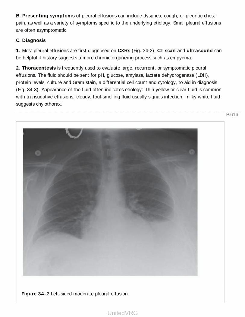
P.616
B. Presenting symptoms of pleural effusions can include dyspnea, cough, or pleuritic chestpain, as well as a variety of symptoms specific to the underlying etiology. Small pleural effusionsare often asymptomatic.
C. Diagnosis
1. Most pleural effusions are first diagnosed on CXRs (Fig. 34-2). CT scan and ultrasound canbe helpful if history suggests a more chronic organizing process such as empyema.
2. Thoracentesis is frequently used to evaluate large, recurrent, or symptomatic pleuraleffusions. The fluid should be sent for pH, glucose, amylase, lactate dehydrogenase (LDH),protein levels, culture and Gram stain, a differential cell count and cytology, to aid in diagnosis(Fig. 34-3). Appearance of the fluid often indicates etiology: Thin yellow or clear fluid is commonwith transudative effusions; cloudy, foul-smelling fluid usually signals infection; milky white fluidsuggests chylothorax.
Figure 34-2 Left-sided moderate pleural effusion.
UnitedVRG

P.617
D. Management
1. Transudative pleural effusions are considered a secondary diagnosis; therefore, therapyshould be directed at the underlying problem (e.g., congestive heart failure, cirrhosis, ornephrotic syndrome). PleurX indwelling catheters can be used for drainage of recurrentsymptomatic effusions (Fig. 34-4).
2. Exudative pleural effusions may be broadly classified based on whether the cause isbenign or malignant.
a. Benign exudative effusions are often a result of pneumonia (parapneumonic). They canalso be a true empyema (see next section), tuberculous, chylous, or pancreatic reactive effusions.Treatment of parapneumonic and tuberculous effusions is adequate pleural drainage andappropriate antibiotics. Chylothorax can be controlled with diet changes and drainage,pleurodesis, or thoracic duct ligation. A pancreatic reactive effusion usually disappears withresolution of the pancreatitis.
b. Malignant effusions are most often associated with cancers of the breast, lung, and ovaryand with lymphoma. Diagnosis is often made by cytology. In the event that cytology is notdiagnostic, pleural biopsy may be indicated. Given the overall poor prognosis in these patients,therapy offered by the thoracic surgeon is generally palliative.
(1) Drainage of effusion to alleviate dyspnea and improve pulmonary mechanics by reexpandingthe lung is performed with a PleurX catheter.
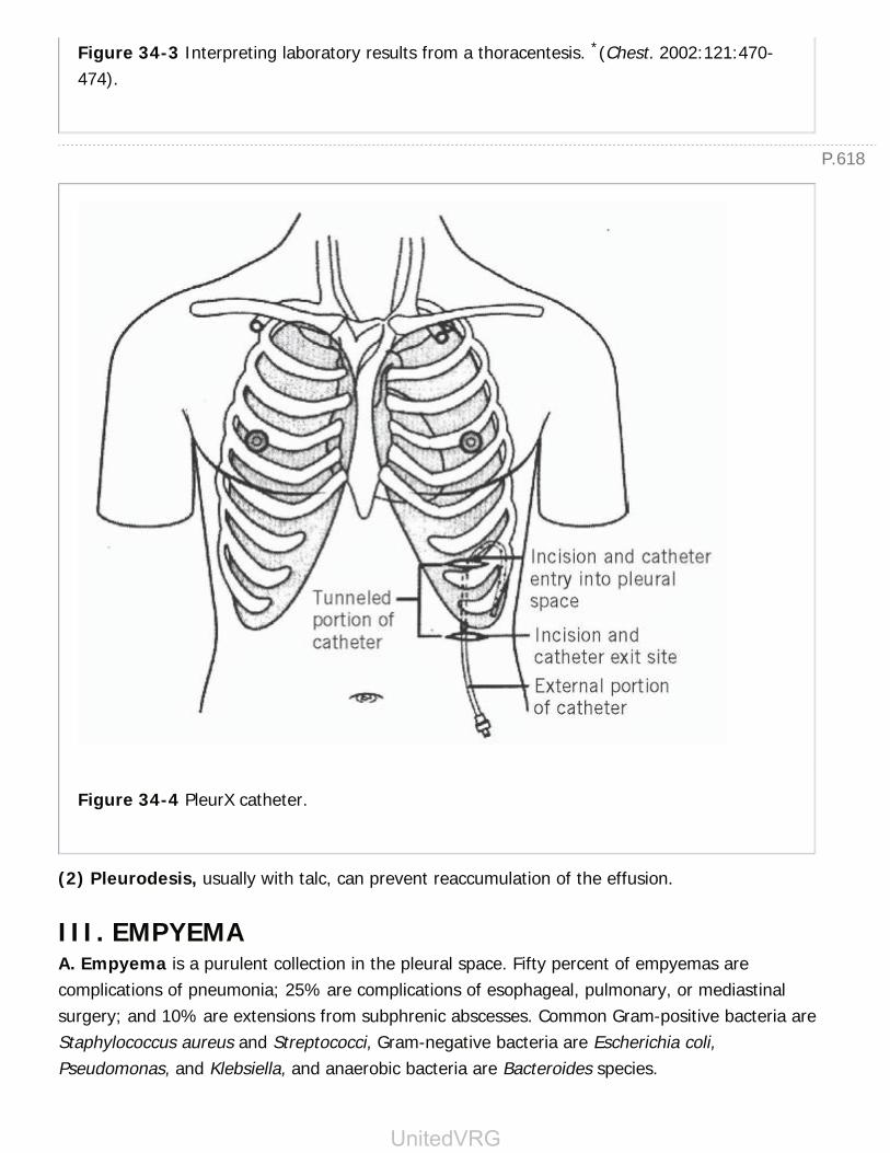
P.618
Figure 34-3 Interpreting laboratory results from a thoracentesis. *(Chest. 2002:121:470-474).
Figure 34-4 PleurX catheter.
(2) Pleurodesis, usually with talc, can prevent reaccumulation of the effusion.
III. EMPYEMAA. Empyema is a purulent collection in the pleural space. Fifty percent of empyemas arecomplications of pneumonia; 25% are complications of esophageal, pulmonary, or mediastinalsurgery; and 10% are extensions from subphrenic abscesses. Common Gram-positive bacteria areStaphylococcus aureus and Streptococci, Gram-negative bacteria are Escherichia coli,Pseudomonas, and Klebsiella, and anaerobic bacteria are Bacteroides species.
UnitedVRG

P.619
B. Presentation of empyema ranges from chronic loculated effusion in a patient with fatigue tosystemic sepsis requiring emergent care. Other symptoms include pleuritic chest pain, fever,cough, and dyspnea. Empyemas are diagnosed via thoracentesis and evaluated by CT scan.
C. Management includes control of the infection by appropriate antibiotics, drainage of thepleural space, and obliteration of the empyema space.
1. Early or exudative empyema is usually adequately treated with simple tube drainage.Fibropurulent empyema may be amenable to tube drainage alone, but the fluid may beloculated and may require thoracoscopic drainage. In advanced or organizing empyema, thefluid is thicker and a fibrous peel encases the lung. Thoracotomy may be
necessary to free the entrapped lung. If a patient has a persistent fluid collection with anadequately placed tube as evidenced by chest CT, intrapleural fibrinolytic therapy may be usefulto break down thin adhesions. Failure of intrapleural fibrinolytics usually requires operativeintervention.
2. A postpneumonectomy empyema can result from a bronchial stump dehiscence withcontamination of the pneumonectomy space. Increasing air in the pneumonectomy space on CXRis often diagnostic. Bronchopleural fistula has a high mortality rate and is managed via drainageof the space, antibiotics, and surgical repair of the fistula. The residual pleural cavity can beobliterated by a muscle transposition, thoracoplasty, or delayed Clagett procedure. Greatcaution should be taken in inserting chest tubes into postpneumonectomy empyemas.
IV. HEMOPTYSISA. Hemoptysis is the expectoration of blood and can originate from a number of causes,including infectious, malignant, and cardiac disorders (e.g., bronchitis or tuberculosis,bronchogenic carcinoma, and mitral stenosis, respectively).
B. Workup of hemoptysis includes a focused history and physical examination to assess forsymptoms that suggest infection or systemic disease, in conjunction with a CXR.
1. If the CXR is abnormal, appropriate workup and management is indicated.
2. In the setting of a negative CXR and hemoptysis that is likely due to acute bronchitis,observation can be appropriate.
3. For negative CXR and recurrent hemoptysis, a CT scan is warranted to further assess theunderlying etiology, and can be used as an adjunct to bronchoscopy to localize and potentiallytreat the source of bleeding.
C. Massive hemoptysis is a surgical emergency. The first steps are to assess the patient andsecure the airway.
1. Bronchoscopy can identify the bleeding side and allow for prompt protection of the

P.620
remaining lung parenchyma, either by controlling the area of bleeding or selective ventilation.Bleeding can be controlled by topical or injected vasoconstrictors or by placement of a balloon-tipped catheter in the lobar orifice. Selective ventilation can be achieved with a double-lumentube or by direct intubation of the contralateral mainstem bronchus.
2. If unable to identify the site of bleeding or after temporizing the bleed, angiographicembolization of a bronchial arterial source may allow for lung salvage without the need forresection. The bronchial circulation is almost always the source of hemoptysis. Bleeding from thepulmonary circulation is seen only in patients with pulmonary hypertension or some lung cancers.
3. Definitive therapy may require thoracotomy with lobar resection or, rarely, pneumonectomy.Rarely, emergent surgical resection is necessary
to control the hemoptysis. The etiology of the bleeding and the pulmonary reserve of the patientare important because many patients are not candidates for surgical resection.
V. LUNG CANCERA. Lung cancer is the second most common nonskin malignancy, and is the leading cause ofcancer death. An estimated 222,520 cases will be diagnosed and 158,040 patients will die of thedisease in 2015 (www.cancer.org). Cigarette smoking is the leading risk factor for thedevelopment of lung cancer.
B. Pathology. The two main classes of lung tumors are small-cell carcinoma and non-small-cellcarcinoma (NSCLC).
1. Small-cell carcinoma accounts for approximately 20% of all lung cancers. It is an aggressivetumor, usually occurs near the hilum, is almost exclusive to smokers, and rarely is amenable tosurgery because of wide dissemination by the time of diagnosis. These cancers initially respond tochemotherapy, but overall 5-year survival remains less than 10%.
2. Non-small-cell carcinomas account for 80% of all lung cancers and make up the vastmajority of those treated by surgery. The three main subtypes are adenocarcinoma (30% to50% of cases), squamous cell (20% to 35%), and large cell (4% to 15%).Bronchioloalveolar carcinoma is a variant of adenocarcinoma, produces mucin, and can bemultifocal. Over the last decade, it has been appreciated that typical carcinoid tumors (grade Ineuroendocrine carcinoma), atypical carcinoid tumors (grade II neuroendocrine carcinoma),large-cell carcinoma, and small-cell tumors represent important subgroups of bronchogenicneuroendocrine carcinoma. This may explain the more aggressive behavior of large-cellcarcinoma relative to other non-smallcell cancers.
C. Radiographic presentation may occur during a diagnostic evaluation for symptoms or as anincidental finding.
1. Solitary pulmonary nodules are circumscribed lung lesions in an asymptomatic individual,
UnitedVRG

P.621
with lesions greater than 3 cm labeled masses. An estimated 200,000 SPNs are diagnosed eachyear on CT scans.
2. Radiographic imaging by CT is used to follow lesions and predict outcome. Factors favoringa benign lesion include no growth over a 2-year period, lesion <2 cm, and certain patterns ofcalcification such as diffuse, centrally located, laminar, or popcornlike calcifications. Malignancy isfavored with intravenous contrast enhancement, irregular borders, and eccentric or stippledcalcifications (Fig. 34-5).
3. Positron emission tomography (PET) scanning is 95% sensitive and 80% specific incharacterizing nodules. However, bronchoalveolar carcinoma and carcinoid tumor can be falselynegative, and inflammatory and infectious processes can be falsely positive. The patient's overallrisk factor profile must also be considered. In the setting of low risk (e.g., young age, nonsmoker,favorable CT finding), a negative PET scan has
a high negative predictive value, but the same result in an elderly smoker is less reassuring, andfurther evaluation is warranted.
Figure 34-5 Solitary pulmonary nodule in right upper lobe.
4. Tissue biopsy remains the gold standard for diagnosis. Tissue may be obtained by
P.622
bronchoscopy in patients with central lung lesions or by CT-guided biopsy. Surgical biopsy byminimally invasive surgical techniques like video thoracoscopy and radial endobronchialultrasound (EBUS) can provide a definitive diagnosis.
D. Symptomatic presentation of lung cancer implies a more advanced stage and is associatedwith an overall lower rate of survival.
1. Bronchopulmonary features include cough or a change in a previously stable smoker'scough, increased sputum production, dyspnea, and wheezing. Minor hemoptysis should beinvestigated with bronchoscopy in patients with a history of smoking who are 40 years of age orolder. Lung cancer may also present with postobstructive pneumonia.
2. Extrapulmonary thoracic symptoms include chest wall pain secondary to local tumorinvasion, hoarseness from invasion of the left recurrent laryngeal nerve near the aorta and leftmain pulmonary artery, shortness of breath secondary to malignant pleural effusion or phrenicnerve invasion, and superior vena cava syndrome causing facial, neck, and upper-extremityswelling. A Pancoast tumor (superior sulcus tumor) can lead to invasion of the brachial plexus orthe cervical sympathetic ganglia, causing an ipsilateral Horner syndrome (ptosis, miosis, andanhidrosis). Rarely, lung cancer can present as dysphagia from compression or invasion of theesophagus by mediastinal nodes or the primary tumor.
3. The most frequent sites of distant metastases include the liver, bone, brain, adrenalglands, and the contralateral lung. Symptoms may
include pathologic fractures from bony involvement. Brain metastasis may cause headache, visionchanges, or changes in mental status. Adrenal involvement infrequently presents with Addisondisease. Lung cancer is the most common tumor causing adrenal dysfunction.
4. Paraneoplastic syndromes are frequent and occur due to release of endocrine substancesby tumor cells. They include Cushing syndrome (ACTH secretion in small-cell carcinoma), SIADH,hypercalcemia (PTH-related protein secreted by squamous cell carcinomas), hypertrophicpulmonary osteoarthropathy (clubbed fingers, joint stiffness, and periosteal thickening on XR),and various myopathies.
E. Accurate clinical and pathologic staging is critical in the management of patients withnon-small-cell carcinoma. Surgery is the primary therapy for many stage I and II patients andselected stage III patients. The essential elements of staging include evaluation for lymph nodeinvolvement and evaluation for distant metastasis. The staging system was most recentlymodified in 2009 (AJCC 7th edition; Table 34-1).
1. Chest CT to include the upper abdomen examines the size, location, and localinvolvement of tumor, identifies mediastinal lymphadenopathy, and evaluates for liver andadrenal metastasis. The sensitivity for identifying metastatic lymph nodes by CT is 65% to 80%and the specificity is only 65%.
UnitedVRG
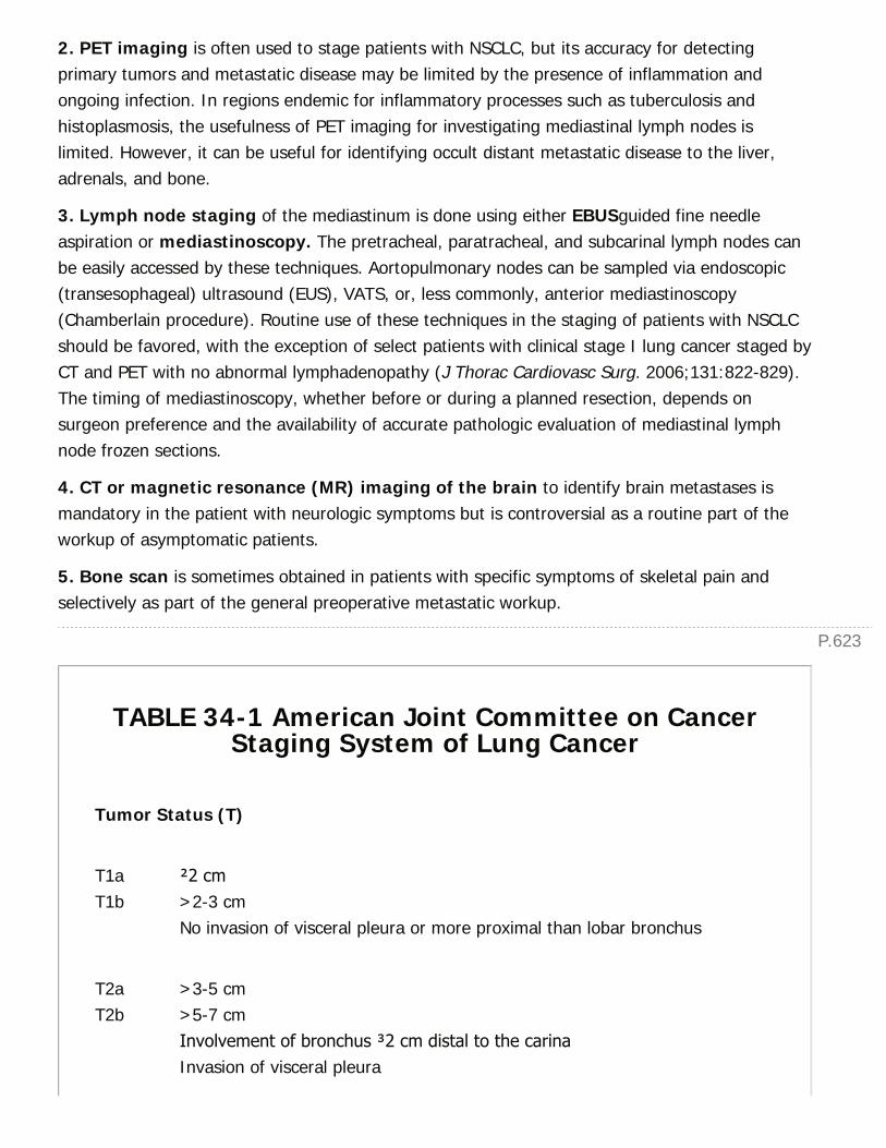
P.623
2. PET imaging is often used to stage patients with NSCLC, but its accuracy for detectingprimary tumors and metastatic disease may be limited by the presence of inflammation andongoing infection. In regions endemic for inflammatory processes such as tuberculosis andhistoplasmosis, the usefulness of PET imaging for investigating mediastinal lymph nodes islimited. However, it can be useful for identifying occult distant metastatic disease to the liver,adrenals, and bone.
3. Lymph node staging of the mediastinum is done using either EBUSguided fine needleaspiration or mediastinoscopy. The pretracheal, paratracheal, and subcarinal lymph nodes canbe easily accessed by these techniques. Aortopulmonary nodes can be sampled via endoscopic(transesophageal) ultrasound (EUS), VATS, or, less commonly, anterior mediastinoscopy(Chamberlain procedure). Routine use of these techniques in the staging of patients with NSCLCshould be favored, with the exception of select patients with clinical stage I lung cancer staged byCT and PET with no abnormal lymphadenopathy (J Thorac Cardiovasc Surg. 2006;131:822-829).The timing of mediastinoscopy, whether before or during a planned resection, depends onsurgeon preference and the availability of accurate pathologic evaluation of mediastinal lymphnode frozen sections.
4. CT or magnetic resonance (MR) imaging of the brain to identify brain metastases ismandatory in the patient with neurologic symptoms but is controversial as a routine part of theworkup of asymptomatic patients.
5. Bone scan is sometimes obtained in patients with specific symptoms of skeletal pain andselectively as part of the general preoperative metastatic workup.
TABLE 34-1 American Joint Committee on CancerStaging System of Lung Cancer
Tumor Status (T)
T1aT1b
≤2 cm>2-3 cmNo invasion of visceral pleura or more proximal than lobar bronchus
T2aT2b
>3-5 cm>5-7 cmInvolvement of bronchus ≥2 cm distal to the carinaInvasion of visceral pleura

Associated atelectasis or obstructive pneumonitis not involving entire lung
T3 >7 cm or tumor with any of the following characteristics:Invasion of chest wall, diaphragm, phrenic nerveInvasion of mediastinal pleura or parietal pericardiumAssociated atelectasis or obstructive pneumonitis of entire lungTumor within main bronchus <2 cm from carina but does not involve carinaSatellite nodules in the same lobe
T4 Tumor with any of the following characteristics:Mediastinal invasionInvasion of heart of great vesselsInvasion of carina, trachea, esophagus, or recurrent laryngeal nerveInvasion of vertebral bodySeparate tumor nodules in a different but ipsilateral lobe
Nodal Involvement (N)
N0 None
N1 Hilar, interlobar, or peripheral lymph node zones
N2 Ipsilateral mediastinal lymph nodes, subcarinal, or aortopulmonary lymphnodes
N3 Contralateral mediastinal, hilar, or aortopulmonary lymph nodes; ipsilateralor contralateral scalene or supraclavicular lymph nodes
Distant Metastases (M)
M0 None
M1 Distant metastases present
Stage 5-yr Survival
UnitedVRG
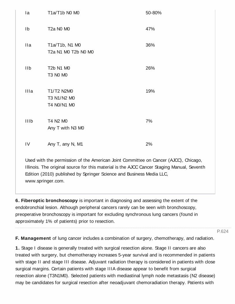
P.624
Ia T1a/T1b N0 M0 50-80%
Ib T2a N0 M0 47%
IIa T1a/T1b, N1 M0T2a N1 M0 T2b N0 M0
36%
IIb T2b N1 M0T3 N0 M0
26%
IIIa T1/T2 N2M0T3 N1/N2 M0T4 N0/N1 M0
19%
IIIb T4 N2 M0Any T with N3 M0
7%
IV Any T, any N, M1 2%
Used with the permission of the American Joint Committee on Cancer (AJCC), Chicago,Illinois. The original source for this material is the AJCC Cancer Staging Manual, SeventhEdition (2010) published by Springer Science and Business Media LLC,www.springer.com.
6. Fiberoptic bronchoscopy is important in diagnosing and assessing the extent of theendobronchial lesion. Although peripheral cancers rarely can be seen with bronchoscopy,preoperative bronchoscopy is important for excluding synchronous lung cancers (found inapproximately 1% of patients) prior to resection.
F. Management of lung cancer includes a combination of surgery, chemotherapy, and radiation.
1. Stage I disease is generally treated with surgical resection alone. Stage II cancers are alsotreated with surgery, but chemotherapy increases 5-year survival and is recommended in patientswith stage II and stage III disease. Adjuvant radiation therapy is considered in patients with closesurgical margins. Certain patients with stage IIIA disease appear to benefit from surgicalresection alone (T3N1M0). Selected patients with mediastinal lymph node metastasis (N2 disease)may be candidates for surgical resection after neoadjuvant chemoradiation therapy. Patients with
P.625
bulky, diffuse mediastinal lymphadenopathy are typically treated using definitive chemoradiation.Stage IV tumors have distant metastases and are considered unresectable, though some patientswith node-negative lung cancer and a solitary brain metastasis have achieved long-term survivalwith combined resection.
2. Operative principles. In the patient able to tolerate any resection (see Section IX.A), theminimum extent of resection is usually an anatomic lobectomy. Even in stage I disease, a wedgeresection results in a threefold higher local recurrence rate and a decreased overall and disease-free survival. Patients with limited pulmonary reserve may be treated by segmental or wedgeresection. Most centers report operative mortality of less than 2% with lobectomy and 6% with
pneumonectomy. VATS has become a widespread technique for lung resections.
3. Stereotactic body radiation therapy (SBRT) is external beam radiation therapy thatprecisely delivers high-dose radiation to a target in the body over one or more treatments. SBRThas been shown to be effective for early stage lung cancer in patients that are poor candidatesfor surgical resection.
G. Five-year survival rates are summarized in Table 34-1.
VI. TUMORS OF THE PLEURAA. The most common tumor of the pleura is mesothelioma. Malignant mesothelioma is a rareand aggressive cancer that has been linked to asbestos exposure with a latency period ofdecades.
1. Patient presentation varies, but can include chest pain, malaise, cough, weakness, weight loss,and shortness of breath with pleural effusions. One-third of patients report paraneoplasticsymptoms of osteoarthropathy, hypoglycemia, and fever.
2. Tissue for diagnosis can be obtained via cytology of pleural fluid, needle biopsy of the pleura,or biopsy via thoracoscopy. The most favorable subtype is epithelioid (50%), followed bysarcomatoid (20%), mixed, and desmoplastic.
3. CT scans can differentiate pleural from parenchymal disease. Routine use of MRI is notrecommended, while PET scan is used to identify distant metastatic disease (J Thorac CardiovascSurg. 2003;126: 11-15).
4. Treatment consists of a multimodal therapy comprising surgery, chemotherapy, and radiation.Surgical options include extrapleural pneumonectomy (EPP) or pleurectomy/decortication. Forearly stage cases, EPP may offer the best chance of cure. The best reported 5-year survivalfollowing completion of the multimodal therapy in patients without nodal metastasis is 53% butsurvival is typically lower than that (J Clin Oncol. 2009;27:1413-1418).
B. Less common tumors include lipomas, angiomas, soft-tissue sarcomas, and fibroushistiocytomas.
UnitedVRG
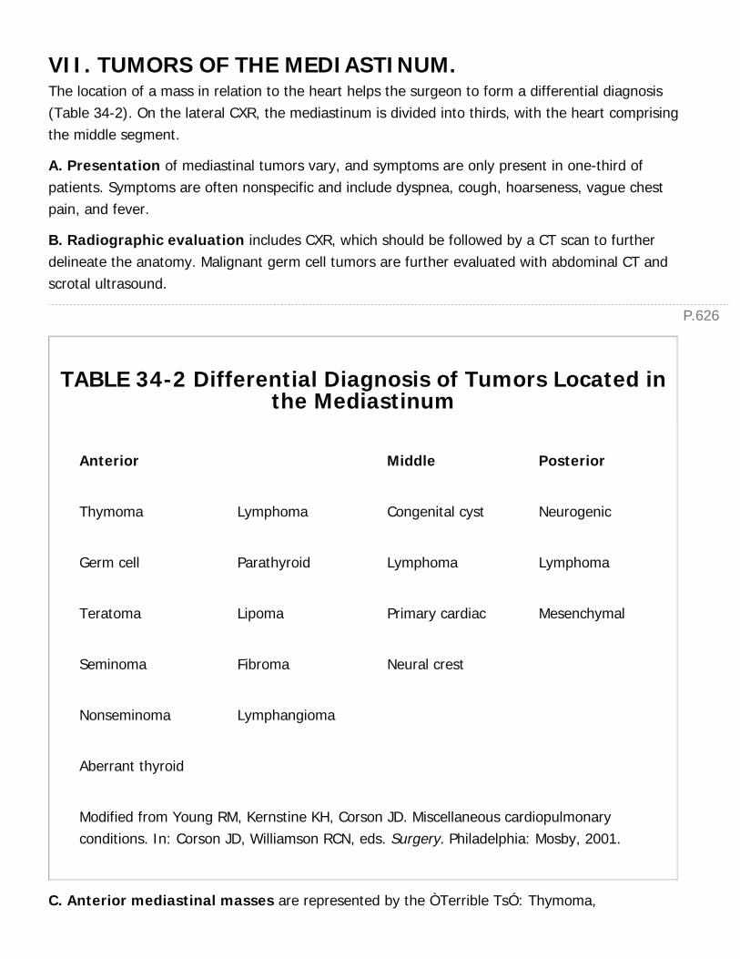
P.626
VII. TUMORS OF THE MEDIASTINUM.The location of a mass in relation to the heart helps the surgeon to form a differential diagnosis(Table 34-2). On the lateral CXR, the mediastinum is divided into thirds, with the heart comprisingthe middle segment.
A. Presentation of mediastinal tumors vary, and symptoms are only present in one-third ofpatients. Symptoms are often nonspecific and include dyspnea, cough, hoarseness, vague chestpain, and fever.
B. Radiographic evaluation includes CXR, which should be followed by a CT scan to furtherdelineate the anatomy. Malignant germ cell tumors are further evaluated with abdominal CT andscrotal ultrasound.
TABLE 34-2 Differential Diagnosis of Tumors Located inthe Mediastinum
Anterior Middle Posterior
Thymoma Lymphoma Congenital cyst Neurogenic
Germ cell Parathyroid Lymphoma Lymphoma
Teratoma Lipoma Primary cardiac Mesenchymal
Seminoma Fibroma Neural crest
Nonseminoma Lymphangioma
Aberrant thyroid
Modified from Young RM, Kernstine KH, Corson JD. Miscellaneous cardiopulmonaryconditions. In: Corson JD, Williamson RCN, eds. Surgery. Philadelphia: Mosby, 2001.
C. Anterior mediastinal masses are represented by the ÒTerrible TsÓ: Thymoma,
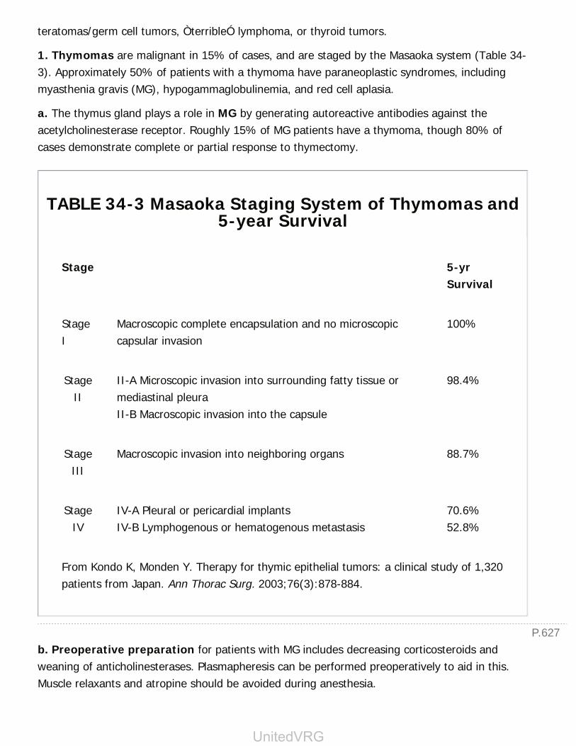
P.627
teratomas/germ cell tumors, ÒterribleÓ lymphoma, or thyroid tumors.
1. Thymomas are malignant in 15% of cases, and are staged by the Masaoka system (Table 34-3). Approximately 50% of patients with a thymoma have paraneoplastic syndromes, includingmyasthenia gravis (MG), hypogammaglobulinemia, and red cell aplasia.
a. The thymus gland plays a role in MG by generating autoreactive antibodies against theacetylcholinesterase receptor. Roughly 15% of MG patients have a thymoma, though 80% ofcases demonstrate complete or partial response to thymectomy.
TABLE 34-3 Masaoka Staging System of Thymomas and5-year Survival
Stage 5-yrSurvival
StageI
Macroscopic complete encapsulation and no microscopiccapsular invasion
100%
StageII
II-A Microscopic invasion into surrounding fatty tissue ormediastinal pleuraII-B Macroscopic invasion into the capsule
98.4%
StageIII
Macroscopic invasion into neighboring organs 88.7%
StageIV
IV-A Pleural or pericardial implantsIV-B Lymphogenous or hematogenous metastasis
70.6%52.8%
From Kondo K, Monden Y. Therapy for thymic epithelial tumors: a clinical study of 1,320patients from Japan. Ann Thorac Surg. 2003;76(3):878-884.
b. Preoperative preparation for patients with MG includes decreasing corticosteroids andweaning of anticholinesterases. Plasmapheresis can be performed preoperatively to aid in this.Muscle relaxants and atropine should be avoided during anesthesia.
UnitedVRG
P.628
c. Operative approach includes transcervical thymectomy, median sternotomy, VATS resection,and robotic resection. For more advanced stages of thymoma, neoadjuvant chemotherapy maybe indicated.
2. Biochemical evaluation with β-human chorionic gonadotropin (β-HCG) and α-fetoprotein(AFP) if the lesion is suspected to be germ cell tumors.
a. Teratomas are usually benign and often contain ectodermal components such as hair, teeth,and bone. Elevation of both β-HCG and AFP is very rare, and suggests a malignant teratoma.Treatment is surgical resection.
b. Seminomas do not have elevated AFP, and fewer than 10% present have elevated β-HCG.Their treatment is primarily nonsurgical (radiation and chemotherapy), except in the case oflocalized disease.
c. Nonseminomatous germ cell tumors present with an elevation of both tumor markers. Thetreatment is with platinum-based chemotherapy, with resection of residual masses after definitivechemotherapy.
3. Tissue diagnosis is often crucial for the diagnosis and treatment of lymphoma. Treatment isprimarily nonsurgical. Cervical lymph node biopsy, CT-guided biopsy, or mediastinoscopy withbiopsy may be required. These lesions often present as irregular masses on CT scan.
D. Bronchogenic and enteric cysts present as middle mediastinal masses, and should beresected for tissue diagnosis and due to risk of future infection or malignant degeneration.
E. Patients with posterior mediastinal masses or paravertebral masses should have theircatecholamine levels measured to rule out pheochromocytomas.
VIII. LUNG TRANSPLANTATIONA. Lung transplantation has been increasing steadily over the last decade, with the leadingindications for lung transplant being chronic obstructive pulmonary disease (COPD), idiopathicpulmonary fibrosis (IPF), and CF. Other common indications include α1-antitrypsin deficiency
(alpha-1) and pulmonary arterial hypertension (PAH).
B. COPD is a long-term consequence of smoking and is the result of nonuniform destruction oflung parenchyma. As lung tissue loses its elastic recoil, the areas of destruction expand. Thisexpansion of diseased areas, in combination with inflammation, leads to poor ventilation ofrelatively normal lung.
1. This leads to the typical findings of hyperexpanded lungs on CXR: Flattened diaphragms,widened intercostal spaces, and horizontal ribs. On pulmonary function testing, patients presentwith increased residual volumes and decreased FEV1 (forced expiratory volume in 1 second).
Surgical treatment is generally reserved for the symptomatic patient who has failed maximal
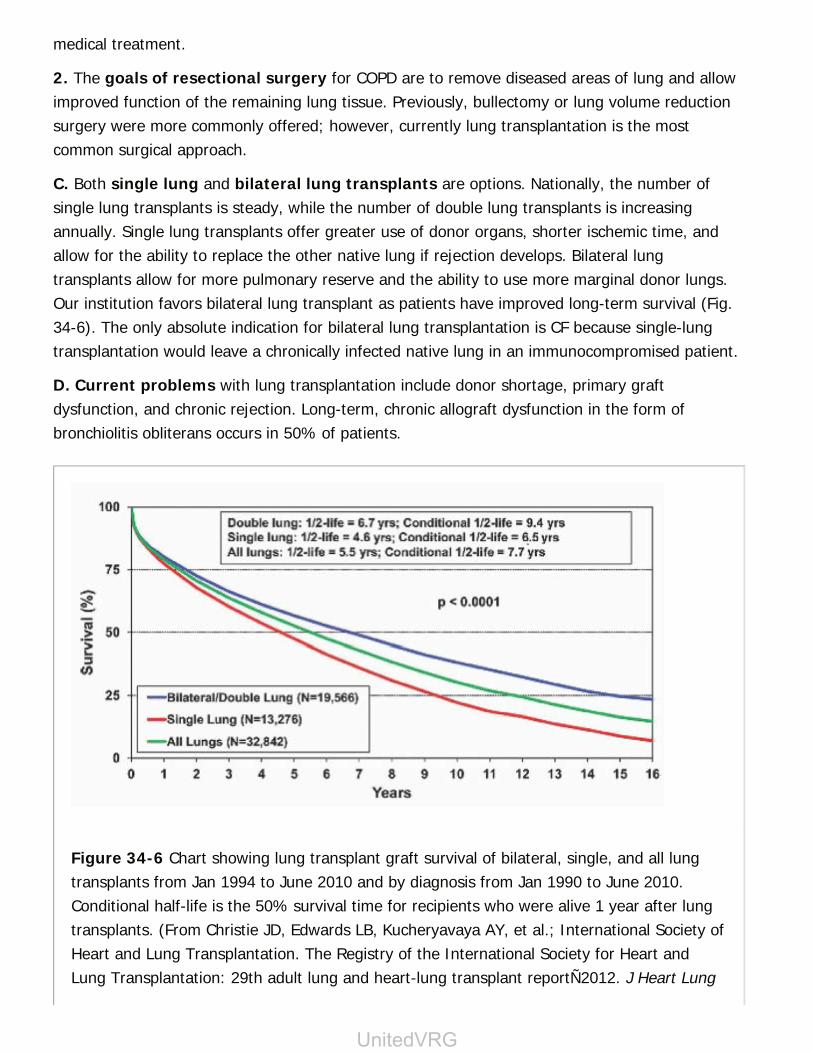
medical treatment.
2. The goals of resectional surgery for COPD are to remove diseased areas of lung and allowimproved function of the remaining lung tissue. Previously, bullectomy or lung volume reductionsurgery were more commonly offered; however, currently lung transplantation is the mostcommon surgical approach.
C. Both single lung and bilateral lung transplants are options. Nationally, the number ofsingle lung transplants is steady, while the number of double lung transplants is increasingannually. Single lung transplants offer greater use of donor organs, shorter ischemic time, andallow for the ability to replace the other native lung if rejection develops. Bilateral lungtransplants allow for more pulmonary reserve and the ability to use more marginal donor lungs.Our institution favors bilateral lung transplant as patients have improved long-term survival (Fig.34-6). The only absolute indication for bilateral lung transplantation is CF because single-lungtransplantation would leave a chronically infected native lung in an immunocompromised patient.
D. Current problems with lung transplantation include donor shortage, primary graftdysfunction, and chronic rejection. Long-term, chronic allograft dysfunction in the form ofbronchiolitis obliterans occurs in 50% of patients.
Figure 34-6 Chart showing lung transplant graft survival of bilateral, single, and all lungtransplants from Jan 1994 to June 2010 and by diagnosis from Jan 1990 to June 2010.Conditional half-life is the 50% survival time for recipients who were alive 1 year after lungtransplants. (From Christie JD, Edwards LB, Kucheryavaya AY, et al.; International Society ofHeart and Lung Transplantation. The Registry of the International Society for Heart andLung Transplantation: 29th adult lung and heart-lung transplant reportÑ2012. J Heart Lung
UnitedVRG

P.629
Transplant. 2012;31(10):1052-1064, with permission.)
IX. MANAGEMENT OF THORACIC SURGICAL PATIENTSA. Preoperative assessment of pulmonary function and estimation of postoperative pulmonaryassessment is the most critical factor in planning lung resection.
1. Pulmonary function tests (PFTs), which include FEV1, and arterial blood gas (ABG) are
used to assess risk of postoperative pulmonary failure (Fig. 34-7). Diffusion capacity (DLCO)and cardiopulmonary exercise testing (CPET) are indicated in patients with marginalfunction for accurate assessment of postoperative function. In general, average risk forpulmonary resection is associated with a 1% to 2% mortality.
Preoperative hypercapnia (arterial carbon dioxide tension >45 mm Hg) may precluderesection.
2. Evaluation of cardiac disease starts with a detailed history and physical examination toelicit symptoms of ischemia and a baseline EKG. Abnormal findings should be pursued with stresstests or coronary catheterization.
3. Smoking cessation preoperatively for as little as 2 weeks can aid in the regeneration of themucociliary function and has been associated with fewer postoperative respiratory complications.
B. Postoperative care of the thoracic surgery patient focuses on pain control, fluidmanagement, and maintenance of pulmonary function.
1. The thoracotomy incision is one of the most painful and debilitating in surgery.Inadequate pain control contributes heavily to postoperative complications. Chest wall splintingcontributes to atelectasis and poor pulmonary toilet. Pain increases sympathetic tone andmyocardial oxygen demand, provoking arrhythmias and cardiac ischemic episodes. The routineuse of epidural catheter anesthesia during the early recovery period significantly improves painmanagement. Other effective analgesic maneuvers include intercostal blocks with long-actinglocal anesthetic before closure of the chest and subpleural administration of local anesthetic viacatheters placed adjacent to intercostal nerves at the time of thoracotomy or thoracoscopy.
2. Perioperative fluid management of thoracic surgery patients differs from that of patientsafter abdominal surgery. Pulmonary surgery does not induce large fluid shifts, while collapse andreexpansion of lungs during surgery can lead to pulmonary edema. Judicious fluid managementto avoid fluid overload and pulmonary edema is critical in patients with limited pulmonary reserve.Discussions regarding intraoperative fluid management should be held with the anesthesiologistbefore surgery. Physicians may need to accept transiently decreased urine output and increasedserum creatinine. Mild hypotension may be treated with intravenous α-agonists such as

P.630
phenylephrine. Cardiac dysfunction may also be the source of postoperative oliguria, pulmonaryedema, and hypotension and should always be considered in patients who are not respondingnormally. Echocardiography or placement of a Swan-Ganz catheter may guide treatment.
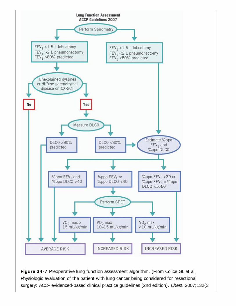
Figure 34-7 Preoperative lung function assessment algorithm. (From Colice GL et al.Physiologic evaluation of the patient with lung cancer being considered for resectionalsurgery: ACCP evidenced-based clinical practice guidelines (2nd edition). Chest. 2007;132(3

P.631
P.632
Suppl): 161S-177S, with permission.)
3. Maintenance of good bronchial hygiene is often the most difficult challenge facing thepostthoracotomy patient. A lengthy smoking history, decreased ciliary function, chronic bronchitis,and significant postoperative pain all contribute to the ineffective clearance of pulmonarysecretions. Adequate analgesia along with aggressive pulmonary toilet, incentive spirometry, andchest physiotherapy delivered by the respiratory therapist are essential. Occasionally nasotrachealsuctioning or bedside flexible bronchoscopy can be indicated for bronchial toilet.
4. Cardiovascular complications are common because the population that develops lungcancer is at high risk for heart disease. The three most common sources of cardiac morbidity arearrhythmias, myocardial infarctions, and congestive heart failure.
a. Cardiac arrhythmias occur in up to 30% of patients undergoing pulmonary surgery. Themost common arrhythmia is atrial fibrillation. The highest incidence occurs in elderly patientsundergoing pneumonectomy or intrapericardial pulmonary artery ligation.
b. Treatment of any rhythm disturbance begins with an assessment of the patient'shemodynamic status. Manifestations of these arrhythmias vary in acuity from palpitations tohemodynamic collapse. If the patient is hemodynamically unstable, the advanced cardiac lifesupport protocol should be followed. After the patient has been examined and hemodynamicstability confirmed, an ECG, ABG sample, and serum electrolyte panel should be obtained.Frequently, supplementary oxygen and aggressive potassium and magnesium replenishment arethe only treatment necessary. They should be treated expediently with electrolyte correction,optimization of oxygenation, and evaluation for cardiac ischemia.
CHAPTER 34: LUNG AND MEDIASTINAL DISEASESMultiple Choice Questions1. Which of the following patients can be initially managed byobservation?
a. A 45-year-old pilot with a small pneumothorax on room air.
b. A 62-year-old teacher with a small pneumothorax on room air.
c. An 18-year-old college student with a recurrent small pneumothorax onroom air.
d. A 31-year-old police officer with a small pneumothorax and shortnessof breath on room air.
View Answer

P.633
2. A patient with COPD has acute respiratory failure from pneumoniaand is intubated in the ICU. The nurse calls you to inform you that thepatient's oxygen requirement has increased, in addition to his bloodpressure slowly trending down, with his MAP going from 70 to 60 overthe last hour. On physical examination, you notice significantlydecreased breath sounds on the right. The next step is:
a. Obtain a CXR
b. Needle decompression
c. Percutaneous catheter placement
d. Chest tube placement
View Answer
3. A 50-year-old female with hepatitis C cirrhosis has a recurrentrightsided pleural effusion that is being managed with drainage from aPleurX catheter. His serum laboratory results are: Protein 6.8, LDH 100,amylase 20, WBC 9,000. Which of the following laboratory resultswould be expected in this effusion?
a. Pleural fluid protein is 2.0
b. Pleural fluid LDH is 75
c. Pleural amylase is 250
d. Pleural WBC is 12,000
View Answer
4. A 35-year-old male presents with an incidentally found mediastinalmass on CXR. Further workup with a CT scan demonstrates an anteriormediastinal mass. AFP and β-HCG are both elevated. Treatment of thismass is:
a. Observation as he is asymptomatic
b. Radiation and chemotherapy without surgery
c. Surgical resection
d. Chemotherapy followed by surgery
View Answer
5. A 70-year-old patient presents to the ER after a motor vehiclecollision. In the ER, he undergoes a chest CT that demonstrates anincidentally found 3 cm peripheral mass in the RUL. What is the nextstep in management?

a. Observation with repeat CT in 6 to 12 months
b. PET scan
c. Navigational bronchoscopy
d. Wedge resection
View Answer
6. A 65-year-old patient had a chronic cough with a 40 pack yearsmoking history. CXR demonstrated a RLL mass with staging by PET/CTconcerning for a T1, N2, M0 lesion. A percutaneous biopsy indicatesthat the patient has lung adenocarcinoma. What is the appropriatemanagement?
a. Mediastinoscopy or EBUS
b. Surgical resection alone
c. Surgical resection followed by chemotherapy
d. Neoadjuvant chemotherapy
View Answer
7. Which of the following lung function evaluations has the highest riskfor undergoing a lobectomy?
a. FEV1 80% predicted, DLCO 40% predicted
b. FEV1 40% predicted, DLCO 70% predicted
c. FEV1 20% predicted, VO2 max 12 mL/kg/minute
d. FEV1 30% predicted, VO2 max 15 mL/kg/minute
View Answer
35Breast
Oluwadamilola M. Fayanju
Julie A. Margenthaler
ANATOMY
I. THE BREAST.Located between the subcutaneous fat and the fascia of the pectoralis major and serratusanterior muscles, it is bounded superiorly by the second/third rib, inferiorly by theinframammary fold, and medially by the lateral edge of the sternum, with its lateral border lyingbetween the anterior and midaxillary lines. Suspensory ligaments (Cooper ligaments) runningthrough the breasts from the deep fascia to the skin may cause skin dimpling when associatedwith a malignancy.
A. Vasculature. Arterial supply is predominantly internal thoracic artery (or internalmammary artery) via perforating branches (perforators). Venous drainage is mainly to theaxillary vein.
B. Lymphatic Drainage. The superficial Sappey plexus converges with a deep lymphaticplexus, and they ultimately drain into the axillary (75%) and internal mammary lymph nodes.
C. Innervation. Lateral and anterior cutaneous branches of the second to sixth intercostalnerves provide sensory innervation.
II. THE AXILLA.The borders of the axilla are defined as the axillary vein superiorly, latissimus dorsi laterally,and the serratus anterior muscle medially.
A. Axillary lymph nodes are classified according to their anatomic location relative to thepectoralis minor muscle.
1. Level I nodes: Lateral to the pectoralis minor muscle.
2. Level II nodes: Posterior to the pectoralis minor muscle.
3. Level III nodes: Medial to the pectoralis minor muscle and most accessible with division ofthe muscle.
4. Rotter nodes: Between the pectoralis major and the minor muscles.
> Table of Contents > 35 - Breast
P.635
B. Axillary Nerves. Three motor and several sensory nerves are located in the axilla.Preservation of all is preferred during an axillary lymph node dissection (ALND); however, directtumor invasion may require resection along with the specimen.
1. Long thoracic nerve travels superior to inferior along chest wall and medially within theaxilla, innervating the serratus anterior muscle. Injury to this nerve causes a ÒwingedÓscapula.
2. Thoracodorsal nerve courses along the posterior border of the axilla superior to inferior onthe subscapularis muscle and innervates the
latissimus dorsi. Injury to this nerve causes weakness in arm adduction and medial rotation.
3. Medial pectoral nerve travels from the posterior aspect of the pectoralis minor musclearound the lateral border of the pectoralis minor to the posterior aspect of the pectoralis majormuscle. It innervates the lateral third of the pectoralis major; injury to this nerve results inatrophy of the lateral pectoralis major muscle.
4. Intercostal brachial sensory nerves travel laterally in the axilla from the second intercostalspace to the medial upper arm. Transection causes numbness in the posterior and medialsurfaces of the upper arm.
CLINICAL ASSESSMENT
I. HISTORY.Patients seek medical attention most commonly for an abnormal mammogram, a breastmass, breast pain, nipple discharge, or skin changes. History should include:
Description and duration of signs and symptoms and their temporal relationship to pregnancy,menstrual cycle, or previous trauma.
Date of last menstrual period and regularity of the menstrual cycle.
Age of menarche.
Number of pregnancies and age at first full-term pregnancy.
Lactational history.
Age at natural or surgical menopause (i.e., oophorectomy).
Previous history of breast biopsies and mammoplasties.
Mammogram history.
History of oral contraceptive and/or hormone replacement therapy (HRT).
Personal and family history of breast and gynecologic cancer, including age at diagnosis. This

P.636
should include at least two generations as well as any associated cancers, such as ovary,colon, prostate, gastric, or pancreatic.
A. Assessment of Cancer Risks
1. Risk factors. Hormonal and environmental exposures, genetics, and certain types of breasttissue histology can all be associated with an increased risk for breast cancer (Table 35-1).
2. Hereditary breast cancer. Approximately 5% to 10% of breast cancers are hereditary, thatis, can be attributed to a mutation in a single, highly penetrant gene, and approximately 80% ofhereditary breast cancers are the result of mutations in BRCA1 and BRCA2. Women with BRCA1mutations have an estimated risk of 85% for breast cancer by the age of 70 years, a 50% chanceof developing a second primary breast cancer, and a 20% to 40% chance of developing ovariancancer. BRCA2 mutations carry a slightly lower risk for breast and ovarian cancer and account for4% to 6% of all male breast cancers. Screening for BRCA gene mutations should be reserved forwomen who have a strong family history of breast or ovarian cancer, and referral for geneticcounseling should be based on the National Comprehensive Cancer Network (NCCN) guidelines (JNatl Compr Canc Netw. 2014;12:1326-1338).
TABLE 35-1 Risk Factors for Breast Cancer andApproximate Strength of Association
Reproductive HormonalNutritional/Lifestyle/BodyHabitus Other
Earlymenarche [+]
OC use (currentvs. none) [+]
Obesity (>30 BMI vs. <25)Premenopausal [-]Postmenopausal [+]
Family history(mother and
sister)a [+++]
Age at firstbirth (>35 vs.<20) [++]
Estrogenreplacement(10+ yr vs.none) [+]
Adult weight gain(postmenopausal) [++]
Family history(first-degree
relative)b [++]
No. of births(0 vs. 1 child)[+]
Estrogen plusprogesteronereplacement (>5yrs vs. none)
Alcohol (1 or more drink/dayvs. none) [+]
Jewishheritage (yesvs. no) [+]

P.637
[++]
Age atmenopause (5-yr increment)[+]
High bloodestrogens orandrogens(postmenopause)[+++]
Height (>5 ft 7 in) [+] Ionizingradiation (yesvs. no) [+]
Breast-feeding(>1 yr vs.none) [-]
High bloodprolactin [++]
Physical activity (>3 hrs/wk)[-]
Monounsaturated fatc (vs.saturated fat) [-]
Low intake of fruits and
vegetablesc (specifically forER-breast cancer) [+]
Benign breastdisease (MD
diagnosed)d
[++]Mammographicdensity(highestcategory vs.lowest) [+++]
a Two first-degree relatives who have a history of breast cancer before age 65 years versusno relative.b First-degree relative who has a history of breast cancer before age 65 years versus norelative.c Upper quartile (top 25%) versus lower quartile (lowest 25%).d Clinically recognized chronic cystic, fibrocystic, or other benign breast disease versusnone.BMI, body mass index; OC, oral contraceptives;[+] = relative risk (RR) 1.1-1.4; [++] = RR 1.5-2.9; [+++] = RR 3.0-6.9; [-] = RR 0.7-0.8.Adapted with permission from Table 18.2 in Chapter 18 : Nongenetic Factors in theCausation of Breast Cancer, Diseases of the Breast, LWW 2014.
3. Modeling breast cancer risk. The Gail model is one of several prediction modelsÑincludingthe BOADICEA, BRCAPRO, Claus, and Tyrer-Cuzick, modelsÑthat estimate the absolute risk(probability) that a woman in a program of annual screening will develop breast cancer over adefined age interval (J Nat Cancer Inst. 1989; 81:1879-1886). A modified Gail model focusingonly on the risk of invasive cancer has been used to define eligibility criteria for entry intochemoprevention trials. The National Surgical Adjuvant Breast and Bowel Project (NSABP) and the
P.638
National Cancer Institute (NCI) offer an interactive online risk assessment tool(http://www.cancer.gov/bcrisktool). In addition, a number of genome-based prediction tools havealso been developedÑthe most widely used being Oncotype DxÑin order to help predict who isat risk for malignancy and who would benefit from chemotherapy.
II. PHYSICAL EXAMINATION.Inspect the patient's breasts in both the upright and supine positions. With the patient in theupright position, examine with the patient's arms relaxed and then raised, looking for shapeasymmetry, deformity, skin changes (erythema, edema, dimpling), nipple changes or discharge,and lymphadenopathy (axillary, supraclavicular, and infraclavicular). With the patient in thesupine position, examine the entire breast systematically with the patient's ipsilateral arm raisedabove and behind the head.
III. BREAST IMAGINGA. Screening mammograms are performed in the asymptomatic patient, and consist of twostandard views, mediolateral oblique (MLO) and craniocaudal (CC). Tomosynthesis becameavailable in the United States in 2011 and improves the sensitivity and specificity ofmammography, particularly for women with nonfatty breasts and in the assessment ofnoncalcified lesions. Although the United States Preventative Task Force (USPTF) changed itsscreening recommendation in 2009 to annually for women beginning at age 50, the currentrecommendation from the NCI, the American Congress of Obstetricians and Gynecologists(ACOG), and the American Cancer Society (ACS) is annual screening mammography forwomen aged 40 years and older, and we agree with these societies' recommendation. Breastlesions on mammograms are classified according to the American College of Radiology by BI-RADS (Breast Imaging Reporting and Database System) scores:
0 = Needs further imaging; assessment incomplete.
1 = Normal; continue annual followup (risk of malignancy: 1/2,000).
2 = Benign lesion; no risk of malignancy; continue annual followup (risk of malignancy:1/2,000).
3 = Probably benign lesion; needs 4 to 6 months followup (risk of malignancy: 1% to 2%).
4 = Suspicious for breast cancer; biopsy recommended. This category can be furthersubdivided into
4A = Low suspicion for malignancy (>2% to ≤ 10%)
4B = Moderate suspicion for malignancy (>10% to ≤ 50%)
4C = Finding of moderate concern of being cancer, but not as high as Category 5 (>50% to

≤95%)
5 = Highly suspicious for breast cancer; biopsy strongly recommended (≥95% are malignant).
6 = Known biopsy-proven malignancy.
1. Malignant mammographic findings
a. New or spiculated masses.
b. Clustered microcalcifications in linear or branching array.
c. Architectural distortion.
2. Benign mammographic findings that might be mistaken for malignancy include radialscar (biopsy needed), fat necrosis (i.e., oil cysts; biopsy may be needed), and milk of calcium(no biopsy needed). Cysts cannot be distinguished from solid masses by mammography;ultrasound is needed to make this distinction.
3. Screening in high-risk patients. For patients with known BRCA mutations, annualmammograms and semiannual physical examinations should begin at the age of 25 to 30years. In patients with a strong family history of breast cancer but undocumented geneticmutation, annual mammograms and semiannual physical examinations should begin 10 yearsearlier than the age at diagnosis of the youngest affected relative and no later thanthe age of 40 years.
4. Magnetic resonance imaging (MRI) is recommended for screening in select high-riskpatients including those with an elevated lifetime risk per a validated risk assessment model, apersonal or family history of BRCA mutations or other predisposing genetic syndromes (Li-Fraumeni, Cowden, or BannayanÑRileyÑRuvalcaba), or a history of chest wall radiation betweenthe ages of 10 and 30.
B. Diagnostic Imaging
1. Diagnostic mammograms are performed in the symptomatic patient or to follow up anabnormality noted on a screening mammogram. Additional views (spot-compression viewsor magnification views) may be used to further characterize any lesion. A normal mammogram inthe presence of a palpable mass does not exclude malignancy and further workup should beperformed with an ultrasound, MRI, and/or biopsy.
2. Ultrasonography can determine whether a lesion is solid or cystic and can define the size,contour, or internal texture of the lesion. Although not a useful screening modality by itself due tosignificant false-positive rates, when used as an adjunct with mammography, ultrasonographymay improve diagnostic sensitivity of benign findings to greater than 90%, especially amongyounger patients for whom mammographic sensitivity is lower due to denser breasttissue. In patients with known cancer, ultrasound is sometimes used to detect additionalsuspicious lesions and/or to map the extent of disease.
P.639
3. MRI is useful as an adjunct to mammography to determine extent of disease, to detectmulticentric disease in the dense breast, to assess the contralateral breast, to evaluate patientswith axillary metastases and an
unknown primary (i.e., occult primary breast cancer), and in patients in whom mammogram,ultrasound, and clinical findings are inconclusive. It is also useful for assessing chest wallinvolvement. Patients should be counseled about the relatively high false-positive rates associatedwith this modality (Fig. 35-1).
IV. BREAST BIOPSYA. Palpable masses (Figs. 35-2 and 35-3)
1. Fine-needle aspiration biopsy (FNAB) is reliable and accurate, with sensitivity greater than90%. FNAB can determine the presence of malignant cells and estrogen receptor (ER) andprogesterone receptor (PR) status but does not give information on tumor grade or the presenceof invasion. Nondiagnostic aspirates require an additional biopsy, either surgical, that is,excisional or core needle biopsy (Am J Surg. 1997;174:371-385).

Figure 35-1 Magnetic resonance imaging (MRI)-detected multiple enhancing Òlesions.Ó(Reproduced with permission from Harris JR, Lippman ME, Osborne CK, et al., eds. Diseasesof the Breast. Philadelphia, PA: Wolters Kluwer Health, 2014.)
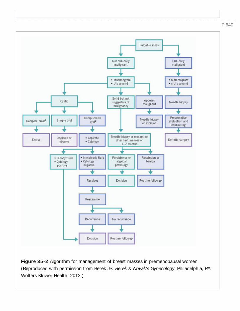
P.640
Figure 35-2 Algorithm for management of breast masses in premenopausal women.(Reproduced with permission from Berek JS. Berek & Novak's Gynecology. Philadelphia, PA:Wolters Kluwer Health, 2012.)
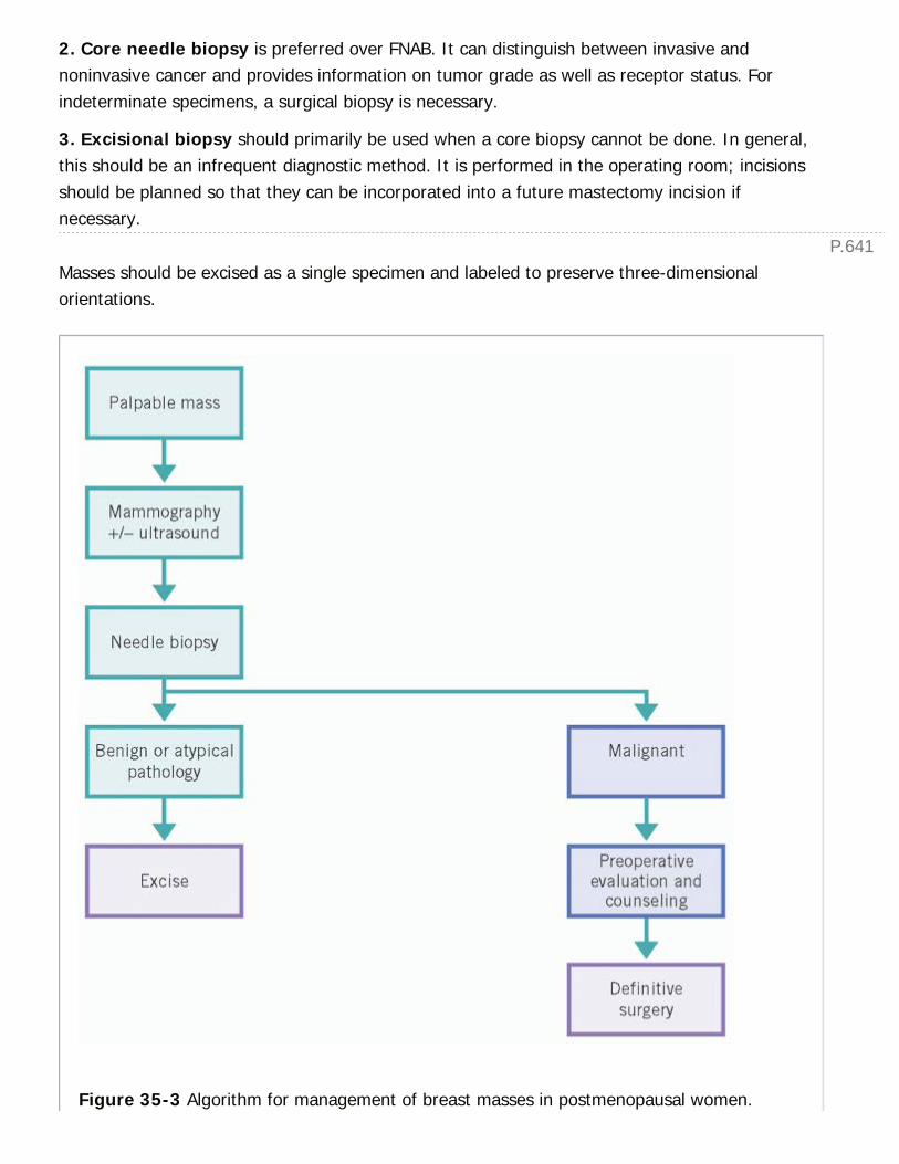
P.641
2. Core needle biopsy is preferred over FNAB. It can distinguish between invasive andnoninvasive cancer and provides information on tumor grade as well as receptor status. Forindeterminate specimens, a surgical biopsy is necessary.
3. Excisional biopsy should primarily be used when a core biopsy cannot be done. In general,this should be an infrequent diagnostic method. It is performed in the operating room; incisionsshould be planned so that they can be incorporated into a future mastectomy incision ifnecessary.
Masses should be excised as a single specimen and labeled to preserve three-dimensionalorientations.
Figure 35-3 Algorithm for management of breast masses in postmenopausal women.

P.642
(Reproduced with permission from Berek JS. Berek & Novak's Gynecology. Philadelphia, PA:Wolters Kluwer Health, 2012.)
4. Incisional biopsy is indicated for the evaluation of a large breast mass suspicious formalignancy but for which a definitive diagnosis cannot be made by FNAB or core biopsy. Forinflammatory breast cancer with skin involvement, an incisional biopsy can consist of a skinpunch biopsy.
B. Nonpalpable Lesions. Minimally invasive breast biopsy is the optimal initial tissue acquisitionmethod and procedure of choice for obtaining a pathologic diagnosis of image-detectedabnormalities. Correlation between pathology results and imaging findings is mandatory. Patientswith histologically benign findings on percutaneous biopsy do not require open biopsy if imagingand pathologic findings are concordant. Patients with
high-risk lesions on image-guided biopsy (atypical ductal hyperplasia [ADH], atypicallobular hyperplasia [ALH], lobular carcinoma in situ [LCIS], radial scar) may havemalignancy at the same site and should undergo a surgical biopsy.
1. Stereotactic core biopsy is used for nonpalpable mammographically detected lesions, suchas microcalcifications that cannot be seen with ultrasonography. In a vacuum-assisted approach,a metallic marking clip is usually placed through the probe after sampling is complete to allowfor identification of the biopsy site if excisional biopsy or partial mastectomy becomes necessary;this is the preferred approach for lesions presenting with microcalcifications without a visible orpalpable mass. Contraindications include lesions close to the chest wall or in the axillary tail andthin breasts that may allow needle strikethrough into the thorax. Superficial lesions and lesionsdirectly beneath the nippleÑareolar complex are also often not approachable with stereotactictechniques. Nondiagnostic and insufficient specimens necessitate needle-localized excisionalbiopsy (NLB, see later discussion).
2. Ultrasound-guided biopsy is generally easier to perform than stereotactic core biopsy andis the preferred method for lesions with a cystic component, as it can be used to aspirate the cystas well as to provide core biopsy specimens.
3. NLB. A needle and hookwire are placed into the breast adjacent to the concerning lesionunder mammographic guidance. The patient is then brought to the operating room for anexcisional biopsy. Using localization mammograms as a map, the whole hookwire, breast lesion,and a rim of normal breast tissue is removed en bloc. The specimen is oriented, and a radiographis performed to confirm the presence of the lesion within the specimen.
BENIGN BREAST CONDITIONS
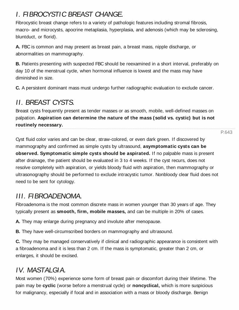
P.643
I. FIBROCYSTIC BREAST CHANGE.Fibrocystic breast change refers to a variety of pathologic features including stromal fibrosis,macro- and microcysts, apocrine metaplasia, hyperplasia, and adenosis (which may be sclerosing,bluntduct, or florid).
A. FBC is common and may present as breast pain, a breast mass, nipple discharge, orabnormalities on mammography.
B. Patients presenting with suspected FBC should be reexamined in a short interval, preferably onday 10 of the menstrual cycle, when hormonal influence is lowest and the mass may havediminished in size.
C. A persistent dominant mass must undergo further radiographic evaluation to exclude cancer.
II. BREAST CYSTS.Breast cysts frequently present as tender masses or as smooth, mobile, well-defined masses onpalpation. Aspiration can determine the nature of the mass (solid vs. cystic) but is notroutinely necessary.
Cyst fluid color varies and can be clear, straw-colored, or even dark green. If discovered bymammography and confirmed as simple cysts by ultrasound, asymptomatic cysts can beobserved. Symptomatic simple cysts should be aspirated. If no palpable mass is presentafter drainage, the patient should be evaluated in 3 to 4 weeks. If the cyst recurs, does notresolve completely with aspiration, or yields bloody fluid with aspiration, then mammography orultrasonography should be performed to exclude intracystic tumor. Nonbloody clear fluid does notneed to be sent for cytology.
III. FIBROADENOMA.Fibroadenoma is the most common discrete mass in women younger than 30 years of age. Theytypically present as smooth, firm, mobile masses, and can be multiple in 20% of cases.
A. They may enlarge during pregnancy and involute after menopause.
B. They have well-circumscribed borders on mammography and ultrasound.
C. They may be managed conservatively if clinical and radiographic appearance is consistent witha fibroadenoma and it is less than 2 cm. If the mass is symptomatic, greater than 2 cm, orenlarges, it should be excised.
IV. MASTALGIA.Most women (70%) experience some form of breast pain or discomfort during their lifetime. Thepain may be cyclic (worse before a menstrual cycle) or noncyclical, which is more suspiciousfor malignancy, especially if focal and in association with a mass or bloody discharge. Benign

P.644
disease is the etiology in the majority of cases. However, pain may be associated with cancer inup to 10% of patients. Once cancer has been excluded, most patients can be managedsuccessfully with symptomatic therapy and reassurance, as it resolves in up to 30% of women,though it does recur in 60%. In 15% of patients, the pain may be so disabling that it interfereswith activities of daily living. A well-fitting supportive bra is an important first step in painrelief. Topical nonsteroidal anti-inflammatory drugs (NSAIDs, e.g., diclofenac gel) isconsidered first-line therapy, having been proven effective with minimal side effects in arandomized trial (J Am Coll Surg. 2003;196:525-530). Lowdose tamoxifen (an estrogenantagonist) has been shown to provide good pain relief in placebo-controlled trials with tolerableside effects (Br J Surg. 1988; 75: 845Ñ846), although concerns over increased risks ofendometrial cancer limit long-term use. Danazol (a derivative of testosterone), bromocriptine,and gonadorelin analogs have significant side effects, and their use should be limited torefractory cases. Many patients experience symptomatic relief by reducing caffeine intake or bytaking vitamin E or evening primrose oil, although there is no scientific evidence supportingthese lifestyle modifications.
A. Superficial thrombophlebitis of the veins overlying the breast (Mondor disease) maypresent as breast pain. Treatment is conservative with NSAIDs and hot compresses.Antibiotics are not generally indicated.
B. Breast pain in pregnancy and lactation can occur from engorgement, clogged ducts,trauma to the areola and nipple from pumping or nursing,
or any of the aforementioned sources. Clogged ducts are usually treated with warmcompresses, soaks, and massage.
V. NIPPLE DISCHARGEA. Lactation is the most common physiologic cause of nipple discharge and may continue for upto 2 years after cessation of breastfeeding. In parous nonlactating women, a small amount ofmilk may be expressed from multiple ducts. This requires no treatment.
B. Galactorrhea is milky discharge unrelated to breastfeeding. Physiologic galactorrhea is thecontinued production of milk after lactation has ceased and menses has resumed and is oftencaused by continued mechanical stimulation of the nipples.
1. Drug-related galactorrhea is caused by medications that affect the hypothalamicÑpituitaryaxis by depleting dopamine (tricyclic antidepressants, reserpine, methyldopa, cimetidine,and benzodiazepines), blocking the dopamine receptor (phenothiazine, metoclopramide,and haloperidol), or having an estrogenic effect (digitalis). Discharge is generally bilateraland nonbloody.
2. Spontaneous galactorrhea in a nonlactating patient may be due to a pituitaryprolactinoma, and may be associated with amenorrhea. The diagnosis is established by
P.645
measuring the serum prolactin level and performing a computed tomography (CT) or MRIscan of the pituitary gland. Treatment is bromocriptine or resection of the prolactinoma.
C. Pathologic nipple discharge is usually bloody (can be confirmed with guaiac test),spontaneous, unilateral, and/or originates from a single duct. Normal physiologic dischargeis usually nonbloody, is from multiple ducts, can be a variety of colors (clear to yellow to green),and requires breast manipulation to produce. Cytologic evaluation is generally not useful.
1. Malignancy is the underlying cause in 10% of patients.
2. If not associated with a mass, the most likely etiologies are benign intraductal papilloma(peripheral papillomas put patients at slightly higher risk of malignancy), duct ectasia, andfibrocystic changes. In lactating women, serosanguineous or bloody discharge can beassociated with duct trauma, infection, or epithelial proliferation associated with breastenlargement.
3. Patients with persistent spontaneous discharge from a single duct require a surgicalmicrodochectomy (excision of a single duct and its associated lobule) using a ductogram orductoscopy or major duct excision (excision of all retroareolar ducts).
VI. BREAST INFECTIONSA. Lactational Mastitis
1. The most common causative organism is Staphylococcus aureus.
2. It presents as a swollen, erythematous, and tender breast; purulent discharge from the nippleis uncommon.
3. In the early cellulitic phase, the treatment is antibiotics, and the frequency of nursing orpumping should be increased. Approximately 25% progress to abscess formation.
4. Breast abscesses occur in the later stages and are often not fluctuant. The diagnosis is madeby failure to improve on antibiotics, abscess cavity seen on ultrasound, or aspiration of pus.Treatment is cessation of nursing and surgical drainage.
B. Nonpuerperal abscesses result from duct ectasia with periductal mastitis, infected cysts,infected hematoma, or hematogenous spread from another source.
1. They usually are located in the peri/retroareolar area.
2. Anaerobes are the most common causative agent, although antibiotics should cover bothanaerobic and aerobic organisms.
3. Treatment is surgical drainage.
4. Unresolved or recurring infection requires biopsy to exclude cancer. These patientsoften have a chronic relapsing course with multiple infections requiring surgical drainage.

P.646
5. Repeated infections can result in a chronically draining periareolar lesion or amammary fistula lined with squamous epithelium. Treatment is excision of the central ductalong with the fistula once the acute infection resolves. The fistula can recur even aftersurgery.
VII. GYNECOMASTIA.Gynecomastia is hypertrophy of breast tissue in men that is usually secondary to an imbalancebetween the breast stimulatory effects of estrogen and the inhibitory effects of androgens.
A. Pubertal hypertrophy occurs in adolescent boys, is usually bilateral, and resolvesspontaneously in 6 to 12 months.
B. Senescent gynecomastia is commonly seen after the age of 70 years, as testosterone levelsdecrease.
C. Drugs associated with this are similar to those that cause galactorrhea in women: Digoxin,spironolactone, methyldopa, cimetidine, tricyclic antidepressants, phenothiazine,reserpine, and marijuana. Drugs used for androgen blockade, such as luteinizing hormonereleasing hormone analogues for the treatment of prostate cancer and 5-alpha reductaseinhibitors (e.g., finasteride) for the management of benign prostatic hypertrophy, may alsoresult in gynecomastia.
D. Tumors can cause gynecomastia secondary to excess secretion of estrogens. These includetesticular teratomas and seminomas, bronchogenic carcinomas, adrenal tumors, and tumors ofthe pituitary and hypothalamus.
E. Gynecomastia may be a manifestation of systemic diseases such as hepatic cirrhosis, renalfailure, hyperthyroidism, and malnutrition.
F. During the workup of gynecomastia, cancer should be excluded by mammography andsubsequently by biopsy if a mass is found. If workup fails
to reveal a medically treatable cause or if the enlargement fails to regress, excision of breasttissue via a periareolar incision can be performed.
VIII. HIGH-RISK AND PREMALIGNANT CONDITIONSA. ADH and ALH are proliferative lesions with cell atypia that arise within breast ducts andlobules, respectively. ADH confers a 4 to 5 times increased relative risk of developing an invasivebreast malignancy. Historically, ALH was thought to have a weaker association with malignancy,but a recent study suggested that both lesions confer equal levels of risk (Cancer Prev Res.2014;7:211-217). If atypical hyperplasia is found on needle biopsy, excisional biopsy iswarranted to rule out associated malignancy. If no malignancy is found on postoperativepathology, patients with these conditions can simply undergo surveillance with imaging and
P.647
physician examination at increased intervals as compared to low-risk patients.Chemoprevention with tamoxifen is also an option.
B. LCIS is not considered a preinvasive lesion but rather an indicator for increased breast cancerrisk of approximately 1% per year (÷20% to 30% at 15 years) (J Natl Compr Canc Netw.2006;4:511-522).
1. It may be multifocal and/or bilateral.
2. The cancer that develops may be invasive ductal or lobular and may occur in eitherbreast.
3. LCIS has loss of E-cadherin (involved in cellÑcell adhesion), which can be stained for onpathology slides to clarify cases that are borderline ductal carcinoma in situ (DCIS) from LCIS.
4. Pleomorphic LCIS is a particularly aggressive subtype of LCIS that is treated more like DCIS;it tends to have less favorable biologic markers.
5. Treatment options are (1) lifelong close surveillance, (2) bilateral total mastectomieswith immediate reconstruction for selected women with a strong family history after appropriatecounseling, or (3) chemoprevention with tamoxifen, raloxifene (which has been validated inthe postmenopausal setting), or an aromatase inhibitor.
MALIGNANCY OF THE BREAST
I. EPIDEMIOLOGY.Breast cancer is the most common noncutaneous cancer in women, with a lifetime risk ofone in eight women. In the United States in 2014, an estimated 235,030 new cases of invasivebreast cancer and 62,570 new cases of noninvasive in situ carcinoma of the breast werediagnosed (CA Cancer J Clin. 2014;64:9-29). In that same period, approximately 40,000 womendied of breast cancer, making it the second-leading cause of cancer death in women, exceededonly by lung cancer.
II. STAGING.Breast cancer is staged by using the American Joint Committee on Cancer (AJCC) system,which is both a clinical and pathologic staging system and is based on the TNM system(tumor, node, and metastasis) system (Tables 35-2, 35-3, 35-4, 35-5 and 35-6). The most recentseventh edition marks a significant departure from the previous version with its inclusion ofpathologic staging, which can be performed in patients treated initially with definitive surgery orin patients treated with neoadjuvant systemic therapy followed by definitive surgery. Workupshould include the following in addition to breast-specific imaging:

TABLE 35-2 AJCC StagingÑPrimary Tumor (T)a
TX Primary tumor cannot be assessed
T0 No evidence of primary tumor
Tis Carcinoma in situ
Tis(DCIS)
DCIS
Tis(LCIS)
LCIS
Tis(Paget)
Paget disease of the nipple NOT associated with invasive carcinomaand/or carcinoma in situ (DCIS and/or LCIS) in the underlying breastparenchyma. Carcinomas in the breast parenchyma associated withPaget disease are categorized based on the size and characteristics ofthe parenchymal disease, although the presence of Paget diseaseshould still be noted.
T1 Tumor ≤20 mm in greatest dimension
T1mi Tumor ≤1 mm in greatest dimension
T1a Tumor >1 mm but ≤5 mm in greatest dimension
T1b Tumor >5 mm but ≤10 mm in greatest dimension
T1c Tumor >10 mm but ≤20 mm in greatest dimension
T2 Tumor >20 mm but ≤50 mm in greatest dimension
T3 Tumor >50 mm in greatest dimension

P.648
T4 Tumor of any size with direct extension to the chest wall and/or to the
skin (ulceration or skin nodules)b
T4a Extension to the chest wall, not including only pectoralis muscleadherence/invasion
T4b Ulceration and/or ipsilateral satellite nodules and/or edema (includingpeau d'orange) of the skin, which do not meet the criteria forinflammatory carcinoma
T4c Both T4a and T4b
T4d Inflammatory carcinoma
aThe T classification of the primary tumor is the same regardless of whether it is basedon clinical or pathologic criteria, or both. Size should be measured to the nearestmillimeter. If the tumor size is slightly less than or greater than a cutoff for a given Tclassification, it is recommended that the size be rounded to the millimeter reading thatis closest to the cutoff. For example, a reported size of 1.1 mm is reported as 1 mm, ora size of 2.01 cm is reported as 2 cm. Designation should be made with the subscriptÒcÓ or ÒpÓ modifier to indicate whether the T classification was determined by clinical(physical examination or radiologic) or pathologic measurements, respectively. Ingeneral, pathologic determination should take precedence over clinical determination ofT size.b Invasion of the dermis alone does not qualify as T4.DCIS, ductal carcinoma in situ; LCIS, lobular carcinoma in situ.Used with the permission of the American Joint Committee on Cancer (AJCC), Chicago,Illinois. The original source for this material is the AJCC Cancer Staging Manual, SeventhEdition (2010) published by Springer Science and Business Media LLC,www.springer.com.
TABLE 35-3 AJCC StagingÑRegional Lymph Nodes (N)
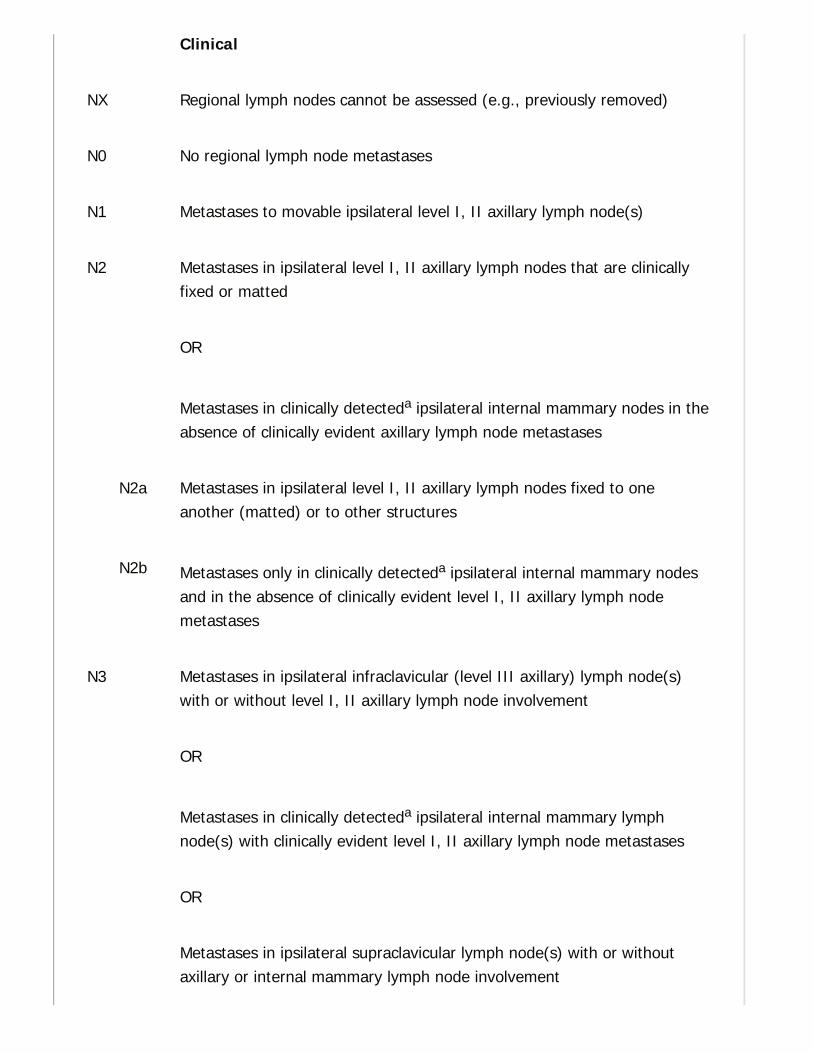
Clinical
NX Regional lymph nodes cannot be assessed (e.g., previously removed)
N0 No regional lymph node metastases
N1 Metastases to movable ipsilateral level I, II axillary lymph node(s)
N2 Metastases in ipsilateral level I, II axillary lymph nodes that are clinicallyfixed or matted
OR
Metastases in clinically detecteda ipsilateral internal mammary nodes in theabsence of clinically evident axillary lymph node metastases
N2a Metastases in ipsilateral level I, II axillary lymph nodes fixed to oneanother (matted) or to other structures
N2b Metastases only in clinically detecteda ipsilateral internal mammary nodesand in the absence of clinically evident level I, II axillary lymph nodemetastases
N3 Metastases in ipsilateral infraclavicular (level III axillary) lymph node(s)with or without level I, II axillary lymph node involvement
OR
Metastases in clinically detecteda ipsilateral internal mammary lymphnode(s) with clinically evident level I, II axillary lymph node metastases
OR
Metastases in ipsilateral supraclavicular lymph node(s) with or withoutaxillary or internal mammary lymph node involvement

P.649P.650
N3a Metastases in ipsilateral infraclavicular lymph node(s)
N3b Metastases in ipsilateral internal mammary lymph node(s) and axillarylymph node(s)
N3c Metastases in ipsilateral supraclavicular lymph node(s)
a Clinically detected is defined as detected by imaging studies (excludinglymphoscintigraphy) or by clinical examination and having characteristics highlysuspicious for malignancy or a presumed pathologic macrometastasis based on fine-needle aspiration biopsy with cytologic examination. Confirmation of clinically detectedmetastatic disease by fine-needle aspiration without excision biopsy is designated withan (f) suffix, for example, cN3a(f). Excisional biopsy of a lymph node or biopsy of asentinel node, in the absence of assignment of a pT, is classified as a clinical N, forexample, cN1. Information regarding the confirmation of the nodal status will bedesignated in site-specific factors as clinical, fine-needle aspiration, core biopsy, orsentinel lymph node biopsy. Pathologic classification (pN) is used for excision or sentinellymph node biopsy only in conjunction with a pathologic T assignment.Used with the permission of the American Joint Committee on Cancer (AJCC), Chicago,Illinois. The original source for this material is the AJCC Cancer Staging Manual, SeventhEdition (2010) published by Springer Science and Business Media LLC,www.springer.com.
TABLE 35-4 AJCC StagingÑPathologic Lymph NodeStatus (pN)a,b,c
pNX Regional lymph nodes cannot be assessed (e.g., previously removedor not removed for pathologic study)
pN0 No regional lymph node metastasis identified histologically
Note: ITCs are defined as small clusters of cells ≤0.2 mm, or single tumor cells, or a

cluster of <200 cells in a single histologic cross-section. ITCs may be detected byroutine histology or by IHC methods. Nodes containing only ITCs are excluded from thetotal positive node count for purposes of N classification but should be included in thetotal number of nodes evaluated.
pN0(i-) No regional lymph node metastases histologically, negative IHC
pN0(i+) Malignant cells in regional lymph node(s) ≤0.2 mm (detected byH&E or IHC including ITC)
pN0(mol-) No regional lymph node metastases histologically, negativemolecular findings (RT-PCR)
pN0(mol+) Positive molecular findings (RT-PCR), but no regional lymph nodemetastases detected by histology or IHC
pN1 Micrometastases
OR
Metastases in 1-3 axillary lymph nodes
AND/OR
Metastases in internal mammary nodes with metastases detected by
sentinel lymph node biopsy but not clinically detectedb
pN1mi Micrometastases (>0.2 mm and/or >200 cells but none >2.0 mm)
pN1a Metastases in 1-3 axillary lymph nodes, at least one metastasis>2.0 mm
pN1b Metastases in internal mammary nodes with micrometastases ormacrometastases detected by sentinel lymph node biopsy but not
clinically detectedb

pN1c Metastases in 1-3 axillary lymph nodes and in internal mammarylymph nodes with micrometastases or macrometastases detected bysentinel lymph node biopsy but not clinically detected
pN2 Metastases in 4-9 axillary lymph nodes
OR
Metastases in clinically detectedb internal mammary lymph nodes inthe absence of axillary lymph node metastases
pN2a Metastases in 4-9 axillary lymph nodes (at least 1 tumor deposit >2mm)
pN2b Metastases in clinically detectedc internal mammary lymph nodes inthe absence of axillary lymph node metastases
pN3 Metastases in ≥10 axillary lymph nodes
OR
Metastases in infraclavicular (level III axillary) lymph nodes
OR
Metastases in clinically detectedc ipsilateral internal mammarylymph nodes in the presence of one or more positive level I, IIaxillary lymph nodes
OR
Metastases in >3 axillary lymph nodes and in internal mammarylymph nodes with micrometastases or macrometastases detected by
sentinel lymph node biopsy but not clinically detectedb
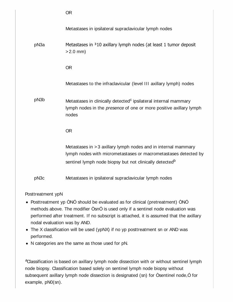
OR
Metastases in ipsilateral supraclavicular lymph nodes
pN3a Metastases in ≥10 axillary lymph nodes (at least 1 tumor deposit>2.0 mm)
OR
Metastases to the infraclavicular (level III axillary lymph) nodes
pN3b Metastases in clinically detectedc ipsilateral internal mammarylymph nodes in the presence of one or more positive axillary lymphnodes
OR
Metastases in >3 axillary lymph nodes and in internal mammarylymph nodes with micrometastases or macrometastases detected by
sentinel lymph node biopsy but not clinically detectedb
pN3c Metastases in ipsilateral supraclavicular lymph nodes
Posttreatment ypN
Posttreatment yp ÒNÓ should be evaluated as for clinical (pretreatment) ÒNÓmethods above. The modifier ÒsnÓ is used only if a sentinel node evaluation wasperformed after treatment. If no subscript is attached, it is assumed that the axillarynodal evaluation was by AND.The X classification will be used (ypNX) if no yp posttreatment sn or AND wasperformed.N categories are the same as those used for pN.
aClassification is based on axillary lymph node dissection with or without sentinel lymphnode biopsy. Classification based solely on sentinel lymph node biopsy withoutsubsequent axillary lymph node dissection is designated (sn) for Òsentinel node,Ó forexample, pN0(sn).
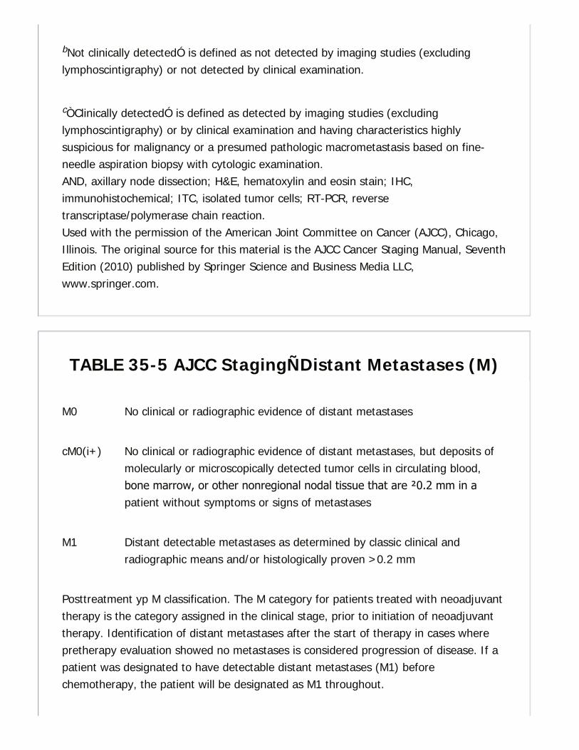
bNot clinically detectedÓ is defined as not detected by imaging studies (excludinglymphoscintigraphy) or not detected by clinical examination.
cÒClinically detectedÓ is defined as detected by imaging studies (excludinglymphoscintigraphy) or by clinical examination and having characteristics highlysuspicious for malignancy or a presumed pathologic macrometastasis based on fine-needle aspiration biopsy with cytologic examination.AND, axillary node dissection; H&E, hematoxylin and eosin stain; IHC,immunohistochemical; ITC, isolated tumor cells; RT-PCR, reversetranscriptase/polymerase chain reaction.Used with the permission of the American Joint Committee on Cancer (AJCC), Chicago,Illinois. The original source for this material is the AJCC Cancer Staging Manual, SeventhEdition (2010) published by Springer Science and Business Media LLC,www.springer.com.
TABLE 35-5 AJCC StagingÑDistant Metastases (M)
M0 No clinical or radiographic evidence of distant metastases
cM0(i+) No clinical or radiographic evidence of distant metastases, but deposits ofmolecularly or microscopically detected tumor cells in circulating blood,bone marrow, or other nonregional nodal tissue that are ≤0.2 mm in apatient without symptoms or signs of metastases
M1 Distant detectable metastases as determined by classic clinical andradiographic means and/or histologically proven >0.2 mm
Posttreatment yp M classification. The M category for patients treated with neoadjuvanttherapy is the category assigned in the clinical stage, prior to initiation of neoadjuvanttherapy. Identification of distant metastases after the start of therapy in cases wherepretherapy evaluation showed no metastases is considered progression of disease. If apatient was designated to have detectable distant metastases (M1) beforechemotherapy, the patient will be designated as M1 throughout.

P.651
P.652
Used with the permission of the American Joint Committee on Cancer (AJCC), Chicago,Illinois. The original source for this material is the AJCC Cancer Staging Manual, SeventhEdition (2010) published by Springer Science and Business Media LLC,www.springer.com.
A. Complete blood cell count (CBC), complete metabolic panel including liver functiontests (LFTs), and chest x-ray.
B. A bone scan, if the alkaline phosphatase or calcium level is elevated.
C. CT scan of the abdomen if LFTS are abnormal.
D. Patients with clinical stage III or IV disease should undergo bone scan and CT scan ofthe chest/abdomen/pelvis due to a high probability of distant metastases.
III. TUMOR BIOMARKERS AND PROGNOSTIC FACTORS.These should be evaluated on all tumor specimens. The presence or absence of disease inaxillary lymph nodes is the single most important prognostic factor in breast cancer. Tumorsize and grade (which is based on degrees of glandular differentiation, mitotic count, andnuclear grade) are the most reliable pathologic predictors of outcome for patients without axillarynodal involvement. High grade (i.e., grade 3 on a scale of 1 to 3) is a poor prognostic factor.
A. Hormone Receptors. Expression of ERs and progesterone (PRs) should be evaluated byimmunohistochemistry. Intense ER and PR staining is a good prognostic factor.
TABLE 35-6 AJCC Anatomic Stage/Prognostic Groupsa,b
Stage T N M
0 Tis N0 M0
IA T1a N0 M0
IB T0 N1mi M0

T1b N1mi M0
IIA T0 N1b M0
T1a N1b M0
T2 N0 M0
IIB T2 N1 M0
T3 N0 M0
IIIA T0 N2 M0
T1a N2 M0
T2 N2 M0
T3 N1 M0
T3 N2 M0
IIIB T4 N0 M0
T4 N1 M0
T4 N2 M0
IIIC Any T N3 M0
IV Any T Any N M1
M0 includes M0(i+).The designation pM0 is not valid; any M0 should be clinical. If a patient presents with
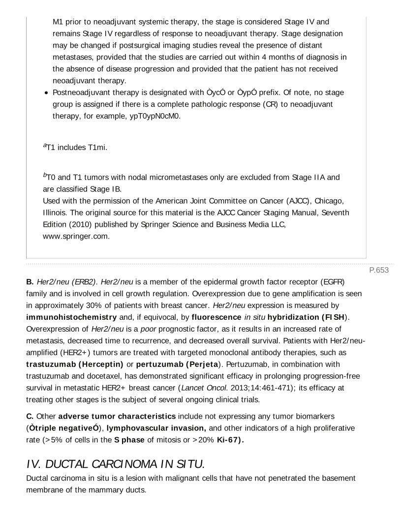
P.653
M1 prior to neoadjuvant systemic therapy, the stage is considered Stage IV andremains Stage IV regardless of response to neoadjuvant therapy. Stage designationmay be changed if postsurgical imaging studies reveal the presence of distantmetastases, provided that the studies are carried out within 4 months of diagnosis inthe absence of disease progression and provided that the patient has not receivedneoadjuvant therapy.Postneoadjuvant therapy is designated with ÒycÓ or ÒypÓ prefix. Of note, no stagegroup is assigned if there is a complete pathologic response (CR) to neoadjuvanttherapy, for example, ypT0ypN0cM0.
aT1 includes T1mi.
bT0 and T1 tumors with nodal micrometastases only are excluded from Stage IIA andare classified Stage IB.Used with the permission of the American Joint Committee on Cancer (AJCC), Chicago,Illinois. The original source for this material is the AJCC Cancer Staging Manual, SeventhEdition (2010) published by Springer Science and Business Media LLC,www.springer.com.
B. Her2/neu (ERB2). Her2/neu is a member of the epidermal growth factor receptor (EGFR)family and is involved in cell growth regulation. Overexpression due to gene amplification is seenin approximately 30% of patients with breast cancer. Her2/neu expression is measured byimmunohistochemistry and, if equivocal, by fluorescence in situ hybridization (FISH).Overexpression of Her2/neu is a poor prognostic factor, as it results in an increased rate ofmetastasis, decreased time to recurrence, and decreased overall survival. Patients with Her2/neu-amplified (HER2+) tumors are treated with targeted monoclonal antibody therapies, such astrastuzumab (Herceptin) or pertuzumab (Perjeta). Pertuzumab, in combination withtrastuzumab and docetaxel, has demonstrated significant efficacy in prolonging progression-freesurvival in metastatic HER2+ breast cancer (Lancet Oncol. 2013;14:461-471); its efficacy attreating other stages is the subject of several ongoing clinical trials.
C. Other adverse tumor characteristics include not expressing any tumor biomarkers(Òtriple negativeÓ), lymphovascular invasion, and other indicators of a high proliferativerate (>5% of cells in the S phase of mitosis or >20% Ki-67).
IV. DUCTAL CARCINOMA IN SITU.Ductal carcinoma in situ is a lesion with malignant cells that have not penetrated the basementmembrane of the mammary ducts.

P.654
A. DCIS is treated as a malignancy because DCIS has the potential to develop into invasivecancer.
It is usually detected by mammography as clustered pleomorphic calcifications (Fig. 35-4).
Physical examination is normal in the majority of patients.
It may advance in a segmental manner, with gaps between disease areas.
It can be multifocal (two or more lesions >5 mm apart within the same index quadrant) ormulticentric (in different quadrants).
1. Histology
a. There are five architectural subtypes: papillary, micropapillary, solid, cribriform, andcomedo (necrosis). Specimens are also grouped as comedo versus noncomedo.
b. The high-grade subtype is often associated with microinvasion, a higher proliferation rate,aneuploidy, gene amplification, and a higher local recurrence rate.
c. ER and PR expression levels should be obtained if hormone therapy is being considered.
2. Treatment
a. Surgical excision alone (via partial mastectomy) with margins greater than 10 mm isassociated with a local recurrence rate of 14% at 12 years (Am J Surg. 2006;192:420-222). Theaddition of adjuvant radiation reduces the local recurrence rate to 2.5%. Approximately half ofthe recurrences present as invasive ductal carcinomas. Surgical options depend on the extentof disease, grade, margin status, multicentricity of disease, and patient age.
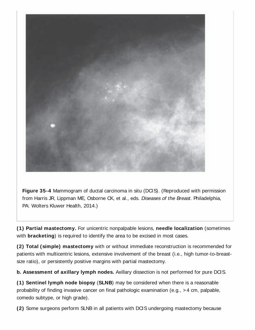
Figure 35-4 Mammogram of ductal carcinoma in situ (DCIS). (Reproduced with permissionfrom Harris JR, Lippman ME, Osborne CK, et al., eds. Diseases of the Breast. Philadelphia,PA: Wolters Kluwer Health, 2014.)
(1) Partial mastectomy. For unicentric nonpalpable lesions, needle localization (sometimeswith bracketing) is required to identify the area to be excised in most cases.
(2) Total (simple) mastectomy with or without immediate reconstruction is recommended forpatients with multicentric lesions, extensive involvement of the breast (i.e., high tumor-to-breast-size ratio), or persistently positive margins with partial mastectomy.
b. Assessment of axillary lymph nodes. Axillary dissection is not performed for pure DCIS.
(1) Sentinel lymph node biopsy (SLNB) may be considered when there is a reasonableprobability of finding invasive cancer on final pathologic examination (e.g., >4 cm, palpable,comedo subtype, or high grade).
(2) Some surgeons perform SLNB in all patients with DCIS undergoing mastectomy because

P.656
P.655
SLNB cannot be performed postmastectomy if an occult invasive cancer is found. This is an areaof ongoing controversy and research.
(3) A positive sentinel node indicates invasive breast cancer and changes the stage of thedisease. Historically, a completion axillary dissection was indicated for patients with a positivesentinel node, but the findings of the American College of Surgeons Oncology Group (ACOSOG)trial Z011, have contributed to a paradigm shift in the management of axillary disease (see laterdiscussion in ÒManagement of the AxillaÓ).
c. Adjuvant therapy
(1) For pure DCIS, there is no added benefit from systemic chemotherapy because the disease isconfined to the ducts of the breast. However, in those patients with ER-positive (ER+) DCIS,adjuvant tamoxifen can reduce the risk of breast cancer recurrence by 36% over 5 years andthe risk of developing a new contralateral breast cancer (NSABP B-24 trial, J Clin Oncol.2012;30:1268-1273) but confers no survival benefit. Aromatase inhibitors (e.g., anastrozole,exemestane, letrozole), which block the peripheral conversion of androgens into estrogens byinhibiting the enzyme aromatase but do not affect estrogen produced by the ovaries, aresometimes used as an alternative in postmenopausal patients.
(2) Adjuvant radiation should be given to patients with DCIS treated with partial mastectomyto decrease the rate of local recurrence (NSABP B-17 trial, J Natl Cancer Inst. 2011;103:478-488). This is especially true for younger women with close margins or large tumors. However,there is no survival benefit. For older patients with smaller, widely excised DCIS of low orintermediate grade, the benefit of radiation therapy is less clear and adjuvant radiation may notbe necessary (Ann Surg Oncol. 2013;20:3175-3179).
d. The University of Southern California/Van Nuys Prognostic Index (Table 35-7) is anumerical algorithm used to determine which patients with DCIS are at greatest risk forrecurrence and would therefore benefit from aggressive treatment. In order to
achieve a local recurrence rate of less than 20% at 12 years, surgical excision alone isrecommended for all patients scoring 4, 5, or 6 and patients who score 7 but have margin widths≥3 mm; surgical excision plus radiation therapy for those who score 7 and have margins <3mm, patients who score 8 and have margins ≥3 mm, and for patients who score 9 and havemargins >5 mm; and mastectomy for patients who score 8 and have margins <3 mm, whoscore 9 and have margins <5 mm, and for all patients who score 10, 11, or 12 (J Natl Cancer InstMonogr. 2010;2010(41):193-196).
TABLE 35-7 University of Southern California/Van Nuysa
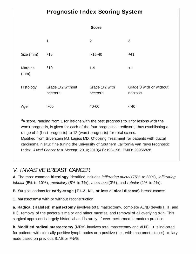
Prognostic Index Scoring System
Score
1 2 3
Size (mm) ≤15 >15-40 ≥41
Margins(mm)
≤10 1-9 <1
Histology Grade 1/2 withoutnecrosis
Grade 1/2 withnecrosis
Grade 3 with or withoutnecrosis
Age >60 40-60 <40
aA score, ranging from 1 for lesions with the best prognosis to 3 for lesions with theworst prognosis, is given for each of the four prognostic predictors, thus establishing arange of 4 (best prognosis) to 12 (worst prognosis) for total scores.Modified from Silverstein MJ, Lagios MD. Choosing Treatment for patients with ductalcarcinoma in situ: fine tuning the University of Southern California/Van Nuys PrognosticIndex. J Natl Cancer Inst Monogr. 2010;2010(41):193-196. PMID: 20956828.
V. INVASIVE BREAST CANCERA. The most common histology identified includes infiltrating ductal (75% to 80%), infiltratinglobular (5% to 10%), medullary (5% to 7%), mucinous (3%), and tubular (1% to 2%).
B. Surgical options for early-stage (T1-2, N1, or less clinical disease) breast cancer:
1. Mastectomy with or without reconstruction.
a. Radical (Halsted) mastectomy involves total mastectomy, complete ALND (levels I, II, andIII), removal of the pectoralis major and minor muscles, and removal of all overlying skin. Thissurgical approach is largely historical and is rarely, if ever, performed in modern practice.
b. Modified radical mastectomy (MRM) involves total mastectomy and ALND. It is indicatedfor patients with clinically positive lymph nodes or a positive (i.e., with macrometastases) axillarynode based on previous SLNB or FNAB.
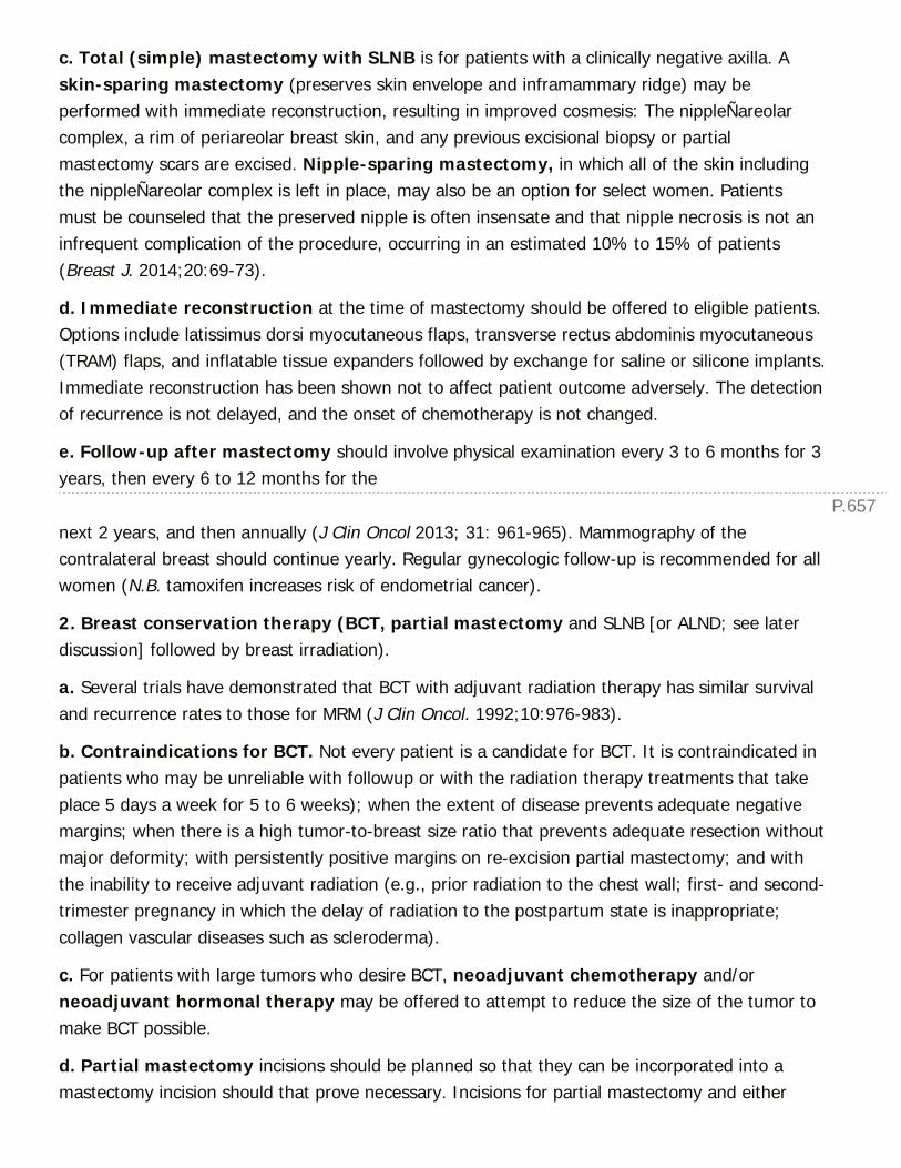
P.657
c. Total (simple) mastectomy with SLNB is for patients with a clinically negative axilla. Askin-sparing mastectomy (preserves skin envelope and inframammary ridge) may beperformed with immediate reconstruction, resulting in improved cosmesis: The nippleÑareolarcomplex, a rim of periareolar breast skin, and any previous excisional biopsy or partialmastectomy scars are excised. Nipple-sparing mastectomy, in which all of the skin includingthe nippleÑareolar complex is left in place, may also be an option for select women. Patientsmust be counseled that the preserved nipple is often insensate and that nipple necrosis is not aninfrequent complication of the procedure, occurring in an estimated 10% to 15% of patients(Breast J. 2014;20:69-73).
d. Immediate reconstruction at the time of mastectomy should be offered to eligible patients.Options include latissimus dorsi myocutaneous flaps, transverse rectus abdominis myocutaneous(TRAM) flaps, and inflatable tissue expanders followed by exchange for saline or silicone implants.Immediate reconstruction has been shown not to affect patient outcome adversely. The detectionof recurrence is not delayed, and the onset of chemotherapy is not changed.
e. Follow-up after mastectomy should involve physical examination every 3 to 6 months for 3years, then every 6 to 12 months for the
next 2 years, and then annually (J Clin Oncol 2013; 31: 961-965). Mammography of thecontralateral breast should continue yearly. Regular gynecologic follow-up is recommended for allwomen (N.B. tamoxifen increases risk of endometrial cancer).
2. Breast conservation therapy (BCT, partial mastectomy and SLNB [or ALND; see laterdiscussion] followed by breast irradiation).
a. Several trials have demonstrated that BCT with adjuvant radiation therapy has similar survivaland recurrence rates to those for MRM (J Clin Oncol. 1992;10:976-983).
b. Contraindications for BCT. Not every patient is a candidate for BCT. It is contraindicated inpatients who may be unreliable with followup or with the radiation therapy treatments that takeplace 5 days a week for 5 to 6 weeks); when the extent of disease prevents adequate negativemargins; when there is a high tumor-to-breast size ratio that prevents adequate resection withoutmajor deformity; with persistently positive margins on re-excision partial mastectomy; and withthe inability to receive adjuvant radiation (e.g., prior radiation to the chest wall; first- and second-trimester pregnancy in which the delay of radiation to the postpartum state is inappropriate;collagen vascular diseases such as scleroderma).
c. For patients with large tumors who desire BCT, neoadjuvant chemotherapy and/orneoadjuvant hormonal therapy may be offered to attempt to reduce the size of the tumor tomake BCT possible.
d. Partial mastectomy incisions should be planned so that they can be incorporated into amastectomy incision should that prove necessary. Incisions for partial mastectomy and either
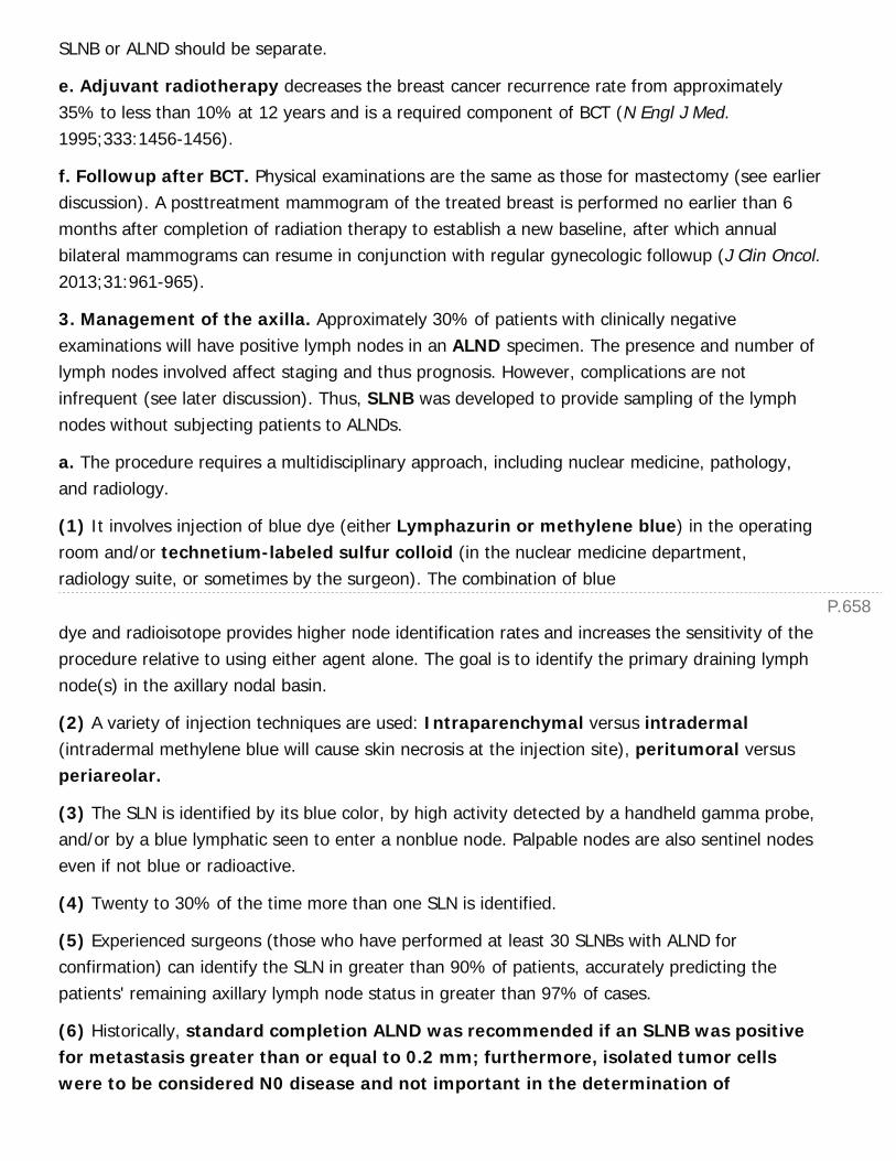
P.658
SLNB or ALND should be separate.
e. Adjuvant radiotherapy decreases the breast cancer recurrence rate from approximately35% to less than 10% at 12 years and is a required component of BCT (N Engl J Med.1995;333:1456-1456).
f. Followup after BCT. Physical examinations are the same as those for mastectomy (see earlierdiscussion). A posttreatment mammogram of the treated breast is performed no earlier than 6months after completion of radiation therapy to establish a new baseline, after which annualbilateral mammograms can resume in conjunction with regular gynecologic followup (J Clin Oncol.2013;31:961-965).
3. Management of the axilla. Approximately 30% of patients with clinically negativeexaminations will have positive lymph nodes in an ALND specimen. The presence and number oflymph nodes involved affect staging and thus prognosis. However, complications are notinfrequent (see later discussion). Thus, SLNB was developed to provide sampling of the lymphnodes without subjecting patients to ALNDs.
a. The procedure requires a multidisciplinary approach, including nuclear medicine, pathology,and radiology.
(1) It involves injection of blue dye (either Lymphazurin or methylene blue) in the operatingroom and/or technetium-labeled sulfur colloid (in the nuclear medicine department,radiology suite, or sometimes by the surgeon). The combination of blue
dye and radioisotope provides higher node identification rates and increases the sensitivity of theprocedure relative to using either agent alone. The goal is to identify the primary draining lymphnode(s) in the axillary nodal basin.
(2) A variety of injection techniques are used: Intraparenchymal versus intradermal(intradermal methylene blue will cause skin necrosis at the injection site), peritumoral versusperiareolar.
(3) The SLN is identified by its blue color, by high activity detected by a handheld gamma probe,and/or by a blue lymphatic seen to enter a nonblue node. Palpable nodes are also sentinel nodeseven if not blue or radioactive.
(4) Twenty to 30% of the time more than one SLN is identified.
(5) Experienced surgeons (those who have performed at least 30 SLNBs with ALND forconfirmation) can identify the SLN in greater than 90% of patients, accurately predicting thepatients' remaining axillary lymph node status in greater than 97% of cases.
(6) Historically, standard completion ALND was recommended if an SLNB was positivefor metastasis greater than or equal to 0.2 mm; furthermore, isolated tumor cellswere to be considered N0 disease and not important in the determination of
P.659
therapeutic decisions. This remains the standard of care in many practices, but thissurgical paradigm has recently been challenged by a number of studies, mostsignificantly by the results of the ACOSOG Z0011 trial. This randomized trial compared theoverall survival and local recurrence rates for patients with T1Ñ2 tumors and limited SLNmetastatic disease who received BCT and systemic therapy and either had ALND or no furtheraxillary procedures (JAMA. 2011;305:569-575.). There was no difference in the two groups,leading many to defer completion ALND for this subgroup of patients. All patients underwentlumpectomy, whole breast radiation, and systemic therapy; thus, the results cannot begeneralized to all patients with a positive SLN. Nevertheless, this study contributed to growingconcern that excess surgery is being performed for axillary disease. This concern has garneredfurther discussion following publication of the results of the AMAROS trial, a Europeanrandomized noninferiority trial that compared completion ALND to axillary radiation therapy for apositive SLN in patients with T1Ñ2 primary breast cancer and no palpable lymphadenopathy;rates of local recurrence were statistically equivalent and radiation was associated with lesslymphedema than ALND (Lancet Oncol. 2014;15:1303-1310). Furthermore, the prognosticsignificance of isolated tumor cells has been brought into question by the Dutch MIRRORcohort study, which demonstrated an association between disease-free survival and isolatedtumor cells in women who had been diagnosed at a young age and in women with triple-negativecancer who did
not undergo either ALND or axillary radiation (N Engl J Med. 2009;361:653-663). Finally, theSOUND trial is an ongoing clinical trial that is examining the extent to which SLNB can beobviated by sonographic staging of the axilla in a subset of patients (Breast. 2012;21:678-681;Fig. 35-5).
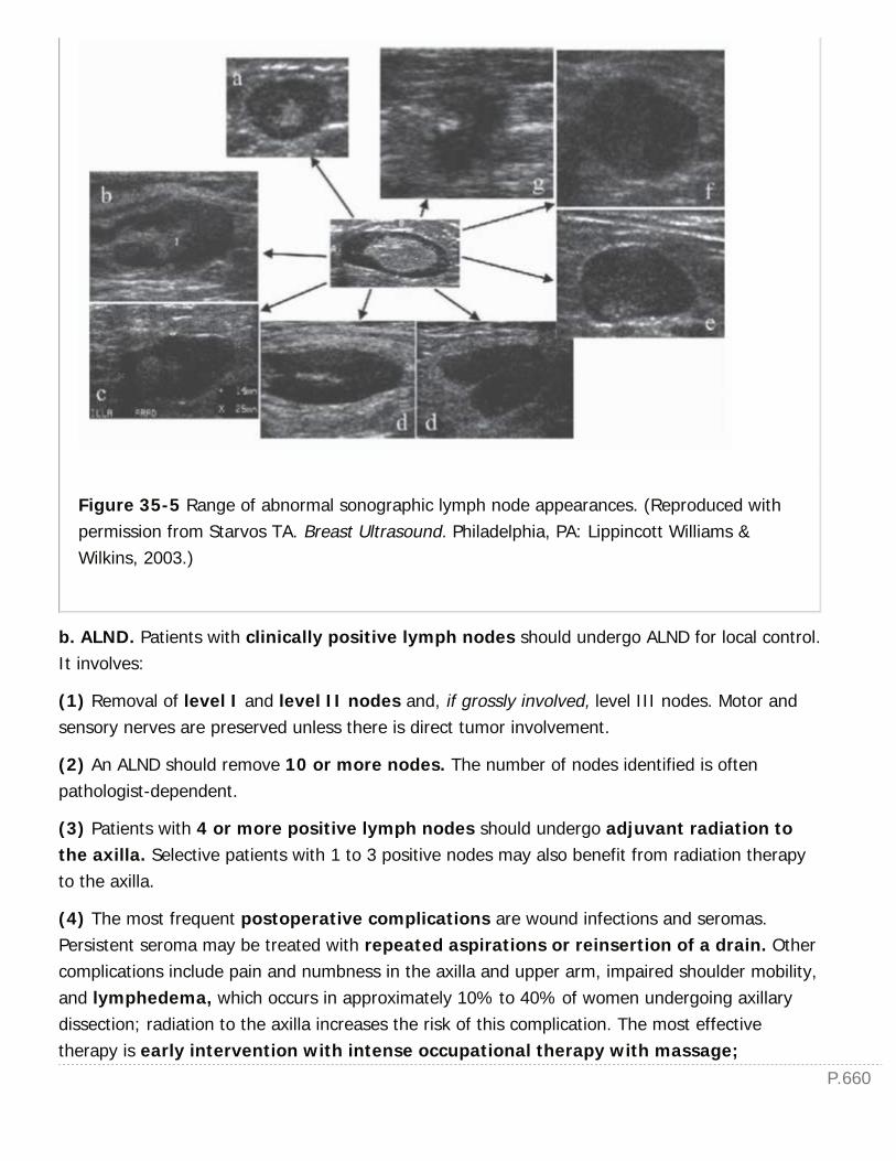
P.660
Figure 35-5 Range of abnormal sonographic lymph node appearances. (Reproduced withpermission from Starvos TA. Breast Ultrasound. Philadelphia, PA: Lippincott Williams &Wilkins, 2003.)
b. ALND. Patients with clinically positive lymph nodes should undergo ALND for local control.It involves:
(1) Removal of level I and level II nodes and, if grossly involved, level III nodes. Motor andsensory nerves are preserved unless there is direct tumor involvement.
(2) An ALND should remove 10 or more nodes. The number of nodes identified is oftenpathologist-dependent.
(3) Patients with 4 or more positive lymph nodes should undergo adjuvant radiation tothe axilla. Selective patients with 1 to 3 positive nodes may also benefit from radiation therapyto the axilla.
(4) The most frequent postoperative complications are wound infections and seromas.Persistent seroma may be treated with repeated aspirations or reinsertion of a drain. Othercomplications include pain and numbness in the axilla and upper arm, impaired shoulder mobility,and lymphedema, which occurs in approximately 10% to 40% of women undergoing axillarydissection; radiation to the axilla increases the risk of this complication. The most effectivetherapy is early intervention with intense occupational therapy with massage;
P.661
graded pneumatic compression devices and a professionally fitted compression sleevecan also provide relief and prevent worsening of lymphedema. Blood draws, blood pressure cuffs,and intravenous lines should be avoided in the affected arm, mainly to avoid infection. Infectionsof the hand or arm should be treated promptly and aggressively with antibiotics and armelevation because infection can damage lymphatics further and cause irreversible lymphedema.Lymphedema increases the risk of developing angiosarcoma.
C. Adjuvant systemic therapy is given in appropriate patients after completion of surgery.
1. All node-positive patients should be considered for adjuvant chemotherapy.
a. Regimens are guided by the tumor biomarkers. Typical regimens consist of four to eight cyclesof a combination of cyclophosphamide and an anthracycline followed by a taxane administeredevery 2 to 3 weeks.
b. Patients with ER-positive tumors receive adjuvant hormonal therapy for 5 to 10 years.Tamoxifen is given to premenopausal women, and aromatase inhibitors are given topostmenopausal women.
c. In postmenopausal women older than 70 years, chemotherapy is performed less frequently. Inpostmenopausal women with ER+ tumors, tamoxifen or an aromatase inhibitor is frequently thesole adjuvant medical therapy.
d. In patients with Her2/neu-positive tumors, polychemotherapy is combined with biologictherapy targeting the Her2/neu protein (see earlier discussion).
2. Node-negative patients may have increased disease-free survival from adjuvantchemotherapy and/or hormonal therapy. An individualized approach is crucial and requiresthorough discussion with the patient regarding the risks of recurrence without adjuvant therapy,the cost and toxicities of treatment, and the expected benefit in risk reduction and survival.
a. Node-negative patients who are at high risk and benefit the most from adjuvantchemotherapy include those with tumors greater than 1 cm, high tumor grade, Her2/neuexpression, aneuploidy, elevated Ki-67 expression, high percentage of cells in S phase,lymphovascular invasion, and ER/PR-negative tumors.
b. The NSABP B-20 trial and the International Breast Cancer Study Group trial IX showed thatpolychemotherapy in combination with tamoxifen was superior to tamoxifen alone inincreasing disease-free and overall survival, especially in ER-negative patients, regardless oftumor size.
c. The St. Gallen Consensus Panel in 1997 suggested that patients who have node-negativedisease and whose tumors are 1 cm or less and ER-positive may be spared adjuvantchemotherapy but still may benefit from adjuvant endocrine therapy.
d. The Web site http://www.adjuvantonline.com provides an online tool for physicians to use to
P.662
calculate the added benefit of hormonal and chemotherapeutic therapies.
D. Adjuvant Radiation
1. Indications for adjuvant radiation to the chest wall and axilla after mastectomy include T3and T4 tumors, attachment to the pectoral fascia, positive surgical margins, skin involvement,involved internal mammary nodes, inadequate or no axillary dissection, four or more positivelymph nodes, and residual tumor on the axillary vein. Presence of one to three positive axillarynodes is a relative indication (N Engl J Med. 1997;337:949-955). (See earlier discussion ofradiation after BCT.)
2. Complications. Radiation to the chest wall can cause skin changes. Infrequent complicationsinclude interstitial pneumonitis, spontaneous rib fracture, breast fibrosis, pericarditis, pleuraleffusion, and chest wall myositis. Radiation to the axilla can increase the incidence oflymphedema and axillary fibrosis.
E. Locally advanced breast cancer (LABC, i.e., T3, T4, N2, and/or N3 clinical disease)
1. Staging in LABC. Because up to 10% to 20% of these patients have distant metastasis at thetime of presentation, all should receive a bone scan and CT scan of the chest and abdomenbefore treatment.
2. Noninflammatory LABC
a. Patients should receive neoadjuvant chemotherapy (often cyclophosphamide combinedwith an anthracycline [e.g., doxorubicin] and a taxane), followed by surgery and radiation asbased on previously discussed criteria. Neoadjuvant chemotherapy also provides informationregarding tumor response to treatment that may aid to guide further adjuvant therapy. Additionaladjuvant chemotherapy is also necessary in select cases. SLNB may be used in selected patientswith a clinically negative axilla, but the accuracy of this practice has been called into question bythe results of ACOSOG Z1071, which demonstrated a false-negative rate of 12.6% in this subsetof patients (JAMA. 2013;310:1455-1461). Work is ongoing to determine how the accuracy ofaxillary staging after neoadjuvant therapy might be improved.
3. Inflammatory LABC (T4d). Characterized by erythema, warmth, tenderness, and edema([peau d'orange]; Fig. 35-6), it is often misdiagnosed initially as mastitis and represents 1% to6% of all breast cancers. Skin punch biopsy confirms the diagnosis: In two-thirds of cases,tumor emboli are seen in dermal lymphatics. An underlying mass is present in 70% of cases.Associated axillary adenopathy occurs in 50% of cases. Approximately 30% of patients havedistant metastasis at the time of diagnosis. Despite aggressive multimodal therapy, mediansurvival is approximately 2 years, with a 5-year survival of only 5%.
4. Followup. Because of high risk for local and distant recurrence, patients should be examinedevery 3 months by all specialists involved in their care.
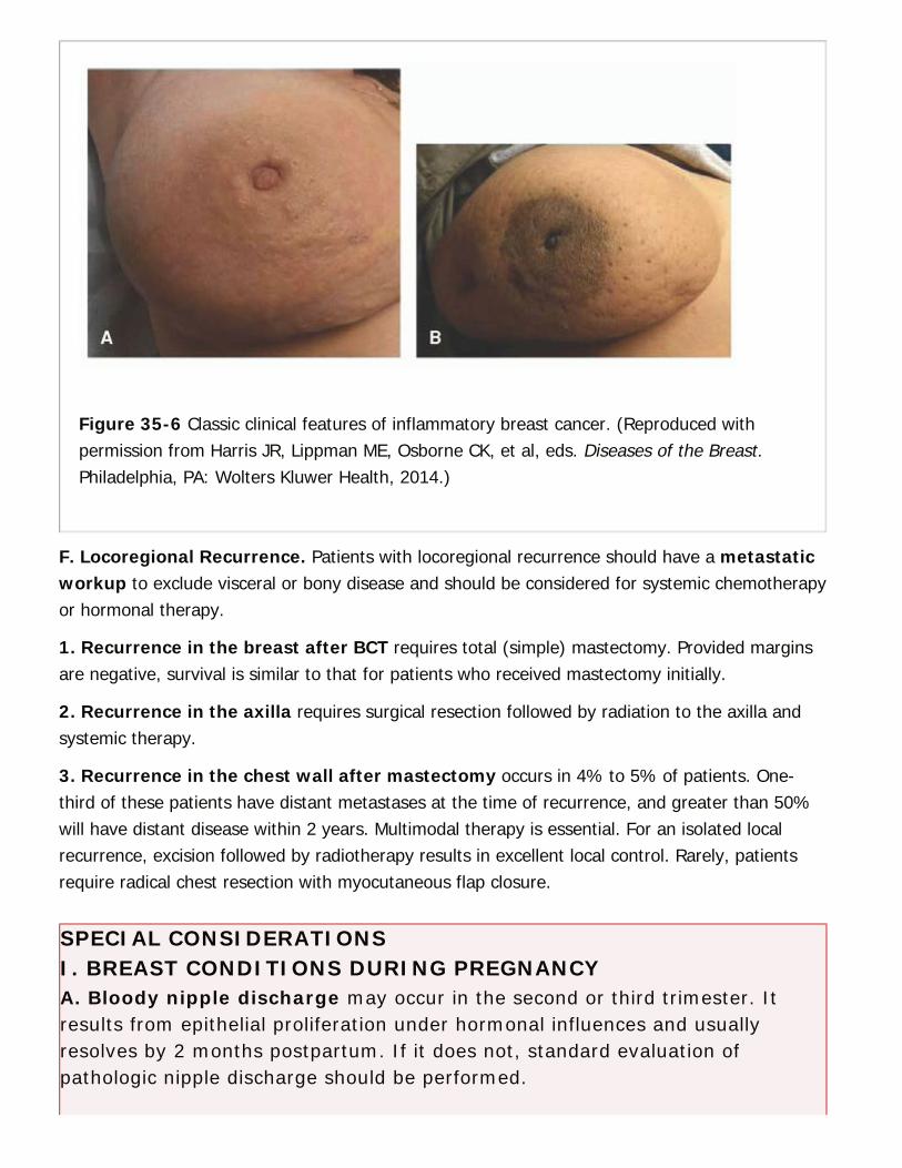
Figure 35-6 Classic clinical features of inflammatory breast cancer. (Reproduced withpermission from Harris JR, Lippman ME, Osborne CK, et al, eds. Diseases of the Breast.Philadelphia, PA: Wolters Kluwer Health, 2014.)
F. Locoregional Recurrence. Patients with locoregional recurrence should have a metastaticworkup to exclude visceral or bony disease and should be considered for systemic chemotherapyor hormonal therapy.
1. Recurrence in the breast after BCT requires total (simple) mastectomy. Provided marginsare negative, survival is similar to that for patients who received mastectomy initially.
2. Recurrence in the axilla requires surgical resection followed by radiation to the axilla andsystemic therapy.
3. Recurrence in the chest wall after mastectomy occurs in 4% to 5% of patients. One-third of these patients have distant metastases at the time of recurrence, and greater than 50%will have distant disease within 2 years. Multimodal therapy is essential. For an isolated localrecurrence, excision followed by radiotherapy results in excellent local control. Rarely, patientsrequire radical chest resection with myocutaneous flap closure.
SPECIAL CONSIDERATIONSI. BREAST CONDITIONS DURING PREGNANCYA. Bloody nipple discharge may occur in the second or third trimester. Itresults from epithelial proliferation under hormonal influences and usuallyresolves by 2 months postpartum. If it does not, standard evaluation ofpathologic nipple discharge should be performed.
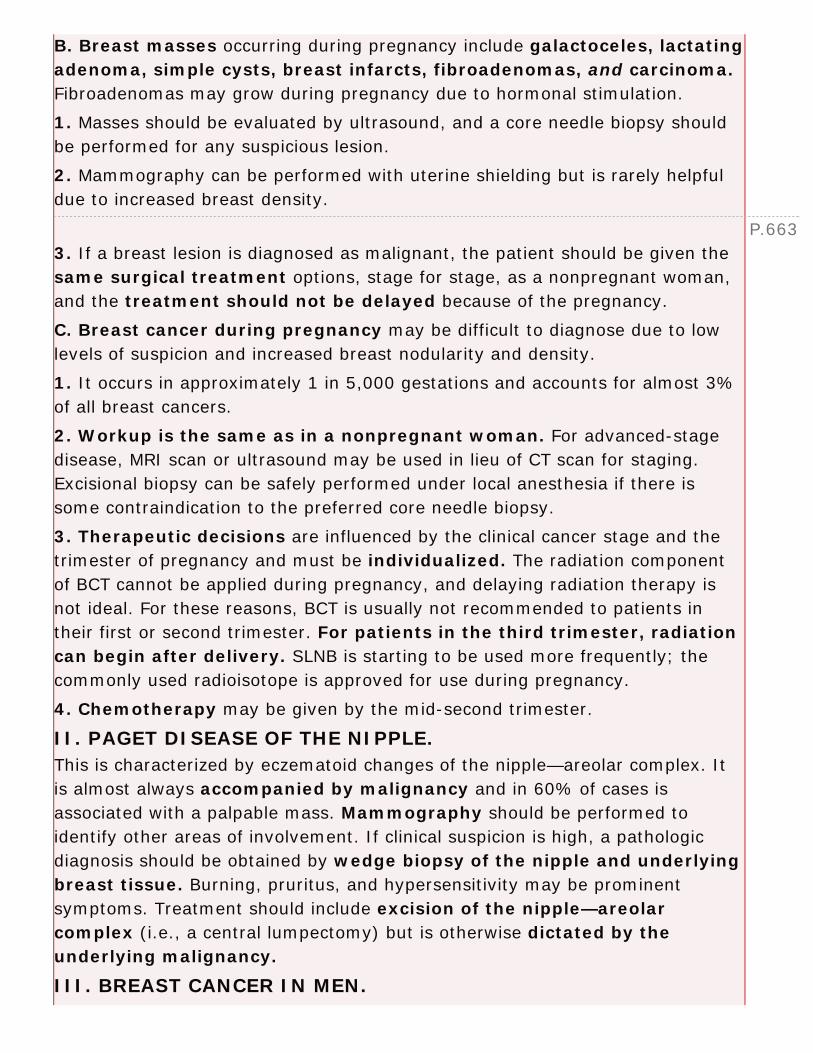
P.663
B. Breast masses occurring during pregnancy include galactoceles, lactatingadenoma, simple cysts, breast infarcts, fibroadenomas, and carcinoma.Fibroadenomas may grow during pregnancy due to hormonal stimulation.
1. Masses should be evaluated by ultrasound, and a core needle biopsy shouldbe performed for any suspicious lesion.
2. Mammography can be performed with uterine shielding but is rarely helpfuldue to increased breast density.
3. If a breast lesion is diagnosed as malignant, the patient should be given thesame surgical treatment options, stage for stage, as a nonpregnant woman,and the treatment should not be delayed because of the pregnancy.
C. Breast cancer during pregnancy may be difficult to diagnose due to lowlevels of suspicion and increased breast nodularity and density.
1. It occurs in approximately 1 in 5,000 gestations and accounts for almost 3%of all breast cancers.
2. Workup is the same as in a nonpregnant woman. For advanced-stagedisease, MRI scan or ultrasound may be used in lieu of CT scan for staging.Excisional biopsy can be safely performed under local anesthesia if there issome contraindication to the preferred core needle biopsy.
3. Therapeutic decisions are influenced by the clinical cancer stage and thetrimester of pregnancy and must be individualized. The radiation componentof BCT cannot be applied during pregnancy, and delaying radiation therapy isnot ideal. For these reasons, BCT is usually not recommended to patients intheir first or second trimester. For patients in the third trimester, radiationcan begin after delivery. SLNB is starting to be used more frequently; thecommonly used radioisotope is approved for use during pregnancy.
4. Chemotherapy may be given by the mid-second trimester.
II. PAGET DISEASE OF THE NIPPLE.This is characterized by eczematoid changes of the nipple—areolar complex. Itis almost always accompanied by malignancy and in 60% of cases isassociated with a palpable mass. Mammography should be performed toidentify other areas of involvement. If clinical suspicion is high, a pathologicdiagnosis should be obtained by wedge biopsy of the nipple and underlyingbreast tissue. Burning, pruritus, and hypersensitivity may be prominentsymptoms. Treatment should include excision of the nipple—areolarcomplex (i.e., a central lumpectomy) but is otherwise dictated by theunderlying malignancy.
III. BREAST CANCER IN MEN.
P.664
P.665
This accounts for less than 1% of male cancers and less than 1% of all breastcancers. BRCA2 mutations are associated with approximately 4% to 6% ofthese cancers. Mammography can be helpful in distinguishing gynecomastiafrom malignancy. Eighty-five percent of malignancies are infiltrating ductalcarcinoma. MRM was traditionally the surgical procedure of choice; however,SLNB has been shown to be effective in men. Thus, total (simple)mastectomy with SLNB is a valid option in men. Adjuvant hormonal,chemotherapy, and radiation treatment criteria are the same as inwomen. Overall survival per stage is comparable to that observed inwomen, although men tend to present in later stages.
IV. PHYLLODES TUMORS.Phyllodes tumors account for 1% of breast neoplasms. They present aslarge, smooth, lobulated masses and may be difficult to distinguish fromfibroadenomas on physical examination. FNAB cannot reliably diagnose thesetumors; at least a core needle biopsy is needed. Ninety percent are benign;10% are malignant, with biologic behavior similar
to that of sarcomas. Treatment is wide local excision to tumor-free marginsor total mastectomy. Axillary assessment is not needed in clinically node-negative patients. Historically, there was no role for adjuvant radiation therapy,but a recent retrospective review demonstrated an association between receiptof radiation and improved local control (Int J Radiat Oncol Biol Phys.2008;70:492-500). Tumors greater than 5 cm in diameter and with evidence ofstromal overgrowth may benefit from adjuvant chemotherapy with doxorubicinand ifosfamide (Cancer. 2000;89:1502-1511). Patients should be followedwith semiannual physical examinations and annual mammograms andchest radiographs.
CHAPTER 35: BREASTMultiple Choice Questions1. Which of the following patients should undergo radiation therapy aspart of her breast cancer management?
a. A 25-year-old woman with 2 cm palpable left breast mass and historyof non-Hodgkin lymphoma at 16
b. An 80-year-old woman with 6 mm of ductal carcinoma in situ (DCIS) inher right breast
c. A 32-year-old pregnant woman with T1 tumor and a suspicious lymph
P.666
node on axillary ultrasound at 33 weeks of gestation
d. A 41-year-old woman diagnosed with multicentric lobular carcinoma at26 weeks of gestation
View Answer
2. Radiation therapy after partial mastectomy improves:
a. Rates of local recurrence
b. Overall survival
c. Risk of lymphedema after axillary lymph node dissection (ALND)
d. Breast-cancer-specific survival
View Answer
3. First-line pharmaceutical treatment for mastalgia is:
a. Oral vitamin E
b. Evening primrose oil
c. Topical vitamin E
d. Topical NSAIDs
View Answer
4. A 32-year-old breastfeeding woman who is 2 weeks postpartumpresents to the emergency department with a 2-day history of warmthand erythema over the inferomedial right breast. She is afebrile and herskin is red but not edematous. Your next step in management is:
a. Incision and drainage (I&D)
b. Antibiotics, instruction to increase breastfeeding frequency
c. Antibiotics, instruction to cease breastfeeding
d. Skin punch biopsy
View Answer
5. Breast cancer in men:
a. Is associated with the BRCA2 mutation in about 5% of cases
b. Is more lethal when compared to stage-matched female controls
c. Is more likely to be infiltrating lobular carcinoma
d. Mandates radical mastectomy
View Answer
6. The most common cause of pathologic nipple discharge is:
a. DCIS
b. Lobular carcinoma in situ (LCIS)
c. Intraductal papilloma
d. Atypical ductal hyperplasia (ADH)
View Answer
7. A 75-year-old man presents with a chief complaint of bilateral breastenlargement over the past 4 months. His past medical history issignificant for a history of congestive heart failure; atrial fibrillation, forwhich he takes digitalis; hypertension; benign prostatic hypertrophy,for which he takes finasteride; and stage 3 chronic kidney disease. Yournext step in management is:
a. To perform a punch biopsy in the clinic
b. Order a stereotactic core needle biopsy
c. To call his primary care physician (PCP)
d. To inform him there is nothing to be done
View Answer
8. A 44-year-old G3P2 female whose mother was diagnosed with breastcancer at 65 comes to see you in clinic after being told that her mostrecent screening mammogram was read as BIRADS 3, probably benign.How do you counsel her?
a. Tell her she has nothing to worry about
b. Ask her to come back and see you in 6 months with repeatmammography
c. Start her on low-dose tamoxifen
d. Perform an ultrasound in clinic
View Answer
9. A 41-year-old female with a known history of lobular carcinoma insitu (LCIS) found on an excisional biopsy 4 months ago presents toyour clinic after being referred to you by her primary care physician.She would like to undergo bilateral prophylactic mastectomy withreconstruction. Options for managing LCIS include:
a. Chemoprevention with an estrogen antagonist
b. Bilateral mastectomy

c. Close surveillance
d. All of the above
View Answer
10. Pertuzumab:
a. Is an aromatase inhibitor used in the treatment of ER+ breast cancer
b. Is associated with progression-free survival in women with metastaticHER2+ cancer when used with cyclophosphamide
c. Is associated with progression-free survival in women with metastaticHER2+ cancer when used with trastuzumab and docetaxel
d. Is still an experimental drug and not yet available in the United States(US)
View Answer
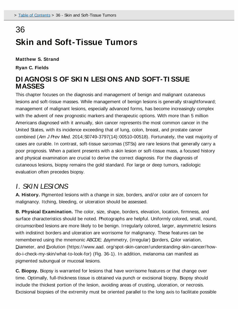
36Skin and Soft-Tissue Tumors
Matthew S. Strand
Ryan C. Fields
DIAGNOSIS OF SKIN LESIONS AND SOFT-TISSUEMASSESThis chapter focuses on the diagnosis and management of benign and malignant cutaneouslesions and soft-tissue masses. While management of benign lesions is generally straightforward;management of malignant lesions, especially advanced forms, has become increasingly complexwith the advent of new prognostic markers and therapeutic options. With more than 5 millionAmericans diagnosed with it annually, skin cancer represents the most common cancer in theUnited States, with its incidence exceeding that of lung, colon, breast, and prostate cancercombined (Am J Prev Med. 2014;S0749-3797(14):00510-00518). Fortunately, the vast majority ofcases are curable. In contrast, soft-tissue sarcomas (STSs) are rare lesions that generally carry apoor prognosis. When a patient presents with a skin lesion or soft-tissue mass, a focused historyand physical examination are crucial to derive the correct diagnosis. For the diagnosis ofcutaneous lesions, biopsy remains the gold standard. For large or deep tumors, radiologicevaluation often precedes biopsy.
I. SKIN LESIONSA. History. Pigmented lesions with a change in size, borders, and/or color are of concern formalignancy. Itching, bleeding, or ulceration should be assessed.
B. Physical Examination. The color, size, shape, borders, elevation, location, firmness, andsurface characteristics should be noted. Photographs are helpful. Uniformly colored, small, round,circumscribed lesions are more likely to be benign. Irregularly colored, larger, asymmetric lesionswith indistinct borders and ulceration are worrisome for malignancy. These features can beremembered using the mnemonic ABCDE: Asymmetry, (irregular) Borders, Color variation,Diameter, and Evolution (https://www.aad. org/spot-skin-cancer/understanding-skin-cancer/how-do-i-check-my-skin/what-to-look-for) (Fig. 36-1). In addition, melanoma can manifest aspigmented subungual or mucosal lesions.
C. Biopsy. Biopsy is warranted for lesions that have worrisome features or that change overtime. Optimally, full-thickness tissue is obtained via punch or excisional biopsy. Biopsy shouldinclude the thickest portion of the lesion, avoiding areas of crusting, ulceration, or necrosis.Excisional biopsies of the extremity must be oriented parallel to the long axis to facilitate possible
> Table of Contents > 36 - Skin and Soft-Tissue Tumors

P.668
need for subsequent definitive resection. Critical to the performance of the biopsy is thedetermination of the Breslow depth; hence superficial shave biopsies should be avoided whiledeep shave biopsies are usually adequate (J Am Coll Surg. 2011;212(4):454-460).

Figure 36-1 ABCDEs of Melanoma.
UnitedVRG
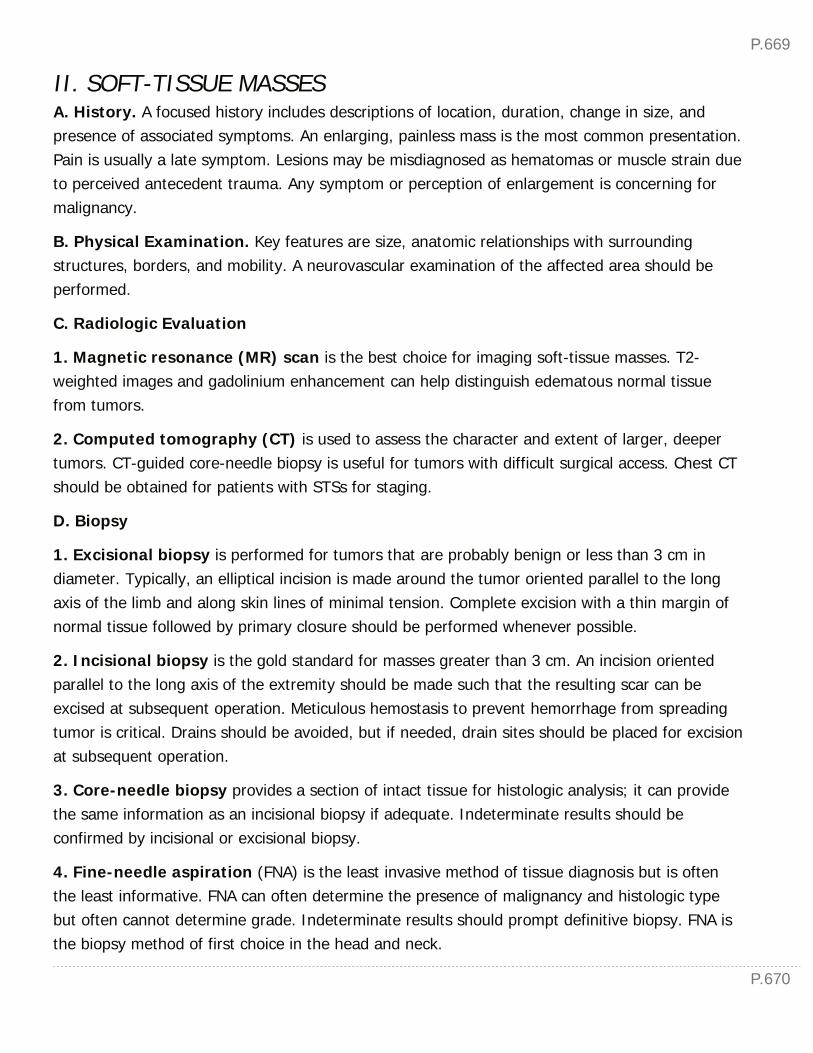
P.669
P.670
II. SOFT-TISSUE MASSESA. History. A focused history includes descriptions of location, duration, change in size, andpresence of associated symptoms. An enlarging, painless mass is the most common presentation.Pain is usually a late symptom. Lesions may be misdiagnosed as hematomas or muscle strain dueto perceived antecedent trauma. Any symptom or perception of enlargement is concerning formalignancy.
B. Physical Examination. Key features are size, anatomic relationships with surroundingstructures, borders, and mobility. A neurovascular examination of the affected area should beperformed.
C. Radiologic Evaluation
1. Magnetic resonance (MR) scan is the best choice for imaging soft-tissue masses. T2-weighted images and gadolinium enhancement can help distinguish edematous normal tissuefrom tumors.
2. Computed tomography (CT) is used to assess the character and extent of larger, deepertumors. CT-guided core-needle biopsy is useful for tumors with difficult surgical access. Chest CTshould be obtained for patients with STSs for staging.
D. Biopsy
1. Excisional biopsy is performed for tumors that are probably benign or less than 3 cm indiameter. Typically, an elliptical incision is made around the tumor oriented parallel to the longaxis of the limb and along skin lines of minimal tension. Complete excision with a thin margin ofnormal tissue followed by primary closure should be performed whenever possible.
2. Incisional biopsy is the gold standard for masses greater than 3 cm. An incision orientedparallel to the long axis of the extremity should be made such that the resulting scar can beexcised at subsequent operation. Meticulous hemostasis to prevent hemorrhage from spreadingtumor is critical. Drains should be avoided, but if needed, drain sites should be placed for excisionat subsequent operation.
3. Core-needle biopsy provides a section of intact tissue for histologic analysis; it can providethe same information as an incisional biopsy if adequate. Indeterminate results should beconfirmed by incisional or excisional biopsy.
4. Fine-needle aspiration (FNA) is the least invasive method of tissue diagnosis but is oftenthe least informative. FNA can often determine the presence of malignancy and histologic typebut often cannot determine grade. Indeterminate results should prompt definitive biopsy. FNA isthe biopsy method of first choice in the head and neck.
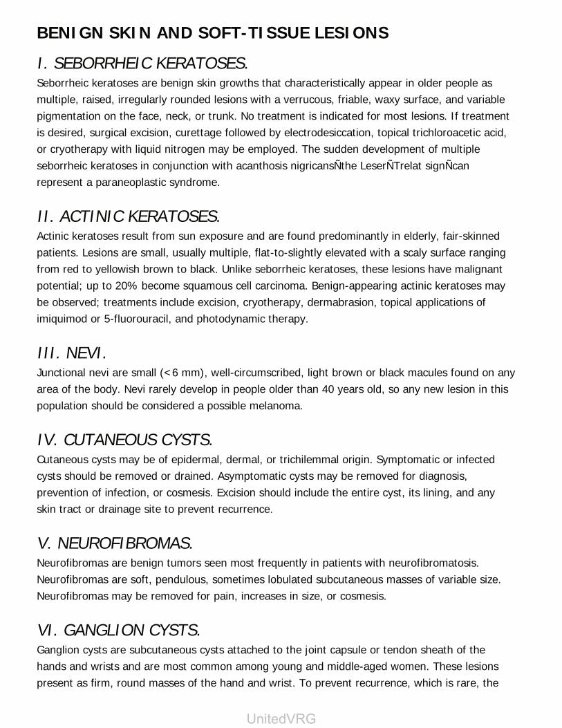
BENIGN SKIN AND SOFT-TISSUE LESIONS
I. SEBORRHEIC KERATOSES.Seborrheic keratoses are benign skin growths that characteristically appear in older people asmultiple, raised, irregularly rounded lesions with a verrucous, friable, waxy surface, and variablepigmentation on the face, neck, or trunk. No treatment is indicated for most lesions. If treatmentis desired, surgical excision, curettage followed by electrodesiccation, topical trichloroacetic acid,or cryotherapy with liquid nitrogen may be employed. The sudden development of multipleseborrheic keratoses in conjunction with acanthosis nigricansÑthe LeserÑTrelat signÑcanrepresent a paraneoplastic syndrome.
II. ACTINIC KERATOSES.Actinic keratoses result from sun exposure and are found predominantly in elderly, fair-skinnedpatients. Lesions are small, usually multiple, flat-to-slightly elevated with a scaly surface rangingfrom red to yellowish brown to black. Unlike seborrheic keratoses, these lesions have malignantpotential; up to 20% become squamous cell carcinoma. Benign-appearing actinic keratoses maybe observed; treatments include excision, cryotherapy, dermabrasion, topical applications ofimiquimod or 5-fluorouracil, and photodynamic therapy.
III. NEVI.Junctional nevi are small (<6 mm), well-circumscribed, light brown or black macules found on anyarea of the body. Nevi rarely develop in people older than 40 years old, so any new lesion in thispopulation should be considered a possible melanoma.
IV. CUTANEOUS CYSTS.Cutaneous cysts may be of epidermal, dermal, or trichilemmal origin. Symptomatic or infectedcysts should be removed or drained. Asymptomatic cysts may be removed for diagnosis,prevention of infection, or cosmesis. Excision should include the entire cyst, its lining, and anyskin tract or drainage site to prevent recurrence.
V. NEUROFIBROMAS.Neurofibromas are benign tumors seen most frequently in patients with neurofibromatosis.Neurofibromas are soft, pendulous, sometimes lobulated subcutaneous masses of variable size.Neurofibromas may be removed for pain, increases in size, or cosmesis.
VI. GANGLION CYSTS.Ganglion cysts are subcutaneous cysts attached to the joint capsule or tendon sheath of thehands and wrists and are most common among young and middle-aged women. These lesionspresent as firm, round masses of the hand and wrist. To prevent recurrence, which is rare, the
UnitedVRG

P.671
capsular attachment and a small portion of the joint capsule should be removed.
VII. LIPOMAS.Lipomas are benign tumors consisting of fat and are perhaps the most common humanneoplasms. Malignant potential is rare; sarcomatous elements occur in less than 1%. They aresoft, mobile subcutaneous masses of variable size. Asymptomatic small tumors can be observed,but large (>5 cm), symptomatic, or growing tumors are concerning and should be biopsied orremoved. Lipomas must be excised cleanly at the first operation to prevent recurrence.
MALIGNANT LESIONS
I. DERMATOFIBROSARCOMA PROTUBERANS.Dermatofibrosarcoma protuberans (DFSP) is a locally aggressive cutaneous sarcoma that rarelymetastasizes but can recur locally. Because of the infiltrative nature of the tumor, excision shouldinclude 2 to 3 cm margins to achieve a high rate of R0 resection. Tumor depth for primary DFSPand margin positivity in recurrent DFSP are independent predictors for recurrence (Ann SurgOncol. 2011;18(2):328-336). Radiation therapy can be employed for patients with microscopicallypositive margins but only one in three patients may benefit; therefore, the morbidity of radiationmust be weighed against the possible need for additional resection. Mohs microsurgery can beemployed to improve tissue conservation, cosmesis, and may actually improve recurrence rates(Arch Dermatol. 2012;148(9):1055).
II. DESMOID TUMORS.Desmoid tumors are nonmetastasizing, locally aggressive tumors that arise from connectivetissue. Excision with a margin of normal tissue should be performed. Local recurrences arecommon, requiring reexcision. Desmoids are radiosensitive, and radiation therapy can be used inthe primary, adjuvant, or neoadjuvant setting. Anti-estrogen-receptor agents (e.g., Tamoxifen),nonsteroidal anti-inflammatory drugs (NSAIDs, e.g., sulindac), and a combination of both types ofmedications have been used with anecdotal success and may be used for recurrent orunresectable disease. Patients with a desmoid tumor require colonoscopy to exclude familialadenomatous polyposis (FAP; Fam Cancer. 2006;5:275).
III. MELANOMA.The incidence of melanoma continues to rise at an epidemic rate. Melanoma represents the fifth-most common type of cancer in the United States (CA Cancer J Clin. 2014;64(1):9-29).
A. Lesions. Most pigmented lesions are benign, but approximately one-third of all melanomasarise from pigmented nevi. Amelanotic melanoma represents 2% of all melanomas.
1. Premalignant lesions

P.672
a. Dysplastic nevi have variegated color (tan to brown on a pink base), are large (5 to 12 mm),appear indistinct with irregular edges, and have macular and papular components. Congenitalnevi (ÒbirthmarksÓ) are associated with an increased risk of melanoma developing from theselesions, particularly for giant congenital nevi, which have diameters greater than 20 cm and aresometimes hairy. Biopsied nevi that reveal moderate or severe dysplasia should be excised tonegative margins.
2. Malignant lesions
a. While malignant melanoma was historically classified into histological subtypes (superficialspreading, nodular, lentigo maligna, and acral lentiginous), this convention is falling out of favordue to the recognition that all subtypes have similar outcomes when controlled for stage, Breslowdepth, and presence of ulceration and mitoses (J Clin Oncol. 2009;27:6199-6206; Ann SurgOncol. 2010;17:1475-1477; Ann Surg Oncol. 2010;17:2006-2014). Instead, new molecular
classification schemes including mutational analysis for genes such as BRAF, NRAS, c-Kit, andCDK4 are becoming relevant in the development of targeted therapies.
UnitedVRG
P.673
Figure 36-2 Cutaneous melanoma of the thigh with satellitosis.
b. In-transit metastases and satellite lesions (Fig 36-2) signify a poor prognosis with ahigh risk of local recurrence and distant metastasis. The distinction between the two is historical,as there is no difference in mechanism or outcome.
B. Risk Factors. A history for melanoma should include an assessment of risk factors and familyhistory.
1. Each of the risk factors listed below carries at least a three-fold increase in the risk formelanoma; the presence of three or more risk factors carries approximately 20 times the risk(Curr Probl Surg. 2006;43:781).
a. Family or personal history of melanoma.
b. Blond or red hair.
c. Freckling of the upper back.
d. Three or more blistering sunburns before age 20.
e. Presence of actinic keratosis.
f. Blue, green, or gray eyes.
A recent meta-analysis demonstrated that individuals with at least one dysplastic nevus had arelative risk of 3.63 for melanoma compared to individuals who had none (Cancer Prev Res.2010;3(2):233-245). Melanoma is familial in approximately 10% of cases, and in these cases, it isoften associated with multiple atypical moles. Familial atypical multiple-mole melanomasyndrome (FAMMM) is a syndrome that causes a predisposition for melanoma. FAMMM isdefined using the following criteria: (1) Malignant melanoma in one or more first- or second-degree relatives, (2) a large number of
melanocytic nevi, usually more than 50, some of which are atypical and variable in size, and (3)melanocytic nevi with particular histopathologic features including architectural disorder withasymmetry, subepidermal fibroplasia, and lentiginous melanocytic hyperplasia with spindle orepithelial melanocyte nests. For patients who have a large number of moles, baselinephotographs or computerized scanning are helpful. Patients with FAMMM should avoid sunexposure and undergo regular ophthalmic examinations due to the increased risk of ocularinvolvement. A proportion of those with FAMMM have germline mutations in p16, a cyclin-dependent kinase inhibitor, accounting for about 40% of melanoma-prone families (J Med Genet.44:99-106).
C. Clinical Features. See ÒSkin Lesions, Physical ExaminationÓ above.
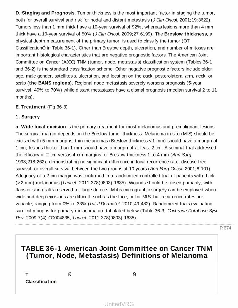
P.674
D. Staging and Prognosis. Tumor thickness is the most important factor in staging the tumor,both for overall survival and risk for nodal and distant metastasis (J Clin Oncol. 2001;19:3622).Tumors less than 1 mm thick have a 10-year survival of 92%, whereas lesions more than 4 mmthick have a 10-year survival of 50% (J Clin Oncol. 2009;27:6199). The Breslow thickness, aphysical depth measurement of the primary tumor, is used to classify the tumor (ÒTClassificationÓ in Table 36-1). Other than Breslow depth, ulceration, and number of mitoses areimportant histological characteristics that are negative prognostic factors. The American JointCommittee on Cancer (AJCC) TNM (tumor, node, metastasis) classification system (Tables 36-1and 36-2) is the standard classification scheme. Other negative prognostic factors include olderage, male gender, satellitosis, ulceration, and location on the back, posterolateral arm, neck, orscalp (the BANS regions). Regional node metastasis severely worsens prognosis (5-yearsurvival, 40% to 70%) while distant metastases have a dismal prognosis (median survival 2 to 11months).
E. Treatment (Fig 36-3)
1. Surgery
a. Wide local excision is the primary treatment for most melanomas and premalignant lesions.The surgical margin depends on the Breslow tumor thickness: Melanoma in situ (MIS) should beexcised with 5 mm margins, thin melanomas (Breslow thickness <1 mm) should have a margin of1 cm; lesions thicker than 1 mm should have a margin of at least 2 cm. A seminal trial addressedthe efficacy of 2-cm versus 4-cm margins for Breslow thickness 1 to 4 mm (Ann Surg.1993;218:262), demonstrating no significant difference in local recurrence rate, disease-freesurvival, or overall survival between the two groups at 10 years (Ann Surg Oncol. 2001;8:101).Adequacy of a 2-cm margin was confirmed in a randomized controlled trial of patients with thick(>2 mm) melanomas (Lancet. 2011;378(9803):1635). Wounds should be closed primarily, withflaps or skin grafts reserved for large defects. Mohs micrographic surgery can be employed wherewide and deep excisions are difficult, such as the face, or for MIS, but recurrence rates arevariable, ranging from 0% to 33% (Int J Dermatol. 2010;49:482). Randomized trials evaluatingsurgical margins for primary melanoma are tabulated below (Table 36-3; Cochrane Database SystRev. 2009;7(4):CD004835; Lancet. 2011;378(9803):1635).
TABLE 36-1 American Joint Committee on Cancer TNM(Tumor, Node, Metastasis) Definitions of Melanoma
TClassification
Ñ Ñ
UnitedVRG

Tis Melanoma in situ Ñ
T1 ≤1.0 mm a. Without ulceration and mitosis
<1/mm2
Ñ b. With ulceration or mitoses
≥1/mm2
T2 1.01-2.0 mm a. Without ulceration
Ñ b. With ulceration
T3 2.01-4.0 mm a. Without ulceration
Ñ b. With ulceration
T4 >4.0 mm a. Without ulceration
Ñ b. With ulceration
RegionalLymph Nodes(N)
Ñ Ñ
N1 One lymph node a. Micrometastasisa
Ñ b. Macrometastasisb
N2 2-3 lymph nodes a. Micrometastasisa
Ñ b. Macrometastasisb
Ñ c. In-transit met(s)/satellite(s)without metastatic lymph node(s)
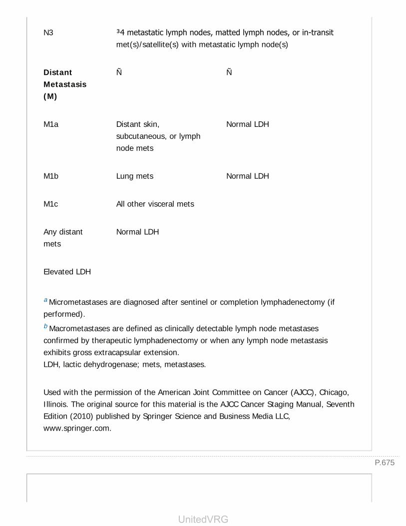
P.675
N3 ≥4 metastatic lymph nodes, matted lymph nodes, or in-transitmet(s)/satellite(s) with metastatic lymph node(s)
DistantMetastasis(M)
Ñ Ñ
M1a Distant skin,subcutaneous, or lymphnode mets
Normal LDH
M1b Lung mets Normal LDH
M1c All other visceral mets
Any distantmets
Normal LDH
Elevated LDH
a Micrometastases are diagnosed after sentinel or completion lymphadenectomy (ifperformed).b Macrometastases are defined as clinically detectable lymph node metastasesconfirmed by therapeutic lymphadenectomy or when any lymph node metastasisexhibits gross extracapsular extension.LDH, lactic dehydrogenase; mets, metastases.
Used with the permission of the American Joint Committee on Cancer (AJCC), Chicago,Illinois. The original source for this material is the AJCC Cancer Staging Manual, SeventhEdition (2010) published by Springer Science and Business Media LLC,www.springer.com.
UnitedVRG
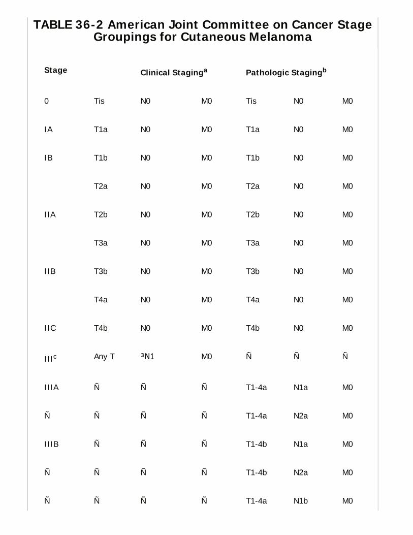
TABLE 36-2 American Joint Committee on Cancer StageGroupings for Cutaneous Melanoma
Stage Clinical Staginga Pathologic Stagingb
0 Tis N0 M0 Tis N0 M0
IA T1a N0 M0 T1a N0 M0
IB T1b N0 M0 T1b N0 M0
T2a N0 M0 T2a N0 M0
IIA T2b N0 M0 T2b N0 M0
T3a N0 M0 T3a N0 M0
IIB T3b N0 M0 T3b N0 M0
T4a N0 M0 T4a N0 M0
IIC T4b N0 M0 T4b N0 M0
IIIc Any T ≥N1 M0 Ñ Ñ Ñ
IIIA Ñ Ñ Ñ T1-4a N1a M0
Ñ Ñ Ñ Ñ T1-4a N2a M0
IIIB Ñ Ñ Ñ T1-4b N1a M0
Ñ Ñ Ñ Ñ T1-4b N2a M0
Ñ Ñ Ñ Ñ T1-4a N1b M0
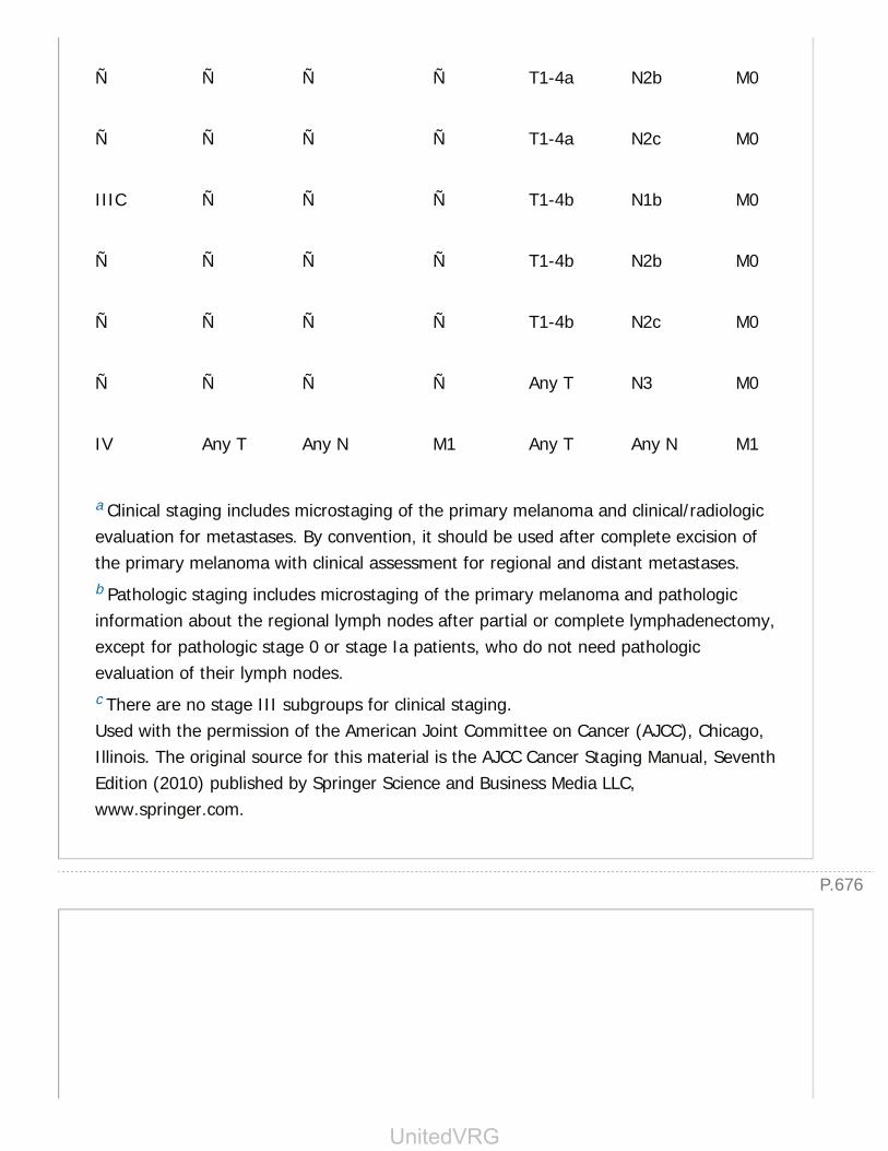
P.676
Ñ Ñ Ñ Ñ T1-4a N2b M0
Ñ Ñ Ñ Ñ T1-4a N2c M0
IIIC Ñ Ñ Ñ T1-4b N1b M0
Ñ Ñ Ñ Ñ T1-4b N2b M0
Ñ Ñ Ñ Ñ T1-4b N2c M0
Ñ Ñ Ñ Ñ Any T N3 M0
IV Any T Any N M1 Any T Any N M1
a Clinical staging includes microstaging of the primary melanoma and clinical/radiologicevaluation for metastases. By convention, it should be used after complete excision ofthe primary melanoma with clinical assessment for regional and distant metastases.b Pathologic staging includes microstaging of the primary melanoma and pathologicinformation about the regional lymph nodes after partial or complete lymphadenectomy,except for pathologic stage 0 or stage Ia patients, who do not need pathologicevaluation of their lymph nodes.c There are no stage III subgroups for clinical staging.Used with the permission of the American Joint Committee on Cancer (AJCC), Chicago,Illinois. The original source for this material is the AJCC Cancer Staging Manual, SeventhEdition (2010) published by Springer Science and Business Media LLC,www.springer.com.
UnitedVRG

P.678
P.677
Figure 36-3 Therapeutic Decision Algorithm for Cutaneous Melanoma.
b. Sentinel lymph node biopsy (SLNB). SLNB has greatly enhanced accurate staging ofpatients with melanoma and has replaced elective lymph node dissection (ELND), which is lymph-node dissection
performed in the absence of clinically positive nodes and which has not been associated with anysurvival benefit. This technique is based on the lymphatic drainage of melanomas to specific,initial lymph node(s), termed the sentinel lymph node(s), before further spread. The histology ofthe SLN is highly reflective of the rest of the nodal basinÑthe incidence of a positive nonsentinelnode in the setting of a negative sentinel node is less than 1% (Arch Surg. 1992;127(4):392). Anegative SLN obviates radical and morbid lymphadenectomy. The SLN can be accurately identified96% of the time using radiolymphoscintigraphy and intraoperative dye injection with radioprobeguidance. SLNB is most beneficial for intermediate-thickness melanomas (Breslow thickness 1 to4 mm) (Ann Surg. 2001;233:250). Data from the Multicenter Sentinel Lymphadenectomy Trial(MSLT)-I support the role of SLN and immediate (vs. delayed) complete lymphadenectomy if theSLN is positive. In MSLT-I, 1,269 patients with intermediate-thickness melanomas (1.2 to 3.5mm) were randomized to either wide excision only followed by observation or to wide excisionand SLNB. In the wide-excision plus observation-only group, complete lymphadenectomy wasperformed only when there was clinical evidence of nodal recurrence (delayed), whereas theSLNB group underwent a complete (immediate) lymphadenectomy if nodal micrometastases were
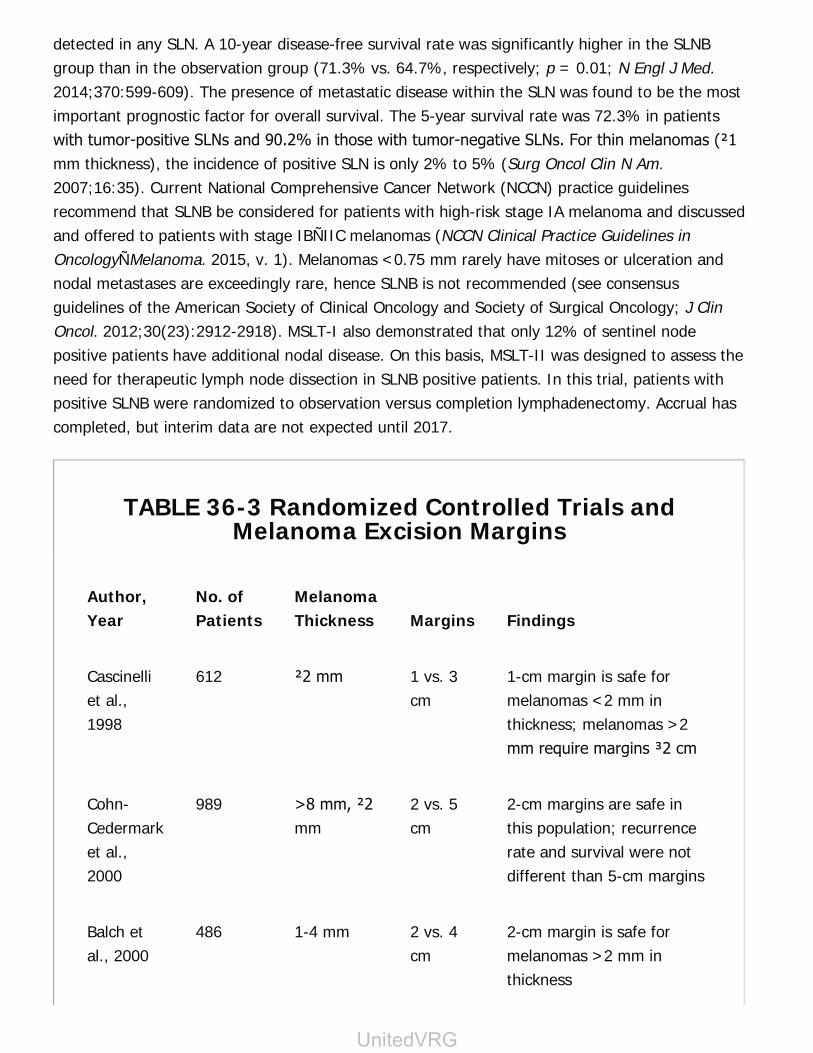
detected in any SLN. A 10-year disease-free survival rate was significantly higher in the SLNBgroup than in the observation group (71.3% vs. 64.7%, respectively; p = 0.01; N Engl J Med.2014;370:599-609). The presence of metastatic disease within the SLN was found to be the mostimportant prognostic factor for overall survival. The 5-year survival rate was 72.3% in patientswith tumor-positive SLNs and 90.2% in those with tumor-negative SLNs. For thin melanomas (≤1mm thickness), the incidence of positive SLN is only 2% to 5% (Surg Oncol Clin N Am.2007;16:35). Current National Comprehensive Cancer Network (NCCN) practice guidelinesrecommend that SLNB be considered for patients with high-risk stage IA melanoma and discussedand offered to patients with stage IBÑIIC melanomas (NCCN Clinical Practice Guidelines inOncologyÑMelanoma. 2015, v. 1). Melanomas <0.75 mm rarely have mitoses or ulceration andnodal metastases are exceedingly rare, hence SLNB is not recommended (see consensusguidelines of the American Society of Clinical Oncology and Society of Surgical Oncology; J ClinOncol. 2012;30(23):2912-2918). MSLT-I also demonstrated that only 12% of sentinel nodepositive patients have additional nodal disease. On this basis, MSLT-II was designed to assess theneed for therapeutic lymph node dissection in SLNB positive patients. In this trial, patients withpositive SLNB were randomized to observation versus completion lymphadenectomy. Accrual hascompleted, but interim data are not expected until 2017.
TABLE 36-3 Randomized Controlled Trials andMelanoma Excision Margins
Author,Year
No. ofPatients
MelanomaThickness Margins Findings
Cascinelliet al.,1998
612 ≤2 mm 1 vs. 3cm
1-cm margin is safe formelanomas <2 mm inthickness; melanomas >2mm require margins ≥2 cm
Cohn-Cedermarket al.,2000
989 >8 mm, ≤2mm
2 vs. 5cm
2-cm margins are safe inthis population; recurrencerate and survival were notdifferent than 5-cm margins
Balch etal., 2000
486 1-4 mm 2 vs. 4cm
2-cm margin is safe formelanomas >2 mm inthickness
UnitedVRG
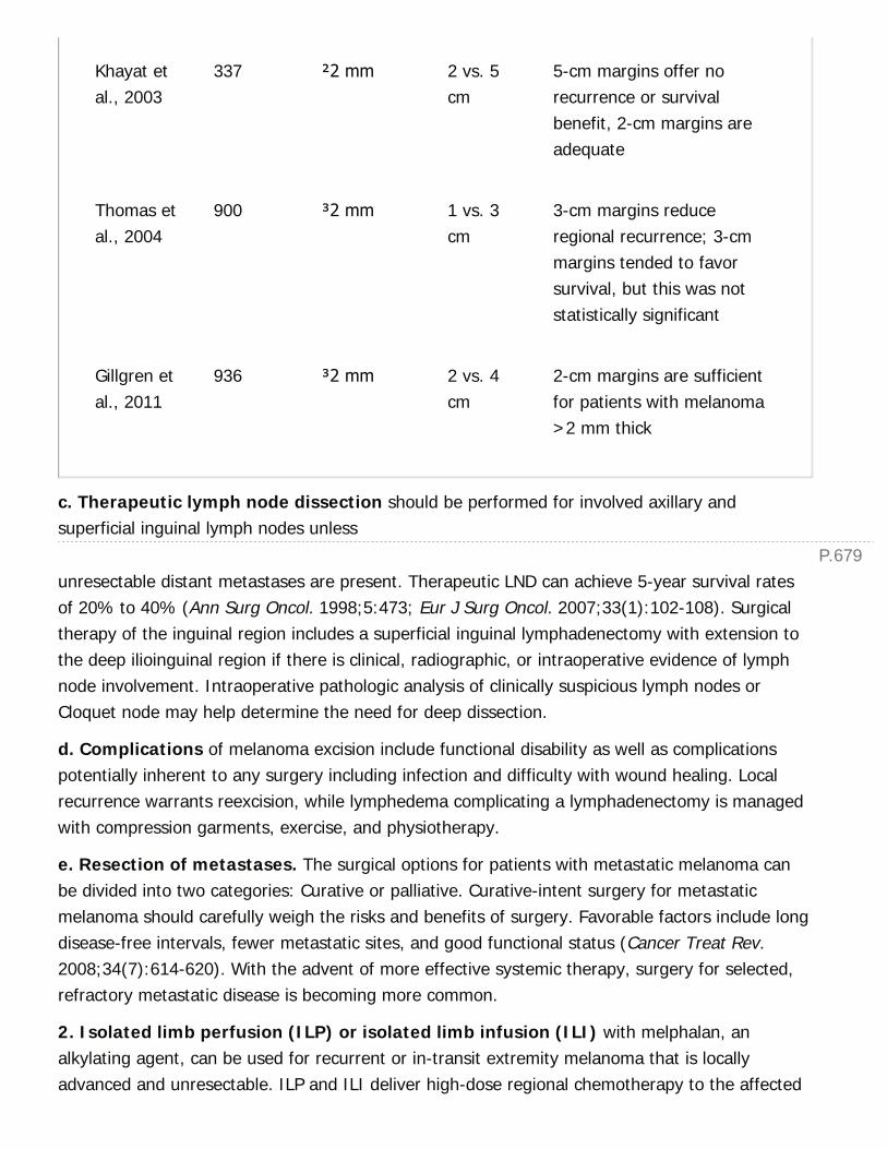
P.679
Khayat etal., 2003
337 ≤2 mm 2 vs. 5cm
5-cm margins offer norecurrence or survivalbenefit, 2-cm margins areadequate
Thomas etal., 2004
900 ≥2 mm 1 vs. 3cm
3-cm margins reduceregional recurrence; 3-cmmargins tended to favorsurvival, but this was notstatistically significant
Gillgren etal., 2011
936 ≥2 mm 2 vs. 4cm
2-cm margins are sufficientfor patients with melanoma>2 mm thick
c. Therapeutic lymph node dissection should be performed for involved axillary andsuperficial inguinal lymph nodes unless
unresectable distant metastases are present. Therapeutic LND can achieve 5-year survival ratesof 20% to 40% (Ann Surg Oncol. 1998;5:473; Eur J Surg Oncol. 2007;33(1):102-108). Surgicaltherapy of the inguinal region includes a superficial inguinal lymphadenectomy with extension tothe deep ilioinguinal region if there is clinical, radiographic, or intraoperative evidence of lymphnode involvement. Intraoperative pathologic analysis of clinically suspicious lymph nodes orCloquet node may help determine the need for deep dissection.
d. Complications of melanoma excision include functional disability as well as complicationspotentially inherent to any surgery including infection and difficulty with wound healing. Localrecurrence warrants reexcision, while lymphedema complicating a lymphadenectomy is managedwith compression garments, exercise, and physiotherapy.
e. Resection of metastases. The surgical options for patients with metastatic melanoma canbe divided into two categories: Curative or palliative. Curative-intent surgery for metastaticmelanoma should carefully weigh the risks and benefits of surgery. Favorable factors include longdisease-free intervals, fewer metastatic sites, and good functional status (Cancer Treat Rev.2008;34(7):614-620). With the advent of more effective systemic therapy, surgery for selected,refractory metastatic disease is becoming more common.
2. Isolated limb perfusion (ILP) or isolated limb infusion (ILI) with melphalan, analkylating agent, can be used for recurrent or in-transit extremity melanoma that is locallyadvanced and unresectable. ILP and ILI deliver high-dose regional chemotherapy to the affected

P.680
extremity while minimizing systemic toxicity. ILI is simpler and less toxic than ILP, but itsresponse rates are lower (J Am Coll Surg. 2009;208(5):706). A review of ILP and ILI including theagents used, technique employed, and treatment algorithms was published by Tyler et al. in 2011(Surg Oncol Clin N Am. 2011;20(1):79-103).
3. Immunotherapy. Ipilimumab, a monoclonal antibody against CTLA-4, andpembrolizumab, a monoclonal antibody against PD-1, have shown clinical efficacy in thetreatment of melanoma and have been approved by the FDA. A placebo-controlled phase III trialof ipilimumab including 676 patients with metastatic melanoma showed a significant survivalbenefit. In this trial, patients received ipilimumab alone, ipilimumab plus glycoprotein 100 (gp100)vaccine, or gp100 vaccine alone. At 12 months, survival rates were 44%, 46%, and 25%respectively and at 24 months they were 22%, 24%, and 14% (N Engl J Med. 2010;363(8):711).An additional phase III trial was conducted comparing ipilimumab plus dacarbazine to placeboplus dacarbazine. Survival rates were significantly higher in the group receiving ipilimumab plusdacarbazine at 1-, 2-, and 3-year followup (47% vs. 36%, 29% vs. 18%, and 21% vs. 12%,respectively; N Engl J Med. 2011;364(26):2517). Randomized trials have also demonstrated theefficacy of pembrolizumab, especially in patients with
ipilimumab-refractory disease. A trial in which 540 ipilimumab-refractory patients wererandomized to receive pembrolizumab 2 mg/kg, pembrolizumab10 mg/kg, and chemotherapyshowed that pembrolizumab significantly reduced progression at 6 months compared tochemotherapy (J Transl Med. 2015;13:2062). Studies combining agents that target CTLA-4 andPD-1 or PDL-1 in conjunction with vaccines are currently ongoing and may improve therapeuticresponse.
Interferon-alpha (IFN-α) therapy is considered standard adjuvant therapy for patients with knownnodal disease or at high risk for regional recurrence (primary melanoma >4 mm thick, ulceration,or elevated mitoses. Two randomized controlled trials have shown the benefit of IFN-α in selectpopulations. Garbe et al. compared surgery alone to adjuvant INF-2α with or withoutdacarbazine. Overall survival in the IFN-2α group was significantly higher than in the surgeryalone group at the 4-year mark (59 vs. 42%, P = 0.0045; Ann Oncol. 2008;19(6):1195-1201).Pegylated IFN-α2b adjuvant therapy versus surgery alone was compared in a phase IIIrandomized trial of patients with resected stage III melanoma. At 4 years, patients in the IFN-α2bhad improved recurrence-free survival (45.6% vs. 38.9%, P = 0.1); however, no survival benefitwas seen (Lancet. 2008;372(9633):117-126). Combination therapy with ipilimumab as adjuvanttherapy is currently ongoing in ECOG 1609.
4. Targeted molecular therapy. Testing for BRAF and NRAS mutations is now standard forpatients with metastatic melanoma due to the advent of targeted therapies. BRAF and NRAS areproto-oncogenes that have been found to be activated about 40% and 15% of melanomas,respectively (J Clin Oncol. 2012;30(20):2522-2529). Vemurafenib and dabrafenib are smallmolecule inhibitors of BRAF that have shown clinical efficacy in patients with the V600 mutation in
UnitedVRG
P.681
BRAF. Cobimetinib and Trametinib are small molecule inhibitors of MEK, an effector justdownstream of BRAF. Combination therapies with BRAF and MEK inhibitors have demonstratedimproved efficacy over BRAF therapy alone for patients with V600 BRAF mutations in two recentrandomized controlled trials (N Engl J Med. 2014;371(20):1877-1888; N Engl J Med.2014;371(20):1867-1876). Another small molecule inhibitor, imatinib has also been efficaciousin patients with a KIT mutation more prevalent in acral and mucosal melanomas. Generally thesetreatments are reserved as second-line therapy after immunotherapy but represent importantadvances in the application of genetic data to individualize care.
OTHER MALIGNANT SKIN TUMORS
I. BASAL CELL CARCINOMA.Basal cell carcinoma is the most common skin cancer. They are slow-growing, may be large,disfiguring, and locally invasive, but rarely metastasize (<0.1%). Sun exposure is the mostsignificant epidemiologic factor; consequently, this neoplasm is found most commonly on sun-exposed areas in fair-skinned patients older than 40 years.
A. Lesions. Lesions are flat but indurated with a smooth, whitish, waxy surface and indistinctborders. The noduloulcerative form is the most common and is characterized by shiny,translucent nodules with a central umbilication that often becomes ulcerated, with pearly, rolled,telangiectatic edges. The morpheaform variant is important to identify because it is moreaggressive.
B. Treatment
1. Excisional biopsy is adequate for small tumors, with intraoperative frozen-section analysis(to confirm negative margins) and primary closure. Larger tumors may require incisional or punchbiopsy followed by excision. A margin of 2 to 4 mm should be obtained, and positive marginsshould be reexcised.
2. Other options for treatment include Mohs micrographic surgery (useful for recurrenttumors or cosmetically sensitive areas), curettage with electrodessication, liquid nitrogen therapy,topical 5-FU or imiquimod (for small tumors), or radiation or photodynamic therapy forunresectable lesions or if treatment intent is palliative. The small molecule inhibitor Vismodegibis a relatively new therapy that has shown efficacy in patients with inoperable BCC (N Engl J Med.2012;366(23):2171-2179). This has proven very useful in patients with Gorlin syndrome, afamilial form of BCC.
II. SQUAMOUS CELL CARCINOMA.Squamous cell carcinoma is the second-most common skin cancer. Sunlight is the major etiology,and elderly men with a history of chronic sun exposure are the most at risk. Squamous cell
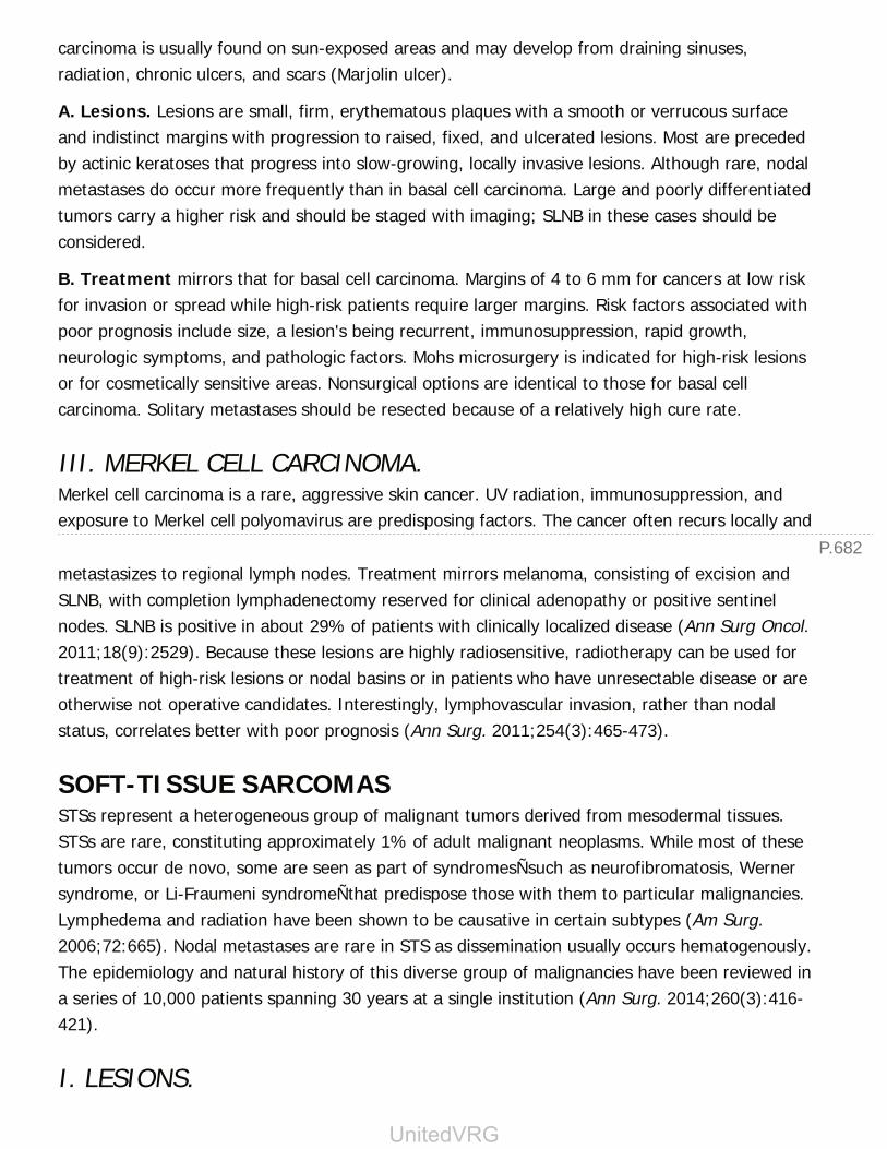
P.682
carcinoma is usually found on sun-exposed areas and may develop from draining sinuses,radiation, chronic ulcers, and scars (Marjolin ulcer).
A. Lesions. Lesions are small, firm, erythematous plaques with a smooth or verrucous surfaceand indistinct margins with progression to raised, fixed, and ulcerated lesions. Most are precededby actinic keratoses that progress into slow-growing, locally invasive lesions. Although rare, nodalmetastases do occur more frequently than in basal cell carcinoma. Large and poorly differentiatedtumors carry a higher risk and should be staged with imaging; SLNB in these cases should beconsidered.
B. Treatment mirrors that for basal cell carcinoma. Margins of 4 to 6 mm for cancers at low riskfor invasion or spread while high-risk patients require larger margins. Risk factors associated withpoor prognosis include size, a lesion's being recurrent, immunosuppression, rapid growth,neurologic symptoms, and pathologic factors. Mohs microsurgery is indicated for high-risk lesionsor for cosmetically sensitive areas. Nonsurgical options are identical to those for basal cellcarcinoma. Solitary metastases should be resected because of a relatively high cure rate.
III. MERKEL CELL CARCINOMA.Merkel cell carcinoma is a rare, aggressive skin cancer. UV radiation, immunosuppression, andexposure to Merkel cell polyomavirus are predisposing factors. The cancer often recurs locally and
metastasizes to regional lymph nodes. Treatment mirrors melanoma, consisting of excision andSLNB, with completion lymphadenectomy reserved for clinical adenopathy or positive sentinelnodes. SLNB is positive in about 29% of patients with clinically localized disease (Ann Surg Oncol.2011;18(9):2529). Because these lesions are highly radiosensitive, radiotherapy can be used fortreatment of high-risk lesions or nodal basins or in patients who have unresectable disease or areotherwise not operative candidates. Interestingly, lymphovascular invasion, rather than nodalstatus, correlates better with poor prognosis (Ann Surg. 2011;254(3):465-473).
SOFT-TISSUE SARCOMASSTSs represent a heterogeneous group of malignant tumors derived from mesodermal tissues.STSs are rare, constituting approximately 1% of adult malignant neoplasms. While most of thesetumors occur de novo, some are seen as part of syndromesÑsuch as neurofibromatosis, Wernersyndrome, or Li-Fraumeni syndromeÑthat predispose those with them to particular malignancies.Lymphedema and radiation have been shown to be causative in certain subtypes (Am Surg.2006;72:665). Nodal metastases are rare in STS as dissemination usually occurs hematogenously.The epidemiology and natural history of this diverse group of malignancies have been reviewed ina series of 10,000 patients spanning 30 years at a single institution (Ann Surg. 2014;260(3):416-421).
I. LESIONS.
UnitedVRG

P.683
Sarcomas are classified by histologic cell type of origin and grade. Pleomorphic undifferentiatedsarcoma (previously malignant fibrous histiocytoma, 40%) is most common, followed byliposarcoma (25%). Patients typically present with an asymptomatic mass that becomes visible orpalpable. Retroperitoneal tumors can become massive before symptoms prompt presentation.
II. DIAGNOSIS.Biopsy (usually core or incisional) is necessary for diagnosis. Care is needed to orient incisions toaid in definitive resection.
III. STAGING AND PROGNOSIS.The AJCC staging system is based on tumor size and depth, nodal status, histologic grade, andmetastasis (Table 36-4). Of these, tumor grade is the major prognostic factor and is based onmitotic index, nuclear morphology, and degree of anaplasia.
A. Staging includes physical examination and CT or MR scan to assess the size and extent oftumor. As hematogenous spread to the lungs is the most common form of metastasis, chest CT isrequired for grade II and III lesions. Abdominal CT scan is needed to evaluate retroperitonealsarcomas, in which hepatic metastases are more common than in other STS.
B. Prognosis. Almost 80% of metastases are to the lungs and occur within 2 to 3 years ofdiagnosis. If pulmonary disease is resectable, overall survival is 30% at 3 years. In addition,tumor size, grade, tumor rupture during surgery, margins after resection, and anatomic locationall influence local recurrence, overall survival, and tumor-free survival. Retroperitoneal and truncalSTS have worse prognoses than extremity STS. While mortality from extremity STS is usually dueto metastasis, retroperitoneal STS is usually fatal secondary to local recurrence.
TABLE 36-4 American Joint Committee on CancerStaging System for Soft-Tissue Sarcoma
Tumor Grade (G) Stage IA
GX: Grade cannot be assessed G1/GX, T1a, N0, M0
G1: Well differentiated G1/GX, T1b, N0, M0
G2: Moderately differentiated Stage IB

G3: Poorly differentiated G1/GX, T2a, N0, M0
G4: Undifferentiated G1/GX, T2b, N0, M0
Primary Tumor (T) Stage IIA
TX: Primary tumor cannot be assessed G2-3, T1a, N0, M0
T0: No evidence of primary tumor G2-3, T1b, N0, M0
T1: Tumor >5 cm in greatest dimension Stage IIB
T1a: Superficial tumora G2, T2a, N0, M0
T1b: Deep tumora G2, T2b, N0, M0
T2: Tumor >5 cm in greatest dimension Stage III
T2a: Superficial tumora G3, T2a, N0, M0
T2b: Deep tumora G3, T2b, N0, M0
Regional Lymph Nodes (N) Any G, any T, N1, M0
NX: Regional lymph nodes cannot be assessed Stage IV
N0: No regional lymph node metastasis Any G, any T, any N, M1
N1: Regional lymph node metastasis Ñ
Distant Metastasis (M) Ñ
MX: Distant metastasis cannot be assessed Ñ
UnitedVRG

P.684
M0: No distant metastasis Ñ
M1: Distant metastasis Ñ
a Superficial tumor is located exclusively above the superficial fascia without invasion ofthe fascia; deep tumor either is located exclusively beneath the superficial fascia orsuperficial to the fascia with invasion of or through the fascia or is located superficialand beneath the fascia. Retroperitoneal, mediastinal, and pelvic sarcomas are classifiedas deep tumors.
Used with the permission of the American Joint Committee on Cancer (AJCC), Chicago,Illinois. The original source for this material is the AJCC Cancer Staging Manual, SeventhEdition (2010) published by Springer Science and Business Media LLC,www.springer.com.
IV. TREATMENTA. Surgical Resection. Smaller, grade I tumors can be excised with aminimum 1-cm margin and usually do not require adjuvant radiation. Largertumors may benefit from a larger margin or from radiation to preventrecurrence. Grade II and III tumors, in general, require adjuvant radiationtherapy to prevent recurrence. Local recurrence should be resectedaggressively, and long-term followup is required. Amputation is rarelynecessary.
Limb-sparing resection combined with radiation therapy offers rates ofsurvival equivalent to those achieved with amputation (Ann Surg.1982;196:305). Limb-sparing procedures have a psychological and functionaladvantage over amputations and are the procedures of choice for most tumors.Resection should include the area of previous incision. Intraoperative use ofclips can aid in postoperative radiation therapy.
Retroperitoneal sarcomas are considerably more difficult to treat because thetumors often involve vital structures. Overall and disease free survival forretroperitoneal STS are 56.8% and 39.4%, respectively (J Clin Oncol.2013;31(13):1649-1655). Organs associated with the tumor should be resecteden bloc because 5-year recurrence rates are lower for en bloc resections thanfor cases of tumor resection alone (28% vs. 48%, respectively; J Clin Oncol.2009;27:24). Postoperative irradiation may be used but is associated with
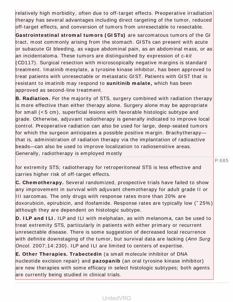
P.685
relatively high morbidity, often due to off-target effects. Preoperative irradiationtherapy has several advantages including direct targeting of the tumor, reducedoff-target effects, and conversion of tumors from unresectable to resectable.
Gastrointestinal stromal tumors (GISTs) are sarcomatous tumors of the GItract, most commonly arising from the stomach. GISTs can present with acuteor subacute GI bleeding, as vague abdominal pain, as an abdominal mass, or asan incidentaloma. These tumors are distinguished by expression of c-kit(CD117). Surgical resection with microscopically negative margins is standardtreatment. Imatinib mesylate, a tyrosine kinase inhibitor, has been approved totreat patients with unresectable or metastatic GIST. Patients with GIST that isresistant to imatinib may respond to sunitinib malate, which has beenapproved as second-line treatment.
B. Radiation. For the majority of STS, surgery combined with radiation therapyis more effective than either therapy alone. Surgery alone may be appropriatefor small (<5 cm), superficial lesions with favorable histologic subtype andgrade. Otherwise, adjuvant radiotherapy is generally indicated to improve localcontrol. Preoperative radiation can also be used for large, deep-seated tumorsfor which the surgeon anticipates a possible positive margin. Brachytherapy—that is, administration of radiation therapy via the implantation of radioactivebeads—can also be used to improve localization to radiosensitive areas.Generally, radiotherapy is employed mostly
for extremity STS; radiotherapy for retroperitoneal STS is less effective andcarries higher risk of off-target effects.
C. Chemotherapy. Several randomized, prospective trials have failed to showany improvement in survival with adjuvant chemotherapy for adult grade II orIII sarcomas. The only drugs with response rates more than 20% aredoxorubicin, epirubicin, and ifosfamide. Response rates are typically low (˜25%)although they are dependent on histologic subtype.
D. ILP and ILI. ILP and ILI with melphalan, as with melanoma, can be used totreat extremity STS, particularly in patients with either primary or recurrentunresectable disease. There is some suggestion of decreased local recurrencewith definite downstaging of the tumor, but survival data are lacking (Ann SurgOncol. 2007;14:230). ILP and ILI are limited to centers of expertise.
E. Other Therapies. Trabectedin (a small molecule inhibitor of DNAnucleotide excision repair) and pazopanib (an oral tyrosine kinase inhibitor)are new therapies with some efficacy in select histologic subtypes; both agentsare currently being studied in clinical trials.
UnitedVRG

P.686
CHAPTER 36: SKIN AND SOFT-TISSUE TUMORSMultiple Choice Questions1. For which of the following patients is sentinel lymph node biopsy(SLNB) indicated?
a. A 55-year-old male with 0.6 mm thick melanoma of the back with no
ulceration and 3 mitoses/mm2
b. A 28-year-old female with melanoma in situ of the right back
c. A 66-year-old male with 1.2 mm thick acral melanoma of the left lowerextremity with left inguinal adenopathy
d. A 54-year-old female with 3.2 mm thick right upper extremitymelanoma with two pulmonary metastases
e. A 35-year-old male with 1.4 mm thick melanoma of the back with noulceration or mitoses
View Answer
2. A 68-year-old male presents with a growing, smooth, hypopigmented2-cm lesion on the right cheek that he says occasionally stings andbleeds. The best next step is:
a. Mohs microsurgery
b. Excisional biopsy
c. FNA
d. Punch biopsy
e. Shave biopsy
View Answer
3. A 38-year-old firefighter is referred to your clinic with a nonhealingwound that arose after sustaining a burn injury 4 months ago. His pastmedical history is significant for diabetes mellitus type II controlled oninsulin; a recent HgbA1c is 6.8%. He smokes about 1 pack weekly. He isotherwise healthy. He denies any fever but complains of oozing fromhis lowerextremity wound. Examination of the left lower extremityreveals a 4-cm linear scar with a 1.5-cm central ulcer withserosanguineous drainage and mild local edema. Peripheral pulses arepalpable. There is mild tenderness but no warmth or erythema. Thebest course of action is to:
a. Prescribe topical mupirocin
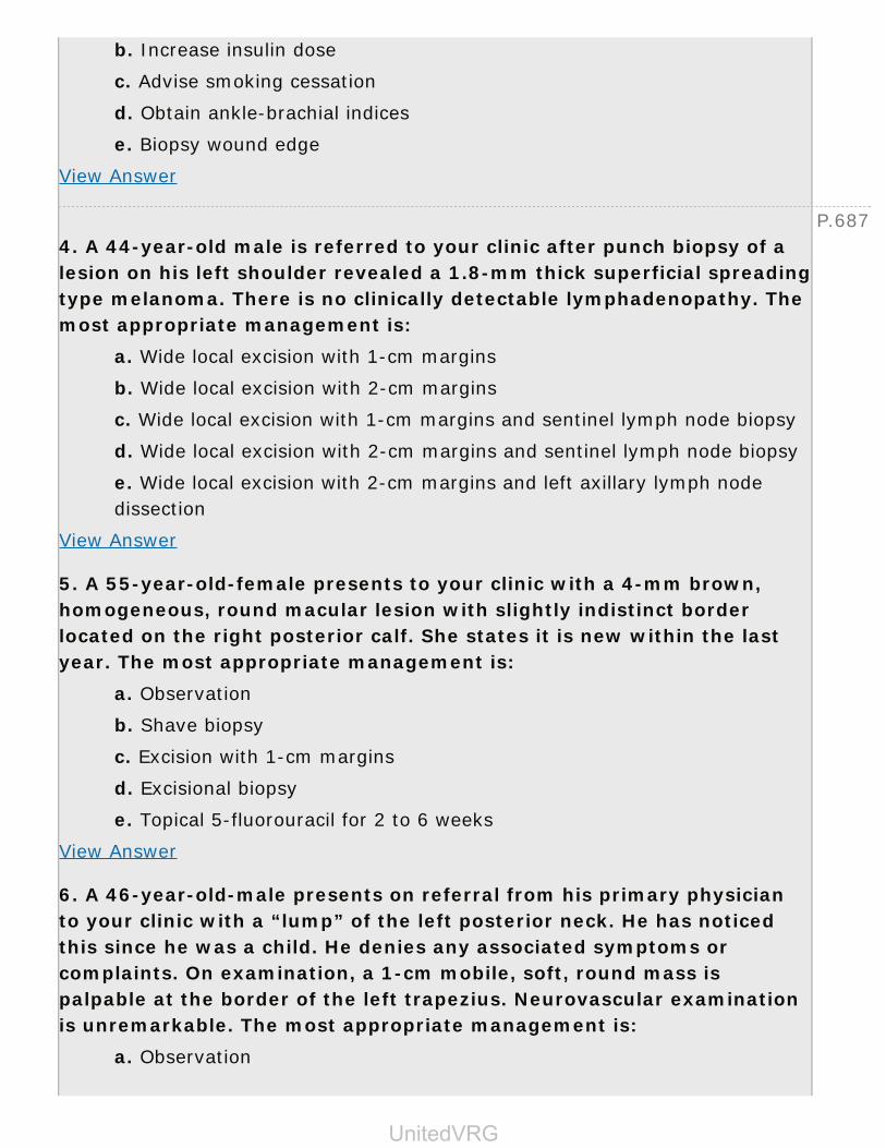
P.687
b. Increase insulin dose
c. Advise smoking cessation
d. Obtain ankle-brachial indices
e. Biopsy wound edge
View Answer
4. A 44-year-old male is referred to your clinic after punch biopsy of alesion on his left shoulder revealed a 1.8-mm thick superficial spreadingtype melanoma. There is no clinically detectable lymphadenopathy. Themost appropriate management is:
a. Wide local excision with 1-cm margins
b. Wide local excision with 2-cm margins
c. Wide local excision with 1-cm margins and sentinel lymph node biopsy
d. Wide local excision with 2-cm margins and sentinel lymph node biopsy
e. Wide local excision with 2-cm margins and left axillary lymph nodedissection
View Answer
5. A 55-year-old-female presents to your clinic with a 4-mm brown,homogeneous, round macular lesion with slightly indistinct borderlocated on the right posterior calf. She states it is new within the lastyear. The most appropriate management is:
a. Observation
b. Shave biopsy
c. Excision with 1-cm margins
d. Excisional biopsy
e. Topical 5-fluorouracil for 2 to 6 weeks
View Answer
6. A 46-year-old-male presents on referral from his primary physicianto your clinic with a “lump” of the left posterior neck. He has noticedthis since he was a child. He denies any associated symptoms orcomplaints. On examination, a 1-cm mobile, soft, round mass ispalpable at the border of the left trapezius. Neurovascular examinationis unremarkable. The most appropriate management is:
a. Observation
UnitedVRG
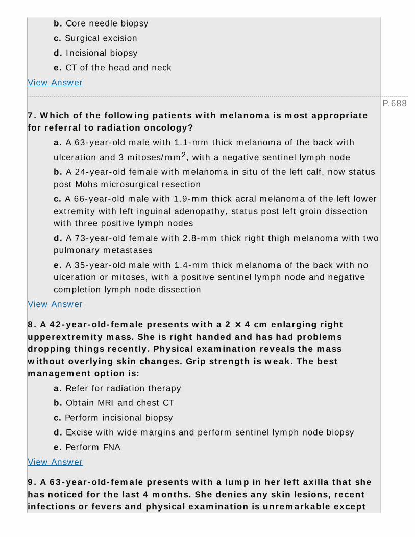
P.688
b. Core needle biopsy
c. Surgical excision
d. Incisional biopsy
e. CT of the head and neck
View Answer
7. Which of the following patients with melanoma is most appropriatefor referral to radiation oncology?
a. A 63-year-old male with 1.1-mm thick melanoma of the back with
ulceration and 3 mitoses/mm2, with a negative sentinel lymph node
b. A 24-year-old female with melanoma in situ of the left calf, now statuspost Mohs microsurgical resection
c. A 66-year-old male with 1.9-mm thick acral melanoma of the left lowerextremity with left inguinal adenopathy, status post left groin dissectionwith three positive lymph nodes
d. A 73-year-old female with 2.8-mm thick right thigh melanoma with twopulmonary metastases
e. A 35-year-old male with 1.4-mm thick melanoma of the back with noulceration or mitoses, with a positive sentinel lymph node and negativecompletion lymph node dissection
View Answer
8. A 42-year-old-female presents with a 2 × 4 cm enlarging rightupperextremity mass. She is right handed and has had problemsdropping things recently. Physical examination reveals the masswithout overlying skin changes. Grip strength is weak. The bestmanagement option is:
a. Refer for radiation therapy
b. Obtain MRI and chest CT
c. Perform incisional biopsy
d. Excise with wide margins and perform sentinel lymph node biopsy
e. Perform FNA
View Answer
9. A 63-year-old-female presents with a lump in her left axilla that shehas noticed for the last 4 months. She denies any skin lesions, recentinfections or fevers and physical examination is unremarkable except

P.689
for axillary adenopathy. Mammogram obtained 3 weeks ago isunremarkable. The most appropriate next step is:
a. Core needle biopsy
b. PET scan
c. Observe, followup in 1 month
d. 10-day course of Keflex
e. Radiation therapy
View Answer
10. Which of the following is the most important prognostic factor forsofttissue sarcomas?
a. Histologic subtype
b. Age at presentation
c. Comorbidities
d. Necrosis
e. Grade
View Answer
11. A 73-year-old female was recently diagnosed with a left distalfemoral malignant fibrous histiocytoma abutting the distal superficialfemoral artery. The best next step is:
a. Wide local excision
b. Left above the knee amputation
c. Chest CT
d. Isolated limb perfusion
e. Radiation therapy
View Answer
12. A 44-year-old male is taken to the operating room for resection of alarge retroperitoneal liposarcoma. He has no significant past medicalhistory and preoperative laboratory studies were normal. Afterlaparotomy, the tumor is discovered to encase the left renal artery andvein. The most appropriate course of action is:
a. Tumor debulking followed by adjuvant chemotherapy
b. Placement of clips, closure, and referral for radiation therapy
c. Closure and subsequent chemotherapy
UnitedVRG

P.690
d. Resection of the mass en bloc with the left kidney
e. Resection of the mass with a positive gross margin, percutaneousnephrostomy tube placement
View Answer
13. The worst prognostic factor in Merkel cell carcinoma is:
a. Age at diagnosis
b. Size of primary tumor
c. Elevated mitotic index
d. Presence of microscopic nodal disease
e. Lymphovascular invasion
View Answer
14. A 67-year-old male is diagnosed with a 2.2-cm basal cell carcinomaof the right shoulder. The next step is:
a. Chest CT
b. Resection with 0.4-cm margins
c. Mohs microsurgery
d. Resection with 1-cm margins, right axillary sentinel lymph node biopsy
e. Radiation therapy
View Answer
15. A 61-year-old male presents with a 6-cm squamous cell cancer ofthe left thigh arising from a chronic wound. He has no other medicalcomorbidities. Preoperative laboratory studies demonstrated LFTabnormalities and ultrasound reveals a single 4-cm hepatic lesion,which is confirmed by CT scan. Biopsy reveals squamous cell carcinomawithout cirrhosis. A PET demonstrates avidity in the liver lesion and theleft thigh and no other sites. Appropriate management consists of:
a. Resection of both lesions
b. Excision of the thigh lesion, radiation to the liver
c. Cryotherapy of the thigh lesion, radiation to the liver
d. Systemic chemotherapy
e. Topical imiquimod for the thigh lesion, resection of the liver lesion
View Answer

UnitedVRG
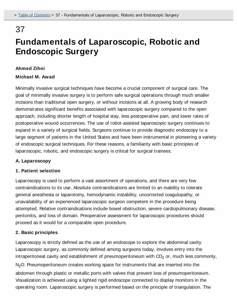
37Fundamentals of Laparoscopic, Robotic andEndoscopic Surgery
Ahmed Zihni
Michael M. Awad
Minimally invasive surgical techniques have become a crucial component of surgical care. Thegoal of minimally invasive surgery is to perform safe surgical operations through much smallerincisions than traditional open surgery, or without incisions at all. A growing body of researchdemonstrates significant benefits associated with laparoscopic surgery compared to the openapproach, including shorter length of hospital stay, less postoperative pain, and lower rates ofpostoperative wound occurrences. The use of robot-assisted laparoscopic surgery continues toexpand in a variety of surgical fields. Surgeons continue to provide diagnostic endoscopy to alarge segment of patients in the United States and have been instrumental in pioneering a varietyof endoscopic surgical techniques. For these reasons, a familiarity with basic principles oflaparoscopic, robotic, and endoscopic surgery is critical for surgical trainees.
A. Laparoscopy
1. Patient selection
Laparoscopy is used to perform a vast assortment of operations, and there are very fewcontraindications to its use. Absolute contraindications are limited to an inability to tolerategeneral anesthesia or laparotomy, hemodynamic instability, uncorrected coagulopathy, orunavailability of an experienced laparoscopic surgeon competent in the procedure beingattempted. Relative contraindications include bowel obstruction, severe cardiopulmonary disease,peritonitis, and loss of domain. Preoperative assessment for laparoscopic procedures shouldproceed as it would for a comparable open procedure.
2. Basic principles
Laparoscopy is strictly defined as the use of an endoscope to explore the abdominal cavity.Laparoscopic surgery, as commonly defined among surgeons today, involves entry into theintraperitoneal cavity and establishment of pneumoperitoneum with CO2 or, much less commonly,
N2O. Pneumoperitoneum creates working space for instruments that are inserted into the
abdomen through plastic or metallic ports with valves that prevent loss of pneumoperitoneum.Visualization is achieved using a lighted rigid endoscope connected to display monitors in theoperating room. Laparoscopic surgery is performed based on the principle of triangulation. The
> Table of Contents > 37 - Fundamentals of Laparoscopic, Robotic and Endoscopic Surgery

P.692
camera port is placed centrally, with working ports to either side, allowing the long rigidinstruments to converge at the surgical target
without colliding with the laparoscope or with one another. Laparoscopic surgery has severallimitations. The surgical field is a three-dimensional space, but most laparoscopes project animage onto a traditional twodimensional monitor, creating limited depth perception for theoperating surgeon. Most laparoscopic instruments can rotate around their long axis and somemay have some degree of articulation at the instrument tip, but they have significantly limiteddexterity compared to the human hand and wrist. In addition, laparoscopic instruments cantransmit some haptic input through the instrument shaft and handle to the hand of the operator,but much of the haptic feedback that is essential to handling tissue in open surgery is lost duringlaparoscopic surgery.
3. Equipment and troubleshooting
a. Room setup and positioning. All necessary equipment should be in the operating roomprior to patient arrival, including an insufflator and gas tank, light source, camera, laparoscope,monitors, laparoscopic ports and trocars, laparoscopic instrumentation, and instruments for openconversion if necessary. If intraoperative fluoroscopy or radiography is likely to be employed, thepatient and equipment should be positioned to allow additional hardware to access the operatingtable. The patient, surgeon, and monitor should be positioned to place the operative fieldbetween the surgeon and the monitor. Patient position is dictated by the surgical procedure beingundertaken. Most abdominal operations require the patient be placed in the supine position. Thisposition is sometimes modified to split the legs and allow the surgeon to operate from betweenthe legs to access the upper abdomen. Footboards, suction beanbags, and safety straps are oftenemployed to secure the patient to the operating table, allowing for steep angles to be employedintraoperatively. Pelvic procedures in which a perineal approach may be required typically employthe lithotomy position. Lateral decubitus positioning is used to perform thoracoscopic surgery orlaparoscopic retroperitoneal procedures. Monitors should be positioned to allow the operatingsurgeon to view them clearly without turning their head and with a 15-degree downward gaze,minimizing neck extension and postural fatigue. The operating table height and port placementshould allow the operating surgeon to keep both arms at their sides with their elbows flexed to 90to 120 degrees. A preoperative safety and equipment check is essential prior to any laparoscopicoperation (Fig. 37-1).
b. Insufflation. A pressure-limited insufflator is typically used in laparoscopic surgery. Thisdevice controls the flow of CO2 into the abdominal cavity. Most insufflators display the pressure in
the system, which reflects the pressure in the target body cavity when the two are in continuity,as well as the flow rate of gas through the insufflator and the total volume of gas insufflated. Theinsufflator allows a target pressure to be selected and the rate of gas flow to be modulated. Theminimum pressure that allows adequate working space should be employed. Loss ofpneumoperitoneum or an increase in pressure can occur due to factors at the level of the gas
UnitedVRG

P.693
P.694
source, insufflator, tubing, ports, or change in patient sedation level (Fig. 37-1).
Figure 37-1 Troubleshooting Algorithm for Laparoscopic Surgery. (Airan M (2012)Equipment Setup and Troubleshooting. In: Soper NJ, Scott-Connor CEH, eds. The SAGESmanual: Volume 1. Basic laparoscopy and endoscopy. New York, NY: Springer, 21-43.)(continued)
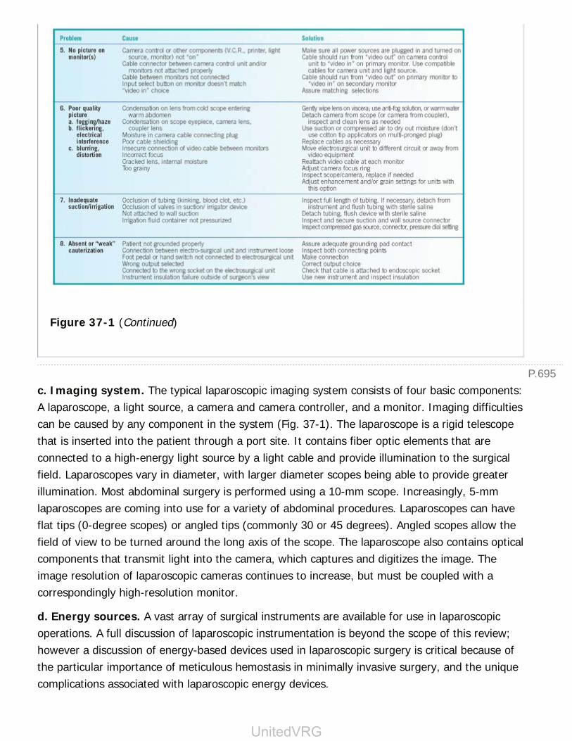
P.695
Figure 37-1 (Continued)
c. Imaging system. The typical laparoscopic imaging system consists of four basic components:A laparoscope, a light source, a camera and camera controller, and a monitor. Imaging difficultiescan be caused by any component in the system (Fig. 37-1). The laparoscope is a rigid telescopethat is inserted into the patient through a port site. It contains fiber optic elements that areconnected to a high-energy light source by a light cable and provide illumination to the surgicalfield. Laparoscopes vary in diameter, with larger diameter scopes being able to provide greaterillumination. Most abdominal surgery is performed using a 10-mm scope. Increasingly, 5-mmlaparoscopes are coming into use for a variety of abdominal procedures. Laparoscopes can haveflat tips (0-degree scopes) or angled tips (commonly 30 or 45 degrees). Angled scopes allow thefield of view to be turned around the long axis of the scope. The laparoscope also contains opticalcomponents that transmit light into the camera, which captures and digitizes the image. Theimage resolution of laparoscopic cameras continues to increase, but must be coupled with acorrespondingly high-resolution monitor.
d. Energy sources. A vast array of surgical instruments are available for use in laparoscopicoperations. A full discussion of laparoscopic instrumentation is beyond the scope of this review;however a discussion of energy-based devices used in laparoscopic surgery is critical because ofthe particular importance of meticulous hemostasis in minimally invasive surgery, and the uniquecomplications associated with laparoscopic energy devices.
UnitedVRG

P.696
(1) Monopolar energy devices use a current of electrons that originates at an electrode at thesurgical site and travel to a dispersive electrode elsewhere on the patient's body, usually a pad ontheir leg or back. The density of current at the site of the surgical electrode generates heat,leading to tissue disruption. There are two commonly used modalities of monopolar energy.ÒCuttingÓ mode is a continuous high-frequency, low-voltage current that rapidly produces veryhigh temperatures and tissue vaporization at the target. This allows the instrument to cut throughtissue easily without achieving hemostasis, and is therefore rarely used in laparoscopic surgerywhere hemostasis is crucial for adequate visualization of tissue. ÒCoagulatingÓ mode is a pulsedlow-frequency high-wavelength current that causes a slower, lower temperature heating of tissuethat causes coagulative necrosis of tissue and hemostasis at the surgical site. There are uniquerisks associated with laparoscopic monopolar electrosurgery. Many monopolar devices do nothave shielded instrument tips or require a connection to be made between two electrodes for thedevice to be activated, allowing for injuries to occur if the device is inadvertently activated whilethe instrument tip is out of the laparoscopic field of view. Also, current can pass through
breaks in the insulation, leading to injuries if points of insulation failure come into contact withtissue. Direct coupling is a phenomenon that occurs when current passes from a monopolarinstrument through another conductive instrument or material. This is often used purposefullyduring surgery, but can also occur inadvertently when current is transmitted through metallicports or other instruments to tissue. Capacitive coupling is a similar phenomenon that occurswhen an active electrode passes through an uninsulated conductor, such as a plastic and metallictrocar, causing current to be stored and potentially discharged into tissue.
(2) Bipolar energy devices use a current that originates in a grounded energy source andtravels between two electrodes that are contained within the working tip of the instrument.Because target tissue is contained between the two electrodes, there is very little thermal spreadbeyond this target. There is also a much lower risk of capacitive coupling associated with bipolarinstruments. Bipolar devices are commonly used as vessel sealing devices, or to divide highlyvascular tissue, because their ability to deliver a current between two instrument tines that areheld together with pressure can effectively fuse the walls of vessels up to 7 mm in diameter.
(3) Argon beam coagulation is a form of monopolar electrosurgery in which a stream ofionized argon gas is emitted at the surgical site and completes the circuit between an activeelectrode and the distant dispersive electrode. A high current density flows through the argongas, causing thermal damage to tissue contacted by the gas. This technology is very useful forachieving hemostasis across a large raw bleeding surface where bleeding would otherwise bedifficult to control. Employing argon beam coagulation laparoscopically, however, can increaseintra-abdominal pressure due to the additional gas being introduced to the abdomen.
(4) Ultrasonic energy devices employ an active blade that vibrates at very high frequency,generating thermal energy that can divide tissue that is compressed against the active blade byan inactive blade. They can be used to divide or dissect tissues, including vascularized tissue, and

P.697
can seal vessels up to 5 mm in diameter. These systems do not have any risk of direct orcapacitive coupling. In addition, ultrasonic devices coagulate tissue at lower temperatures andgenerate much less thermal spread than monopolar devices. However, since the active blade ofan ultrasonic device is heated through use, this thermal energy takes a few seconds to dissipateafter the active blade is employed, and inadvertent injuries may occur if the active blade isbrought into contact with tissue immediately after activation.
e. Abdominal access. Safe access to the abdomen is the crucial first step to any laparoscopicprocedure. Two strategies to achieve access
to the abdomen are generally used: The ÒclosedÓ technique using a Veress needle, and theÒopenÓ technique using a Hasson cannula.
(1) Closed Veress needle technique. The Veress needle is a 7 to 12 cm long 14-gauge needleattached to a valve and stopcock that allow gas to be insufflated through the needle. The needlehas a sharp beveled tip with a spring-loaded retractable blunt tip that projects beyond the sharptip and retracts when pressed against a surface. To achieve access to the abdomen using aVeress needle, the patient is first positioned appropriately and their abdomen prepped forsurgery, and the site of Veress insertion selected. In a patient without prior abdominal surgery, aperiumbilical insertion site is used. In patients with prior midline abdominal surgery, alternativesites on the abdomen remote from the prior incision may be used. The left upper quadrant andright lower quadrant are frequently used alternate sites of needle insertion and initial portplacement. A stab incision is made at the site of Veress insertion. It is recommended to lift theabdominal wall away from the underlying viscera prior to needle insertion. The needle is theninserted into the abdomen, generally at an angle perpendicular to the abdominal wall, while heldat the shaft rather than its hub. The needle encounters resistance followed by a sensation of giveat two separate points, first as it crosses the midline fascia, then as it traverses the peritoneumand the blunt tip springs out of the needle. When performing Veress entry away from the midline,three ÒpopsÓ are typically encountered due to the presence of anterior and posterior fasciallayers along with the peritoneum. Once the Veress needle has entered the abdomen, its positionmay be confirmed. First, a syringe containing saline is attached to the hub and aspiratedÑnoblood or succus should be aspirated. Saline should easily instill into the abdomen without anyreturn of saline on subsequent aspiration. After instilling 3 to 5 cc of saline, the syringe should beremoved with the stopcock open and the saline remaining in the Veress needle should flow easilyinto the abdomen. This constitutes the ÒdropÓ test. The insufflation tubing is then connected tothe hub and the gas flow initiated at 1 to 2 L/minute. Initial pressure should be no more than 10mm Hg. Elevated initial pressure is caused by malposition of the Veress needle tip, either dueplacement of the needle in the preperitoneal space or due to the needle opening abutting astructure or sitting in a pocket with limited continuity with the rest of the abdomen. If this occurs,the needle may be gently rotated and slightly advanced or withdrawn. If the pressure does notquickly drop to the expected level, the needle should be withdrawn and another attempt made.
UnitedVRG

P.698
P.699
This may be safely repeated until an intra-abdominal position of the needle and normalinsufflation are confirmed.
Insufflation of the abdomen proceeds at 1 to 2 L/minute for 1 minute; then the flow rate isincreased to the maximal rate
the insufflator can support until the intra-abdominal pressure reaches the set target of 12 to 15mm Hg. Once the abdomen is adequately insufflated, the Veress needle is removed, the skin isincised at the camera port site, and the first trocar is inserted. This can be done blindly, or underdirect vision using an optical trocar. These trocars have a clear tip and allow a 0-degree scope tobe inserted into the trocar. As it is pushed into the abdomen, the layers of the abdominal wall andintraperitoneal space can be visualized. Once the port is placed into the abdomen, the trocar isremoved and the laparoscope inserted to the abdomen to confirm placement and perform aninitial exploration. Particular attention is paid to the area directly below the initial port, whereinjury may have been caused by the Veress needle or the trocar. Afterward, subsequent portsmay be placed with direct visualization using the camera. Ports should be placed 8 to 10 cm apartand adhere to the principle of triangulation described above.
(2) Open Hasson cannula technique. This technique involves entry into the abdomen at theinitial trocar site under direct vision. This technique is particularly useful when safety concerns arepresent due to prior surgery and the possibility of viscera adherent to the abdominal wall. Somesurgeons preferentially use an open access technique for all laparoscopic procedures. Open entryis usually performed at a periumbilical site which is used as the camera port during surgery.However, in patients with extensive prior midline abdominal surgery, an alternative site may beused, such as the left upper or right lower quadrants. After the skin is prepped, a 1 to 3 cmincision is made at the site of entry and carried down to the subcutaneous tissue using sharpdissection and electrocautery. Sweeping away subcutaneous fat using retractors, the fascia isexposed, pulled away from underlying viscera and incised. The peritoneum is then similarlyelevated and opened. The abdominal cavity is then visualized and gently explored with a finger,and fascial sutures placed. Then the Hasson cannula is inserted into the intraperitoneal space.The Hasson cannula has a blunt obturator to prevent insertion trauma, a cone-shaped sleeve thatsits in the fascial incision, and two struts that are fixed to the fascial stitches to hold the system inplace. Some systems employ a balloon that sits over the intraperitoneal portion of the cannulaand is inflated upon insertion, holding the system in place without fascial sutures when the sleeveis brought down into position. Once the cannula is positioned, the camera is inserted for an initialexploration of the abdomen and placement of subsequent ports as in the Veress technique.
f. Complications of abdominal access. A variety of complications can occur during abdominalaccess, particularly during blind portions such as Veress needle or blind trocar insertion. Visceralor vascular injury may occur during Veress needle insertion. If bowel contents or blood areaspirated during Veress insertion, the needle should be
P.700
withdrawn and reinserted and laparoscopic access should proceed. Upon entry to the abdomen,bowel or other injured organs should be examined and managed per established protocols fortraumatic injury to intraperitoneal organs. In most cases, a Veress needle stick to intraperitonealviscera does not require any intervention, but inadvertent intraluminal insufflation with the Veressneedle can cause significant injury requiring bowel resection. The retroperitoneum should also beclosely examined on abdominal exploration, particularly in situations where blood wasencountered on initial Veress insertion. A stable hematoma in the mesentery or lateralretroperitoneum can usually be observed. However, central or rapidly expanding hematomasrequire laparotomy and exploration of the retroperitoneum.
Trocar insertion can cause injuries far more severe than Veress insertion. Bowel and visceralinjuries should be considered penetrating traumas and managed accordingly, and any central orrapidly expanding retroperitoneal hematoma should be explored. If a rush of blood isencountered on trocar insertion, or open cannula placement, the surgeon must assume that acritical vascular injury has occurred and conversion to laparotomy must be performedimmediately, without removing the trocar which may partially tamponade the injury.
Abdominal wall hemorrhage is a common complication of laparoscopic surgery that may becaused by injury to the epigastric arteries, veins, or their tributaries. These hemorrhages areoften selflimited and cease with the tamponading effect of the port. If this fails to control thebleeding, control may be attempted using electrocautery or with a suture passing device.
g. Port removal and closure. At the conclusion of surgery, instruments and ports are removedunder direct visualization, with the camera port being removed last, after the abdomen isdesufflated. Fascial incisions 5 mm or smaller do not need to be closed. Ports of 10 mm or largermay be sutured to prevent hernia formation and incarceration of bowel if a bladed trocar wasused or the site was dilated. Skin incisions are closed with subcuticular absorbable sutures,staples, or simply with skin tape or glue in the case of incisions 5 mm or smaller.
h. Physiologic effects of laparoscopy. Laparoscopic surgery requires the creation ofpneumoperitoneum and a resultant increase in intra-abdominal pressure, which is associated witha variety of physiological effects
(1) Cardiovascular. Elevated intra-abdominal pressure leads to an increase in systemic vascularresistance due to compression of the aorta and visceral arteries, as well as the resultantactivation of the renin-angiotensin-aldosterone axis. Inferior vena cava compression leads toreduced preload and lower cardiac output. Cephalad displacement of the diaphragm causesincreased intrathoracic pressure, which leads to increased pulmonary vascular resistance.
(2) Respiratory. Increased intra-abdominal pressure limits diaphragmatic excursion, decreasingpulmonary compliance, and functional residual capacity, and increasing thoracic pressure. Thesechanges are associated with the development of atelectasis. In addition, absorption of insufflatedCO2 can contribute to hypercarbia and acidosis.
UnitedVRG
P.701
(3) Renal. Increased intra-abdominal pressure leads to a reduced glomerular filtration rate dueto reduced afferent arterial flow and elevated renal venous pressure.
(4) Neurologic. The increased intrathoracic and central venous pressure associated withpneumoperitoneum leads to a decrease in venous return from the brain. This leads to an increasein intracranial pressure and may contribute to temporary confusion on emergence from longlaparoscopic operations.
4. Fundamentals of laparoscopic surgery. The Fundamentals of Laparoscopic Surgery (FLS)curriculum and examination are a validated program developed by the Society of AmericanGastrointestinal and Endoscopic Surgeons (SAGES) for the teaching and evaluation of didacticknowledge and technical skills associated with laparoscopic surgery. The program is composed ofa web-based didactic curriculum, a written examination, and a skill-based examination.
The didactic portion of the FLS curriculum is a web-based series of modules that learners are ableto navigate and study at their own pace. These modules cover basic preoperative considerations,intraoperative considerations, basic laparoscopic procedures, postoperative care andcomplications, and an explanation of the technical skills portion of the examination. After studyingthe didactic modules, the learner takes a 90-minute 75-question examination in a proctoredsetting, based on the content of the didactic modules.
The manual skills examination is a test of five tasks that are performed in a laparoscopic trainerbox. These tasks are: Peg transfer, Pattern cutting, Endoloop, Extracorporeal stitch, andIntracorporeal stitch. Each task is scored for efficiency, and has a time limit to complete the task.In addition, each task is scored for performance, with specifically defined task errors leading tolower scores. Passing the FLS written and technical examinations is now a prerequisite foreligibility for board certification in General Surgery in the United States.
B. Robot-assisted Surgery. Robot-assisted laparoscopic surgery is used in a variety of surgicalfields, particularly general surgery, urologic surgery, and gynecologic surgery, and continues toexpand. Robotic surgery holds great promise in the advancement of minimally invasive surgery,but also has significant current limitations.
1. Equipment and basic principles. Strictly speaking, current ÒroboticÓ surgical systems arenot robots, as the term implies some degree of automation and current robotic surgical platformsdo not perform any autonomous functions. More accurately, the most common robotic surgicalplatform (daVinci Surgical System, Intuitive Surgical Inc.,
Sunnyvale, CA) can be described as a computer-assisted surgical system. This platform employs aÒmaster-slaveÓ configuration, in which the surgeon is seated at a console (surgeon cart) andmanipulates hand-held controllers (masters), whose motions are mapped directly to the surgicalinstrument tip. The instruments are mounted to mechanical arms on a platform that is ÒdockedÓto the patient at the laparoscopic port sites (patient cart). The laparoscope is also attached to an

P.702
arm of the patient cart and is controlled by the surgeon from the surgeon cart. Commonly usedrobotic surgical platforms employ a scope very similar to a traditional 10-mm high-definitionlaparoscope, but which captures two separate images that are projected to the binoculareyepieces the surgeon looks into at the surgeon cart, creating a three-dimensional display. Inmost cases, a nonrobotic assistant port is also placed, allowing an assistant surgeon to participateusing traditional laparoscopic instruments and to assist with robotic instrument exchanges.Abdominal access for robot-assisted laparoscopic surgery is achieved in much the same way as intraditional laparoscopy. Triangulation of the surgical target remains the guiding principle for portplacement, though robotic ports are generally arranged along a straight line, rather than theÒbaseball diamondÓ configuration that is commonly used in laparoscopic surgery, to facilitate theaction of the robotic arms.
2. Advantages. Robot-assisted laparoscopic surgery has a variety of benefits for operatingsurgeons compared to laparoscopy. Robotic instruments are wristed, allowing for more degrees offreedom compared to laparoscopic instruments. In addition, the master-slave configuration allowsthe movements of the surgeon to be scaled down when they are converted to instrument actions,allowing for finer movements than laparoscopic instruments can achieve. Robot-assisted surgeryhas also been shown to generate less ergonomic stress on operating surgeons than laparoscopicsurgery, both objectively and subjectively (Surg Endosc. 2014;28:3379-3384).
3. Limitations. Robot-assisted laparoscopic surgery has several limitations. The robotic platformrequires docking of the patient cart to the laparoscopic ports, limiting surgery to one or two bodyquadrants. The newest iteration of the daVinci Surgical System (Xi) attempts to address thislimitation by streamlining undocking and redocking with a different surgical target, but traditionallaparoscopy still allows for greater freedom to explore the abdomen. In addition, the currentsurgeon cart provides minimal haptic feedback to the operating surgeon. This compels surgeonsto increasingly rely on visual cues to safely manipulate tissue. Finally, robotic surgery carriesadditional expense compared to laparoscopic surgery, and this added expense has not alwaysbeen shown to contribute to superior patient outcomes.
4. Fundamentals of Robotic Surgery. Fundamentals of Robotic Surgery (FRS) is aneducational curriculum for training and assessing surgical learners in the use of robot-assistedsurgery. This program has been developed by a group of experts in robotic surgery and surgicaleducation, and is modeled on the FLS program, though its technical
component has yet to be implemented. The curriculum contains modules focusing on introducinglearners to robotic surgical systems, learning about important psychomotor skills for robot-assisted surgery, and understanding the team-based approach and communication skillsnecessary to effectively perform robotic surgery.
D. Flexible Endoscopy and Endoscopic Surgery
1. Equipment and troubleshooting. Flexible endoscopy is performed using a flexible fiberoptic
UnitedVRG
P.703
endoscope, connected to a source of water or saline, a suction source, and an air or gasinsufflator. Most modern endoscopes are operated with a CCD chip camera that captures animage from the endoscope tip, digitizes it, and projects it to a monitor. Commonly usedendoscopes also have working channels that allow instrumentation to be passed through thescope to the target, where biopsies can be taken or interventions performed. The most commonlyused endoscopes among surgeons are gastroscopes and colonoscopes. Gastroscopes are used fordiagnostic esophagogastroduodenoscopy and have a typical working length of 92.5 to 110 cmand a thickness of 9.0 to 9.2 mm, and usually have a single working channel. Colonoscopes arelonger (133 to 170 cm) and thicker (11.1 to 13.7 mm diameter) and also have a working channel.Diagnostic endoscopy is performed by inserting an endoscope into the gastrointestinal tract andmeticulously examining the target mucosa while advancing or withdrawing the scope. Insufflationis used to dilate the gastrointestinal lumen to facilitate scope advancement or mucosalvisualization. Irrigation is used to clean the tip of the endoscope or wash debris from segments ofmucosa. All of these elements are subject to malfunction, and a systematic approach totroubleshooting is critical (Fig. 37-2).
2. Endoscopic surgery. A variety of graspers, electrosurgical hemostatic devices, biopsy snares,ablative technologies, stents, clips, and other devices are available for use in interventionalendoscopy. A full description of interventional endoscopy is beyond the scope of this chapter, butthis expansion of technology has made possible a variety of endoluminal therapies for disordersthat would have required open or laparoscopic surgical interventions. These include palliativestenting for gastrointestinal cancers, endoluminal therapies for reflux, complications of bariatricsurgery, and achalasia.
3. Fundamentals of endoscopic surgery. Fundamentals of Endoscopic Surgery (FES) is adidactic curriculum and technical exam developed by SAGES to train surgical learners in thetheory and technical skills necessary to perform diagnostic and interventional endoscopy. Thedidactic portion is a web-based modular curriculum similar to the model established with FLS.Modules focus on technology, patient preparation, patient sedation, upper and lower diagnosticendoscopy, biopsy techniques, enteral access procedures, hemostatic techniques, and ERCP. Aproctored written exam tests knowledge of these topics,
and is accompanied by a technical exam that assesses fundamental endoscopic skillsin a simulator. Passage of the FES written and technical exam is a prerequisite forboard eligibility in General Surgery in the United States beginning with graduates in2018.

P.704
Figure 37-2 Troubleshooting for Endoscopy. (From Vitale GC, Davis BR (2012) FlexibleEndoscopes: Characteristics, Troubleshooting, and Equipment Care. In: Soper NJ, Scott-Connor CEH, eds. The SAGES manual: Volume 1. Basic laparoscopy and endoscopy. NewYork, NY: Springer, 497-507.)
CHAPTER 37: FUNDAMENTALS OF LAPAROSCOPIC,ROBOTIC AND ENDOSCOPIC SURGERYMultiple Choice Questions1. Which of the following is an absolute contraindication to laparoscopicsurgery?
UnitedVRG
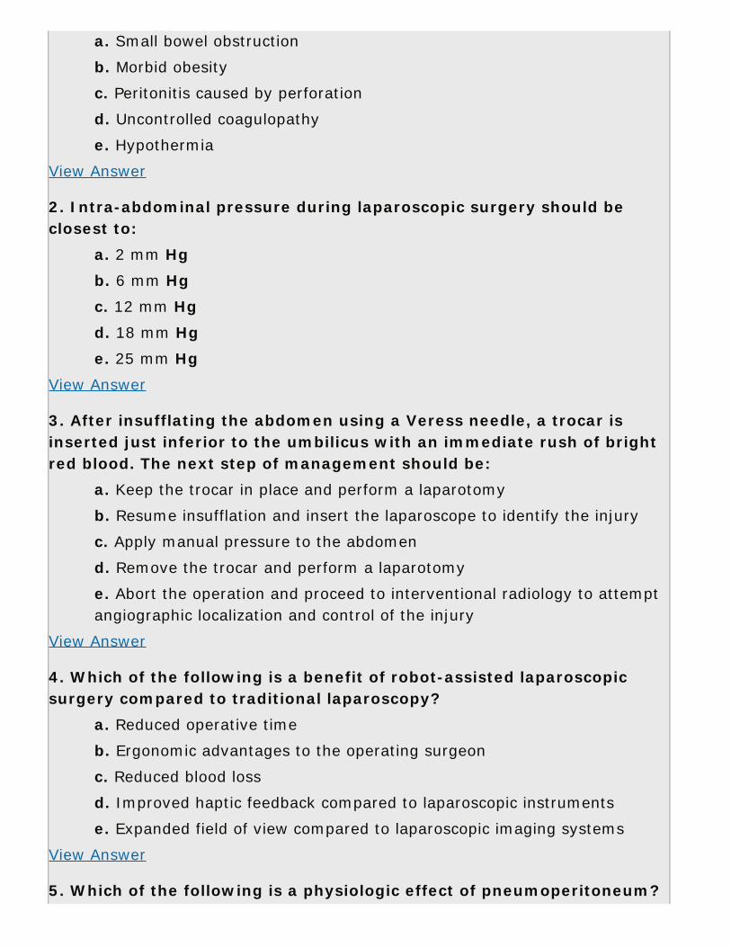
a. Small bowel obstruction
b. Morbid obesity
c. Peritonitis caused by perforation
d. Uncontrolled coagulopathy
e. Hypothermia
View Answer
2. Intra-abdominal pressure during laparoscopic surgery should beclosest to:
a. 2 mm Hg
b. 6 mm Hg
c. 12 mm Hg
d. 18 mm Hg
e. 25 mm Hg
View Answer
3. After insufflating the abdomen using a Veress needle, a trocar isinserted just inferior to the umbilicus with an immediate rush of brightred blood. The next step of management should be:
a. Keep the trocar in place and perform a laparotomy
b. Resume insufflation and insert the laparoscope to identify the injury
c. Apply manual pressure to the abdomen
d. Remove the trocar and perform a laparotomy
e. Abort the operation and proceed to interventional radiology to attemptangiographic localization and control of the injury
View Answer
4. Which of the following is a benefit of robot-assisted laparoscopicsurgery compared to traditional laparoscopy?
a. Reduced operative time
b. Ergonomic advantages to the operating surgeon
c. Reduced blood loss
d. Improved haptic feedback compared to laparoscopic instruments
e. Expanded field of view compared to laparoscopic imaging systems
View Answer
5. Which of the following is a physiologic effect of pneumoperitoneum?

a. Increased preload
b. Metabolic alkalosis
c. Increased pulmonary compliance
d. Decreased intracranial pressure
e. Decreased glomerular filtration rate
View Answer
UnitedVRG
38Hernias
Thomas J. Wade
L. Michael Brunt
I. INGUINAL HERNIAA. Incidence. The true incidence and prevalence of inguinal hernia worldwide isunknown. Inguinal hernia formation is a by-product of genetic, environmental, and metabolicfactors, combined with individual patient factors that can vary over time such as activity level,immune status, infection(s), medications, personal habits (e.g., smoking), and changes in bodymass index (Surg Clin North Am. 2008;88:179-201). Laparoscopic studies have reported rates ofcontralateral defects as high as 22%, with 28% of these going on to become symptomatic duringshort-term followup. The male-to-female ratio is greater than 10:1. The lifetime prevalence isestimated to be 25% in men and 2% in women. Two-thirds of incident inguinal hernias areindirect whereas nearly two-thirds of recurrent hernias are direct. Approximately 10% of inguinalhernias will become incarcerated, and a portion of these may become strangulated. Recurrencerates after surgical repair are less than 1% in children and vary in adults related to the method ofhernia repair.
B. Terminology and Anatomy
1. The inguinal canal (Fig. 38-1) is a tunnel that traverses the layers of the abdominal wallmusculature, bounded on the lateral deep aspect by an opening in the transversalisfascia/transversus abdominis muscle (internal inguinal ring), and travels along the fused edges ofthe transversus abdominis/internal oblique/inguinal ligament and iliopubic tract posteriorly andlayers of the external oblique musculature anteriorly, ending on the medial superficial aspect atan opening in the external oblique aponeurosis (external inguinal ring). The inguinal canal housesthe spermatic cord (males) or the round ligament (females) and is subject to hernia formationdue primarily to decreased mechanical integrity of the internal ring and/or transversalis fascia,allowing intra-abdominal contents to encroach into this space and form the characteristic bulge ofa groin hernia.
2. Direct hernias occur as a result of weakness in the posterior wall of the inguinal canal, whichis usually a result of attenuation of the transversalis fascia. The hernia sac protrudes throughHesselbach triangle, which is the space bounded by the inferior epigastric artery, the lateral edgeof the rectus sheath, and the inguinal ligament.
3. Indirect hernias pass through the internal inguinal ring lateral to the inferior epigastric
> Table of Contents > 38 - Hernias

P.706
vessels and Hesselbach triangle and follow the spermatic cord in males and the round ligament infemales. During
dissection, an indirect hernia sac is typically found on the anteromedial aspect of the spermaticcord. Indirect hernias may become incarcerated at either the internal or external ring.
Figure 38-1 Anatomy of the right inguinal region.
4. In combined (pantaloon) hernias, direct and indirect hernias coexist.
5. Variants of inguinal hernias
a. Sliding hernia: (Usually indirect inguinal) denotes that a part of the wall of the hernia sac isformed by an intra-abdominal viscus (usually colon, sometimes bladder).
b. Richter hernia: A portion of (rather than the entire circumference) of the bowel wall isincarcerated.
c. Littré hernia: Contains a Meckel diverticulum.
UnitedVRG
P.707
d. Amyand hernia: An inguinal hernia that contains the appendix.
6. Incarcerated inguinal hernias cannot be reduced into the abdominal cavity and may ormay not be symptomatic. Strangulated hernias are incarcerated with vascular compromise of theherniated contents. Frequently, intense pain is caused by ischemia of the incarcerated segment.
C. Diagnosis
1. Clinical presentation
a. Most inguinal hernias present as an intermittent bulge that appears in the groin. In males, itmay extend into the scrotal sac. Symptoms
are usually related to exertion or long periods of standing. The patient may complain of unilateraldiscomfort without noting a mass. Often, a purposeful Valsalva maneuver can reproduce thesymptoms and/or the presence of a bulge. In infants and children, a groin bulge is often noticedby caregivers during episodes of crying or defecation. Only in rare cases do patients present withbowel obstruction without the presence of a groin abnormality. All patients presenting with smallbowel obstruction must be questioned carefully and examined for all types of hernia (i.e.,inguinal, umbilical, incisional, obturator, etc.) as a possible etiology of obstruction.
b. Physical examination. The main diagnostic maneuver for inguinal hernias is palpation of theinguinal region. The patient is best examined while standing and straining (cough or Valsalva).Hernias manifest as bulges with smooth, rounded surfaces that become more evident withstraining. It may be necessary to invaginate the hemiscrotum to introduce an index fingerthrough the external inguinal ring if the hernia is not apparent, but this maneuver is oftenuncomfortable for the patient and is unnecessary if an obvious bulge is present. It is difficult todetermine whether the hernia is direct or indirect based solely on physical examination, althoughmost hernias that extend into the scrotum are indirect. Incarcerated inguinal hernias present withpain, abdominal distention, nausea, and vomiting due to intestinal obstruction.
2. Radiographic evaluation. X-ray studies are rarely indicated. Ultrasonography or computedtomographic (CT) scan may occasionally be used to diagnose an occult groin hernia, particularlyin the obese patient. Plain abdominal radiographs may verify intestinal obstruction in cases ofincarceration.
D. Differential Diagnosis. Inguinal hernias should be distinguished from femoral hernias, whichprotrude below the inguinal ligament. Inguinal adenopathy, lipomas, hydrocele, epididymitis,testicular torsion, groin abscess, and vascular aneurysms/pseudoaneurysms all should beconsidered in the differential diagnosis.
E. Treatment
1. Preoperative evaluation and preparation. Most patients with inguinal hernias should betreated surgically although watchful waiting may be appropriate for individuals with asymptomatichernias or for elderly patients with minimally symptomatic hernias (J Am Coll Surg. 2006;203:458-

P.708
468). Recently, Fitzgibbons et al. found that 68% of male patients who undergo watchful waitingwill undergo repair within 10 years of presentation (Ann Surg. 2013; 258(3):508-515). Therefore,surgical repair should be considered in patients with asymptomatic or minimally symptomaticinguinal hernia. Associated conditions that lead to increased intra-abdominal pressure such aschronic cough, constipation, or bladder outlet obstruction should be evaluated and remedied tothe extent possible before elective herniorrhaphy. In patients with symptoms of altered bowelhabits (i.e., frequent straining/constipation),
one must assess the risk of underlying colorectal malignancy. In incarcerated hernias withintestinal obstruction and possible strangulation, broad-spectrum antibiotics should be given andnasogastric suction may be necessary. Correction of volume status and electrolyte abnormalitiesis also important when there is associated small bowel obstruction.
2. Reduction. In uncomplicated cases, the hernia should reduce with palpation over the inguinalcanal while the patient is supine. If this does not occur, the physician should apply gentlepressure over the hernia with the concavity of the palm of his/her hand and fingers and exert asteady but gentle pressure as follows: Craniad and lateral for direct and indirect hernias, craniadand posterior for femoral hernias. If this is not successful, gentle traction over the mass withcompression may allow bowel gas to leave the herniated segment, making the mass reducible.Sedation and Trendelenburg position may be required for reduction of an incarcerated hernia, butit may be difficult to distinguish between acute incarceration and strangulation, as the inguinalcanal can become quite tender with or without ischemic contents. When an incarcerated hernia isreduced nonsurgically, the patient should be observed for the potential development of peritonitiscaused by perforation or ischemic necrosis of a loop of strangulated bowel. Strong suspicion ofstrangulation (i.e., erythema over hernia site, pain out of proportion to examination) is a surgicalemergency, and the patient should be taken expeditiously to the operating room.
3. Surgical treatment
a. Choice of anesthetic. Local anesthesia with sedation and monitored anesthesia care (MAC),which has several advantages over general or regional (spinal or epidural) anesthesia, is thepreferred anesthetic for elective open repair for small- to moderate-sized hernias. This approachresults in better postoperative analgesia, a shorter recovery room stay, and a negligible rate ofpostoperative urinary retention; it is also the lowest-risk anesthetic for patients with underlyingcardiopulmonary disorders. Commonly, a mixture of a short-acting agent (lidocaine 1%) andlonger-acting agent (bupivacaine 0.25% to 0.50%) is used. The dose limits for local anesthesiaare 4.5 mg/kg of plain lidocaine or 7 mg/kg of lidocaine with 1:100,000 epinephrine and 2 mg/kgplain bupivacaine or 3 mg/kg bupivacaine with 1:100,000 epinephrine. Use of local anesthesia forherniorrhaphy in our hospital is routinely supplemented by MAC and administration of intravenousmidazolam and propofol. Virtually all patients who undergo hernia repair under local anesthesiacan be managed as outpatients unless associated medical conditions or extenuating socialcircumstances necessitate overnight observation in the hospital. In contrast, laparoscopic inguinal
UnitedVRG

P.709
hernia repair is performed under general anesthesia to facilitate tolerance of pneumoperitoneum.
b. Treatment of the hernia sac. For indirect hernias, the sac (peritonealized abdominalcontents) is dissected from the spermatic cord and cremasteric fibers (Fig. 38-1). The sac caneither be ligated deep into the internal ring with an absorbable suture after reduction of
herniated contents or invaginated back into the abdomen without ligation; the latter approach isthought to be associated with less postoperative pain because it avoids transection of innervatedperitoneum (Hernia. 2014;18(2):199-204). Large, indirect sacs that extend into the scrotumshould not be dissected beyond the pubic tubercle because of an increased risk of ischemicorchitis. Similarly, one should avoid translocating the testicle into the inguinal canal during herniarepair owing to the risk of ischemia or torsion. Cord lipomas are frequently encountered duringrepair and should be excised or reduced into the preperitoneal space to avoid future confusionwith a recurrent hernia. Sliding hernia sacs can usually be managed by reducing the sac andattached viscera without sac ligation. Direct sacs are usually too broadly based for ligation and donot need to be opened. The redundant attenuated tissue is inverted and if the defect is small, itmay be closed with a few interrupted sutures before placement of mesh.
c. Primary tissue repairs without mesh were the mainstay of hernia surgery prior to thedevelopment of synthetic-mesh approaches. While primary repair avoids placement of foreignprosthetic material, disadvantages of this approach include higher recurrence rates (5% to 10%for primary repairs and 15% to 30% for repair of recurrent hernias) due to tension on the repairand a slower return to unrestricted physical activity. Although the vast majority of hernias arenow treated with a tension-free mesh repair, a primary tissue repair can be considered incontaminated wounds (e.g., if strangulated bowel is resected), as placement of synthetic materialwould be contraindicated. The principal features of the more commonly performed tissue repairsare as follows:
(1) Bassini repair. The inferior arch of the transversalis fascia or conjoint tendon isapproximated to the shelving portion of the inguinal ligament (iliopubic tract) with interrupted,nonabsorbable sutures. The Bassini repair has been used primarily for indirect hernias, includinginguinal hernias in women.
(2) McVay repair. The transversalis fascia is sutured to Cooper ligament medial to the femoralvein and to the inguinal ligament at the level of, and lateral to, the femoral vein. This operationrequires placement of a relaxing incision medially on the aponeuroses of the internal obliquemuscle to avoid undue tension on the repair. Historically, this approach was used more commonlyfor direct hernias. The McVay repair closes the femoral space and therefore, unlike the Bassinirepair, is also effective for femoral hernias.
(3) Shouldice repair. In this repair, the transversalis fascia is incised (and partially excised ifweakened) and reapproximated. The overlying tissues (the conjoint tendon, iliopubic tract, andinguinal ligament) are approximated in multiple, imbricated layers of running nonabsorbable
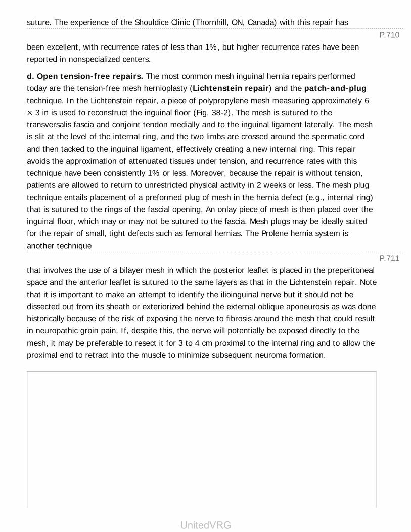
P.710
P.711
suture. The experience of the Shouldice Clinic (Thornhill, ON, Canada) with this repair has
been excellent, with recurrence rates of less than 1%, but higher recurrence rates have beenreported in nonspecialized centers.
d. Open tension-free repairs. The most common mesh inguinal hernia repairs performedtoday are the tension-free mesh hernioplasty (Lichtenstein repair) and the patch-and-plugtechnique. In the Lichtenstein repair, a piece of polypropylene mesh measuring approximately 6× 3 in is used to reconstruct the inguinal floor (Fig. 38-2). The mesh is sutured to thetransversalis fascia and conjoint tendon medially and to the inguinal ligament laterally. The meshis slit at the level of the internal ring, and the two limbs are crossed around the spermatic cordand then tacked to the inguinal ligament, effectively creating a new internal ring. This repairavoids the approximation of attenuated tissues under tension, and recurrence rates with thistechnique have been consistently 1% or less. Moreover, because the repair is without tension,patients are allowed to return to unrestricted physical activity in 2 weeks or less. The mesh plugtechnique entails placement of a preformed plug of mesh in the hernia defect (e.g., internal ring)that is sutured to the rings of the fascial opening. An onlay piece of mesh is then placed over theinguinal floor, which may or may not be sutured to the fascia. Mesh plugs may be ideally suitedfor the repair of small, tight defects such as femoral hernias. The Prolene hernia system isanother technique
that involves the use of a bilayer mesh in which the posterior leaflet is placed in the preperitonealspace and the anterior leaflet is sutured to the same layers as that in the Lichtenstein repair. Notethat it is important to make an attempt to identify the ilioinguinal nerve but it should not bedissected out from its sheath or exteriorized behind the external oblique aponeurosis as was donehistorically because of the risk of exposing the nerve to fibrosis around the mesh that could resultin neuropathic groin pain. If, despite this, the nerve will potentially be exposed directly to themesh, it may be preferable to resect it for 3 to 4 cm proximal to the internal ring and to allow theproximal end to retract into the muscle to minimize subsequent neuroma formation.
UnitedVRG
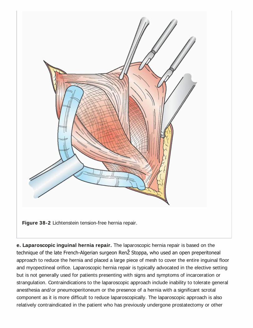
Figure 38-2 Lichtenstein tension-free hernia repair.
e. Laparoscopic inguinal hernia repair. The laparoscopic hernia repair is based on thetechnique of the late French-Algerian surgeon René Stoppa, who used an open preperitonealapproach to reduce the hernia and placed a large piece of mesh to cover the entire inguinal floorand myopectineal orifice. Laparoscopic hernia repair is typically advocated in the elective settingbut is not generally used for patients presenting with signs and symptoms of incarceration orstrangulation. Contraindications to the laparoscopic approach include inability to tolerate generalanesthesia and/or pneumoperitoneum or the presence of a hernia with a significant scrotalcomponent as it is more difficult to reduce laparoscopically. The laparoscopic approach is alsorelatively contraindicated in the patient who has previously undergone prostatectomy or other

P.712
lower midline abdominal surgery due to scarring in the preperitoneal space. There are twoapproaches to laparoscopic repair of inguinal hernias:
(1) Transabdominal preperitoneal (TAPP) repair. In the TAPP technique, the peritonealspace is entered by conventional means at the umbilicus, the peritoneum overlying the inguinalfloor is dissected away as a flap, the hernia is reduced, mesh is fixed over the internal ringopening in the preperitoneal space, and the peritoneum is reapproximated. The advantages ofthe TAPP approach are that a large working space is retained, familiar anatomic landmarks arevisible, and the contralateral groin can be examined for an occult hernia.
(2) Totally extraperitoneal repair (TEP). In the TEP technique, the preperitoneal space isdeveloped with a dissecting balloon inserted between the posterior rectus sheath and the rectusabdominis and directed toward the pelvis inferior to the arcuate ligament (Fig. 38-3). The otherports are inserted into this preperitoneal space without entering the peritoneal cavity. Theadvantages of the TEP repair are that the peritoneum is not opened and, therefore, does notneed to be closed and the operation is also typically faster to perform.
In either the TAPP or TEP technique, a large piece of mesh (6 × 4 in) is placed over the inguinalfloor and fixed (1) superiorly to the posterior abdominal wall fascia on either side of
the inferior epigastric vessels, (2) inferiorly to Cooper ligament, (3) medially to the midline fascia,and (4) superolaterally to the fascia above the internal ring. Staples/tacks must not be placedinferomedial to the internal ring or inferior to the iliopubic tract because of the risk of injury to theexternal iliac vessels (triangle of doom) and ilioinguinal, genitofemoral, lateral femoral cutaneous,and femoral nerves (triangle of pain).
UnitedVRG
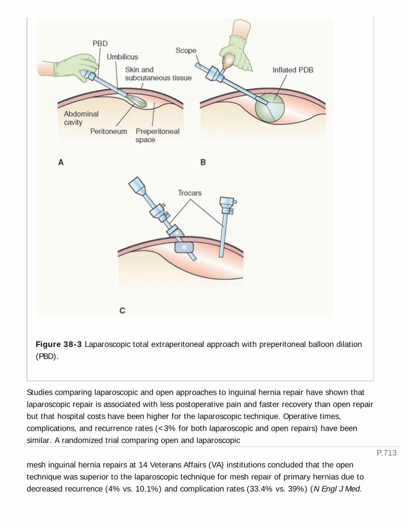
P.713
Figure 38-3 Laparoscopic total extraperitoneal approach with preperitoneal balloon dilation(PBD).
Studies comparing laparoscopic and open approaches to inguinal hernia repair have shown thatlaparoscopic repair is associated with less postoperative pain and faster recovery than open repairbut that hospital costs have been higher for the laparoscopic technique. Operative times,complications, and recurrence rates (<3% for both laparoscopic and open repairs) have beensimilar. A randomized trial comparing open and laparoscopic
mesh inguinal hernia repairs at 14 Veterans Affairs (VA) institutions concluded that the opentechnique was superior to the laparoscopic technique for mesh repair of primary hernias due todecreased recurrence (4% vs. 10.1%) and complication rates (33.4% vs. 39%) (N Engl J Med.
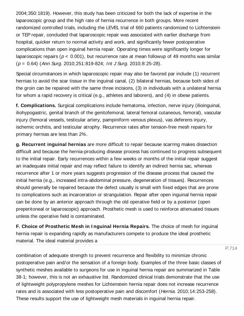
P.714
2004;350:1819). However, this study has been criticized for both the lack of expertise in thelaparoscopic group and the high rate of hernia recurrence in both groups. More recentrandomized controlled trials, including the LEVEL trial of 660 patients randomized to Lichtensteinor TEP repair, concluded that laparoscopic repair was associated with earlier discharge fromhospital, quicker return to normal activity and work, and significantly fewer postoperativecomplications than open inguinal hernia repair. Operating times were significantly longer forlaparoscopic repairs (p < 0.001), but recurrence rate at mean followup of 49 months was similar(p = 0.64) (Ann Surg. 2010;251:819-824; Int J Surg. 2010;8:25-28).
Special circumstances in which laparoscopic repair may also be favored par include (1) recurrenthernias to avoid the scar tissue in the inguinal canal, (2) bilateral hernias, because both sides ofthe groin can be repaired with the same three incisions, (3) in individuals with a unilateral herniafor whom a rapid recovery is critical (e.g., athletes and laborers), and (4) in obese patients.
f. Complications. Surgical complications include hematoma, infection, nerve injury (ilioinguinal,iliohypogastric, genital branch of the genitofemoral, lateral femoral cutaneous, femoral), vascularinjury (femoral vessels, testicular artery, pampiniform venous plexus), vas deferens injury,ischemic orchitis, and testicular atrophy. Recurrence rates after tension-free mesh repairs forprimary hernias are less than 2%.
g. Recurrent inguinal hernias are more difficult to repair because scarring makes dissectiondifficult and because the hernia-producing disease process has continued to progress subsequentto the initial repair. Early recurrences within a few weeks or months of the initial repair suggestan inadequate initial repair and may reflect failure to identify an indirect hernia sac, whereasrecurrence after 1 or more years suggests progression of the disease process that caused theinitial hernia (e.g., increased intra-abdominal pressure, degeneration of tissues). Recurrencesshould generally be repaired because the defect usually is small with fixed edges that are proneto complications such as incarceration or strangulation. Repair after open inguinal hernia repaircan be done by an anterior approach through the old operative field or by a posterior (openpreperitoneal or laparoscopic) approach. Prosthetic mesh is used to reinforce attenuated tissuesunless the operative field is contaminated.
F. Choice of Prosthetic Mesh in Inguinal Hernia Repairs. The choice of mesh for inguinalhernia repair is expanding rapidly as manufacturers compete to produce the ideal prostheticmaterial. The ideal material provides a
combination of adequate strength to prevent recurrence and flexibility to minimize chronicpostoperative pain and/or the sensation of a foreign body. Examples of the three basic classes ofsynthetic meshes available to surgeons for use in inguinal hernia repair are summarized in Table38-1; however, this is not an exhaustive list. Randomized clinical trials demonstrate that the useof lightweight polypropylene meshes for Lichtenstein hernia repair does not increase recurrencerates and is associated with less postoperative pain and discomfort (Hernia. 2010;14:253-258).These results support the use of lightweight mesh materials in inguinal hernia repair.
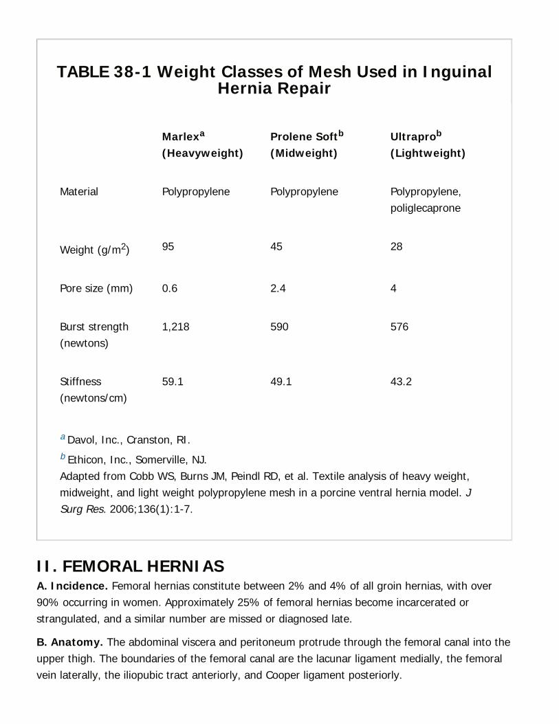
TABLE 38-1 Weight Classes of Mesh Used in InguinalHernia Repair
Marlexa
(Heavyweight)Prolene Softb
(Midweight)Ultraprob
(Lightweight)
Material Polypropylene Polypropylene Polypropylene,poliglecaprone
Weight (g/m2) 95 45 28
Pore size (mm) 0.6 2.4 4
Burst strength(newtons)
1,218 590 576
Stiffness(newtons/cm)
59.1 49.1 43.2
a Davol, Inc., Cranston, RI.b Ethicon, Inc., Somerville, NJ.Adapted from Cobb WS, Burns JM, Peindl RD, et al. Textile analysis of heavy weight,midweight, and light weight polypropylene mesh in a porcine ventral hernia model. JSurg Res. 2006;136(1):1-7.
II. FEMORAL HERNIASA. Incidence. Femoral hernias constitute between 2% and 4% of all groin hernias, with over90% occurring in women. Approximately 25% of femoral hernias become incarcerated orstrangulated, and a similar number are missed or diagnosed late.
B. Anatomy. The abdominal viscera and peritoneum protrude through the femoral canal into theupper thigh. The boundaries of the femoral canal are the lacunar ligament medially, the femoralvein laterally, the iliopubic tract anteriorly, and Cooper ligament posteriorly.

P.715
C. Diagnosis
1. Clinical presentation
a. Symptoms. Patients may complain of an intermittent groin bulge or a groin mass that may betender. Femoral hernias have a high
incidence of incarceration and small bowel obstruction may be the presenting feature in somepatients. Elderly patients, in whom femoral hernias occur most commonly, may not complain ofgroin pain even in the setting of incarceration. Therefore, an occult femoral hernia should beconsidered in the differential diagnosis of any patient with small bowel obstruction, especially ifthere is no history of previous abdominal surgery.
b. Physical examination. The characteristic finding is a small, rounded bulge that appears inthe upper thigh just below the inguinal ligament. An incarcerated femoral hernia usually presentsas a firm, tender mass. The differential diagnosis is the same as that for inguinal hernia.
c. Radiographic evaluation. Radiographic studies are rarely indicated. Occasionally, a femoralhernia is found on a CT scan or gastrointestinal contrast study performed to evaluate a smallbowel obstruction.
D. Treatment. The surgical approach can be inguinal, preperitoneal, or femoral.
1. Inguinal approach. A Cooper ligament repair (McVay) using the inguinal canal approachallows reduction of the hernia sac with visualization from above the inguinal ligament and closureof the femoral space. Occasionally, it may be necessary to divide the inguinal ligament to reducethe hernia. The repair can be performed with or without mesh.
2. Preperitoneal approach. A transverse suprainguinal incision permits access to theextraperitoneal spaces of Bogros and Retzius. The hernia is reduced from inside the femoralspace, and the hernia defect is repaired preperitoneally, usually with mesh, but can be repairedprimarily. This approach is especially useful for incarcerated or strangulated femoral hernias.Uncomplicated femoral hernias can also be repaired laparoscopically.
3. Femoral approach. A horizontal incision is made over the hernia, inferior and parallel to theinguinal ligament. After the hernia sac is dissected free, it can be resected or invaginated. Thefemoral canal is closed by placing interrupted stitches to approximate Cooper ligament to theinguinal ligament or by using a plug of prosthetic material.
4. Complications. Complications are similar to those for inguinal hernia repair. The femoral veinmay be especially susceptible to injury because it forms the lateral border of the femoral canal.
III. INTERNAL HERNIAA. Incidence. Of patients who present with acute intestinal obstruction, less than 5% have aninternal hernia. When internal hernias are complicated by intestinal volvulus, there is an 80%
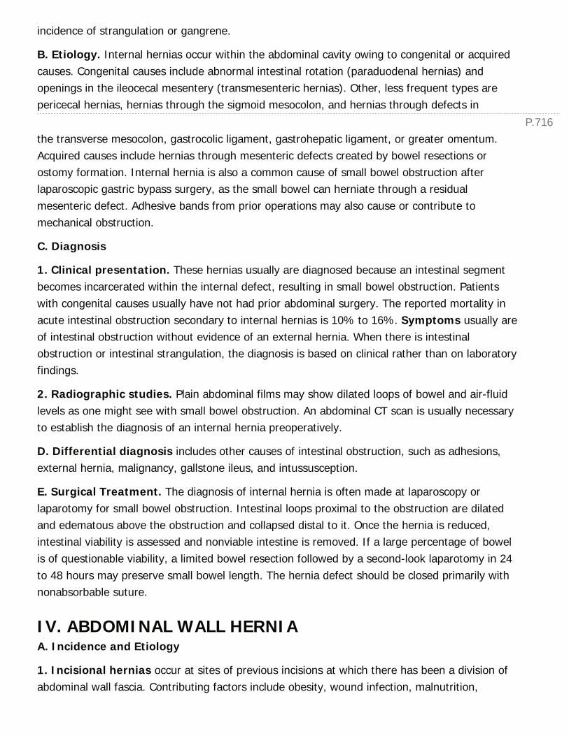
P.716
incidence of strangulation or gangrene.
B. Etiology. Internal hernias occur within the abdominal cavity owing to congenital or acquiredcauses. Congenital causes include abnormal intestinal rotation (paraduodenal hernias) andopenings in the ileocecal mesentery (transmesenteric hernias). Other, less frequent types arepericecal hernias, hernias through the sigmoid mesocolon, and hernias through defects in
the transverse mesocolon, gastrocolic ligament, gastrohepatic ligament, or greater omentum.Acquired causes include hernias through mesenteric defects created by bowel resections orostomy formation. Internal hernia is also a common cause of small bowel obstruction afterlaparoscopic gastric bypass surgery, as the small bowel can herniate through a residualmesenteric defect. Adhesive bands from prior operations may also cause or contribute tomechanical obstruction.
C. Diagnosis
1. Clinical presentation. These hernias usually are diagnosed because an intestinal segmentbecomes incarcerated within the internal defect, resulting in small bowel obstruction. Patientswith congenital causes usually have not had prior abdominal surgery. The reported mortality inacute intestinal obstruction secondary to internal hernias is 10% to 16%. Symptoms usually areof intestinal obstruction without evidence of an external hernia. When there is intestinalobstruction or intestinal strangulation, the diagnosis is based on clinical rather than on laboratoryfindings.
2. Radiographic studies. Plain abdominal films may show dilated loops of bowel and air-fluidlevels as one might see with small bowel obstruction. An abdominal CT scan is usually necessaryto establish the diagnosis of an internal hernia preoperatively.
D. Differential diagnosis includes other causes of intestinal obstruction, such as adhesions,external hernia, malignancy, gallstone ileus, and intussusception.
E. Surgical Treatment. The diagnosis of internal hernia is often made at laparoscopy orlaparotomy for small bowel obstruction. Intestinal loops proximal to the obstruction are dilatedand edematous above the obstruction and collapsed distal to it. Once the hernia is reduced,intestinal viability is assessed and nonviable intestine is removed. If a large percentage of bowelis of questionable viability, a limited bowel resection followed by a second-look laparotomy in 24to 48 hours may preserve small bowel length. The hernia defect should be closed primarily withnonabsorbable suture.
IV. ABDOMINAL WALL HERNIAA. Incidence and Etiology
1. Incisional hernias occur at sites of previous incisions at which there has been a division ofabdominal wall fascia. Contributing factors include obesity, wound infection, malnutrition,

P.717
smoking, and technical errors in wound closure. Hernias occur in up to 20% to 30% of patientsundergoing abdominal operations and are most commonly seen with midline incisions. Mostincisional hernias are now repaired with a mesh prosthetic via open or laparoscopic approach.
2. Umbilical hernias are congenital defects that may enlarge over time and becomeprotuberant and symptomatic. They are more frequent in people of African ancestry. Mostnewborn umbilical hernias close
spontaneously by the second year of life. However, umbilical hernias are also common in adults.Patients with ascites have a high incidence of umbilical hernias. Small umbilical hernias can bepresent for years without causing symptoms and may even go unnoticed. Over time, however,these hernias can enlarge and become incarcerated, usually with preperitoneal fat or omentum.Umbilical hernias greater than 3 cm should be repaired with a prosthetic mesh.
3. Epigastric hernias are hernias of the linea alba above the umbilicus. They occur morefrequently in athletically active young men or women. When small or in obese individuals,epigastric hernias may be hard to palpate and difficult to diagnosis. Usually, they produceepigastric pain that may be falsely attributed to other abdominal diagnoses. The diagnosis ismade by palpation of a subcutaneous epigastric mass; most such hernias occur within a fewcentimeters of the umbilicus and are associated with a small (1 to 2 cm) fascial defect.
4. Spigelian hernias protrude through the Spigelian fascia, near the termination of thetransversus abdominis muscle along the lateral edge of the rectus abdominis near the junction ofthe linea semilunaris and linea semicircularis. Because the herniated visceral contents areintraparietal (between the abdominal wall muscles), these hernias can be difficult to diagnoseand, therefore, are included in the differential diagnosis of obscure abdominal pain.Ultrasonography, CT scan, or laparoscopy can be useful confirmatory tools in patients with focalsymptoms in the appropriate region.
5. The most common type of lumbar hernia is an incisional hernia from a previousretroperitoneal or flank incision. Lumbar hernias may also occur in two different triangles: ThePetit (inferior lumbar) triangle and the GrynfelttÑLesshaft (superior lumbar) triangle,although these hernias are quite rare. Petit hernias are located in an area limited posteriorly bythe latissimus dorsi, anteriorly by the external oblique muscle, and inferiorly by the iliac crest; thefloor is formed by the internal abdominal oblique muscle. GrynfelttÑLesshaft hernias are borderedsuperiorly by the 12th rib, medially by the quadratus lumborum muscle, and laterally by theinternal abdominal oblique muscle, while the floor and roof of the triangle are formed by thetransversalis fascia and the external abdominal oblique muscle, respectively.
6. Obturator hernias are very rare hernias that occur predominantly in thin, older women andare difficult to diagnose. Patients classically present with bowel obstruction and focal tendernesson rectal examination. Pain along the medial aspect of the thigh with internal rotation of thethigh, known as the HowshipÑRomberg sign, results from obturator nerve compression and,

P.718
when present, may aid in the clinical diagnosis of an obturator hernia.
B. Treatment and Operative Management. Small epigastric, umbilical, obturator, andspigelian hernias may be repaired primarily. Most incisional hernias as well as lumbar andobturator hernias require the use of a prosthetic mesh because of their size and high recurrencerates after primary repair.
1. Prosthetic mesh in abdominal wall hernia repairs. The recurrence rate for ventralincisional hernia repair is 31% to 54% when primarily repaired. The technique of placingprosthetic biomaterials in the retrorectus preperitoneal space to repair ventral incisional herniaswas popularized by Jean Rives and René Stoppa and has reduced the recurrence rate of thesehernias to between 4% and 24%. Long-term followup of a randomized controlled trial showedthat the use of mesh results in a lower recurrence rate and less abdominal pain and does notresult in more complications than primary repair (Hernia. 2006;10:236-242). The choice of meshfor incisional hernia repair should be based on location and mesh properties. Uncoatedpolypropylene mesh should not be placed in an intraperitoneal position because it can formdense adhesions to the intestine and precipitate fistulization. For intraperitoneal placement, eithermicroporous polytetrafluoroethylene (PTFE) mesh or a barrier-coated mesh should be used.Microporous PTFE mesh has a microporous architecture and hydrophobicity that preventcellular penetration of intestine or abdominal viscera and may reduce the density ofintraperitoneal adhesions. There are also several absorbable barrier-coated meshes with apolypropylene or polyester construction. Different types of mesh are summarized in Table 38-2.Currently, there are limited data to support the use of one product over another. What is clear,however, is that the use of mesh is superior to primary repair for incisional hernias.
2. Open repairs. The principles for ventral hernia repair include dissection and identification ofall defects and repair with nonabsorbable sutures placed in healthy tissue. Incisional herniasshould be repaired with mesh prosthesis that should be anchored by nonabsorbable suturesplaced at least 3 cm beyond the margins of the defect. The placement of mesh in the retrorectusspace, that is, the RivesÑStoppa technique, has the lowest reported recurrence rate forhernia repair. This technique includes closure of the posterior rectus sheath beneath the mesh,allowing for the use of uncoated mesh. The principles of this repair have been expanded toinclude transverse abdominis release for the repair of giant ventral/incisional hernias. The meshutilized for these repairs should be durable and well tolerated by the patient, with a low risk forinfection. A variety of mesh products are available for repair, including polypropylene, PTFE,Gore-Tex, and a composite mesh of polypropylene and PTFE. Several composite mesh products(Table 38-2), with absorbable barriers coating and external to polypropylene or polyester meshcores, are available to minimize tissue attachment to intra-abdominal structures.
3. Laparoscopic repairs. The laparoscopic approach is another method for repair of incisionalhernias. The repair involves placement of a mesh prosthesis to cover the hernia defect. Thisapproach requires adhesiolysis of the entire prior incision, reduction of herniated abdominal
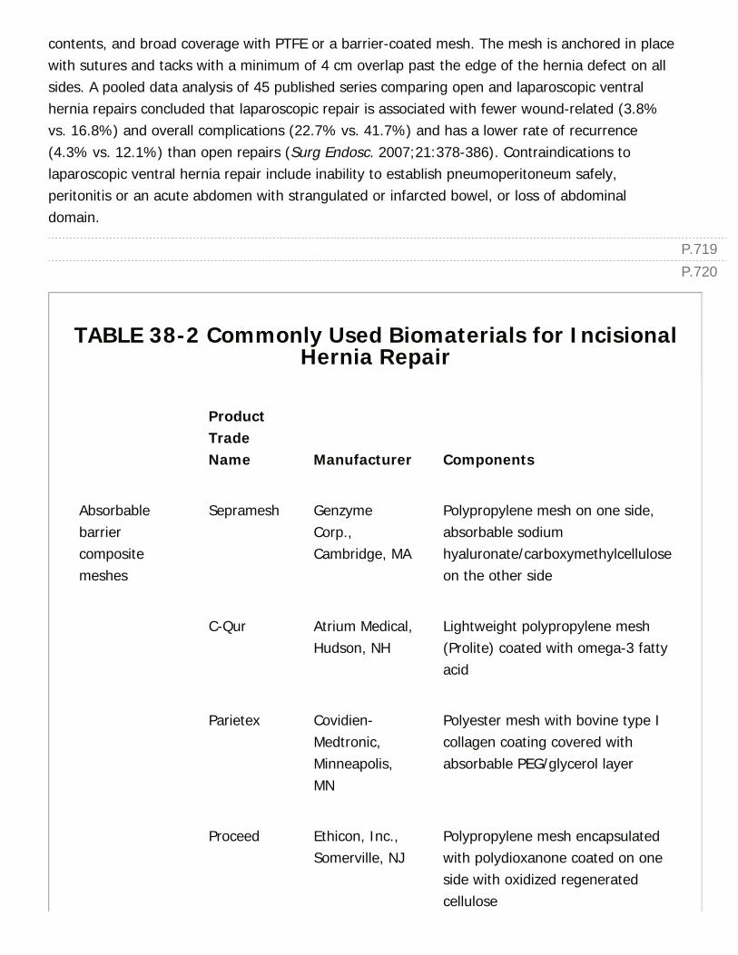
P.719P.720
contents, and broad coverage with PTFE or a barrier-coated mesh. The mesh is anchored in placewith sutures and tacks with a minimum of 4 cm overlap past the edge of the hernia defect on allsides. A pooled data analysis of 45 published series comparing open and laparoscopic ventralhernia repairs concluded that laparoscopic repair is associated with fewer wound-related (3.8%vs. 16.8%) and overall complications (22.7% vs. 41.7%) and has a lower rate of recurrence(4.3% vs. 12.1%) than open repairs (Surg Endosc. 2007;21:378-386). Contraindications tolaparoscopic ventral hernia repair include inability to establish pneumoperitoneum safely,peritonitis or an acute abdomen with strangulated or infarcted bowel, or loss of abdominaldomain.
TABLE 38-2 Commonly Used Biomaterials for IncisionalHernia Repair
ProductTradeName Manufacturer Components
Absorbablebarriercompositemeshes
Sepramesh GenzymeCorp.,Cambridge, MA
Polypropylene mesh on one side,absorbable sodiumhyaluronate/carboxymethylcelluloseon the other side
C-Qur Atrium Medical,Hudson, NH
Lightweight polypropylene mesh(Prolite) coated with omega-3 fattyacid
Parietex Covidien-Medtronic,Minneapolis,MN
Polyester mesh with bovine type Icollagen coating covered withabsorbable PEG/glycerol layer
Proceed Ethicon, Inc.,Somerville, NJ
Polypropylene mesh encapsulatedwith polydioxanone coated on oneside with oxidized regeneratedcellulose
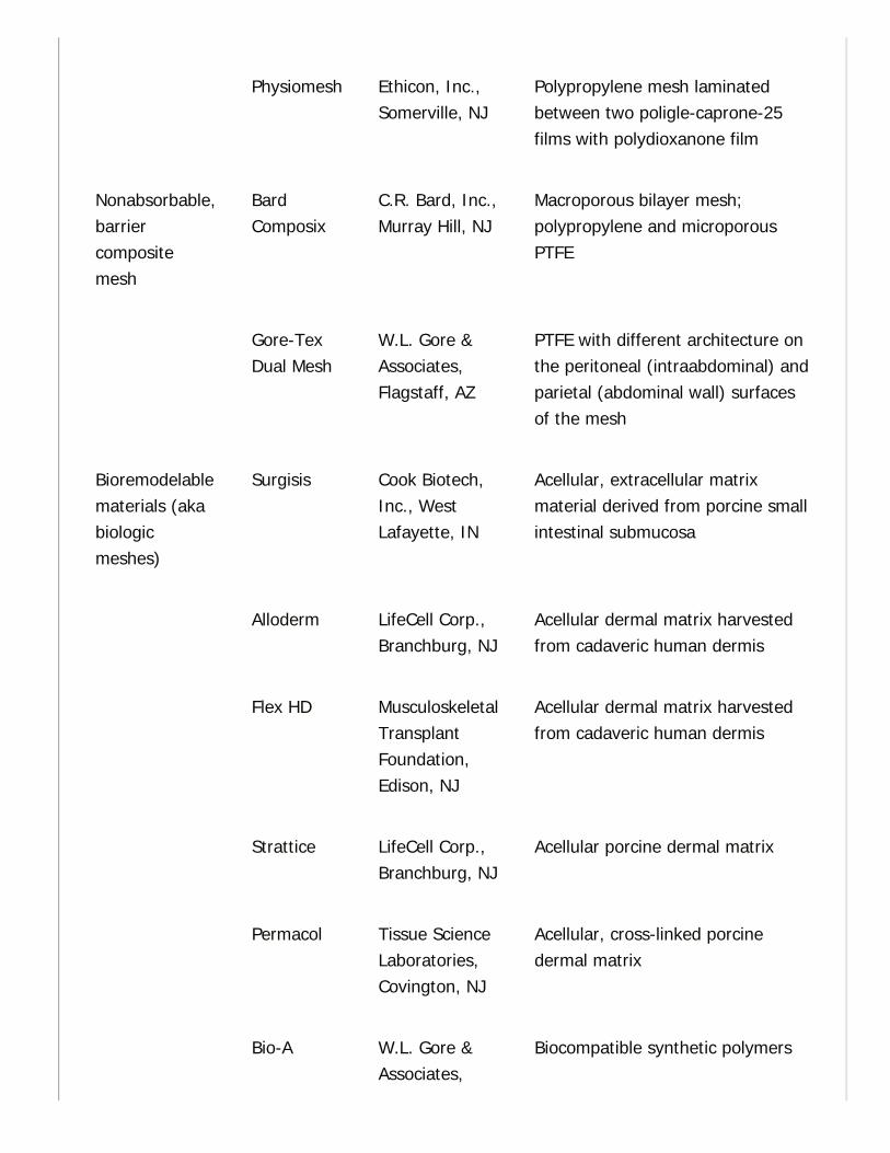
Physiomesh Ethicon, Inc.,Somerville, NJ
Polypropylene mesh laminatedbetween two poligle-caprone-25films with polydioxanone film
Nonabsorbable,barriercompositemesh
BardComposix
C.R. Bard, Inc.,Murray Hill, NJ
Macroporous bilayer mesh;polypropylene and microporousPTFE
Gore-TexDual Mesh
W.L. Gore &Associates,Flagstaff, AZ
PTFE with different architecture onthe peritoneal (intraabdominal) andparietal (abdominal wall) surfacesof the mesh
Bioremodelablematerials (akabiologicmeshes)
Surgisis Cook Biotech,Inc., WestLafayette, IN
Acellular, extracellular matrixmaterial derived from porcine smallintestinal submucosa
Alloderm LifeCell Corp.,Branchburg, NJ
Acellular dermal matrix harvestedfrom cadaveric human dermis
Flex HD MusculoskeletalTransplantFoundation,Edison, NJ
Acellular dermal matrix harvestedfrom cadaveric human dermis
Strattice LifeCell Corp.,Branchburg, NJ
Acellular porcine dermal matrix
Permacol Tissue ScienceLaboratories,Covington, NJ
Acellular, cross-linked porcinedermal matrix
Bio-A W.L. Gore &Associates,
Biocompatible synthetic polymers
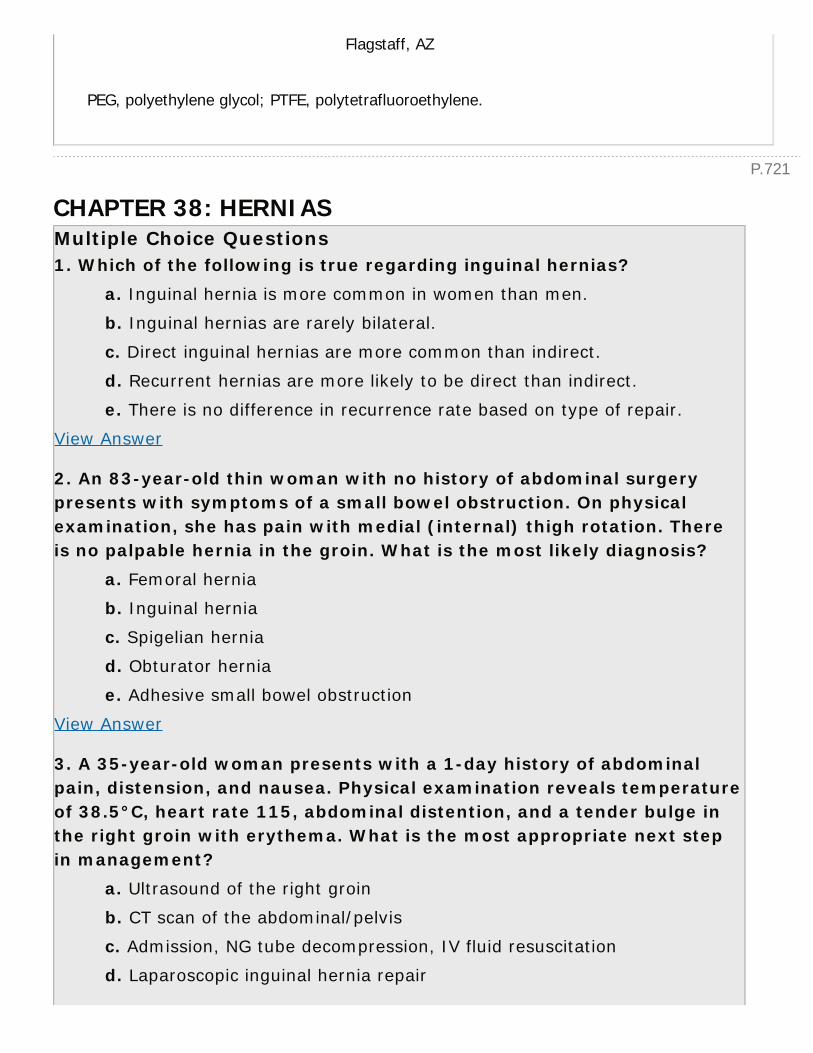
P.721
Flagstaff, AZ
PEG, polyethylene glycol; PTFE, polytetrafluoroethylene.
CHAPTER 38: HERNIASMultiple Choice Questions1. Which of the following is true regarding inguinal hernias?
a. Inguinal hernia is more common in women than men.
b. Inguinal hernias are rarely bilateral.
c. Direct inguinal hernias are more common than indirect.
d. Recurrent hernias are more likely to be direct than indirect.
e. There is no difference in recurrence rate based on type of repair.
View Answer
2. An 83-year-old thin woman with no history of abdominal surgerypresents with symptoms of a small bowel obstruction. On physicalexamination, she has pain with medial (internal) thigh rotation. Thereis no palpable hernia in the groin. What is the most likely diagnosis?
a. Femoral hernia
b. Inguinal hernia
c. Spigelian hernia
d. Obturator hernia
e. Adhesive small bowel obstruction
View Answer
3. A 35-year-old woman presents with a 1-day history of abdominalpain, distension, and nausea. Physical examination reveals temperatureof 38.5°C, heart rate 115, abdominal distention, and a tender bulge inthe right groin with erythema. What is the most appropriate next stepin management?
a. Ultrasound of the right groin
b. CT scan of the abdominal/pelvis
c. Admission, NG tube decompression, IV fluid resuscitation
d. Laparoscopic inguinal hernia repair
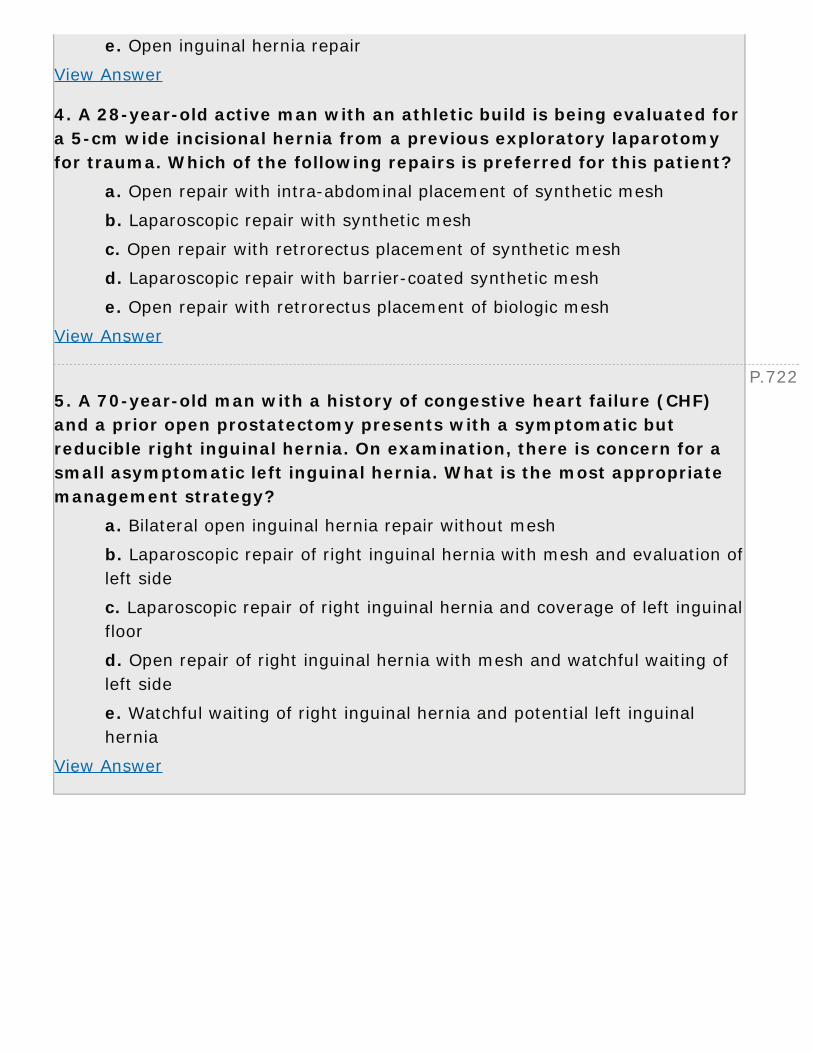
P.722
e. Open inguinal hernia repair
View Answer
4. A 28-year-old active man with an athletic build is being evaluated fora 5-cm wide incisional hernia from a previous exploratory laparotomyfor trauma. Which of the following repairs is preferred for this patient?
a. Open repair with intra-abdominal placement of synthetic mesh
b. Laparoscopic repair with synthetic mesh
c. Open repair with retrorectus placement of synthetic mesh
d. Laparoscopic repair with barrier-coated synthetic mesh
e. Open repair with retrorectus placement of biologic mesh
View Answer
5. A 70-year-old man with a history of congestive heart failure (CHF)and a prior open prostatectomy presents with a symptomatic butreducible right inguinal hernia. On examination, there is concern for asmall asymptomatic left inguinal hernia. What is the most appropriatemanagement strategy?
a. Bilateral open inguinal hernia repair without mesh
b. Laparoscopic repair of right inguinal hernia with mesh and evaluation ofleft side
c. Laparoscopic repair of right inguinal hernia and coverage of left inguinalfloor
d. Open repair of right inguinal hernia with mesh and watchful waiting ofleft side
e. Watchful waiting of right inguinal hernia and potential left inguinalhernia
View Answer
39Diseases of the Adrenal and Pituitary Glandand Hereditary Endocrine Syndromes
Kathryn J. Rowland
Jeffrey F. Moley
ADRENAL-PITUITARY AXIS
I. EMBRYOLOGY, ANATOMY, AND PHYSIOLOGYA. Embryology. The adrenal cortex arises from the coelomic mesoderm around the fifth weekof gestation. The adrenal medulla is populated by the neural crest cells originating from theneural ectoderm. The consequence of this migration is evident by the existence ofparagangliomas (extra-adrenal pheochromocytomas) all along the paraspinal axis.
B. Anatomy. The adrenal glands are located in the retroperitoneum superior to the kidney andlateral to the vena cava (on the right) and aorta (on the left). This relationship is important indetermining the vascular supply to the adrenals. Each adrenal is supplied by three arteries:Superior adrenal artery (arises from the inferior phrenic artery), middle adrenal artery(branch of the aorta), and inferior adrenal artery (branch of the renal artery). The rightadrenal vein drains directly into the vena cava. The left adrenal vein drains into the left renalvein.
C. Physiology. The adrenal gland is histologically composed of four layers, each with their ownbiosynthetic products.
1. Adrenal cortex
a. Zona glomerulosa is responsible for mineralocorticoid production, of which aldosterone isthe primary product. Aldosterone production is stimulated by angiotensin II and increased levelsof serum potassium. Aldosterone acts to increase circulating blood volume by increasing sodiumand chloride reabsorption in the distal tubule of the kidney.
b. Zona fasciculata produces the glucocorticoids of the adrenal glands, of which cortisol is theprimary product. Cortisol production is stimulated by the release of adrenocorticotropichormone (ACTH) by the anterior pituitary gland. ACTH itself is stimulated by the release ofcorticotropin-releasing hormone (CRH) by the hypothalamus. Glucocorticoids haveextremely broad effects with the overall goal of inducing a catabolic state in the body in responseto stress. Glucocorticoids increase blood glucose concentrations, stimulate lipolysis, enhance
> Table of Contents > 39 - Diseases of the Adrenal and Pituitary Gland and Hereditary Endocrine Syndromes

P.724
adrenergic stimulation of the cardiovascular system, and reduce the inflammatory response of theimmune system.
c. Zona reticularis produces the adrenal sex hormone androstenedione and DHEA. Thesehormones support the gonadal production of testosterone and estrogen.
2. The adrenal medulla produces the catecholamines norepinephrine (noradrenaline) andepinephrine (adrenaline) that act on peripheral α and β-adrenergic receptors. α Receptorstimulation produces peripheral vasoconstriction. β Simulation of the myocardium via β1 receptors
increases heart rate and contractility. Stimulation of peripheral β2 receptors causes relaxation of
smooth muscles.
II. BIOCHEMICAL EVALUATION AND IMAGING OF ADRENALMASSESA. Patients presenting with an adrenal mass should undergo a complete biochemical workupincluding (Endocr Pract. 2009;15:450-453):
1. Overnight dexamethasone suppression test with measurement of plasma cortisol toevaluate for a cortisol secreting mass.
2. Basic metabolic panel with measurement of serum potassium in hypertensive patients toevaluate for an aldosterone secreting mass.
3. Plasma metanephrines and normetanephrines to evaluate for a pheochromocytoma.
B. Malignant and benign adrenal masses can be distinguished on the basis of characteristic CT,MRI, and PET imaging (Table 39-1) (Cancer Imaging. 2010;10:102-113).
III. FUNCTIONAL ADRENAL MASSESA. Cushing SyndromeÑHypercortisolism
1. The clinical manifestations of Cushing syndrome include hypertension, edema, muscleweakness, glucose intolerance, osteoporosis, easy bruising, cutaneous striae, and truncal obesity(buffalo hump, moon facies). Women may develop acne, hirsutism, and amenorrhea as a resultof adrenal androgen excess.
2. Pathophysiology of excess circulating glucocorticoids.
a. Iatrogenic. Cushing syndrome is usually of iatrogenic etiology and secondary to theadministration of exogenous glucocorticoids or ACTH.
b. Cushing disease. Hypersecretion of ACTH from the anterior pituitary gland (Cushing disease)is the most common pathologic cause (65% to 70% of cases) of endogenous hypercortisolism.The adrenal glands respond normally to the elevated ACTH, and the result is bilateral adrenal
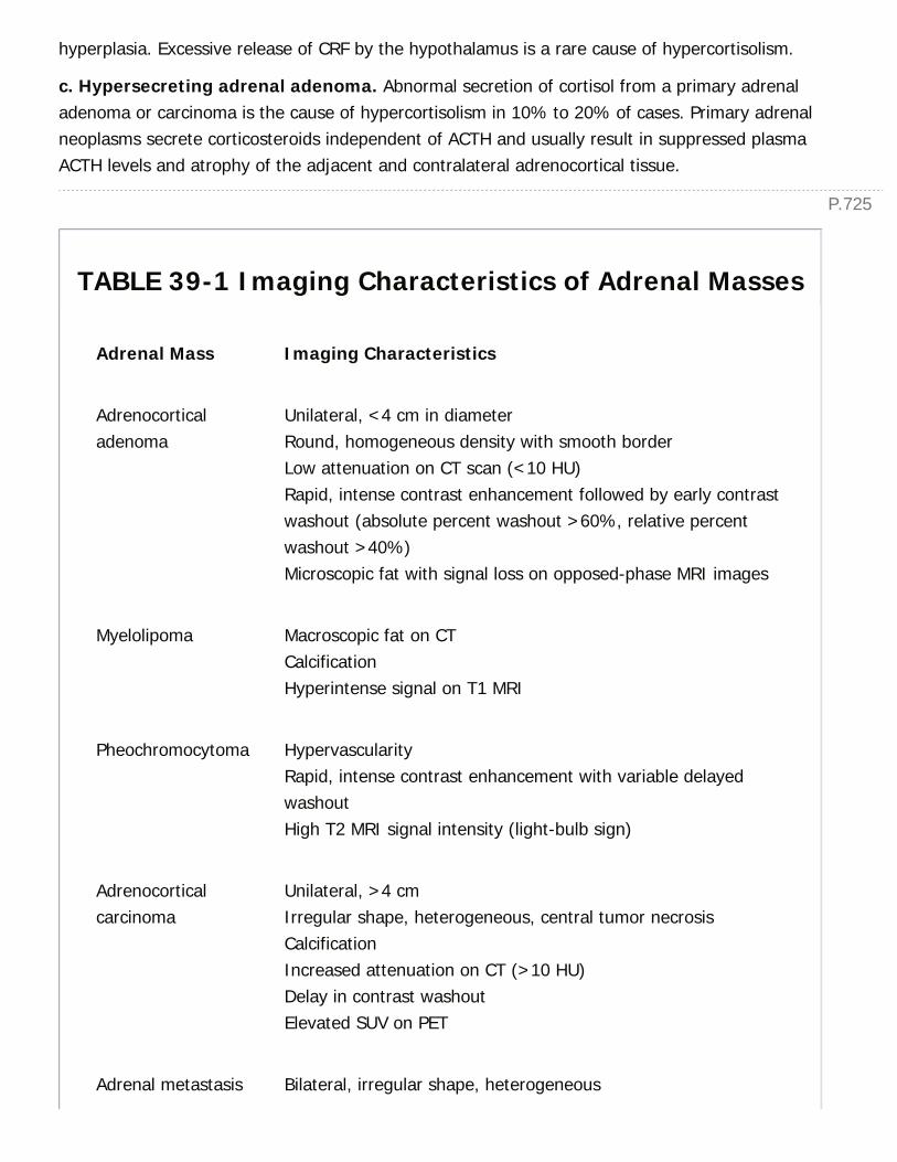
P.725
hyperplasia. Excessive release of CRF by the hypothalamus is a rare cause of hypercortisolism.
c. Hypersecreting adrenal adenoma. Abnormal secretion of cortisol from a primary adrenaladenoma or carcinoma is the cause of hypercortisolism in 10% to 20% of cases. Primary adrenalneoplasms secrete corticosteroids independent of ACTH and usually result in suppressed plasmaACTH levels and atrophy of the adjacent and contralateral adrenocortical tissue.
TABLE 39-1 Imaging Characteristics of Adrenal Masses
Adrenal Mass Imaging Characteristics
Adrenocorticaladenoma
Unilateral, <4 cm in diameterRound, homogeneous density with smooth borderLow attenuation on CT scan (<10 HU)Rapid, intense contrast enhancement followed by early contrastwashout (absolute percent washout >60%, relative percentwashout >40%)Microscopic fat with signal loss on opposed-phase MRI images
Myelolipoma Macroscopic fat on CTCalcificationHyperintense signal on T1 MRI
Pheochromocytoma HypervascularityRapid, intense contrast enhancement with variable delayedwashoutHigh T2 MRI signal intensity (light-bulb sign)
Adrenocorticalcarcinoma
Unilateral, >4 cmIrregular shape, heterogeneous, central tumor necrosisCalcificationIncreased attenuation on CT (>10 HU)Delay in contrast washoutElevated SUV on PET
Adrenal metastasis Bilateral, irregular shape, heterogeneous
P.726
Increased attenuation on CT (>10 HU)Delay in contrast washoutElevated SUV on PET
CT, computed tomography; HU, Hounsfield units; MRI, magnetic resonance image; PET,positron emission tomography; SUV, standardized uptake values.
d. Ectopic ACTH production. In approximately 15% of cases, Cushing syndrome is caused byectopic secretion of ACTH or an ACTHlike substance from a small-cell bronchogenic carcinoma,carcinoid tumor, pancreatic carcinoma, thymic carcinoma, medullary thyroid cancer, or otherneuroendocrine neoplasm. Patients with ectopic ACTH-secreting neoplasms can present primarilywith hypokalemia, glucose intolerance, and hyperpigmentation but with few other chronic signs ofCushing syndrome.
3. Diagnosis of Cushing syndrome is biochemical (Fig. 39-1). The goals are to first establishhypercortisolism and then identify the source.

Figure 39-1 Diagnosis of Cushing syndrome is biochemical.
a. Establishing the presence of hypercortisolism:
(1) The best screening test for hypercortisolism is a 24-hour measurement of the urinaryexcretion of free cortisol. Urinary excretion of more than 100 µg/day of free cortisol in twoindependent collections is virtually diagnostic of Cushing syndrome. Measurement of randomplasma cortisol levels alone is not a reliable method of diagnosing Cushing syndrome due tooverlap of the levels in normal and abnormal patients.
(2) A low-dose overnight dexamethasone suppression test (dexamethasone 1 mg orally isadministered at 11 pm and plasma cortisol is measured at 8 am) is used to confirm Cushingsyndrome, especially in obese or depressed patients who may have marginally elevated urinarycortisol levels. Patients with true hypercortisolism have lost normal adrenalÑpituitary feedback
P.727
and usually fail to suppress the morning plasma cortisol level to less than 5 µg/dL.
b. Localization of the cause of hypercortisolism:
(1) Determination of basal ACTH by immunoradiometric assay is the best method of determiningthe cause of hypercortisolism. Suppression of the absolute level of ACTH to below 5 pg/mL isnearly diagnostic of adrenocortical neoplasms (ACTH-independent Cushing syndrome). ACTHlevels in Cushing disease may range from the upper limits of to significantly above normal (15pg/mL to 500 pg/mL). The highest plasma levels of ACTH (1,000 pg/mL) have been observed inpatients with ectopic ACTH syndrome.
(2) Standard high-dose dexamethasone suppression testing is used to distinguish apituitary source from an ectopic source of ACTH. Normal individuals and most patients with apituitary ACTH-producing neoplasm respond to a high-dose dexamethasone suppression test (2mg orally every 6 hours for 48 hours) with a reduction in urinary free cortisol and urinary 17-hydroxysteroids to less than 50% of basal values. Most patients with a primary adrenal tumor oran ectopic source of ACTH production fail to suppress to this level. However, this test does notseparate clearly pituitary and ectopic ACTH hypersecretion because 25% of patients with theectopic ACTH syndrome also have suppressible tumors.
(3) CRH stimulation test may also be used to distinguish a pituitary source of ACTH from anectopic source. Forty-five minutes following intravenous CRH administration, ACTH and cortisollevels are increased in patients with Cushing disease. Patients with a suppressed hypothalamicÑpituitary axis (primary adrenal tumor, ectopic ACTH syndrome) usually do not have acompensatory rise in ACTH and cortisol levels in response to CRH.
c. Imaging tests are useful for identifying lesions suspected on the basis of biochemical testing.
(1) Patients with ACTH-independent hypercortisolism require thin-section CT scan or MRI scan ofthe adrenal gland, both of which identify adrenal abnormalities with more than 95% sensitivity.
(2) Gadolinium-enhanced MRI scan of the sella turcica is the best imaging test for pituitaryadenomas suspected of causing ACTH-dependent hypercortisolism.
(3) Patients with ACTH-dependent hypercortisolism and either markedly elevated ACTH or anegative pituitary MRI scan should have CT scan of the chest to identify a tumor-producingectopic ACTH.
(4) Bilateral inferior petrosal sinus sampling can delineate unclear cases of Cushing diseasefrom other causes of hypercortisolism. Simultaneous bilateral petrosal sinus and peripheral bloodsamples are obtained before and after peripheral intravenous injection of 1 µg/kg of CRH. Levelsof ACTH in the inferior petrosal sinus are compared to peripheral plasma ACTH levels: A basalratio of ≥2 or a ratio of ≥3 after CRH administration is 100% sensitive and specific for pituitaryadenoma.

P.7284. Surgical treatment of Cushing syndrome involves removing the cause of cortisol excess.
a. Transsphenoidal resection of an ACTH-producing pituitary tumor is successful in 80%or more of cases of Cushing disease.
b. Treatment of ectopic ACTH syndrome involves resection of the primary lesion, if possible.
c. Primary adrenal causes of Cushing syndrome are treated by removal of the adrenal glandcontaining the tumor. All patients who undergo adrenalectomy for primary adrenal causes ofCushing syndrome require perioperative and postoperative glucocorticoid replacement becausethe pituitaryÑadrenal axis is suppressed. Recovery of the pituitaryÑadrenal axis may take as longas 6 to 18 months following unilateral adrenalectomy.
B. Conn SyndromeÑHyperaldosteronism
1. Primary hyperaldosteronism is a syndrome of hypertension and hypokalemia caused byhypersecretion of the mineralocorticoid aldosterone. Secondary aldosteronism is a physiologicresponse of the reninÑangiotensin system to renal artery stenosis, cirrhosis, congestive heartfailure, and normal pregnancy. In these conditions, the adrenal gland functions normally.
2. Pathophysiology of primary hyperaldosteronism
a. An aldosterone-producing adrenal adenoma (APA) is the cause of primary aldosteronismin two-thirds of cases and is one of the few surgically correctable causes of hypertension.
b. Idiopathic bilateral adrenal hyperplasia (IHA) causes 30% to 40% of cases of primaryaldosteronism.
c. Adrenocortical carcinoma and autosomal dominant glucocorticoid-suppressiblealdosteronism are rare causes of primary aldosteronism.
3. Diagnosis
a. Given the prevalence of essential hypertension, it is not cost-effective to screen all adults withhypertension for primary hyperaldosteronism. Adults who should be evaluated include those withnew onset severe hypertension at a young age and those whose blood pressure is labile or poorlycontrolled on three or more antihypertensives.
b. Laboratory diagnosis of primary aldosteronism begins with the demonstration of hypokalemia(<3.5 mEq/L), inappropriate kaliuresis (>30 mEq/day), and elevated aldosterone (>15 ng/dL)with normal cortisol. Aldosterone to renin ratio (ARR) is determined by the measurement ofplasma renin activity (PRA) and plasma aldosterone concentration (PAC). This test is bestperformed upright, midmorning. An ARR of >20 is suggestive of primary hyperaldosteronism.Drugs that stimulate renin such as spironolactone, angiotensin-converting enzyme inhibitors (ACEinhibitors), angiotensin receptor antagonists (ARBs), or dihydropyridine calcium blockers (e.g.,amlodipine) can produce false negatives. β-blockers, clonidine, and nonsteroidal anti-
P.729inflammatory drugs suppress renin and may result
in false positives (Horm Metab Res. 2012;44:170-176). Primary aldosteronism is confirmed bylack of aldosterone suppression (PAC >10 ng/dL) with salt loading. This is performed by havingpatients add a teaspoon of salt to each meal for 72 hours prior to PAC and PRA measurement.Alternatively, a 500 mL normal saline infusion can be administered.
c. Localization. High-resolution adrenal CT scan should be the initial step in localization of anadrenal tumor. CT scanning localizes an adrenal adenoma in 90% of cases overall, and thepresence of a unilateral adenoma larger than 1 cm on CT scan and supportive biochemicalevidence of an aldosteronoma are generally all that is needed to make the diagnosis of Connsyndrome. Differentiation between APA and IHA is important because unilateral adenomas aretreated by surgical excision, whereas bilateral hyperplasia is treated medically. Uncertaintyregarding APA versus IHA after biochemical testing and noninvasive localization may bedefinitively resolved by bilateral adrenal venous sampling for aldosterone and cortisol.Simultaneous adrenal vein blood samples for aldosterone and cortisol are taken. The ratio ofaldosterone to cortisol is greater than 4:1 for a diagnosis of aldosteronoma and less than 4:1 fora diagnosis of IHA.
4. Treatment. Surgical removal of an APA through a posterior or laparoscopic approach resultsin immediate cure or substantial improvement in hypertension and hypokalemia in more than90% of patients with Conn syndrome. The patient should be treated with spironolactone (200to 400 mg/day) preoperatively for 2 to 3 weeks to control blood pressure and to correcthypokalemia. Patients with IHA should be treated medically with spironolactone (200 to 400mg/day). A potassium-sparing diuretic, such as amiloride (5 to 20 mg/day), and calciumchannel blockers have also been used. Surgical excision rarely cures bilateral hyperplasia.
C. Pheochromocytoma
1. The clinical manifestations of pheochromocytoma include paroxysms of pounding frontalheadache, diaphoresis, palpitations, flushing, or anxiety related to the excess sympatheticstimulation from catecholamines. The most common sign is episodic or sustained hypertension,but pheochromocytoma accounts for only 0.1% to 0.2% of patients with sustained diastolichypertension. Uncommonly, patients present with complications of prolonged uncontrolledhypertension (e.g., myocardial infarction, cerebrovascular accident, or renal disease).
2. Pathophysiology of pheochromocytoma (Table 39-2).
a. Pheochromocytomas are neoplasms derived from the chromaffin cells of the sympathoadrenalsystem that engage in unregulated, episodic oversecretion of catecholamines.
b. Approximately 80% to 85% of pheochromocytomas in adults arise in the adrenal medulla,whereas 10% to 15% arise in the extraadrenal chromaffin tissue, including the paravertebralganglia, posterior mediastinum, organ of Zuckerkandl, and urinary bladder.
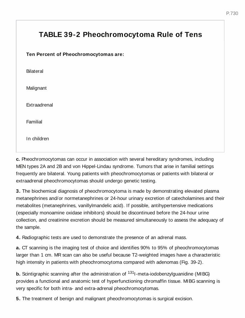
P.730
TABLE 39-2 Pheochromocytoma Rule of Tens
Ten Percent of Pheochromocytomas are:
Bilateral
Malignant
Extraadrenal
Familial
In children
c. Pheochromocytomas can occur in association with several hereditary syndromes, includingMEN types 2A and 2B and von Hippel-Lindau syndrome. Tumors that arise in familial settingsfrequently are bilateral. Young patients with pheochromocytomas or patients with bilateral orextraadrenal pheochromocytomas should undergo genetic testing.
3. The biochemical diagnosis of pheochromocytoma is made by demonstrating elevated plasmametanephrines and/or normetanephrines or 24-hour urinary excretion of catecholamines and theirmetabolites (metanephrines, vanillylmandelic acid). If possible, antihypertensive medications(especially monoamine oxidase inhibitors) should be discontinued before the 24-hour urinecollection, and creatinine excretion should be measured simultaneously to assess the adequacy ofthe sample.
4. Radiographic tests are used to demonstrate the presence of an adrenal mass.
a. CT scanning is the imaging test of choice and identifies 90% to 95% of pheochromocytomaslarger than 1 cm. MR scan can also be useful because T2-weighted images have a characteristichigh intensity in patients with pheochromocytoma compared with adenomas (Fig. 39-2).
b. Scintigraphic scanning after the administration of 131I-meta-iodobenzylguanidine (MIBG)provides a functional and anatomic test of hyperfunctioning chromaffin tissue. MIBG scanning isvery specific for both intra- and extra-adrenal pheochromocytomas.
5. The treatment of benign and malignant pheochromocytomas is surgical excision.
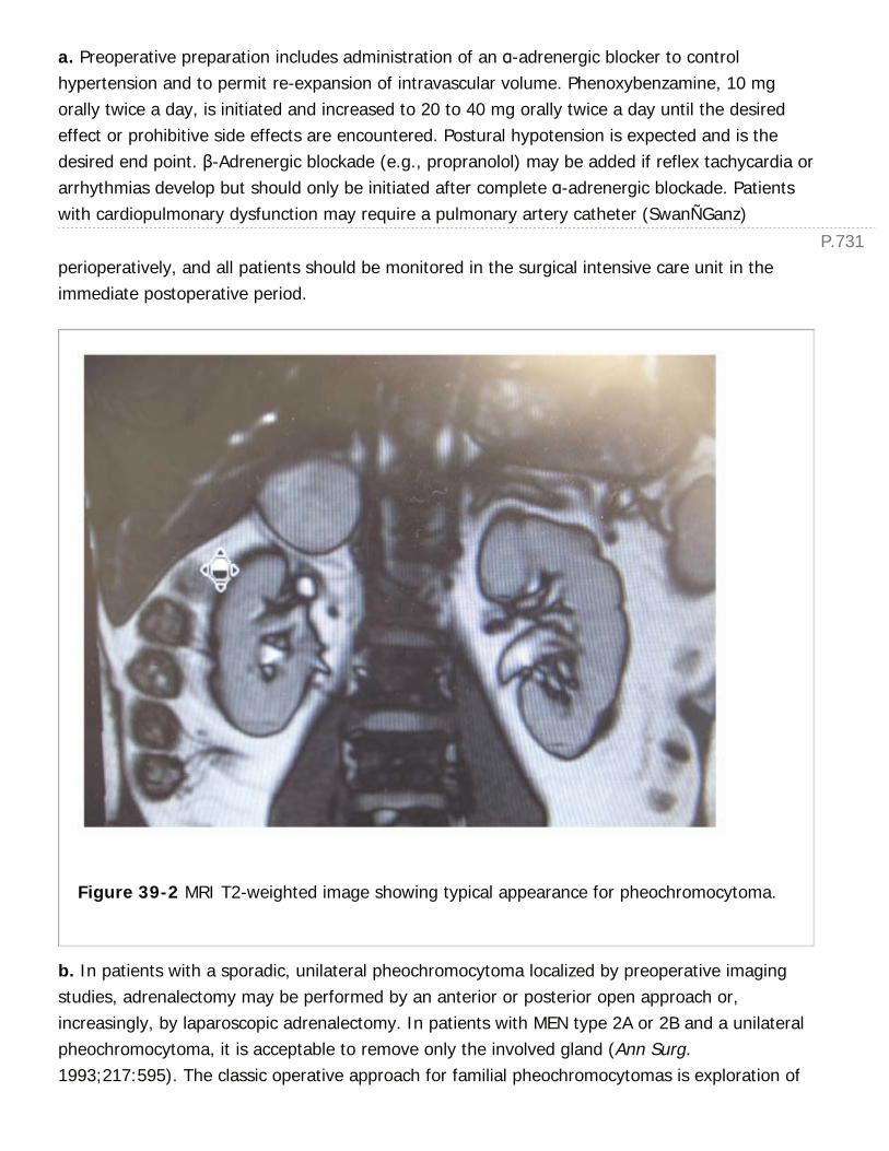
P.731
a. Preoperative preparation includes administration of an α-adrenergic blocker to controlhypertension and to permit re-expansion of intravascular volume. Phenoxybenzamine, 10 mgorally twice a day, is initiated and increased to 20 to 40 mg orally twice a day until the desiredeffect or prohibitive side effects are encountered. Postural hypotension is expected and is thedesired end point. β-Adrenergic blockade (e.g., propranolol) may be added if reflex tachycardia orarrhythmias develop but should only be initiated after complete α-adrenergic blockade. Patientswith cardiopulmonary dysfunction may require a pulmonary artery catheter (SwanÑGanz)
perioperatively, and all patients should be monitored in the surgical intensive care unit in theimmediate postoperative period.
Figure 39-2 MRI T2-weighted image showing typical appearance for pheochromocytoma.
b. In patients with a sporadic, unilateral pheochromocytoma localized by preoperative imagingstudies, adrenalectomy may be performed by an anterior or posterior open approach or,increasingly, by laparoscopic adrenalectomy. In patients with MEN type 2A or 2B and a unilateralpheochromocytoma, it is acceptable to remove only the involved gland (Ann Surg.1993;217:595). The classic operative approach for familial pheochromocytomas is exploration of

P.732
adrenal glands, the preaortic and paravertebral areas, and the organ of Zuckerkandl through amidline or bilateral subcostal incision.
c. Intraoperative labile hypertension can occur during resection of pheochromocytoma. This canbe prevented by minimal manipulation of the tumor but can be controlled most effectively withintravenous sodium nitroprusside (0.5 to 10 µg/kg/minute) or phentolamine (5 mg).
d. Following pheochromocytoma excision, 10% to 15% of patients will develop recurrence. Long-term followup with measurement of plasma metanephrines and normetanephrines is necessary.
IV. NONFUNCTIONAL ADRENAL MASSESA. Adrenocortical adenomas comprise the majority of incidentally discovered adrenal masses(incidentalomas). Incidental adrenal masses are detected
in 0.4% to 4.4% of abdominal CT scans obtained for other reasons. Adrenocortical adenomas arebenign, and nonfunctioning, with the majority having no clinical significance.
1. Surgery should be performed in patients with nonfunctional adrenal adenomas greater than 6cm in diameter given the increased probability of adrenocortical carcinoma in masses ofincreasing size. Strong consideration for removal of tumors between 4 and 6 cm should be basedupon clinical scenario, patient age and comorbidities, and imaging characteristics. Thisrecommendation is based upon the National Institutes of Health Consensus Statement (NIHConsens State Sci Statements. 2002;19:1-25). In contrast, the American Association of ClinicalEndocrinologists and American Association of Endocrine Surgeons guidelines recommendresection of any mass greater than or equal to 4 cm (Endocr Pract. 2009;15:450-453). Tumorsless than 4 cm should be monitored clinically and radiologically in 3 to 6 months followed byannually for 2 years. Any tumor that enlarges by more than 1 cm during the followup periodshould be removed. Biochemical testing should be performed annually for 5 years.
2. Either open or laparoscopic adrenalectomy is acceptable. Laparoscopic adrenalectomy hasbeen associated with shorter hospitalization, faster recovery, and lower morbidity (Br J Surg.2004;91:1259-1274). Its use is generally limited to malignant lesions less than 5 cm in diameterand benign-appearing lesions up to 10 cm in diameter. Both laparoscopic transperitonealadrenalectomy and laparoscopic retroperitoneal adrenalectomy have been described with nodifference in terms of operative time, blood loss, length of hospitalization, time to oral intake,morbidity, or mortality (Surgery. 2013;153:111-119).
B. Adrenal myelolipoma is a benign tumor composed of mature fat and hematopoieticelements.
1. Myelolipomas have characteristic macroscopic fat on CT imaging.
2. Myelolipomas may enlarge over time. Routine followup imaging is not necessary. Surgicalexcision is only indicated for masses causing local mass-effect symptoms. Spontaneous

P.733
retroperitoneal hemorrhage may occur with large masses. Approximately 4% of patientsdiagnosed with a myelolipoma will require adrenalectomy (J Surg Oncol. 2012;106:557-564).
C. Adrenocortical carcinoma is a rare but aggressive malignancy. Most patients with thiscancer present with locally advanced disease.
1. Syndromes of adrenal hormone overproduction may include rapidly progressivehypercortisolism, hyperaldosteronism, or virilization (Fig. 39-3). Large (>6 cm) adrenal massesthat extend to nearby structures on CT scanning likely represent carcinoma.
2. Complete surgical resection of locally confined tumor is the only chance for cure ofadrenocortical carcinoma. Definitive diagnosis of adrenocortical carcinoma requires operative andpathologic demonstration of nodal or distant metastases. Any adrenal neoplasm weighing morethan 50 g should be considered malignant.
Figure 39-3 Adrenocortical carcinoma.
P.734
3. Often, patients with adrenocortical carcinoma present with metastatic disease, most ofteninvolving the lung, lymph nodes, liver, or bone. Palliative surgical debulking of locally advanced ormetastatic adrenocortical carcinoma may provide these patients with symptomatic relief fromsome slow-growing, hormone-producing cancers. Chemotherapy with mitotane may besomewhat effective. Overall, the prognosis for patients with adrenocortical carcinoma is poor.
D. Adrenal metastases are the most common malignant lesions involving the adrenal gland.Frequently bilateral lesions are present.
1. Lung, breast, melanoma, colorectal, thyroid, and pancreatic cancer all metastasize to theadrenal glands. Renal cell carcinoma and hepatocellular carcinoma may also metastasize to theadrenals.
2. Diagnosis of metastatic disease can often be made from imaging and a history of cancer. Needfor pathologic confirmation of metastatic disease is rare, but if necessary, biochemical testing forpheochromocytoma should be performed prior to biopsy.
3. Patients with bilateral metastatic disease should be evaluated for adrenal insufficiency.
4. Adrenalectomy for metastatic disease is rarely indicated but may be considered for an isolatedadrenal lesion.
E. Collision tumor is a rare tumor that represents the coexistence of two histologically distinctbut adjacent adrenal masses (Cancer Imaging. 2013;13:602-610).
1. Collision tumors may be composed of an adenoma with myelolipoma, adenoma withmetastases, and adrenocortical carcinoma with myelolipoma.
2. Hemorrhage into an existing adrenal mass can mimic a collision tumor.
V. ACUTE ADRENAL INSUFFICIENCYA. Acute adrenal insufficiency is an emergency and should be suspected in physiologicallystressed patients with a history of either adrenal insufficiency or exogenous steroid use.Adrenocortical insufficiency is most often caused by acute withdrawal of chronic corticosteroidtherapy but can occur in the postoperative setting following adrenal surgery or from autoimmunedestruction of the adrenal cortex, adrenal hemorrhage (Waterhouse-Friderichsen syndrome),or, rarely, infiltration with metastatic carcinoma. The diagnosis and treatment of acute adrenalinsufficiency in patients in septic shock is very controversial. Two prospective randomized trialshave shown different effects in the use of hydrocortisone in patients with septic shock (N Engl JMed. 2003;348:727-734; N Engl J Med. 2008;358:111-124). Current Surviving Sepsis Guidelinescall for the use of corticosteroids only in cases of septic shock where the blood pressure is notresponsive to fluid administration or vasopressor therapy (Crit Care Med. 2013;41:580-637).
1. Signs and symptoms include fever, nausea, vomiting, severe hypotension, and lethargy.
P.735
Characteristic laboratory findings of adrenal insufficiency include hyponatremia, hyperkalemia,azotemia, and fasting or reactive hypoglycemia.
2. Diagnosis. A rapid ACTH stimulation test is used to test for adrenal insufficiency.Cosyntropin (i.e., synthetic ACTH, 250 µg) is administered intravenously, and plasma cortisollevels are measured on completion of the administration and then 30 and 60 minutes later.Normal peak cortisol response should exceed 20 µg/dL.
3. Treatment of adrenal crisis must be immediate and based on clinical suspicion, beforelaboratory confirmation is available. Intravenous volume replacement with normal or hypertonicsaline and dextrose is essential, as is immediate intravenous steroid replacement therapy with 4mg of dexamethasone. Thereafter, 50 mg of hydrocortisone is administered intravenously every 8hours and is tapered to standard replacement doses as the patient's condition stabilizes.Mineralocorticoid replacement is not required until intravenous fluids are discontinued and oralintake resumes.
4. Prevention. Patients who have known adrenal insufficiency or have received supraphysiologicdoses of steroid for at least 1 week in the year preceding surgery should receive 100 mg ofhydrocortisone the evening before and the morning of major surgery, followed by 100 mg ofhydrocortisone every 8 hours during the first postoperative 24 hours.
HEREDITARY ENDOCRINE TUMOR SYNDROMES (Table39-3)
I. MULTIPLE ENDOCRINE NEOPLASIA TYPE I (MEN-1)A. MEN-1 is an autosomal-dominant syndrome characterized by tumors of the parathyroid glands,pancreatic islet cells, and pituitary gland. Hyperparathyroidism occurs in virtually all patients.Clinical evidence of pancreatic islet cell and pituitary tumors develops in 50% and 25% ofpatients, respectively. Lipomas, thymic, or bronchial carcinoid tumors, and tumors of the thyroid,adrenal cortex, and central nervous system (CNS) may also develop. The gene responsible forMEN-1, MENIN, is located on chromosome 11q13 and appears to act through transcription factors(Science. 1997;276:404-407). Genetic testing is available. Screening of affected family membersshould begin in their early teens, including yearly determinations of plasma calcium, glucose,gastrin, fasting insulin, vasoactive intestinal polypeptide (VIP), pancreatic polypeptide, prolactin,growth hormone, and β-human gonadotropin hormone levels.
1. Hyperparathyroidism. Because hyperparathyroidism is frequently the first detectableabnormality in patients with MEN-1, yearly calcium screening of asymptomatic kindred membersis recommended. Patients with hyperparathyroidism and MEN-1 usually have generalized(fourgland) parathyroid enlargement. Surgery should consist of 3.5-gland parathyroidectomy or atotal parathyroidectomy with autotransplantation of parathyroid tissue to the sternocleidomastoid
P.736
muscle or forearm. This method achieves cure in more than 90% of cases and results inhypoparathyroidism in less than 5%. Graft-dependent recurrent hyperparathyroidism, however, isseen in up to 50% of cases, and is managed by debulking of the autografted material (Ann Surg.1980;192:451).
2. Pituitary tumors occur in up to 40% of MEN-1 patients and most commonly are benignprolactin-producing adenomas. Growth hormone-producing, ACTH-producing, and nonfunctioningtumors are also seen. Patients may present with headache, diplopia, or symptoms referable tohormone overproduction. Bromocriptine inhibits prolactin production and may reduce tumorbulk and obviate the need
for surgical intervention. Transsphenoidal hypophysectomy may be necessary if medicaltreatment fails.
TABLE 39-3 Hereditary Endocrine Tumor Syndromes
MEN-1 MEN-2A MEN-2B FMTC
Gene mutation MENIN Ret Ret Ret
Endocrinopathy HPTPituitaryPanc
MTCPheoHPT
MTCPheo
MTC
HPT, hyperparathyroidism; Panc, pancreatic islet cell tumor; MTC, medullary thyroidcarcinoma; Pheo, pheochromocytoma.
3. Pancreatic islet cell tumors pose the most difficult clinical challenge and account for most ofthe morbidity and mortality of the syndrome. Gastrinomas (i.e., ZollingerÑEllisonSyndrome, ZES) are most common, but VIP-secreting tumors, insulinomas, glucagonomas, andsomatostatinomas are also encountered. The pancreas is usually diffusely involved, with islet cellhyperplasia and multifocal tumors. Tumors may be found in the proximal duodenum andperipancreatic areas (gastrinoma triangle) and are virtually always malignant. The treatment goalis relief of symptoms related to excessive hormone production and cure or palliation of themalignant process. Patients frequently require medical and surgical therapy.
II. MULTIPLE ENDOCRINE NEOPLASIA TYPE 2 (MEN-2)

P.737
A. MEN2 is characterized by medullary thyroid carcinoma (MTC) and includes MEN-2A, MEN-2B,and familial MTC (FMTC). These autosomal-dominant syndromes are caused by gain-of-function mutations in the RET proto-oncogene, which encodes a transmembrane tyrosinekinase receptor. Mutations in RET lead to constitutive activation (tyrosine phosphorylation) of theRET protein, which drives tumorigenesis. Genetic testing should be performed on all suspectedindividuals. Because MTC occurs universally in all MEN-2 variants, thyroidectomy is indicated forall RET-mutation carriers. Calcitonin serves as a tumor marker for MTC and can be used toguide timing of thyroidectomy as well as postoperative monitoring for disease recurrence. Whenpossible, prophylactic thyroidectomy should be performed prior to the presence of biochemicalevidence of disease in order to reduce the risk of spread outside the thyroid that may lead todisease persistence or recurrence. Current guidelines call for prophylactic thyroidectomy in thefirst year of life for MEN-2B and/or RET codon 883, 918, or 922 mutation carriers; thyroidectomybefore age 5 years in MEN-2A codon 611, 618, 620, or 634 mutation carriers; and thyroidectomybetween ages 5 and 10 in MEN-2A codon 609, 768, 790, 791, 804, and 891 mutation carriers (JClin Endocrinol Metab. 2001;86:5658-5671). A rising calcitonin level should prompt earlierintervention.
1. MEN-2A. All patients with MEN-2A will develop MTC, but the course of MTC is variable andcan be predicted based upon codon mutation. Mutations in codon 611, 618, 620, 634 exposepatients to an increasing, cumulative age-related risk of lymph node metastasis, starting from themid-teens and reaching a greater than 40% cumulative risk by the age of 20. MEN-2A withmutations in codons 768, 790, 804, or 891 is less aggressive and often presents with MTC in thesecond or third decade of life. The penetrance of other features of the syndrome is variable.Pheochromocytomas arise in approximately 40% to 50% of patients, and hyperplasia of theparathyroid glands arises in approximately 25% to 35%. Patients with MEN-2A also developgastrointestinal manifestations, including abdominal pain, distention, and
constipation as well as Hirschsprung disease (Ann Surg. 2002;235:648). On genetic analysis,patients with MEN-2A and Hirschsprung disease (MEN-2A-HD) share common mutations in eithercodon 609, 618, or 620 of exon 10 of the RET proto-oncogene. MTC generally occurs earlier thanpheochromocytoma or hyperparathyroidism. Nonetheless, biochemical testing to excludepheochromocytoma is mandatory in all MEN-2 and MTC patients prior to any elective surgicaloperation. Pheochromocytoma in children as young as age 8 has been reported (J Surg Oncol.2013;108:203-206).
2. MEN-2B. MTC develops in all patients with MEN-2B and is particularly aggressive. MTC maybe present at birth in these patients and invasive disease with lymph node metastasis occurs atan early age. Pheochromocytoma penetrance is variable. Patients also developganglioneuromatosis and a characteristic physical appearance, with hypergnathism of themidface, marfanoid body habitus, and multiple mucosal neuromas. MEN-2B patients maydemonstrate multiple gastrointestinal symptoms and megacolon.
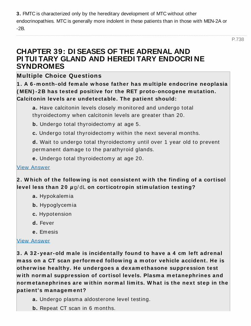
P.738
3. FMTC is characterized only by the hereditary development of MTC without otherendocrinopathies. MTC is generally more indolent in these patients than in those with MEN-2A or-2B.
CHAPTER 39: DISEASES OF THE ADRENAL ANDPITUITARY GLAND AND HEREDITARY ENDOCRINESYNDROMESMultiple Choice Questions1. A 6-month-old female whose father has multiple endocrine neoplasia(MEN)-2B has tested positive for the RET proto-oncogene mutation.Calcitonin levels are undetectable. The patient should:
a. Have calcitonin levels closely monitored and undergo totalthyroidectomy when calcitonin levels are greater than 20.
b. Undergo total thyroidectomy at age 5.
c. Undergo total thyroidectomy within the next several months.
d. Wait to undergo total thyroidectomy until over 1 year old to preventpermanent damage to the parathyroid glands.
e. Undergo total thyroidectomy at age 20.
View Answer
2. Which of the following is not consistent with the finding of a cortisollevel less than 20 µg/dL on corticotropin stimulation testing?
a. Hypokalemia
b. Hypoglycemia
c. Hypotension
d. Fever
e. Emesis
View Answer
3. A 32-year-old male is incidentally found to have a 4 cm left adrenalmass on a CT scan performed following a motor vehicle accident. He isotherwise healthy. He undergoes a dexamethasone suppression testwith normal suppression of cortisol levels. Plasma metanephrines andnormetanephrines are within normal limits. What is the next step in thepatient's management?
a. Undergo plasma aldosterone level testing.
b. Repeat CT scan in 6 months.

P.739
c. Perform laparoscopic left adrenalectomy.
d. Undergo MRI to further evaluate mass.
e. Undergo Ret proto-oncogene testing to rule out multiple endocrineneoplasia type 2.
View Answer
4. A 50-year-old male with persistent hypertension despiteantihypertensive combination therapy with a beta blocker, calciumchannel blocker, and ACE inhibitor presents with hypokalemia andelevations in plasma aldosterone levels. CT and MRI imagingdemonstrate a 1-cm left-sided adrenal mass and a 6-cm right-sidedadrenal mass, both benign appearing. Dexamethasone suppressiontesting and plasma metanephrines and normetanephrines are normal.The next step in management is:
a. Right adrenalectomy
b. Medical management alone with addition of spironolactone
c. Repeat imaging in 6 months
d. Bilateral adrenal vein sampling
e. Bilateral adrenalectomy
View Answer
5. A 65-year-old female with hypertension and coronary artery diseasestatus post placement of drug eluting stent 3 weeks ago presents withan 8-cm right adrenal mass with macroscopic fat found incidentally onCT. Biochemical evaluation reveals a normal dexamethasonesuppression test, normal plasma metanephrines and normetanephrines,and normal metabolic panel. Appropriate management of this patientincludes:
a. Repeat CT imaging at 6 months
b. MRI
c. Immediate right adrenalectomy with patient on Plavix
d. Right adrenalectomy 1 year after coronary stent placement with patientoff of Plavix
e. No further imaging or intervention
View Answer

40Thyroid and Parathyroid Glands
Jennifer Yu
William E. Gillanders
THYROID
I. EMBRYOLOGY, ANATOMY, AND PHYSIOLOGYA. Embryology. The thyroid gland develops from the endoderm of the primitive foregut andarises in the ventral pharynx near the base of the tongue, which ultimately becomes the foramencecum. The thyroid then descends in the midline of the neck anterior to the hyoid bone andlaryngeal cartilages. Congenital anomalies such as ectopic thyroid tissue or thyroglossal duct cystsare directly related to variations in this process. The parafollicular cells, or C cells, are derivedfrom the neural crest and migrate to the thyroid to produce calcitonin.
B. Anatomy. The adult thyroid is a bilobar structure connected by an isthmus that lies anteriorto the trachea. The thyroid gland's blood supply arises mainly from the superior and inferiorthyroid arteries, which are branches of the external carotid artery and thyrocervical trunk,respectively. Important structures in close proximity to the thyroid include the external branch ofthe superior laryngeal nerve (SLN, located near the superior pole of the thyroid) and therecurrent laryngeal nerve (RLN, located just anterior or posterior to the inferior thyroid artery inthe tracheoesophageal groove). Careful dissection around the inferior thyroid artery is necessaryto avoid injury to the RLN.
C. Physiology. Thyrotropin-releasing hormone (TRH) secreted by the hypothalamus stimulatesthyroid-stimulating hormone (TSH) secretion by the anterior pituitary gland. TSH then stimulatesthyroid hormone secretion by the thyroid gland. The process of thyroid hormone synthesis beginswhen dietary iodide is ingested, actively transported into the thyroid, and oxidized by thyroidperoxidase (TPO) into iodine. Iodination of tyrosine residues in thyroglobulin createsmonoiodotyrosine and diiodotyrosine. Coupling reactions of monoiodotyrosine and diiodotyrosineresult in the formation of triiodothyronine (T3) and thyroxine (T4), both of which are bound to
thyroglobulin and stored in thyroid follicles. When released into plasma, more than 99% of T3 and
T4 is bound to carrier proteins, including thyroxine-binding globulin (TBG), thyroxine-binding
prealbumin, and albumin. Only the unbound or ÒfreeÓ hormones are active (i.e., available totissues), and T4 is converted to T3 by deiodinases in the peripheral tissues. Compared to T3, T4
has a 20-fold higher circulating concentration but is
> Table of Contents > 40 - Thyroid and Parathyroid Glands

P.7413 to 5 times less potent. The half-life of T4 (7 days) is significantly longer than the half-life of T3
(1 day).
Assessment of thyroid function requires biochemical evaluation and interpretation in the contextof clinical findings. Measurement of TSH (0.3 to 4.12 mIU/L) is the most useful biochemical test inthe assessment of thyroid function. In most patients without hypothalamic or pituitary disease(rare), TSH and free T4 (FT4) vary inversely around a euthyroid state: Increased TSH and low FT4
signify hypothyroidism, while suppressed TSH and high FT4 suggest hyperthyroidism. Assessment
of FT4 (0.8 to 1.8 ng/dL) supports identified abnormalities in TSH and provides an index of
severity of thyroid dysfunction. Total T4 (5 to 12 µg/dL) is affected by changes in hormone
production or binding and does not directly reflect the small FT4 fraction.
Measurement of total T3 (80 to 200 ng/dL) is unreliable as a test for hypothyroidism but is useful
in the patient with suspected hyperthyroidism, suppressed TSH, and normal FT4 in order to rule
out T3 thyrotoxicosis. A newer assay for free T3 (2 to 3.5 pg/dL) may offer additional benefit in
directly measuring the fraction of unbound active T3. The American Thyroid Association (ATA) has
published evidence-based guidelines on thyroid disease evaluation and recommends serum TSHas an initial screening test for thyroid dysfunction (Thyroid. 2012; 22(12):1200-1235).
II. BENIGN THYROID DISEASEA. Thyroid Nodule. A solitary thyroid nodule is defined as a discrete lesion that is distinct fromthe surrounding thyroid parenchyma.
1. Epidemiology. Thyroid nodules are common: 4% to 7% of all adults have palpable thyroidnodules, with an even higher prevalence on ultrasound. Although commonly benign, up to 15%of thyroid nodules may harbor malignancy depending on patient factors such as age, sex, andhistory of radiation exposure; thus, diagnostic testing is recommended to separate patients withmalignancy from the larger population with benign nodules (N Engl J Med. 2004;351(17):1764-1771). Revised management guidelines from the ATA Guidelines Task Force for the evaluationand treatment of thyroid nodules are expected in 2015 (http://www.thyroid.org/thyroid-guidelines/).
2. Clinical evaluation. A thorough history and physical examination are critical in the workup ofa thyroid nodule. Risk of malignancy is higher at the extremes of age (especially in older men)and in those with a personal history of ionizing radiation exposure, a positive family history ofthyroid malignancy, familial polyposis, or other endocrine diseases. Rapid nodule growth, pain,compressive symptoms, or hoarseness increase the likelihood of malignancy but are nonspecific.Physical findings of a solitary nodule with firm or irregular texture, fixation to surroundingstructures, or associated enlarged cervical lymph nodes may also suggest malignancy. Generally,

P.742
only nodules >1 cm should undergo further investigation as these have a greater potential ofbeing
clinically significant cancers. Figure 40-1 illustrates the algorithm for workup of a newly diagnosedthyroid nodule.
Figure 40-1 Thyroid nodule initial evaluation algorithm.
3. Biochemical evaluation. For patients with nodules >1 cm in size, a serum TSH level will

P.743
determine the subsequent diagnostic pathway. In patients with suppressed TSH, an Iofetamine
(123I) radionuclide scan should be performed. Malignancy is uncommon in hyperfunctioningnodules, that is, those that take up more tracer than surrounding normal thyroid, and no furtherworkup is necessary in these lesions. However, patients with hypofunctioning nodules or withnormal or high serum TSH should undergo diagnostic ultrasound.
4. Diagnostic imaging
a. Thyroid ultrasound. All patients with known or suspected thyroid nodules should undergodiagnostic ultrasonography, which can accurately determine gland volume and the number,character, and size of thyroid nodules. Ultrasound can also help guide fine needle aspiration(FNA) biopsy and cyst aspiration (Radiol Clin North Am. 2014;52(6):1283-1294). Purely cysticthyroid nodules have very low malignant potential, whereas nodules that are solid or hypoechoicand/or have an incomplete peripheral halo, irregular margins, and/or microcalcifications are moresuspicious for malignancy. Importantly, FNA is not indicated for most nodules unless size andsonographic pattern criteria are satisfied. The ATA recommends FNA for nodules >1 cm withintermediate or high-suspicion sonographic patterns, nodules >1.5 cm with low-suspicionsonographic patterns, and nodules >2 cm with very low-suspicion sonographic patterns. Cervicallymph nodes should be evaluated concurrently, and FNA of suspicious lymph nodes and anyassociated thyroid nodule should be performed regardless of nodule size. For nodules that do notmeet FNA criteria, repeat ultrasound is indicated in 6 to 12 months for high-suspicion sonographicpatterns or 12 to 24 months for low-intermediate suspicion sonographic patterns. Table 40-1summarizes common ultrasound characteristics of thyroid nodules and recommendations formanagement of each risk category.
b. Thyroid scan. Thyroid scans cannot differentiate benign from malignant lesions and aregenerally not recommended in the initial workup of a thyroid nodule. Following ultrasound,
technetium (99mTc) pertechnetate or 123I thyroid scanning can help distinguish solitaryfunctioning nodules from multinodular goiter or Graves disease. Hypofunctioning areas (cyst,neoplasm, or suppressed tissue adjacent to autonomous nodules) are Òcold,Ó whereas areas ofincreased uptake are Òhot.Ó Cold nodules are more likely to be malignant than hot nodules, butthis is an unreliable measure, as most cold nodules are benign. Whole body scanning at 4 to 24
hours after administration of 123I or iodine-131 (131I) is useful for identifying metastatic
differentiated thyroid tumors or predicting a response to 131I radioablation.
c. Other imaging studies. CT and MR imaging of the thyroid are generally reserved forassessing substernal or retrosternal masses, for staging known malignancy, or for evaluating localinvasion that may change the operative approach (Radiol Clin North Am. 2015;53(1):145Ñ161).Routine use of preoperative fluorodeoxyglucose-positron emission tomography (FDG-PET) scan isnot recommended.
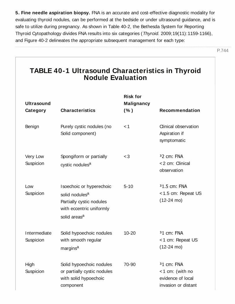
P.744
5. Fine needle aspiration biopsy. FNA is an accurate and cost-effective diagnostic modality forevaluating thyroid nodules, can be performed at the bedside or under ultrasound guidance, and issafe to utilize during pregnancy. As shown in Table 40-2, the Bethesda System for ReportingThyroid Cytopathology divides FNA results into six categories (Thyroid. 2009;19(11):1159-1166),and Figure 40-2 delineates the appropriate subsequent management for each type:
TABLE 40-1 Ultrasound Characteristics in ThyroidNodule Evaluation
UltrasoundCategory Characteristics
Risk forMalignancy(%) Recommendation
Benign Purely cystic nodules (noSolid component)
<1 Clinical observationAspiration ifsymptomatic
Very LowSuspicion
Spongiform or partially
cystic nodulesa
<3 ≥2 cm: FNA<2 cm: Clinicalobservation
LowSuspicion
Isoechoic or hyperechoic
solid nodulesa
Partially cystic noduleswith eccentric uniformly
solid areasa
5-10 ≥1.5 cm: FNA<1.5 cm: Repeat US(12-24 mo)
IntermediateSuspicion
Solid hypoechoic noduleswith smooth regular
marginsa
10-20 ≥1 cm: FNA<1 cm: Repeat US(12-24 mo)
HighSuspicion
Solid hypoechoic nodulesor partially cystic noduleswith solid hypoechoiccomponent
70-90 ≥1 cm: FNA<1 cm: (with noevidence of localinvasion or distant
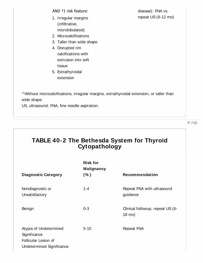
P.745
AND ≥1 risk feature:
1. Irregular margins(infiltrative,microlobulated)
2. Microcalcifications3. Taller than wide shape4. Disrupted rim
calcifications withextrusion into softtissue
5. Extrathyroidalextension
disease): FNA vs.repeat US (6-12 mo)
a Without microcalcifications, irregular margins, extrathyroidal extension, or taller thanwide shape.US, ultrasound; FNA, fine needle aspiration.
TABLE 40-2 The Bethesda System for ThyroidCytopathology
Diagnostic Category
Risk forMalignancy(%) Recommendation
Nondiagnostic orUnsatisfactory
1-4 Repeat FNA with ultrasoundguidance
Benign 0-3 Clinical followup, repeat US (6-18 mo)
Atypia of UndeterminedSignificanceFollicular Lesion ofUndetermined Significance
5-15 Repeat FNA
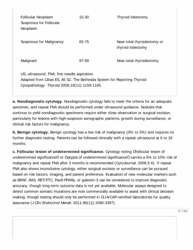
P.746
Follicular NeoplasmSuspicious for FollicularNeoplasm
15-30 Thyroid lobectomy
Suspicious for Malignancy 60-75 Near-total thyroidectomy orthyroid lobectomy
Malignant 97-99 Near-total thyroidectomy
US, ultrasound; FNA, fine needle aspiration.Adapted from Cibas ES, Ali SZ. The Bethesda System for Reporting ThyroidCytopathology. Thyroid 2009;19(11):1159-1165.
a. Nondiagnostic cytology. Nondiagnostic cytology fails to meet the criteria for an adequatespecimen, and repeat FNA should be performed under ultrasound guidance. Nodules thatcontinue to yield nondiagnostic specimens require either close observation or surgical excision,particularly for lesions with high-suspicion sonographic patterns, growth during surveillance, orclinical risk factors for malignancy.
b. Benign cytology. Benign cytology has a low risk of malignancy (0% to 3%) and requires nofurther diagnostic testing. Patients can be followed clinically with a repeat ultrasound at 6 to 18months.
c. Follicular lesion of undetermined significance. Cytology noting Òfollicular lesion ofundetermined significanceÓ or Òatypia of undetermined significanceÓ carries a 5% to 10% risk ofmalignancy and repeat FNA after 3 months is recommended (CytoJournal. 2008;5:6). If repeatFNA also shows inconclusive cytology, either surgical excision or surveillance can be pursuedbased on risk factors, imaging, and patient preference. Evaluation of new molecular markers suchas BRAF, RAS, RET/PTC, Pax8-PPARγ, or galectin-3 can be considered to improve diagnosticaccuracy, though long-term outcome data is not yet available. Molecular assays designed todetect common somatic mutations are now commercially available to assist with clinical decisionmaking, though testing should only be performed in CLIA/CAP-certified laboratories for qualityassurance (J Clin Endocrinol Metab. 2011;96(11):3390-3397).
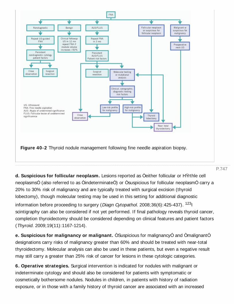
P.747
Figure 40-2 Thyroid nodule management following fine needle aspiration biopsy.
d. Suspicious for follicular neoplasm. Lesions reported as Òeither follicular or HŸrthle cellneoplasmsÓ (also referred to as ÒindeterminateÓ) or Òsuspicious for follicular neoplasmÓ carry a20% to 30% risk of malignancy and are typically treated with surgical excision (thyroidlobectomy), though molecular testing may be used in this setting for additional diagnostic
information before proceeding to surgery (Diagn Cytopathol. 2008;36(6):425-437). 123Iscintigraphy can also be considered if not yet performed. If final pathology reveals thyroid cancer,completion thyroidectomy should be considered depending on clinical features and patient factors(Thyroid. 2009;19(11):1167-1214).
e. Suspicious for malignancy or malignant. ÒSuspicious for malignancyÓ and ÒmalignantÓdesignations carry risks of malignancy greater than 60% and should be treated with near-totalthyroidectomy. Molecular analysis can also be used in these patients, but even a negative resultmay still carry a greater than 25% risk of cancer for lesions in these cytologic categories.
6. Operative strategies. Surgical intervention is indicated for nodules with malignant orindeterminate cytology and should also be considered for patients with symptomatic orcosmetically bothersome nodules. Nodules in children, in patients with history of radiationexposure, or in those with a family history of thyroid cancer are associated with an increased
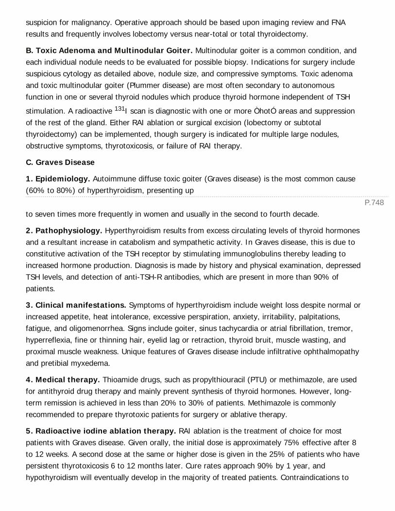
P.748
suspicion for malignancy. Operative approach should be based upon imaging review and FNAresults and frequently involves lobectomy versus near-total or total thyroidectomy.
B. Toxic Adenoma and Multinodular Goiter. Multinodular goiter is a common condition, andeach individual nodule needs to be evaluated for possible biopsy. Indications for surgery includesuspicious cytology as detailed above, nodule size, and compressive symptoms. Toxic adenomaand toxic multinodular goiter (Plummer disease) are most often secondary to autonomousfunction in one or several thyroid nodules which produce thyroid hormone independent of TSH
stimulation. A radioactive 131I scan is diagnostic with one or more ÒhotÓ areas and suppressionof the rest of the gland. Either RAI ablation or surgical excision (lobectomy or subtotalthyroidectomy) can be implemented, though surgery is indicated for multiple large nodules,obstructive symptoms, thyrotoxicosis, or failure of RAI therapy.
C. Graves Disease
1. Epidemiology. Autoimmune diffuse toxic goiter (Graves disease) is the most common cause(60% to 80%) of hyperthyroidism, presenting up
to seven times more frequently in women and usually in the second to fourth decade.
2. Pathophysiology. Hyperthyroidism results from excess circulating levels of thyroid hormonesand a resultant increase in catabolism and sympathetic activity. In Graves disease, this is due toconstitutive activation of the TSH receptor by stimulating immunoglobulins thereby leading toincreased hormone production. Diagnosis is made by history and physical examination, depressedTSH levels, and detection of anti-TSH-R antibodies, which are present in more than 90% ofpatients.
3. Clinical manifestations. Symptoms of hyperthyroidism include weight loss despite normal orincreased appetite, heat intolerance, excessive perspiration, anxiety, irritability, palpitations,fatigue, and oligomenorrhea. Signs include goiter, sinus tachycardia or atrial fibrillation, tremor,hyperreflexia, fine or thinning hair, eyelid lag or retraction, thyroid bruit, muscle wasting, andproximal muscle weakness. Unique features of Graves disease include infiltrative ophthalmopathyand pretibial myxedema.
4. Medical therapy. Thioamide drugs, such as propylthiouracil (PTU) or methimazole, are usedfor antithyroid drug therapy and mainly prevent synthesis of thyroid hormones. However, long-term remission is achieved in less than 20% to 30% of patients. Methimazole is commonlyrecommended to prepare thyrotoxic patients for surgery or ablative therapy.
5. Radioactive iodine ablation therapy. RAI ablation is the treatment of choice for mostpatients with Graves disease. Given orally, the initial dose is approximately 75% effective after 8to 12 weeks. A second dose at the same or higher dose is given in the 25% of patients who havepersistent thyrotoxicosis 6 to 12 months later. Cure rates approach 90% by 1 year, andhypothyroidism will eventually develop in the majority of treated patients. Contraindications to
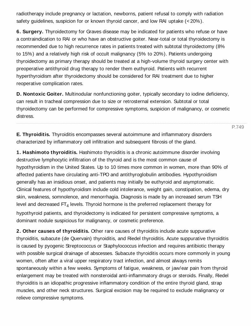
P.749
radiotherapy include pregnancy or lactation, newborns, patient refusal to comply with radiationsafety guidelines, suspicion for or known thyroid cancer, and low RAI uptake (<20%).
6. Surgery. Thyroidectomy for Graves disease may be indicated for patients who refuse or havea contraindication to RAI or who have an obstructive goiter. Near-total or total thyroidectomy isrecommended due to high recurrence rates in patients treated with subtotal thyroidectomy (8%to 15%) and a relatively high risk of occult malignancy (5% to 20%). Patients undergoingthyroidectomy as primary therapy should be treated at a high-volume thyroid surgery center withpreoperative antithyroid drug therapy to render them euthyroid. Patients with recurrenthyperthyroidism after thyroidectomy should be considered for RAI treatment due to higherreoperative complication rates.
D. Nontoxic Goiter. Multinodular nonfunctioning goiter, typically secondary to iodine deficiency,can result in tracheal compression due to size or retrosternal extension. Subtotal or totalthyroidectomy can be performed for compressive symptoms, suspicion of malignancy, or cosmeticdistress.
E. Thyroiditis. Thyroiditis encompasses several autoimmune and inflammatory disorderscharacterized by inflammatory cell infiltration and subsequent fibrosis of the gland.
1. Hashimoto thyroiditis. Hashimoto thyroiditis is a chronic autoimmune disorder involvingdestructive lymphocytic infiltration of the thyroid and is the most common cause ofhypothyroidism in the United States. Up to 10 times more common in women, more than 90% ofaffected patients have circulating anti-TPO and antithyroglobulin antibodies. Hypothyroidismgenerally has an insidious onset, and patients may initially be euthyroid and asymptomatic.Clinical features of hypothyroidism include cold intolerance, weight gain, constipation, edema, dryskin, weakness, somnolence, and menorrhagia. Diagnosis is made by an increased serum TSHlevel and decreased FT4 levels. Thyroid hormone is the preferred replacement therapy for
hypothyroid patients, and thyroidectomy is indicated for persistent compressive symptoms, adominant nodule suspicious for malignancy, or cosmetic preference.
2. Other causes of thyroiditis. Other rare causes of thyroiditis include acute suppurativethyroiditis, subacute (de Quervain) thyroiditis, and Riedel thyroiditis. Acute suppurative thyroiditisis caused by pyogenic Streptococcus or Staphylococcus infection and requires antibiotic therapywith possible surgical drainage of abscesses. Subacute thyroiditis occurs more commonly in youngwomen, often after a viral upper respiratory tract infection, and almost always remitsspontaneously within a few weeks. Symptoms of fatigue, weakness, or jaw/ear pain from thyroidenlargement may be treated with nonsteroidal anti-inflammatory drugs or steroids. Finally, Riedelthyroiditis is an idiopathic progressive inflammatory condition of the entire thyroid gland, strapmuscles, and other neck structures. Surgical excision may be required to exclude malignancy orrelieve compressive symptoms.

P.750
III. THYROID CANCER.Differentiated thyroid cancers (papillary, follicular, and HŸrthle cells) are relatively indolentcancers arising from follicular epithelial cells and are the most common types of thyroidmalignancy. Overall, these cancers are rare in children, more common in females (2.5:1), andhave a peak incidence in the fourth decade. Exposure to ionizing radiation is the best documentedenvironmental factor, and prognosis depends on the patient's age as well as the extent andhistologic subtype of the disease (85% to 90% of patients fall into a low-risk category withfavorable prognosis). As of January 2015, the ATA Guidelines Task Force has issued draft versionsof the new management guidelines for the workup and treatment of differentiated thyroidcancers (http://www.thyroid.org/thyroid-guidelines/). Table 40-3 reviews the major categories ofthyroid malignancies with clinical characteristics and management recommendations for eachtype.
A. Papillary Thyroid Cancer
1. Epidemiology. Papillary thyroid carcinoma (PTC) represents 85% of thyroid carcinomas, isoften multifocal, and frequently metastasizes to cervical lymph nodes. Occult, clinicallyinsignificant foci of microscopic PTC are found in 5% to 30% of autopsies or in thyroidectomyspecimens for benign diseases. Features deemed to be high risk and associated with worseprognosis include male gender, primary tumor size >4 cm, gross local invasion and extrathyroidalextension, age >45 years, certain histologic subtypes (specifically, the tall cell variant),lymphovascular invasion, or known metastatic disease (Cancer Control. 2011;18(2):96-103).
TABLE 40-3 Categories of Thyroid Malignancies
Category
Percent of AllThyroidMalignancies Clinical Features Management
Papillarycarcinoma
70-80 Indolent, oftenmultifocalFrequently metastaticto cervical lymphnodes
Surgery ± RAI ablationtherapy± thyroid hormonesuppression therapyFollowup: Serumthyroglobulin, neckultrasound
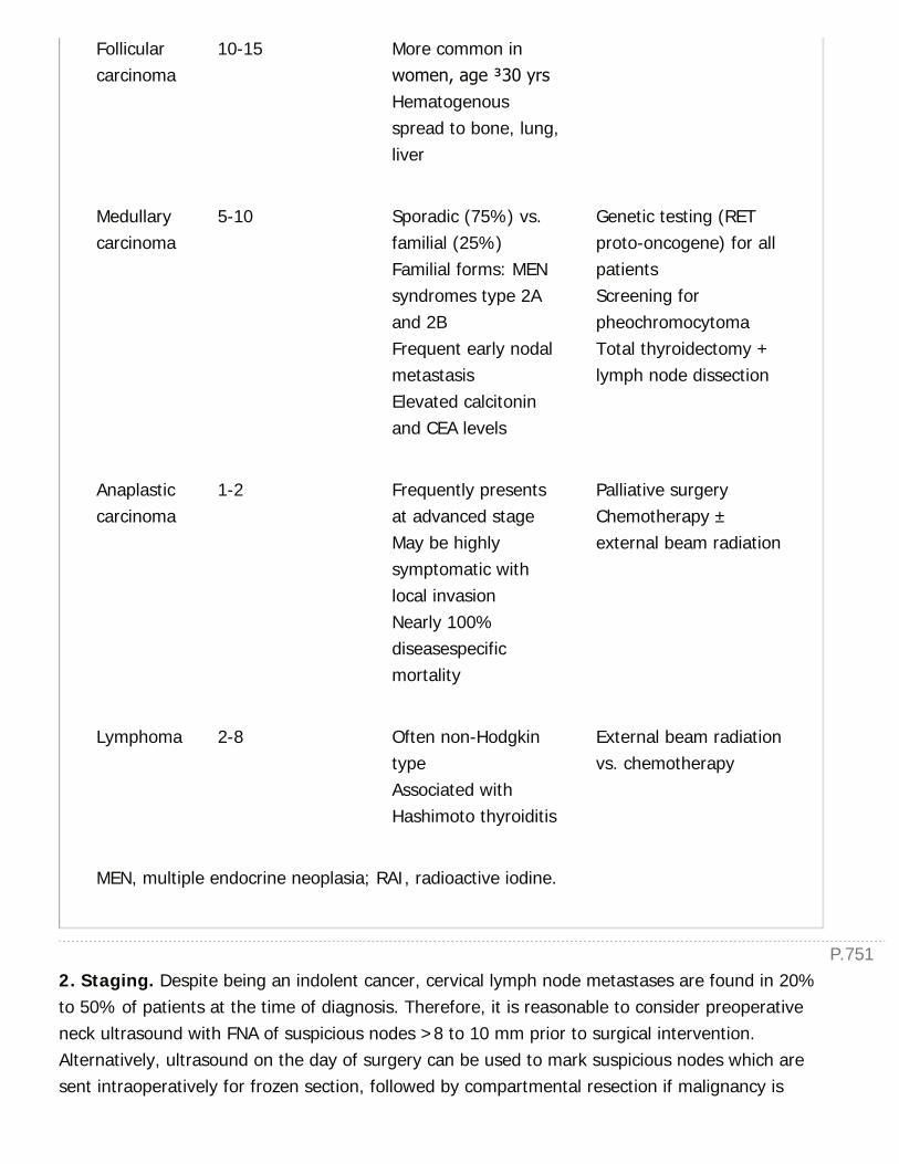
P.751
Follicularcarcinoma
10-15 More common inwomen, age ≥30 yrsHematogenousspread to bone, lung,liver
Medullarycarcinoma
5-10 Sporadic (75%) vs.familial (25%)Familial forms: MENsyndromes type 2Aand 2BFrequent early nodalmetastasisElevated calcitoninand CEA levels
Genetic testing (RETproto-oncogene) for allpatientsScreening forpheochromocytomaTotal thyroidectomy +lymph node dissection
Anaplasticcarcinoma
1-2 Frequently presentsat advanced stageMay be highlysymptomatic withlocal invasionNearly 100%diseasespecificmortality
Palliative surgeryChemotherapy ±external beam radiation
Lymphoma 2-8 Often non-HodgkintypeAssociated withHashimoto thyroiditis
External beam radiationvs. chemotherapy
MEN, multiple endocrine neoplasia; RAI, radioactive iodine.
2. Staging. Despite being an indolent cancer, cervical lymph node metastases are found in 20%to 50% of patients at the time of diagnosis. Therefore, it is reasonable to consider preoperativeneck ultrasound with FNA of suspicious nodes >8 to 10 mm prior to surgical intervention.Alternatively, ultrasound on the day of surgery can be used to mark suspicious nodes which aresent intraoperatively for frozen section, followed by compartmental resection if malignancy is

P.752
detected.
3. Operative strategies
a. Total thyroidectomy. Total thyroidectomy is recommended in several conditions: Tumorsgreater than 4 cm, presence of bilateral nodules, regional or metastatic disease, personal historyof head/neck radiation exposure, or first-degree relatives with PTC.
b. Thyroid lobectomy. Thyroid lobectomy is appropriate for low-risk patients with small (<1cm), intrathyroidal, unifocal tumors without evidence of regional or metastatic disease. For similarpatients with tumor size 1 to 4 cm, either thyroid lobectomy or total thyroidectomy can beconsidered.
c. Lymph node dissection. Lymph node dissection of the central neck compartment (level VI)and/or the ipsilateral lateral neck compartment (levels II, III, IV) should be performed in allpatients with biopsy-proven nodal disease. Furthermore, prophylactic central neck dissectionshould be considered for high-risk patients (e.g., large tumors, bilateral tumors, radiationexposure) even in the setting of clinically uninvolved lymph nodes, since up to 90% of patientswith PTC will have micrometastases at the time of surgery.
4. Complications
a. Hemorrhage. Hemorrhage is a rare but serious complication (0.3% to 1%) of thyroidectomythat usually occurs within 6 hours of surgery. Management typically requires airway control byendotracheal intubation but may necessitate immediate opening of the incision and/or anemergent surgical airway before return to the operating room for wound irrigation and control ofbleeding.
b. Hypocalcemia. Transient hypocalcemia may occur 24 to 48 hours after total or near-totalthyroidectomy. We start patients routinely on oral calcium carbonate (1 gram TID) for 2 weeksafter total thyroidectomy. If symptoms occur, additional oral calcium and calcitriol (0.25 µg/day)can be implemented, or if severe, intravenous replacement is achieved using six ampules of 10%calcium gluconate (93 mg elemental calcium in 10 mL) mixed in 500 mL of 5% dextrose in water(D5W) at a goal infusion of elemental calcium at 0.5 to 1.5 mg/kg/hour. Permanenthypoparathyroidism
is uncommon after total thyroidectomy. Normal parathyroid tissue removed or devascularizedduring surgery should be minced into 1 mm fragments and autotransplanted into individualmuscle pockets in the sternocleidomastoid muscle to minimize risk of postoperativehypoparathyroidism (Ann Surg. 1996;223(5):472-478).
c. Recurrent laryngeal nerve injury. RLN injury is a significant complication of thyroidectomythat should rarely occur (<1%). Unilateral RLN injury causes hoarseness, which often presentsimmediately but may appear days to weeks after surgery, and bilateral injury compromises theairway, usually manifesting immediately postoperatively and potentially requiring tracheostomy.
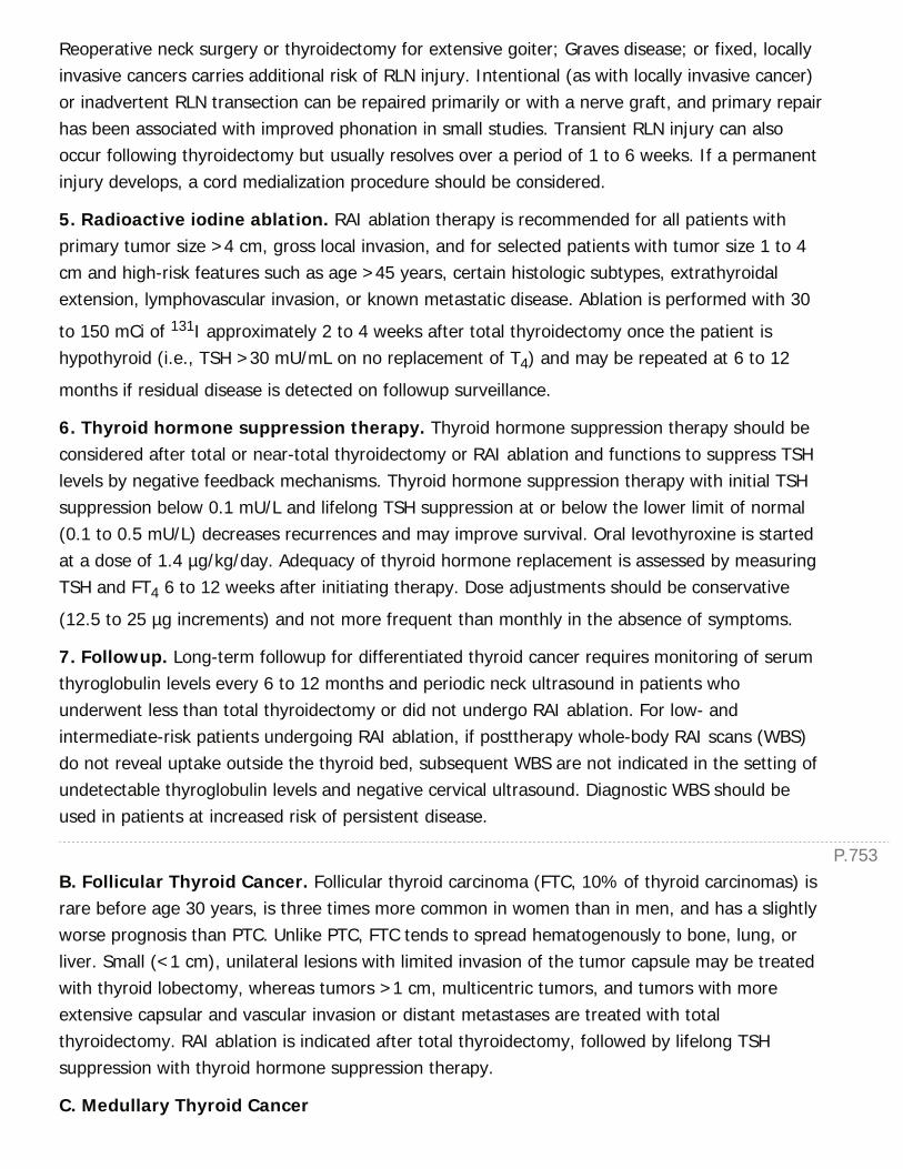
P.753
Reoperative neck surgery or thyroidectomy for extensive goiter; Graves disease; or fixed, locallyinvasive cancers carries additional risk of RLN injury. Intentional (as with locally invasive cancer)or inadvertent RLN transection can be repaired primarily or with a nerve graft, and primary repairhas been associated with improved phonation in small studies. Transient RLN injury can alsooccur following thyroidectomy but usually resolves over a period of 1 to 6 weeks. If a permanentinjury develops, a cord medialization procedure should be considered.
5. Radioactive iodine ablation. RAI ablation therapy is recommended for all patients withprimary tumor size >4 cm, gross local invasion, and for selected patients with tumor size 1 to 4cm and high-risk features such as age >45 years, certain histologic subtypes, extrathyroidalextension, lymphovascular invasion, or known metastatic disease. Ablation is performed with 30
to 150 mCi of 131I approximately 2 to 4 weeks after total thyroidectomy once the patient ishypothyroid (i.e., TSH >30 mU/mL on no replacement of T4) and may be repeated at 6 to 12
months if residual disease is detected on followup surveillance.
6. Thyroid hormone suppression therapy. Thyroid hormone suppression therapy should beconsidered after total or near-total thyroidectomy or RAI ablation and functions to suppress TSHlevels by negative feedback mechanisms. Thyroid hormone suppression therapy with initial TSHsuppression below 0.1 mU/L and lifelong TSH suppression at or below the lower limit of normal(0.1 to 0.5 mU/L) decreases recurrences and may improve survival. Oral levothyroxine is startedat a dose of 1.4 µg/kg/day. Adequacy of thyroid hormone replacement is assessed by measuringTSH and FT4 6 to 12 weeks after initiating therapy. Dose adjustments should be conservative
(12.5 to 25 µg increments) and not more frequent than monthly in the absence of symptoms.
7. Followup. Long-term followup for differentiated thyroid cancer requires monitoring of serumthyroglobulin levels every 6 to 12 months and periodic neck ultrasound in patients whounderwent less than total thyroidectomy or did not undergo RAI ablation. For low- andintermediate-risk patients undergoing RAI ablation, if posttherapy whole-body RAI scans (WBS)do not reveal uptake outside the thyroid bed, subsequent WBS are not indicated in the setting ofundetectable thyroglobulin levels and negative cervical ultrasound. Diagnostic WBS should beused in patients at increased risk of persistent disease.
B. Follicular Thyroid Cancer. Follicular thyroid carcinoma (FTC, 10% of thyroid carcinomas) israre before age 30 years, is three times more common in women than in men, and has a slightlyworse prognosis than PTC. Unlike PTC, FTC tends to spread hematogenously to bone, lung, orliver. Small (<1 cm), unilateral lesions with limited invasion of the tumor capsule may be treatedwith thyroid lobectomy, whereas tumors >1 cm, multicentric tumors, and tumors with moreextensive capsular and vascular invasion or distant metastases are treated with totalthyroidectomy. RAI ablation is indicated after total thyroidectomy, followed by lifelong TSHsuppression with thyroid hormone suppression therapy.
C. Medullary Thyroid Cancer

P.754
1. Epidemiology, pathophysiology, and clinical manifestations. Medullary thyroidcarcinoma (MTC) arises from the thyroid parafollicular C cells and accounts for 5% to 10% of allthyroid cancers in the United States. MTC may occur sporadically (75%) or may be associatedwith a familial syndrome, either alone or as a component of multiple endocrine neoplasia (MEN)syndromes type 2A or 2B (25%). Sporadic MTC generally presents as a firm, palpable, unilateralnodule with or without involved cervical lymph nodes, whereas hereditary MTC more oftendevelops as bilateral, multifocal tumors with diagnosis on the basis of genetic or biochemicalscreening. MTC spreads early to cervical lymph nodes and may metastasize to liver, lungs, orbone. All patients with MTC should undergo genetic testing for germline mutations in the RETproto-oncogene to exclude familial medullary thyroid carcinoma (FMTC) or MEN2 syndromes.Similar to other thyroid malignancies, diagnosis is made by FNA of suspicious thyroid nodules.More than 50% of patients presenting with a palpable primary tumor will already have nodalmetastases and elevated basal serum calcitonin levels (>20 to 100 pg/mL). Calcitonin and CEAlevels are important tumor markers for MTC and correlate strongly with extent of disease.Preoperative neck ultrasound is critical for identification of regional metastases and surgicalplanning, and screening for pheochromocytoma with serum or urine metanephrines andcatecholamines should be considered in all patients undergoing surgery for MTC. (For moreinformation on MEN, see Chapter 39: Diseases of the Adrenal and Pituitary Gland and HereditaryEndocrine Syndromes.)
2. Operative strategies. Total thyroidectomy alone is only indicated for MEN 2 patients whohave thyroid nodules <5 mm and calcitonin levels <40 pg/mL. Otherwise, treatment of bothhereditary and sporadic MTC requires at least total thyroidectomy with central neck compartment(Level VI) lymph node dissection, and additional dissection of ipsilateral lateral compartmentnodes should be performed in patients with palpable primary tumors. Surgical management ofresidual or recurrent disease remains the standard of care based upon patient clinical factors.
3. Medical therapy. MTC does not concentrate radioactive iodine, and postoperative RAItreatment is not recommended in the absence of
concurrent differentiated thyroid cancer. Adjuvant external beam radiation and systemicchemotherapy options have not shown significant benefit. However, clinical trials using tyrosinekinase inhibitors targeting the RET receptor have demonstrated improvement in progression-freesurvival, and two agents (vandetanib and cabozantinib) have been approved by the FDA. TheATA Guidelines Task Force issued a newly revised statement for the management of medullarythyroid cancer in 2015 (http://www.thyroid.org/thyroid-guidelines/).
D. Anaplastic Thyroid Cancer. Undifferentiated or anaplastic thyroid carcinoma (1% to 2% ofthyroid cancers) carries an extremely poor prognosis with nearly 100% disease-specific mortality.Disease usually presents as a fixed, sometimes painful goiter in patients older than 50 years, andover 90% of patients have regional or distant disease at the time of diagnosis. Invasion of local

P.755
structures can preclude resection, and local symptoms such as dysphagia, respiratorycompromise, or hoarseness may occur due to RLN involvement. External beam radiation orchemotherapy may provide limited palliation.
E. Primary Thyroid Lymphoma. Primary malignant lymphoma of the thyroid is usually non-Hodgkin type and is frequently associated with Hashimoto thyroiditis. Surgical resection is usuallynot indicated following diagnosis, and radiation or chemotherapy regimens are effective.
PARATHYROID
I. EMBRYOLOGY, ANATOMY, AND PHYSIOLOGYA. Embryology. The inferior and superior parathyroid glands are derived from the endoderm ofthe third and fourth pharyngeal pouches, respectively. The inferior parathyroids are intimatelyassociated with the thymus, which also develops from the third pharyngeal pouch, and ectopicinferior glands can be found anywhere along the tract of descent by the thymus into the chestthat becomes the thyrothymic ligament. The superior glands have a limited descent from the neckand are much less variable in position.
B. Anatomy. Typically, the inferior parathyroid glands are found inferior to the inferior thyroidartery and anterior to the RLN. The superior glands are usually found at the posterolateral aspectof the superior thyroid lobe, posterior to the RLN, and superior to the inferior thyroid artery,which is the main blood supply for all of the parathyroids. Because the embryologic path ofdescent of the inferior parathyroid crosses that of the superior glands, the glands can rarely befound at the same level, above or below the crossing of the inferior thyroid artery and RLN.
C. Physiology. Serum calcium levels are maintained within normal range (8.2 to 10.2 mg/dL) bythe interplay of parathyroid hormone (PTH) and vitamin D. Upon stimulation, chief cells of theparathyroid glands secrete PTH that (1) stimulates calcium and phosphate release from bone, (2)increases calcium and inhibits phosphate reabsorption in the kidneys,
and (3) enhances intestinal absorption of calcium through increased renal activation of vitamin D.Vitamin D is initially absorbed through the small intestine, undergoes hydroxylation in the liver to25(OH)D3, and then undergoes a second hydroxylation in the kidney under the influence of PTH
to its active form, 1,25(OH)D3. While vitamin D stimulates bone and intestinal calcium resorption,
calcitonin antagonizes the effect of PTH in the bones and kidneys.
II. BENIGN PARATHYROID DISEASEA. Primary Hyperparathyroidism
1. Epidemiology. Primary hyperparathyroidism (HPT) has an incidence of 0.25 to 1 per 1,000 inthe United States and is especially common in postmenopausal women. It most often occurssporadically but can be inherited alone or as a component of familial endocrinopathies, including
P.756
MEN types 1 and 2A.
2. Clinical manifestations. Common clinical findings associated with HPT includenephrolithiasis, osteoporosis, hypertension, and emotional disturbances. Patients may also havesubtle symptoms such as muscle weakness, polyuria, anorexia, fatigue, bone/joint pain, poorsleep, reflux, and nausea.
3. Biochemical evaluation. Diagnosis of primary HPT typically requires documentation ofhypercalcemia (serum calcium >10.5 mg/dL) and an elevated PTH level. The assay of choice forPTH is the highly sensitive and specific intact PTH immunoassay, and ionized calcium is a moresensitive test of physiologically active calcium. In addition, multiple biochemical abnormalitiesmay be present concurrently with hypercalcemia and require correction prior to surgicalintervention. Metabolic acidosis and hypophosphatemia are more commonly associated withprimary hyperparathyroidism due to increased urinary excretion of bicarbonate and phosphate.Patients are also more likely to present with hyperchloremia due to the increased urinaryexcretion of bicarbonate. Hypomagnesemia can occur in 5% to 10% of patients with primaryhyperparathyroidism. Serum alkaline phosphatase levels are often elevated in patients with bonedisease from hyperparathyroidism due to an increase in osteoclastic bone resorption.
4. Differential diagnosis. Hypercalcemia can be due to a variety of causes (e.g., malignancy,Paget disease, sarcoidosis, and milk-alkali syndrome), which are typically associated with low PTHlevels. Familial hypocalciuric hypercalcemia (FHH) commonly presents with mild hypercalcemiaand low urine calcium, with elevated PTH in 15% to 20% of patients. Caused by loss-of-functionmutations in renal and parathyroid calcium-sensing receptors, FHH patients have loss of feedbackinhibition of PTH secretion and inadequate clearance of calcium in the urine. FHH can bedistinguished from HPT by a 24-hour measurement of urine calcium or by measuring the renalcalcium/creatinine clearance ratio. A ratio less than 0.01 suggests FHH while the ratio seen inHPT is usually much higher. Parathyroidectomy is ineffective and not indicated for FHH.
5. Preoperative localization studies. Parathyroid imaging has no role in the diagnosis of HPTbut is critical in operative planning to facilitate limited neck exploration. Technetium-99 sestamibiscintigraphy has historically been utilized to localize hyperfunctioning parathyroid tissue, thoughnewer modalities of scintigraphy in combination with single photon emission computedtomography (SPECT), SPECT with computed tomography (SPECT/CT), and four-dimensional CThave demonstrated higher sensitivity and positive predictive values (J Nucl Med.2007;48(7):1084-1089). Ultrasonography with color Doppler examination complements thesestamibi scan and can assist in precise localization of adenomas, assessment of concomitantthyroid pathology, or FNA of equivocal lesions.
6. Indications for surgery. Parathyroidectomy is indicated for patients with classic symptomsof primary HPT (i.e., nephrolithiasis, pathologic fracture, neuromuscular disturbances, andhypercalcemic crisis). Management of asymptomatic patients is more controversial, but recent
P.757
guidelines from an expert consensus panel recommend parathyroidectomy for those meeting oneof the following criteria: (1) Age less than 50 years; (2) unable to participate in appropriatefollowup; (3) serum calcium level >1 mg/dL above normal range; (4) urine calcium >400 mg per24 hours; (5) creatinine clearance <60 mL/min; or (6) complications of primary HPT (J ClinEndocrinol Metab. 2014;99(10):3561-3569). If patients do not meet one of these criteria, surgeryis not required, but remains an option. Many patients choose surgery if they have significantnonspecific symptoms. Nephrolithiasis, bone disease, and neuromuscular symptoms are improvedfollowing surgery more often than renal insufficiency, hypertension, and psychiatricmanifestations.
7. Operative strategies
a. Minimally invasive parathyroidectomy. Preoperative localization studies used with rapidintraoperative PTH measurement now enable several minimally invasive techniques, includingopen, radioguided, video-assisted, and endoscopic methods. Since the majority (>85%) ofpatients with primary HPT have a single parathyroid adenoma, successful preoperativelocalization allows for directed unilateral neck exploration while normal parathyroids do not needto be identified. The rapid intraoperative PTH assay allows surgeons to verify the adequacy ofresection: following adenoma removal; a 50% decrease in PTH levels at 10 minutes is highlyindicative of cure.
b. Conventional neck exploration. Historically, bilateral neck exploration and identification ofall four parathyroids has been the cornerstone of surgical management in HPT, with resultantnormocalcemia in more than 95% of patients. If an abnormally enlarged parathyroid or all fourparathyroids cannot be found, exploration for ectopic or supernumerary glands should beperformed. Ectopic superior glands may be found posterior and deep to the thyroid, in thetracheoesophageal groove, or between the carotid artery and the esophagus. Ectopic inferiorglands are most likely found embedded in the thymus in the anterior mediastinum. Occasionally,multiple
parathyroid adenomas are found and should be removed, leaving at least one normal parathyroidbehind. Four-gland parathyroid hyperplasia is rare, and acceptable management options includetotal parathyroidectomy with parathyroid autotransplantation or 3.5-gland parathyroidectomy. Aclear factual operative note detailing the identification and position of each gland is essential.
c. Parathyroid autotransplantation. Total parathyroidectomy with heterotopic parathyroidautotransplantation should be considered in patients with renal failure and secondaryhyperparathyroidism, four-gland parathyroid hyperplasia, and those undergoing neck re-exploration in which the adenoma is the only remaining parathyroid gland. Thesternocleidomastoid or the brachioradialis muscles of the patient's non-dominant forearm arecommon sites for autotransplantation. Parathyroid autotransplantation into the forearm isadvantageous if recurrent HPT is a possibility (e.g., MEN type 1 or 2A) because the transplantedhyperfunctioning parathyroid tissue can easily be localized and excised under local anesthesia
P.758
with sedation.
Freshly removed parathyroid tissue is finely minced (approximately 1 × 1 × 2 mm) and placed insterile iced saline. Separate intramuscular beds are created by spreading the fibers of thebrachioradialis or the sternocleidomastoid with a fine forceps. Four to five pieces of parathyroidtissue are placed in each site for a total transplant volume of approximately 100 mg.Nonabsorbable suture is used to close the beds and to mark the site of transplanted tissue.Transplanted parathyroid tissue begins to function within 14 to 21 days of surgery.
Cryopreservation of parathyroid glands is performed in all patients who are at risk for permanenthypoparathyroidism after repeat exploration. Approximately 200 mg of minced parathyroid tissueis frozen in vials containing autologous serum, dimethyl sulfoxide, and cell culture media.Cryopreserved tissue can be used for autotransplantation in patients with failure of the initialgraft. Viable cryopreservation and subsequent thawing must be performed in a Food and DrugAdministration (FDA)-approved facility.
8. Management of postoperative hypocalcemia
a. Clinical manifestations. Transient hypocalcemia commonly occurs after total thyroidectomyor parathyroidectomy and requires treatment if it is severe (total serum calcium <7.5 mg/dL) or ifthe patient is symptomatic. Symptoms may involve numbness/paresthesias in the distalextremities, perioral numbness, or hyperactive tendon reflexes. Chvostek sign (twitching of thefacial muscles with tapping over the facial nerve anterior to the ear) indicates relativehypocalcemia, but it is present in up to 15% of the normal population and does not necessarilyrequire calcium replacement.
b. Oral calcium supplementation. Hypocalcemic patients may require postoperativesupplementation for 6 to 8 weeks and are given oral calcium carbonate (500 to 1,000 mg TID)and calcitriol (0.25 µg/day).
c. Intravenous calcium supplementation. Intravenous administration of calcium gluconate orcalcium chloride may be necessary in persistently symptomatic patients or in emergent situationssuch as hypocalcemic tetany. Ten to 20 mL of 10% calcium gluconate are given intravenouslyover 10 minutes and may be repeated every 15 to 20 minutes as required until symptomresolution. Subsequently, a continuous infusion of calcium gluconate in D5W is initiated at 0.5 to1.5 mg/kg/hour with correction of any concurrent hypomagnesemia.
B. Secondary and Tertiary Hyperparathyroidism
1. Biochemical evaluation. Most commonly due to chronic renal failure, secondary HPTmanifests as increased PTH levels in response to hypocalcemia. Decreased serum calcium levelsare a terminal feature of kidney dysfunction, which becomes evident through phosphateretention, decreased vitamin D activation, and poor calcium absorption. Intestinal malabsorptionof calcium or vitamin D can also result in elevated PTH levels and secondary HPT. Thus, patients
P.759
with secondary HPT have high PTH levels and low calcium levels. Tertiary hyperparathyroidismcan be seen in patients who have undergone a kidney transplant for renal failure. Typically,parathyroid gland function returns to normal within one year after kidney transplant, but inpatients with tertiary HPT, the parathyroid glands fail to respond to normal signals for PTHsecretion and regulation of calcium homeostasis. In these patients, both the PTH and calciumlevels are high.
2. Medical management. Hypercalcemia from secondary and tertiary HPT is treated initiallywith dietary phosphate restriction, phosphate binders, and vitamin D supplementation. Cinacalcet,a calcimimetic, is also commonly used. Although it does not impact mortality, it may decrease theneed for parathyroidectomy.
3. Operative strategies. Patients with medically unresponsive, symptomatic HPT (e.g., bonepain, osteopenia, ectopic calcification, or pruritus) may undergo total parathyroidectomy withautotransplantation or subtotal parathyroidectomy. Controversy exists regarding type of surgeryand appropriate postoperative PTH level to prevent adynamic bone disease.
C. Recurrent or Persistent Hyperparathyroidism
1. Biochemical evaluation. In all cases of persistent hypercalcemia, the diagnosis of HPTshould be confirmed. In addition to calcium and intact PTH levels, a 24-hour urine calcium shouldbe obtained to rule out FHH. Factors associated with recurrent or persistent disease include failedpreoperative localization studies, multiple gland disease, ectopic or supernumerary glands,malignancy, and surgeon inexperience.
2. Preoperative localization. Preoperative localization is mandatory in patients being
considered for reoperative parathyroidectomy and may include 99mTc-sestamibi scintigraphy withSPECT/CT, ultrasound with FNA, or 4D CT scan. Approximately 70% to 80% of patientsundergoing reexploration have a missed gland that is accessible through a
cervical incision. Noninvasive imaging is successful in gland localization in 25% to 75% of cases.For patients with negative or discordant noninvasive studies, selective venous sampling with rapidPTH assessment can be considered.
3. Operative strategy. The goal of reexploration is to perform an orderly search based oninformation from the initial operation and from preoperative localization studies. Reoperativeparathyroid surgery carries a substantially higher risk of RLN injury and of hypocalcemia due topostoperative scarring and disruption of normal tissue planes.
a. Missed parathyroid glands can be found in either normal anatomic location or in ectopic sites.They may occasionally be intrathyroidal (especially in patients with multinodular goiter), andthyroid lobectomy can be performed if an exhaustive search fails to identify a parathyroidadenoma. If four normal glands have been located, a supernumerary gland is likely responsible.Intraoperative ultrasound and/or venous sampling from the right and left internal jugular veins
P.760
P.761
can sometimes be useful in localizing adenomas.
b. Mediastinal adenomas within the thymus are managed by resecting the cranial portion viagentle traction on the thyrothymic ligament or by a complete transcervical thymectomy using aspecialized substernal retractor (Ann Surg. 1991;214:555). Median sternotomy is associated withhigher morbidity and increased postoperative pain, and the possibility that this procedure may berequired should be discussed with the patient preoperatively.
III. PARATHYROID CANCERA. Clinical Manifestations. Parathyroid cancer is a rare disease, accounting for <1% ofpatients with primary HPT. Most (>90%) of parathyroid carcinomas are biochemically functional.Approximately 50% of patients have a palpable neck mass, and serum calcium levels may exceed
15 mg/dL. Ultrasound and 99mTc sestamibi imaging can help localize disease, but diagnosisdepends on histologic findings of vascular or capsular invasion, metastases, or gross invasion oflocal structures.
B. Medical Management of Hypercalcemic Crisis. Patients with parathyroid cancer andsome patients with benign HPT may develop hypercalcemic crisis with serum calcium levels of 16to 20 mg/dL and azotemia. Symptoms of this acute, sometimes fatal illness, include profoundmuscular weakness, nausea and vomiting, drowsiness, and confusion. Ultimate treatment isparathyroidectomy, but volume and electrolyte abnormalities should be addressed first. First-linetherapy involves intravenous infusion of 0.9% sodium chloride at 300 to 500 mL/hour to restoreintravascular volume and to promote renal excretion of calcium. After urinary output exceeds 100mL/hour, furosemide (80 to 100 mg intravenously every 2 to 6 hours) may be given to promotefurther renal sodium and calcium excretion, though consequent hypokalemia andhypomagnesemia should be corrected. Thiazide diuretics impair calcium excretion and should beavoided. If diuresis alone is unsuccessful in lowering the serum calcium,
other agents may be used, including the bisphosphonates pamidronate and etidronate,mithramycin, and salmon calcitonin. Orthophosphate, gallium nitrate, and glucocorticoids alsohave calcium-lowering effects.
C. Operative Strategy. Surgical treatment of parathyroid cancer entails radical local excision ofthe tumor, surrounding soft tissue, lymph nodes, and ipsilateral thyroid lobe. Reoperation isindicated for local recurrence or to control malignant hypercalcemia.
CHAPTER 40: THYROID AND PARATHYROID GLANDSMultiple Choice Questions1. A 56-year-old man presents to clinic for evaluation of a small rightneck mass. He has no significant past medical history and denies any
history of smoking. The mass has been slowly enlarging over the last 2years but is not painful. The patient is normotensive with a negativereview of systems, and he denies any dyspnea, choking sensations, orhoarseness. What is the first diagnostic study that should be performedin the workup of this mass?
a. Ultrasonography of the thyroid
b. Serum thyroid stimulating hormone (TSH) level
c. Fine-needle aspiration (FNA)
d. Computed tomography (CT) scan of the neck and chest
e. Thyroid scintigraphy
View Answer
2. A 64-year-old woman presents to the emergency department withvague abdominal pain, nausea, confusion, and muscle weakness. AnEKG shows a short QT interval. A serum calcium level is 15.2 mg/dL.What etiology does this suggest?
a. Thiazide use
b. Secondary hyperparathyroidism
c. Parathyroid carcinoma
d. Single parathyroid adenoma
e. Factitious hypercalcemia
View Answer
3. Following total thyroidectomy, a 50-year-old male presents for his 1year followup visit. He is currently on daily levothyroxine therapy. Thebest method to monitor the adequacy of replacement therapy is:
a. Radioactive iodine (RAI) uptake
b. Thyroglobulin
c. Triiodothyronine resin uptake (RT3U)
d. Serum TSH level
e. Total thyroxine level (total T4)
View Answer
4. A 72-year-old woman with recently diagnosed primaryhyperparathyroidism presents for surgical evaluation. Her serumcalcium level is found to be 13.6 mg/dL. Other biochemicalabnormalities
UnitedVRG

P.762
that may accompany hypercalcemia in patients with primaryhyperparathyroidism include which of the following?
a. Metabolic alkalosis
b. Hyperphosphatemia
c. Hypochloremia
d. Hypermagnesemia
e. Elevated alkaline phosphatase
View Answer
5. A 63-year-old patient with primary hyperparathyroidism andnonlocalizing studies undergoes neck exploration. He is found to havenormal right and left superior parathyroids and a normal left inferiorparathyroid gland.
Of the following, which is the most likely location for the missing rightinferior parathyroid?
a. Tracheoesophageal groove
b. Right thyroid lobe
c. Superior thymus
d. Posterior mediastinum
e. Pharyngeal mucosa
View Answer
6. Which of the following patients with thyroid gland enlargement isLEAST likely to have a diagnosis of thyroid cancer?
a. A 5-year-old boy with two family members with medullary thyroidcarcinoma
b. A 75-year-old man with a solitary nodule and hoarseness
c. A 56-year-old woman with a solitary nodule and a history of radiationtherapy to the neck
d. A 43-year-old woman with a multinodular goiter and tremor
e. A 14-year-old girl with an asymptomatic solitary nodule
View Answer
7. A 47-year-old woman presents to clinic for evaluation of weight gain,thinning hair, constant fatigue, constipation and muscle weakness overthe past year. She denies any prior history of thyroid disorders and
P.763
currently takes no medications. Serum TSH level is elevated at greaterthan 30 mIU/L. What is the most likely cause of the patient'ssymptoms?
a. Thyroid adenoma
b. Self-administration of thyroid hormone
c. Papillary thyroid carcinoma
d. Radioactive iodine administration
e. Hashimoto thyroiditis
View Answer
8. A 65-year-old female presents for her first postoperative visitfollowing total thyroidectomy for papillary thyroid carcinoma. Which ofthe following features on the pathology report may indicate tumoraggressiveness and poor outcome?
a. Tumor size larger than 4 cm
b. Macroscopic lymph node metastasis
c. Local invasion through capsule into surrounding soft tissue
d. Poorly differentiated histologic grade
e. All of the above
View Answer
9. A young family brings a 1-month-old female into your clinic forevaluation after referral from their pediatrician. The child's fatherunderwent total thyroidectomy in infancy and multiple family membershave required adrenalectomy for pheochromocytomas. Which of thefollowing is the most sensitive method for detecting medullary thyroidcarcinoma in patients with multiple endocrine neoplasia (MEN) type 2syndromes?
a. Genetic evaluation
b. Plasma carcinoembryonic antigen level
c. Plasma catecholamine level
d. Serum thyroglobulin level
e. Serum calcium level
View Answer
10. A 34-year-old female presents to clinic for evaluation of a large
UnitedVRG

thyroid goiter. She complains of an intermittent tremor, increasedappetite, palpitations, and increased perspiration. What other featuresmay also be present on history and physical examination?
a. Exophthalmos and pretibial myxedema
b. Suppressed thyroid stimulating hormone level
c. Cold intolerance and somnolence
d. Answers A and B only
e. All of the above
View Answer

41Otolaryngology for the General Surgeon
Lauren T. Roland
John S. Schneider
I. THE NECKA. Anatomy and Physiology
1. The cervical fascia provides planes for passage of infection, hemorrhage, and surgicaldissection (Fig. 41-1).
2. The lymphatic system of the neck is divided into six levels. Level I contains the submentaland submandibular lymph nodes, level II through IV parallel the jugular vein, level V consists ofthe posterior triangle, and level VI is the central compartment. These levels are important forpredicting spread of cancer from the head and neck, as well as determining the extent of surgerynecessary during a neck dissection (Fig. 41-2).
B. Neck Masses. History for a neck mass should focus on duration, location, and symptoms(pain, fevers, weight loss, dysphagia, voice changes, otalgia), past medical history, and socialhistory (tobacco and alcohol use, travel history, animal exposures, sick contacts). Differentialdiagnosis is strongly influenced by age. Neck masses in adults are presumed malignant untilproven otherwise. In contrast, neck masses in children are usually inflammatory or congenital,and neoplasms are rare.
1. Adult neck masses. Adult neck masses are commonly metastatic squamous cell carcinoma(SCC) from a primary tumor of the oral cavity, pharynx, or larynx. See Figure 41-3 for analgorithm describing workup of a neck mass in an adult.
2. Pediatric neck masses. Children often have palpable lymph nodes; however, large,persistent masses should be investigated through ultrasound (US), as this is safe andnoninvasive. CT should be reserved for deep neck space infections. Additional studies includewhite blood cell count (WBC) with differential and specific serologic tests for infectious etiologies.See Figure 41-4 for an algorithm describing workup of a neck mass in children.
C. Congenital Neck Lesions. Congenital masses may swell during an upper respiratoryinfection (URI). The acute infection should be treated with antibiotics. If necessary, needleaspiration may be performed for decompression.
1. Branchial cleft anomalies. These congenital masses may result in cysts, sinuses, or fistulae.The most common anomaly is of the second branchial cleft, which presents as a nontender,
> Table of Contents > 41 - Otolaryngology for the General Surgeon
UnitedVRG
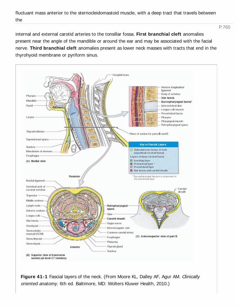
P.765
fluctuant mass anterior to the sternocleidomastoid muscle, with a deep tract that travels betweenthe
internal and external carotid arteries to the tonsillar fossa. First branchial cleft anomaliespresent near the angle of the mandible or around the ear and may be associated with the facialnerve. Third branchial cleft anomalies present as lower neck masses with tracts that end in thethyrohyoid membrane or pyriform sinus.
Figure 41-1 Fascial layers of the neck. (From Moore KL, Dalley AF, Agur AM. Clinicallyoriented anatomy. 6th ed. Baltimore, MD: Wolters Kluwer Health, 2010.)

P.766
2. Thyroglossal duct cysts. These masses are due to lack of obliteration of the thyroglossalduct after descent of the thyroid from the base of the tongue during embryologic development.Due to attachment to the tongue, these midline masses often move with tongue protrusion.Before surgery, the physician should perform an US to ensure that the mass is not the solethyroid tissue, as removal would cause hypothyroidism. The
definitive treatment is the Sistrunk procedure and involves removal of the tissue as well as themidline portion of the hyoid bone that the embryologic tissue passes through.
Figure 41-2 Neck Node Levels. IA: Submental, IB: Submandibular, II: Upper jugular, III:Middle jugular, IV: Lower jugular, V: Posterior triangle, VI: Anterior compartment. (FromMulholland, MW, ed. Operative techniques in surgery. Philadelphia, PA: Wolters KluwerHealth, 2015.)
3. Hemangioma. This lesion presents as a reddish-bluish compressible mass in infancy.Hemangiomas typically grow rapidly during the first year of life and then slowly involute; themajority do not require treatment. However, cervical hemangiomas may be associated withsubglottic hemangiomas or airway masses which may be symptomatic and require emergent
UnitedVRG

P.767
attention. Propranolol, steroids, laser therapy, and surgical resection are treatment options.
4. Lymphatic malformations. These lesions are soft, doughy, compressible lesions.Treatments include sclerotherapy and surgery.
5. Other congenital masses include laryngoceles, dermoid cysts, teratomas, plungingranulas, and thymic cysts.
D. Infectious/Inflammatory Disorders
1. Suppurative bacterial lymphadenitis. This condition is common in children and is usuallythe result of S. aureus or group A streptococcal infections. Treatment includes incision anddrainage (I&D) and intravenous (IV) antibiotics.
2. Acute mononucleosis. Mononucleosis is caused by EpsteinÑBarr (EBV) infection andcommonly causes lymphadenopathy. It is associated with fevers, tonsillitis, andhepatosplenomegaly. Treatment is supportive.
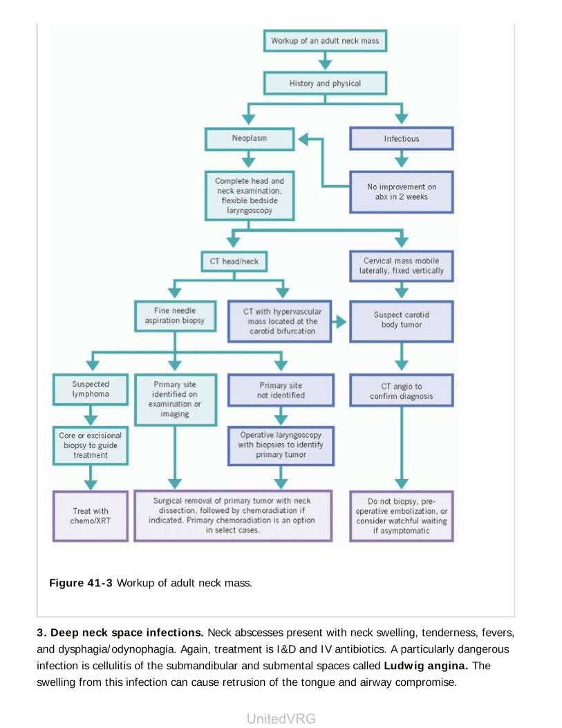
Figure 41-3 Workup of adult neck mass.
3. Deep neck space infections. Neck abscesses present with neck swelling, tenderness, fevers,and dysphagia/odynophagia. Again, treatment is I&D and IV antibiotics. A particularly dangerousinfection is cellulitis of the submandibular and submental spaces called Ludwig angina. Theswelling from this infection can cause retrusion of the tongue and airway compromise.
UnitedVRG
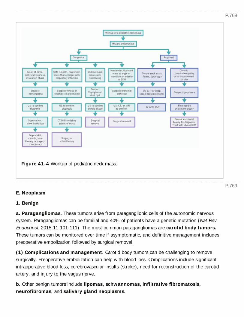
P.768
P.769
Figure 41-4 Workup of pediatric neck mass.
E. Neoplasm
1. Benign
a. Paragangliomas. These tumors arise from paraganglionic cells of the autonomic nervoussystem. Paragangliomas can be familial and 40% of patients have a genetic mutation (Nat RevEndocrinol. 2015;11:101-111). The most common paragangliomas are carotid body tumors.These tumors can be monitored over time if asymptomatic, and definitive management includespreoperative embolization followed by surgical removal.
(1) Complications and management. Carotid body tumors can be challenging to removesurgically. Preoperative embolization can help with blood loss. Complications include significantintraoperative blood loss, cerebrovascular insults (stroke), need for reconstruction of the carotidartery, and injury to the vagus nerve.
b. Other benign tumors include lipomas, schwannomas, infiltrative fibromatosis,neurofibromas, and salivary gland neoplasms.
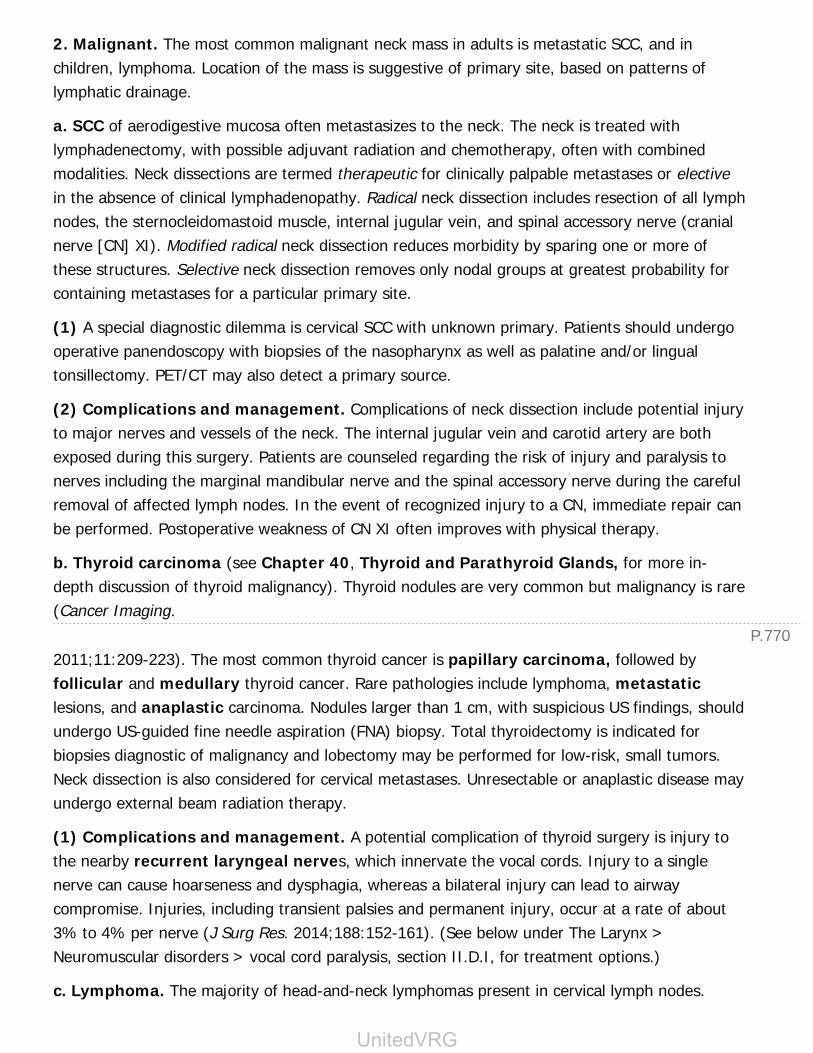
P.770
2. Malignant. The most common malignant neck mass in adults is metastatic SCC, and inchildren, lymphoma. Location of the mass is suggestive of primary site, based on patterns oflymphatic drainage.
a. SCC of aerodigestive mucosa often metastasizes to the neck. The neck is treated withlymphadenectomy, with possible adjuvant radiation and chemotherapy, often with combinedmodalities. Neck dissections are termed therapeutic for clinically palpable metastases or electivein the absence of clinical lymphadenopathy. Radical neck dissection includes resection of all lymphnodes, the sternocleidomastoid muscle, internal jugular vein, and spinal accessory nerve (cranialnerve [CN] XI). Modified radical neck dissection reduces morbidity by sparing one or more ofthese structures. Selective neck dissection removes only nodal groups at greatest probability forcontaining metastases for a particular primary site.
(1) A special diagnostic dilemma is cervical SCC with unknown primary. Patients should undergooperative panendoscopy with biopsies of the nasopharynx as well as palatine and/or lingualtonsillectomy. PET/CT may also detect a primary source.
(2) Complications and management. Complications of neck dissection include potential injuryto major nerves and vessels of the neck. The internal jugular vein and carotid artery are bothexposed during this surgery. Patients are counseled regarding the risk of injury and paralysis tonerves including the marginal mandibular nerve and the spinal accessory nerve during the carefulremoval of affected lymph nodes. In the event of recognized injury to a CN, immediate repair canbe performed. Postoperative weakness of CN XI often improves with physical therapy.
b. Thyroid carcinoma (see Chapter 40, Thyroid and Parathyroid Glands, for more in-depth discussion of thyroid malignancy). Thyroid nodules are very common but malignancy is rare(Cancer Imaging.
2011;11:209-223). The most common thyroid cancer is papillary carcinoma, followed byfollicular and medullary thyroid cancer. Rare pathologies include lymphoma, metastaticlesions, and anaplastic carcinoma. Nodules larger than 1 cm, with suspicious US findings, shouldundergo US-guided fine needle aspiration (FNA) biopsy. Total thyroidectomy is indicated forbiopsies diagnostic of malignancy and lobectomy may be performed for low-risk, small tumors.Neck dissection is also considered for cervical metastases. Unresectable or anaplastic disease mayundergo external beam radiation therapy.
(1) Complications and management. A potential complication of thyroid surgery is injury tothe nearby recurrent laryngeal nerves, which innervate the vocal cords. Injury to a singlenerve can cause hoarseness and dysphagia, whereas a bilateral injury can lead to airwaycompromise. Injuries, including transient palsies and permanent injury, occur at a rate of about3% to 4% per nerve (J Surg Res. 2014;188:152-161). (See below under The Larynx >Neuromuscular disorders > vocal cord paralysis, section II.D.I, for treatment options.)
c. Lymphoma. The majority of head-and-neck lymphomas present in cervical lymph nodes.
UnitedVRG
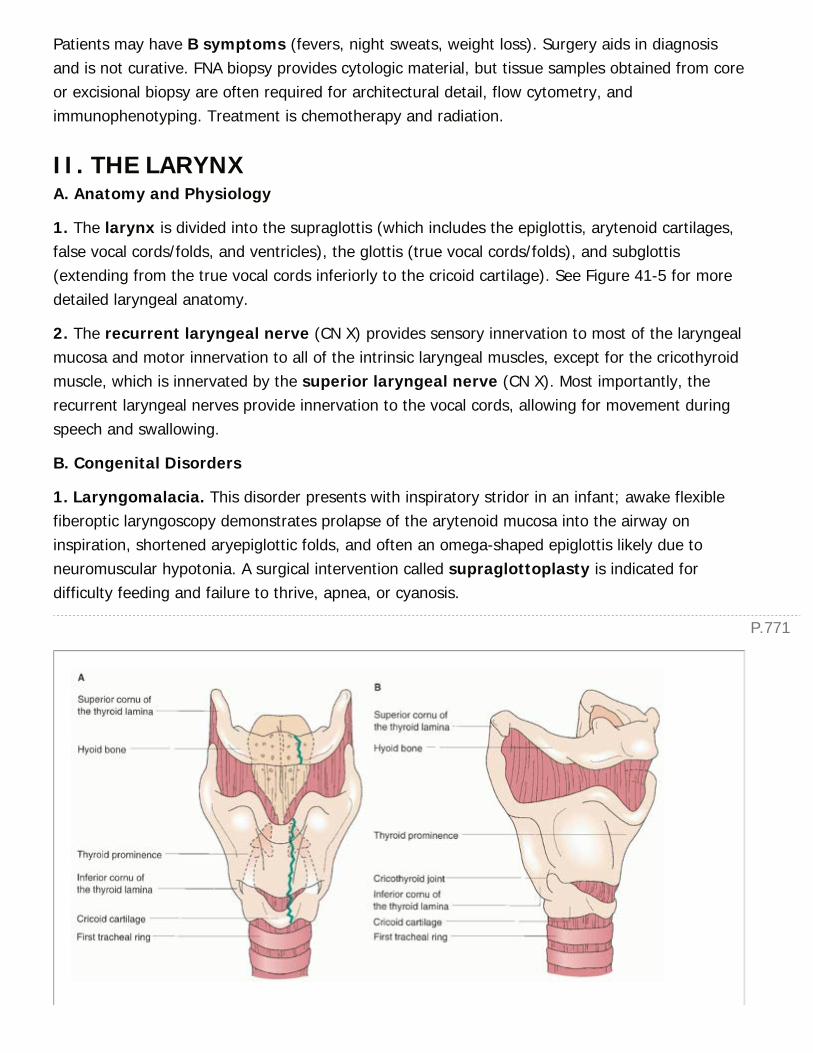
P.771
Patients may have B symptoms (fevers, night sweats, weight loss). Surgery aids in diagnosisand is not curative. FNA biopsy provides cytologic material, but tissue samples obtained from coreor excisional biopsy are often required for architectural detail, flow cytometry, andimmunophenotyping. Treatment is chemotherapy and radiation.
II. THE LARYNXA. Anatomy and Physiology
1. The larynx is divided into the supraglottis (which includes the epiglottis, arytenoid cartilages,false vocal cords/folds, and ventricles), the glottis (true vocal cords/folds), and subglottis(extending from the true vocal cords inferiorly to the cricoid cartilage). See Figure 41-5 for moredetailed laryngeal anatomy.
2. The recurrent laryngeal nerve (CN X) provides sensory innervation to most of the laryngealmucosa and motor innervation to all of the intrinsic laryngeal muscles, except for the cricothyroidmuscle, which is innervated by the superior laryngeal nerve (CN X). Most importantly, therecurrent laryngeal nerves provide innervation to the vocal cords, allowing for movement duringspeech and swallowing.
B. Congenital Disorders
1. Laryngomalacia. This disorder presents with inspiratory stridor in an infant; awake flexiblefiberoptic laryngoscopy demonstrates prolapse of the arytenoid mucosa into the airway oninspiration, shortened aryepiglottic folds, and often an omega-shaped epiglottis likely due toneuromuscular hypotonia. A surgical intervention called supraglottoplasty is indicated fordifficulty feeding and failure to thrive, apnea, or cyanosis.
P.772
Figure 41-5 A, B: External Larynx Anatomy. (From Mulholland, MW et al., eds. Greenfield'ssurgery: scientific principles and practice. Philadelphia, PA: Wolters Kluwer Health, 2011.)
2. Vocal cord paralysis. Vocal cord paralysis causes inspiratory or biphasic stridor, aspiration,and weak cry. Etiologies include birth trauma, neurologic disease, and iatrogenic injury. Diagnosisis made by awake flexible laryngoscopy. Most noniatrogenic unilateral paralysis resolvesspontaneously within the first year of life. Bilateral cord paralysis often requires tracheotomy.
3. Subglottic stenosis. Congenital subglottic stenosis is due to abnormally formed cricoidcartilage. Acquired stenosis is usually due to intubation. If stenosis is mild, patients may improveas they grow. Otherwise, surgical laryngotracheal reconstruction or cricotracheal resection maybe necessary.
C. Infectious/Inflammatory Disorders
1. Viral croup. Viral laryngotracheitis is glottic and subglottic inflammation from parainfluenzavirus. Patients present with a barking cough, hoarseness, and inspiratory stridor. Lateral airway x-ray may show the steeple sign from subglottic edema. Treatment includes humidified air,glucocorticoids for moderate to severe croup, racemic epinephrine, and heliox.
2. Epiglottitis. This infection is usually caused by H. influenzae type B. Patients present withfever, muffled voice, drooling, and stridor. Treatment is urgent airway management and IVantibiotics.
D. Neuromuscular Disorders
1. Vocal cord paralysis. This disorder occurs secondary to recurrent laryngeal nerve injury,often due to surgery, neoplasm, or trauma to the neck or thorax. Iatrogenic injuries duringsurgery should be repaired by primary epineural anastomosis or cable grafting. Treatment forunilateral paralysis consists of speech therapy and observation, as recovery often occurs overseveral months. Temporary vocal cord medialization via injection of a resorbable material(e.g., hyaluronic acid, Gelfoam) helps prevent aspiration and improves voice during nerverecovery, with effects lasting up to 6 months. If significant problems persist, thyroplasty andlaryngeal reinnervation are other surgical options. Bilateral paralysis can cause stridor fromairway obstruction and is treated with arytenoidectomy, cordectomy, or tracheostomy.
E. Neoplasm
1. Benign. Recurrent respiratory papillomatosis. RRP is characterized by bulky papillomas,caused by HPV 6 and 11 on the larynx and tracheobronchial tree, causing hoarseness andairway obstruction. Treatment is repeated excision.
2. Malignant. Laryngeal malignancies are most commonly SCC. Patients present with
UnitedVRG
P.773
hoarseness, dyspnea, stridor, dysphagia, and neck mass (from metastases). Patients are treatedwith a combination of surgery (either total laryngectomy or partial laryngectomy ± neckdissection), chemotherapy, and radiation, depending on stage and location (glottic, supraglottic,subglottic) of the tumor.
F. Trauma
1. Blunt or penetrating laryngeal trauma requires rapid airway assessment and management,often requiring intubation or awake tracheostomy. Workup involves fiberoptic laryngoscopy, CT,and operative endoscopy. Laryngeal hematomas and small lacerations are managedconservatively with airway observation and humidified air. Displaced fractures and laryngealinstability require urgent tracheostomy followed by open reduction and internal fixation.
III. ORAL CAVITY AND PHARYNXA. Anatomy and Physiology
1. The oral cavity. The oral cavity extends from the vermillion border of the lips anteriorly tothe circumvallate papillae and junction of the hard and soft palate posteriorly. The pharynx isdivided into the nasopharynx, oropharynx, and hypopharynx.
B. Congenital Disorders
1. PierreÑRobin sequence. This congenital disorder is characterized by micrognathia,glossoptosis, and a U-shaped cleft palate. Treatment may be needed for airway obstruction orfeeding difficulties and includes prone positioning, glossopexy, mandibular advancement, andtracheostomy.
2. Cleft lip and palate. Clefts are caused by an embryologic failure of midface fusion. Inaddition to cosmetic and feeding issues, cleft palate causes abnormal insertion of the tensor velipalatini muscle, resulting in eustachian tube dysfunction and recurrent ear infections.
C. Infectious/Inflammatory Disorders
1. Tonsillopharyngitis. This common childhood infection is usually viral; bacterial infections aretypically caused by group A β-hemolytic streptococci. Current guidelines recommend tonsillectomy(± adenoidectomy) in children with 7 or more episodes in a year, or 5 episodes per year for 2years, or 3 episodes per year for 3 years (Otolaryngol Head Neck Surg. 2011;144:S1-S30).
a. Complications and management. Postoperative tonsillectomy bleeding occurs in up toabout 10% of cases (J Laryngol Otol. 2004;118:937Ñ 940). These events are usually within 2weeks of surgery and require close monitoring; they may even necessitate a second surgicalintervention.
2. Peritonsillar abscess (PTA). A PTA is a collection of purulence between the tonsil bed andcapsule. Patients have often had a recent episode of tonsillitis and present with fever, pain,

P.774
trismus, and a muffled Òhot potato voice.Ó Examination reveals a bulging, erythematous softpalate with deviation of the uvula. Treatment is needle aspiration or I&D and antibiotics.
D. Neoplasms
1. Benign
a. Ameloblastoma. This benign tumor arises from odontogenic epithelium, most frequentlyoccurring in the mandible. It often requires segmental mandibulectomy with reconstruction.
b. Premalignant lesions include leukoplakia (white hyperkeratotic patches) and erythroplakia(velvet-red patches).
2. Malignant
a. SCC is the most common neoplasm of the head and neck, and can arise in the oral cavity orpharynx and metastasize to the neck. Tobacco and alcohol use increase the risk for SCC, andrecently HPV genotypes 16 and 18 have been found to increase the risk of SCC in young adultswithout a smoking or alcohol history. Staging follows the TNM (primary Tumor, regional Nodalmetastases, distant Metastasis) site-specific guidelines from the American Joint Committee onCancer. Treatment of SCC is complex and based on location, nodal involvement, local invasion,and metastasis. Treatment options include a combination of surgery (either open, robotic ortransoral laser microsurgery), chemotherapy, and radiation.
b. Other oral cavity and oropharyngeal cancers include minor salivary gland carcinomas,verrucous carcinoma, lymphoma, mucosal melanoma, and Kaposi sarcoma.
E. Trauma
1. Mandible fractures occur most commonly at the angle and parasymphysis, as well as at thecondylar neck. Fractures present with dental malocclusion, halitosis, and pain with crepitus whilechewing or on manipulation. Panorex radiographs are usually sufficient to diagnose and visualizethem postreduction; however, high-resolution maxillofacial CT may be more sensitive. Fracturescan be treated by closed reduction and external fixation (Òwiring the jaw shutÓ) or by openreduction and internal fixation with lag screws and/or plates. Complications include woundinfection, malocclusion, nonunion, tooth loss, temporomandibular joint ankylosis, andparesthesias.
IV. THE SALIVARY GLANDSA. Anatomy and Physiology
1. There are three pairs of major salivary glands (parotid, submandibular, and sublingual) andmany minor salivary glands in the mucosa of the oral cavity, oropharynx, and nasopharynx.
2. The parotid gland, which lies over the masseter muscle, is the largest salivary gland. The
UnitedVRG
P.775
facial nerve travels through this gland, dividing it into superficial and deep lobes. CN IX providesparasympathetic innervation, which helps to regulate saliva flow.
3. The submandibular gland is inferomedial to the mandible and the sublingual gland liesbeneath the floor of the mouth mucosa. Secretomotor innervation to these glands is provided bythe chorda tympani nerve (CN VII).
B. Inflammatory Diseases
1. Acute sialadenitis. This infectious process usually involves the parotid gland, presenting as atender, preauricular swelling with purulence expressible from the parotid duct (Stensen duct). Itoccurs as a result
of retrograde bacterial contamination from the oral cavity due to stasis of saliva or stones.Treatment is hydration, warm compresses, massage, antibiotics, and sialogogues to stimulatesaliva flow.
2. Sialolithiasis. Ductal calculi, or stones, are most commonly found in the submandibularglands. The obstruction causes swelling and pain. Stones are removed transorally using probinginstruments, via open excision, or minimally invasively via sialoendoscopy.
C. Neoplasms. Salivary neoplasms are most frequent in the parotid gland (70%), but mostparotid tumors are benign. A larger percentage of submandibular tumors are malignant (50%malignant), and sublingual tumors have a high likelihood of malignancy (Sem Rad Oncol.2012;22:245-253).
1. Benign. The most common neoplasm is pleomorphic adenoma, followed by Warthintumor. These tumors grow slowly, are painless, and usually occur in the parotid gland.Treatment is excision with a cuff of normal parotid tissue, sometimes necessitating superficialparotidectomy, with facial nerve preservation.
2. Malignancy. The most common malignancy is mucoepidermoid carcinoma, followed byadenoid cystic carcinoma. Other types include acinic cell carcinoma, adenocarcinoma, andprimary SCC. Treatment is parotidectomy, with facial nerve sacrifice if involved in tumor, possibleneck dissection, and possible adjuvant radiation therapy.
a. Complications and management. An important potential risk of parotid surgery is injuryto the facial nerve. The facial nerve divides the superficial and deep parotid lobes, and is at riskduring surgery, requiring intraoperative facial nerve monitoring. Iatrogenic nerve injury can berepaired primarily if recognized. In the event that the facial nerve is sacrificed due to tumorinvolvement, postoperative care to protect the exposed eye (eye patch, drops for moisture, goldweight implant in eyelid), and cosmetic surgery to improve facial symmetry may be indicated inthe future.
V. THE EAR

P.776
A. Anatomy and Physiology
1. External ear. The auricle (pinna) is composed of elastic cartilage and channels sound wavesto the external auditory canal.
2. Middle ear. The middle ear is a mucosa-lined sinus in the temporal bone containing theossicular chain, which consists of the malleus, incus, and stapes.
3. Inner ear. The end-organs of hearing and balance are surrounded by thick bone, the oticcapsule. The cochlea is a snail-shaped structure containing the organ of Corti. The vestibularsystem consists of three semicircular canals, which sense angular acceleration, and the sacculeand utricle, which sense linear acceleration.
4. The facial nerve (CN VII) travels a complex path through the temporal bone, where it isvulnerable to trauma. It innervates the facial musculature, stapedius muscle, and taste sensationfor the anterior two-thirds of
the tongue via the chorda tympani. It then exits the skull base to enter the parotid gland andinnervate the muscles of facial expression.
B. Infectious/Inflammatory Disorders
1. Otitis externa, or ÒSwimmer's ear,Ó is inflammation of the external auditory canal causingsevere ear pain, drainage, pruritus, canal swelling, and conductive hearing loss (CHL).
2. Acute otitis media (AOM) is acute inflammation of the middle ear, usually of infectiousetiology. Tympanostomy tubes are recommended for recurrent infections: Three episodes in 6months, or four in a year (Curr Opin Otolaryngol Head Neck Surg. 2008;16:406-411).
3. Otitis media with effusion (OME) is fluid present in the middle ear without acute infection.Patients present with hearing loss, aural fullness/pressure, and a dull, gray, or yellow tympanicmembrane (TM) with reduced mobility. Tympanostomy tube placement is indicated for bilateralOME for greater than 3 months, unilateral OME for more than 6 months, and in children withhearing loss and concerns for speech/language delay (Curr Opin Otolaryngol Head Neck Surg.2008;16:406-411). Adults with persistent unilateral OME should undergo evaluation of thenasopharynx for masses causing eustachian tube obstruction.
4. Mastoiditis. This infectious process presents with fevers, otalgia, postauricular tendernessand swelling, and a proptotic pinna. CT can help demonstrate the severity of infection, which caninclude subperiosteal abscess and intracranial complications. Treatment includes IV antibiotics,tympanostomy, and/or mastoidectomy.
a. Potential complications. Tympanomastoidectomy, which is performed for mastoiditis,requires exposure of the middle ear very close to the pathway of the facial nerve. Facial nerveinjury is rare, and facial nerve monitoring is routinely used. Inadvertent entry into the tegmentympani bone, which separates the mastoid from the middle cranial fossa, can lead to
UnitedVRG

P.777
cerebrospinal fluid (CSF) leaks or meningitis.
C. Hearing loss can be conductive, sensorineural, or mixed.
1. CHL is caused by pathology of the external auditory canal, TM, and middle ear, resulting inattenuation of sound energy delivered to the inner ear.
2. Sensorineural hearing loss (SNHL) involves the cochlea or auditory neural pathway.Congenital SNHL occurs in 1 in 1,000 newborns, with approximately 50% from genetic etiology(AJNR Am J Neuroradiol. 2012;33:211-217).
3. Treatment of hearing loss
a. Hearing loss is commonly treated with hearing aids. Idiopathic sudden SNHL can be treatedwith high-dose systemic or intratympanic steroids.
b. Profoundly deaf individuals who do not benefit from hearing aids are candidates for cochlearimplant (CI), in which an electrode array is placed into the cochlea.
D. Vestibular schwannomas, also known as acoustic neuromas, are benign neoplasms arisingfrom CN VIII. Patients can present with hearing loss, vertigo, and/or facial nerve paralysis.Workup includes auditory brainstem response (ABR) and MRI, and definitive treatment includessurgical excision, usually requiring a craniotomy. Many are slow-growing and may be closelymonitored, particularly in patients not suitable for surgery.
E. Vertigo. True vertigo originates from the inner ear and is described as a sensation of spinningor moving. Diagnosis is made by history, physical examination, and vestibular testing. Please seeFigure 41-6 for an algorithm describing the workup and management of vertigo.

P.778
Figure 41-6 Algorithm for workup of dizziness.
VI. THE NOSE AND PARANASAL SINUSESA. Anatomy and Physiology
1. The nose and septum are composed of bone superiorly and posteriorly and cartilage anteriorly.The turbinates are mucosa-covered bony prominences from the lateral nasal cavity that humidify,warm, and filter inhaled air. The nasopharynx contains the eustachian tube orifices bilaterally,and the adenoid pad centrally, which involute in late childhood.
2. The paranasal sinuses are pneumatized cavities in the skull named for the bone in which they
UnitedVRG

P.779
lie (frontal, sphenoid, ethmoid, or maxillary). They reduce the weight of the skull, contribute tothe resonance of a person's voice, and cushion the cranial contents against trauma. They arelined with ciliated respiratory epithelium.
B. Infectious/Inflammatory Disorders
1. Acute rhinosinusitis. The etiology of this process may be infectious or allergic. Althoughmost acute rhinosinusitis is viral, bacterial superinfection may occur, typically with S. pneumoniae,H. influenzae, M. catarrhalis, or S. aureus. Treatment consists of saline nasal irrigations toimprove mucociliary clearance, mucolytics to thin secretions, hydration, and antibiotics forbacterial sinusitis (first-line agent is amoxicillin). Nasal topical steroids reduce symptom duration.Topical decongestion (e.g., oxymetazoline) aids in symptomatic relief, but use should not exceed3 days, as it may result in rebound congestion, that is, rhinitis medicamentosa. Intraorbital orintracranial infectious complications may rarely occur.
2. Chronic rhinosinusitis is diagnosed when the signs and symptoms of rhinosinusitis persistfor more than 12 weeks. CT may show mucosal thickening, sinus opacification, obstruction of theosteomeatal complex, and/or anatomic/bony abnormalities. Treatment typically includes acombination of antibiotics, nasal saline irrigation, and topical nasal and/or oral steroids. Patientsrefractory to medical therapy benefit from functional endoscopic sinus surgery (FESS). Theobjectives of FESS are to reestablish the patency of the sinus ostia, ventilate the sinuses, andselectively remove diseased mucosa or polyps.
3. Fungal sinusitis
a. Noninvasive fungal sinusitis, typically associated with Aspergillus species, includesmycetoma (fungus ball) and allergic fungal sinusitis. It is treated with steroids, saline irrigations,and FESS.
b. Acute invasive fungal sinusitis is an aggressive, potentially fatal angioinvasion of fungalspecies that results in tissue death and is seen in immunocompromised patients. Commonpathogens include Aspergillus in neutropenic patients, Zygomycetes in diabetic patients as well asRhizopus and Mucor species. Management is IV antifungal therapy and surgical debridement ofnecrotic tissue.
C. Nasal Airway Obstruction
1. Adenoid hypertrophy. The adenoids are foci of lymphoid tissue present in the posteriornasopharynx that hypertrophy during childhood
and then atrophy with age. Adenoid hypertrophy can cause nasal obstruction, snoring, sleep-disordered breathing, and recurrent AOM.
2. Nasal polyposis. Nasal polyps are inflammatory, edematous, hyperplastic regions of nasalmucosa that often obstruct sinus drainage. Nasal polyps are usually treated with nasal orsystemic steroids and surgical debulking.

P.780
3. Nasal septal deviation. Deviation of the septum results from nasal trauma or differentialgrowth. Septal deviation can be corrected in conjunction with FESS if nasal obstruction and sinusdisease are present.
D. Epistaxis. Nose bleeds are caused by trauma, neoplasm, environmental irritants, rhinitis,coagulopathies, and granulomatous diseases. Patients should be instructed to pinch thecartilaginous nose and lean forward to avoid swallowing blood. A vasoconstrictor such asoxymetazoline (Afrin) spray should be used and bleeding can be cauterized with silver nitrate orelectrocautery if necessary. The nose can also be packed using epistaxis balloons, gauze, orabsorbable hemostatic agents. Arterial embolization is reserved for refractory cases. Preventativemeasures involve moisturization via nasal saline spray, Vaseline application, and humidification ofair.
E. Neoplasms
1. Benign
a. Inverted papilloma. The most common benign lesion is the inverted papilloma, a wart-likegrowth usually arising from the lateral nasal wall. It has a 5% to 10% incidence of malignanttransformation into SCC (Laryngoscope. 2014;124:1981-1982). Wide local excision is necessary toprevent recurrence, which is very common.
b. Juvenile nasopharyngeal angiofibromas. These vascular tumors usually present inadolescent boys as nasal obstruction and recurrent epistaxis. Treatment is surgical excision(either open or endoscopic), with preoperative embolization to reduce blood loss. Bleeding andrecurrence are both complications to be discussed with patients (Laryngoscope. 2013;123:859-869).
2. Malignant
a. Nasopharyngeal SCC. These tumors are most common in Asia and Africa, where it is oftenassociated with EBV. Treatment is chemotherapy and radiation, with surgical resection reservedfor residual disease.
CHAPTER 41: OTOLARYNGOLOGY FOR THE GENERALSURGEONMultiple Choice Questions1. A 5-year-old, otherwise healthy male presents to clinic with a neckmass that appears to move vertically when he swallows. The mass isnontender and midline. What important diagnostic test/study should beperformed before surgical intervention?
a. Fine needle aspiration biopsy
UnitedVRG

P.781
b. MRI of the head and neck
c. Ultrasound of the neck
d. CT of the neck and chest
e. EBV testing for mononucleosis
View Answer
2. A 45-year-old woman is found to have papillary thyroid cancer andundergoes a total thyroidectomy. Immediately following surgery inPACU, the patient complains of difficulty breathing. On examination,she is noted to have stridor and desaturations. An awake scopeexamination is performed in PACU, and it appears that both vocal cordsare paralyzed. Which of the following options below would provide thebest care for this patient?
a. Speech therapy
b. Oxygen via nasal cannula
c. Temporary vocal cord injection
d. Thyroplasty
e. Tracheostomy
View Answer
3. A 12-year-old female with a history of asthma presents to clinic withlarge bulky persistent cervical lymph nodes, despite a 2-week course ofantibiotics. A fine needle aspiration (FNA) biopsy is suspicious forlymphoma. What is the neck best step in management and treatmentfor this patient?
a. Bilateral radical neck dissection
b. Selective neck dissection of affected nodes
c. Radiation without chemotherapy
d. Core or excisional biopsy for flow cytometry, to direct further treatment
e. Fine needle aspiration of a second lymph node to verify diagnosis
View Answer
4. A 40-year-old female, who was previously healthy, presents to clinicwith a slowly growing, painless cheek mass, lying over her massetermuscle. You plan to perform a fine needle aspiration biopsy. What is themost common diagnosis for the parotid mass described here?
a. Adenoid cystic carcinoma

b. Adenocarcinoma
c. Pleomorphic adenoma
d. Squamous cell carcinoma
e. Warthin tumor
View Answer
5. A 25-year-old female presents with a slowly growing deep neckmass. She reports that she has been having episodes oflightheadedness with fainting and she remembers a family memberhaving neck surgery in the past. The radiologist reviews her CT andnotifies you that the mass appears to be splaying the internal andexternal carotid arteries, and you are considering a diagnosis of carotidbody tumor. What is your next step in management?
a. Fine needle aspiration biopsy
b. Core or open biopsy to confirm diagnosis
c. Watch and wait
d. Surgical resection
e. Angiography with preoperative embolization
View Answer
6. A 50-year-old male with a history of intermittent smoking over thepast 10 years and arthritis presents to clinic with a right-sided neckmass. Examination reveals asymmetry of his tonsils with bulkiness ofthe right tonsil. You suspect squamous cell carcinoma, as you know thiscommonly metastasizes to the neck. What is the next best step inmanagement?
a. Core or open biopsy to confirm diagnosis
b. Watch and wait
c. Removal of right tonsil, and selective right neck dissection
d. Fine needle aspiration biopsy of the right neck node
e. Chemoradiation
View Answer
UnitedVRG
42Plastic and Hand Surgery
Gwendolyn Hoben
Kamlesh B. Patel
Plastic surgery has no defined anatomic territory and thus is built on principles and techniquesrather than specific procedures. In this chapter, we discuss topics pertinent to the generalsurgeon.
BASIC TECHNIQUES AND PRINCIPLES
I. THE RECONSTRUCTIVE LADDER.When planning reconstruction, the simplest approach is often the best, but it is critical to knowwhen a more complex solution is appropriate.
A. Healing by Secondary Intention. Dressings should be utilized to maintain a clean protectedwound. Contraindications include exposed critical structures, prolonged (>3 weeks) anticipatedperiod of healing, undesirable aesthetic consequences. Negative pressure wound therapy(NPWT) can be used to accelerate the healing process; contraindications include the presence ofmalignancy, ischemic wounds, infected wounds, or inadequately debrided tissue beds.
B. Primary closure will often have best aesthetic result but care must be taken to eliminatetension and prevent displacement of neighboring structures (e.g., lower eyelid) or necrosis of theskin flaps.
C. Skin grafting requires a healthy, vascularized, uninfected wound bed and an available donorsite. Bare tendon, desiccated bone or cartilage, radiation-damaged tissue, or infected wounds willnot support skin graft survival. NPWT may be used to promote granulation tissue and create agraft-able wound.
D. Local tissue transfers of skin, fascia, and muscle may be used in regions with nearbyhealthy tissue.
E. Distant tissue transfers move healthy tissue into the wound bed while leaving it attached toits native blood supply and requires subsequent division of the pedicle. These are rarely used asthey require long-term immobilization and multiple procedures.
F. Free tissue transfer requires microvascular anastomosis but may allow single-stage woundclosure and often an acceptable aesthetic outcome.
> Table of Contents > 42 - Plastic and Hand Surgery

P.783
Figure 42-1 Skin Graft Thickness. (From Thorne CH, et al., eds. Grabb and Smith's PlasticSurgery, 7th ed. Philadelphia, PA: Wolters Kluwer Health, 2014.)
II. TYPES OF GRAFTSA. Skin Grafts (Fig. 42-1)
1. Split-thickness grafts (STSGs) include epidermis and a portion of dermis.
a. 20% primary contracture, 40% secondary contracture (thicker grafts will have reducedcontracture)
b. Donor sites: Thigh, buttock, and scalp
(1) Heals by secondary intention
(2) Use a moist occlusive dressing to reduce pain and facilitate healing
c. Meshing can be used to increase coverage surface area and reduce risk of fluid accumulation(a ratio of 1.5:1 is commonly used)
d. Relative contraindications: Covering a joint, aesthetically demanding location
2. Full-thickness grafts include epidermis and dermis.
a. >40% primary contraction, ÷0% secondary contraction
b. Donor sites: Groin, postauricular, supraclavicular, abdomen
UnitedVRG

P.784
(1) Heal by primary closure or split thickness grafting
c. Critical to de-fat the graft to allow for optimal take
d. Cutting slits in graft to allow fluid egress and bolstering may reduce the risk of graft loss
3. Graft healing
a. Days 1 and 2: Imbibition (diffusion of nutrients from the wound bed)
b. Days 3 and 5: Inosculation (graft and wound bed capillaries begin to align)
c. Day 4: Graft well anchored by collagen (earliest time at which bolster should be removed)
d. >5 days: Graft revascularized by capillary ingrowth
e. 6 weeks: 90% of maximal tensile strength
4. Graft failures are most often the result of hematoma, seroma, infection, or shearing.
B. Other Graft Sources
1. Tendon grafts. Palmaris longus, plantaris tendons.
2. Bone grafts
a. Cancellous: Iliac crest
b. Cortical: Ribs, outer table of cranium, fibula
3. Cartilage grafts. Costal cartilage, concha of ear, nasal septum.
4. Nerve grafts. Sural nerve, lateral or medial antebrachial cutaneous nerves. Cadavericallograft also available.
5. Fat grafts. Liposuction of abdomen, thigh, buttocks.
III. TYPES OF FLAPS.A flap is any tissue that is transferred to another site with an intact blood supply.
A. Classification Based on Blood Supply
1. Random cutaneous flaps are used to cover adjacent defects.
a. Blood supply from the dermal and subdermal plexus
b. Length-to-width ratio (usually 3:1), varies by anatomic region (e.g., the face has a ratio of upto 5:1).
c. Flaps that rotate about a pivot point include rotation flaps (Fig. 42-2) and transpositionflaps (Fig. 42-3).
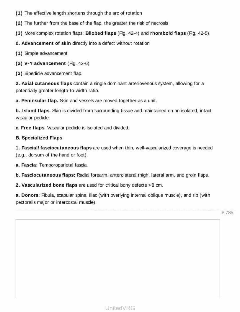
P.785
(1) The effective length shortens through the arc of rotation
(2) The further from the base of the flap, the greater the risk of necrosis
(3) More complex rotation flaps: Bilobed flaps (Fig. 42-4) and rhomboid flaps (Fig. 42-5).
d. Advancement of skin directly into a defect without rotation
(1) Simple advancement
(2) V-Y advancement (Fig. 42-6)
(3) Bipedicle advancement flap.
2. Axial cutaneous flaps contain a single dominant arteriovenous system, allowing for apotentially greater length-to-width ratio.
a. Peninsular flap. Skin and vessels are moved together as a unit.
b. Island flaps. Skin is divided from surrounding tissue and maintained on an isolated, intactvascular pedicle.
c. Free flaps. Vascular pedicle is isolated and divided.
B. Specialized Flaps
1. Fascial/fasciocutaneous flaps are used when thin, well-vascularized coverage is needed(e.g., dorsum of the hand or foot).
a. Fascia: Temporoparietal fascia.
b. Fasciocutaneous flaps: Radial forearm, anterolateral thigh, lateral arm, and groin flaps.
2. Vascularized bone flaps are used for critical bony defects >8 cm.
a. Donors: Fibula, scapular spine, iliac (with overlying internal oblique muscle), and rib (withpectoralis major or intercostal muscle).
UnitedVRG

Figure 42-2 Rotation flap. A: The edge of the flap is four to five times the length of thebase of the defect triangle. B, C: A backcut or Burow triangle can be useful if the flap isunder tension.

P.786
3. Functional muscle may be transferred with its accompanying dominant nerve.
a. Donors. Gracilis, latissimus dorsi.
IV. TISSUE EXPANSION.Tissue expansion uses an inflatable silicone balloon to serially expand surrounding skin. Duringexpansion, the dermis thins, collagen re-aligns, and the epidermis thickens. Common indicationsinclude burn alopecia, congenital nevi, and postmastectomy breast reconstruction.
UnitedVRG
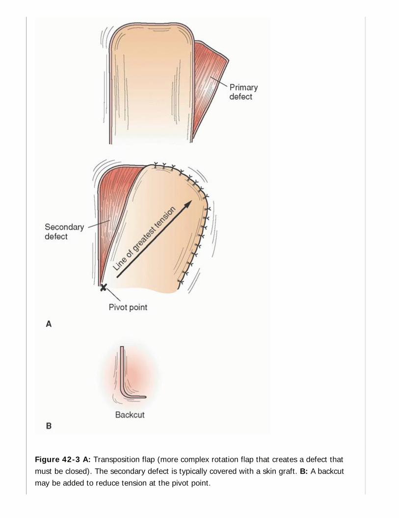
Figure 42-3 A: Transposition flap (more complex rotation flap that creates a defect thatmust be closed). The secondary defect is typically covered with a skin graft. B: A backcutmay be added to reduce tension at the pivot point.

P.787
UnitedVRG

P.788
Figure 42-4 Bilobed flap. After the lesion is excised, the primary flap (P) is transposed intothe initial defect, and the secondary flap (S) is moved to the site vacated by the primaryflap. The bed of the secondary flap is then closed primarily. The primary flap is slightlynarrower than the initial defect, whereas the secondary flap is half the width of the primaryflap. To be effective, this must be planned in an area where loose skin surrounds thesecondary flap site. Three choices for the secondary flap are shown (S1, S2, S3).
Figure 42-5 Rhomboid or Limberg. The rhomboid defect must have 60- and 120-degreeangles so that the length of the short diagonal is the same as the length of the sides. Theshort diagonal is extended by its own length to point E. The line EF is parallel to CD, andthey are equal in length. There are four possible Limberg flaps for any rhomboid defect; theflap should be planned in an area where loose skin is available to close the donor defectprimarily. The greatest tension will be between points F and D.
A. Advantages. Low donor-site morbidity, provision of donor tissue of similar color, texture,thickness, and sensation to the recipient tissue (like for like).
B. Disadvantages. Staged technique, there is a visible deformity during the period ofexpansion, frequent visits for expansion, and a relatively high rate of complications (infection,extrusion).

P.789
Figure 42-6 V-Y advancement. The skin to the sides of the V is advanced.
C. Technique Pearls
1. Goal of expanded surface area is 130% of the defect size.
2. Expander placement should be through an incision at the junction of the defect and the area ofproposed expansion, placed perpendicular to the direction of expansion.
3. Ports
a. External ports: Minimize anxiety and pain during filling, especially in the pediatric population.
b. Internal ports: Associated with reduced infection risk, commonly used when overlying skin isinsensate.
4. The expansion phase begins 2 to 3 weeks after expander placement.
a. Inflate weekly/bi-weekly using saline and sterile technique.
b. Amount infused depends on patient comfort, skin tension, and blanching of overlying skin.
c. Waiting 2 to 3 weeks after the desired volume is achieved allows the expanded skin to softenand decreases the contraction at the time of flap transposition.
ACUTE INJURIES
I. FACIAL TRAUMAA. Examination Pearls
1. C-spine. 10% of facial trauma patients have associated c-spine injury.
UnitedVRG

P.790
2. Oral examination. Malocclusion or limited mouth opening can indicate fractures; assess forloose teeth that are at risk for aspiration.
3. Facial nerve. Assess the five branches (temporal, zygomatic, buccal, marginal mandibular,cervical) by having the patient raise the eyebrows, squeeze the eyelids shut, smile, and frown,noting any paresis or asymmetry. Facial nerve injuries located lateral to the lateral canthus shouldbe explored and repaired within 72 hours; more medial injuries can be managed expectantly asthe facial nerve is highly arborized in the medial face.
4. Ocular examination
a. Assess extra-ocular movements; limited up-gaze may indicate inferior rectus entrapmentin an orbital fracture (acute indication for surgery).
b. Orbital compartment syndrome. Results from retrobulbar hematoma and requiresemergent lateral cantholysis. Look for proptosis, acute vision loss, and severe eye pain.
5. Nasal examination. Examine the nasal septum with an otoscope to assess for septalhematoma. Acute evacuation of the hematoma will prevent permanent deformity.
B. Imaging
1. CT with <3 mm cuts through the facial bones is a fast, sensitive, and specific means ofdetermining the location and orientation of facial fractures.
2. Panorex radiograph is useful in the setting of isolated mandible fractures or dental injuries.
C. Facial Hemorrhage. Nasal hemorrhage will generally cease with packing. Massivehemorrhage that does not stop with appropriate pressure is likely a branch of the external carotidand will require embolization.
D. Soft-tissue Repair
1. Local anesthesia may be used to block facial nerve branches
2. Copiously irrigate, debride devitalized tissue
3. Deep dermal closure: Interrupted resorbable suture
4. Epidermal closure: Running suture, generally better scarring with permanent monofilament
E. Fractures
1. Most can be repaired electively within 1 to 2 weeks
2. Exceptions: Airway compromise, ocular muscle entrapment
3. Indications for fixation
a. Mandible. Malocclusion

P.791
b. Orbital. Persistent diplopia, enophthalmos, extraocular muscle entrapment, hypoglobus
c. Midface. Cosmetic deformity, trismus/malocclusion
d. Nasal fracture. Cosmetic deformity, septal hematoma, airway obstruction
II. HAND TRAUMAA. Assessment Pearls
1. History. Hand dominance and patient occupation
2. Examination
a. Inspect the resting position of the patient's hand looking for swelling or asymmetrycompared with the contralateral hand.
b. Vascular assessment. Color, temperature, capillary refill, and the presence of pulses at thewrist and individual digits (palpable or Doppler)
(1) Allen test. Verifies integrity of the palmar arches. Have the patient make a fist, manuallyocclude the radial and ulnar arteries by applying pressure at the wrist, have the patient open theirhand and observe the color. Remove pressure from the radial artery: Color will return to the handif the superficial arch is intact. Repeat with the ulnar artery to assess the deep arch.
(2) Control bleeding with direct pressure. Tourniquets should be reserved for life-threatening exsanguinations only and should be placed as distally as possible.
c. Motor examination
(1) Flexor digitorum profundus (FDP) is tested by stabilizing the proximal interphalangeal(PIP) joint in extension and having the patient flex the distal interphalangeal (DIP) joint.
(2) Flexor digitorum superficialis (FDS) is tested by stabilizing all other fingers in fullextension and asking the patient to flex the finger being tested at the PIP joint.
(3) Extensor tendons. Place hand on a flat surface, palm down. Have patient lift each finger offthe surface individually.
TABLE 42-1 Unambiguous Tests of Hand NerveFunction
Test RadialNerve
Median Nerve Ulnar Nerve
UnitedVRG
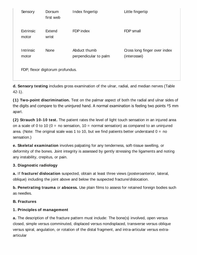
Sensory Dorsumfirst web
Index fingertip Little fingertip
Extrinsicmotor
Extendwrist
FDP index FDP small
Intrinsicmotor
None Abduct thumbperpendicular to palm
Cross long finger over index(interossei)
FDP, flexor digitorum profundus.
d. Sensory testing includes gross examination of the ulnar, radial, and median nerves (Table42-1).
(1) Two-point discrimination. Test on the palmar aspect of both the radial and ulnar sides ofthe digits and compare to the uninjured hand. A normal examination is feeling two points ≤5 mmapart.
(2) Strauch 10-10 test. The patient rates the level of light touch sensation in an injured areaon a scale of 0 to 10 (0 = no sensation, 10 = normal sensation) as compared to an uninjuredarea. (Note: The original scale was 1 to 10, but we find patients better understand 0 = nosensation.)
e. Skeletal examination involves palpating for any tenderness, soft-tissue swelling, ordeformity of the bones. Joint integrity is assessed by gently stressing the ligaments and notingany instability, crepitus, or pain.
3. Diagnostic radiology
a. If fracture/dislocation suspected, obtain at least three views (posteroanterior, lateral,oblique) including the joint above and below the suspected fracture/dislocation.
b. Penetrating trauma or abscess. Use plain films to assess for retained foreign bodies suchas needles.
B. Fractures
1. Principles of management
a. The description of the fracture pattern must include: The bone(s) involved, open versusclosed, simple versus comminuted, displaced versus nondisplaced, transverse versus obliqueversus spiral, angulation, or rotation of the distal fragment, and intra-articular versus extra-articular

P.792
b. Postreduction radiographs should be done for all fractures after splinting or casting.
c. Splinting the fracture
(1) Use plaster or fiberglass with appropriate padding; in the acute situation, the splint shouldnot be circumferential to allow for swelling.
(2) Positioning: ÒIntrinsic-plusÓ position may be used for almost all hand injuries. It placesligaments in their longest position. The interphalangeal (IP) joints are in full extension, themetacarpophalangeal (MCP) joints are at 60 to 90 degrees of flexion, and the wrist is in 20 to 30degrees of extension. The thumb should be abducted with the MCP extended.
(3) The splint should stabilize the joints above and below the fracture while avoidingimmobilization of the remainder of the patient's hand and wrist.
d. Indications for operative intervention. Reduction is not maintained after closed reduction,contaminated open fractures, associated soft-tissue injuries, malalignment (uncorrected rotated,angulated, or shortened deformities of the digit), and articular incongruity >1 mm.
e. Operative repair
(1) Closed-reduction, percutaneous pinning (CRPP) is used when a fracture is closed butreduction is inadequate or unstable. Reduction is performed under anesthesia with the aid offluoroscopy. Kirschner wires (K-wires) are placed to hold the reduction.
(2) Open reduction-internal fixation (ORIF). An incision is made over the fracture andplates/screws are used to hold the reduction. Although more invasive, fixation is rigid and allowsfor early active motion and passive range of motion, thus reducing stiffness.
(3) External fixation is used when significant contamination or soft tissue loss does not allowfor safe placement of permanent hardware.
C. Dislocations and ligament injuries should be assessed by stressing the periarticularstructures and ranging the joint. If instability is demonstrated, operative management should beconsidered. A stable joint is managed with protective splinting and early range-of-motionexercise. Perform neurovascular exam before and after manipulation.
D. Tendon Injuries
1. Assessment. Look for a change in the resting tone of the digits, which normally lie in a radial-to-ulnar cascade of flexion. Assess both flexor tendons and extensor tendons separately. Ifmovement against resistance elicits pain, suspect a partial laceration.
2. Flexor tendons are frequently lacerated during everyday activities.
a. Flexor tendon zones (Fig. 42-7)
(1) Zone I. At the DIP level, distal to the FDS insertion.
UnitedVRG
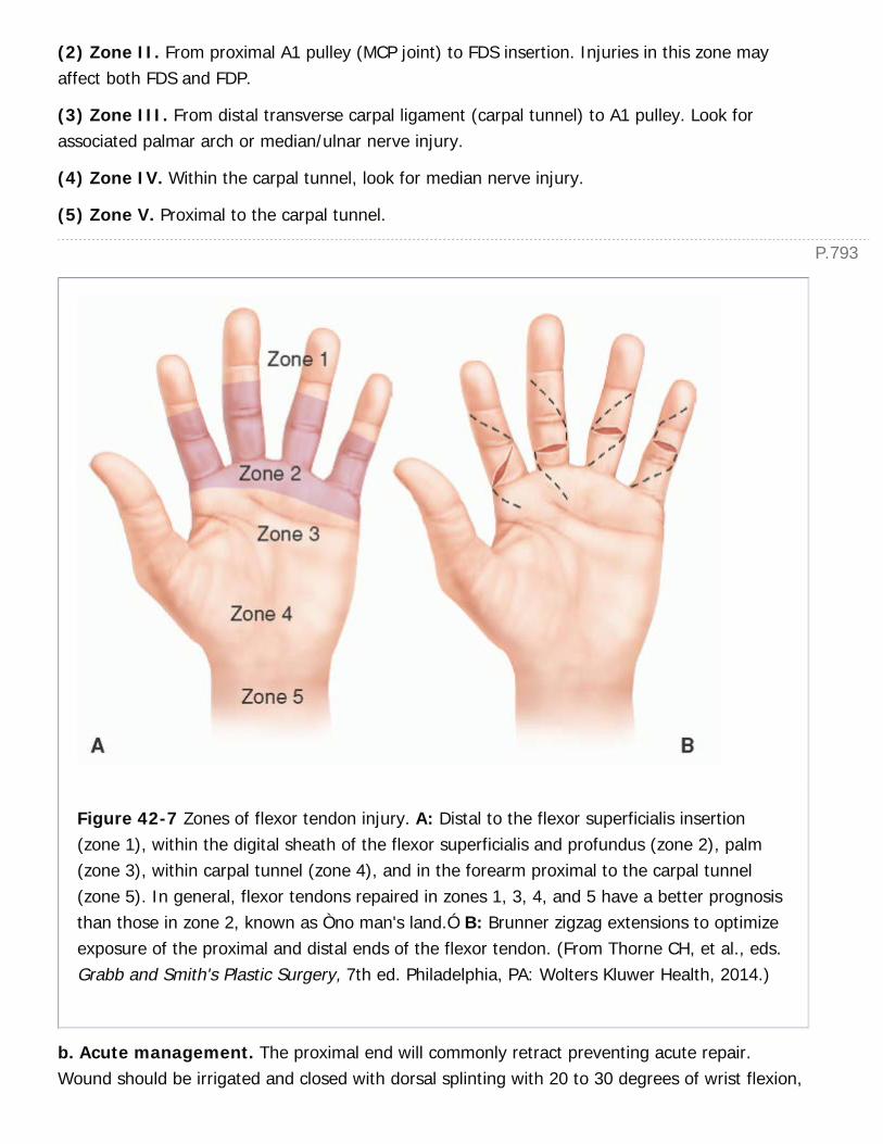
P.793
(2) Zone II. From proximal A1 pulley (MCP joint) to FDS insertion. Injuries in this zone mayaffect both FDS and FDP.
(3) Zone III. From distal transverse carpal ligament (carpal tunnel) to A1 pulley. Look forassociated palmar arch or median/ulnar nerve injury.
(4) Zone IV. Within the carpal tunnel, look for median nerve injury.
(5) Zone V. Proximal to the carpal tunnel.
Figure 42-7 Zones of flexor tendon injury. A: Distal to the flexor superficialis insertion(zone 1), within the digital sheath of the flexor superficialis and profundus (zone 2), palm(zone 3), within carpal tunnel (zone 4), and in the forearm proximal to the carpal tunnel(zone 5). In general, flexor tendons repaired in zones 1, 3, 4, and 5 have a better prognosisthan those in zone 2, known as Òno man's land.Ó B: Brunner zigzag extensions to optimizeexposure of the proximal and distal ends of the flexor tendon. (From Thorne CH, et al., eds.Grabb and Smith's Plastic Surgery, 7th ed. Philadelphia, PA: Wolters Kluwer Health, 2014.)
b. Acute management. The proximal end will commonly retract preventing acute repair.Wound should be irrigated and closed with dorsal splinting with 20 to 30 degrees of wrist flexion,

P.794
90-degree MCP flexion, and IP joints in extension. Repair within 10 days to avoid the need fortendon grafting.
c. Definitive repair involves a core, locking suture and an epitendinous repair. Injuries in zoneII to IV have a worse prognosis because of scarring and reduced tendon glide within the sheath.
3. Extensor tendon injuries result from lacerations and closed axial loading of the digits.
a. Extensor tendon zones
(1) Zone I. Over the DIP joint. Rupture of the terminal tendon secondary to forced flexionresults in Mallet finger, or inability to extend the DIP.
(2) Zone II. Over the middle phalanx.
(3) Zone III. Over the PIP joint. May injure the central slip and/or lateral bands and ifuntreated, can result in a boutonnière deformity (PIP flexion and DIP hyperextension).
(4) Zone IV. Over the proximal phalanx. The lacerations are often partial because of thewidth of the tendon at this level.
(5) Zone V. Over the MCP joint. Carefully explore to assess for sagittal band injury at thislevel as this must be repaired to prevent tendon subluxation. These injuries can occur secondaryto delivering a punch to the mouth (see Section Local infections > Human bites/fightbites).
b. Management. Can be repaired acutely as the tendons generally do not retract as much asflexors secondary to the juncturae tendinum, which are the narrow, fibrous bands of tissue thatpass obliquely between the diverging tendons of the extensor digitorum on the dorsal hand.Mallet injuries can be treated in a closed fashion with extension splinting of the DIP for 6 weeks.
4. Local infections
a. Management. Most finger infections heal well if all the purulence is drained and the wound isirrigated 2 to 3 times daily with soapy water and allowed to heal from the inside out. Splinting forcomfort and elevation to reduce edema are important adjuncts.
(1) Antibiotics may be needed if there is associated cellulitis, in the case of human/animal bites,and if there is involvement of the tendon sheath or joint space.
(2) Bites and joint injuries may require multiple washouts in the OR.
(3) Always obtain plain films prior to exploration to assess for foreign bodies (i.e., needles).
b. Paronychia is a localized infection of the skin and lateral nail fold, often due to nail biting orforeign-body penetration, such as a needlestick injury. Treatment requires incision and drainage(I&D), with removal of the nail when the infection extends deep to the nail plate. Chronicparonychia is sometimes associated with underlying osteomyelitis or fungal organisms (Fig. 42-
UnitedVRG

P.795
8).
c. Felon is a local infection of the finger pulp commonly due to a puncture wound. Adequatedrainage requires dividing all involved septae.
d. Cellulitis in the hand usually arises secondary to a laceration, abrasion, or other soft-tissueinjury. Management involves draining an abscess, if present, and treating with antibiotics.
e. In animal bites, the wound must be thoroughly irrigated to decrease the bacterial load and toremove any foreign body, such as a tooth. Bite wounds should be treated with oral antibioticsprophylactically and with intravenous antibiotics when an established infection is present.
f. Human bites/fight bites are most commonly associated with a punch to the mouth.Aggressive wound exploration, generally in the operating room, must be undertaken as the skinwill retract proximally and cover a deeper wound; 75% of fight bites result inbone/tendon/cartilage injury. Treat with intravenous (IV) antibiotics acutely. May require multiplewashouts.
E. Surgical Emergencies
1. Compartment syndrome in the hand and forearm results from increased pressure within anosseofascial space, leading to decreased
perfusion pressure. Left untreated, it will result in muscle and nerve ischemia and necrosis.
Figure 42-8 Acute paronychia. A: As seen in the emergency room. B: Incision is made
P.796
through the most fluctuant region. (From Thorne CH, et al., eds. Grabb and Smith's PlasticSurgery, 7th ed. Philadelphia, PA: Wolters Kluwer Health, 2014.)
a. Etiology. Fractures that cause bleeding, crush and vascular injuries, circumferential burns,bleeding dyscrasias, reperfusion after ischemia, or tight dressings.
b. Diagnosis. It is critical to have a high index of suspicion based on the type of injury. Painwith passive stretch of the compartment musculature and paresthesias are early indicators andshould be followed up aggressively. Paralysis and pulselessness occur later once the damage islikely irreversible. Measurement with a pressure monitor of a compartment pressure >30 mmHg confirms diagnosis.
c. Treatment of incipient compartment syndrome involves close observation, frequentexaminations, removal of tight casts and dressings, and elevation of the extremity above the levelof the heart. Acute suspected compartment syndrome requires urgent fasciotomies within 6 hoursto prevent irreversible muscle ischemia.
2. Suppurative tenosynovitis is infection of the flexor tendon sheath, usually due to apuncture wound to the volar aspect of the digit or palm.
a. Diagnosis: Cardinal signs of Kanavel
(1) Finger held in flexion
(2) Fusiform swelling of the finger
(3) Tenderness along the tendon sheath
(4) Pain on passive extension
b. Management involves urgent I&D in the operating room and IV antibiotics.
3. Palmar abscess is usually associated with a puncture wound. The fascia divides the palm intothenar, midpalmar, and hypothenar spaces; each involved space must be incised and drained.
4. Necrotizing infections threaten both limb and life. Aggressive and repeated surgicaldebridement, broad spectrum antibiotics, and supportive management are the mainstays oftreatment.
5. High-pressure injection injuries result from grease or paint injected at up to 10,000 lb/in2.Although the external wounds are often small and unassuming, deep-tissue injury can be severe.Management involves urgent, thorough debridement, irrigation, decompression, systemicantibiotics, and splinting.
GENERAL RECONSTRUCTIVE PLASTIC SURGERY
UnitedVRG
P.797
I. SCALP, CALVARIAL, AND FOREHEAD RECONSTRUCTIONA. Scalp Layers. Skin, subcutaneous tissue, galea aponeurotica, loose areolar tissue, andpericranium.
B. Scalp Lacerations
1. Often associated with blunt head trauma: Assess for associated skull, cervical spine, orintracranial injuries.
2. The rich blood supply to the scalp can produce significant blood loss so expedient managementis critical. Hemostasis is achieved with closure of the galea and skin following thorough irrigationand debridement of devitalized tissue. Stapled closure of the hair-bearing scalp is expedient andresults in reduced trauma to hair follicles compared to sutures.
C. Partial-thickness scalp loss from avulsion usually occurs at the subaponeurotic layer. Largeavulsions may be skin grafted acutely. Definitive management often requires tissue expanderplacement to replace hair-bearing scalp.
D. Full-thickness scalp loss can occur from trauma or tumor extirpation. The optimaltreatment varies depending on the size of the defect.
1. Small defects (<3 cm) can often be closed primarily after undermining of flaps.
2. Medium-sized defects (3 to 10 cm) can be covered with a skin graft or skin substitute (e.g.,Integra) by burring the bone down to the vascular diploic space or using a scalp flap combinedwith skin grafting of the donor pericranium. Tissue expansion can later be used to replace hair-bearing scalp.
3. Large defects (>10 cm) often require free tissue transfer with omentum or latissimus dorsi(Fig. 42-9).
II. TRUNKA. Breast
1. Postmastectomy breast reconstruction is an important part of recovery for many womenand can lead to a significant improvement in body image (J Natl Cancer Inst. 2000;92:1422-1429).
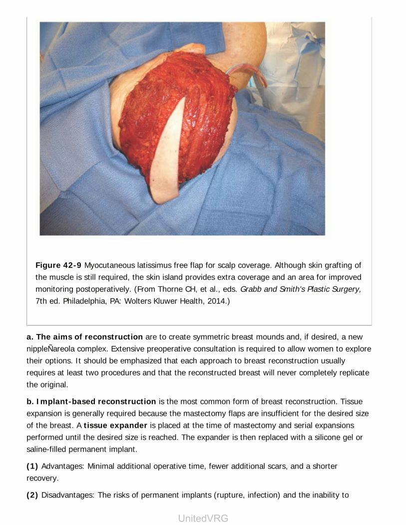
Figure 42-9 Myocutaneous latissimus free flap for scalp coverage. Although skin grafting ofthe muscle is still required, the skin island provides extra coverage and an area for improvedmonitoring postoperatively. (From Thorne CH, et al., eds. Grabb and Smith's Plastic Surgery,7th ed. Philadelphia, PA: Wolters Kluwer Health, 2014.)
a. The aims of reconstruction are to create symmetric breast mounds and, if desired, a newnippleÑareola complex. Extensive preoperative consultation is required to allow women to exploretheir options. It should be emphasized that each approach to breast reconstruction usuallyrequires at least two procedures and that the reconstructed breast will never completely replicatethe original.
b. Implant-based reconstruction is the most common form of breast reconstruction. Tissueexpansion is generally required because the mastectomy flaps are insufficient for the desired sizeof the breast. A tissue expander is placed at the time of mastectomy and serial expansionsperformed until the desired size is reached. The expander is then replaced with a silicone gel orsaline-filled permanent implant.
(1) Advantages: Minimal additional operative time, fewer additional scars, and a shorterrecovery.
(2) Disadvantages: The risks of permanent implants (rupture, infection) and the inability to
UnitedVRG

P.798
reproduce certain natural contours.
(3) Anaplastic large cell lymphoma (ALCL) is an extremely rare malignancy associated withimplants (<1/1,000,000). Most commonly found in association with a late-onset seroma.Diagnosis is via seroma fluid cytology.
c. Autologous tissue can be used to recreate a breast mound in the form of pedicled (rectusabdominis, latissimus dorsi) or free (rectus abdominis, gluteus maximus)myocutaneous flaps.

Figure 42-10 Chest wall reconstruction algorithm.
UnitedVRG
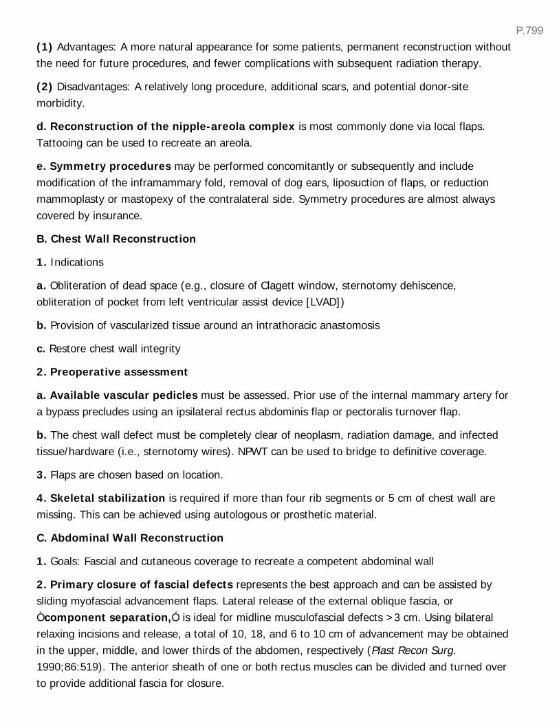
P.799(1) Advantages: A more natural appearance for some patients, permanent reconstruction withoutthe need for future procedures, and fewer complications with subsequent radiation therapy.
(2) Disadvantages: A relatively long procedure, additional scars, and potential donor-sitemorbidity.
d. Reconstruction of the nipple-areola complex is most commonly done via local flaps.Tattooing can be used to recreate an areola.
e. Symmetry procedures may be performed concomitantly or subsequently and includemodification of the inframammary fold, removal of dog ears, liposuction of flaps, or reductionmammoplasty or mastopexy of the contralateral side. Symmetry procedures are almost alwayscovered by insurance.
B. Chest Wall Reconstruction
1. Indications
a. Obliteration of dead space (e.g., closure of Clagett window, sternotomy dehiscence,obliteration of pocket from left ventricular assist device [LVAD])
b. Provision of vascularized tissue around an intrathoracic anastomosis
c. Restore chest wall integrity
2. Preoperative assessment
a. Available vascular pedicles must be assessed. Prior use of the internal mammary artery fora bypass precludes using an ipsilateral rectus abdominis flap or pectoralis turnover flap.
b. The chest wall defect must be completely clear of neoplasm, radiation damage, and infectedtissue/hardware (i.e., sternotomy wires). NPWT can be used to bridge to definitive coverage.
3. Flaps are chosen based on location.
4. Skeletal stabilization is required if more than four rib segments or 5 cm of chest wall aremissing. This can be achieved using autologous or prosthetic material.
C. Abdominal Wall Reconstruction
1. Goals: Fascial and cutaneous coverage to recreate a competent abdominal wall
2. Primary closure of fascial defects represents the best approach and can be assisted bysliding myofascial advancement flaps. Lateral release of the external oblique fascia, orÒcomponent separation,Ó is ideal for midline musculofascial defects >3 cm. Using bilateralrelaxing incisions and release, a total of 10, 18, and 6 to 10 cm of advancement may be obtainedin the upper, middle, and lower thirds of the abdomen, respectively (Plast Recon Surg.1990;86:519). The anterior sheath of one or both rectus muscles can be divided and turned overto provide additional fascia for closure.
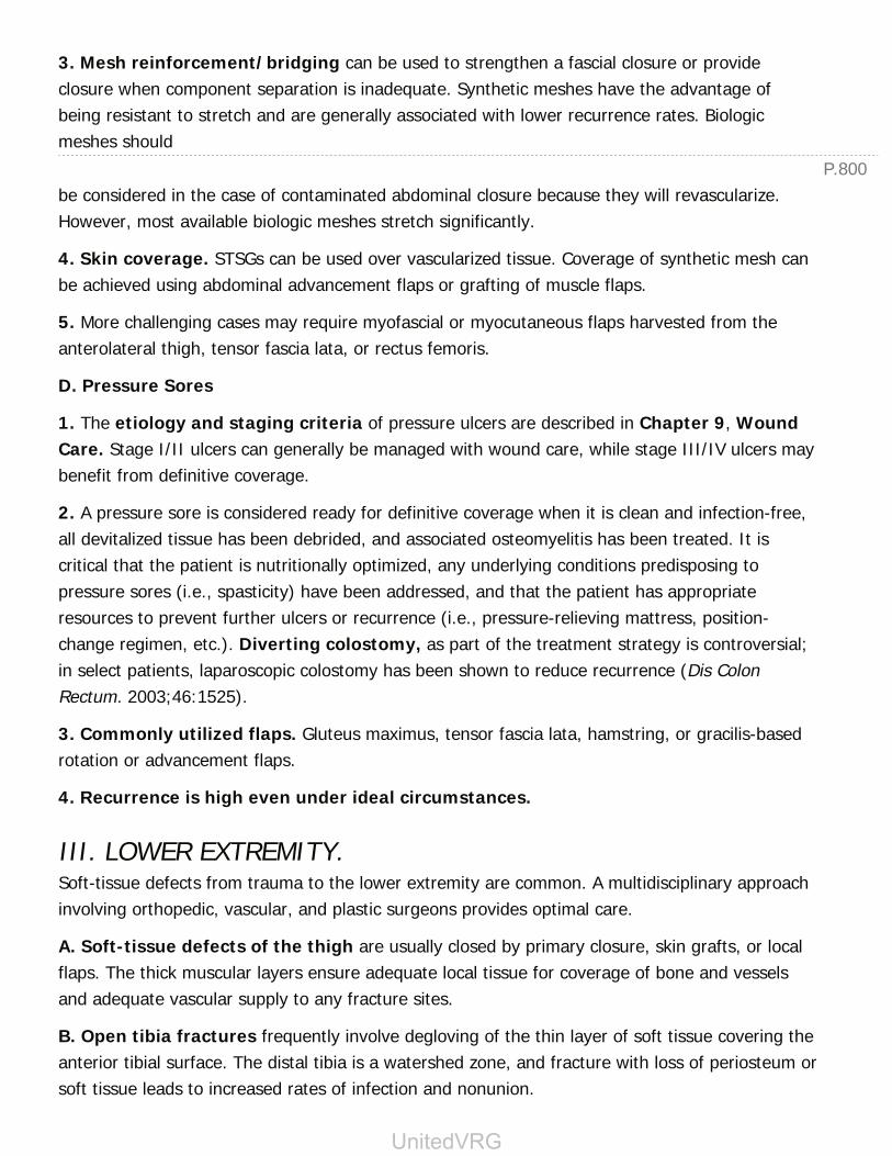
P.800
3. Mesh reinforcement/bridging can be used to strengthen a fascial closure or provideclosure when component separation is inadequate. Synthetic meshes have the advantage ofbeing resistant to stretch and are generally associated with lower recurrence rates. Biologicmeshes should
be considered in the case of contaminated abdominal closure because they will revascularize.However, most available biologic meshes stretch significantly.
4. Skin coverage. STSGs can be used over vascularized tissue. Coverage of synthetic mesh canbe achieved using abdominal advancement flaps or grafting of muscle flaps.
5. More challenging cases may require myofascial or myocutaneous flaps harvested from theanterolateral thigh, tensor fascia lata, or rectus femoris.
D. Pressure Sores
1. The etiology and staging criteria of pressure ulcers are described in Chapter 9, WoundCare. Stage I/II ulcers can generally be managed with wound care, while stage III/IV ulcers maybenefit from definitive coverage.
2. A pressure sore is considered ready for definitive coverage when it is clean and infection-free,all devitalized tissue has been debrided, and associated osteomyelitis has been treated. It iscritical that the patient is nutritionally optimized, any underlying conditions predisposing topressure sores (i.e., spasticity) have been addressed, and that the patient has appropriateresources to prevent further ulcers or recurrence (i.e., pressure-relieving mattress, position-change regimen, etc.). Diverting colostomy, as part of the treatment strategy is controversial;in select patients, laparoscopic colostomy has been shown to reduce recurrence (Dis ColonRectum. 2003;46:1525).
3. Commonly utilized flaps. Gluteus maximus, tensor fascia lata, hamstring, or gracilis-basedrotation or advancement flaps.
4. Recurrence is high even under ideal circumstances.
III. LOWER EXTREMITY.Soft-tissue defects from trauma to the lower extremity are common. A multidisciplinary approachinvolving orthopedic, vascular, and plastic surgeons provides optimal care.
A. Soft-tissue defects of the thigh are usually closed by primary closure, skin grafts, or localflaps. The thick muscular layers ensure adequate local tissue for coverage of bone and vesselsand adequate vascular supply to any fracture sites.
B. Open tibia fractures frequently involve degloving of the thin layer of soft tissue covering theanterior tibial surface. The distal tibia is a watershed zone, and fracture with loss of periosteum orsoft tissue leads to increased rates of infection and nonunion.
UnitedVRG
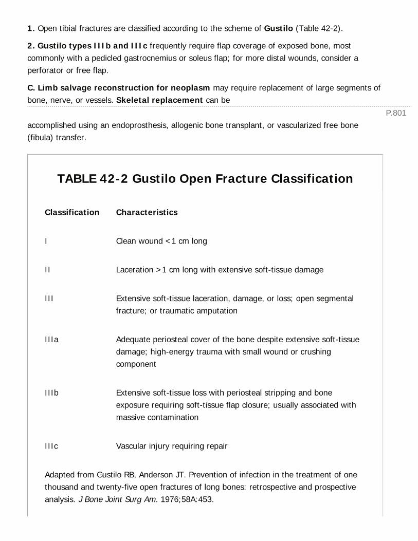
P.801
1. Open tibial fractures are classified according to the scheme of Gustilo (Table 42-2).
2. Gustilo types IIIb and IIIc frequently require flap coverage of exposed bone, mostcommonly with a pedicled gastrocnemius or soleus flap; for more distal wounds, consider aperforator or free flap.
C. Limb salvage reconstruction for neoplasm may require replacement of large segments ofbone, nerve, or vessels. Skeletal replacement can be
accomplished using an endoprosthesis, allogenic bone transplant, or vascularized free bone(fibula) transfer.
TABLE 42-2 Gustilo Open Fracture Classification
Classification Characteristics
I Clean wound <1 cm long
II Laceration >1 cm long with extensive soft-tissue damage
III Extensive soft-tissue laceration, damage, or loss; open segmentalfracture; or traumatic amputation
IIIa Adequate periosteal cover of the bone despite extensive soft-tissuedamage; high-energy trauma with small wound or crushingcomponent
IIIb Extensive soft-tissue loss with periosteal stripping and boneexposure requiring soft-tissue flap closure; usually associated withmassive contamination
IIIc Vascular injury requiring repair
Adapted from Gustilo RB, Anderson JT. Prevention of infection in the treatment of onethousand and twenty-five open fractures of long bones: retrospective and prospectiveanalysis. J Bone Joint Surg Am. 1976;58A:453.
P.802
D. Foot wound coverage may be necessary due to trauma, neoplasm, or ulceration. The etiologyof ulcers (e.g., arterial insufficiency requiring extremity revascularization in the case of ischemiculcers) should be addressed prior to coverage.
1. Plantar surface. Optimal coverage of the plantar surface provides a durable, sensateplatform; options include dermal substitutes, rotation of instep-tissue for small wounds, or freeflaps.
2. Dorsal foot. Coverage should be thin to allow for standard footwear. In the presence ofperitenon, a skin graft can be used; otherwise, consider a dermal substitute or thin fascial freeflaps (temporoparietal, parascapular, or radial forearm) covered by skin grafts.
IV. PERIPHERAL NERVEA. Clinical assessment of neuropathy requires evaluation of both motor and sensory functionas well as electrodiagnostic evaluation of nerve conduction and muscle innervation.
1. Evaluation of motor nerve function is standardized (Table 42-3).
2. Diagnostic studies
a. Nerve conduction studies (NCS) characterizes the conduction of large-diameter,myelinated nerves and can help determine the degree
of nerve dysfunction, the presence of segmental demyelination or axonal degeneration, the site ofinjury, and whether the injury is unifocal, multifocal, or diffuse.
TABLE 42-3 Classification of Motor Function
Grade Motor Function
M0 No contraction
M1 Perceptible contraction in proximal muscles
M2 Perceptible contraction in proximal and distal muscles
M3 All important muscles powerful enough to act against gravity
UnitedVRG

P.803
M4 Muscles act against strong resistance; some independent movement possible
M5 Normal strength and function
Adapted from Mackinnon SE, Dellon AL. Surgery of the Peripheral Nerve. New York, NY:Thieme, 1988:118.
b. Electromyography (EMG) samples the action potentials from muscle fibers. Motor unitpotentials (MUPs) can indicate early reinnervation, while fibrillations may represent denervation.
3. Nerve injuries are classified according to severity (Table 42-4).
B. Management of Acute Injuries
1. Sharp transection (trauma, iatrogenic). Nerve should be repaired urgently.
a. Debride injured nerve to healthy fascicles.
b. If possible, a tension-free primary repair should be performed.
c. If gap is too large for tension free repair, consider grafting.
2. Acute postoperative nerve deficit unrelated to location of surgery
a. Commonly due to patient positioning or traction, especially at anatomically restrictivesites (e.g., the ulnar nerve at the elbow or the common peroneal nerve at the knee).
b. Usually a first-, second-, or third-degree injury, and full recovery can be expected in mostcases.
c. If no improvement by 6 weeks, obtain baseline NCS and EMG. If there is no evidence of returnof function at 3 months, obtain repeat studies.
(1) If MUPs are present but there are reduced conduction velocities at known compressionpoints, decompression of the nerve will aid recovery.
(2) If there are no MUPS on EMG, the nerve should be explored and repaired.
3. Loss of nerve function after gunshot or open blunt trauma is usually the result of first-or second-degree injury.
TABLE 42-4 Classification of Nerve Injuries

Sunderlanda Seddonb Structure Injured Prognosis
First degree Neurapraxia Schwann cell(demyelination)
Complete recovery within12 wks
Seconddegree
Axonotmesis Axon (Walleriandegeneration)
Complete recoveryregeneration 1 mm/day
Third degree Endoneurium Incomplete recovery
Fourth degree Perineurium No recovery
Fifth degree Neurotmesis Epineurium No recovery
Sixth degree Mixed injury,neuroma
incontinuityc
Unpredictable recovery
a Sunderland S. A classification of peripheral nerve injuries producing loss of function.Brain. 1951;74:491.b Seddon HJ. Three types of nerve injury. Brain. 1943;66:237.c Mackinnon SE. New direction in peripheral nerve surgery. Ann Plast Surg.1989;22(3):257-273.
a. Treat as a closed injury, observing for improvement and obtaining EMG and NCS at 6 weeksand 3 months, as needed.
b. If the nerve is visible or the wound is explored for other reasons (e.g., vascular repair), thenerve is explored. If the nerve is in continuity, manage as one would for a closed injury. If thenerve is not in continuity, it is usually best to tag the ends of the nerve for ease of identificationand delay definitive repair until the zone of injury to the nerve is clearer (generally by 3 weeks).
4. Nerve deficit from compartment syndrome is treated by emergent fasciotomy. Ifdecompressed early (within 6 hours), there is usually a rapid return of function.
C. Compression neuropathy due to compression or repetitive trauma is a common clinicalproblem. Typically involved nerves include the median nerve at the wrist (carpal tunnelsyndrome), the ulnar nerve at the elbow or wrist (cubital tunnel syndrome), the anterior or

P.804
posterior interosseous nerves in the forearm, the brachial plexus at the thoracic outlet(neurogenic thoracic outlet syndrome), the common peroneal nerve at the knee, and theposterior tibial nerve at the ankle (tarsal tunnel syndrome).
1. Initial management is usually physical therapy, behavior modification, and splinting to avoidrepetitive compression for at least 6 weeks.
2. If nonsurgical intervention fails, open decompression and transposition is indicated.
AESTHETIC SURGERY PEARLS FOR THE GENERALSURGEON
I. AUGMENTATION MAMMOPLASTYA. Implants
1. Placement. Subglandular, submuscular or a combination (dual plane) via periareolar,transaxillary, or inframammary fold incisions
2. Monitoring
a. Cancer screening. Use standard mammography plus Eklund views (pushes implantagainst the chest wall and breast tissue is pulled forward)
b. Rupture. Saline implant rupture presents as a deflated breast. Silicone implants must bemonitored by MRI with an initial study 3 years after placement and every 2 years thereafter.
B. Fat transfer may be used in large volume to recreate a whole breast or to fill the upper polefollowing implant placement, most commonly in the case of postmastectomy reconstruction.
1. Fat necrosis on standard mammography requires careful examination to determine if furtherinvestigation to rule out cancer is needed. Most commonly appear as scattered microcalcificationson mammography or oil cysts on gross inspection. Generally, it is a late finding occurring monthsto years postprocedure.
II. ABDOMINOPLASTYA. Involves elevation of large abdominal flaps extending from umbilicus to xiphoid, transpositionof the umbilicus, with or without rectus plication. Blood supply to the flaps comes from lateralintercostals, subcostals, and lumbar vessels. Large abdominal scars (e.g., subcostal incision afteropen cholecystectomy) can result in flap necrosis.
B. Risk of pulmonary embolism is between 1/300 and 1/1000, with the highest risk periodbeing the first postoperative week. Accordingly, patients are commonly discharged on 5 to 10days of anticoagulation therapy.
P.805
III. POSTBARIATRIC BODY CONTOURINGA. Generally not covered by insurance.
B. Timing. Weight should be stable for at least 3 to 4 months; generally, this occurs 12 to 18months after a bariatric operation. The lower the BMI, the lower the risk of complications and thebetter the cosmetic outcome.
C. Patients commonly have nutritional deficiencies that must be corrected before further surgery:The most common is low iron, but it is also worthwhile to check levels of cyanocobalamin(vitamin B12), calcium, folate, and albumin.
CHAPTER 42: PLASTIC AND HAND SURGERYMultiple Choice Questions1. An 83-year-old man undergoes excision of a basal cell carcinoma ofthe cheek. The defect is 3 × 3 cm with subcutaneous fat exposed at thebase. Reconstruction with skin grafting is planned. Which of thefollowing will minimize long-term graft contracture?
a. Split thickness skin grafting with meshing
b. Split thickness skin grafting without meshing
c. Full thickness skin grafting
d. Cultured epidermal autografting
e. A skin graft is inappropriate in this situation
View Answer
2. A 23-year-old man is brought to the trauma bay by emergencyservices following a motor vehicle accident. He has multiple foreheadlacerations, bilateral orbital ecchymoses and reports he cannot see outof his right eye. What is the first step in management?
a. Maxillofacial CT
b. Intraocular examination
c. Assessment of pupillary response
d. Completion of the primary survey
e. Immediate lateral cantholysis
View Answer
3. Following an altercation at a bar, a 20-year-old male presents with acut over the third metacarpal head on the dorsum of his right hand. Onphysical examination he has full range of motion but reports pain on

P.806
extending his middle finger. Radiographs show no fracture. To minimizethis patient's risk of metacarpal joint destruction, which of thefollowing is most appropriate?
a. Wound exploration and wash-out in the operating room
b. Irrigate wound thoroughly and allow to heal by secondary intention
c. Begin oral antibiotics immediately
d. Instruct the patient in range of motion exercises
e. Obtain an MRI of the hand
View Answer
4. A 46-year-old female is brought to the emergency room by herhusband. He reports she had an abdominoplasty at an ambulatorysurgery center 3 days ago. The patient appears confused and lethargic.On examination, she is hypotensive and tachycardic and pulse oximetryon room air is 98%. Her abdomen is tender, incisions are intact, anddressings are clean. Her drains contain sanguineous fluid and a fewclots. The husband is not sure when the drains were last emptied.Following initial resuscitation and stabilization, which of the followingstudies is most appropriate?
a. Spiral CT of the chest
b. Type and cross
c. Lower-extremity duplex ultrasound
d. Chest x-ray
e. Urine drug screen
View Answer
5. A polytrauma patient has a thigh wound measuring 15 × 23 cm.There is heavy dirt and gravel contamination and frayed tendon andmuscle at the wound base. An external fixator has been placed on theassociated femur fracture. Following the initial wash-out anddebridement, what is the most appropriate therapy?
a. “Wet to dry” wound care
b. Negative pressure wound therapy
c. Split thickness skin grafting
d. Rotation flap coverage
e. Free flap

P.807
View Answer
6. A 56-year-old male with a history of bladder cancer and pelvicradiation, was found to have metastases in the previous incision,abdominal wall, and bladder dome. Urology has planned full-thicknessabdominal wall resection and radical cystectomy. The abdominal walldefect is 12 × 12 cm in the lower abdomen and includes portions of thebilateral rectus abdominis. Which of the following options will providethe best abdominal wall reconstruction?
a. Vicryl mesh
b. Component separation
c. Component separation with split thickness skin graft
d. Pedicled anterolateral thigh fasciocutaneous flaps
e. Free latissimus dorsi muscle flap with split thickness skin graft
View Answer
7. A 76-year-old male underwent coronary artery bypass surgery usingthe left internal mammary artery and left saphenous vein for grafts. Hiscourse was complicated by mediastinitis and sternal dehiscence with aresultant 3 × 7 cm central chest defect with exposed sternum.Following multiple debridements and negative pressure wound therapy,wound cultures are negative. Which of the following is mostappropriate for coverage?
a. Skin graft
b. Left pedicled rectus abdominis flap with skin graft
c. Left turn-over pectoralis flap with skin graft
d. Pedicled omentum with skin graft
e. Free anterolateral thigh flap
View Answer
8. A 20-year-old suffered multiple gunshot wounds to the chest,abdomen, and right arm. He is stabilized and able to participate in anupperextremity examination 1 week following presentation. Onexamination, he is unable to extend his wrist, fingers, and thumb. Whatis the most appropriate management of this patient?
a. Obtain electromyography and nerve conduction studies, explore nerveif evidence of denervation
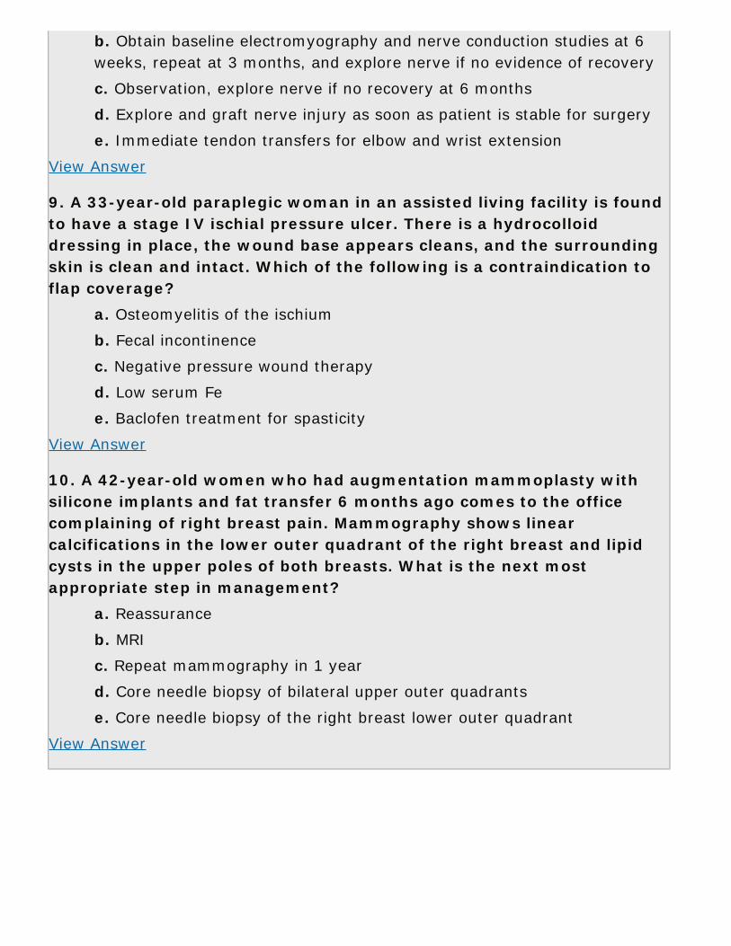
b. Obtain baseline electromyography and nerve conduction studies at 6weeks, repeat at 3 months, and explore nerve if no evidence of recovery
c. Observation, explore nerve if no recovery at 6 months
d. Explore and graft nerve injury as soon as patient is stable for surgery
e. Immediate tendon transfers for elbow and wrist extension
View Answer
9. A 33-year-old paraplegic woman in an assisted living facility is foundto have a stage IV ischial pressure ulcer. There is a hydrocolloiddressing in place, the wound base appears cleans, and the surroundingskin is clean and intact. Which of the following is a contraindication toflap coverage?
a. Osteomyelitis of the ischium
b. Fecal incontinence
c. Negative pressure wound therapy
d. Low serum Fe
e. Baclofen treatment for spasticity
View Answer
10. A 42-year-old women who had augmentation mammoplasty withsilicone implants and fat transfer 6 months ago comes to the officecomplaining of right breast pain. Mammography shows linearcalcifications in the lower outer quadrant of the right breast and lipidcysts in the upper poles of both breasts. What is the next mostappropriate step in management?
a. Reassurance
b. MRI
c. Repeat mammography in 1 year
d. Core needle biopsy of bilateral upper outer quadrants
e. Core needle biopsy of the right breast lower outer quadrant
View Answer
43Urology for the General Surgeon
Jennifer A. Robles
Alana C. Desai
The discipline of urologic surgery encompasses the management of benign and malignantconditions of the genitourinary system including the kidneys, ureters, bladder, urethra, and themale external genitalia.
I. HEMATURIA.Hematuria or blood in the urine, warrants a complete urologic workup. Gross hematuria isvisibly bloody urine, whereas microscopic hematuria is defined as 3 or more red blood cellsper high-power field. Common etiologies include: Urinary tract infection (UTI), stones, benignprostatic hypertrophy, malignancy, and recent trauma or instrumentation.
A. Evaluation for hematuria (gross or microscopic) consists of the following:
1. Urinalysis and urine culture from a freshly voided mid-stream specimen. Cytology shouldalso be obtained for gross hematuria.
2. Computed tomography (CT) urogram: Consisting of a three-phase CT of theabdomen/pelvis without contrast, with contrast, and with delayed contrast imagesÑis thepreferred imaging modality. However, magnetic resonance (MR) urogram and renal ultrasoundwith retrograde pyelograms are adequate in appropriately selected patients.
3. Cystoscopy is required for a complete evaluation of the urethra and bladder as imaging canmiss small lesions or strictures.
4. If the etiology of hematuria remains unclear, repeat a UA in 1 year.
a. For recurrent gross hematuria: Annual complete workup.
b. For recurrent microscopic hematuria, repeat workup Q3Ñ5 years.
B. Treatment of Symptomatic Gross Hematuria
1. Patients in urinary retention due to clots require urgent urologic consultation and a 22F to24F three-way Foley catheter. The catheter should be manually irrigated and aspirated withsterile saline or water until the bladder is clot-free. Continuous bladder irrigation (CBI) shouldnot be initiated until the bladder is clot free.
> Table of Contents > 43 - Urology for the General Surgeon

P.809
2. Persistent gross hematuria from a lower urinary tract source (bladder, prostate, urethra)despite conservative measures requires operative management via cystoscopy and fulguration ofthe bleeding source.
3. Persistent hematuria from an upper urinary tract source (kidney, ureter) may be due to ahemorrhagic renal mass, angiomyolipoma, arteriovenous fistula, or renal trauma.These patients may require surgical intervention by urology or angiography ± embolization byinterventional radiology.
TABLE 43-1 Bosniak Classification System of RenalCystic Masses
BosniakCategory Characteristics
Risk ofMalignancy Management
I Simple cyst. Thin wall, no septa.Low attenuation, noenhancement.
0% None
II Few thin septa or finecalcifications. Can be highattenuation (if <3 cm) but noenhancement.
5-18.5%a None
IIF Multiple thin septa, minimal wallthickening or thick calcifications.Can be high attenuation (>3 cm)but no enhancement.
15-25% Surveillanceimaging Q6-12mo
III Walls or septa with measurableenhancement
33+% Resection/ablation
IV Contains enhancing soft tissuecomponents
92.5% Resection/ablation
a Note: There is limited data on Category II cysts as most studies combine II and IIF
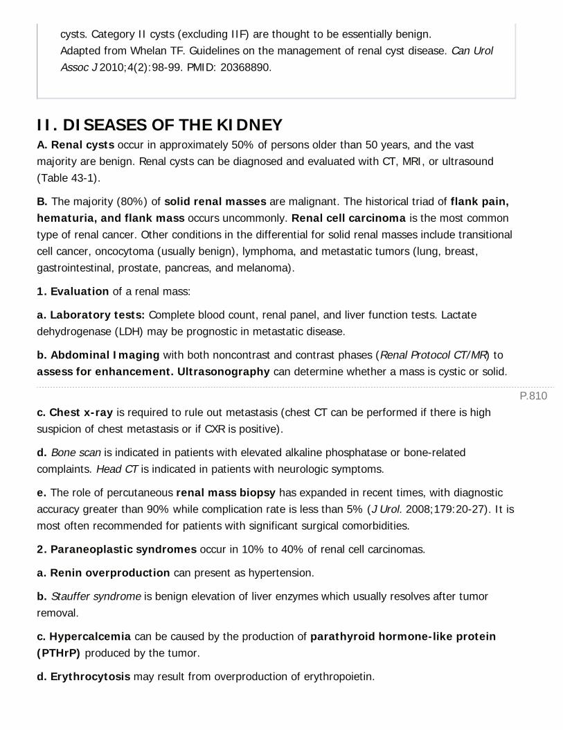
P.810
cysts. Category II cysts (excluding IIF) are thought to be essentially benign.Adapted from Whelan TF. Guidelines on the management of renal cyst disease. Can UrolAssoc J 2010;4(2):98-99. PMID: 20368890.
II. DISEASES OF THE KIDNEYA. Renal cysts occur in approximately 50% of persons older than 50 years, and the vastmajority are benign. Renal cysts can be diagnosed and evaluated with CT, MRI, or ultrasound(Table 43-1).
B. The majority (80%) of solid renal masses are malignant. The historical triad of flank pain,hematuria, and flank mass occurs uncommonly. Renal cell carcinoma is the most commontype of renal cancer. Other conditions in the differential for solid renal masses include transitionalcell cancer, oncocytoma (usually benign), lymphoma, and metastatic tumors (lung, breast,gastrointestinal, prostate, pancreas, and melanoma).
1. Evaluation of a renal mass:
a. Laboratory tests: Complete blood count, renal panel, and liver function tests. Lactatedehydrogenase (LDH) may be prognostic in metastatic disease.
b. Abdominal Imaging with both noncontrast and contrast phases (Renal Protocol CT/MR) toassess for enhancement. Ultrasonography can determine whether a mass is cystic or solid.
c. Chest x-ray is required to rule out metastasis (chest CT can be performed if there is highsuspicion of chest metastasis or if CXR is positive).
d. Bone scan is indicated in patients with elevated alkaline phosphatase or bone-relatedcomplaints. Head CT is indicated in patients with neurologic symptoms.
e. The role of percutaneous renal mass biopsy has expanded in recent times, with diagnosticaccuracy greater than 90% while complication rate is less than 5% (J Urol. 2008;179:20-27). It ismost often recommended for patients with significant surgical comorbidities.
2. Paraneoplastic syndromes occur in 10% to 40% of renal cell carcinomas.
a. Renin overproduction can present as hypertension.
b. Stauffer syndrome is benign elevation of liver enzymes which usually resolves after tumorremoval.
c. Hypercalcemia can be caused by the production of parathyroid hormone-like protein(PTHrP) produced by the tumor.
d. Erythrocytosis may result from overproduction of erythropoietin.

P.811
C. Management of Renal Masses
1. The management of solid renal masses depends on the tumor stage (Table 43-2).
a. T1 and T2 lesions. Nephron-sparing surgery via Partial Nephrectomy is preferred wheneverpossible; otherwise, radical nephrectomy is considered the standard of care.
b. T3 and T4 lesions mandate radical nephrectomy.
2. Metastatic renal cell carcinoma is resistant to radiation and chemotherapy. It isunclear how beneficial cytoreductive nephrectomy is in the modern era; studies are pending.Targeted therapy with agents such as tyrosine kinase inhibitors, VEGF inhibitors, as well as IL-2 immunotherapy has shown survival benefit in some patients (BMC Cancer. 2009;9:34).
III. DISEASES OF THE URETERA. Hydronephrosis is commonly caused by ureteral obstruction, which can be intrinsic orextrinsic in origin.
1. Common intrinsic causes include stones, ureteropelvic junction obstruction, stricture, andurothelial carcinoma. Common extrinsic causes include compression from a crossing vessel,pregnancy, abdominal mass, or retroperitoneal fibrosis.
2. Short-term management includes placement of a ureteral stent or percutaneousnephrostomy tube to relieve obstruction. Long-term management is based on the etiology.
3. The best imaging modality depends on the suspected etiology. Ultrasound and noncontrastCT are best to evaluate urolithiasis (see below). Intrinsic compression is best defined by CT/MRurogram. CT/MR with contrast can distinguish most forms of extrinsic compression.
TABLE 43-2 AJCC 2010 TNM Staging for Renal CellCarcinoma
Primary Tumor (T)
Tx Primary tumor cannot be assessed
T0 No evidence of primary tumor
T1 Tumor 7 cm or less in greatest dimension, limited to kidney
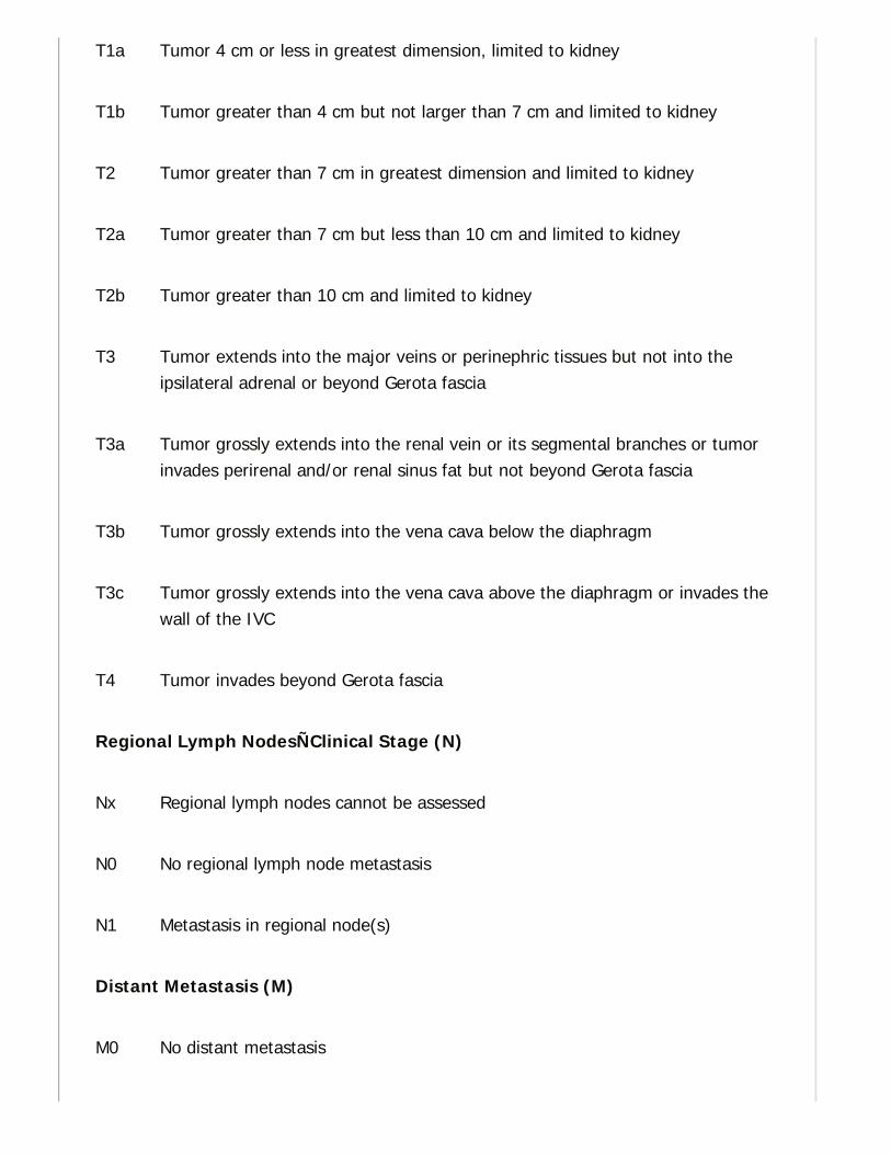
T1a Tumor 4 cm or less in greatest dimension, limited to kidney
T1b Tumor greater than 4 cm but not larger than 7 cm and limited to kidney
T2 Tumor greater than 7 cm in greatest dimension and limited to kidney
T2a Tumor greater than 7 cm but less than 10 cm and limited to kidney
T2b Tumor greater than 10 cm and limited to kidney
T3 Tumor extends into the major veins or perinephric tissues but not into theipsilateral adrenal or beyond Gerota fascia
T3a Tumor grossly extends into the renal vein or its segmental branches or tumorinvades perirenal and/or renal sinus fat but not beyond Gerota fascia
T3b Tumor grossly extends into the vena cava below the diaphragm
T3c Tumor grossly extends into the vena cava above the diaphragm or invades thewall of the IVC
T4 Tumor invades beyond Gerota fascia
Regional Lymph NodesÑClinical Stage (N)
Nx Regional lymph nodes cannot be assessed
N0 No regional lymph node metastasis
N1 Metastasis in regional node(s)
Distant Metastasis (M)
M0 No distant metastasis
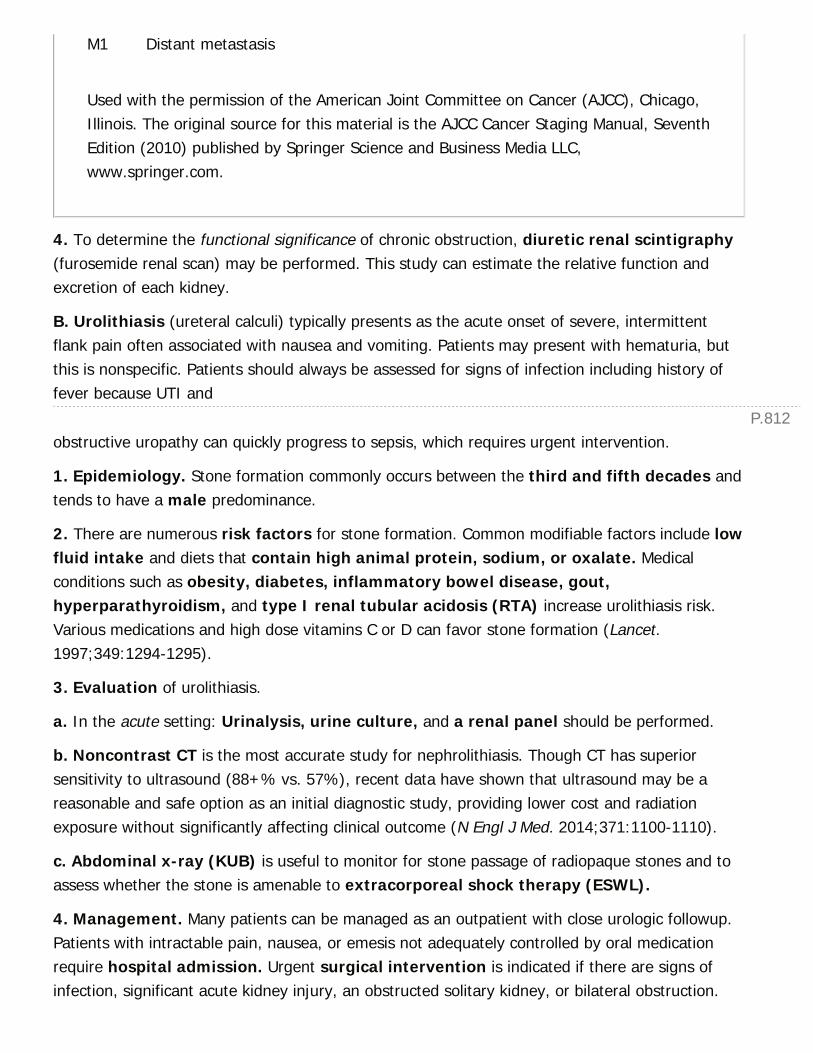
P.812
M1 Distant metastasis
Used with the permission of the American Joint Committee on Cancer (AJCC), Chicago,Illinois. The original source for this material is the AJCC Cancer Staging Manual, SeventhEdition (2010) published by Springer Science and Business Media LLC,www.springer.com.
4. To determine the functional significance of chronic obstruction, diuretic renal scintigraphy(furosemide renal scan) may be performed. This study can estimate the relative function andexcretion of each kidney.
B. Urolithiasis (ureteral calculi) typically presents as the acute onset of severe, intermittentflank pain often associated with nausea and vomiting. Patients may present with hematuria, butthis is nonspecific. Patients should always be assessed for signs of infection including history offever because UTI and
obstructive uropathy can quickly progress to sepsis, which requires urgent intervention.
1. Epidemiology. Stone formation commonly occurs between the third and fifth decades andtends to have a male predominance.
2. There are numerous risk factors for stone formation. Common modifiable factors include lowfluid intake and diets that contain high animal protein, sodium, or oxalate. Medicalconditions such as obesity, diabetes, inflammatory bowel disease, gout,hyperparathyroidism, and type I renal tubular acidosis (RTA) increase urolithiasis risk.Various medications and high dose vitamins C or D can favor stone formation (Lancet.1997;349:1294-1295).
3. Evaluation of urolithiasis.
a. In the acute setting: Urinalysis, urine culture, and a renal panel should be performed.
b. Noncontrast CT is the most accurate study for nephrolithiasis. Though CT has superiorsensitivity to ultrasound (88+% vs. 57%), recent data have shown that ultrasound may be areasonable and safe option as an initial diagnostic study, providing lower cost and radiationexposure without significantly affecting clinical outcome (N Engl J Med. 2014;371:1100-1110).
c. Abdominal x-ray (KUB) is useful to monitor for stone passage of radiopaque stones and toassess whether the stone is amenable to extracorporeal shock therapy (ESWL).
4. Management. Many patients can be managed as an outpatient with close urologic followup.Patients with intractable pain, nausea, or emesis not adequately controlled by oral medicationrequire hospital admission. Urgent surgical intervention is indicated if there are signs ofinfection, significant acute kidney injury, an obstructed solitary kidney, or bilateral obstruction.
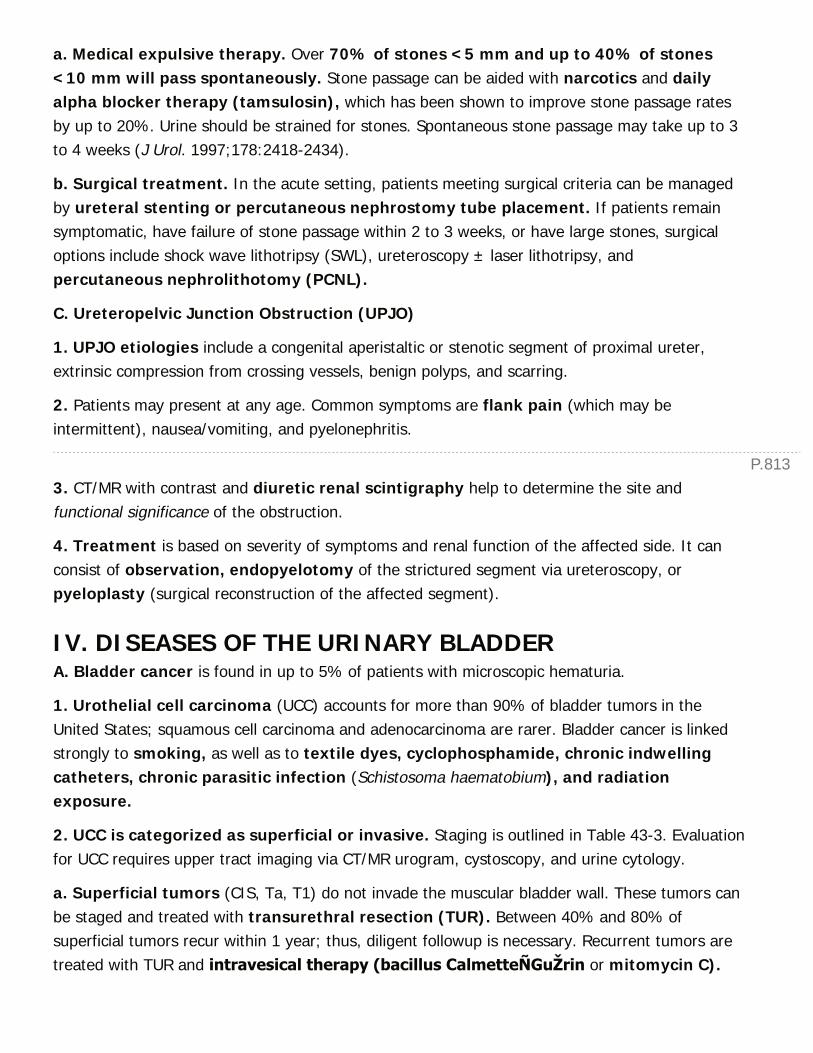
P.813
a. Medical expulsive therapy. Over 70% of stones <5 mm and up to 40% of stones<10 mm will pass spontaneously. Stone passage can be aided with narcotics and dailyalpha blocker therapy (tamsulosin), which has been shown to improve stone passage ratesby up to 20%. Urine should be strained for stones. Spontaneous stone passage may take up to 3to 4 weeks (J Urol. 1997;178:2418-2434).
b. Surgical treatment. In the acute setting, patients meeting surgical criteria can be managedby ureteral stenting or percutaneous nephrostomy tube placement. If patients remainsymptomatic, have failure of stone passage within 2 to 3 weeks, or have large stones, surgicaloptions include shock wave lithotripsy (SWL), ureteroscopy ± laser lithotripsy, andpercutaneous nephrolithotomy (PCNL).
C. Ureteropelvic Junction Obstruction (UPJO)
1. UPJO etiologies include a congenital aperistaltic or stenotic segment of proximal ureter,extrinsic compression from crossing vessels, benign polyps, and scarring.
2. Patients may present at any age. Common symptoms are flank pain (which may beintermittent), nausea/vomiting, and pyelonephritis.
3. CT/MR with contrast and diuretic renal scintigraphy help to determine the site andfunctional significance of the obstruction.
4. Treatment is based on severity of symptoms and renal function of the affected side. It canconsist of observation, endopyelotomy of the strictured segment via ureteroscopy, orpyeloplasty (surgical reconstruction of the affected segment).
IV. DISEASES OF THE URINARY BLADDERA. Bladder cancer is found in up to 5% of patients with microscopic hematuria.
1. Urothelial cell carcinoma (UCC) accounts for more than 90% of bladder tumors in theUnited States; squamous cell carcinoma and adenocarcinoma are rarer. Bladder cancer is linkedstrongly to smoking, as well as to textile dyes, cyclophosphamide, chronic indwellingcatheters, chronic parasitic infection (Schistosoma haematobium), and radiationexposure.
2. UCC is categorized as superficial or invasive. Staging is outlined in Table 43-3. Evaluationfor UCC requires upper tract imaging via CT/MR urogram, cystoscopy, and urine cytology.
a. Superficial tumors (CIS, Ta, T1) do not invade the muscular bladder wall. These tumors canbe staged and treated with transurethral resection (TUR). Between 40% and 80% ofsuperficial tumors recur within 1 year; thus, diligent followup is necessary. Recurrent tumors aretreated with TUR and intravesical therapy (bacillus Calmette—Guérin or mitomycin C).
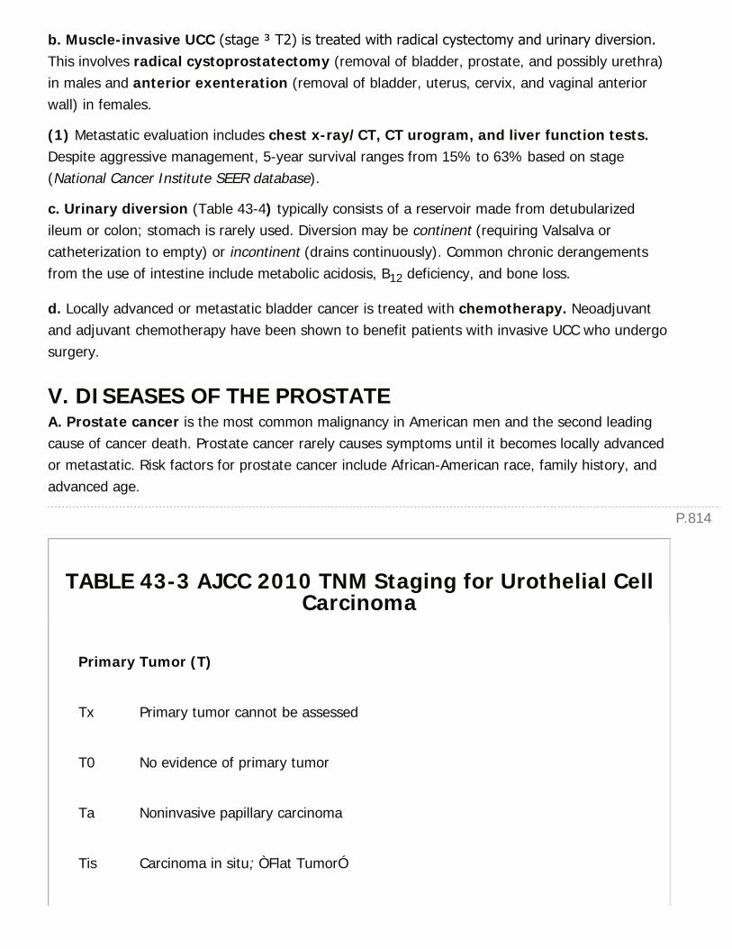
P.814
b. Muscle-invasive UCC (stage ≥ T2) is treated with radical cystectomy and urinary diversion.This involves radical cystoprostatectomy (removal of bladder, prostate, and possibly urethra)in males and anterior exenteration (removal of bladder, uterus, cervix, and vaginal anteriorwall) in females.
(1) Metastatic evaluation includes chest x-ray/CT, CT urogram, and liver function tests.Despite aggressive management, 5-year survival ranges from 15% to 63% based on stage(National Cancer Institute SEER database).
c. Urinary diversion (Table 43-4) typically consists of a reservoir made from detubularizedileum or colon; stomach is rarely used. Diversion may be continent (requiring Valsalva orcatheterization to empty) or incontinent (drains continuously). Common chronic derangementsfrom the use of intestine include metabolic acidosis, B12 deficiency, and bone loss.
d. Locally advanced or metastatic bladder cancer is treated with chemotherapy. Neoadjuvantand adjuvant chemotherapy have been shown to benefit patients with invasive UCC who undergosurgery.
V. DISEASES OF THE PROSTATEA. Prostate cancer is the most common malignancy in American men and the second leadingcause of cancer death. Prostate cancer rarely causes symptoms until it becomes locally advancedor metastatic. Risk factors for prostate cancer include African-American race, family history, andadvanced age.
TABLE 43-3 AJCC 2010 TNM Staging for Urothelial CellCarcinoma
Primary Tumor (T)
Tx Primary tumor cannot be assessed
T0 No evidence of primary tumor
Ta Noninvasive papillary carcinoma
Tis Carcinoma in situ; ÒFlat TumorÓ

T1 Tumor invades the subepithelial connective tissue
T2 Tumor invades the muscularis propria
pT2a Tumor invades superficial muscularis propria
pT2b Tumor invades deep muscularis propria
T3 Tumor invades perivesical tissue
pT3a Microscopic invasion
pT3b Macroscopic invasion
T4 Tumor invades any of the following: prostatic stroma, seminal vesicles, uterus,vagina, pelvic/abdominal wall
Regional Lymph NodesÑClinical Stage (N)
Nx Regional lymph nodes cannot be assessed
N0 No regional lymph node metastasis
N1 Single regional lymph node in the true pelvis
N2 Multiple regional lymph nodes in the true pelvis
N3 Lymph node metastasis to the common iliac lymph nodes
Distant Metastasis (M)
M0 No distant metastasis
M1 Distant metastasis
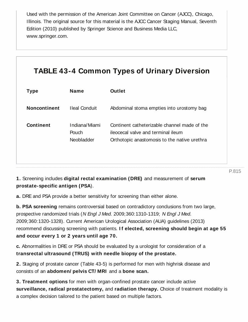
P.815
Used with the permission of the American Joint Committee on Cancer (AJCC), Chicago,Illinois. The original source for this material is the AJCC Cancer Staging Manual, SeventhEdition (2010) published by Springer Science and Business Media LLC,www.springer.com.
TABLE 43-4 Common Types of Urinary Diversion
Type Name Outlet
Noncontinent Ileal Conduit Abdominal stoma empties into urostomy bag
Continent Indiana/MiamiPouchNeobladder
Continent catheterizable channel made of theileocecal valve and terminal ileumOrthotopic anastomosis to the native urethra
1. Screening includes digital rectal examination (DRE) and measurement of serumprostate-specific antigen (PSA).
a. DRE and PSA provide a better sensitivity for screening than either alone.
b. PSA screening remains controversial based on contradictory conclusions from two large,prospective randomized trials (N Engl J Med. 2009;360:1310-1319; N Engl J Med.2009;360:1320-1328). Current American Urological Association (AUA) guidelines (2013)recommend discussing screening with patients. If elected, screening should begin at age 55and occur every 1 or 2 years until age 70.
c. Abnormalities in DRE or PSA should be evaluated by a urologist for consideration of atransrectal ultrasound (TRUS) with needle biopsy of the prostate.
2. Staging of prostate cancer (Table 43-5) is performed for men with highrisk disease andconsists of an abdomen/pelvis CT/MRI and a bone scan.
3. Treatment options for men with organ-confined prostate cancer include activesurveillance, radical prostatectomy, and radiation therapy. Choice of treatment modality isa complex decision tailored to the patient based on multiple factors.
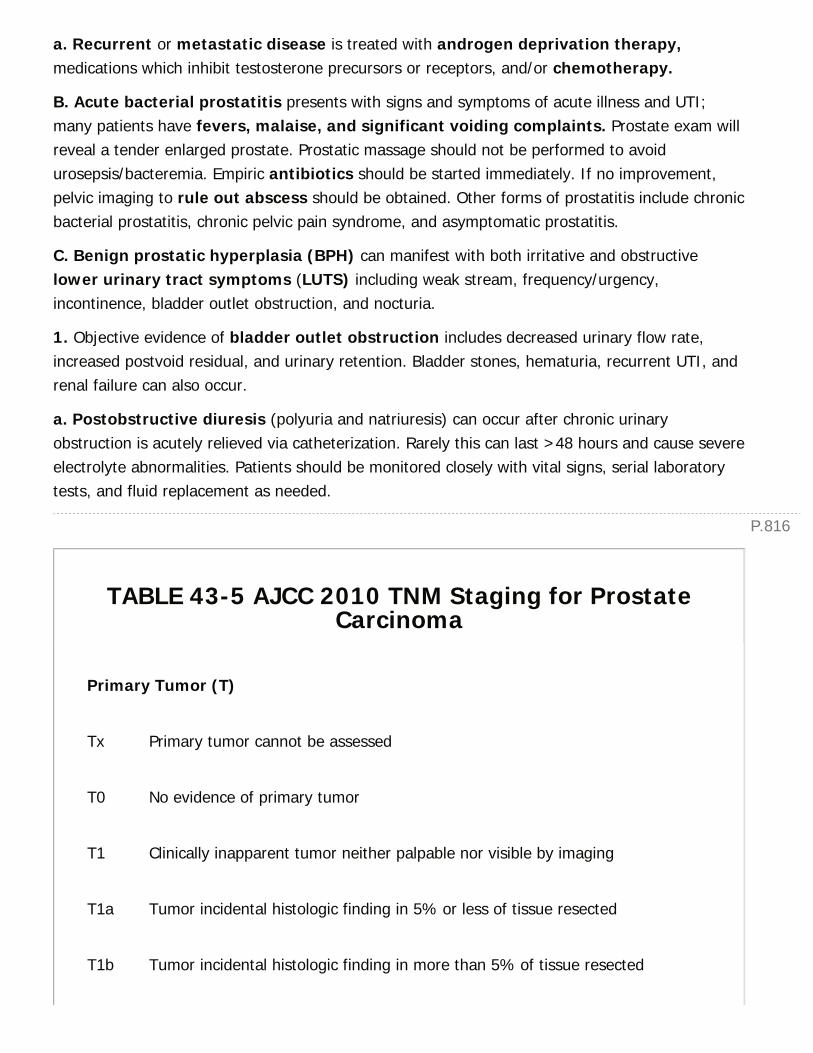
P.816
a. Recurrent or metastatic disease is treated with androgen deprivation therapy,medications which inhibit testosterone precursors or receptors, and/or chemotherapy.
B. Acute bacterial prostatitis presents with signs and symptoms of acute illness and UTI;many patients have fevers, malaise, and significant voiding complaints. Prostate exam willreveal a tender enlarged prostate. Prostatic massage should not be performed to avoidurosepsis/bacteremia. Empiric antibiotics should be started immediately. If no improvement,pelvic imaging to rule out abscess should be obtained. Other forms of prostatitis include chronicbacterial prostatitis, chronic pelvic pain syndrome, and asymptomatic prostatitis.
C. Benign prostatic hyperplasia (BPH) can manifest with both irritative and obstructivelower urinary tract symptoms (LUTS) including weak stream, frequency/urgency,incontinence, bladder outlet obstruction, and nocturia.
1. Objective evidence of bladder outlet obstruction includes decreased urinary flow rate,increased postvoid residual, and urinary retention. Bladder stones, hematuria, recurrent UTI, andrenal failure can also occur.
a. Postobstructive diuresis (polyuria and natriuresis) can occur after chronic urinaryobstruction is acutely relieved via catheterization. Rarely this can last >48 hours and cause severeelectrolyte abnormalities. Patients should be monitored closely with vital signs, serial laboratorytests, and fluid replacement as needed.
TABLE 43-5 AJCC 2010 TNM Staging for ProstateCarcinoma
Primary Tumor (T)
Tx Primary tumor cannot be assessed
T0 No evidence of primary tumor
T1 Clinically inapparent tumor neither palpable nor visible by imaging
T1a Tumor incidental histologic finding in 5% or less of tissue resected
T1b Tumor incidental histologic finding in more than 5% of tissue resected

T1c Tumor identified by needle biopsy via screening (PSA, DRE)
T2 Tumor confined to the prostate
T2a Tumor involves one-half of one lobe or less
T2b Tumor involves more than one-half of one lobe, but not both lobes
T2c Tumor involves both lobes
T3 Tumor extends through the prostatic capsule
T3a Extracapsular extension
T3b Seminal vesicle invasion
T4 Tumor is fixed or invades adjacent structures other than the SVs, such asexternal sphincter, rectum, bladder, levator muscles, and/or pelvic wall
Regional Lymph NodesÑClinical Stage (N)
Nx Regional lymph nodes cannot be assessed
N0 No regional lymph node metastasis
N1 Metastasis in regional node(s)
Distant Metastasis (M)
M0 No distant metastasis
M1 Distant metastasis
M1a Nonregional lymph node(s)
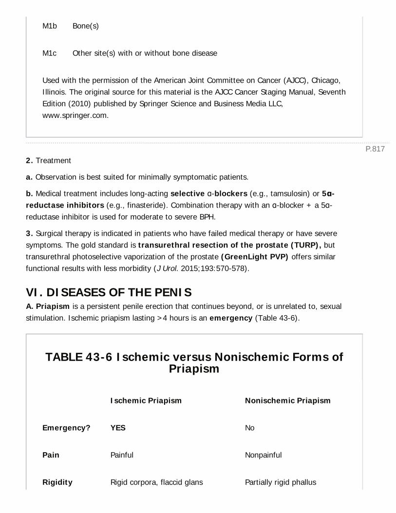
P.817
M1b Bone(s)
M1c Other site(s) with or without bone disease
Used with the permission of the American Joint Committee on Cancer (AJCC), Chicago,Illinois. The original source for this material is the AJCC Cancer Staging Manual, SeventhEdition (2010) published by Springer Science and Business Media LLC,www.springer.com.
2. Treatment
a. Observation is best suited for minimally symptomatic patients.
b. Medical treatment includes long-acting selective α-blockers (e.g., tamsulosin) or 5α-reductase inhibitors (e.g., finasteride). Combination therapy with an α-blocker + a 5α-reductase inhibitor is used for moderate to severe BPH.
3. Surgical therapy is indicated in patients who have failed medical therapy or have severesymptoms. The gold standard is transurethral resection of the prostate (TURP), buttransurethral photoselective vaporization of the prostate (GreenLight PVP) offers similarfunctional results with less morbidity (J Urol. 2015;193:570-578).
VI. DISEASES OF THE PENISA. Priapism is a persistent penile erection that continues beyond, or is unrelated to, sexualstimulation. Ischemic priapism lasting >4 hours is an emergency (Table 43-6).
TABLE 43-6 Ischemic versus Nonischemic Forms ofPriapism
Ischemic Priapism Nonischemic Priapism
Emergency? YES No
Pain Painful Nonpainful
Rigidity Rigid corpora, flaccid glans Partially rigid phallus
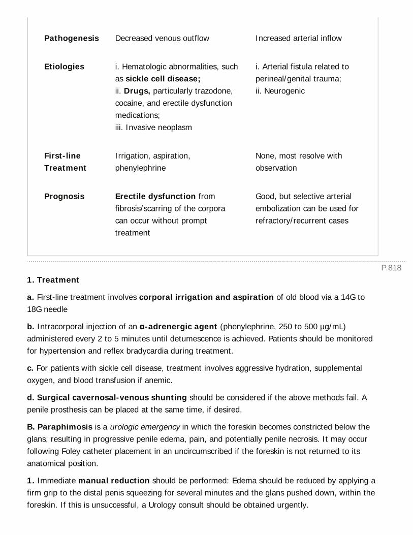
P.818
Pathogenesis Decreased venous outflow Increased arterial inflow
Etiologies i. Hematologic abnormalities, suchas sickle cell disease;ii. Drugs, particularly trazodone,cocaine, and erectile dysfunctionmedications;iii. Invasive neoplasm
i. Arterial fistula related toperineal/genital trauma;ii. Neurogenic
First-lineTreatment
Irrigation, aspiration,phenylephrine
None, most resolve withobservation
Prognosis Erectile dysfunction fromfibrosis/scarring of the corporacan occur without prompttreatment
Good, but selective arterialembolization can be used forrefractory/recurrent cases
1. Treatment
a. First-line treatment involves corporal irrigation and aspiration of old blood via a 14G to18G needle
b. Intracorporal injection of an α-adrenergic agent (phenylephrine, 250 to 500 µg/mL)administered every 2 to 5 minutes until detumescence is achieved. Patients should be monitoredfor hypertension and reflex bradycardia during treatment.
c. For patients with sickle cell disease, treatment involves aggressive hydration, supplementaloxygen, and blood transfusion if anemic.
d. Surgical cavernosal-venous shunting should be considered if the above methods fail. Apenile prosthesis can be placed at the same time, if desired.
B. Paraphimosis is a urologic emergency in which the foreskin becomes constricted below theglans, resulting in progressive penile edema, pain, and potentially penile necrosis. It may occurfollowing Foley catheter placement in an uncircumscribed if the foreskin is not returned to itsanatomical position.
1. Immediate manual reduction should be performed: Edema should be reduced by applying afirm grip to the distal penis squeezing for several minutes and the glans pushed down, within theforeskin. If this is unsuccessful, a Urology consult should be obtained urgently.

P.819
C. Phimosis is typically a benign condition where the foreskin is unable to be fully retracted overthe glans. Many patients are asymptomatic but phimosis can result in glans/foreskin infections orurinary retention (rare).
VII. DISEASES OF THE SCROTUM AND TESTICLESA. Testicular torsion is the rotation of the testicle on its vascular pedicle, resulting in ischemia.This is a true emergency because testicular viability depends on detorsion within a few hours.
1. History. Acute onset of severe testicular pain and swelling often associated with abdominalpain, nausea, and/or vomiting.
2. Examination reveals an extremely tender, swollen testicle which may be high riding in thescrotum with a transverse lie. The cremasteric reflex is usually absent on the affected side.Normal urinalysis and the absence of leukocytosis may help to rule out epididymitis.
3. Testicular torsion is a clinical diagnosis and treatment should not be delayed to obtainimaging. However, Doppler ultrasound can help to confirm or exclude the diagnosis with reportedsensitivity and specificity >95%.
4. Immediate scrotal exploration and bilateral orchiopexy is required if torsion issuspected. Manual detorsion of the testicle may be attempted; however, bilateral orchiopexy isstill necessary.
B. Torsion of testicular/epididymal appendage (appendix testis) presents with mild tomoderate testicular pain and onset over 12 to 24 hours without abdominal symptoms. Onexamination, the testicle has a normal
position but tenderness in the superior testicle and/or epididymis. The testicle may be mildlyswollen due to reactive inflammation. There may be a palpable Òpea-likeÓ nodule on the superiortesticle or epididymis, which can sometimes be visible through the scrotal skin (Òblue dotÓ sign).Diagnosis can be confirmed with ultrasound. Treatment consists of NSAIDs, light physicalactivity and scrotal support until resolution over 7 to 14 days.
C. Epididymitis usually presents with a 1- to 2-day onset of unilateral testicular pain andswelling sometimes associated with dysuria, urethral discharge, or LUTS.
1. Findings include a painful, indurated epididymis, and pyuria. Urinalysis, urine culture, and CBCare obtained. When clinically indicated, urethral swabs for gonococci and chlamydiae areperformed.
2. If sexually transmitted infection (STI) is unlikely (e.g., if >35 years), treat empirically with anoral fluoroquinolone, then a culture-specific antibiotic for 2 weeks. NSAIDs can reduceinflammation and provide symptomatic relief. For moderate to severe cases with fever and/orleukocytosis, ultrasonography can be useful to rule out abscess formation and assess testicular
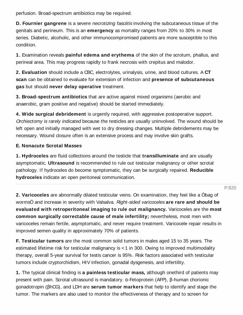
P.820
perfusion. Broad-spectrum antibiotics may be required.
D. Fournier gangrene is a severe necrotizing fasciitis involving the subcutaneous tissue of thegenitals and perineum. This is an emergency as mortality ranges from 20% to 30% in mostseries. Diabetic, alcoholic, and other immunocompromised patients are more susceptible to thiscondition.
1. Examination reveals painful edema and erythema of the skin of the scrotum, phallus, andperineal area. This may progress rapidly to frank necrosis with crepitus and malodor.
2. Evaluation should include a CBC, electrolytes, urinalysis, urine, and blood cultures. A CTscan can be obtained to evaluate for extension of infection and presence of subcutaneousgas but should never delay operative treatment.
3. Broad-spectrum antibiotics that are active against mixed organisms (aerobic andanaerobic, gram positive and negative) should be started immediately.
4. Wide surgical debridement is urgently required, with aggressive postoperative support.Orchiectomy is rarely indicated because the testicles are usually uninvolved. The wound should beleft open and initially managed with wet to dry dressing changes. Multiple debridements may benecessary. Wound closure often is an extensive process and may involve skin grafts.
E. Nonacute Scrotal Masses
1. Hydroceles are fluid collections around the testicle that transilluminate and are usuallyasymptomatic. Ultrasound is recommended to rule out testicular malignancy or other scrotalpathology. If hydroceles do become symptomatic, they can be surgically repaired. Reduciblehydroceles indicate an open peritoneal communication.
2. Varicoceles are abnormally dilated testicular veins. On examination, they feel like a Òbag ofwormsÓ and increase in severity with Valsalva. Right-sided varicoceles are rare and should beevaluated with retroperitoneal imaging to rule out malignancy. Varicoceles are the mostcommon surgically correctable cause of male infertility; nevertheless, most men withvaricoceles remain fertile, asymptomatic, and never require treatment. Varicocele repair results inimproved semen quality in approximately 70% of patients.
F. Testicular tumors are the most common solid tumors in males aged 15 to 35 years. Theestimated lifetime risk for testicular malignancy is <1 in 300. Owing to improved multimodalitytherapy, overall 5-year survival for testis cancer is 95%. Risk factors associated with testiculartumors include cryptorchidism, HIV infection, gonadal dysgenesis, and infertility.
1. The typical clinical finding is a painless testicular mass, although onethird of patients maypresent with pain. Scrotal ultrasound is mandatory. α-Fetoprotein (AFP), β-human chorionicgonadotropin (βhCG), and LDH are serum tumor markers that help to identify and stage thetumor. The markers are also used to monitor the effectiveness of therapy and to screen for
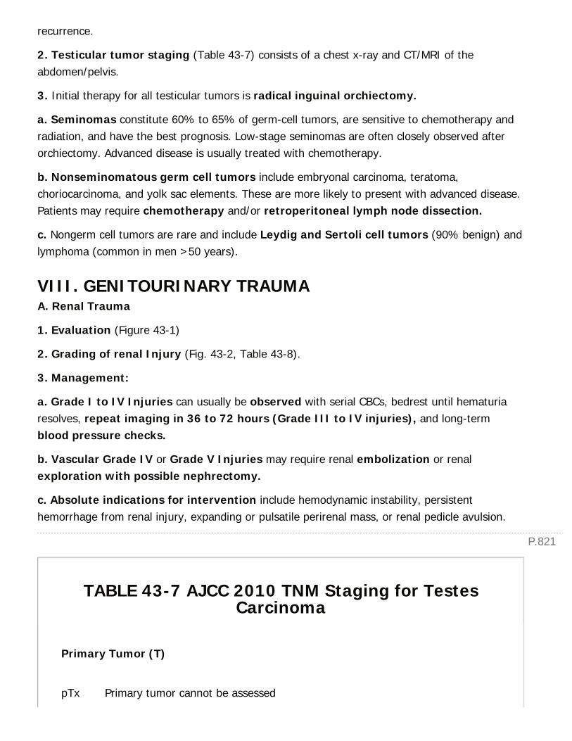
P.821
recurrence.
2. Testicular tumor staging (Table 43-7) consists of a chest x-ray and CT/MRI of theabdomen/pelvis.
3. Initial therapy for all testicular tumors is radical inguinal orchiectomy.
a. Seminomas constitute 60% to 65% of germ-cell tumors, are sensitive to chemotherapy andradiation, and have the best prognosis. Low-stage seminomas are often closely observed afterorchiectomy. Advanced disease is usually treated with chemotherapy.
b. Nonseminomatous germ cell tumors include embryonal carcinoma, teratoma,choriocarcinoma, and yolk sac elements. These are more likely to present with advanced disease.Patients may require chemotherapy and/or retroperitoneal lymph node dissection.
c. Nongerm cell tumors are rare and include Leydig and Sertoli cell tumors (90% benign) andlymphoma (common in men >50 years).
VIII. GENITOURINARY TRAUMAA. Renal Trauma
1. Evaluation (Figure 43-1)
2. Grading of renal Injury (Fig. 43-2, Table 43-8).
3. Management:
a. Grade I to IV Injuries can usually be observed with serial CBCs, bedrest until hematuriaresolves, repeat imaging in 36 to 72 hours (Grade III to IV injuries), and long-termblood pressure checks.
b. Vascular Grade IV or Grade V Injuries may require renal embolization or renalexploration with possible nephrectomy.
c. Absolute indications for intervention include hemodynamic instability, persistenthemorrhage from renal injury, expanding or pulsatile perirenal mass, or renal pedicle avulsion.
TABLE 43-7 AJCC 2010 TNM Staging for TestesCarcinoma
Primary Tumor (T)
pTx Primary tumor cannot be assessed
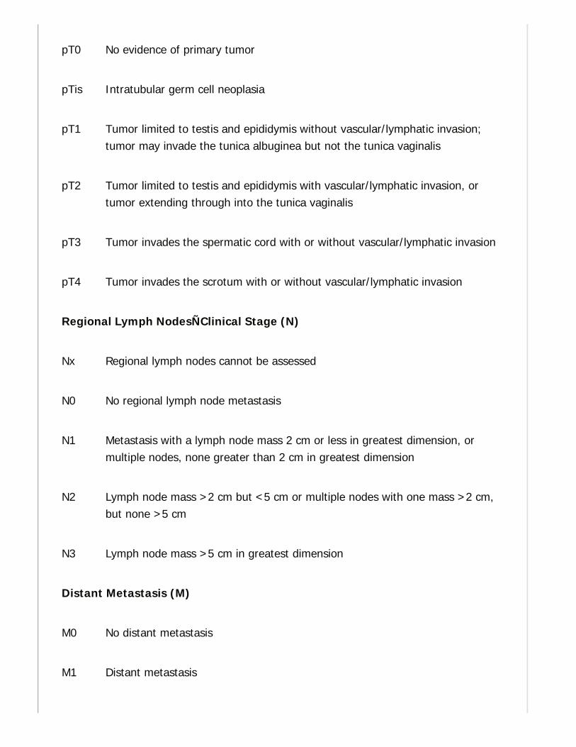
pT0 No evidence of primary tumor
pTis Intratubular germ cell neoplasia
pT1 Tumor limited to testis and epididymis without vascular/lymphatic invasion;tumor may invade the tunica albuginea but not the tunica vaginalis
pT2 Tumor limited to testis and epididymis with vascular/lymphatic invasion, ortumor extending through into the tunica vaginalis
pT3 Tumor invades the spermatic cord with or without vascular/lymphatic invasion
pT4 Tumor invades the scrotum with or without vascular/lymphatic invasion
Regional Lymph NodesÑClinical Stage (N)
Nx Regional lymph nodes cannot be assessed
N0 No regional lymph node metastasis
N1 Metastasis with a lymph node mass 2 cm or less in greatest dimension, ormultiple nodes, none greater than 2 cm in greatest dimension
N2 Lymph node mass >2 cm but <5 cm or multiple nodes with one mass >2 cm,but none >5 cm
N3 Lymph node mass >5 cm in greatest dimension
Distant Metastasis (M)
M0 No distant metastasis
M1 Distant metastasis

P.822
M1a Non-regional nodal or pulmonary metastasis
M1b Distant metastasis other than to non-regional nodes and lung
Serum Tumor Markers (S)
S0 Marker study levels within normal limits
S1 LDH × normal and hCG <5,000 and AFP <1,000
S2 LDH 1.5 to 10× normal or hCG 5-50,000 or AFP 1,000-10,000
S3 LDH > 10× normal or hCG >50,000, or AFP >10,000
Sx Marker studies not available or not performed
Used with the permission of the American Joint Committee on Cancer (AJCC), Chicago,Illinois. The original source for this material is the AJCC Cancer Staging Manual, SeventhEdition (2010) published by Springer Science and Business Media LLC,www.springer.com.
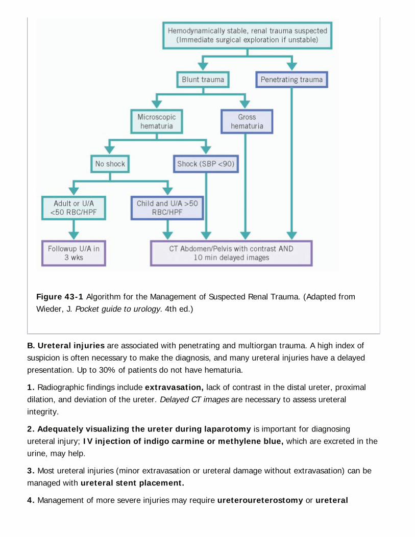
Figure 43-1 Algorithm for the Management of Suspected Renal Trauma. (Adapted fromWieder, J. Pocket guide to urology. 4th ed.)
B. Ureteral injuries are associated with penetrating and multiorgan trauma. A high index ofsuspicion is often necessary to make the diagnosis, and many ureteral injuries have a delayedpresentation. Up to 30% of patients do not have hematuria.
1. Radiographic findings include extravasation, lack of contrast in the distal ureter, proximaldilation, and deviation of the ureter. Delayed CT images are necessary to assess ureteralintegrity.
2. Adequately visualizing the ureter during laparotomy is important for diagnosingureteral injury; IV injection of indigo carmine or methylene blue, which are excreted in theurine, may help.
3. Most ureteral injuries (minor extravasation or ureteral damage without extravasation) can bemanaged with ureteral stent placement.
4. Management of more severe injuries may require ureteroureterostomy or ureteral

P.823
reimplantation, depending on location.
C. Bladder Injuries
1. The majority of bladder injuries present with gross hematuria. CT cystogram is necessary inany patient with gross hematuria and pelvic fracture and should be considered for (i) patientswith recent pelvic trauma and hematuria or suprapubic pain or (ii) pelvic fracture withouthematuria.
Figure 43-2 American Association for the Surgery of Trauma Grading. (From Britt LD, etal., eds. Acute Care Surgery. Philadelphia, PA: Wolters Kluwer Health, 2012.)
2. CT cystogram is the most sensitive imaging modality for bladder injury. The bladder shouldbe filled retrograde by gravity via a catheter. Filling with excreted contrast does not constitute anadequate study.
3. Treatment
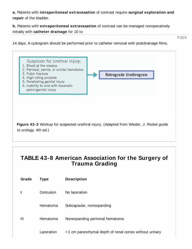
P.824
a. Patients with intraperitoneal extravasation of contrast require surgical exploration andrepair of the bladder.
b. Patients with extraperitoneal extravasation of contrast can be managed nonoperativelyinitially with catheter drainage for 10 to
14 days. A cystogram should be performed prior to catheter removal with postdrainage films.
Figure 43-3 Workup for suspected urethral injury. (Adapted from Wieder, J. Pocket guideto urology. 4th ed.)
TABLE 43-8 American Association for the Surgery ofTrauma Grading
Grade Type Description
I Contusion No laceration
Hematoma Subcapsular, nonexpanding
II Hematoma Nonexpanding perirenal hematoma
Laceration <1 cm parenchymal depth of renal cortex without urinary

P.825
extravasation
III Laceration >1 cm parenchymal depth of renal cortex without collectingsystem rupture or urinary extravasation
IV LacerationVascular
Parenchymal laceration extending through renal cortex,medulla, and collecting systemMain renal artery or vein injury with contained hemorrhage
V LacerationVascular
Completely shattered kidneyAvulsion of renal hilum, devascularizing the kidney
D. Urethral Injuries
1. Posterior urethral injuries involve the prostatic and membranous urethra (from thebladder neck to the external sphincter).
a. Acute management involves urologic consultation with careful urethral catheterization.
b. If catheterization is unsuccessful, recommend suprapubic catheter placement withattempt at cystoscopic primary realignment within 72 hours.
c. Surgical repair of a posterior urethral injury is not recommended in the acute settingas it is complicated by higher rates of impotence, incontinence, and stricture (Urol Clin North Am.2006;33:87-95).
d. Delayed repair in 3 to 6 months is recommended.
2. Anterior urethral injuries include injuries to the bulbous and penile urethra distal toexternal sphincter. Examination may show diffuse penile ecchymosis or a ÒbutterflyÓpattern of perineal ecchymosis. The 2014 AUA Urotrauma guidelines recommend immediatesurgical repair for anterior injuries unless the patient is unstable or there is concern abouttissue viability.
E. Penile Trauma
1. Penile fracture occurs when excessive bending force is applied to the erect penis resulting ina tear of the tunica albuginea. Patients describe an auditory ÒpopÓ heard during intercoursefollowed by rapid detumescence and swelling of the penis/scrotum. Inability to void or blood atthe meatus indicates concomitant urethral injury (20% of cases).
a. Examination demonstrates edema and ecchymosis/hematoma of the penis (ÒeggplantdeformityÓ) and/or perineum.
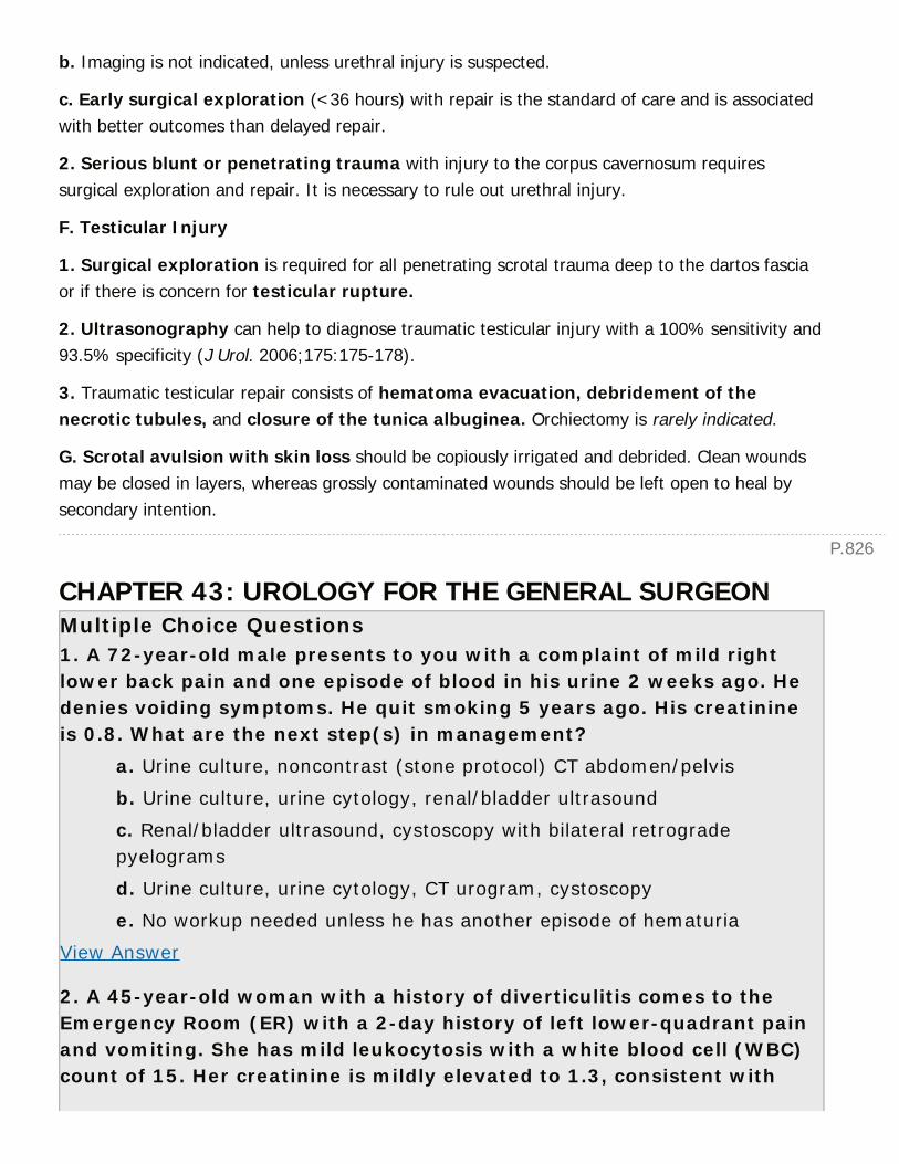
P.826
b. Imaging is not indicated, unless urethral injury is suspected.
c. Early surgical exploration (<36 hours) with repair is the standard of care and is associatedwith better outcomes than delayed repair.
2. Serious blunt or penetrating trauma with injury to the corpus cavernosum requiressurgical exploration and repair. It is necessary to rule out urethral injury.
F. Testicular Injury
1. Surgical exploration is required for all penetrating scrotal trauma deep to the dartos fasciaor if there is concern for testicular rupture.
2. Ultrasonography can help to diagnose traumatic testicular injury with a 100% sensitivity and93.5% specificity (J Urol. 2006;175:175-178).
3. Traumatic testicular repair consists of hematoma evacuation, debridement of thenecrotic tubules, and closure of the tunica albuginea. Orchiectomy is rarely indicated.
G. Scrotal avulsion with skin loss should be copiously irrigated and debrided. Clean woundsmay be closed in layers, whereas grossly contaminated wounds should be left open to heal bysecondary intention.
CHAPTER 43: UROLOGY FOR THE GENERAL SURGEONMultiple Choice Questions1. A 72-year-old male presents to you with a complaint of mild rightlower back pain and one episode of blood in his urine 2 weeks ago. Hedenies voiding symptoms. He quit smoking 5 years ago. His creatinineis 0.8. What are the next step(s) in management?
a. Urine culture, noncontrast (stone protocol) CT abdomen/pelvis
b. Urine culture, urine cytology, renal/bladder ultrasound
c. Renal/bladder ultrasound, cystoscopy with bilateral retrogradepyelograms
d. Urine culture, urine cytology, CT urogram, cystoscopy
e. No workup needed unless he has another episode of hematuria
View Answer
2. A 45-year-old woman with a history of diverticulitis comes to theEmergency Room (ER) with a 2-day history of left lower-quadrant painand vomiting. She has mild leukocytosis with a white blood cell (WBC)count of 15. Her creatinine is mildly elevated to 1.3, consistent with
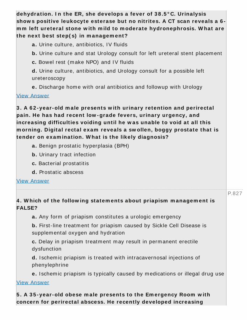
P.827
dehydration. In the ER, she develops a fever of 38.5°C. Urinalysisshows positive leukocyte esterase but no nitrites. A CT scan reveals a 6-mm left ureteral stone with mild to moderate hydronephrosis. What arethe next best step(s) in management?
a. Urine culture, antibiotics, IV fluids
b. Urine culture and stat Urology consult for left ureteral stent placement
c. Bowel rest (make NPO) and IV fluids
d. Urine culture, antibiotics, and Urology consult for a possible leftureteroscopy
e. Discharge home with oral antibiotics and followup with Urology
View Answer
3. A 62-year-old male presents with urinary retention and perirectalpain. He has had recent low-grade fevers, urinary urgency, andincreasing difficulties voiding until he was unable to void at all thismorning. Digital rectal exam reveals a swollen, boggy prostate that istender on examination. What is the likely diagnosis?
a. Benign prostatic hyperplasia (BPH)
b. Urinary tract infection
c. Bacterial prostatitis
d. Prostatic abscess
View Answer
4. Which of the following statements about priapism management isFALSE?
a. Any form of priapism constitutes a urologic emergency
b. First-line treatment for priapism caused by Sickle Cell Disease issupplemental oxygen and hydration
c. Delay in priapism treatment may result in permanent erectiledysfunction
d. Ischemic priapism is treated with intracavernosal injections ofphenylephrine
e. Ischemic priapism is typically caused by medications or illegal drug use
View Answer
5. A 35-year-old obese male presents to the Emergency Room withconcern for perirectal abscess. He recently developed increasing
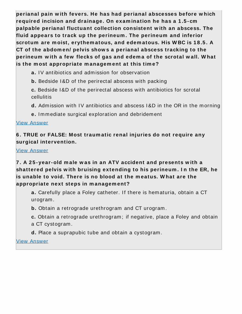
perianal pain with fevers. He has had perianal abscesses before whichrequired incision and drainage. On examination he has a 1.5-cmpalpable perianal fluctuant collection consistent with an abscess. Thefluid appears to track up the perineum. The perineum and inferiorscrotum are moist, erythematous, and edematous. His WBC is 18.5. ACT of the abdomen/pelvis shows a perianal abscess tracking to theperineum with a few flecks of gas and edema of the scrotal wall. Whatis the most appropriate management at this time?
a. IV antibiotics and admission for observation
b. Bedside I&D of the perirectal abscess with packing
c. Bedside I&D of the perirectal abscess with antibiotics for scrotalcellulitis
d. Admission with IV antibiotics and abscess I&D in the OR in the morning
e. Immediate surgical exploration and debridement
View Answer
6. TRUE or FALSE: Most traumatic renal injuries do not require anysurgical intervention.
View Answer
7. A 25-year-old male was in an ATV accident and presents with ashattered pelvis with bruising extending to his perineum. In the ER, heis unable to void. There is no blood at the meatus. What are theappropriate next steps in management?
a. Carefully place a Foley catheter. If there is hematuria, obtain a CTurogram.
b. Obtain a retrograde urethrogram and CT urogram.
c. Obtain a retrograde urethrogram; if negative, place a Foley and obtaina CT cystogram.
d. Place a suprapubic tube and obtain a cystogram.
View Answer
44Obstetrics and Gynecology for the GeneralSurgeon
Ivy Wilkinson-Ryan
Andrea R. Hagemann
I. OBSTETRIC AND GYNECOLOGIC DISORDERS.Vaginal Bleeding (Fig. 44-1, Table 44-1). Gather a thorough history including pattern andintensity of bleeding and date of last menstrual period. Obtain urine beta-human chorionicgonadotropin (β-hCG) level, complete blood count (CBC), coagulation studies and blood type.Physical examination should include a speculum and bimanual pelvic exam. Pelvic andtransvaginal ultrasound (U/S) is the most sensitive imaging modality for pelvic organs in thepregnant and non-pregnant patient.
A. Obstetric Etiologies. Forty percent of all pregnancies are associated with vaginal bleedingand approximately half of these result in spontaneous abortions (SABs).
First trimester: SABs, postcoital bleeding, ectopic pregnancy (see Section IIA Nonobstetric surgeryin the pregnant patient, Perioperative considerations), lower genital tract lesions/lacerations, andexpulsion of a molar pregnancy.
Third trimester: Placenta previa, placental abruption, vasa previa, preterm labor, and lowergenital tract lesions/lacerations.
1. Terminology
a. Threatened abortion: Any vaginal bleeding <20 weeks of gestation without expulsion ofproducts of contraception (POCs); cervix closed
b. Missed abortion: Nonviable gestation <20 weeks of gestation with retention of POCs; cervixclosed
c. Inevitable abortion: Cervical dilation with or without ruptured membranes
d. Incomplete abortion: Partial passage of POCs; cervix open
e. Complete abortion: Expulsion of all POCs; cervix closed
f. Septic abortion: Retained infected POC
2. Transvaginal ultrasound
> Table of Contents > 44 - Obstetrics and Gynecology for the General Surgeon
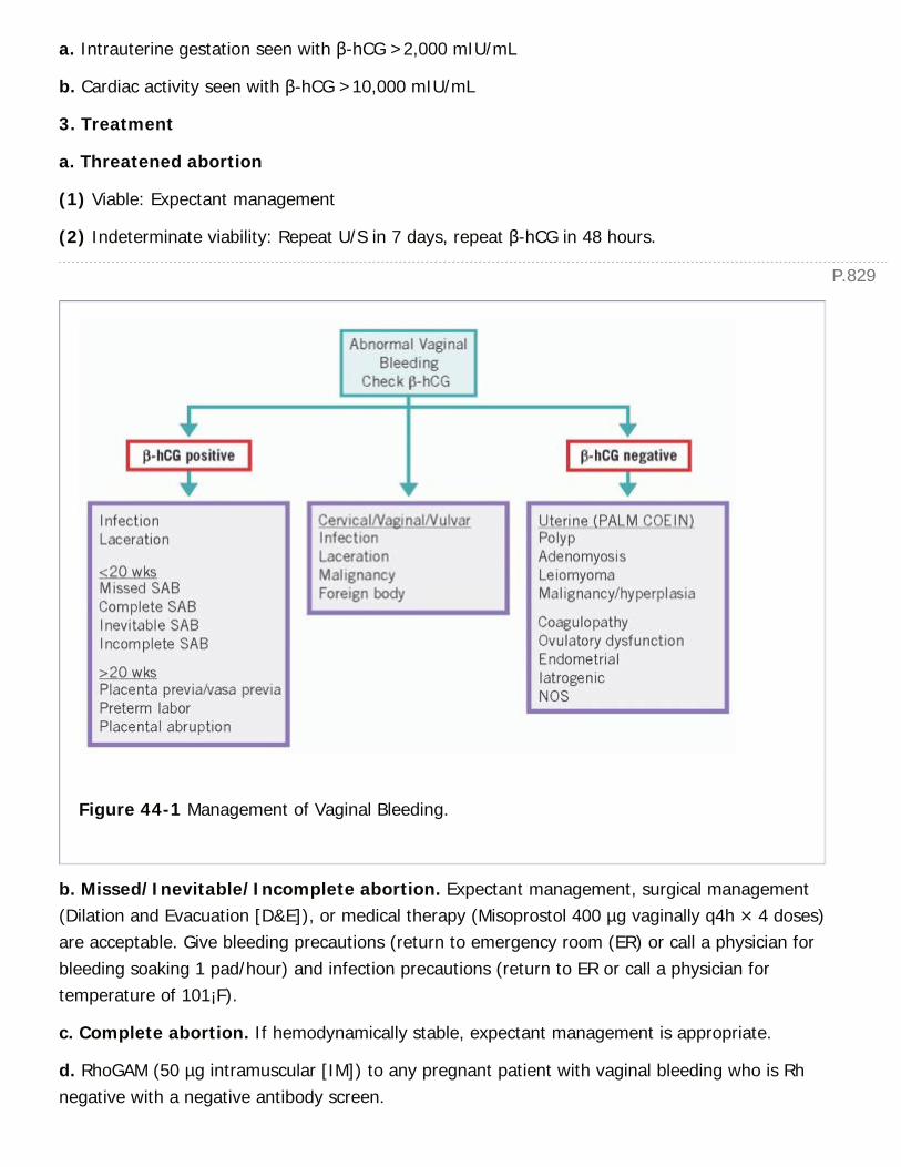
P.829
a. Intrauterine gestation seen with β-hCG >2,000 mIU/mL
b. Cardiac activity seen with β-hCG >10,000 mIU/mL
3. Treatment
a. Threatened abortion
(1) Viable: Expectant management
(2) Indeterminate viability: Repeat U/S in 7 days, repeat β-hCG in 48 hours.
Figure 44-1 Management of Vaginal Bleeding.
b. Missed/Inevitable/Incomplete abortion. Expectant management, surgical management(Dilation and Evacuation [D&E]), or medical therapy (Misoprostol 400 µg vaginally q4h × 4 doses)are acceptable. Give bleeding precautions (return to emergency room (ER) or call a physician forbleeding soaking 1 pad/hour) and infection precautions (return to ER or call a physician fortemperature of 101¡F).
c. Complete abortion. If hemodynamically stable, expectant management is appropriate.
d. RhoGAM (50 µg intramuscular [IM]) to any pregnant patient with vaginal bleeding who is Rhnegative with a negative antibody screen.
P.830
B. Nonobstetric Etiologies of Vaginal Bleeding. See Figure 44-1 for diagnostic workup andTable 44-1 for treatment options.
C. Risks and Complications of D&E
1. Uterine perforation: Perform laparoscopy/cystoscopy if concerned for bowel/bladder injury.Manage expectantly if hemodynamically stable and no injury to surrounding organs.
2. Endometritis: If unrelated to gonorrhea/chlamydia, Doxycycline 100 mg BID × 7 days. Ifhigh level of suspicion for gonorrhea/chlamydia infection, use pelvic inflammatory disease (PID)treatment regimen.
II. NONOBSTETRIC SURGERY IN THE PREGNANTPATIENT.If nonemergent, it is safest to proceed with surgery in the second trimester. Surgery inthe first trimester carries a risk of spontaneous abortion. Surgery in the third trimester carries arisk of inducing preterm labor and injury to the enlarging uterus. Risk of pregnancy interruption(any trimester) from nonobstetric
surgery in the pregnant patient is approximately 3% to 11%. Pregnancy is not a contraindicationto indicated/emergent surgery (Clin Obstet Gynecol. 2009;52:586-596). Once the need forsurgery has been identified, communication with the obstetrics and anesthesia teams is critical.The age and weight threshold for viability is controversial and may differ between institutions.Traditionally, 24 weeks or 500 g is used as the cut-off for fetal viability. However, someinstitutions are offering interventions after 23 weeks or 400 g.
TABLE 44-1 Nonobstetric Causes of Vaginal Bleeding
DifferentialDiagnosis
LaboratoryData
Signs andSymptoms Treatment
Menses CBC count,urine hCG
Cyclic bleedingevery 21-35days
Iron therapy ifindicated
Dysfunctional uterinebleeding
CBC count,urine hCG,endometrialbiopsy if >45
Non-cyclicbleeding; mayhaveassociated
Hormonal therapy ifpatient ishemodynamicallystable; if unstable,
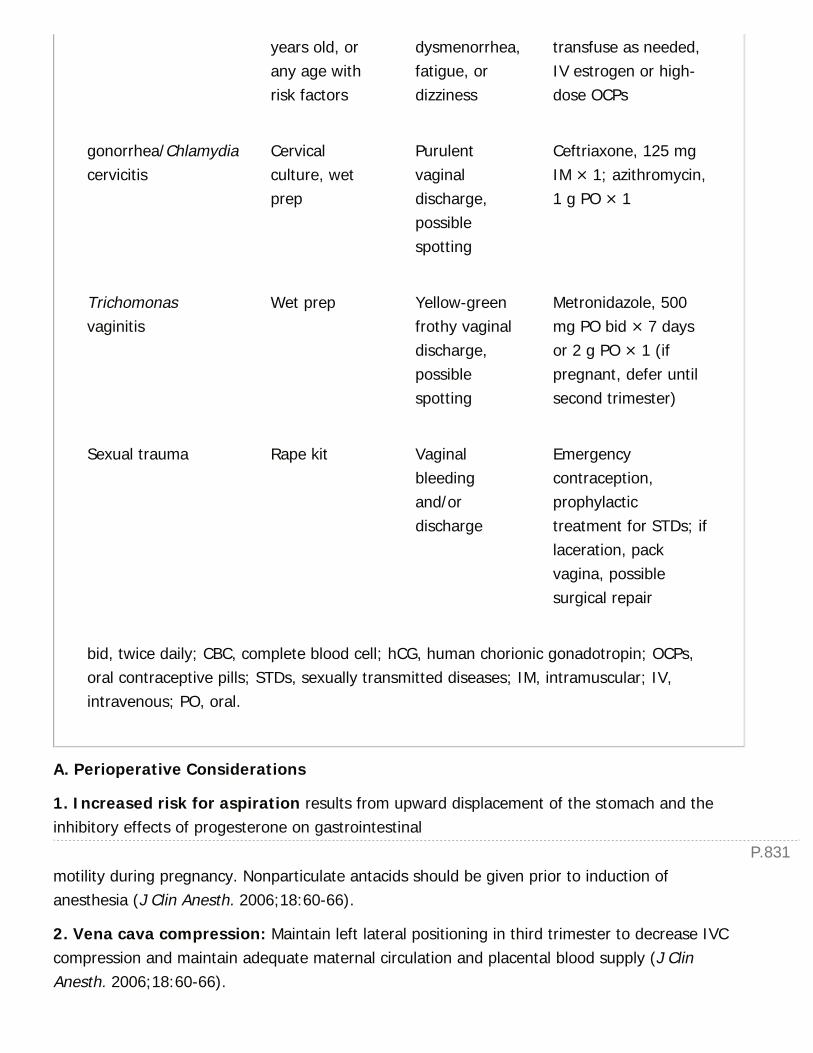
P.831
years old, orany age withrisk factors
dysmenorrhea,fatigue, ordizziness
transfuse as needed,IV estrogen or high-dose OCPs
gonorrhea/Chlamydiacervicitis
Cervicalculture, wetprep
Purulentvaginaldischarge,possiblespotting
Ceftriaxone, 125 mgIM × 1; azithromycin,1 g PO × 1
Trichomonasvaginitis
Wet prep Yellow-greenfrothy vaginaldischarge,possiblespotting
Metronidazole, 500mg PO bid × 7 daysor 2 g PO × 1 (ifpregnant, defer untilsecond trimester)
Sexual trauma Rape kit Vaginalbleedingand/ordischarge
Emergencycontraception,prophylactictreatment for STDs; iflaceration, packvagina, possiblesurgical repair
bid, twice daily; CBC, complete blood cell; hCG, human chorionic gonadotropin; OCPs,oral contraceptive pills; STDs, sexually transmitted diseases; IM, intramuscular; IV,intravenous; PO, oral.
A. Perioperative Considerations
1. Increased risk for aspiration results from upward displacement of the stomach and theinhibitory effects of progesterone on gastrointestinal
motility during pregnancy. Nonparticulate antacids should be given prior to induction ofanesthesia (J Clin Anesth. 2006;18:60-66).
2. Vena cava compression: Maintain left lateral positioning in third trimester to decrease IVCcompression and maintain adequate maternal circulation and placental blood supply (J ClinAnesth. 2006;18:60-66).

P.832
3. Fetal monitoring: If previable (<23 weeks, <400 g), Doptone maternal abdomen prior toand following the procedure. If pregnancy is viable, intraoperative continuous fetal monitoringand uterine contraction monitoring is recommended if the following conditions are met: (1)Monitoring is compatible with patient position and planned procedure, (2) there are appropriatestaff in the operating room (OR) to interpret fetal monitoring, and (3) there are appropriate staffimmediately available to perform an emergency cesarean section. If intraoperative continuousmonitoring is not feasible, monitor fetal heart rate and uterine contractions pre- andpostoperatively. (ACOG Committee Opinion Number 474, Feb 2011, http://www.acog.org/-/media/Committee-Opinions/Committee-on-Obstetric-Practice/co474.pdf?dmc = 1&ts =20141013T1551357449. Clin Obstet Gynecol. 2009;52:586-96, PMID: 20393411.)
4. Antenatal steroids: If gestation is between 23 and 34 weeks, start antenatal steroids(Betamethasone 12 mg IM q24h × 2 doses) prior to proceeding to OR to decrease risk ofrespiratory distress syndrome, intraventricular hemorrhage, necrotizing enterocolitis, neonatalinfection, and neonatal mortality (NIH Consensus Statement. 1994;12:1-24).
B. Laparoscopy in Pregnancy. Based on the Society of American Gastrointestinal EndoscopicSurgeons (SAGES) Guidelines of Laparoscopic Surgery During Pregnancy (Surg Endosc.1998;12:189), laparoscopy is safe and has similar advantages over open surgery as in thenonpregnant patient. Initial trocar placement is best accomplished by an open Hasson techniqueand/or left upper quadrant entry. Avoid Veress needle entry. Carbon dioxide pneumoperitoneumup to 15 mm Hg is safe and is unlikely to result in fetal hypoxia or acidosis. Avoid cervical oruterine manipulation.
III. GYNECOLOGIC SURGICAL ANATOMYA. Uterus and Cervix. The primary blood supply to the uterus and cervix comes from theuterine branch of the hypogastric artery. After passing through the cardinal ligament the uterineartery has ascending and descending branches (Fig. 44-2). The uterine suspensory ligamentsinclude the uterosacral ligaments, cardinal ligaments, round ligaments, and utero-ovarianligaments. The ureter travels through the cardinal ligament passing under the uterine artery andvein within the ureteric tunnel. Hypogastric or uterine artery ligation either surgically or byinterventional radiology (IR) can be used to treat uncontrolled uterine hemorrhage. The uteruscan be amputated from the cervix and the cervix can be left in situ, for instance in a supracervicalhysterectomy, or the cervix can be removed with the uterus in the case of a total abdominalhysterectomy.
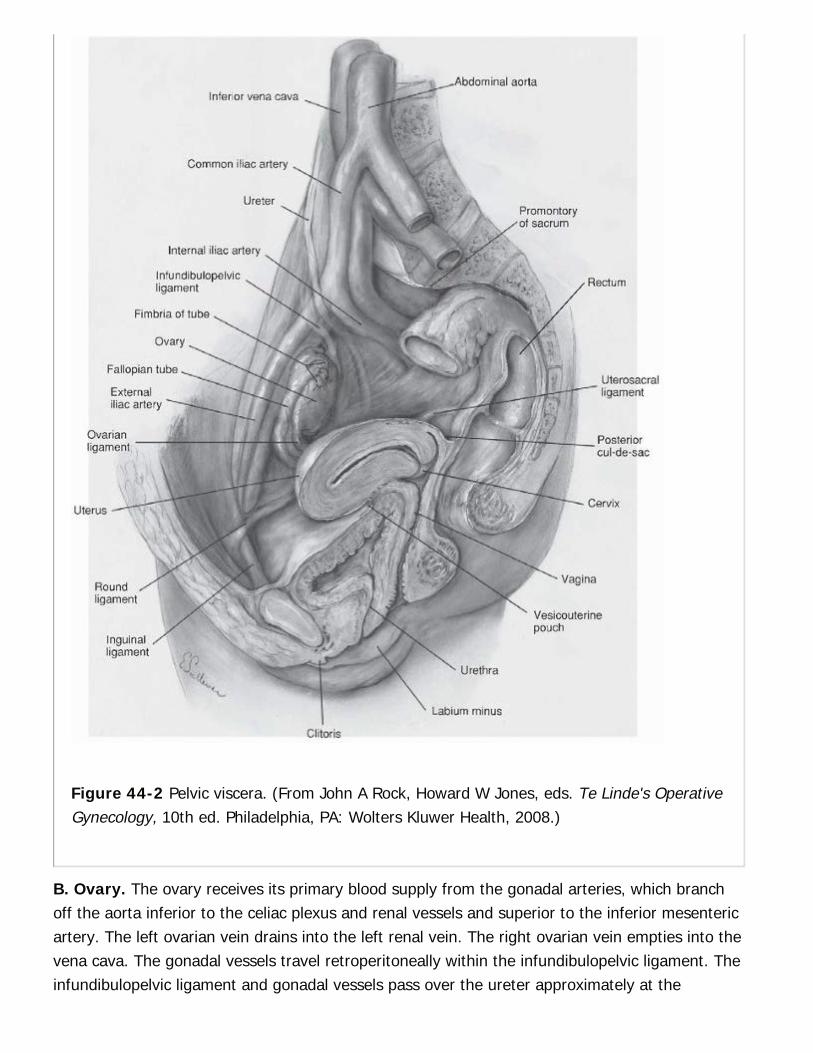
Figure 44-2 Pelvic viscera. (From John A Rock, Howard W Jones, eds. Te Linde's OperativeGynecology, 10th ed. Philadelphia, PA: Wolters Kluwer Health, 2008.)
B. Ovary. The ovary receives its primary blood supply from the gonadal arteries, which branchoff the aorta inferior to the celiac plexus and renal vessels and superior to the inferior mesentericartery. The left ovarian vein drains into the left renal vein. The right ovarian vein empties into thevena cava. The gonadal vessels travel retroperitoneally within the infundibulopelvic ligament. Theinfundibulopelvic ligament and gonadal vessels pass over the ureter approximately at the
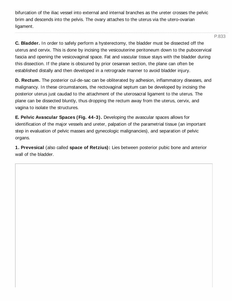
P.833
bifurcation of the iliac vessel into external and internal branches as the ureter crosses the pelvicbrim and descends into the pelvis. The ovary attaches to the uterus via the utero-ovarianligament.
C. Bladder. In order to safely perform a hysterectomy, the bladder must be dissected off theuterus and cervix. This is done by incising the vesicouterine peritoneum down to the pubocervicalfascia and opening the vesicovaginal space. Fat and vascular tissue stays with the bladder duringthis dissection. If the plane is obscured by prior cesarean section, the plane can often beestablished distally and then developed in a retrograde manner to avoid bladder injury.
D. Rectum. The posterior cul-de-sac can be obliterated by adhesion, inflammatory diseases, andmalignancy. In these circumstances, the rectovaginal septum can be developed by incising theposterior uterus just caudad to the attachment of the uterosacral ligament to the uterus. Theplane can be dissected bluntly, thus dropping the rectum away from the uterus, cervix, andvagina to isolate the structures.
E. Pelvic Avascular Spaces (Fig. 44-3). Developing the avascular spaces allows foridentification of the major vessels and ureter, palpation of the parametrial tissue (an importantstep in evaluation of pelvic masses and gynecologic malignancies), and separation of pelvicorgans.
1. Prevesical (also called space of Retzius): Lies between posterior pubic bone and anteriorwall of the bladder.

P.834
Figure 44-3 Cross-section of the pelvis showing avascular planes. (From John A Rock,Howard W Jones, eds. Te Linde's Operative Gynecology, 10th ed. Philadelphia, PA: WoltersKluwer Health, 2008.)
2. Vesicovaginal space: Lies between vagina posteriorly and bladder anteriorly. Laterallydefined by bladder pillars.
3. Paravesical space: Bordered by bladder pillars medially and pelvic sidewall/obturatorinternus laterally.
4. Pararectal space: Identified between the ureter and the hypogastric artery.
5. Rectovaginal space: Potential space between the rectum and vagina. Laterally defined bythe rectal pillars/uterosacral ligaments.
P.835
Recommended Text: Te Linde's Operative Gynecology, 10th ed. LWW.
IV. GYNECOLOGIC MALIGNANCIES.Over 94,000 new cases of gynecologic malignancy were diagnosed in the United States in 2014(CA Cancer J Clin. 2014;64:9-29). A brief overview of vulvar, cervical, endometrial, and ovariancancers is presented with emphasis on diagnosis and initial management. If a gynecologicmalignancy is suspected, all effort should be made to obtain an intraoperative consult with agynecologic oncologist.
A. Vulvar Carcinoma
1. Epidemiology. There were an estimated 4,850 new cases and 1,030 deaths in the UnitedStates in 2014. The mean age at diagnosis is 68 years old. Vulvar cancer is associated with HPVin 60% of cases. Other risk factors for vulvar cancer include cigarette smoking, vulvar andcervical dysplasia, vulvar dystrophy, and immunodeficiency.
2. Presentation and clinical features. The most common symptom is itching, but lesions canbe pruritic, painful, or asymptomatic. Vulvar examination typically yields an ulcerated, hyper- orhypopigmented, or exophytic lesion. A biopsy should be taken any time such a lesion is noted onthe vulva.
3. Standard of care. See Table 44-2 for treatment by stage. Vulvar cancer is staged using bothclinical and surgical assessment. Size of the lesion and regional or distant metastasis must beassessed. Radical vulvectomy is defined as complete resection of the lesion with at least 1 cmmargins and deep excision to the inferior fascia of the urogenital diaphragm (coplanar with thefascia lata and fascia over the pubic symphysis). Adjuvant radiation is reserved for cancersconfined to the vulva at high risk for recurrence or tumors that metastasize to the inguinofemorallymph nodes (Fig. 44-4).
4. What to do if a lesion is identified pre- or intraoperatively. As above, it is essential toobtain a biopsy for pathologic diagnosis. A 3 to 5 mm Keyes punch biopsy is usually adequate.
B. Cervical Carcinoma
1. Epidemiology. There were an estimated 12,360 new cases and 4,020 deaths in the UnitedStates in 2014. The mean age of diagnosis is 48 years old. HPV is detected in over 99% ofcervical cancer cases. Other risk factors for cervical cancer include cigarette smoking,immunosuppression, history of multiple sexual partners, early sexual debut (i.e., onset of sexualactivity), history of sexually transmitted infections (STIs), and vulvar or vaginal dysplasia.
TABLE 44-2 Vulvar Cancer Staging

TNM FIGO Definition Treatment
T1 I Tumor confined to the vulva
T1a IA Lesions ≤2 cm in size with stromalinvasion ≤1 mm
Radical excision
T1b IB Lesions >2 cm in size or withstromal invasion >1 mm
Radical excision andinguinofemorallymphadenectomyIpsilaterallymphadenectomy if >2cm from midlineBilaterallymphadenectomy if <2cm from midlineResection of distal 1 cmor urethra for periurethrallesionsSentinel lymph noderesection acceptable forpalpably negative inguinallymph nodes
T2 II Tumor of any size, extending toadjacent perineal structures (lowerone-third urethra, lower one-thirdvagina, anus) with negative nodes
Stage IIÑIVA radicalexcision and bilateralinguinofemorallymphadenectomy ifresection is feasiblePrimary radiation therapyfor unresectable lesions
T3 III Tumor of any size with or withoutextension to adjacent perinealstructures (lower onethird urethra,lower one-third vagina, anus) withpositive inguinofemoral lymph nodes
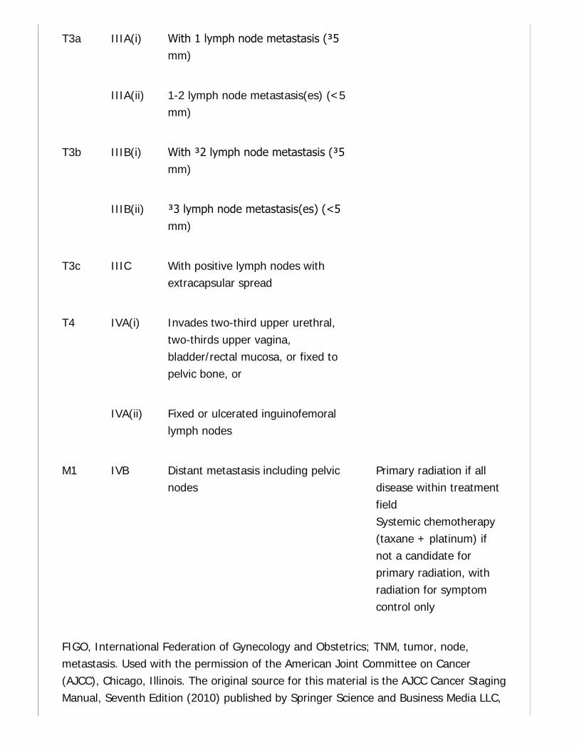
T3a IIIA(i) With 1 lymph node metastasis (≥5mm)
IIIA(ii) 1-2 lymph node metastasis(es) (<5mm)
T3b IIIB(i) With ≥2 lymph node metastasis (≥5mm)
IIIB(ii) ≥3 lymph node metastasis(es) (<5mm)
T3c IIIC With positive lymph nodes withextracapsular spread
T4 IVA(i) Invades two-third upper urethral,two-thirds upper vagina,bladder/rectal mucosa, or fixed topelvic bone, or
IVA(ii) Fixed or ulcerated inguinofemorallymph nodes
M1 IVB Distant metastasis including pelvicnodes
Primary radiation if alldisease within treatmentfieldSystemic chemotherapy(taxane + platinum) ifnot a candidate forprimary radiation, withradiation for symptomcontrol only
FIGO, International Federation of Gynecology and Obstetrics; TNM, tumor, node,metastasis. Used with the permission of the American Joint Committee on Cancer(AJCC), Chicago, Illinois. The original source for this material is the AJCC Cancer StagingManual, Seventh Edition (2010) published by Springer Science and Business Media LLC,

P.837
P.836
www.springer.com.
2. Presentation and clinical features. Irregular or postcoital vaginal bleeding andmalodorous, watery discharge are the most common symptoms. Advanced stage disease maypresent with leg pain (sciatic nerve involvement), flank pain (ureteral obstruction), renal failure,or rectal bleeding from a pelvic mass.
3. Standard of care. See Table 44-3 for treatment by stage. Cervical cancer is clinically, notsurgically, staged. The World Health Organization (WHO) recognizes a standard worldwidestaging system for cervical cancer that includes a pelvic examination, x-ray, intravenouspyelogram, endoscopy (hysteroscopy, cystoscopy, proctoscopy), and cervical biopsy. A speculumshould be placed prior to a bimanual exam to obtain visualization and approximate measurementsof the tumor. Rectovaginal examination is essential to characterize parametrial involvement. Inthe
United States, a positron emission tomography (PET) scan, magnetic resonance imaging (MRI), orcomputerized tomography (CT) is typically performed to assess for pelvic or para-aorticlymphadenopathy and guide treatment planning, but this does not change clinical stage as it isnot available worldwide.
Figure 44-4 Lymphatic drainage of the vulva and femoral triangle. (From John A Rock,Howard W Jones, eds. Te Linde's Operative Gynecology, 10th ed. Philadelphia, PA: Wolters
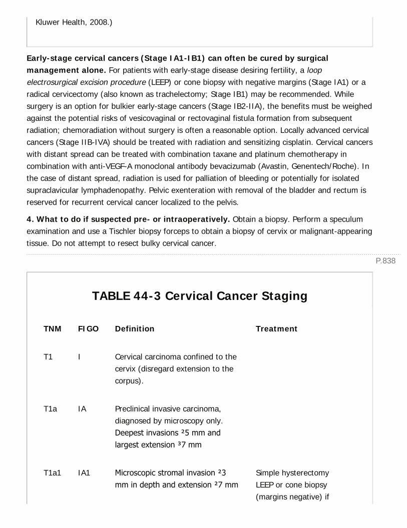
P.838
Kluwer Health, 2008.)
Early-stage cervical cancers (Stage IA1-IB1) can often be cured by surgicalmanagement alone. For patients with early-stage disease desiring fertility, a loopelectrosurgical excision procedure (LEEP) or cone biopsy with negative margins (Stage IA1) or aradical cervicectomy (also known as trachelectomy; Stage IB1) may be recommended. Whilesurgery is an option for bulkier early-stage cancers (Stage IB2-IIA), the benefits must be weighedagainst the potential risks of vesicovaginal or rectovaginal fistula formation from subsequentradiation; chemoradiation without surgery is often a reasonable option. Locally advanced cervicalcancers (Stage IIB-IVA) should be treated with radiation and sensitizing cisplatin. Cervical cancerswith distant spread can be treated with combination taxane and platinum chemotherapy incombination with anti-VEGF-A monoclonal antibody bevacizumab (Avastin, Genentech/Roche). Inthe case of distant spread, radiation is used for palliation of bleeding or potentially for isolatedsupraclavicular lymphadenopathy. Pelvic exenteration with removal of the bladder and rectum isreserved for recurrent cervical cancer localized to the pelvis.
4. What to do if suspected pre- or intraoperatively. Obtain a biopsy. Perform a speculumexamination and use a Tischler biopsy forceps to obtain a biopsy of cervix or malignant-appearingtissue. Do not attempt to resect bulky cervical cancer.
TABLE 44-3 Cervical Cancer Staging
TNM FIGO Definition Treatment
T1 I Cervical carcinoma confined to thecervix (disregard extension to thecorpus).
T1a IA Preclinical invasive carcinoma,diagnosed by microscopy only.Deepest invasions ≤5 mm andlargest extension ≥7 mm
T1a1 IA1 Microscopic stromal invasion ≤3mm in depth and extension ≤7 mm
Simple hysterectomyLEEP or cone biopsy(margins negative) if
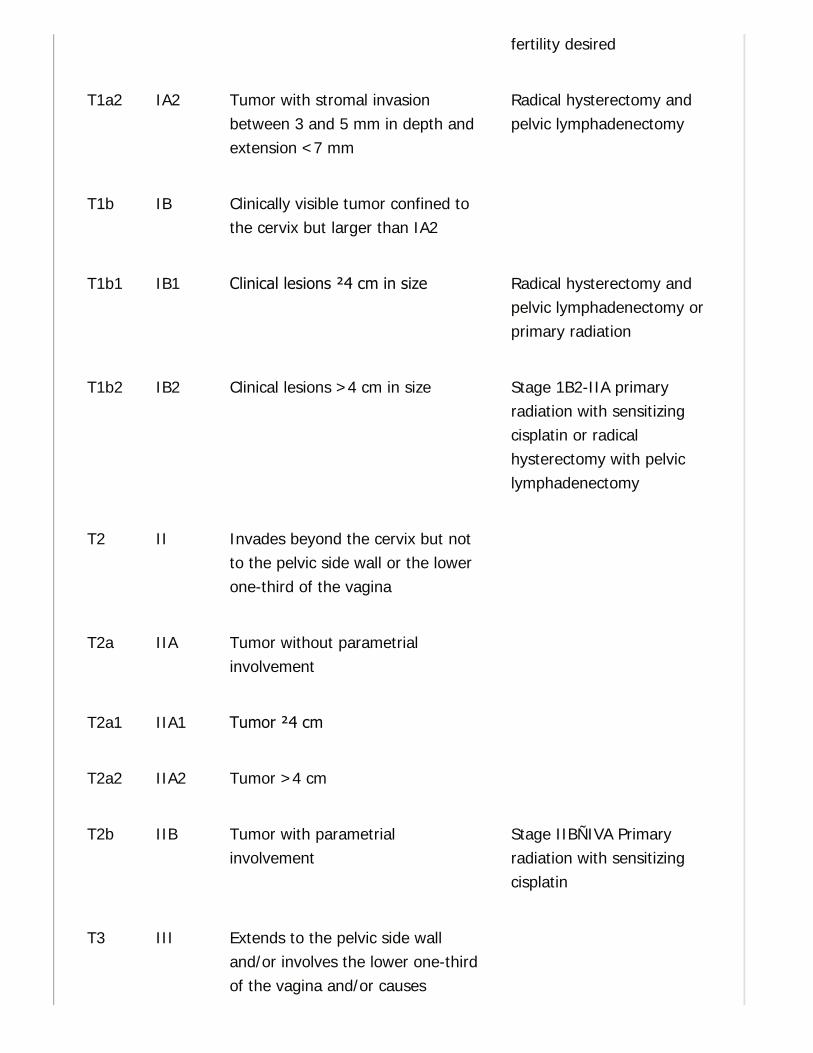
fertility desired
T1a2 IA2 Tumor with stromal invasionbetween 3 and 5 mm in depth andextension <7 mm
Radical hysterectomy andpelvic lymphadenectomy
T1b IB Clinically visible tumor confined tothe cervix but larger than IA2
T1b1 IB1 Clinical lesions ≤4 cm in size Radical hysterectomy andpelvic lymphadenectomy orprimary radiation
T1b2 IB2 Clinical lesions >4 cm in size Stage 1B2-IIA primaryradiation with sensitizingcisplatin or radicalhysterectomy with pelviclymphadenectomy
T2 II Invades beyond the cervix but notto the pelvic side wall or the lowerone-third of the vagina
T2a IIA Tumor without parametrialinvolvement
T2a1 IIA1 Tumor ≤4 cm
T2a2 IIA2 Tumor >4 cm
T2b IIB Tumor with parametrialinvolvement
Stage IIBÑIVA Primaryradiation with sensitizingcisplatin
T3 III Extends to the pelvic side walland/or involves the lower one-thirdof the vagina and/or causes

P.840
P.839
hydronephrosis or nonfunctioningkidney
T3a IIIA Invades lower one-third of thevagina with no extension to thepelvic side wall
T3b IIIB Extends to the pelvic side walland/or causes hydronephrosis or anonfunctioning kidney
T4 IVA Invades mucosa of thebladder/rectum and/or extendsbeyond the true pelvis
M1 IVB Distant metastasis Taxane, Platinum,Bevacizumab with orwithout pelvic radiation forsymptom control
Used with the permission of the American Joint Committee on Cancer (AJCC), Chicago,Illinois. The original source for this material is the AJCC Cancer Staging Manual, SeventhEdition (2010) published by Springer Science and Business Media LLC,www.springer.com.
5. Uncontrolled vaginal bleeding. Heavy vaginal bleeding from cervical cancer canoccasionally be encountered in the emergency department. Place a tight vaginal packing and atransurethral Foley catheter. Acetone-soaked gauze is the most effective packing for vesselsclerosis and control of hemorrhage from necrotic tumor. If ineffective at controlling bleeding, IRembolization may be necessary. Prompt consultation with radiation oncology is also indicatedonce a biopsy has confirmed invasive cancer.
C. Endometrial Cancers
1. Epidemiology. Endometrial carcinoma is the most common gynecologic cancer in the UnitedStates with 52,630 new cases and 8,590 deaths in 2014. The average age of diagnosis is 61 yearsold. Risk factors
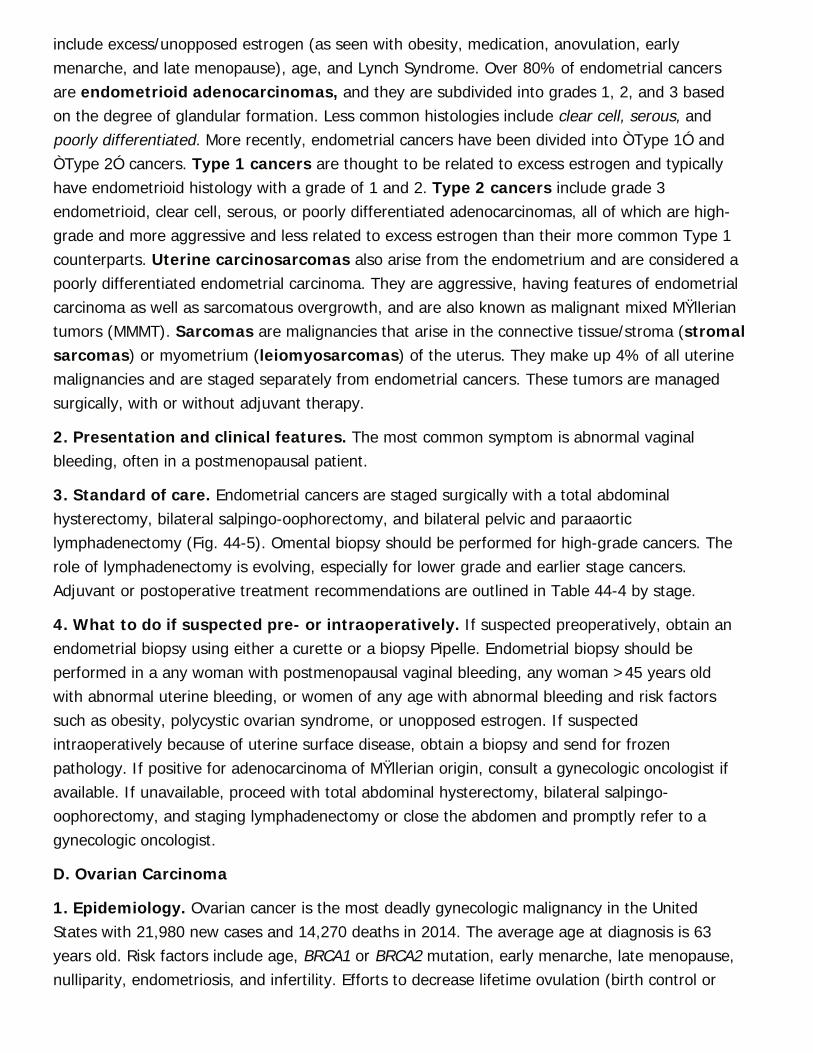
include excess/unopposed estrogen (as seen with obesity, medication, anovulation, earlymenarche, and late menopause), age, and Lynch Syndrome. Over 80% of endometrial cancersare endometrioid adenocarcinomas, and they are subdivided into grades 1, 2, and 3 basedon the degree of glandular formation. Less common histologies include clear cell, serous, andpoorly differentiated. More recently, endometrial cancers have been divided into ÒType 1Ó andÒType 2Ó cancers. Type 1 cancers are thought to be related to excess estrogen and typicallyhave endometrioid histology with a grade of 1 and 2. Type 2 cancers include grade 3endometrioid, clear cell, serous, or poorly differentiated adenocarcinomas, all of which are high-grade and more aggressive and less related to excess estrogen than their more common Type 1counterparts. Uterine carcinosarcomas also arise from the endometrium and are considered apoorly differentiated endometrial carcinoma. They are aggressive, having features of endometrialcarcinoma as well as sarcomatous overgrowth, and are also known as malignant mixed MŸlleriantumors (MMMT). Sarcomas are malignancies that arise in the connective tissue/stroma (stromalsarcomas) or myometrium (leiomyosarcomas) of the uterus. They make up 4% of all uterinemalignancies and are staged separately from endometrial cancers. These tumors are managedsurgically, with or without adjuvant therapy.
2. Presentation and clinical features. The most common symptom is abnormal vaginalbleeding, often in a postmenopausal patient.
3. Standard of care. Endometrial cancers are staged surgically with a total abdominalhysterectomy, bilateral salpingo-oophorectomy, and bilateral pelvic and paraaorticlymphadenectomy (Fig. 44-5). Omental biopsy should be performed for high-grade cancers. Therole of lymphadenectomy is evolving, especially for lower grade and earlier stage cancers.Adjuvant or postoperative treatment recommendations are outlined in Table 44-4 by stage.
4. What to do if suspected pre- or intraoperatively. If suspected preoperatively, obtain anendometrial biopsy using either a curette or a biopsy Pipelle. Endometrial biopsy should beperformed in a any woman with postmenopausal vaginal bleeding, any woman >45 years oldwith abnormal uterine bleeding, or women of any age with abnormal bleeding and risk factorssuch as obesity, polycystic ovarian syndrome, or unopposed estrogen. If suspectedintraoperatively because of uterine surface disease, obtain a biopsy and send for frozenpathology. If positive for adenocarcinoma of MŸllerian origin, consult a gynecologic oncologist ifavailable. If unavailable, proceed with total abdominal hysterectomy, bilateral salpingo-oophorectomy, and staging lymphadenectomy or close the abdomen and promptly refer to agynecologic oncologist.
D. Ovarian Carcinoma
1. Epidemiology. Ovarian cancer is the most deadly gynecologic malignancy in the UnitedStates with 21,980 new cases and 14,270 deaths in 2014. The average age at diagnosis is 63years old. Risk factors include age, BRCA1 or BRCA2 mutation, early menarche, late menopause,nulliparity, endometriosis, and infertility. Efforts to decrease lifetime ovulation (birth control or
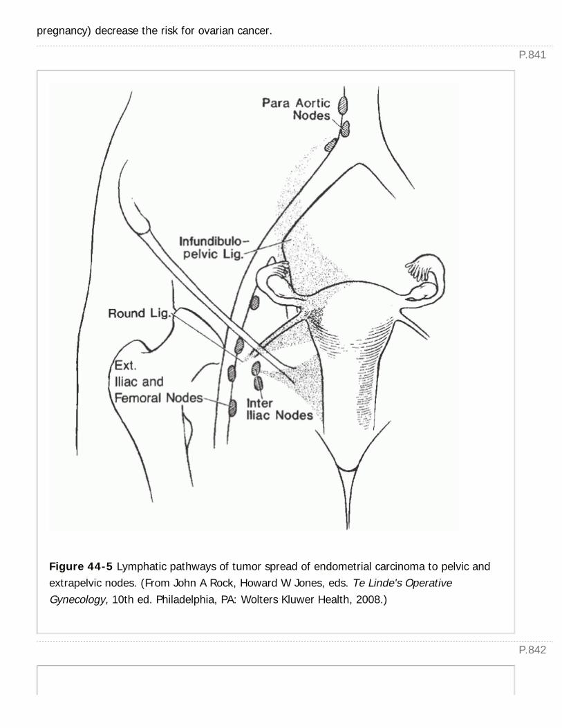
P.841
P.842
pregnancy) decrease the risk for ovarian cancer.
Figure 44-5 Lymphatic pathways of tumor spread of endometrial carcinoma to pelvic andextrapelvic nodes. (From John A Rock, Howard W Jones, eds. Te Linde's OperativeGynecology, 10th ed. Philadelphia, PA: Wolters Kluwer Health, 2008.)

TABLE 44-4 Endometrial Cancer Staging
TNM FIGO Definition Adjuvant Treatment
T1 I Tumor confined to thecorpus uteri
T1a IA Invades one-half or less ofthe myometrium
Observation for most Type 1carcinomasConsider chemotherapy (platinum+ taxane) with radiation for Type2 carcinomas
T1b IB Invades more than one-halfof the myometrium
Vaginal Brachytherapy or PelvicRadiationConsider chemotherapy (platinum+ taxane) with radiation for Type2 carcinomas
T2 II Invades cervical stroma butdoes not extend beyond theuterus
Pelvic radiationPlatinum and taxanechemotherapy with radiation forType 2 carcinomas
T3 III Local and/or regional spreadas specified
Radiation of involved field (pelvic+/- paraaortic) and chemotherapy(platinum + taxane)
T3a IIIA Involves the serosa and/oradnexa
T3b IIIB Vaginal and/or parametrialinvolvement
N1 IIIC Metastasis to the pelvicand/or para-aortic lymphnodes
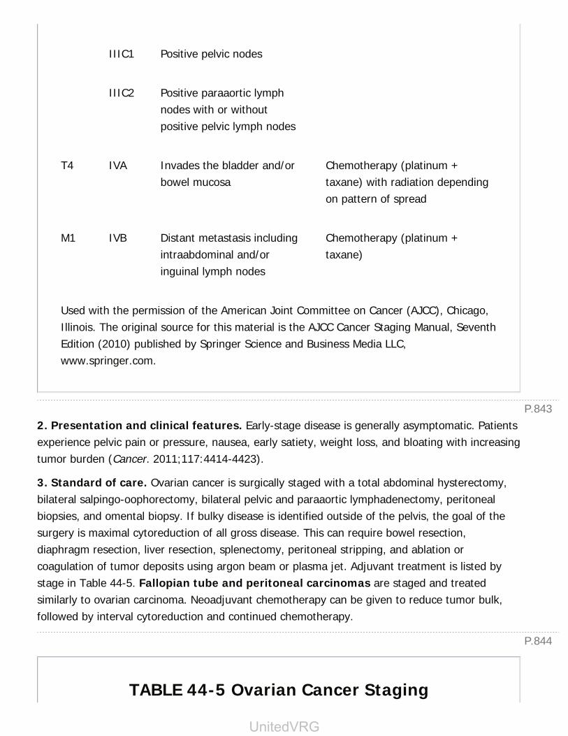
P.843
P.844
IIIC1 Positive pelvic nodes
IIIC2 Positive paraaortic lymphnodes with or withoutpositive pelvic lymph nodes
T4 IVA Invades the bladder and/orbowel mucosa
Chemotherapy (platinum +taxane) with radiation dependingon pattern of spread
M1 IVB Distant metastasis includingintraabdominal and/oringuinal lymph nodes
Chemotherapy (platinum +taxane)
Used with the permission of the American Joint Committee on Cancer (AJCC), Chicago,Illinois. The original source for this material is the AJCC Cancer Staging Manual, SeventhEdition (2010) published by Springer Science and Business Media LLC,www.springer.com.
2. Presentation and clinical features. Early-stage disease is generally asymptomatic. Patientsexperience pelvic pain or pressure, nausea, early satiety, weight loss, and bloating with increasingtumor burden (Cancer. 2011;117:4414-4423).
3. Standard of care. Ovarian cancer is surgically staged with a total abdominal hysterectomy,bilateral salpingo-oophorectomy, bilateral pelvic and paraaortic lymphadenectomy, peritonealbiopsies, and omental biopsy. If bulky disease is identified outside of the pelvis, the goal of thesurgery is maximal cytoreduction of all gross disease. This can require bowel resection,diaphragm resection, liver resection, splenectomy, peritoneal stripping, and ablation orcoagulation of tumor deposits using argon beam or plasma jet. Adjuvant treatment is listed bystage in Table 44-5. Fallopian tube and peritoneal carcinomas are staged and treatedsimilarly to ovarian carcinoma. Neoadjuvant chemotherapy can be given to reduce tumor bulk,followed by interval cytoreduction and continued chemotherapy.
TABLE 44-5 Ovarian Cancer Staging
UnitedVRG

TNM FIGO DefinitionAdjuvantTreatment
T1 I Tumor limited to one or both ovaries
T1a IA Limited to one ovary; capsule intact, notumor on ovarian surface, no malignantcells in ascites or peritoneal washings
Carboplatin andPaclitaxel for highgrade or clear cellcarcinomas
T1b IB Limited to both ovaries; capsule intact, notumor on ovarian surface, no malignantcells in ascites or peritoneal washings
Carboplatin andPaclitaxel for highgrade or clear cellcarcinomas
T1c IC Limited to one or both ovaries with any ofthe following: capsule ruptured, tumor onovarian surface, malignant cells in ascites,or peritoneal washings
Carboplatin andPaclitaxel
T2 II Tumor involves one or both ovaries withpelvic extension
Combinationintravenous andintraperitonealplatinum and taxanechemotherapy
T2a IIA Extension and/or implants on uterusand/or tubes; no malignant cells in ascitesor peritoneal washings
T2b IIB Extension to other pelvic tissues; nomalignant cells in ascites or peritonealwashings
T2c IIC Pelvic extension with malignant cells inascites or peritoneal washings
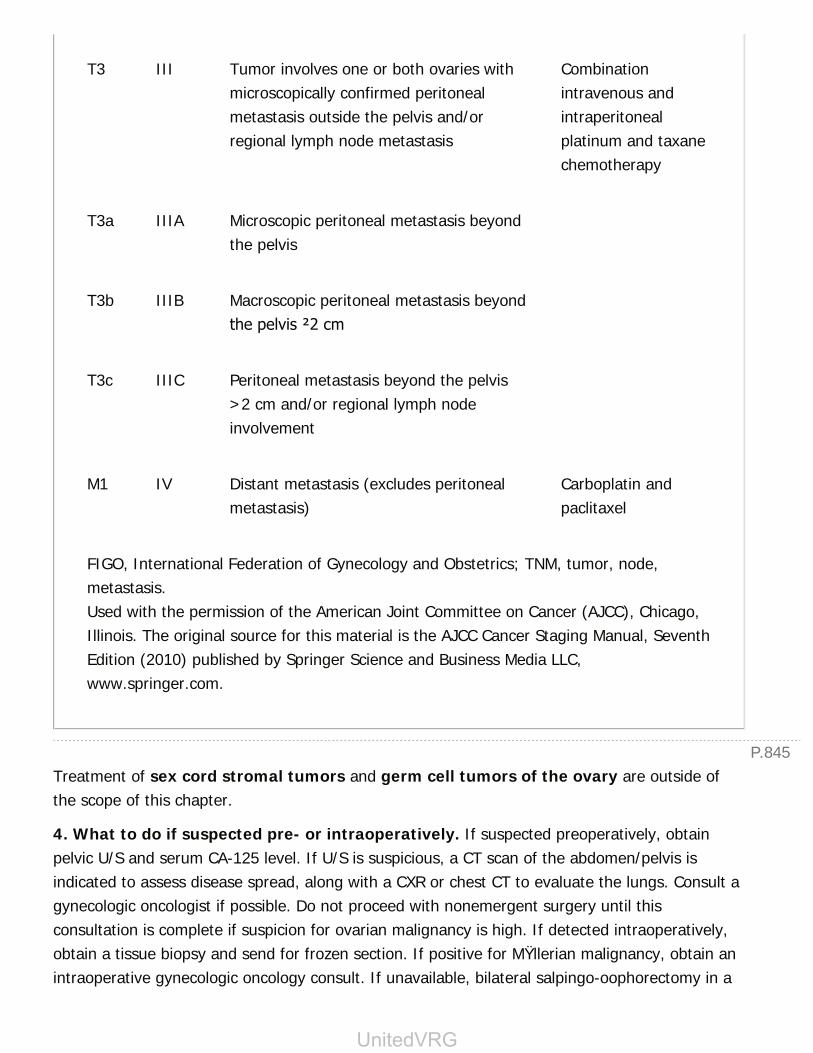
P.845
T3 III Tumor involves one or both ovaries withmicroscopically confirmed peritonealmetastasis outside the pelvis and/orregional lymph node metastasis
Combinationintravenous andintraperitonealplatinum and taxanechemotherapy
T3a IIIA Microscopic peritoneal metastasis beyondthe pelvis
T3b IIIB Macroscopic peritoneal metastasis beyondthe pelvis ≤2 cm
T3c IIIC Peritoneal metastasis beyond the pelvis>2 cm and/or regional lymph nodeinvolvement
M1 IV Distant metastasis (excludes peritonealmetastasis)
Carboplatin andpaclitaxel
FIGO, International Federation of Gynecology and Obstetrics; TNM, tumor, node,metastasis.Used with the permission of the American Joint Committee on Cancer (AJCC), Chicago,Illinois. The original source for this material is the AJCC Cancer Staging Manual, SeventhEdition (2010) published by Springer Science and Business Media LLC,www.springer.com.
Treatment of sex cord stromal tumors and germ cell tumors of the ovary are outside ofthe scope of this chapter.
4. What to do if suspected pre- or intraoperatively. If suspected preoperatively, obtainpelvic U/S and serum CA-125 level. If U/S is suspicious, a CT scan of the abdomen/pelvis isindicated to assess disease spread, along with a CXR or chest CT to evaluate the lungs. Consult agynecologic oncologist if possible. Do not proceed with nonemergent surgery until thisconsultation is complete if suspicion for ovarian malignancy is high. If detected intraoperatively,obtain a tissue biopsy and send for frozen section. If positive for MŸllerian malignancy, obtain anintraoperative gynecologic oncology consult. If unavailable, bilateral salpingo-oophorectomy in a
UnitedVRG
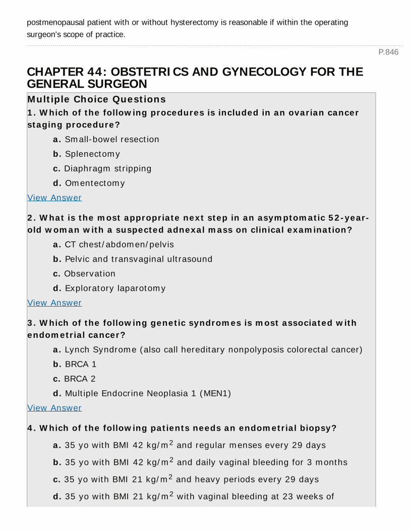
P.846
postmenopausal patient with or without hysterectomy is reasonable if within the operatingsurgeon's scope of practice.
CHAPTER 44: OBSTETRICS AND GYNECOLOGY FOR THEGENERAL SURGEONMultiple Choice Questions1. Which of the following procedures is included in an ovarian cancerstaging procedure?
a. Small-bowel resection
b. Splenectomy
c. Diaphragm stripping
d. Omentectomy
View Answer
2. What is the most appropriate next step in an asymptomatic 52-year-old woman with a suspected adnexal mass on clinical examination?
a. CT chest/abdomen/pelvis
b. Pelvic and transvaginal ultrasound
c. Observation
d. Exploratory laparotomy
View Answer
3. Which of the following genetic syndromes is most associated withendometrial cancer?
a. Lynch Syndrome (also call hereditary nonpolyposis colorectal cancer)
b. BRCA 1
c. BRCA 2
d. Multiple Endocrine Neoplasia 1 (MEN1)
View Answer
4. Which of the following patients needs an endometrial biopsy?
a. 35 yo with BMI 42 kg/m2 and regular menses every 29 days
b. 35 yo with BMI 42 kg/m2 and daily vaginal bleeding for 3 months
c. 35 yo with BMI 21 kg/m2 and heavy periods every 29 days
d. 35 yo with BMI 21 kg/m2 with vaginal bleeding at 23 weeks of
P.847
gestation
View Answer
5. If feasible, what is ideal positioning for a gravid patient undergoingsurgery?
a. Supine with a left lateral tilt
b. Dorsal lithotomy
c. Prone
d. Supine in Trendelenburg
View Answer
6. Which of the following is the most appropriate treatment for stageIIIB cervical cancer with no distant metastatic disease?
a. Radical surgical resection
b. Simple hysterectomy followed by radiation
c. Radiation with sensitizing cisplatin
d. Chemotherapy with cisplatin, taxane, and bevacizumab
View Answer
7. Which of the following scenarios describes an incomplete abortion?
a. Vaginal bleeding, cervix closed, gestational sac appropriate forestimated gestational age identified on transvaginal U/S
b. Vaginal bleeding, cervix open, thickened endometrial strip withheterogeneous echogenicity and no viable intrauterine pregnancy ontransvaginal U/S
c. Lower abdominal cramping, cervix closed, fetal pole identifiedmeasuring 8 weeks with no heart beat on transvaginal U/S
d. Vaginal bleeding, cervix closed, no identifiable intrauterine pregnancyand normal appearing endometrium on transvaginal U/S withdowntrending β-hCG
View Answer
UnitedVRG

45Biostatistics for the General Surgeon
Leisha C. Elmore
Graham A. Colditz
Evidence-based medicine aims to use information gleaned from the existing and ever-expandingbody of published research to inform clinical practice. Whether contributing to the literature viathe conduct and publication of original research, critically evaluating existing data to incorporatechanges into day-to-day healthcare delivery, or evaluating practice patterns to improve patientoutcomes at a systems level, a basic knowledge of biostatistics is important for the generalsurgeon.
I. STUDY DESIGN.Study design can be stratified into two separate categories based on the aims of inquiry:Observational and experimental. In an observational study, participants are not assigned totreatment or control groups. Inferences are made by surveillance of a group of participants.Observational studies can determine associations between exposures and outcomes, butcausation cannot be established. In contrast, in experimental studies, investigators assignparticipants to treatment and nontreatment groups and observe a specified effect. In a properlycrafted prospective trial, experimental studies can establish a causal relationship betweenexposure and outcome (Fig. 45-1).
A. Observational Studies
1. Case-control study. In this study design, participants are selected based on the presence orabsence of the outcome of interest. In order to determine the relationship of exposure tooutcome, a group of individuals with the disease (cases) are compared to a group of individualswithout the disease (controls). Evaluation of exposures in both cases and controls is conducted todetermine systematic differences between the groups. A case-control study can be a quick andrelatively inexpensive first step in evaluating whether an exposure is associated with a givendisease. Additionally, this study design is useful for investigation of rare diseases. This approachcan also be used to sample cases and controls from a much larger randomized trial or cohortstudy in which biomarkers or other expensive laboratory measures are being considered foradditional analysis (Fig. 45-2).
2. Cohort study. In cohort studies, individuals are divided into groups based on exposure andfollowed over time to document incidence of disease or the development of the outcome of
> Table of Contents > 45 - Biostatistics for the General Surgeon
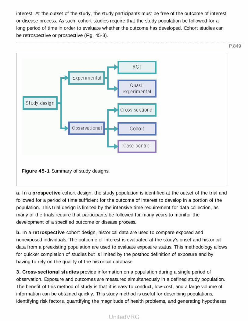
P.849
interest. At the outset of the study, the study participants must be free of the outcome of interestor disease process. As such, cohort studies require that the study population be followed for along period of time in order to evaluate whether the outcome has developed. Cohort studies canbe retrospective or prospective (Fig. 45-3).
Figure 45-1 Summary of study designs.
a. In a prospective cohort design, the study population is identified at the outset of the trial andfollowed for a period of time sufficient for the outcome of interest to develop in a portion of thepopulation. This trial design is limited by the intensive time requirement for data collection, asmany of the trials require that participants be followed for many years to monitor thedevelopment of a specified outcome or disease process.
b. In a retrospective cohort design, historical data are used to compare exposed andnonexposed individuals. The outcome of interest is evaluated at the study's onset and historicaldata from a preexisting population are used to evaluate exposure status. This methodology allowsfor quicker completion of studies but is limited by the posthoc definition of exposure and byhaving to rely on the quality of the historical database.
3. Cross-sectional studies provide information on a population during a single period ofobservation. Exposure and outcomes are measured simultaneously in a defined study population.The benefit of this method of study is that it is easy to conduct, low-cost, and a large volume ofinformation can be obtained quickly. This study method is useful for describing populations,identifying risk factors, quantifying the magnitude of health problems, and generating hypotheses
UnitedVRG
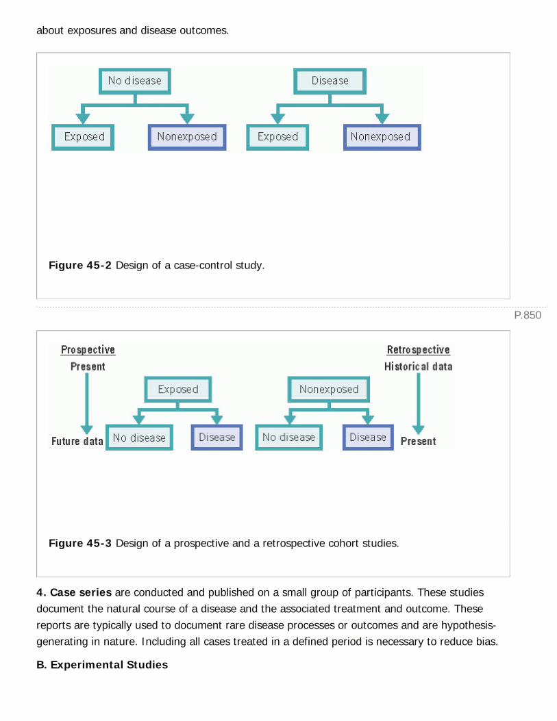
P.850
about exposures and disease outcomes.
Figure 45-2 Design of a case-control study.
Figure 45-3 Design of a prospective and a retrospective cohort studies.
4. Case series are conducted and published on a small group of participants. These studiesdocument the natural course of a disease and the associated treatment and outcome. Thesereports are typically used to document rare disease processes or outcomes and are hypothesis-generating in nature. Including all cases treated in a defined period is necessary to reduce bias.
B. Experimental Studies

P.851
1. Randomized controlled trials (RCT). In an RCT, patients are randomly assigned tointervention and control groups or arms. This trial design is the gold standard for clinicalresearch.
a. Randomization is a key element in this study design. The random allocation of participantsfacilitates unbiased distribution to the study arms of both known/measured andunknown/unmeasured participant covariates, that is, observed, immutable characteristics thatmay or may not be associated with the outcome of interest and that may bias an observer'sinterpretation of the relationship between the intervention being studied and the outcome to beobserved.
b. Blinding is the concealment of participant allocation to treatment and control groups. It canbe utilized by the trial researchers with respect to both the investigators and the participants.From the patient perspective, blinding means that participants do not know if they are in thetreatment or control group. Investigators can also be blinded so they are not aware of patientassignment. When neither investigators nor participants are informed of participants' allocationstatus, an experiment is considered double-blinded.
2. Quasi-experimental studies. This study design is similar to the RCT in that patients areassigned to treatment and control groups and observed for a specific outcome. The distinguishingcharacteristic is that patients are not randomly assigned to treatment groups. This design is usedwhen randomization is impractical or unethical.
C. Systematic review and meta-analysis are methods to critically appraise existing publishedliterature and develop a consensus amongst the data. The terms are often used interchangeably,but they are distinct entities. A systematic review is a qualitative literature review. It isconducted by establishing a clear clinical question, completing an exhaustive search of publisheddata, identifying relevant studies, and analyzing the data.
Analysis is largely qualitative but may have quantitative components. A quantitative statisticalanalysis of results gleaned from a literature search of published data is referred to as a meta-analysis; furthermore, if individual patient data are abstracted from these results, they can beanalyzed in a pooled analysis, which reduces heterogeneity among studies by using a moreuniform classification of participant covariates.
II. DATA ANALYSISA. Basic Statistical Principles
1. The null hypothesis (Ho) is the premise that there is no difference between the control group
and the treatment group. In the simplest terms, the aim of the typical study is to reject the nullhypothesis and demonstrate a statistically significant difference between the control andtreatment groups.
UnitedVRG

P.852
2. p-value (p) is defined as the probability of rejecting the null when the null hypothesis is infact true. In the typical clinical study, authors refer to a finding as statistically significant when p<0.05.
3. A confidence interval (CI) indicates the precision of a study measure. It is expressed as alower limit (A) and an upper limit (B) bounded by parentheses (A,B). When the p-value is set at0.05, the CI is 95%, indicating that if a study were repeated multiple times, the true sampleparameter or measure would fall within the CI 95% of the time.
B. Estimating Risk
1. Prevalence is the proportion of individuals in a given population with a specific diseaseprocess or risk factor at a particular point in time.
2. Incidence is the number of new cases developing in a population over a discrete period oftime.
3. Relative risk (RR) is the measure of association for cohort studies. It is calculated byevaluating the incidence of a disease in those who were exposed to a certain risk factor andcomparing it to the incidence of that disease in those who were not exposed to the same riskfactor.
a. Interpreting relative risk
(1) If RR = 1, the risk of developing the outcome is the same for individuals with and without therisk factor, that is, exposed and unexposed individuals.
(2) If RR > 1, the risk of developing the outcome is greater among the exposed individuals.
(3) If RR <1, the risk of developing the outcome is less in the exposed group.
4. Odds ratio (OR) is the measure of association for case-control studies. This ratio comparesthe likelihood that people with the outcome of interest might have been exposed to a particularrisk factor to the likelihood that people who did NOT have the outcome of interest were exposed.The odds ratio represents a snapshot of cases and controls and does not evaluate the incidenceof disease over time as seen in a cohort study.
TABLE 45-1 Sample 2 × 2 Table
Outcome (Disease) No Outcome (No Disease)
Exposed A B
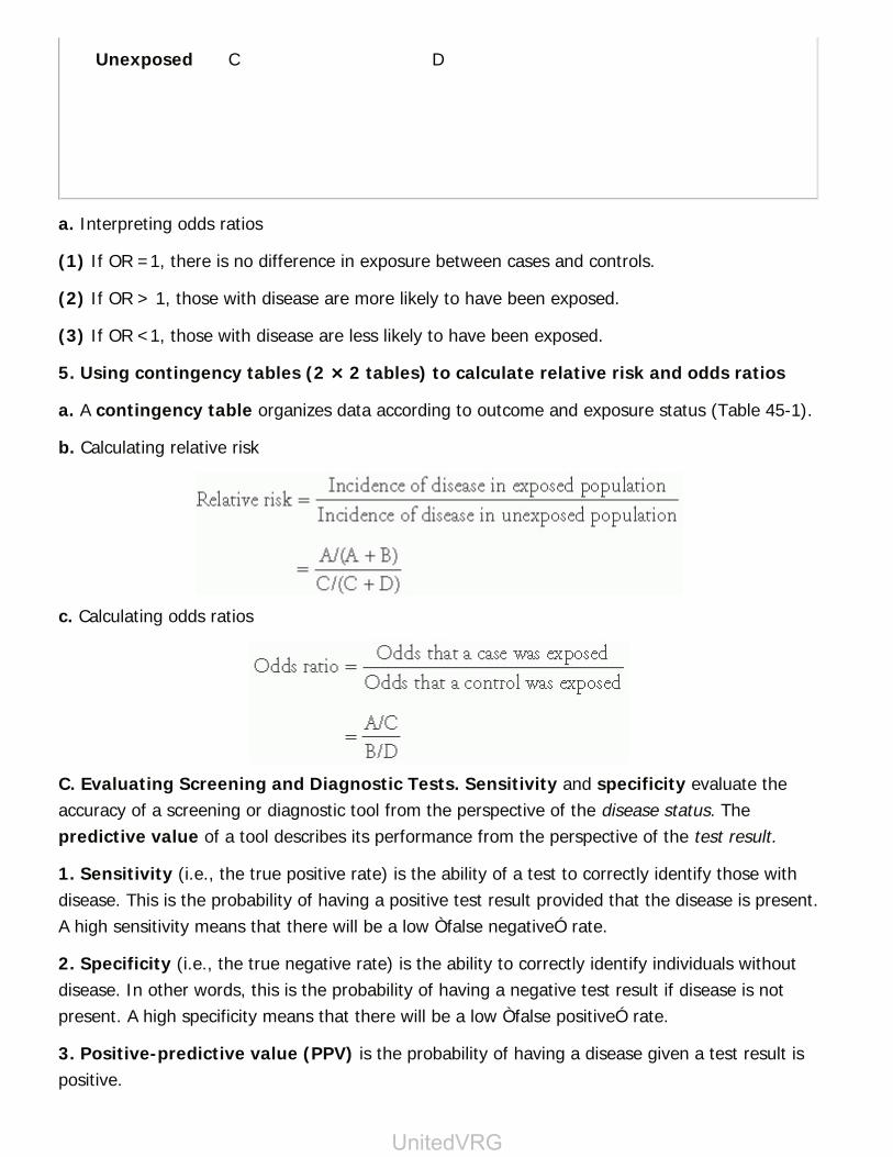
Unexposed C D
a. Interpreting odds ratios
(1) If OR =1, there is no difference in exposure between cases and controls.
(2) If OR > 1, those with disease are more likely to have been exposed.
(3) If OR <1, those with disease are less likely to have been exposed.
5. Using contingency tables (2 × 2 tables) to calculate relative risk and odds ratios
a. A contingency table organizes data according to outcome and exposure status (Table 45-1).
b. Calculating relative risk
c. Calculating odds ratios
C. Evaluating Screening and Diagnostic Tests. Sensitivity and specificity evaluate theaccuracy of a screening or diagnostic tool from the perspective of the disease status. Thepredictive value of a tool describes its performance from the perspective of the test result.
1. Sensitivity (i.e., the true positive rate) is the ability of a test to correctly identify those withdisease. This is the probability of having a positive test result provided that the disease is present.A high sensitivity means that there will be a low Òfalse negativeÓ rate.
2. Specificity (i.e., the true negative rate) is the ability to correctly identify individuals withoutdisease. In other words, this is the probability of having a negative test result if disease is notpresent. A high specificity means that there will be a low Òfalse positiveÓ rate.
3. Positive-predictive value (PPV) is the probability of having a disease given a test result ispositive.
UnitedVRG

P.853
4. Negative-predictive value (NPV) is the probability of not having a disease given a negativetest result.
TABLE 45-2 Calculating Sensitivity, Specificity, PPV,and NPV Using a 2 × 2 Table
Disease Present Disease Absent
Positive test True positive (A) False positive (B) PPV = A/(A + B)
Negativetest
False negative (C)Sensitivity = A/(A +C)
True negative (D)Specificity = D/(B +D)
NPV = D/(C +D)
PPV, positive predictive value; NPV, negative predictive value.
5. It is worth noting that while sensitivity and specificity are not affected by the prevalence ofdisease, the predictive value of a test is related to the prevalence of a disease process. As theprevalence of a disease process decreases, the positive predictive value also decreases and thenegative predictive value increases (Table 45-2).
D. Commonly Used Statistical Tests and Analytic Approaches
1. t-tests allow for comparison of a continuous, normally distributed variable of interest in twogroups. For example, a t-test could be used to compare hospital lengths of stay after laparoscopicversus open colectomy.
2. Analysis of variance (ANOVA) allows for comparison of a continuous, normally distributedvariable of interest in more than two groups. Thus, ANOVA could be used to compare hospitallengths of stay after laparoscopic versus open versus robotic colectomy.
3. The Chi-Square test is used to analyze categorical data and contingency tables.
4. Survival analyses allow for modeling and comparison of differences in time-to-eventbetween groups. Time-to-event is the duration of time between entry into a study or diagnosis ofdisease and a specified endpoint, which in medicine is often death or recurrence of disease. Apatient is censored if her time-to-event is unknown either because the patient or her informationcannot be accounted for (e.g., she becomes lost to follow-up) or because the study ends before
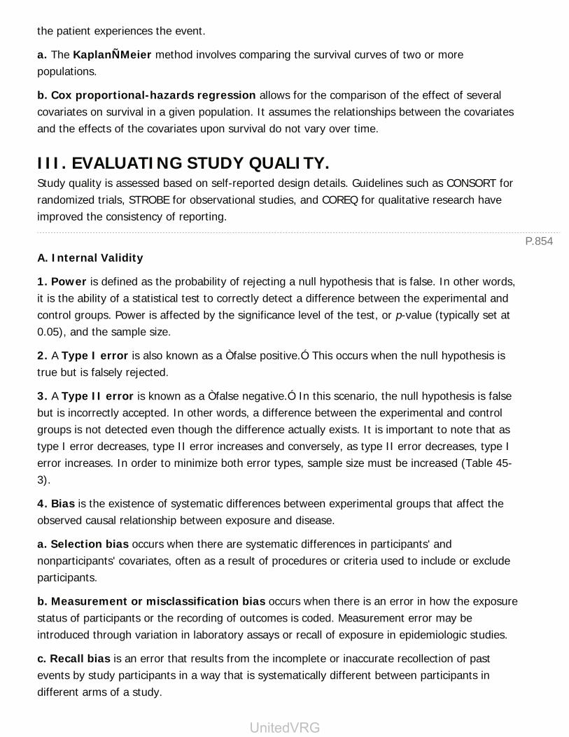
P.854
the patient experiences the event.
a. The KaplanÑMeier method involves comparing the survival curves of two or morepopulations.
b. Cox proportional-hazards regression allows for the comparison of the effect of severalcovariates on survival in a given population. It assumes the relationships between the covariatesand the effects of the covariates upon survival do not vary over time.
III. EVALUATING STUDY QUALITY.Study quality is assessed based on self-reported design details. Guidelines such as CONSORT forrandomized trials, STROBE for observational studies, and COREQ for qualitative research haveimproved the consistency of reporting.
A. Internal Validity
1. Power is defined as the probability of rejecting a null hypothesis that is false. In other words,it is the ability of a statistical test to correctly detect a difference between the experimental andcontrol groups. Power is affected by the significance level of the test, or p-value (typically set at0.05), and the sample size.
2. A Type I error is also known as a Òfalse positive.Ó This occurs when the null hypothesis istrue but is falsely rejected.
3. A Type II error is known as a Òfalse negative.Ó In this scenario, the null hypothesis is falsebut is incorrectly accepted. In other words, a difference between the experimental and controlgroups is not detected even though the difference actually exists. It is important to note that astype I error decreases, type II error increases and conversely, as type II error decreases, type Ierror increases. In order to minimize both error types, sample size must be increased (Table 45-3).
4. Bias is the existence of systematic differences between experimental groups that affect theobserved causal relationship between exposure and disease.
a. Selection bias occurs when there are systematic differences in participants' andnonparticipants' covariates, often as a result of procedures or criteria used to include or excludeparticipants.
b. Measurement or misclassification bias occurs when there is an error in how the exposurestatus of participants or the recording of outcomes is coded. Measurement error may beintroduced through variation in laboratory assays or recall of exposure in epidemiologic studies.
c. Recall bias is an error that results from the incomplete or inaccurate recollection of pastevents by study participants in a way that is systematically different between participants indifferent arms of a study.
UnitedVRG
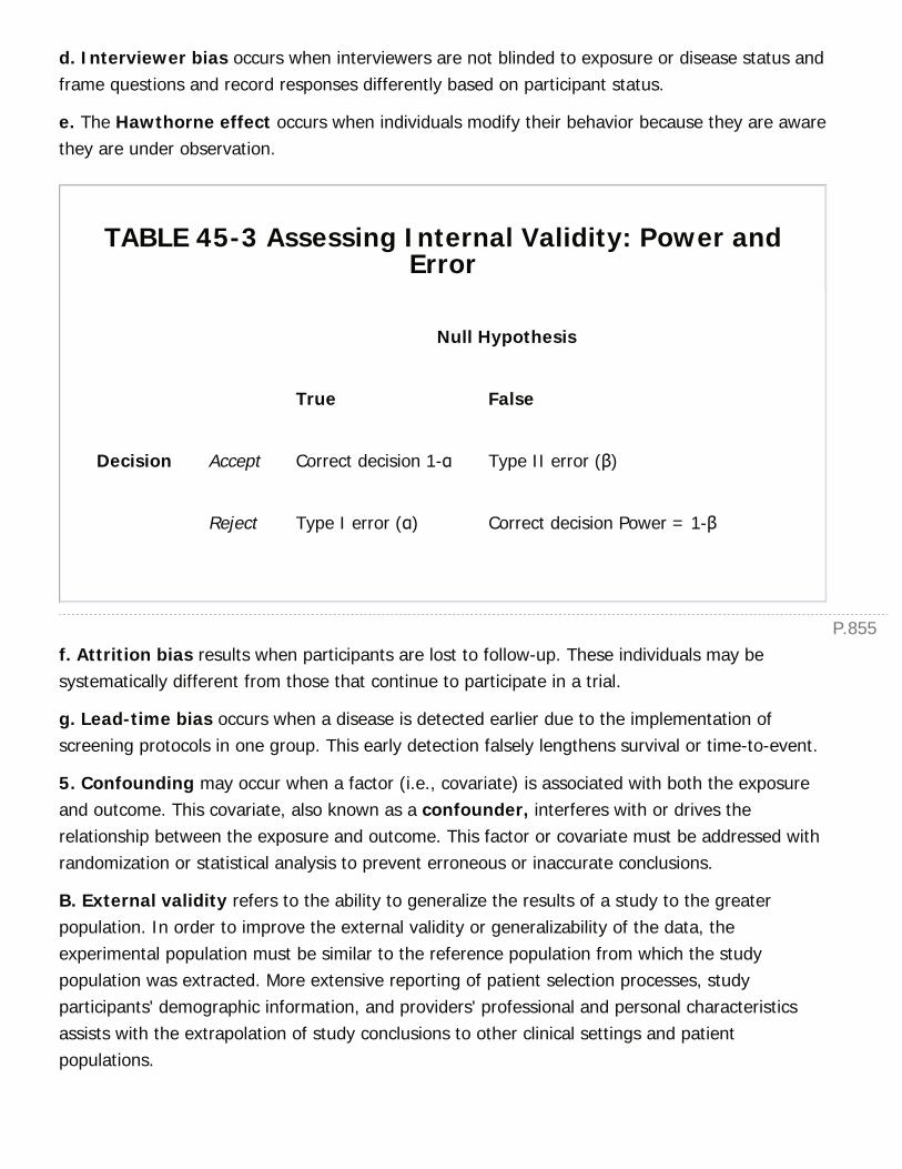
P.855
d. Interviewer bias occurs when interviewers are not blinded to exposure or disease status andframe questions and record responses differently based on participant status.
e. The Hawthorne effect occurs when individuals modify their behavior because they are awarethey are under observation.
TABLE 45-3 Assessing Internal Validity: Power andError
Null Hypothesis
True False
Decision Accept Correct decision 1-α Type II error (β)
Reject Type I error (α) Correct decision Power = 1-β
f. Attrition bias results when participants are lost to follow-up. These individuals may besystematically different from those that continue to participate in a trial.
g. Lead-time bias occurs when a disease is detected earlier due to the implementation ofscreening protocols in one group. This early detection falsely lengthens survival or time-to-event.
5. Confounding may occur when a factor (i.e., covariate) is associated with both the exposureand outcome. This covariate, also known as a confounder, interferes with or drives therelationship between the exposure and outcome. This factor or covariate must be addressed withrandomization or statistical analysis to prevent erroneous or inaccurate conclusions.
B. External validity refers to the ability to generalize the results of a study to the greaterpopulation. In order to improve the external validity or generalizability of the data, theexperimental population must be similar to the reference population from which the studypopulation was extracted. More extensive reporting of patient selection processes, studyparticipants' demographic information, and providers' professional and personal characteristicsassists with the extrapolation of study conclusions to other clinical settings and patientpopulations.
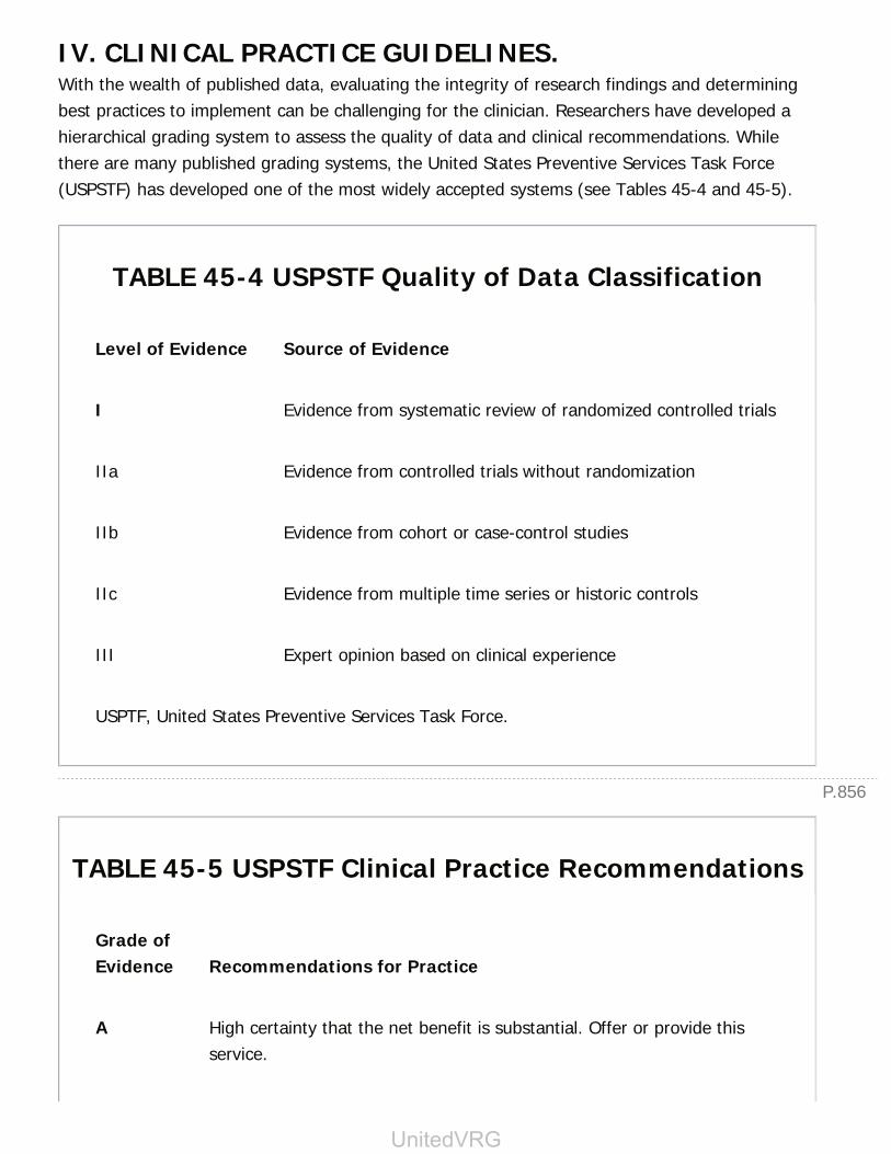
P.856
IV. CLINICAL PRACTICE GUIDELINES.With the wealth of published data, evaluating the integrity of research findings and determiningbest practices to implement can be challenging for the clinician. Researchers have developed ahierarchical grading system to assess the quality of data and clinical recommendations. Whilethere are many published grading systems, the United States Preventive Services Task Force(USPSTF) has developed one of the most widely accepted systems (see Tables 45-4 and 45-5).
TABLE 45-4 USPSTF Quality of Data Classification
Level of Evidence Source of Evidence
I Evidence from systematic review of randomized controlled trials
IIa Evidence from controlled trials without randomization
IIb Evidence from cohort or case-control studies
IIc Evidence from multiple time series or historic controls
III Expert opinion based on clinical experience
USPTF, United States Preventive Services Task Force.
TABLE 45-5 USPSTF Clinical Practice Recommendations
Grade ofEvidence Recommendations for Practice
A High certainty that the net benefit is substantial. Offer or provide thisservice.
UnitedVRG
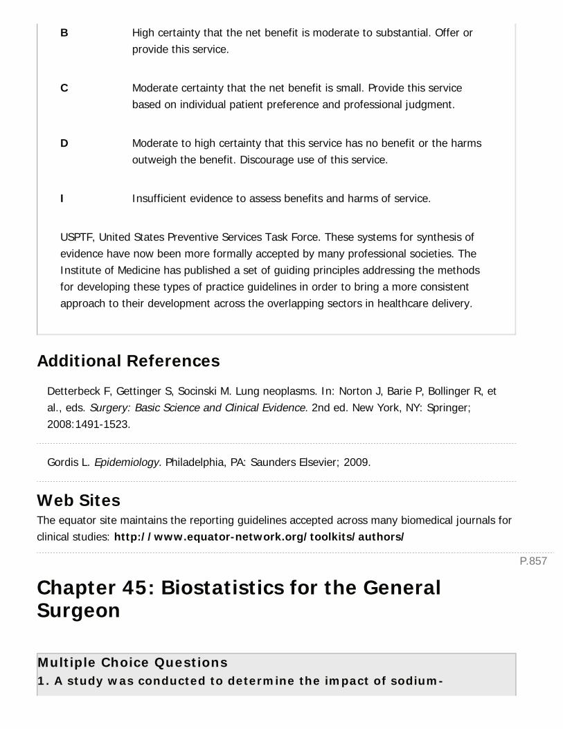
P.857
B High certainty that the net benefit is moderate to substantial. Offer orprovide this service.
C Moderate certainty that the net benefit is small. Provide this servicebased on individual patient preference and professional judgment.
D Moderate to high certainty that this service has no benefit or the harmsoutweigh the benefit. Discourage use of this service.
I Insufficient evidence to assess benefits and harms of service.
USPTF, United States Preventive Services Task Force. These systems for synthesis ofevidence have now been more formally accepted by many professional societies. TheInstitute of Medicine has published a set of guiding principles addressing the methodsfor developing these types of practice guidelines in order to bring a more consistentapproach to their development across the overlapping sectors in healthcare delivery.
Additional References
Detterbeck F, Gettinger S, Socinski M. Lung neoplasms. In: Norton J, Barie P, Bollinger R, etal., eds. Surgery: Basic Science and Clinical Evidence. 2nd ed. New York, NY: Springer;2008:1491-1523.
Gordis L. Epidemiology. Philadelphia, PA: Saunders Elsevier; 2009.
Web SitesThe equator site maintains the reporting guidelines accepted across many biomedical journals forclinical studies: http://www.equator-network.org/toolkits/authors/
Chapter 45: Biostatistics for the GeneralSurgeon
Multiple Choice Questions1. A study was conducted to determine the impact of sodium-

bicarbonate preprocedural hydration on the incidence of contrast-induced nephropathy. Which of the following analytic techniques wouldbe best suited to analyze this data?
a. ANO VA
b. Cox proportional-hazards analysis
c. Chi-square test
d. t-test
View Answer
2. A colorectal surgeon wants to investigate the role of Alvimopan(Entereg), a peripherally acting µ-opioid antagonist, on the number ofdays until return of bowel function after open colectomy. Which of thefollowing statistical methods would be used to evaluate the impact ofthis drug?
a. Wilcoxon rank sum test
b. t-test
c. ANO VA
d. Chi-square test
View Answer
3. As the prevalence of a disease increases, which of the following istrue?
a. Positive predictive decreases
b. Positive predictive value does not change
c. Sensitivity increases
d. Sensitivity does not change
View Answer
4. Pseudomyxoma peritonei is a form of cancer that produces mucinousascites and is most commonly secondary to a primary tumor of theappendix. Researchers are interested in conducting a study todetermine factors associated with the development of pseudomyxomaperitonei in patients with appendiceal cancer. Which of the followingstudy designs is most appropriate to investigate this clinical question?
a. Case-Control Study
b. Cohort Study
c. Randomized Controlled Trial
UnitedVRG
P.858
d. Cross-Sectional Study
View Answer
The following scenario applies to questions 5 to 7:
Researchers are developing a new diagnostic test to identify patients with lungcancer. A total of 150 patients with lung cancer are tested and 300 patientswithout lung cancer are included for study. A total of 125 of the patients withlung cancer and 20 of the patients without lung cancer receive positive testsresults.
5. What is the sensitivity and specificity of the diagnostic test,respectively?
a. 93% and 83%
b. 86% and 83%
c. 83% and 93%
d. 92% and 86%
View Answer
6. What are the positive predictive value and the appropriateinterpretation of the result?
a. Given a positive test results, the likelihood of having lung cancer is86%.
b. Given a positive test results, the likelihood of having lung cancer is83%.
c. In patients with lung cancer, the probability of a positive test is 86%.
d. In patients with lung cancer, the probability of a positive test is 83%.
View Answer
7. What is the false positive rate?
a. 17%
b. 13%
c. 7%
d. 8%
View Answer

46Patient Safety and Quality Improvement inSurgery
Shuddhadeb Ray
Bruce Lee Hall
Jacqueline M. Saito
I. PATIENT SAFETYA. Introduction: What is Patient Safety?
1. Institute of Medicine (IOM): To Err is Human (National Academies Press (US); 2000. BookshelfID: NBK225179)
a. In 1999, the IOM reviewed medical errors in the United States and estimated that up to 98,000patients die in hospitals each year due to preventable medical errors.
b. The majority of errors do not result from individual recklessness, but rather from issues withinhospital systems and processes that create an environment prone or vulnerable to error. Forexample, the storage of adult and pediatric doses of heparin next to each other in similarcontainers could lead to improper dosing.
c. Medical errors are defined as failure of planned action to be completed as intended or theuse of a wrong plan to achieve an aim.
d. The Swiss Cheese Model of Accident Causation (Fig. 46-1):
(1) Psychologist James Reason suggested the existence of latent errors, defects in the layers oforganization of systems that could lead to active errors that then can cause harm. (CambridgeUniversity Press; 1990. ISBN: 05214194)
(2) In this model, processes are represented by slices of cheese; each slice represents a differentprocess stage or aspect, and holes represent weaknesses in each layer/process step that couldlead to failures. Weaknesses are either latent errors present in organizational influences (e.g.,inadequate institutional structure to promote safety training), improper levels of processsupervision, conditions predisposing to errors, or active errors consisting of unsafe acts. Systemsfail to prevent hazards from propagating to actual losses/errors when the ÒholesÓ (potential forerror) in all layers (process steps) align. (Nat Rev Urol. 2013;10(3):161-173.)
e. Examples: Wrong-site surgery, falls, adverse drug events, improper transfusions
> Table of Contents > 46 - Patient Safety and Quality Improvement in Surgery
UnitedVRG

P.860
f. Types of error
(1) Diagnostic
(2) Treatment
(3) Preventative (e.g., inadequate follow-up or failure to provide prophylactic treatment)
Figure 46-1 Swiss Cheese Model of Accident Causation. (From Mulholland MW, LillemoeKD, Doherty GM, et al., eds. Greenfield's surgery: scientific principles and practice. 5th ed.Philadelphia, PA: Wolters Kluwer Health, 2010.)
(4) Commission and OmissionÑErrors of commission result from Òdoing the wrong thingÓwhile errors of omission result from Ònot doing the right thing.Ó Errors of omission are moredifficult to detect and are likely more common (e.g., failure to provide/perform diagnostic,therapeutic, or preventative measures that could improve care)
(5) Other (e.g., communication or equipment failure)
2. Normalization of deviance in healthcare
a. Medical personnel repetitively and often progressively violate standard care practices until thedeviant practice becomes the new norm, creating conditions that can cause harm to patients (BusHoriz. 2010;53(2):139). An example of this phenomenon is clinicians' not washing or sanitizingtheir hands properly and routinely before seeing patients.
B. Detecting and Reporting Medical Errors
1. Errors represent opportunities to improve processes in medical practice, but they must berecognized and addressed in a systematic fashion in order for organizations to appropriately and

P.861
consistently benefit from error reporting.
2. ÒNear missesÓ are events in which an error occurs but does not result in an adverse event.These are also important to identify as opportunities for improvement in patient safety.
3. Many medical institutions use voluntary, often anonymous, reporting systems (e.g.,paper- or computer-based error reports, hotlines) open to anyone working at the institution. Thereports are then analyzed by hospital risk management personnel or a patient safety and/orquality improvement department (Institute for Healthcare Improvement, http://www.ihi.org,2015).
a. Only 10% to 20% of medical errors are caught using such a system due to lack of errorrecognition, fear of punishment, difficulty in
using reporting systems, and perceptions that error-reporting is punitive.
b. In addition to hospital-based systems, reporting systems for medical errors can be in place atthe regional, state, or national levels.
4. Human factors in medical errors (Institute for Healthcare Improvement,http://www.ihi.org, 2015)
a. Human factors engineering is a field of science that takes into consideration humancharacteristics within processes. A multidisciplinary approach (physiology, anatomy, ergonomics,and psychology) is used to understand how humans perform under varying conditions.
b. Human factors that can potentially increase the risk of a medical error include
(1) Pre-existing psychological and/or physiological states including stress, fatigue, andboredom
(2) Decision-making factors including memory, reasoning, and judgment
(3) Action factors including communication skills, influence, and technique to perform a specificaction
c. Example: Pattern Matching during ReadingÑHumans visually read the beginning and endingletters of each word. When looking at ÒDo___mine,Ó some individuals automatically think ofÒDopamine,Ó others think of ÒDobutamineÓ; as a result, these two medications could beinadvertently substituted.
5. Root cause analysis (RCA)
a. A method to retrospectively review medical errors to find latent errors within a systemwith the overall goal of preventing these errors from recurring
b. Often initiated after an adverse event or a Ònear missÓ
c. A misnomer since multiple Òroot causesÓ are often found with each analysis
UnitedVRG
P.862
d. RCA is conducted in a team-based fashion with goals to identify strategies to improveprocesses and not to blame particular individuals
(1) The team generally consists of 4 to 6 members from all aspects of care within a healthsystem and members from a Risk Management or Safety/Quality Office. Although practices varybetween institutions, generally personnel directly involved in the evaluated incident are excludedfrom the RCA team in order to minimize bias.
e. RCA steps:
(1) Define the problem: Gather all information relevant to the incident using chart review,debriefing of involved individuals, and development of a detailed timeline
(2) Develop a cause-and-effect chart: Often referred to as a Fishbone or IshikawaDiagram (see Fig. 46-3), this will help organize possible cause-and-effect relationships thatmight have contributed to the investigated error. Ask Òwhy?Ó five times for each possible causeidentified to help identify a root cause.
(3) Identify possible solutions: As a group, find possible systemic fixes to prevent the errorfrom occurring again, categorize the fixes as high or low impact, and assess the resourcerequirements for each fix. In this manner, tasks with the highest impact but lowest resourceutilization can be the focus for the short term, whereas resource-intensive tasks can beconsidered after more discussion and research.
(4) Implementation and review: Initiate any solutions found to be appropriate from theprevious step and intermittently review the effectiveness of new processes.
C. Communication and Handoffs
1. Communicating adverse events
a. Adverse event: An injury or suboptimal outcome caused by medical management rather thanthe patient's underlying disease. (Lippincotts Case Manag. 2006;11(4):193-194.)
b. Healthcare workers might avoid reporting adverse events due to fear of censure, colleaguedisapproval, negative publicity, or anger from patients and/or employers; threat of medicalmalpractice; and the effort required to report.
c. Important steps that should be taken following an adverse event:
(1) First and foremost address medical needs of the patient.
(2) Communicate what is known about the adverse event to the patient or patientrepresentative with a statement of empathy.
(3) Notify departments, entities, specific people about the adverse event per facilitypolicies in your institution.
P.863
(4) Document adverse event and communication following the adverse event with thepatient and other entities in the medical record.
d. An apology to the patient by the physician following a medical error can decrease blame,anger, and the risk of litigation and may also increase trust in the doctorÑpatient relationship.Despite this evidence, some institutions view apologies to patients as risky and an admission ofguilt that could subsequently increase the risk of litigation; therefore, it is important tounderstand your institution's specific guidance on apologizing to patients. (Clin Orthop Relat Res.2009;467(2):376-382.)
2. Patient health education
a. The American Hospital Association adopted the Patients' Bill of Rights in 1973, stating thatpatients have the right to relevant, current, and understandable information of their diagnosis,treatment, and prognosis.
b. Evidence shows that following informed consent, a significant number of surgical patients donot adequately understand all aspects of the planned surgical procedure. (Am J Surg.2009;198(3):420-435.)
c. Effective communication of health information to the patient is essential to respect patientautonomy, manage patient expectations following a surgical procedure, and improve patientsatisfaction.
d. The informed consent process can be personalized and made more patient-centered throughuse of the Request for Treatment Form; following the consent process, the patient completesthe form in his or her own words at home, and then returns the form to the physician so that thepatient's understanding of the planned treatment can be confirmed and any questions ormisconceptions can be answered or clarified. (Ann R Coll Surg Engl. 2010;92(2):93-100.)
3. Handoffs in surgical residency
a. Resident duty-hour restrictions established in 2010 have resulted in a greater number ofhandoffs between clinical providers. (N Engl J Med. 2010;363(2):e3.)
b. Handoffs, if conducted improperly, can lead to an increase in adverse events, worse patientoutcomes, longer hospital stays, and increased admissions to the intensive care unit. (Am J Med.2012;125(1):104-110.)
c. Communication breakdown during handoffs is one of the most common factors recognizedas causing errors that lead to adverse events. (Surgery. 2003;133(6):614-621.)
d. Due to the unique needs of different care teams and surgical subspecialties, a universalstandardized handoff tool might not be feasible.
e. Handoffs can occur between two individuals but are often most efficient if carried out as a
UnitedVRG

P.864
group with the supervision of senior residents, fellows, or faculty.
f. Mnemonics and tools have been created to help standardize handoffs and thereby prevent theomission of the most important information:
(1) ÒSHOUTÓÑSick or not, History, Objective data, Upcoming plan/disposition, To do. (PracticalQI: The Basics of Quality Improvement. American College of Surgeons; 2014.)
(2) ÒSBARÓÑSituation, Background, Assessment, Recommendation. (Jt Comm J Qual PatientSaf. 2006;32(3):167-175.)
(3) ÒI-PASSÓÑIllness severity, Patient summary, Action list, Situational awareness/contingencyplan. (Pediatrics. 2012;129(2):201-204.)
D. Culture of Safety
1. Healthcare organizations establish processes that focus on the continuing improvement ofreliability and safety in caring for patients.
a. The following aspects must be addressed by organizations to attain a culture of safety,which requires psychological safety in which employees understand that their concerns will beaddressed, active leadership that encourages open expression of concerns from healthcareworkers, transparency in addressing patient safety issues, and fairness in dealing with medicalerrors in which people are not blamed for systems-based errors. (Institute for HealthcareImprovement, http://www.ihi.org, 2015)
b. Discussing patient safety can be difficult in healthcare settings due to the hierarchical nature ofmedicine and the relative ease of staying quiet rather than voicing concerns about patient safetyissues.
c. Surveys of U.S. hospitals show that a strong culture of safety is promoted by organizations thatvalue teamwork, whereas weak safety culture is associated with organizations focused onhierarchy. (Health Care Manage Rev. 2009;34(4):300-311.) Organizations with a strong culture ofsafety reported better performance on patient safety indicators. (Health Care Manage Rev.2009;34(4):300-311.)
2. Physician peer support (second victim)
a. Following an adverse event, patients and their families receive support from most medicalteams, but physicians involved in the event are rarely treated in the same manner. Physiciansinvolved in adverse events report anxiety about future errors, loss of confidence, sleep difficulties,reduced job satisfaction, and increase in job-related stress. (Jt Comm J Qual Patient Saf.2007;33(8):467-476.)
b. A physician involved with and affected by an adverse event, is the Òsecond victim.Ó Withappropriate institutional support, he or she will pass through the following phases en route to
P.865
recovery: Accident response, managing intrusive reflections, restoring personal integrity, enduringthe review of the case by the institution, getting proper emotional support from peers, andmoving on from the adverse event. (Qual Saf Health Care. 2009;18(5):325-330.)
3. Professionalism
a. American College of Surgeons (ACS) Code of Professional Conduct notes thatprofessionalism Òserves as the basis of the social contract between medicine and the society thatit serves.Ó (American College of Surgeons, https://www.facs.org/about-acs/statements/stonprin,2014)
b. The ACS Code of Professional Conduct defines responsibilities related to patient safetyincluding
(1) Full disclosure adverse events and medical errors
(2) Participation in self-regulation by setting, maintaining, and enforcing practice standards
(3) Improvement of care by evaluating its processes and outcomes
II. QUALITY IMPROVEMENTA. Introduction
1. Quality improvement (QI) consists of systematic and continuous improvements inhealthcare settings that can be measured, especially in reference to the health status of targetedpatient groups (Health Resource and Services Administration,http://www.hrsa.gov/quality/toolbox/methodology/qualityimprovement/, 2015)
2. In QI, the focus is typically on improvement of systems instead of individual performance
3. IOM: Crossing the Quality Chasm (National Academies Press (US); 2001. Bookshelf ID:NBK222271)
a. Following IOM's 1999 report, titled To Err is Human, on errors in the medical system, Crossingthe Quality Chasm was published in 2001 as a shared vision of six aims to help healthcare bemore:
(1) SafeÑavoid injuries
(2) EffectiveÑprovide care based on scientific evidence
(3) Patient-centeredÑprovide care respectful to patient values
(4) TimelyÑreduce wait-time and harmful delays
(5) EfficientÑavoid waste of equipment, ideas, and energy
(6) EquitableÑprovide care that does not vary in quality because of personal characteristics,
UnitedVRG
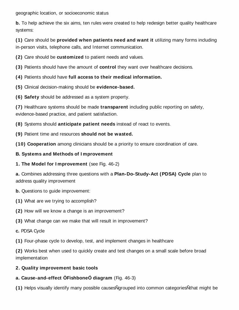
geographic location, or socioeconomic status
b. To help achieve the six aims, ten rules were created to help redesign better quality healthcaresystems:
(1) Care should be provided when patients need and want it utilizing many forms includingin-person visits, telephone calls, and Internet communication.
(2) Care should be customized to patient needs and values.
(3) Patients should have the amount of control they want over healthcare decisions.
(4) Patients should have full access to their medical information.
(5) Clinical decision-making should be evidence-based.
(6) Safety should be addressed as a system property.
(7) Healthcare systems should be made transparent including public reporting on safety,evidence-based practice, and patient satisfaction.
(8) Systems should anticipate patient needs instead of react to events.
(9) Patient time and resources should not be wasted.
(10) Cooperation among clinicians should be a priority to ensure coordination of care.
B. Systems and Methods of Improvement
1. The Model for Improvement (see Fig. 46-2)
a. Combines addressing three questions with a Plan-Do-Study-Act (PDSA) Cycle plan toaddress quality improvement
b. Questions to guide improvement:
(1) What are we trying to accomplish?
(2) How will we know a change is an improvement?
(3) What change can we make that will result in improvement?
c. PDSA Cycle
(1) Four-phase cycle to develop, test, and implement changes in healthcare
(2) Works best when used to quickly create and test changes on a small scale before broadimplementation
2. Quality improvement basic tools
a. Cause-and-effect ÒFishboneÓ diagram (Fig. 46-3)
(1) Helps visually identify many possible causesÑgrouped into common categoriesÑthat might be
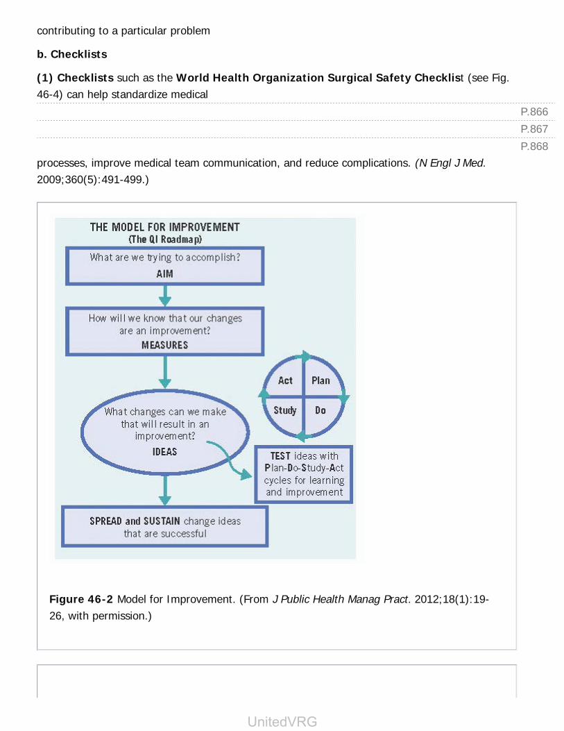
P.866P.867P.868
contributing to a particular problem
b. Checklists
(1) Checklists such as the World Health Organization Surgical Safety Checklist (see Fig.46-4) can help standardize medical
processes, improve medical team communication, and reduce complications. (N Engl J Med.2009;360(5):491-499.)
Figure 46-2 Model for Improvement. (From J Public Health Manag Pract. 2012;18(1):19-26, with permission.)
UnitedVRG
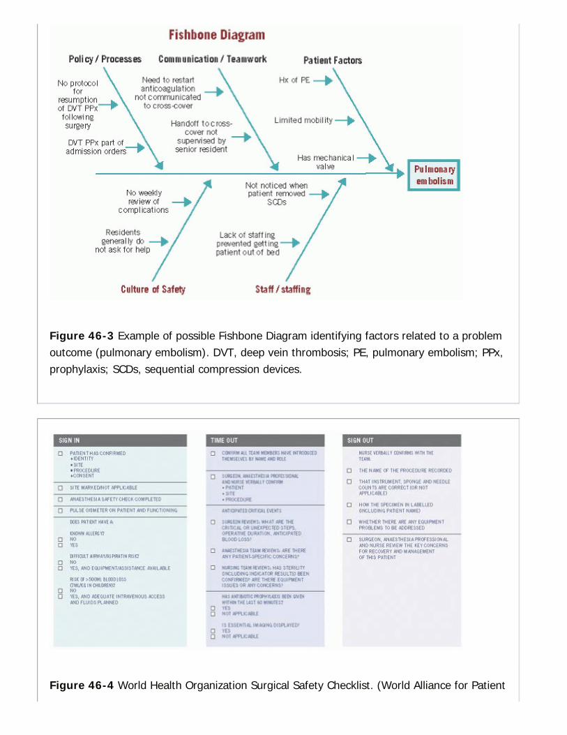
Figure 46-3 Example of possible Fishbone Diagram identifying factors related to a problemoutcome (pulmonary embolism). DVT, deep vein thrombosis; PE, pulmonary embolism; PPx,prophylaxis; SCDs, sequential compression devices.
Figure 46-4 World Health Organization Surgical Safety Checklist. (World Alliance for Patient

Safety, World Health Organization; 2008.)
c. Pareto charts
(1) A combination of a bar graph and a line graph in which the bars represent frequency of anevent or cost with largest bars (most significant factors) on the left side of the graph and the linerepresents cumulative number of events (Tague NR. The Quality Toolbox. 2nd ed. ASQ QualityPress; 2010)
(2) Helpful to display the most significant factors or causes of a problem when there are many
d. Run charts
(1) A graph that shows a measured value plotted sequentially in time (Institute for HealthcareImprovement, http://www.ihi.org, 2015)
(2) Useful for displaying the measured effect of a quality intervention
(3) A median should be calculated from measured values before the onset of the interventionand plotted as a horizontal ÒbaselineÓ from which changes can be measured to gauge success ofthe intervention
3. Morbidity and mortality conference
a. One of the first forums to review medical errors, it is a format widely used in current surgicaltraining and practice.
b. The following practices can help improve the effectiveness of the conference (Practical QI: TheBasics of Quality Improvement. American College of Surgeons; 2014):
(1) Should be held regularly, at least once a month
(2) Must have high level of attendance/engagement from faculty and residents
(3) A list of potential cases for discussion are selected prior to the conference
(4) Method of case reporting should be as complete and accurate as possible. Hospitaldatasets that track outcomes often provide more complete case lists than resident-reported caselists
4. Six Sigma and Lean
a. Two frameworks for quality improvement focused on specific concepts to improve quality
b. Six Sigma, born out of Motorola in 1986, focuses on decreasing process errors to six standarddeviations (i.e., sigma or σ) below the mean. However, most medical processes have error ratesthat average 3 to 4 sigma around the mean. Six Sigma consists of five steps (Milbank Q.1998;76(4):565-591, 510):
UnitedVRG

P.869
(1) DefineÑthe problem in detail
(2) MeasureÑerrors (per million in the original system)
(3) AnalyzeÑprocess measures to determine the conditions under which errors occur
(4) ImproveÑdesign changes aimed at decreasing errors and measure outcomes
(5) ControlÑtake steps to sustain improvements
c. Lean was created by Toyota in the 1970s as a method of quality improvement that centeredaround maximizing value from the customer's perspective and minimizing waste.
(1) Types of waste: Overproduction, waiting, excess transportation, processing, excess inventory,unnecessary movement, and defective products
(2) Value stream mapping: Processes are mapped as a series of steps for which efficiency isdefined as time during each step in which value is added for the customer (or patient) comparedto time spent without any value added
(3) 5 SÑsystem of steps essential to the Lean process. Below are the English counter-parts toJapanese words for each step:
(a) SortÑkeep only essential tools in the workplace
(b) StraightenÑarrange workplace for ease of use
(c) SweepÑmaintain a clean work environment
(d) StandardizeÑestablish best practices for each process
(e) SustainÑcommit former steps to habit
5. National Surgical Quality Improvement Program (NSQIP)
a. Developed by the American College of Surgeons, NSQIP uses trained surgical clinical reviewersto collect data from patient charts on preoperative risk factors, intraoperative variables, and 30-day postoperative morbidity and mortality. (American College of Surgeons, http://site.acsnsqip.org/, 2015)
b. NSQIP evaluations are risk-adjusted to account for patient characteristics and case-mix-adjusted to account for complexity of surgeries completed at a single center to allow forcomparison of outcomes across different medical centers.
c. Using reports of comparative performance, opportunities to improve outcomes at participatinginstitutions are identified and can be targeted with best practice guidelines and directed efforts toimprove quality of surgical care.
C. High-Reliability Organizations (HRO)

P.870
1. An organization that succeeds in avoiding errors where they are expected due to risk factorsand the complexity of the system. They exhibit five characteristics that help avoid errors and dealwith unexpected situations (Qual Saf Health Care. 2004;13 Suppl 2:ii39-44):
a. Preoccupation with failure
b. Reluctance to simplify interpretations
c. Sensitivity to operations
d. Commitment to resilience
e. Deference to expertise
Additional References
ACS NSQIP. Surgical quality improvement [Internet]. [cited 2015 Mar 5]. Available online athttp://site.acsnsqip.org/
ACS Task Force on Professionalism. Code of professional conduct. J Am Coll Surg 2004;199(5):734-735.
Banja J. The normalization of deviance in healthcare delivery. Bus Horiz 2010;53(2):139.
Chassin MR. Is health care ready for Six Sigma quality? Milbank Q 1998;76(4):565-591, 510.
Falagas ME, Korbila IP, Giannopoulou KP, et al. Informed consent: how much and what dopatients understand? Am J Surg 2009;198(3):420-435.
Gawande AA, Zinner MJ, Studdert DM, et al. Analysis of errors reported by surgeons at threeteaching hospitals. Surgery 2003;133(6):614-621.
Haig KM, Sutton S, Whittington J. SBAR: a shared mental model for improving communicationbetween clinicians. Jt Comm J Qual Patient Saf 2006;32(3):167-175.
Harrison LM, Shook ED, Harris G, et al. Applying the model for improvement in a local healthdepartment: quality improvement as an effective approach in navigating the changinglandscape of public health practice in Buncombe County, North Carolina. J Public HealthManag Pract 2012;18(1):19-26.
UnitedVRG
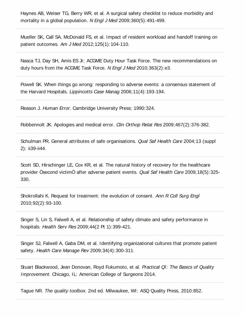
Haynes AB, Weiser TG, Berry WR, et al. A surgical safety checklist to reduce morbidity andmortality in a global population. N Engl J Med 2009;360(5):491-499.
Mueller SK, Call SA, McDonald FS, et al. Impact of resident workload and handoff training onpatient outcomes. Am J Med 2012;125(1):104-110.
Nasca TJ, Day SH, Amis ES Jr; ACGME Duty Hour Task Force. The new recommendations onduty hours from the ACGME Task Force. N Engl J Med 2010;363(2):e3.
Powell SK. When things go wrong: responding to adverse events: a consensus statement ofthe Harvard Hospitals. Lippincotts Case Manag 2006;11(4):193-194.
Reason J. Human Error. Cambridge University Press; 1990:324.
Robbennolt JK. Apologies and medical error. Clin Orthop Relat Res 2009;467(2):376-382.
Schulman PR. General attributes of safe organisations. Qual Saf Health Care 2004;13 (suppl2): ii39-ii44.
Scott SD, Hirschinger LE, Cox KR, et al. The natural history of recovery for the healthcareprovider Òsecond victimÓ after adverse patient events. Qual Saf Health Care 2009;18(5):325-330.
Shokrollahi K. Request for treatment: the evolution of consent. Ann R Coll Surg Engl2010;92(2):93-100.
Singer S, Lin S, Falwell A, et al. Relationship of safety climate and safety performance inhospitals. Health Serv Res 2009;44(2 Pt 1):399-421.
Singer SJ, Falwell A, Gaba DM, et al. Identifying organizational cultures that promote patientsafety. Health Care Manage Rev 2009;34(4):300-311.
Stuart Blackwood, Jean Donovan, Royd Fukumoto, et al. Practical QI: The Basics of QualityImprovement. Chicago, IL: American College of Surgeons 2014.
Tague NR. The quality toolbox. 2nd ed. Milwaukee, WI: ASQ Quality Press, 2010:852.
P.871
P.872
Waterman AD, Garbutt J, Hazel E, et al. The emotional impact of medical errors on practicingphysicians in the United States and Canada. Jt Comm J Qual Patient Saf 2007;33(8):467-476.
Weiser TG, Porter MP, Maier RV. Safety in the operating theatreÑa transition to systems-based care. Nat Rev Urol 2013;10(3):161-173.
WHO. WHO surgical safety checklist and implementation manual [Internet]. WHO. [cited2015 Apr 21]. Available online at http://www.who.int/patientsafety/safesurgery/ss_checklist/en/
Crossing the quality chasm: a new health system for the 21st Century – Institute of Medicine[Internet]. [cited 2015 Apr 20]. Available online athttps://www.iom.edu/Reports/2001/Crossing-the-Quality-Chasm-A-New-Health-System-for-the-21st-Century. aspx
Online learning [Internet]. [cited 2015 Mar 5]. Available online athttp://app.ihi.org/lms/onlinelearning.aspx
Quality improvement [Internet]. [cited 2015 Apr 20]. Available online at http://www.hrsa.gov/quality/toolbox/methodology/qualityimprovement/
To err is human: building a safer health system – Institute of Medicine [Internet]. [cited2015 Mar 5]. Available online at https://www.iom.edu/Reports/1999/To-Err-is-Human-Building-A-Safer-Health-System.aspx
Chapter 46: Patient Safety and QualityImprovement in Surgery
Multiple Choice Questions1. Which of the following tools is best described as a graph that showsa measured value plotted sequentially in time that is useful fordisplaying the measured effect of a quality intervention?
a. Pareto chart
b. Run chart
UnitedVRG
c. Scatter plot
d. Histogram
e. Fishbone diagram
View Answer
2. Value-stream mapping, a method to calculate the efficiency or valueadded to the customer/patient for each step in a system process, is acomponent of which of the following systems of quality improvement?
a. Six Sigma
b. National Surgery Quality Improvement Program
c. Lean
d. Model for Improvement
e. High-reliability organizations
View Answer
3. While driving to work, a physician notes that the majority of driversaround her are consistently driving above the speed limit. The conceptin patient safety that describes this behavior of routinely violatingstandard practices is:
a. Human factors engineering
b. The “second victim” phenomenon
c. Errors of omission
d. Normalization of deviance
e. Preventive error
View Answer
4. In the Model for Improvement, three questions about a quality issuemust be addressed (What are we trying to accomplish? How will weknow a change is an improvement? What change can we make that willresult in improvement?) and which of the following must also beutilized?
a. Plan-Do-Study-Act Cycle
b. Fishbone Diagram
c. Surgical Safety Checklist
d. Morbidity and Mortality Conference
e. Swiss Cheese Model of Accident Causation
View Answer

P.8735. Which of the following represents a team-based approach toretrospective review after a medical error or “near miss” event toidentify latent errors within a system?
a. Swiss Cheese Model of Accident Causation
b. Human Factors Engineering
c. Lean
d. Six Sigma
e. Root Cause Analysis
View Answer
UnitedVRG

Answer Key
CHAPTER 11. Answer: c. Per Table 1-1, patients with a history of TIA are at elevated risk for MACE.Patients with diabetes controlled with oral agents, mild renal insufficiency, and controlledhypertension are not in this elevated risk group.
2. Answer: b. Please see Table 1-2 for details regarding assessment of functional status.
3. Answer: d. Please see Table 1-3. To reduce SSI risk, hair removal should be performed withclippers. Glucose control goal should be <180 mg/dL. Core body temperature should be keptabove 36 degrees.
4. Answer: c. Please see Table 1-5. Protein C deficiency is considered a high-riskhypercoagulable state.
5. Answer: b. Please see Table 1-5. All mitral prostheses are considered high risk forthromboembolism. Bileaflet aortic valves are at lower risk.
6. Answer: c. Please see Figure 1-1. The patient in scenario C has several risk factors and hasclaudication that limits the ability to perform exercise stress testing. The patient in A requiresemergent surgery. The patients in B and D, despite risk factors, have no functional impairmentrequiring testing.
7. Answer: c. Please see Section I.B.7.a. Coumadin should be stopped 5 days prior to electivesurgery. Coumadin causes the production of inactive clotting factors. Once the drug is withheld itcan take up to 5 days for the liver to generate sufficient functional clotting factors.
8. Answer: b. The patient in B is anuric with fluid overload that appears to be leading to arequirement of mechanical ventilatory support. This is an indication for hemodialysis to removefluid and improve pulmonary function. Mild-to-moderate metabolic derangements such as in Aand C can be treated with measures less invasive than hemodialysis. Creatinine level (D) is not anindication for hemodialysis per se.
9. Answer: b. Please see Section I.B.5.b.5. Foley catheters should be removed on postoperativeday 1 unless there is concern for urinary retention, urinary tract injury, or renalinsufficiency/oliguria.
CHAPTER 21. Answer: a. In the oliguric patient, the fractional excretion of sodium helps to distinguishbetween prerenal and intrinsic renal causes of renal failure. A FENa of <1% is consistent with a
> Table of Contents > Answer Key
P.876
P.877
prerenal cause of renal failure.
2. Answer: b. Acute delirium is best treated with sleep hygiene and minimizing mood-alteringmedications. For patients with acute, hyperactive delirium, haloperidol can be used to preventself-injurious behavior. Quetiapine has been demonstrated to improve delirium and can helprestore day/night sleep wake cycling. Benzodiazepines can exacerbate delirium and should bestrictly avoided.
3. Answer: g. Oliguria and tachycardia in the early postoperative period is a red flag for possiblebleeding. This patient is in stage II shock with oliguria and mild tachycardia. The initial workupand treatment are to restore intravascular volume with a bolus of crystalloid and check ahematocrit to assess for bleeding. This patient may need transfer to the intensive care unit, butthe first step is to give the fluid bolus and assess for bleeding. Prior to returning to the operatingroom, the patient should receive resuscitation and the cause of his oliguria need to beestablished. Although he could be bleeding, he may also be under-resuscitated.
4. Answer: d. The Surgical Care Improvement Project recommends a maximum of 24 hours ofperioperative antibiotic coverage for routine general surgery procedures.
5. Answer: b. Antibiotic impregnated central venous catheters are not part of the Surgical CareImprovement Project guidelines for the prevention of perioperative infections. The CDC doesrecommend considering antibiotic impregnated catheters as an alternative method of decreasinghospital-acquired infections.
6. Answer: d. The first step in evaluating this agitated patient is to check her vital signs andpulse oximetry. It is more likely a deficit in executive function than a focal neural deficit, thereforea to c should be considered after the vital signs, but at this time e is not indicated.
7. Answer: b. In addition to sleep hygiene, constant reorientation, and exterior views, forrecalcitrant delirium, Quetiapine 25 to 50 mg can be effective adjunctive therapy. Haldol 1 to 5mg can be used for hyperactive delirium.
8. Answer: c. This patient is most likely suffering from alcohol withdrawal, given his time pointof 3 days (72 hours) since his last drink, his hallucinations, and autonomic instability (diaphoresisand tachycardia) he most likely has delirium tremens. Alcoholic seizures usually occur before 72hours, benzodiazepines are used to treat alcohol withdrawal and an overdose would not producethese symptoms, delirium's symptoms are not listed.
9. Answer: e. While a cause for hypovolemia, sepsis typically is not on the differential until afterpostoperative day 2, unless the patient has recently been operated on, or was septic prior to theoperation.
10. Answer: c. Classically most postoperative MI's occur on postoperative day 2. Any new or
UnitedVRG
P.878
worsening chest pain deserves a full myocardial infarction workup.
11. Answer: a. Since his oxygen saturation is low, he should be placed on supplemental oxygenand monitored for the need for intubation. With his surgery 2 days prior he may now bemobilizing his third space fluids and furosemide will aid in diuresis of excess fluid
12. Answer: b. The patient who recently started eating after an operation is elderly and had twobouts of emesis putting him at high risk for a gastric aspiration. Frequently an immediatepostaspiration chest x-ray will not show significant changes. Eventually, it will show diffuseinterstitial infiltrates in the affected lobe/s.
13. Answer: d. Patients with inability to void after 6 hours, especially after a hernia repair,should undergo a bladder scan. If there is significant volume they can be straight catheterized orhave a Foley inserted. If there is no volume in the bladder, a Foley can be inserted to monitorurinary output, or a fluid challenge and brief waiting period can be tried, knowing that if thepatient fails the fluid challenge they will need a Foley catheter.
14. Answer: d. The patient's low normal urinary output and his FeNa less than 1% both indicateprerenal failure. The next step would be a fluid challenge and close monitoring.
15. Answer: e. In the first 24 hours after surgery nausea is typically attributed to anesthesiaand other medications. After that time, ileus must be considered. A complete ileus often leads todistended small bowel and stomach which require decompression to avoid aspiration or emesis,and to encourage return of bowel function.
CHAPTER 31. Answer: b. (Section 1.B). Carbohydrate digestion is initiated by the action of salivary amylase,and salivary and pancreatic enzymes cleave starches into oligosaccharides. Ketone bodies formwhen dietary carbohydrates are not ingested after starvation. Cholecystokinin is stimulated by fatin the duodenum. Muscle breakdown occurs when energy needs are unmet by nutrition, andglycogen is depleted during starvation and stress.
2. Answer: d. (Section Stress Metabolism, A). After 12 hours carbohydrate stores are exhaustedand fat and protein are broken down for calories.
3. Answer: a. (Section III.C). Nonstressed adults typically require 0.8 to 1.2 g/kg/day of protein,those who are critically ill need 1.2 to 1.5 g/kg/day. Burn, septic, and obese patients need 1.5 to2 g/kg/day.
4. Answer: b. (Section II.B). Clinical signs of inflammation include fever, hypothermia,tachycardia, and hyperglycemia.
5. Answer: e. (Section III.B). BMI can be used to estimate caloric requirements in hospitalized
patients. It is calculated by the following equations: Weight (kg)/m2. Daily calorie estimates are

P.879
below:
BMI Daily Energy Needs (kcal/kg/day)
<15 35-40
15-19 30-35
20-25 20-25
26-29 15-17
>29 15
6. Answer: b. (Section Nutrition Administration, C diet selection, 2 surgery-specific diets).Patients that undergo a pancreaticoduodenectomy are at a higher risk of developing dumpingsyndrome due to the reduction of the reservoir capacity of the stomach. When undigested,hyperosmolar food reaches the jejunum, fluid shifts into the intestine to equalize osmoticpressure lead to nausea, diaphoresis, tachycardia, bloating, diarrhea, and abdominal cramping.Patients should have small frequent meals that limit beverages, liquids, and simple carbohydratesand sugars.
7. Answer: d. (Section Routes of Nutritional Support, A). Enteral feedings may becontraindicated in patients with an intestinal obstruction, upper GI bleeding, severe diarrhea,intractable vomiting, enterocolitis, high output enterocutaneous fistula, and severe inflammatorydisease. In this patient, he does have enteral access with an NGT, and none of the other choicesare contraindications.
8. Answer: d. (Section Routes of Nutritional Support, A Enteral, 5 complications). Patients whosuffer a CVA who have dysphagia and are unable to eat will need feeding access. Many skillednursing facilities require a more permanent feeding tube than a small bowel feeding tube. In thispatient with a large hiatal hernia with intermittent large volume emesis, a functional gastric outletobstruction should be considered. Feeding via a nasogastric or gastrostomy tube can lead to highresiduals, and he is at a higher aspiration risk after his stroke. It is unlikely if he has failed hisswallow study that he will improve enough quickly to take in adequate calories. Therefore, ajejunostomy tube is the best option for long-term feeding in this patient.
9. Answer: a. (Section Routes of Nutritional Support, A Enteral, 5 complications). Refeedingsyndrome is a potentially lethal complication in patients who are severely malnourished.Alterations in phosphate, potassium, and magnesium can be seen which can lead to harmful
effects on the cardiac, respiratory, hepatic, neuromuscular and hematologic systems.
10. Answer: b. (Section Routes of Nutritional Support, B Parenteral Nutrition, 2 TPN). TPN
UnitedVRG
P.880
solutions are generally administered as a 3-in-1 admixture of protein, carbohydrates, and fat.Typical dosing may be based using the patient's BMI to estimate the total daily caloricrequirement.
BMI Daily Energy Needs (kcal/kg/day)
<15 35-40
15-19 30-35
20-25 20-25
26-29 15-17
>29 15
Patients who are not stressed typically need 0.75 g/kg/IBW of protein per day. Patients whoare critically ill without renal or hepatic dysfunction need 1 to 1.5 g/kg/IBW of protein per day.
11. Answer: c. (Section Routes of Nutritional Support, B Parenteral Nutrition, E Complicationswith TPN). Metabolic complications with TPN include electrolyte abnormalities and glucosehomeostasis. While it was previously thought that strict maintenance of serum glucose levelsbelow 110mg/dL improves mortality, it was shown in the NICESUGAR study that intensive glucosecontrol actually increased mortality. Therefore blood glucose levels should be kept below 180mg/dL.
12. Answer: e. (Table 3-2 Minerals). Zinc deficiency can lead to impaired wound healing, inaddition to alopecia, hypogonadism, olfactory and gustatory dysfunction, and acrodermatitis.Manganese deficiency leads to dermatitis, weight loss, nausea, vomiting, and coagulopathy.Fluorine deficiency leads to dental caries, selenium deficiency leads to cardiomyopathy andcopper deficiency leads to hypochromic microcytic anemia, neutropenia, bone demineralization,and diarrhea.
13. Answer: a. (Nutrition Administration, A Diet selection, 2 Surgery Specific diets, C Low-fiberdiet). A low-fiber diet <10 g/day is recommended for patients going through an acute phase ofIBD. A regular diet will likely contain too much fiber for this patient. A high-fat, high-protein dietis recommended for patients with high catabolism such as burn patients. She should be able totake an oral diet and not require tube feedings. A dumping syndrome diet is recommended forpatients who underwent a procedure that reduced the reservoir
capacity of the stomach, compromised the pyloric sphincter or altered secretion of GI hormonessuch as a standard Whipple procedure, partial or total gastrectomy or antrectomy,esophagectomy or pyloromyotomy.
P.881
14. Answer: c. (Routes of Nutritional Support, B Parenteral Nutrition, 2 TPN, C Additives).Electrolytes such as potassium should be adjusted daily in the TPN. Vitamin K is added in astandard dose as are H2-receptor antagonists, copper, and zinc.
15. Answer: d. (Routes of Nutritional Support, A Enteral, 5 Complications, F diarrhea). Diarrheaoccurs in 10% to 20% of patients. After an infectious source is ruled out, an antidiarrhealmedication may be started. Antibiotics will not be useful in this patient as Clostridium difficile wasruled out. A more concentrated tube feeding formula will cause increased diarrhea, a decreasedconcentration formula may be beneficial. Adding soluble fiber is an option, but should be usedwith caution within 7 days of a bowel resection as this patient had. Parenteral nutrition should bestarted only if the patient is unable to tolerate enteral feeds.
CHAPTER 41. Answer: e. Although the infusion of hypertonic saline solutions may be valuable in headinjured patients or in patients suffering traumatic shock through the reduction of inflammatorymediators, no clinical reduction in mortality has ever been shown by the infusion of hypertonicsolutions.
2. Answer: c. In a septic patient requiring high volume fluid resuscitation, albumin should beadministered if the patient continues to have a low MAP after large volume crystalloidadministration.
3. Answer: b. This patient has hypervolemic hyponatremia associated with congestive heartfailure. Initial management includes fluid restriction to 1 L of free water per day.
4. Answer: a. Diabetes insipidus is characterized by polydipsia, polyuria, and hypotonic urine. Aurine specific gravity less than 1.005 is diagnostic of diabetes insipidus.
5. Answer: b. Hyperkalemia should first prompt an EKG to look for peaked T waves, followed bya confirmatory whole blood level. Insulin and glucose are then administered. Dialysis is last in thealgorithm for refractory hyperkalemia.
6. Answer: d. The most common clinical manifestation of hypomagnesemia is QT prolongationand associated ventricular arrhythmias.
7. Answer: e. The most common causes of metabolic alkalosis in postsurgical patients aredehydration from inadequate fluid resuscitation, followed by acid losses from nasogastric suction.
8. Answer: b. Bicarbonate therapy is indicated as treatment for refractory acidemia whenunderlying causes of acidosis have been addressed.
9. Answer: c. This patient has severe hypocalcemia. Immediate intravenous calcium therapy isindicated because of the severity of her symptoms.
10. Answer: b. Rapid correction of free water deficit can lead to cerebral edema. In order to
UnitedVRG

P.882
avoid this complication, half of the free water deficit should be corrected in the first 24 hours, andthe remaining deficit should be corrected in the next 2 to 3 days.
CHAPTER 51. Answer: c. The patient has developed HIT type II. HIT type II is a severe immune-mediatedsyndrome caused by heparin-dependent antiplatelet antibodies and develops 5 to 10 daysfollowing heparin exposure. The patient needs anticoagulation because of the atrial fibrillationand new DVT; however, this should be achieved with a nonheparin anticoagulant such asbivalirudin. Transfusion of platelets is contraindicated since it will actually worsen the thrombosis.
2. Answer: d. DIC is often marked by low levels of fibrinogen. Patients present with prolongedINRs and PTTs, along with decreased fibrinogen levels. Cryoprecipitate is often used to helpcorrect fibrinogen deficiency in DIC or as second line therapy in vWD. Transfusions of all otherproducts are inappropriate in this case.
3. Answer: c. Antifactor VIII antibodies may develop in hemophiliacs in response to prior factorVIII infusion. Hemophilia A rarely presents with spontaneous bleeding. Since hemophilia A ischaracterized by decreased factor VIII levels, there is no need for transfusion of factor IX prior tosurgery. Hemophilia A is an X-linked recessive disorder and is therefore found mostly in males.Platelet membrane receptors are normal.
4. Answer: e. Active menses in females is not considered an absolute contraindication. All otheroptions are. For a list of both absolute and relative contraindications, see Table 5-4.
5. Answer: e. Von Willebrand disease is the most common inherited bleeding disorder. It ischaracterized by low levels of vWF (type 1) or dysfunctional vWF (types 2 and 3). Type 1 istreated with administration of DDAVP which causes endothelial release of vWF, increasingintravascular levels. Types 2 and 3 require exogenous functional vWF which can be found incryoprecipitate.
6. Answer: b. Factor XIIIa is involved in the cross-linking of fibrin which helps to stabilize newlyformed clots. It has no role in the activation of platelets. Christmas disease, otherwise known asHemophilia B,
is marked by a deficiency in factor IX, not XIIIa. The prothrombinase assembly involves factorsVa, Xa and calcium.
7. Answer: a. Prekallikrein is involved in the intrinsic clotting cascade and its deficiency causesan elevation in the PTT but does not actually cause a hypercoagulability. Protein C, Protein S,Plasminogen, and Antithrombin III are all natural anticoagulants, and deficiencies in these causehypercoagulable states.
8. Answer: a. Antithrombin III is a major inhibitor of thrombin and Xa. Heparin, ananticoagulant, binds to antithrombin III and increases its activity. Argatroban is a direct thrombin
P.883
inhibitor while fondaparinux is an indirect factor Xa inhibitor. Antithrombin III is secreted by theliver, and its synthesis is not inhibited by warfarin.
9. Answer: a. The prothrombinase complex is composed of factor Xa and factor Va bound to anegatively charged phospholipid membrane with calcium. Heparin, an inhibitor of factor Xa andthrombin, works by activating antithrombin III. Therefore the prothrombinase complex isinhibited by heparin. Argatroban directly inhibits thrombin which is not involved. Ionized calciumis required for phospholipid binding. Clopidogrel and aspirin both decrease platelet binding bybinding to the ADP receptor and inhibiting cyclooxygenase respectively.
10. Answer: e. Cryoprecipitate contains factors VIII, XII, and vWF. It is used in patients withDIC, dysfunctional vWF, and hemophilia A. FFP contains factors II, V, VII, IX, X, and XI.
CHAPTER 61. Answer: d. This patient has developed acute signs of local anesthetic systemic toxicity(LAST), presumably from accidental intravascular injection of local anesthetic. The priorities formanagement of LAST include airway management, seizure suppression, management of cardiacarrhythmias, and lipid emulsion therapy. Benzodiazepines are the preferred antiseizuremedications in LAST, as propofol is a cardiovascular depressant that may exacerbatehemodynamic instability and its lipid content is too low to provide benefit (a). Hypotension andcardiovascular collapse should be treated with modified ACLS protocols, including the avoidanceof vasopressin (b) and the use of smaller doses of epinephrine (<1 µg/kg) (c). High doses ofepinephrine can impair resuscitation in LAST and reduce the efficacy of lipid rescue. Twentypercent lipid emulsion therapy is the preferred antidote for local anesthetic toxicity, and althoughthe timing of infusion is controversial, it should be implemented when the clinical severity andrate of progression of symptoms suggest a high likelihood of progression to severe toxicity andcardiovascular compromise (d). Prolonged monitoring for >12 hours after any signs of LAST isrecommended, since cardiovascular depression can persist or recur after treatment (e).
(See American Society of Regional Anesthesia Checklist for Treatment of LAST. Reg Anesth PainMed. 2012;37(1):16-8; PMID: 22189574)
2. Answer: b. The minimum requirements for patient monitoring during anesthesia are definedby the American Society of Anesthesiologists ÒStandards for Basic Anesthesia MonitoringÓ andapply to all general anesthetics, regional anesthetics, and monitored anesthesia care. Thephysiologic variables that should be monitored include the patient's oxygenation (by oxygenanalysis of inspired gas and pulse oximetry), ventilation (preferably by continuous end-tidal CO2
analysis) (e), circulation (by continuous pulse, blood pressure, and ECG monitoring) (a, d), andtemperature (c). During regional or local anesthesia without an endotracheal tube or supraglotticairway, ventilation should be assessed by noninvasive end-tidal CO2 monitoring or, if not feasible,
by continuous observation of qualitative clinical signs. Cerebral oximetry may reflect cerebral
UnitedVRG
P.884
tissue oxygenation more accurately than pulse oximetry and is gaining more widespread use inmajor cardiac and vascular surgery, however its use remains controversial and is not included inthe standards for basic anesthesia monitoring (b). (See American Society of AnesthesiologistsStandards for Basic Anesthesia Monitoring, July 2011; http://www.asahq.org/÷/media/Sites/ASAHQ/ Files/Public/Resources/standards-guidelines/standards-for-basicanesthetic-monitoring.pdf)
3. Answer: d. Cervical blockade is used to achieve anesthesia of the neck and was historicallyused primarily for carotid endarterectomy, although its use is now falling out of favor (a).Interscalene (b), supraclavicular (c), and infraclavicular (d) blockade will all provide anesthesia ofthe upper arm and are acceptable techniques for humerus fixation. However, of these theinfraclavicular approach carries the lowest risk of ipsilateral phrenic nerve palsy and is thereforepreferred to minimize the risk of pulmonary complications in patients with COPD. Axillaryblockade (e) provides reliable anesthesia only for procedures below the elbow.
4. Answer: b. The diaphragm (b) exhibits the most rapid recovery from neuromuscularblockade. Recovery of upper airway, pharyngeal, and flexor hallucis muscles generally parallelsthat of the adductor pollicis, which can be used for twitch monitoring as an indicator ofextubating conditions. Both the muscles of respiration and the muscles that protect the airwaymust recover in order to extubate the patient safely.
5. Answer: c. The patient is exhibiting signs concerning for malignant hyperthermia (MH), whichis most often triggered by the administration of volatile anesthetics (e.g., sevoflurane) orsuccinylcholine and is characterized by hyperthermia, tachycardia, tachypnea, hypertension,acidosis, and skeletal muscle rigidity. The first step in management for an acute MH event is theimmediate cessation of triggering agents (c). Other management steps that should be rapidlyperformed include calling for an MH cart, administration of dantrolene 1 to 2.5 mg/kg IV
(a), and hyperventilation with 100% oxygen to flush volatile anesthetics and lower end-tidal CO2
(b). Monitoring for hyperkalemia and associated electrocardiogram changes is important (d). Afamily history (e) of malignant hyperthermia greatly increases the risk of an MH event, and thishistory should be explicitly sought during preoperative evaluation and the anesthetic plan tailoredaccordingly. (See Emergency treatment of an acute MH event, Malignant HyperthermiaAssociation of the United States (www.mhaus.org))
6. Answer: a. Intraoperative awareness is the unintended consciousness and recall ofintraoperative events, which may result in long-term psychological sequelae for the patient. Riskfactors for awareness include use of neuromuscular blockade (a), use of total intravenousanesthesia (as opposed to inhalational anesthetics [b]), and inadequate anesthetic dosing due toa variety of technical, surgical, or patient-related factors. Patient-related factors increaseresistance to anesthetics and therefore increase the risk of awareness include pyrexia,hyperthyroidism (e), obesity, anxiety, younger age (c), emergency surgery (d), and chronic
P.885
exposure to tobacco, alcohol, recreational drugs, or anesthetic agents.
7. Answer: b. Contraindications to the use of succinylcholine for neuromuscular blockade includepatients with severe burns (a), muscular dystrophy (c), hyperkalemia (d), and a family orpersonal history of malignant hyperthermia (e). Succinylcholine does have a more rapid onset ofaction (30 to 60 seconds) than any of the nondepolarizing neuromuscular blocking agents (90seconds for high-dose rocuronium), and is therefore the preferred agent for rapid sequence (b)and emergent intubation in the absence of contraindications. The short duration of action ofsuccinylcholine (5 to 10 minutes) is also of benefit in the case of blunt head injury or someelective surgeries, when the ability to complete a neurologic examination soon after induction isdesired.
CHAPTER 71. Answer: d. This patient likely had a PA rupture due to balloon inflation. This patient shouldhave the side of the PA catheter in the dependent position and an urgent thoracic surgical consultshould be obtained.
2. Answer: b. The sedating medication and sedation goal should be decided upon andcommunicated to the bedside nurse who will titrate the sedative to reach the desired goal.Sedation should be minimal to keep the patient comfortable and interrupted for a daily sedationvacation. A BIS of 40 to 60 is the goal for a patient receiving a neuromuscular blockade. Propofolleads to hypotension due to increased venous capacitance and decreased preload. Ketamine isoften used in patients with depressed cardiac function due to its lack of cardiac depression.
3. Answer: a. This patient has a TI fistula with a Òherald bleedÓ the day prior. The treatment isas described, and must be done urgently if the patient is to survive.
4. Answer: d. APRV or BiLevel is an advanced ventilatory mode. It uses an inverse I:E ratio sothat there is more time spent at the pressure high to increase the mean airway pressure withoutincreasing the peak. Ventilation occurs during spontaneous breathing over the pressure high andduring the pressure release to pressure low.
5. Answer: c. In a patient with a poorly developed tract after tracheostomy placement, if it isinadvertently removed, the patient should be intubated from above. Once an airway is secured,the tracheostomy can be replaced in a more controlled setting. The blind replacement of atracheostomy tube can result in placement in the pretracheal space and potentially the patient'sdemise.
6. Answer: e. The findings listed above are consistent with cardiogenic shock.
7. Answer: e. The current guidelines suggest that steroids should be given to septic patientswho do not respond to volume or vasoactive medications. A cortisol level does not need to bechecked, they should be given hydrocortisone 50 mg IV Q6 hours. The data seems to show a
UnitedVRG
P.886
quicker duration of sepsis, however, the impact on survival is less clear.
8. Answer: b. This patient most likely has a tension pneumothorax and should be treated withneedle decompression and tube thoracostomy.
9. Answer: a. Stress ulcer prophylaxis should be administered selectively in the ICU to patientswith a high risk because its use does increase the risk of C. difficile infection.
10. Answer: c. Information from the TRICC trial illustrates the futility of blood transfusion whenunnecessary. A restrictive transfusion protocol would necessitate transfusion only if thehemoglobin is <7 mg/dL, unless the patient has had a recent cardiac event.
CHAPTER 81. Answer: c. (Assessment and Management of Burns, Section B.1). As with any trauma, airwayis the first priority. Although the patient is maintaining his oxygen saturation, he is tachypneic andshowing signs of smoke inhalation with facial burns and hoarseness. He should be intubatedbefore proceeding to the rest of the primary and secondary survey.
2. Answer: b. (Burn-specific Secondary Survey, Table 8-1). First-degree burns are red but haveno blisters. Second-degree burns have blisters and are painful. Third- and fourth-degree burnsare insensate.
3. Answer: b. (Burn-specific Secondary Survey, Section G.2). The Òrule of 9Ós helps determinethe percent body surface area. The anterior surface of each leg is 9%, and the anterior torso is18%. Note, children have a different percentage distribution.
4. Answer: e. (Management, Section A.7). Pseudomonas and fungi are the most commoncauses of burn wound sepsis
5. Answer: a. (Management, Section 9.2). Mafenide acetate can lead to a metabolic acidosis dueto carbonic anhydrase inhibition.
6. Answer: b. (Burn Mechanisms: Special Considerations, Section B). Patients with electricalburns are at risk for renal failure due to rhabdomyolysis. The release of myoglobin from injuredcells can lead to precipitation in the renal tubules.
7. Answer: e. (Management). Although burn patients are at risk for infection and sepsis, there isno role for prophylactic antibiotics in the management of burns.
CHAPTER 91. Answer: e. (Acute Wound Healing, Section C.3). Even well-healed wounds never reach theoriginal strength of uninjured tissue.
2. Answer: a. (Chronic Wound Healing, Leg Ulcers, Section B.1). Wounds that are not infecteddo not require antibiotics. It is important to establish if there is any arterial insufficiency that is

P.887
impeding adequate healing. This patient is showing signs of rest pain and tissue loss, suggestiveof an arterial inflow problem that must be addressed before debridement in the setting of anoninfected wound.
3. Answer: e. (Chronic Wound Healing, Pressure Ulcers, Section C.1). Refer to Table 9-1 forpressure ulcer staging. The stage of the ulcer is known until the eschar is incised and the depthof necrosis determined.
4. Answer: b. (Chronic Wound Healing, Pressure Ulcers, Section C.3). In a noninfected wound,there is no reason to use Dakin's or hydrogen peroxide as they impede wound healing. Thepatient is at high risk for recurrence due to being bedridden and should not get amusculocutaneous flap. Normal saline damp to dry dressing changes continue to debride tissuewith every dressing change.
5. Answer: c. (Types of Wound Closure, Section B). This wound will heal by secondary intention.The wound is left open and will heal by contraction and epithelization.
6. Answer: e. (Open Wound Care Options, Section L). NPWT have multiple benefits andincreases wound healing by decreasing edema and increasing capillary growth, thereby increasinggranulation tissue. The VAC also protects the wound from any external contaminants.
7. Answer: d. (Open Wound Care Options, Section L). Contraindications for negative pressuredressings include infected wounds, wounds with exposed blood vessels, and wounds with cancer.
CHAPTER 101. Answer: c. Using the Canadian Head CT rules, this patient qualifies for an NCCT. He has abattle sign (redness behind the ears), which is a sign of basal skull fracture. Other factors whichare concerning in this patient is the mechanism of injury and the bout of emesis. Observing thepatient in the ED for 4 hours and admitting the patient for observation are not advocated due tothe higher risk of an intracranial injury in this patient. The mechanism of injury does not warranta CTA of the head or neck for initial evaluation of the patient.
2. Answer: d. The blown pupil should cause one to be concerned for increased intracranialpressure. Initiating emergent protocols to reverse the ongoing process is critical. The patient isalready intubated, hence hyperventilating the patient, elevating the head of the bed, and bolusingMannitol are actions that could be quickly performed. Delaying therapy for imaging results orconsultation of another service should be avoided. There is no indication for steroid therapy inthis clinical scenario.
3. Answer: c. Using the Denver Screening Criteria this patient qualifies for a CTA of the neck. Hehas a significant mechanism, basilar skull, facial, and cervical spine fractures. The patient is atrisk for a concurrent spine injury; however, a CT scan is the preferred imaging modality. Irrigationand repair of his wound would be needed, but this is not emergent. Diagnosing and treating a
UnitedVRG
P.888
BCVI reduces the morbidity of this patient population. A chest tube may be needed in this patientin the future, but the current size of his pneumothorax does not warrant an intervention. RepeatNCCT and c-spine CT should not be done routinely and should only be considered on patients inwhich the OSH films are not available or are not adequate.
4. Answer: b. Based on the New Orleans criteria and the Canadian CT head rule the patient willneed a CT scan of the c-spine to rule out a potential injury. He will also need to be reassessedwhen sober. However, a cervical collar should be placed initially to prevent the propagation ofany cervical spine injury. An MRI is not the preferred screening modality.
5. Answer: d. The above patient has a possible airway injury (inaudible voice) and impendingairway loss (expanding hematoma). The patient is also hemodynamically unstable. The ABC'sshould be performed before proceeding with any intervention.
CHAPTER 111. Answer: d. The patient is suffering from a tension pneumothorax. This is a life-threateningcondition which leads to obstructive shock. Signs
and symptoms include absent breath sounds on the affected side, tracheal deviation to thecontralateral side, hypotension, tachycardia, hypoxia, and mediastinal shift to the opposite side.Tension pneumothorax should be diagnosed clinically. Initial emergent treatment is needledecompression with a 14-gauge angiocatheter placed in the second intercostal space in themidclavicular line. Thoracostomy tube placement follows initial emergent needle decompression.
2. Answer: b. While the majority of hemopneumothorax in trauma can be managed by simplethoracostomy tube placement, according to trauma.org, emergent thoracotomy in the operatingroom following thoracic trauma should be undertaken if initial chest tube output is 1,500 mL or200 mL/hour of blood is evacuated over the following few hours.
3. Answer: c. Resuscitative thoracotomy is performed in the emergency department in certaininstances of thoracic trauma. The steps include left-sided thoracotomy in the fifth intercostalspace, dissection and division of the inferior pulmonary ligament, incision in the anteriorpericardium with subsequent evacuation of clot and blood, repair of any cardiac injury, and (insome cases) cross-clamp of the thoracic aorta.
4. Answer: a. According to the EAST guidelines, the initial diagnostic test to evaluate for BCI isan EKG. See Table 11-2 for EAST recommendations.
5. Answer: c. Aerodigestive injury can be difficult to diagnose, but can represent significantmorbidity in thoracic trauma. A high index of suspicion for injury to the trachea or esophagus isessential for prompt diagnosis and management. Initial testing includes bronchoscopy andesophagoscopy or esophagography. While CXR is usually performed early in any trauma work-upand can reveal mediastinal gas or pleural fluid, sensitivity for aerodigestive injury is low and directevaluation of the trachea and esophagus are required to make the diagnosis.
P.889
6. Answer: d. Tension pneumothorax, cardiac tamponade, and pulmonary embolism are allcapable of causing obstructive shock. While the etiologies and treatments of these potentially life-threatening conditions are often quite different, each can potentially cause obstruction of cardiacoutflow with subsequent hemodynamic collapse. Because of this, these three disorders have beenlumped into their own separate category of shock.
CHAPTER 121. Answer: b. This patient has an extraperitoneal rectal injury. Although older studies advocateddiversion, distal rectal washout, and presacral drainage (the so-called Ò3 DsÓ), further researchdemonstrated this resulted in overall worse outcomes and no improvement in the rate of pelvicsepsis. However, diversion is still mandatory.
2. Answer: c. This patient has a duodenal hematoma and cannot tolerate oral intake. Given theself-limited nature of most duodenal hematomas, generally nonoperative management withnasogastric drainage (if needed) and TPN is recommended, awaiting resolution of the hematoma.
3. Answer: a. This patient is in severe shock. As all trauma patients are assumed to be inhemorrhagic shock until proven otherwise, you must find the source of bleeding causing hisshock. Given that he has no obvious signs of external bleeding, his chest and pelvis x-rays arenormal, and he has no obvious extremity fractures, the most likely source of bleeding is in hisabdomen. The patient is too unstable for transport to the CT scanner, thus FAST is the bestchoice. DPL would be a reasonable option if FAST was not available or the FAST was negative.
4. Answer: d. This patient suffered a flank GSW and is hemodynamically normal withoutperitonitis. Given that the majority of flank GSWs do not penetrate the peritoneal cavity, the nextbest step is to obtain a CT scan to delineate the bullet's trajectory. If the bullet clearly did notviolate the peritoneum, he can be safely discharged. Thus, neither immediate laparotomy norserial abdominal examinations are indicated.
5. Answer: b. This patient has a Grade IV blunt liver injury. Most blunt hepatic trauma can bemanaged nonoperatively. In fact, operating on hemodynamically normal patients can result indisruption of the clot around the liver and the release of the natural tamponade of a closedabdomen, worsening the degree of hemorrhage. However given the severity of injury on imaging,the patient should be admitted to the ICU for close observation. Evidence of contrastextravasation (i.e., a ÒblushÓ) should trigger referral to IR for angiography and possibleembolization. Ongoing transfusion requirements or the development of hemodynamic instabilityof peritonitis is an indication for immediate laparotomy.
6. Answer: d. Given the patient's profound hypothermia, acidosis, and coagulability, and thatthe bleeding is temporarily controlled, the operation should be aborted and the patient taken tothe ICU for further resuscitation. More formal attempts at controlling bleeding at this time are
UnitedVRG
P.890
likely to be futile.
7. Answer: a. This patient is in hemorrhagic shock and needs to be given blood products assoon as possible. Given that she is a female of child-bearing age, she should be givenuncrossmatched Type O negative blood until crossmatched blood is available in order to preventthe development of Rh sensitization.
8. Answer: b. After splenectomy, patients are susceptible to infections by encapsulatedorganisms, thus B is correct.
9. Answer: c. This patient is hypotensive and has an open-book pelvic fracture. Placing a binderwill help stop bleeding from the pelvic
fracture. The other options may be necessary later, but a binder should be the first step.
10. Answer: e. This patient has a <50% circumferential injury to the colon within minimalcontamination and is hemodynamically normal. Closure of the defect alone is sufficient.
CHAPTER 131. Answer: c. To adequately decompress the deep posterior compartment, the surgeon mustvisualize and incise the fascia over the entire length of the compartment. To accomplish thisproximally, the soleus origin must be partly taken down to gain access to the proximal portion ofthe deep posterior compartment.
2. Answer: b. Using pressure measurements for diagnosis of compartment syndrome has beendone for decades, but benchmark pressures were challenged by a group in Scotland establishingthe Òdelta 30Ó criteria, by which the compartment pressure is compared to the current diastolicblood pressure. A difference of less than 30 mm Hg is diagnostic, and follow-up studies confirmedno sequelae of missed compartment syndrome of the leg in patients with tibia fracture.
3. Answer: c. Patterns of knee injury, including tibial plateau fractures, with knee dislocation arecommonly associated with popliteal artery injury (up to 30%) because of bony displacement ofthe tibia relative to the femur and the tethering effect of the adductor hiatus and the soleal arch.
4. Answer: c. Initial volume containment of the retroperitoneum can be accomplished veryquickly with a sheet or binder following evaluation of the abdomen. A study of practice guidelinesin Australia demonstrated a decrease in mortality comparing pre- and postimplementation, with agoal of abdominal ÒclearanceÓ and noninvasive pelvic binding within 15 minutes.
5. Answer: d. Polytrauma patients with femur fractures, especially those with thoracic injury, arepredisposed to a second-hit phenomenon that can result in ARDS following intramedullary nailfixation of the femur in an under-resuscitated state. To accomplish the goal of femur fracturestabilization within 24 hours of injury, in the setting of ongoing resuscitation requirements,external fixation (Òdamage control orthopedicsÓ) has been recommended to control pain, allowfor comfortable positioning changes, decrease the risk of fat embolism, and avoid the
P.891
complications associated with instrumenting the medullary canal.
6. Answer: c. Most missed injuries occur in the distal extremities and are more common inpatients who have higher ISSs, are intubated, are taken directly from the ER to the OR, or havealtered mental
status. A formal tertiary survey has been demonstrated to reduce missed injuries.
7. Answer: b. The tibia pilon (from the French for pestle or hammer) fracture is usually theresult of a fall from height or an MVC with axial load through the hindfoot resulting in energyabsorbed through the distal end of the tibia and the soft tissues of the distal leg.
8. Answer: c. Two-stage treatment of tibia pilon fractures was demonstrated in the late 1990sto decrease soft tissue compromise (leading to infection) that had been associated with openapproaches to reduction and fixation in the preceding decades through soft tissues that wereseverely traumatized and swollen in the acute phase of injury.
9. Answer: c. Reduction and stabilization of femur fractures decreases the risk of malunion, fatembolism, blood loss at the fracture, and nonunion. Stabilization of these fractures within 24hours was demonstrated to decrease ICU length of stay, and possibly the complication of ARDS.However, more recent publications have demonstrated an increased risk of ARDS forÒborderlineÓ patients who are under-resuscitated prior to intramedullary fixation of femurfractures.
10. Answer: c. Loss of wrist and digit extension due to an injury to the radial nerve is commonlyassociated with humerus shaft fractures. While radial nerve palsy is common with humerus shaftfractures, neurapraxia (stretch but no transection of the nerve) is the most common nerve injury.Most neurapraxia will recover. Transection of the nerve that would benefit from early surgicalintervention is, most commonly, the result of a high energy injury resulting in an open fracture ora direct laceration of the nerve from a stab wound.
CHAPTER 141. Answer: c. Given this patient's age, diabetes, and compromised renal function, his likelihoodof needing dialysis access in the future is high. Therefore, it would be prudent to avoid thesubclavian access sites to minimize the risk central stenosis and loss of potential sites for AVfistula. A femoral CVAD would not be ideal in this patient because his habitus would makemaintaining a sterile dressing difficult and his diabetes increases his risk of infection.
2. Answer: d. This patient will likely require cardiopulmonary resuscitation. A femoral CVAD doesnot interfere with the other procedures of cardiopulmonary resuscitation and is thereforepreferred approach when there is no injury to the vena cava.
3. Answer: a. Catheters placed during a medical emergency or code where sterile techniquecannot be assured should be replaced within 48 hours of insertion to minimize the risk of CRBSI.
P.892
P.893
4. Answer: c. In a patient with an ileus, a midline approach (A) would place the patient at agreater risk for intestinal puncture. The upper quadrants are not advisable for abdominalparacentesis due to the risk of puncturing the liver or the transverse colon. The safest place foran abdominal paracentesis in this patient would be a lower quadrant approach.
5. Answer: c. When a femoral pulse cannot be palpated, as in cardiopulmonary arrest, theposition of the femoral artery can be estimated to be at the midpoint between the anterosuperioriliac spine and the pubic tubercle, with the vein lying 1 to 2 cm medial to this point. In a codesituation, it is unlikely that you will have time to utilize ultrasound guidance and waiting forspontaneous return of circulation would delay central access unnecessarily.
6. Answer: b. Catheters larger than 7F should be removed in a setting in which operative repairof the arteriotomy can be achieved.
7. Answer: d. While answers A, B, and C have relative contraindications to placement of a rightsubclavian CVAD, the patient described in D has symptoms of a right upper extremity DVT.Venous thrombosis is an absolute contraindication to catheter placement at the affected site.
CHAPTER 151. Answer: e. The initial pain in acute appendicitis is poorly localized to the periumbilical region,which is visceral in nature. Once the inflammation involved the parietal peritoneum, the pain isbetter localized to the right lower quadrant.
2. Answer: b. Abdominal pain followed by loss of consciousness suggests an intra-abdominalcatastrophe with associated sepsis and/or shock. The first step is resuscitation, basic labs, and aquick abdominal film to rule out a perforated viscus. Induction of anesthesia of an unresponsivepatient with sepsis or shock will often precipitate cardiovascular collapse.
3. Answer: c. This patient may have acute appendicitis. An ultrasound is the best next step inthe diagnostic workup of a pregnant patient with right lower quadrant pain, when appendicitis issuspected. If the appendix cannot be visualized on ultrasound, an MRI can be performed.
4. Answer: b. Appendectomy is the treatment of choice for acute appendicitis. The risk of fetalloss is as high as 35% if there is progression to perforation. Therefore, urgent appendectomy(laparoscopic or open) is needed.
5. Answer: e. This patient has sigmoid volvulus without evidence of necrotic bowel. The firststep is endoscopic reduction followed by elective sigmoidectomy after bowel preparation.
6. Answer: c. This patient has sigmoid volvulus without evidence of necrotic bowel. The firststep is endoscopic reduction followed by elective sigmoidectomy after bowel preparation.
7. Answer: a. This patient likely has an acute abdomen with peritonitis. A radiographic
P.894
abdominal obstructive series with a chest x-ray is a fast way to determine if the patient has aperforated viscus, and should be the first diagnostic test. If there is intraperitoneal free air, thepatient needs an urgent operation.
8. Answer: c. This patient has a ruptured abdominal aortic aneurysm, which carries anextremely high mortality. Time is of the essence. Given that the patient is hypotensive andacidotic, urgent repair is indicated. There is no time for imaging.
CHAPTER 161. Answer: d. A type III hiatal hernia is more common than a pure type II and involves theherniation of both the greater curvature of the stomach and the GE junction into the chest.
2. Answer: c. Frequently with type III hiatal hernias the esophagus is shortened since the GEjunction and the greater curvature of the stomach have herniated into the chest. In order toperform the repair it may be necessary to perform a lengthening procedure so that the repairmay sit in the abdominal cavity without tension.
3. Answer: a. Characteristics of a manometrically abnormal LES are (1) a resting pressure lessthan 6 mm Hg, (2) an overall length less than 2 cm, and (3) an abdominal length less than 1 cm.A patient with one or more of these abnormal values has a 90% probability of having reflux.
4. Answer: a. The first line of therapy for mild to moderate GERD is lifestyle adjustmentsincluding losing weight, stopping smoking, reducing consumption of alcohol, peppermint, caffeine,and avoiding lying down after meals.
5. Answer: e. The Toupet fundoplication is the preferred posterior fundoplication for GERDpatients with abnormal esophageal motility due to a lower incidence of postoperative dysphagia.
6. Answer: c. The Belsey Mark IV repair consists of a 240-degree fundic wrap around 4 cm ofdistal esophagus. In cases of esophageal neuromotor dysfunction, it produces less dysphagiathan may accompany a 360-degree wrap. The ability to belch is preserved, thereby avoiding gas-bloat syndrome.
7. Answer: e. While all of the options are potential symptoms of achalasia, virtually all patientswill experience progressive dysphagia.
8. Answer: a. Due to the patient's comorbidities, he may be a candidate for peroral endoscopicmyotomy, a less invasive approach than a
modified Heller procedure and more effective than a pure anti-reflux (Toupet) procedure. He doesnot need a gastrostomy tube since achalasia improves with myotomy in ÷90% of patients (AdvSurg. 2014;48:27-41).
9. Answer: c. Nutcracker esophagus is characterized manometrically by prolonged, high-amplitude peristaltic waves associated with chest pain that may mimic cardiac symptoms.
P.895
Treatment with calcium channel blockers and long-acting nitrates has been helpful.
10. Answer: d. Zenker is the most common type of symptomatic diverticulum. A hypertensiveupper esophageal sphincter (UES) or uncoordinated pharyngeal contraction and opening of theUES results in increased pharyngeal intraluminal pressure. Herniation of only the mucosa andsubmucosa results in this false diverticulum.
11. Answer: d. About 75% of esophageal perforations occur secondary to instrumentationinjuries. These are most likely to occur at anatomic sites of narrowing of the esophagus (e.g., atthe cricopharyngeus and GE junction).
12. Answer: a. With low-grade dysplasia, the patient requires frequent follow-up withsurveillance esophagoscopy and biopsy. If the patient has been on medical therapy it shouldcontinue. If the patient is asymptomatic they should start medical therapy once a biopsy showslow-grade dysplasia. The c, d, and e treatments are for high-grade dysplasia or adenocarcinoma.
13. Answer: b. Early detection offers the best opportunity to improve survival after resection,which is 20% at 5 years for all patients with cancer but far higher in those detected bysurveillance and screening.
14. Answer: a. Risk factors for esophageal adenocarcinoma include white race, GERD, Barrettesophagus, obesity, and cigarette smoking. Risk factors for squamous cell carcinoma includeAfrican American race, alcohol and cigarette use, achalasia, caustic esophageal injury, andgeographic locations of China, South Africa, France, and Japan.
15. Answer: b. Intramural tumors, like leiomyomas, are typically asymptomatic, but can producedysphagia or chest pain if large enough. Intramural tumors usually can be enucleated from theesophageal muscular wall without entering the mucosa. Intraluminal tumors, like polyps, causeesophageal obstruction, and patients present with dysphagia, vomiting, and aspiration.Intraluminal tumors can usually be removed endoscopically.
CHAPTER 171. Answer: d. Patients with significant gastric losses, such with prolonged vomiting ornasogastric tube suction, experience a hypochloremic, hypokalemic metabolic alkalosis.
2. Answer: e. Low-grade mucosal associated lymphoid tissue lymphomas of the stomach arethought to arise as a result of H. pylori infection. First-line treatment of this disease begins withH. pylori eradication, which will often result in cure.
3. Answer: b. Bleeding duodenal ulcers are usually located on the posterior wall. In thehemodynamically unstable patient, unable to tolerate endoscopy, duodenotomy with three-pointligation is the treatment of choice. Vagotomy has largely been abandoned due to the addedmorbidity with the high efficacy of proton pump inhibitors.

P.896
4. Answer: c. Gleevec is first-line therapy for metastatic or recurrent gastrointestinal stromaltumors.
5. Answer: d. A minimum of 15 lymph nodes should be resected during gastric cancerlymphadenectomy for adequate staging and possibly therapeutic control.
CHAPTER 181. Answer: b. The indications for bariatric surgery include:
BMI index of 40 or greater
BMI index of 35 or greater with one or more weight-related comorbidities
BMI index of 30 to 34.9 with poorly controlled diabetes or metabolic syndrome
2. Answer: a. Intravenous fluid resuscitation and prompt surgical intervention is warranted in anunstable patient with imaging evidence of obstruction. Observation is inappropriate in this patient.Further imaging is unnecessary and will likely delay delivery of appropriate care.
3. Answer: e. Persistent nausea, vomiting, regurgitation following LAGB should be treated withimmediate removal of the fluid from the adjustable band as these symptoms suggest that theband is too tight. Imaging can be used to assess band positioningÑabdominal x-rays andesophagram with Gastrografin provide the most useful information. If plain films are not helpfulor the patient's symptoms appear unrelated to the adjustable band, a CT scan may be obtained.Nasogastric tube placement is not indicated at this point.
4. Answer: d. In asymptomatic incisional hernias, repair should be deferred until weight loss hasstabilized (typically around 12 to 18 months postoperatively) and nutritional status is optimized(to allow for good healing). If patient is symptomatic, or presents with evidence of incarcerationor strangulation, prompt surgical management is warranted. Decreasing the frequency of exerciseis not necessary and may impair weight loss.
5. Answer: e. This patient has a marginal ulcer, a complication noted in approximately 16% ofpatients undergoing RYGB. Medical therapy
with proton pump inhibitors and sucralfate is employed. Surgical therapy is rarely necessary sincemedical therapy is usually effective.
CHAPTER 191. Answer: c. The most common cause of intestinal obstruction is adhesions from prior surgery.In children and patients without previous surgery, hernias are the most common cause.
2. Answer: b. This patient has peritonitis with signs of systemic inflammatory syndrome and islikely septic from ischemic bowel. He needs an urgent exploration.
P.897
3. Answer: a. This patient has an incarcerated inguinal hernia with resulting intestinalobstruction. Given that there are no obvious signs of strangulation, this patient should undergoattempted reduction followed by hernia repair, preferably during the current hospital admission.
4. Answer: d. This patient has an enterocutaneous fistula, and is 2 weeks out from heroperation. An operation at this point would not be advisable, and ideally should be delayed 4 to 6months if spontaneous closure does not occur.
5. Answer: a. This patient has metastatic gastrointestinal stromal tumor (GIST). Imatinib(Gleevec) has been showed to improve survival.
6. Answer: c. Octreotide is a somatostatin analogue that is useful for controlling the symptomsof neuroendocrine tumors.
7. Answer: b. Patients with short bowel syndrome requiring long-term TPN can have numerouscomplications, such as central venous catheter blood stream infections, liver failure, and centralvenous stenosis.
CHAPTER 201. Answer: c. This patient has an urgent indication for operative repair given the incarceratedbowel in a hernia. Medical optimization would play a larger role in an elective setting. In patientswith platelets >50,000/uL, invasive procedures are safe from spontaneous bleeding events. Aftersuccessful repair, attention must be paid to minimizing the risk of postoperative ascites leak,which can be associated with wound complications, sepsis, and death. Paracentesis prior tooperative repair may achieve this end temporarily, but is no more effective than fluid removalintraoperatively. A drain should be placed intraperitoneally to allow periodic drainage of asciticfluid to promote wound healing. There is no role for antibiotics or conservative management inthis patient, even if she is a cirrhotic.
2. Answer: d. In this patient with likely variceal bleeding, the first step is to insure he isresuscitated and monitored in a higher level of care.
Transfusions may be required with active bleeding, however transfusions for hemoglobin >7 g/dLis not necessary. A nasogastric tube may be placed if the patient continues to have hematemesis;however gastric lavage would likely confirm what is already suspected about the uppergastrointestinal source. Propranolol is best initiated as a prophylaxis and has no role in an acutesetting. Instead, vasoactive substances such as octreotide or vasopressin are required. Endoscopyis the mainstay treatment for acute variceal bleeding since it is diagnostic and therapeutic. TIPSmay be considered if there is recurrence of variceal bleeding after endoscopy.
3. Answer: e. This patient is a favorable candidate for surgical resection of her HCC. Biopsiesare not required if the imaging findings are characteristics of HCC. Although liver transplantationhas better outcomes than surgical resection, in particular with lower recurrence risks, this patientdoes not have evidence of portal hypertension or significant liver dysfunction. Thus, there is a
P.898
benefit to early resection as opposed to permitting disease progression while on the transplantlist. Radiofrequency ablation is useful in patients that are not operative candidates, but in tumorslarger than 3 to 4 cm, there is a high risk of recurrence. In performing the resection, anonanatomic resection with adequate margins of 1 to 2 cm may leave behind larger volumes ofparenchyma than a formal anatomic resection in an already diseased liver.
4. Answer: b. Surgical resection is not advised when there is more than a solitary lesion. TACE,RFA, and sorafenib therapy can all be used to bridge to a liver transplant.
5. Answer: a. A hepatic adenoma >5 cm in size should be excised because of its bleeding riskand malignant potential. Smaller adenomas may be observed, especially after cessation ofcontraceptive use when spontaneous regression is possible. All the other lesions described,including FNH, a hepatic cyst, hemangioma and bile duct hamartomas may be observed if notsymptomatic.
CHAPTER 211. Answer: c. This patient meets the diagnostic criteria for mild acute cholecystitis according tothe Tokyo guidelines, although he does not have an impressive CT scan. In approximately 40% ofpatients, CT shows gallbladder distension, and in 50%, there is mild pericholecystic fat stranding.Other findings include gallbladder wall edema, pericholecystic fluid or abscess. Althoughultrasound is more specific and sensitive as a diagnostic test than CT, it is not uncommon to findthe CT ordered first in the emergency room in the middle of the night prior to surgicalconsultation. Given the imaging findings, symptoms, and laboratory data, there is high suspicionthat this patient has acute cholecystitis, and thus further imaging studies are not necessary,
although commonly an additional test may be ordered if the reads are equivocal. Nevertheless,initial management is to begin antibiotics and fluid resuscitation. Laparoscopic cholecystectomy inthis patient is not an emergent indication, and can typically wait until there is more staff in thehospital for a safer procedure. Finally, although the patient is male and elderly, factors portendinga difficult cholecystectomy, there is no other patient factor (e.g., symptom duration >72 hours,comorbidities and vital sign abnormalities) that preclude early cholecystectomy.
2. Answer: d. This patient likely has biliary pancreatitis. Although she has a mild bilirubinemia,the absence of CBD dilation or stone on imaging and a relative low bilirubin level (<1.8 mg/dL) donot warrant an extra procedure or diagnostic test such as ERCP or MRCP to investigate forcholedocholithiasis. Present studies favor interval LC with IOC as opposed to ERCP for this patientwith intermediate risk for choledocholithiasis. The best treatment is thus offered during the sameadmission after resolution of abdominal symptoms and laboratory abnormalities. Given thelikelihood of recurrence and the morbidity of recurrent pancreatitis, the patient should not bedischarged home without definitive therapy. Although she is obese and recently had a C-section,these findings are not contraindications to laparoscopic surgery.
P.899
3. Answer: a. This patient has signs and symptoms concerning for ascending cholangitis,although he was treated for concern of acalculous cholecystitis as well. In this elderly male withjaundice but without signs of cholelithiasis, there should be suspicion of a biliary tract neoplasmcausing obstruction. The workup for hilar cholangiocarcinoma includes obtaining tumor markerssuch as CEA and CA19-9, MRCP to determine the relationship of the lesion to the biliary anatomy,as well as endoscopy with cytologic brushings. Nevertheless, in this patient who would likely be apoor operative candidate and has a potentially life-threatening problem, a decompressive PTCwould be the next best treatment for his ascending cholangitis.
4. Answer: e. In this patient with a T1a mass, cholecystectomy with a negative cystic marginshould result in less than 5% risk of recurrence. Further interventions such as re-resection of thegallbladder fossa, serial imaging, and laboratory value checks have not been validated in theliterature as additional steps in management.
5. Answer: b. In a suspected bile duct injury during cholecystectomy, an intraoperativecholangiogram is the next best step to facilitate further operative planning. Because the surgeonis already asking for assistance regarding this difficult gallbladder, it would be unreasonable toask him to convert the operation to an open procedure and attempt any kind of repair. On theother end of the spectrum, simply transferring the patient to a specialist before establishing thatthere is in fact an injury is premature. Finally, a suspected bile duct injury should
ideally be treated in an early rather than delayed fashion. Because missed injuries can becomesevere complications, the patient should not be discharged home before an injury is ruled out.
CHAPTER 221. Answer: a. Age greater than 55 is associated with increased mortality according to Ransoncriteria for nongallstone pancreatitis.
2. Answer: b. All patients with pancreatitis and no clear etiology should undergo a right upperquadrant ultrasound to evaluate for gallstones. If gallstones are present, she should undergo acholecystectomy prior to discharge.
3. Answer: c. This patient has an asymptomatic side branch IPMN. He can be observed withserial imaging, since he has no mention of worrisome features.
4. Answer: a. This patient has a mucinous cystic neoplasm (MCN) of the pancreas. All MCNsshould be resected given that they are considered premalignant, with the potential to becomecancer.
5. Answer: c. New onset of blood in the drain of patient 2 weeks after apancreaticoduodenectomy is evidence of a pseudoaneurysm until proven otherwise. The bestcourse of action is an angiogram with embolization, if a pseudoaneurysm is identified.
6. Answer: b. One must maintain a high level of suspicion for cancer in any patient who
P.900
presents with jaundice and a pancreatic head mass. Patients with resectable pancreatic cancer inthe pancreatic head who are good surgical candidates should undergo pancreaticoduodenectomy.
CHAPTER 231. Answer: d. The first-line therapy for TTP is plasmapheresis. Plasmapheresis has improvedinitial response (47% vs. 25%) & 6-month survival (78% vs. 63%) compared with plasmainfusion (N Eng J Med. 1991:325:393-397). Second-line medical therapy includes rituximab,cyclosporin, and increased frequency of plasmapheresis. Splenectomy in patients who do notrespond to medical management has limited utility and has only shown benefit in the setting ofcontinued plasmapheresis. ADAMT-13, a von Willebrand factor cleaving protein, is often severelydeficient and levels have been shown to increase following splenectomy.
2. Answer: d. The majority of splenic abscesses arise from seeding of the spleen by a distantsite, most commonly endocarditis and urinary tract infections. Abdominal CT and/or ultrasoundimaging are the diagnostic modalities of choice. CT images reveal a low intensity lesion that doesnot enhance with contrast. Staphylococcus and streptococcus account for the most commonlyidentified organisms.
Fungal infections are rare, and may resolve with antifungal treatment alone. Percutaneousdrainage may be used in select cases, however splenectomy and appropriate antibiotic therapy isdefinitive treatment.
3. Answer: d. Overwhelming postsplenectomy sepsis is relatively rare, however can be rapidlyfatal if it does develop. OPSS is uncommon following splenectomy for trauma. S. pneumoniae,followed by H. influenzae type B, meningococcus, and group A streptococcus account for themajority of cases. Initial antibiotic therapy should include vancomycin and a third generationcephalosporin such as ceftriaxone or cefotaxime. Children under 5 years of age and theimmunocompromised are at particular risk, likely because they produce an insufficient immuneresponse to the pneumococcal vaccination. Recommendations for children include prophylaxiswith oral penicillin daily until age 5 and/or until 1 year following splenectomy.
4. Answer: d. ITP is the most common reason for elective splenectomy followed by hereditaryspherocytosis, hemolytic anemia, and TTP. Trauma is the most common reason for splenectomyoverall. Previously splenectomy for staging of Hodgkin lymphoma had been a common reason forelective splenectomy.
5. Answer: e. Splenic artery aneurysms are usually found incidentally. In a woman of childbearing age, splenic aneurysm >2 cm should be addressed due to the high maternal and fetalmortality associated with rupture during gestation. For aneurysms in the proximal and middlethird of the splenic artery exclusion by proximal and distal ligation may be performed. Splenicperfusion occurs through collaterals in the short gastrics. For distal aneurysms resection withsplenectomy is the curative.
P.901
6. Answer: b. Patients undergoing splenectomy are at increased risk for thromboticcomplications, particularly portal vein thrombosis. The etiology is a multifactorial combination ofthrombocytosis, alterations in platelet function, and decreased velocity in the splenic veinremnant. Symptoms include low-grade fever, abdominal pain, leukocytosis, and thrombocytosis.CT of the abdomen with contrast is the diagnostic modality of choice, followed by prompttreatment with systemic anticoagulation. Splenomegaly >30 cm and myeloproliferative disordersare the two main risk factors for portal vein thrombosis.
7. Answer: e. Approximately 80% of accessory spleens are found in the splenic hilum. Otherlocations include the gastrocolic ligament, tail of the pancreas, omentum, stomach, andmesentery. Identification of an accessory spleen is critical, particularly in the setting ofhematologic indications, as retained accessory spleen is associated with recurrence.
8. Answer: a. For patients undergoing elective splenectomy pneumococcal vaccination should beplanned for 2 to 3 weeks prior to the operation.
Vaccination less than 14 days preoperatively is associated with an increased requirement forrepeat vaccination. Patients undergoing emergent splenectomy for trauma should receive vaccine14 days postoperatively.
9. Answer: a. A meta-analysis of 2,940 patients across 51 studies found that laparoscopicsplenectomy had significantly fewer complications compared with open (15.5% vs. 26.6%).Laparoscopic operation was associated with significantly less pulmonary, infectious, and woundcomplications as well as shorter length of stay. Laparoscopic splenectomy was found to have anincreased risk of bleeding when conversion to open procedures was included (5% vs. 3%). Therewas no significant difference in overall mortality between laparoscopic (0.6%) or open (1.1%)cases.
10. Answer: e. Vaccination with pneumococcal vaccine should occur 2 to 3 weeks prior or 2weeks after splenectomy. If patient has not had H. influenzae type B vaccine or meningococcalvaccine this should also be administered (if older than 2 years of age). Influenza vaccine isrecommended annually for asplenic patients as it increases susceptibility to bacterial infections.
CHAPTER 241. Answer: a. The Rome criteria for the diagnosis of constipation are as follows:
(1) Straining during at least 25% of defecations.
(2) Lumpy or hard stools in at least 25% of defecations.
(3) Sensation of incomplete evacuation for at least 25% of defecations.
(4) Sensation of anorectal obstruction/blockage for at least 25% of defecations.
(5) Manual maneuvers to facilitate at least 25% of defecations (e.g., digital evacuation, support

P.902
of the pelvic floor).
(6) Fewer than three defecations per week.
2. Answer: c. Neostigmine has potent cholinergic properties that can lead to bradyarrhythmiasrequiring atropine.
3. Answer: b. It is imperative to ensure the patient is adequately resuscitated or continues toreceive appropriate resuscitation as the workup for a source is undertaken.
4. Answer: d. The initial treatment of sigmoid volvulus is endoscopic decompression; however,the risk of recurrence is 40% and mortality of emergent sigmoidectomy is much higher thanelective sigmoidectomy, so all patients should undergo sigmoidectomy if medically able todecrease risk of mortality.
5. Answer: a. Diverticulosis is the most common cause of LGIB in the United States.
6. Answer: b. Hinchey IV diverticulitis is characterized by fecal peritonitis.
7. Answer: c. Distinguishing between ulcerative colitis and Crohn colitis can be difficult.Ulcerative colitis is characterized by mucosal disease that always begins in the rectum andextends proximally without skip lesions. Crohn colitis or disease causes full thickness disease, caninvolve any part of the GI tract and commonly has ÒskipÓ lesions or areas of diseaseinterspersed between normal areas. Crohn disease also commonly causes fistulae and includesperianal disease which distinguish Crohn's from ulcerative colitis. Pyoderma gangrenosum orother systemic manifestations of IBD can be associated with either Crohn disease or ulcerativecolitis.
8. Answer: b. Total abdominal colectomy with end ileostomy or total proctocolectomy arerequired for the treatment of ulcerative colitis as leaving any colon in situ involves riskingrecurrence of disease and potential for dysplasia or malignancy. Segmental colectomy has beenshown to be associated with poor outcomes.
9. Answer: a. It is important to remember that the IMA is ligated in an open AAA repair andcovered by the endograft in an EVAAR, so in this patient your primary concern should be ischemiccolitis. To confirm this diagnosis, the patient should undergo flexible sigmoidoscopy to diagnoseand characterize the extent of disease. CT scan may show colitis, but does not definitivelydiagnose ischemic colitis. Barium enema is contraindicated.
10. Answer: d. Hyperplastic polyps are 10 times more common than adenomatous polyps andare benign. Large polyps (>1 cm) or right side hyperplastic polyps may be a marker of increasedrisk for adenomatous polyps; however, so it is important to consider this when performingendoscopy.
11. Answer: c. The Kudo classification SM3 is an independent risk factor for lymph nodemetastasis in malignant polyps and therefore patients require surgical resection for treatment.
P.903
12. Answer: c. The current recommendations state that screening colonoscopy should beinitiated 10 years prior to the earliest age relative with colon cancer, so the age of the patient'sfather at time of diagnosis is the most important factor to consider.
13. Answer: b. While it is true that patients with stage II disease receive only marginal benefitfrom receiving adjuvant therapy, this patient did not receive an adequate lymph node harvest, sothere is the potential for inappropriate downstaging. Giving this information, the patient should becounseled that adjuvant chemotherapy may be beneficial given his or her inadequate staging.The current recommendations are that all patients with stage III disease receive chemotherapy.
14. Answer: a. To adequately stage rectal cancer a chest x-ray or CT is obtained to confirm thelack of lung metastasis, abdominal CT to
confirm lack of liver metastasis and pelvic MRI or transrectal ultrasound (TRUS) to assess lymphnodes and the level of tumor invasion as this will determine whether the patient is a candidate fortransanal resection, surgery alone or if there is need for preoperative chemoradiation therapy.
15. Answer: b. The two most important principles of resection for rectal cancer limitingrecurrence are to ensure a complete and intact total mesorectal excision and high ligation of thearterial pedicle (IMA) to perform a complete lymphadenectomy. Resection of Denonvilliers fasciais not necessary, resection of the hypogastric nerves could cause issues with continence andsexual function, and as long as a 1 cm distal margin is insured, patients can undergo low anteriorresection with coloproctostomy or coloanal anastomosis.
CHAPTER 251. Answer: d. The patient described has a Grade IV hemorrhoid (see Table 25-1) that would betreated with excisional hemorrhoidectomy. Although all of the answer choices are complicationsof excisional hemorrhoidectomy, urinary retention is the most common. To attempt to preventurinary retention, perioperative intravenous fluids should be limited.
2. Answer: b. Division of the internal sphincter using electrocautery and placement of setonencircling the external sphincter. The patient described has an intersphincteric fistula. Youngerpatients may tolerate transection of internal and external sphincters without loss of analcontinence; however, this technique risks incontinence in older patients and women. The setonpermits resolution of the infection over a minimum period of 6 weeks. A second-stage procedurecan then be performed including use of fibrin glue or an anal advancement flap.
3. Answer: a. 2 cm because there is no correlation between local recurrence and the extent ofdistal margin when it is greater than 2 cm.
4. Answer: b. The increase in pressure during voluntary contraction (squeeze pressure) is due tothe external sphincter. The internal sphincter (involuntary) is innervated by the autonomicnervous system and accounts for 80% of resting pressure. The rectal-anal inhibitory reflex is
P.904
defined by distal rectal distention causing involuntary relaxation of the internal sphincter.
5. Answer: c. Primary chemoradiation therapy. Treatment of anal squamous cell carcinomainvolves chemoradiation according to the Nigro protocol: 3,000-cGy external-beam radiation,mitomycin C, and 5-fluorouracil. Surgical treatment is reserved only for locally persistent orrecurrent disease.
6. Answer: e. Wide local excision is the most appropriate treatment in noninvasive disease.Paget disease is an intraepithelial adenocarcinoma,
most commonly seen in elderly patients. Any patient with suspected Paget disease should alsoundergo colonoscopic evaluation to ensure that the biopsy results are not due to the inferiorspread of a rectal signet-ring cell cancer, as 50% of patients may have a coexisting visceralcarcinoma. Treatment of noninvasive Paget disease is with wide local excision of all lesions.
7. Answer: c. Ninety percent of patients heal with medical treatment, including fiber, sitz baths,and topical nifedipine ointment. Ninety percent of anal fissures occur posteriorly, and an externalskin tag or Òsentinel pileÓ may also be present, but there is not typically an internal hemorrhoidpresent. If surgery is required, lateral internal sphincterotomy is 95% successful with recurrenceand minor incontinence occurring in fewer than 10%.
CHAPTER 261. Answer: c. In general, ischemic strokes are more prevalent among men than among women,although women tend to account for a higher percentage of stroke deaths. This is likely attributedto their greater longevity, compared to men. The risk of stroke increases with advancing age.Atherosclerosis increases with age, subsequently increasing the risk of myocardial infarction andischemic stroke. For each decade after age 55 years, the risk of stroke approximately doubles. Forindividuals greater than 80 years of age the prevalence of stroke is 27%; for those 60 to 79 yearsof age, the prevalence is 13%. Population studies estimate that the incidence of stroke is nearlythree times higher among African-American individuals, and nearly two times higher amongHispanic individuals. The relationship between hypertension and stroke is well established.Observational studies indicate that the risk of stroke death doubles with each 20 mm Hgincremental increase above a systolic blood pressure of 115 mm Hg. Smoking similarly increasesthe risk of stroke, and overall has a relative risk of 1.9 among all smokers. Interestingly, formersmokers continue to have an increased risk of stroke despite cessation, and second-hand smokeexposure nearly doubles the risk of stroke.
2. Answer: d. In 1995, the Asymptomatic Carotid Atherosclerosis Study (ACAS) demonstratedthat asymptomatic patients with at least 60% carotid stenosis, whose general health is suitablefor elective surgery, have a significantly lower 5-year risk of ipsilateral stroke if carotidendarterectomy can be performed with less than 3% perioperative morbidity.
3. Answer: d. In 1991, the North American Symptomatic Carotid Endarterectomy Trial (NASCET)
P.905
P.906
demonstrated that symptomatic patients with at least 70% carotid stenosis have a significantlylower 2-year risk of ipsilateral stroke if carotid endarterectomy is performed. The absolute riskreduction is 17%, when compared to
patients that received medical therapy. The Asymptomatic Carotid Atherosclerosis Study (ACAS)demonstrated that the absolute risk reduction in stroke is 6% over 5 years among asymptomaticpatients if carotid endarterectomy is performed. The European Carotid Study Trial (ECST)revealed that among symptomatic patients with 80% to 99% stenosis (equivalent to 60% to 99%by NASCET criteria), the 3-year absolute risk reduction for stroke is approximately 13% aftercarotid endarterectomy (20% among controls and 7% among CEA patients).
4. Answer: b. Cranial nerve dysfunction is the most common neurologic complication of carotidendarterectomy, and exceeds the risk of perioperative stroke. The incidence of postoperativecranial nerve dysfunction ranges from 5% to 20% in most respective series, and was 4.7%among patients that underwent carotid endarterectomy during the CREST study. The majority ofthese cranial injuries had no significant impact on patients, and seldom represented permanentnerve injuries. Most cranial nerve injuries are transient and resolve within a few weeks to monthsafter carotid endarterectomy. There is considerable variability in the reported incidence of cranialnerve injury, and discrepancy as to which nerve is most commonly injured. Many series, however,suggest that the hypoglossal nerve is the most commonly injured nerve. It is important to identifythis nerve during carotid exposure, particularly before clamping the internal carotid artery. Theposition of the hypoglossal nerve is quite variable; a safe approach is to follow the ansa cervicaliscephalad to its junction with the hypoglossal nerve, and to avoid dissecting tissue along theanterior border of the ansa until the hypoglossal is first identified. The reported incidence ofhypoglossal nerve injury ranges from 4% to 17%.
5. Answer: a. During carotid exposure, the facial vein is identified coursing medially from theinternal jugular vein. The vein is ligated and divided to facilitate exposure of the underlyingcarotid sheath and the bifurcation of the artery. Frequently, the vein has multiple branches thatneed to be ligated. The superior thyroid artery (rather than the vein) is identified medially at thecarotid bifurcation or proximal external carotid artery. This vessel is controlled with a tie or vesselloop, and should not be ligated. The anterior jugular and subclavian veins are not oftenencountered during carotid exposure.
6. Answer: d. The internal carotid artery has no extra cranial branches. The ophthalmic arteryrepresents the first branch of the internal carotid artery (ICA), and branches just after the ICAemerges from the cavernous sinus. This artery is of particular importance when it becomestemporarily occluded, giving rise to the syndrome of amaurosis fugax (i.e., transient monocularvision loss). The superior thyroid and lingual arteries are branches of the external carotid artery.The inferior thyroid artery is a branch of the subclavian artery, and not often encountered duringcarotid exposure.
P.907
7. Answer: a. Among the responses above, only the superior thyroid and lingual arteries arebranches of the external carotid artery. If the surgeon were to dissect cephalad along theexternal carotid artery, the following arterial branches would be encountered (in order): Superiorthyroid, ascending pharyngeal, lingual, facial, occipital, posterior auricular, maxillary, andsuperficial temporal. The superior thyroid artery is identified medially at the carotid bifurcation orproximal external carotid artery. This vessel may be controlled with a tie or vessel loop, andshould be preserved during carotid exposure.
8. Answer: e. According to the North American Symptomatic Carotid Endarterectomy Trial(NASCET), a symptomatic patient with at least 70% carotid stenosis has a significantly lower 2-year risk of ipsilateral stroke after carotid endarterectomy (CEA). Long-term follow-up data fromNASCET demonstrated that symptomatic patients with 50% to 69% carotid stenosis also have asignificantly lower 5-year risk of ipsilateral stroke after CEA, although the absolute risk reductionis only 7%. The patient above should be offered CEA if she is a suitable candidate for electivesurgery, and if the specialist or surgeon performing carotid endarterectomy has perioperativestroke and death rates below 6%.
9. Answer: b. Carotid stenting is reserved for high-risk patients, with severe cardiac disease, oradverse neck conditions that increase the complexity of carotid endarterectomy. The latterincludes a history of prior ipsilateral neck surgery or neck radiation, contralateral vocal cordparalysis, or a surgically inaccessible lesion that extends caudally near the clavicle or cephalad tothe C2 vertebral body. There are conflicting data in the literature demonstrating an increased riskof stroke in patients with contralateral carotid occlusion. Current observational studies do notsupport contralateral carotid occlusion in the absence of other criteria as an absolute indicationfor carotid stenting.
10. Answer: c. Bradycardia and hypotension can manifest during carotid stenting, andfrequently occur during predilatation or after stent deployment. The mechanism is due tostretching of the carotid bulb, which can cause vagal stimulation, resulting in severe bradycardiaand hypotension. Preventive measures consist of fluid administration in the preoperative holdingarea, intraprocedural atropine administration, and infusion of vasopressors. Temporarypacemakers are indicated only in the presence of pre-existing dysrhythmias. It is also critical thatthe interventionist or surgeon inform the anesthesia provider when balloon dilatation and stentdeployment are performed.
CHAPTER 271. Answer: a. Of the listed risk factors, only DM has not been associated with development orenlargement of AAA.
2. Answer: d. Aortography is not sensitive for the diagnosis of AAA because it mayunderestimate the aneurysm size or fail to reveal the aneurysm owing to the presence of mural
P.908
thrombus.
3. Answer: d. Asymptomatic AAAs <5.5 cm in males and <5.0 cm in females can safely beobserved.
4. Answer: d. Cutaneous ischemia following AAA repair can be observed in the setting ofadequate perfusion.
5. Answer: c. Transmural necrosis of the sigmoid colon is a feared complication of AAA repair,and requires emergent resection of the involved segment to prevent perforation and peritonealcontamination.
6. Answer: c. All of the above are supplied by branches of the hypogastrics with the exceptionof small bowel.
7. Answer: a. Surgical repair of ruptured AAAs can be marked by massive fluid resuscitation. Insome instances, abdominal compartment syndrome (ACS) may develop with the triad ofdistended abdomen, high peak airway pressures/elevated bladder pressures, and abdominaldistention. These patients require decompression via laparotomy in the operating room or theICU.
8. Answer: c. The aorta is composed of 3 layers; the intima (lined with endothelium), theadventia (composed of smooth muscle and ECM proteins), and the adventia (composed of looseconnective tissue and fibroblasts). All three layers are involved in aneurysmal degeneration, withthe majority occurring in the media.
9. Answer: c. Of the above, only an occluded left hypogastric artery does not preclude endograftplacement.
10. Answer: c. Type I endoleaks are due to inadequate seal of the proximal or distalcomponents, and are usually treated as soon as identified. Type II endoleaks are due to collateralcirculation and may be watched if there is no sac expansion. Type III endoleaks are due toinadequate seal between components, including fractures. Type IV is due to graft porosity. Thereis no Type IV endoleak.
11. Answer: d. The EVAR1 and DREAM (EVAR 1; Lancet. 2004;364:843-848; DREAM; NEJM.2005;352:2398-2405) studies demonstrated short-term reductions in perioperative morbidity andmortality, and duration of hospitalization. However, DREAM and UK EVAR (DREAM; NEJM.2010;362:1881-1889; UK EVAR; NEJM. 2010;362:1863-1871) did not show a reduction in long-term mortality on long-term follow-up.
12. Answer: d. An asymptomatic TAA can be watched if less than 6 cm. Open repair is done viaa left thoracotomy, often with use of aortofemoral bypass. Thoracic endovascular repair of aorticaneurysm
(TEVAR) is a viable option, and this patient should be evaluated for candidacy.
P.909
13. Answer: b. Rapid surgical repair of Type A dissections has significantly decreased themortality of this condition. At this time there are no endovascular options for treatment of thiscondition.
14. Answer: a. This patient has an uncomplicated, chronic (greater than 14 days duration) TypeB dissection. β-blockade forms the basis of blood pressure management in these patients. TheINSTEAD-XL trial (INSTEAD-XL, Circ Cardiovasc Interv. 2013;6:407-416. PMID 23922146) showedimproved mortality at 5 years in those patients treated with endovascular coverage of the intimaltear.
15. Answer: e. Renovascular HTN should be distinguished from other forms of HTN prior tosurgical intervention. Of the above, age of onset in the middle ages (25 to 55) suggests someother underlying cause.
16. Answer: d. Nonocclusive mesenteric ischemia (NOMI) is characterized by intestinal ischemiain the absence of thromboembolic occlusion. The patients are marked by a low-output cardiacstate, and imaging often reveals minimally diseased vessels. The mortality rate is high, andpatients benefit from optimization of their hemodynamics rather than surgical intervention.
CHAPTER 281. Answer: b. Palpable pulses in the contralateral/unaffected limb with no prior history ofclaudication confirms absence of significant underlying atherosclerotic disease thus suggesting anembolic etiology.
2. Answer: b. Nonoperative management with risk factor modification and structured exercise isalways the initial management for claudication symptoms. Lifestyle limiting claudication despiteconservative management is an indication for an operative intervention. The Society of VascularSurgeons also has practice guidelines for atherosclerotic occlusive disease of the lower extremities(http://www. vascularweb.org).
3. Answer: c. Acute lower extremity compartment syndrome should always be suspected andaggressively managed with adequate 4-compartment fasciotomies when duration of ischemiaexceeds 6 hours.
4. Answer: a. Statins are helpful in reducing adverse cardiovascular events in patients withperipheral atherosclerosis in whom coronary atherosclerosis often coexists (Lancet. 2002;360(9326)). Small studies have also shown to have a positive effect of statins on pain freewalking distance in patients with intermittent claudication (Am J Med. 2003;114(5), Circulation.2003;108(12)).
5. Answer: c. Traumatic joint dislocation can cause mechanical compression or a temporaryspasm of an adjacent artery which is completely
reversible by reduction of the involved joint. This should always be attempted prior to any
P.910
operative intervention for an abnormal distal vascular examination.
CHAPTER 291. Answer: d. The diagnosis of DVT based on physical findings is inaccurate, rather a high indexof suspicion along with risk factor identification results in appropriate referral for venous duplex.In fact, 80% of patients with a DVT have at least one identifiable risk factor. IVF filters may beindicated in certain patients with contraindications to anticoagulation but are certainly notindicated in all patients with a DVT. Finally, DVT is a common complication of orthopedic surgery.
2. Answer: a. This patient has C5 Es As Pr classification of her lower extremity venous disease.
Her ulcer is healed, which is C5. It is secondary to venous reflux diagnosed on the duplex. Her
GSV was involved which is a superficial lower extremity vein. Finally, the pathology as stated inthe body of the text is reflux, no mention was made of obstructive pathology.
3. Answer: c. LDUH reduces the risk of VTE by 50% to 70% (N Engl J Med. 1988;318:18) anddoes not require laboratory monitoring.
4. Answer: d. Current practice as mandated by the CHEST Guidelines, 9th Edition recommendthat proximal DVT provoked by surgery should be treated with a lovenox bridge to Coumadintherapy for a 3-month duration.
5. Answer: e. No surgical procedure is without complications and IVC filter placement is noexception. While many view this relatively simple procedure as benign, some of its complicationsinclude the very thing it is trying to prevent. All of the listed complications are possible after IVCfilter placement.
6. Answer: c. Current recommendations for an asymptomatic below-theknee DVT are for follow-up ultrasound to detect for evolution or resolution of the clot. If the clot has not propagatedwithin the 2-week period it is unlikely to do so later and no treatment is recommended.
CHAPTER 301. Answer: d. 200 to 500 mL/minute is necessary for adequate hemodialysis.
2. Answer: c. Pseudointimal hyperplasia in a graft or neointimal hyperplasia in a native fistulaare the most common causes of dysfunction. Hemodynamically significant stenoses can lead tothrombosis. Evaluation with Duplex ultrasound and fistulogram aid in diagnosis, and interventionwith angioplasty or surgery may be required.
3. Answer: a. Anticipation of the need for dialysis allows for adequate surgical planning. Whenthe serum creatinine reaches 4mg/dL or the creatinine clearance is less than 30 mL/minute,dialysis access planning is indicated. These guidelines should be considered in light of the rate ofworsening renal function as well.
P.911
4. Answer: d. Catheter-associated peritonitis is a serious complication of PD requiring removal ofthe catheter if antibiotic therapy is unsuccessful.
5. Answer: b. Initial empiric antibiotics for catheter-associated peritonitis should cover Gram-positive (vancomycin or first-generation cephalosporin) and Gram-negative (third-generationcephalosporin or aminoglycoside) bacteria.
6. Answer: c. Juxta-anastomotic stenosis occurs secondary to shear stress on the vessel wallsadjacent to the anastomosis (between 2 and 4 cm) proximal on the arterial inflow side of theanastomosis and 2 to 4 cm distal on the venous outflow side of the anastomosis.
7. Answer: d. For ease of access, AV fistulae may require superficialization if the conduit isgreater than 5 mm deep following maturation.
8. Answer: b. Autogenous refers to native tissue. Synthetic grafts are nonautogenous. The graftserves as the conduit or needle access site (NAS).
9. Answer: b. Banding of the access decreases the diameter of the anastomosis, therebyreducing the flow and decreasing the severity of steal.
10. Answer: b. The risk of infection is much greater in catheters than permanent accesses likeAVF and AVG. Tunneled dialysis catheters have a reduced risk of infection compared tonontunneled dialysis catheters because of fibroblast ingrowth forming a microbial barrier aroundthe catheter within the pocket.
CHAPTER 311. Answer: e. The evaluation of potential living donors includes assessment of their overallhealth, co-morbid conditions and psychosocial influences. Compatibility with their intendedrecipient is determined though ABO blood typing and HLA histocompatibility. Donors who are notcompatible with their intended recipient may still donate through paired exchange and ABO-incompatible protocols.
2. Answer: c. ÒHepatic artery thrombosis in the early posttransplantation period may lead tofever, hemodynamic instability, and rapid deterioration, with a marked elevation of thetransaminases. An associated bile leak may be noted soon after liver transplantation due to theloss of the bile ductsÕ main vascular supply. Acute thrombosis may be treated
by attempted thrombectomy; however, this is usually unsuccessful and retransplantation isneeded.Ó The gold standard treatment is to relist this patient for a new liver allograft.
3. Answer: b. Transplantation for hepatic malignancy. Cirrhosis is a risk factor for hepatocellularcarcinoma (HCC). Given that most patients who develop HCC die from their underlying cirrhosisrather than from metastatic disease, it was reasoned that transplantation may be a potentiallycurative approach to the primary tumor as well as the underlying pathology. The Milan Criteriaare outcome driven and establish guidelines for considering OLT in patients who present with
P.912
early stage I or II HCC and underlying cirrhosis. Given the concern for HCC progression whileawaiting transplantation, candidates receive MELD exception points beyond what is calculatedfrom their cirrhosis.
4. Answer: a. Venous hypertension can be caused by the presence or development of outflowvein obstruction. It manifests as swelling, skin discoloration, and hyperpigmentation in the accesslimb. Management consists of fistulogram and correction of stenosis through either balloondilatation and/or stent placement.
5. Answer: b. Most patients are type I diabetics with concomitant nephropathy who areevaluated for a pancreas transplant in conjunction with kidney transplantation. Ninety-fivepercent of all pancreas transplants are performed in conjunction with a kidney transplant. Long-term survival of an SPK recipient is similar to that of a diabetic with ESRD receiving a living donorkidney transplant. Whole organ pancreas transplantation represents the only therapeutic optionfor long-term insulin independence.
6. Answer: d. The peak levels of serum glutamic-oxaloacetic transaminase and serumglutamate-pyruvate transaminase usually are less than 2,000 units/L, and should decrease rapidlyover the first 24 to 48 hours postoperatively. Persistent transaminitis should prompt a liverultrasound to assess vessel patency and flow.
7. Answer: b. Urine leak. The etiology is usually anastomotic leak or ureteral sloughingsecondary to ureteral blood supply disruption. Urine leaks present with pain, rising creatinine, andpossibly urine draining from the wound. A renal scan demonstrates radioisotope outside theurinary tract. Urine leaks are treated by placing a bladder catheter to reduce intravesical pressureand subsequent surgical exploration.
8. Answer: e. Approved indications for intestinal transplant include the life-threateningcomplications associated with PN therapy.
9. Answer: d. Panel reactive antibodies (PRA) help to predict the likelihood of a positivecross-match. It is determined by testing the potential recipient's serum against a panel of cells ofvarious HLA specificities in a manner similar to the cross-match. The percentage of specificities
in the panel with which the patient's sera react is the PRA. Patients who have been exposed toother HLAs via blood transfusion, pregnancy, or prior transplantation will have higher PRAs.
CHAPTER 321. Answer: d. Initial blood transfusion should be in the volume of 10 mL/kg. Her initial fluidboluses should also have been in a volume of 10 mL/kg, with up to 20 mL/kg up to two timesacceptable.
2. Answer: b. Type II choledochal cyst is an isolated cyst arising from the common bile duct andis typically repaired via hepaticojejunostomy.

P.913
3. Answer: c. The injury described on imaging represents a Grade II splenic injury. Per therecommendations of the Liver/Spleen Trauma Study Group of the American Pediatric SurgicalAssociation (Stylianos et al., 2000), the patient should be hospitalized for 3 days and be restrictedto 4 weeks of light activity. Answer choices B, D, and E represent appropriate managementchoices for Grade I, III, and IV splenic injuries in the hemodynamically stable patient. Choice A isnot part of the guidelines.
4. Answer: c. Plain abdominal films can aid in the diagnosis of tracheoesophageal fistula. Coilingof the orogastric tube in the upper chest is sufficient for a presumptive diagnosis of esophagealatresia. Plain abdominal film could aid in the determination of whether or not there is anassociated tracheoesophageal fistula. The presence of air within the bowel would suggest thatthere is a communication between the trachea and the distal esophagus. Other additional imagingmodalities are not essential in the absence of other examination findings. Plain radiographs aresufficient for operative planning.
5. Answer: c. Proximal esophageal atresia and distal tracheoesophageal fistula are the mostcommon type of tracheoesophageal malformation, accounting for 80% to 90% of cases.
6. Answer: b. Gastroschisis is believed to be the result of an intrauterine vascular insult thatcreates a defect in the abdominal wall through which bowel herniates. In contrast to omphalocele(described by choices A, C, and D), gastroschisis usually involves a smaller defect, has a muchless frequent association with anomalies, and is not covered by a peritoneal sac.
7. Answer: c. Clinical examination and imaging are concerning for a delayed presentation of acongenital diaphragmatic hernia. Emergent thoracotomy is not advisable in this situation as thepatient is not stable. Additionally, bedside echocardiogram is insufficient therapy for a hypoxicpatient but likely should be done eventually for cyanotic workup and preoperative evaluations.Finally, ECMO is a
useful, but it is a last-step effort only after the patient's clinical criteria are determined to meetinstitutional ECMO standards, which should always include attempts to stabilize on conventionalor other ventilators as well as ruling out irreversible conditions such as irreparable cardiacabnormalities. Potential for volvulus of herniated intestines should be investigated but only afterthe patient's acute respiratory condition is improved.
8. Answer: d. Meissner plexus resides in the submucosal plane, while the Auerbach plexus isbetween the longitudinal and circular muscular layers.
9. Answer: c. This description is concerning for a mucosal perforation as a result ofpyloromyotomy. Although an incomplete or stenotic myotomy is also a postoperative concern,these patients would not demonstrate abdominal distention to the degree that perforated patientswould. Mucosal perforation can be managed safely in the stable pediatric patient by bowel restand decompression.
P.914
10. Answer: d. Pexying the cecum does not have any benefit in a Ladd procedure.Appendectomy is performed to eliminate any future appendicitis from arising in a nonanatomicalposition.
CHAPTER 331. Answer: b. In a patient with a low cardiac index after open heart surgery, it is important torule out hemorrhage and tamponade. Transfusion would not be the correct answer since there isno evidence of hemorrhage. Other causes of low cardiac index after open heart surgery arehypovolemia and LV dysfunction. Since the CVP is elevated in this scenario, the preload isadequate, and one can increase the cardiac index by increasing the contractility (i.e., addingDobutamine) or reducing afterload (i.e., adding a vasodilator such as nitroglycerin). An intra-aortic balloon pump is reserved for patients who are in cardiogenic shock refractory to inotropesor volume.
2. Answer: e. Since the patient is hemodynamically stable, there is no indication forreexploration in the ICU or operating room. Indiscriminately transfusing platelets or FFP inpatients who are hemodynamically stable is also not recommended. Patients who have justreturned from the operating room may be cold and have inadequate reversal of their heparin.Therefore, warming the patient and giving protamine is the best first step in management of thispatient.
3. Answer: b. The indications for IABP are preoperative low-cardiac-output states, preoperativeunstable angina refractory to medical therapy, intraoperative weaning from cardiopulmonarybypass after inotropic agents alone are maximized, and postoperative low cardiac output states.In the case of aortic insufficiency, a balloon pump is contraindicated because the balloon inflatesduring diastole, which increases the
regurgitant volume, and thereby exacerbating the deleterious effects of the aortic insufficiency.
4. Answer: d. Current guidelines recommend that CABG be performed to improve survival inpatients with significant (≥50% stenosis) left main coronary artery stenosis, in patients withsignificant (≥70% stenosis) in three major coronary arteries or in the proximal LAD plus one othermajor artery, in patients with significant stenosis in two major coronary arteries with severe orextensive myocardial ischemia or target vessels supplying a large area of viable myocardium, inpatients with LV dysfunction (ejection fraction 35% to 50%) and significant stenosis when viablemyocardium is present in the area of intended revascularization, in patients with significantstenosis in the proximal LAD and evidence of extensive ischemia, in patients with complex three-vessel CAD with or without involvement of the LAD who are good candidates for surgery, and inin patients with multivessel CAD with diabetes mellitus. Contraindications include porcelain aorta,no adequate conduits or targets, and unacceptably high perioperative risk.
5. Answer: d. Relative indications for cardiac transplant include refractory cardiogenic shock,
P.915
instability in fluid balance or renal function despite optimal medical therapy, severe persistentangina not amenable to revascularization, markedly reduced exercise capacity (peak VO2 <10 to
14 mL/kg/minute), and recurrent refractory ventricular arrhythmias. Relative contraindications totransplantation include age older than 65 years, irreversible pulmonary hypertension, activeinfection or malignancy, recent pulmonary embolus, excessive comorbidity (renal dysfunction,hepatic dysfunction, systemic disease such as amyloidosis, significant peripheral vascular disease,active peptic ulcer disease, uncontrolled diabetes mellitus, morbid obesity), mental illness, activesubstance abuse, inadequate social support, or psychosocial instability.
CHAPTER 341. Answer: b. The patient has no respiratory distress, has a job that is not high risk, is a firsttime pneumothorax, and is small.
2. Answer: b. This patient is developing a tension pneumothorax, likely from a ruptured bleb inthe setting of positive pressure ventilation. The immediate next step is needle decompression,followed by further evaluation and chest tube placement.
3. Answer: a. This patient has a transudative effusion, so his pleural fluid should have <0.5pleural protein to serum protein, <0.6 pleural LDH to serum LDH, and pleural LDH <2/3 upperlimit of normal (upper limit is 200 to 300).
4. Answer: d. This anterior mediastinal mass is likely a nonseminomatous germ cell tumor, withelevation of both AFP and β-HCG. Treatment
is with platinum-based chemotherapy followed by resection of the remaining masses.
5. Answer: b. Given the size, it is concerning for malignancy. Thus further workup with PET scanis indicated to help determine malignant nature and any regional or distant spread. Navigationalbronchoscopy can be challenging to reach and biopsy peripheral lesions. A biopsy in this scenariomay be obtained by a CT-guided biopsy or a wedge followed by a lobectomy if cancerous,depending on institutional preferences.
6. Answer: a. The patient needs a mediastinoscopy or EBUS to confirm N2 disease. If the N2level nodes are positive, then the patient has stage IIIA cancer, which should be managed initiallywith neoadjuvant therapy.
7. Answer: c. See Figure 34-7 for preoperative lung function assessment.
CHAPTER 351. Answer: c. Radiation therapy is contraindicated in patients who have received past chest wallradiation (A) or in patients in the second or third trimester of pregnancy (D). Radiation therapy isof minimal utility in the management of a small focus of DCIS in an elderly woman (B). However,a woman in the late stages of pregnancy with a small tumor and possible lymph node

P.916
involvement could receive and would benefit from postpartum radiation therapy both as part ofbreast conservation treatment (BCT) and axillary treatment without undue delay.
2. Answer: a. Radiation therapy is NOT associated with improved overall or breast-cancer-specific survival and increases the risk of lymphedema after ALND.
3. Answer: d.
4. Answer: b. First-line treatment for likely lactation mastitis is antibiotics and increasedfrequency of breastfeeding. If her symptoms fail to resolve, inflammatory breast cancer must beexcluded.
5. Answer: a. BRCA2 mutations are associated with approximately 4% to 6% of male breastcancers. Eighty-five percent of malignancies are infiltrating ductal carcinoma. MRM wastraditionally the surgical procedure of choice; however, SLNB has been shown to be effective inmen. Thus, total (simple) mastectomy with SLNB is a valid option in men. Adjuvant hormonal,chemotherapy, and radiation treatment criteria are the same as in women. Overall survival perstage is comparable to that observed in women, although men tend to present at later stages.
6. Answer: c.
7. Answer: c. The patient likely has a combination of senescent and drug-related gynecomastia.Renal failure may also be contributing to the problem. The treating physician should contact hisPCP to discuss potential changes to his medications.
8. Answer: b. Follow-up for BIRADS 3 lesions is 4 to 6 months with repeat imaging.
9. Answer: d.
10. Answer: c. Pertuzumab was approved for use in the United States by the Food and DrugAdministration (FDA) in 2011. When administered with trastuzumab (another anti-Her2monoclonal antibody) and docetaxel (a taxane), pertuzumab has been associated withprogression-free survival in patients with metastatic HER2+ breast cancer.
CHAPTER 361. Answer: e. Melanomas less than 0.75 mm regardless of histology do not require SLNB (choiceA), nor does melanoma in situ (choice B). Regional spread (choice C) warrants lymph nodedissection while SLNB has no role in metastatic disease (choice D). See section Melanoma >Treatment > Sentinel Lymph Node Biopsy or NCCN Clinical Practice Guidelines in Oncology –Melanoma, Version 1.2015.
2. Answer: d. Mohs microsurgery may be appropriate, but not until after diagnosis (choice A).Excisional biopsy is not indicated on cosmetically sensitive areas such as the face (choice B). FNAis reserved for soft-tissue masses, not cutaneous lesions (choice C). Shave biopsy is not thepreferred method since depth of the lesion is difficult to assess and leads to an inferior cosmetic
P.917
result (choice E). See section Skin Lesions > Biopsy.
3. Answer: e. This patient has a chronic nonhealing wound that is concerning for thedevelopment of cancerÑlikely a Marjolin ulcer. There are no signs or symptoms of infection sotopical antibiotics are not indicated (choice A). The patient's diabetes is well controlled and hissmoking is minimal, so they are likely not contributing significantly to his failure to heal (choices Band C). Ankle-brachial indices are likely to be unhelpful in this young patient with palpable pulses(choice D). See section Other Malignant Skin Lesions > Squamous Cell Carcinoma.
4. Answer: d. 1 cm margins are insufficient for melanomas >1 mm in thickness (choices A andC). SLNB is indicated for all lesions >1 mm in thickness in the absence of metastases or clinicaladenopathy, hence choice B is incorrect. Axillary lymph node dissection is not indicated at thisjuncture as he does not have documented adenopathy (choice E). See Therapeutic DecisionAlgorithm for Cutaneous
Melanoma, or NCCN Clinical Practice Guidelines in Oncology – Melanoma, Version 1.2015.
5. Answer: d. Observation (choice A) is not appropriate as new nevi after 40 years of age mustbe considered potentially malignant until proven otherwise. Shave biopsy is suboptimal due todifficulty in assessing depth (choice B). Margins should be dictated by pathology instead ofarbitrarily chosen (choice C). A diagnosis is needed before topical therapy can be instituted(choice E). See section Benign Lesions > Nevi.
6. Answer: a. This lesion is small and has benign characteristics including mobility, soft texture,and perhaps most importantly, the lesion has not grown over time. This presentation is mostconsistent with lipoma. Given these characteristics and the fact that the patient is asymptomatic,this can be safely observed. See section Benign Lesions > Lipoma.
7. Answer: c. Radiation therapy may be indicated for locoregional control in patients withregional disease. It is not indicated for primary melanoma alone, micrometastatic disease limitedto a single sentinel node, or widely metastatic disease (choices A, B, D, E).
8. Answer: b. The patient's presentation is concerning for soft-tissue sarcoma with likelyneurovascular involvement. MRI is the preferred imaging modality for primary disease while CTchest is indicated for staging. A diagnosis must be made prior to intervention (choices A and D).While biopsy will be needed, the preferred method is core tissue biopsy, hence choices C and Eare incorrect.
9. Answer: a. This patient has adenopathy of unknown origin, most consistent with occult orregressed melanoma given normal mammograms. PET scan will not reveal a diagnosis which iswhat this patient needs (choice B). Core needle biopsy is warranted to obtain a diagnosis.Observation is not appropriate in a patient with likely malignancy (choice C). The presentation isnot consistent with an infectious etiology (choice D) and a diagnosis is needed before treatmentcan be considered (choice E).
P.918
10. Answer: e. Grade has been found to be the most important prognostic factor in soft-tissuesarcoma.
11. Answer: c. Patients with soft-tissue sarcoma should be staged before undergoing therapy(choices A, B, D, E).
12. Answer: d. En bloc resection of a soft-tissue sarcoma with involved organs greatly enhancessurvival. This patient has no history of renal failure and has a normal creatinine, so en blocresection is indicated. Chemotherapy is relatively ineffective and gross positive margins worsensurvival, hence answers A, B, C and E are incorrect.
13. Answer: e. Lymphovascular invasion has been shown to portend lower survival than anyother prognostic factor in Merkel cell carcinoma.
14. Answer: b. Chest CT is not indicated as basal cell carcinomas are locally invasive but do notmetastasize (choice A). Mohs surgery is costly and time-consuming and reserved for areas wheretissue conservation is important (choice C). Since basal cell carcinomas do not metastasize, lymphnode biopsies are not indicated (choice D). Radiation therapy can be used if lesions areunresectable or treatment is palliative, however this lesion is amenable to surgical therapy andshould be excised.
15. Answer: a. Squamous cell carcinomas and solitary metastases should be resected whenpossible due to a high chance of cure. Radiation is usually reserved for patients who are notcandidates for surgery (choices B and C). Likewise, systemic and topical therapies are notpreferred in this scenario (choices D and E.)
CHAPTER 371. Answer: d. Uncontrolled coagulopathy is an absolute contraindication to laparoscopic surgery.
2. Answer: c. Pneumoperitoneum should be maintained at 12 to 15 mm Hg.
3. Answer: a. Major intravascular injury, though rare, is a devastating complication oflaparoscopic surgery. The highest risk is during obtaining intra-abdominal access.
4. Answer: b. Robot-assisted laparoscopic surgery is associated with improved ergonomicscompared to laparoscopic surgery alone.
5. Answer: e. Pneumoperitoneum is associated with decreased GFR; however, there is noevidence of kidney injury as result of this transient physiologic change.
CHAPTER 381. Answer: d. Inguinal hernia is a common surgical problem. The ratio of the incidence ofinguinal hernia in men to women is 10:1. Bilateral inguinal hernia is common, present in nearlyone quarter of patients. Indirect inguinal hernias are more common, but recurrent hernias are
P.919
more likely to be direct. Several methods of inguinal hernia repair have been described, with themesh-based Lichtenstein repair commonly reported to have lower recurrence rate than tissue-based repair techniques.
2. Answer: d. Small bowel obstruction is a common surgical problem. Adhesive small bowelobstruction is the most common etiology, however not in a patient without previous surgery. Thepresence of pain
with medial thigh rotation is known as the Howship-Romberg sign and is consistent with anobturator hernia. Femoral, inguinal, and Spigelian hernia etiology for small bowel obstructionwould include palpable masses in the appropriate locations: below the inguinal ligament, alongthe inguinal canal, or in the semilunar line, respectively.
3. Answer: e. The patient presented here has signs and symptoms of a strangulated inguinalhernia with fever, tachycardia, tenderness, and skin changes. Additionally, imaging is notindicated as it will delay but not change management. Admission and NG decompression is notindicated in the setting of potential strangulation. Open hernia repair is preferred to laparoscopicas it will allow tissue-based hernia repair and possible bowel resection in the setting of potentialischemic or necrotic bowel.
4. Answer: c. A number of surgical approaches are available for incisional hernia repair. For ayoung and active patient, reapproximation of the midline with retrorectus placement of syntheticmesh will offer the lowest risk of recurrence with the most favorable functional outcome.Laparoscopic placement of a barrier-coated mesh is also a reasonable option, but it does not offerthe benefits of midline approximation or of a functional abdominal wall, important considerationsin a physically active individual. Intra-abdominal placement of noncoated mesh is contraindicateddue to adhesive complications. Biologic mesh is not indicated in this clean surgical field.
5. Answer: d. The patient's CHF and history of open prostatectomy both favor open overlaparoscopic inguinal hernia repair. Open repair with mesh is favored over open primary repairdue to a lower recurrence rate. Watchful waiting has been advocated for asymptomatic hernias;however, this patient is symptomatic on presentation and would benefit from repair.
CHAPTER 391. Answer: c. MEN-2B patients should undergo prophylactic thyroidectomy within the first yearof life. MTC is particularly aggressive in these patients and may be present at birth. Surgeryshould not be delayed if these patients have a normal calcitonin level as medullary thyroidcarcinoma has been found in patients with a normal calcitonin level.
2. Answer: a. A cortisol level less than 20 µg/dL on corticotropin stimulation testing is consistentwith adrenal insufficiency. Acute adrenal insufficiency may manifest as nausea, emesis, fever,hypotension, lethargy, hypoglycemia, azotemia, hyponatremia, and hyperkalemia. Immediatetreatment with administration of intravenous steroids and hydration with intravenous fluids is

P.920
P.921
critical and should be initiated prior to laboratory confirmation of adrenal insufficiency.
3. Answer: c. A healthy, young patient with a nonfunctional adrenal mass between 4 and 6 cmshould undergo laparoscopic unilateral adrenalectomy. Plasma aldosterone testing is notnecessary if the patient is not hypertensive. MRI to further evaluate the mass may be warrantedin a patient with a mass less than 4 cm or in a patient who is a poor operative candidatesecondary to advanced age, medical comorbidities, or with a strong desire to avoid surgery. Insuch patients, observation with repeat imaging would be a reasonable option if the mass appearsbenign on MRI. Routine Ret proto-oncogene testing should not be performed in patient with anonfunctional adrenal mass. Young patients with pheochromocytomas or patients with extra-adrenal pheochromocytomas should undergo genetic testing to detect mutations in the Ret gene(multiple endocrine neoplasia), VHL gene (von Hippel-Lindau disease), and succinatedehydrogenase genes (Cowden syndrome).
4. Answer: d. Persistent hypertension on a multidrug antihypertensive regimen with elevationsin plasma aldosterone levels is consistent with an aldosterone-producing adenoma. Given CT andMRI findings of bilateral adrenal masses, it is unclear from imaging as to the location of thealdosterone-producing adenoma. Bilateral adrenal vein sampling will help to isolate the side of thealdosterone-producing adenoma and direct operative management. An aldosterone-producingadenoma should be surgically resected, but avoiding bilateral adrenalectomy, if possible, is idealgiven the morbidity of permanent adrenal insufficiency. Spironolactone is used in the preoperativeperiod for control of hypertension and to correct hypokalemia. Long-term medical managementalone is indicated for bilateral adrenal hyperplasia, as surgical resection is rarely curative.
5. Answer: e. Macroscopic fat on CT scan is the characteristic finding of myelolipoma.Myelolipomas are benign lesions that do not require repeat imaging. If identified clearly as amyelolipoma on imaging, resection of masses greater than 6 cm is not necessary as there is nomalignant potential. Surgery is reserved for symptomatic masses resulting in abdominal pain orlocal obstructive effects. Large masses may be resected to prevent spontaneous retroperitonealhemorrhage.
CHAPTER 401. Answer: b. Careful history and physical are critical in the evaluation of a thyroid mass ornodule; subsequently, noninvasive laboratory testing should include a serum TSH level toascertain the patient's thyroid status. Patients with hyperthyroidism generally do not require fine-needle aspiration, while those who are euthyroid or hypothyroid (elevated TSH) should undergodiagnostic ultrasound with fine-needle aspiration if a nodule is seen.
2. Answer: c. Parathyroid carcinoma is more likely to present with hypercalcemia greater thanexpected for other causes of hyperparathyroidism.
P.922
Serum calcium levels tend to be 3 to 4 mg/dL higher than the upper limit of normal versus 1 to 2mg/dL as seen in other etiologies, and patients with parathyroid carcinoma are also more likely tobe symptomatic from hypercalcemia. Arrhythmias are common, and presenting symptoms mayinclude fatigue, weakness, nausea, polyuria, polydipsia, weight loss, anorexia, and vomiting.
3. Answer: d. Levothyroxine is used for treatment of hypothyroidism, both from natural diseaseand following total thyroidectomy, and for the suppression of TSH. Monitoring of thelevothyroxine dosage should be done by checking the serum TSH level, which reflects the effectof thyroid hormone on the peripheral tissues and is the most sensitive test of the adequacy oftherapy. RAI uptake measures percent uptake of administered iodine-131 and approximates theaffinity of thyroid tissue for iodine. Total T4 levels are affected by changes in hormone binding
and do not directly reflect the free T4 fraction. Thyroglobulin is synthesized by both normal and
malignant cells and is used as a tumor marker in patients who have had a thyroidectomy forthyroid cancer. T3 (triiodothyronine) resin uptake measures binding of radioactive-labeled T3 to
thyroid-binding globulin (TBG) and is an indirect measurement of TBG. Neither thyroglobulin norT3 uptake accurately measures the adequacy of levothyroxine therapy. Direct measurements of
serum TSH or free T4 levels are the most accurate methods for evaluating thyroid status.
4. Answer: e. Multiple biochemical abnormalities may be present concurrently withhypercalcemia and require correction prior to surgical intervention. Metabolic acidosis andhypophosphatemia are more commonly associated with primary hyperparathyroidism due toincreased urinary excretion of bicarbonate and phosphate. Patients are also more likely to presentwith hyperchloremia due to the increased urinary excretion of bicarbonate. Hypomagnesemia canoccur in 5% to 10% of patients with primary hyperparathyroidism. Serum alkaline phosphataselevels are often elevated in patients with bone disease from hyperparathyroidism due to anincrease in osteoclastic bone resorption.
5. Answer: c. Abnormalities of descent may lead to ectopic gland positions. The superiorparathyroids are more likely than the inferior glands to be found in the tracheoesophagealgroove, posterior mediastinum, or in an intrathyroidal position. The inferior parathyroids mayoften be found near the thyrothymic ligament and are not uncommonly found embedded in thethymus itself due to the embryologic association of these structures.
6. Answer: d. Though thyroid nodules are very common, thyroid cancer is rare, and certainpatient features may prompt the physician to have additional concern for malignancy. Nodules atthe extremes of age, in patients with a history of radiation exposure, and in patients with familyhistory concerning for possible syndromic association are
more likely to be malignant than those that occur in the third through seventh decade withoutother warning signs in the patient history. A solitary nodule is also more likely to be malignantthan multinodular disease, and malignancy is more common in men than in women after age 60.
P.923
7. Answer: e. Hashimoto thyroiditis, a chronic autoimmune disorder, is the most common causeof hypothyroidism in patients without history of thyroid surgery or RAI therapy and occurs morefrequently in women than men. Symptoms may include a large goiter, cold intolerance, weightgain, constipation, edema, menorrhagia, or muscle weakness. Serum TSH levels will be elevatedwhile free T4 levels will be decreased. Papillary thyroid carcinoma has not been associated with
the development of hypothyroidism, and thyroid adenoma and self-administration of thyroidhormone are more likely to be associated with hyperthyroidism.
8. Answer: e. Representing the majority of all thyroid cancers, papillary thyroid cancer generallyhas an excellent prognosis and good longterm survival with timely surgical intervention. However,some tumors may demonstrate more aggressive features, including poorly differentiatedhistology, size greater than 4 cm, local invasion, and bulky nodal disease. Male gender and agegreater than 45 are also poor prognostic factors.
9. Answer: a. Medullary thyroid carcinoma may occur sporadically or as part of multipleendocrine neoplasia (MEN) syndromes type 2A or 2B. Given the strong family history, the patientshould undergo workup and preoperative evaluation for total thyroidectomy. The patient shouldbe tested for germline mutations in the RET proto-oncogene, and the family should be referredfor genetic counseling. Biochemical testing involves checking the plasma calcitonin level, whichcan be measured either in a basal state or in a stimulated state. Plasma CEA levels may also beelevated in patients with advanced or metastatic medullary thyroid carcinoma, but it is not assensitive for detecting early disease as plasma calcitonin. Plasma catecholamine and serumcalcium levels should also be checked in the biochemical evaluation of these patients, as thesemay be associated with pheochromocytomas or hyperparathyroidism, but these tests do not haveany relationship with the presence of medullary thyroid carcinoma.
10. Answer: d. Graves disease is an autoimmune disorder and the most common cause ofhyperthyroidism. It often demonstrates a genetic predisposition and a higher incidence amongwomen, and the disease is caused by constitutive activation of the TSH receptor due tostimulating immunoglobulins. Increased thyroid hormone production occurs, with resultantdecreased TSH production due to negative feedback. Common signs and symptoms includeweight loss despite normal or increased appetite, heat intolerance, anxiety, irritability,
palpitations, heart arrhythmias, tremor, hyperreflexia, and muscle weakness. Pathognomonicfeatures include exophthalmos and pretibial myxedema.
CHAPTER 411. Answer: c. The mass described here is a thyroglossal duct cyst which is due to lack ofobliteration of the thyroglossal duct after descent of the thyroid from the base of the tongue. Themass moves with tongue movement due to its attachment to the tongue. It is important toperform an US of the neck to ensure that the mass is not the patient's sole thyroid tissue, assurgical removal would cause iatrogenic hypothyroidism.

P.924
2. Answer: e. The situation described here is bilateral vocal cord paralysis following iatrogenicinjury from total thyroidectomy. Speech therapy, vocal cord injections, and thyroplasty areappropriate for unilateral injuries. Tracheostomy will protect the airway.
3. Answer: d. Core or excisional biopsy is needed for immunophenotyping and flow cytometry todirect treatment.
4. Answer: c. Parotid tumors are most commonly benign, and the most common benign tumorof the parotid gland is pleomorphic adenoma. The indicated treatment is removal of the masswith a cuff of normal tissue, which can usually be accomplished with a superficial parotidectomy.
5. Answer: e. This patient has a symptomatic carotid body tumor, therefore watching andwaiting (which is appropriate for asymptomatic slow growing tumors) is not a good option. Biopsyis not recommended for these vascular tumors. Angiography with embolization is recommendedbefore surgery.
6. Answer: d. The next step in management is fine-needle aspiration biopsy. Open/excisionalbiopsy of cervical metastatic SCC is not recommended since it is associated with increased risk ofdistant metastases and late locoregional recurrence.
CHAPTER 421. Answer: c. A full-thickness skin graft includes both the epidermis and dermis. As such, theelastin fibers in the dermis recoil, resulting in up to 40% contracture immediately followingharvest from the donor site. However, there is minimal contracture once the graft is inset. Split-thickness skin grafts, regardless of meshing, do not contract greatly initially, but contract up to40% during the healing process, likely due to the action of myofibroblasts.
2. Answer: d. Although this patient's presentation is concerning for a retrobulbar hematoma, thefirst step in management with any trauma
patient is completion of the primary and secondary surveys. There is a high incidence of c-spineinjury and intracranial injuries in facial trauma patients.
3. Answer: a. This patient presents with a classic Òfight bite.Ó These wounds have a highincidence of tendon, cartilage, and bony injury. In this case, the report of pain with extension issuspicious for a partial tendon laceration. However, the most feared outcome is rampant infectionsecondary to human oral flora in the wound. The presentation is often innocuous, but 75% ofthese wounds have deeper injuries to the cartilage, bone, or tendon. These wounds should beexplored and washed out in the operating room and often require multiple wash-outs.Intravenous antibiotics are also an important part of initial management.
4. Answer: b. This patient's presentation and examination is most concerning for postoperativehemorrhage. Abdominoplasty is associated with the highest rate of venous thromboembolism

P.925
among elective cosmetic surgeries and most patients are discharged on enoxaparin or otheranticoagulant therapies, increasing their risk for postoperative hemorrhage. Drain output can bemisleading because clots in the tube will reduce output, although clots in the drain bulb shouldraise suspicion for sanguineous output. There is a large volume of dead space beneath theabdominoplasty flaps, allowing for a large volume of hemorrhage. Although the index of suspicionfor pulmonary embolism following abdominoplasty should always be high, this presentation ismore consistent with hemorrhage.
5. Answer: a. Heavily contaminated soft tissue wounds often need to be washed out anddebrided multiple times before the infection risk is sufficiently reduced to allow for definitivecoverage. Heavy contamination is also a contraindication to negative pressure wound therapy. Inthis case, a wet to dry dressing will help clean the wound until it is sufficiently clean for coverage.
6. Answer: d. The described defect requires both fascial and cutaneous components in thesetting of a previously irradiated abdomen. Resection of the rectus abdominis precludessuccessful component separation. The pedicled anterolateral thigh flap provides the necessarycoverage without the need for position changes, microsurgery, and skin grafting, as would beneeded with the latissimus free flap.
7. Answer: d. The critical component of this scenario is recognizing that the left internalmammary artery is the vascular pedicle for options (b) and (c). A skin graft would not take overexposed sternum and a free flap is rarely necessary in the chest. Other reasonable options wouldbe a right pedicled rectus flap (right internal mammary artery), pectoralis advancement flap(based on thoracoacromial pedicle), or right turn-over pectoralis flap (right internal mammaryartery).
8. Answer: b. Nerve palsies secondary to gunshot wounds, in the absence of vascular injury orobservation of the nerve in the wound, should be treated as closed injuries. There are no usefulfindings on electromyography and nerve conduction studies in the acute period, so it is best towait until 6 weeks to obtain baseline studies and follow-up in 3 months. If there is functionalrecovery, continued observation and hand therapy is warranted. If there is no functionalrecovery, repeat studies are obtained to determine if operative intervention is indicated.
9. Answer: a. Osteomyelitis must be treated prior to definitive coverage; a bone biopsy showingmore than 10 organisms per gram of tissue is predictive of flap failure. As long as surroundingskin is clean and the wound can be adequately protected, fecal incontinence is not acontraindication. Negative pressure wound therapy can be adequate treatment for stage I/IIulcers, but is unlikely to result in full healing of a stage IV ulcer. Secondary to a chronicinflammatory state, serum iron is low in most patients with pressure sores and cannot bereversed with supplementation. Untreated spasticity would be a contraindication for flapcoverage; baclofen is a standard therapy.
10. Answer: e. Fat grafting can result in fat necrosis-related changes on mammography. The

P.926
most common changes are lipid cysts and scattered microcalcifications. In the case ofaugmentation mammoplasty, fat grafting is most commonly to the upper pole. A finding ofbilateral oil cysts is consistent with fat grafting to bilateral upper poles. However, this patient alsohas pain and a unilateral finding of linear calcifications, findings concerning for malignancy; a coreneedle biopsy should be obtained.
CHAPTER 431. Answer: d. A complete hematuria workup should be performed with any history of hematuria,even if it has resolved. A noncontrast CT or renal/bladder ultrasound would be appropriateimaging modalities for a patient with confirmed kidney stones, but are inadequate for diagnostichematuria workup. Patients with renal failure or an IV contrast allergy require renal ultrasound,cystoscopy, and retrograde pyelograms (performed in the OR) as well as a urine culture andcytology.
2. Answer: b. A patient with an obstructing stone and any signs of infection (fevers, chills,leukocytosis, urinalysis concerning for urinary tract infection [UTI]) is at high risk for developingurosepsis and needs an urgent Urology consult for either a ureteral stent or percutaneousnephrostomy tube to relieve the obstruction. Any patient with an obstructing stone should have aurinalysis with microscopic analysis and a urine culture sent immediately before starting
antibiotics. In a patient with a stone and fever, it is appropriate to start an empiric antibiotic,usually a fluoroquinolone or third- or fourth-generation cephalosporin but this does not replaceobtaining an urgent consult.
3. Answer: c. Urinary retention can be caused by any of the above. BPH alone does not presentwith systemic symptoms like fevers. The prostatic examination is typically normal with a urinarytract infection. Prostatic abscess typically presents with high fevers, leukocytosis, and significantpain. A periprostatic fluid collection may be palpable. While this clinical scenario does not rule outa prostatic abscess, the most likely diagnosis is bacterial prostatitis which is treated with empiricantibiotics.
4. Answer: a. Ischemic priapism is a urologic emergency and can result in permanent erectiledysfunction if untreated. Nonischemic or Òhigh flowÓ priapism is usually caused by increasedarterial flow due to a traumatic fistula; it is not painful, and the penis is only semirigid onexamination. Ischemic priapism is treated initially with irrigation with normal saline, aspiration,and intracavernosal phenylephrine injections. It is most commonly caused by medications(including trazodone and treatments for erectile dysfunction) or use of illegal drugs (e.g., cocaine)that affect vascular contractility.
5. Answer: e. Fournier gangrene is a necrotizing groin infection which initially presents witherythema and edema of the penis, scrotum, and/or perineum. It may originate from a scrotal,perineal, or perianal abscess. If left untreated, it progresses rapidly to frank necrosis with

P.927
significant morbidity and mortality risk. It is a clinical diagnosis, but the presence of gas/free air inthe subcutaneous groin tissues along with the above examination findings should promptimmediate exploration with debridement. Patients often also have fevers and leukocytosis.
6. Answer: TRUE. All Grade I to III renal injuries and most Grade IV renal injuries may be safelyobserved with serial CBCs, bedrest until hematuria resolves, and repeat imaging in 48 to 72hours. Exploration with possible nephrectomy is required when there's hemodynamic instability,an injury to the major renal vessels, and/or persistent hemorrhage. Urinary diversion, usually viaa ureteral stent, is required if there is significant injury to the renal pelvis and/or proximal ureterwith disruption of contrast excretion down the ureter.
7. Answer: c. Suspect bladder and/or urethral injury in any patient with pelvic or pubicfractures, particularly if they have urinary retention, hematuria, perineal or penile bruising. Bloodat the meatus is a sensitive sign for urethral injury, but its absence does not rule it out.Catheterization should never be attempted without a retrograde urethrogram if there is anyconcern for urethral injury. If the RUG is
negative, a Foley catheter can be safely placed. CT cystogram is the most sensitive test forbladder injury. Passive bladder filling via a CT Urogram is not adequate and a standardfluoroscopic cystogram may miss subtle or posterior injuries. Placing a suprapubic tube may beappropriate if there is significant urethral injury but is unnecessarily morbid as initialmanagement.
CHAPTER 441. Answer: d. Surgical staging for ovarian cancer involves collecting pelvic washings, totalabdominal hysterectomy, bilateral salpingo-oophorectomy, pelvic and paraaorticlymphadenectomy, peritoneal biopsies, and omentectomy. If bulky disease is present in theintraperitoneal cavity, it should be removed to accomplish a debulking surgery to no grossresidual disease. This can require splenectomy, bowel or bowel mesentery resection, diaphragmand peritoneal resection or stripping, and argon beam or plasma jet coagulation of tumordeposits. These procedures are not standard staging procedures.
2. Answer: b. Ultrasound is the best first-line imaging to evaluate the pelvic anatomy anddifferentiate physiologic adnexal structures from abnormal or potentially malignant masses. CTand MRI can be helpful in further classifying pelvic masses once the presence of a mass isestablished with ultrasound.
3. Answer: a. Women with Lynch Syndrome have a 15% to 66% lifetime risk of endometrialcancer depending on the mutation. There is some preliminary data to suggest that women withBRCA mutations may be at increased risk for endometrial cancer but not to the degree of womenwith Lynch Syndrome.
4. Answer: b. Women with postmenopausal bleeding, women >45 yo with abnormal bleeding,

P.928
or women of any age with risk factors for endometrial cancer and abnormal bleeding arerecommended to undergo an endometrial biopsy. In this case, the patient has abnormal bleeding(daily) and a risk factor (obesity).
5. Answer: a. The gravid uterus can compress the inferior vena cava resulting in decreasedblood return to the mother and poor placental perfusion resulting in maternal hypotension andfetal hypoxia.
6. Answer: c. The standard of care for stage IIIB cervical cancer is radiation therapy withsensitizing cisplatin. Stage IIIB cervical cancer by definition extends to the sidewall or obstructsureteral flow and is therefore not resectable without significant morbidity.
7. Answer: b. An incomplete abortion is an ongoing spontaneous abortion such that the cervix isopen and there continue to be products of conception within the endometrial canal. The cervix isclosed in a missed abortion, inevitable abortion, and complete abortion.
CHAPTER 451. Answer: c. Chi-square test is used for categorical or nominal data. In this instance, patientseither received prehydration with sodium bicarbonate or they did not. The outcome representsthe presence or absence of contrast-induced nephropathy. This data can be summarized in a 2×2contingency table.
2. Answer: c. A t-test allows for comparison of means between two separate treatment groups.If more than two treatment groups were compared, ANOVA would be the statistical test ofchoice. In order to use a t-test, data must be normally distributed.
3. Answer: d. While positive and negative predictive values are affected by the prevalence of adisease, sensitivity and specificity are not. As the prevalence of a disease increases, the positivepredictive value increases and negative predictive value decreases.
4. Answer: a. Case-control studies are useful for rare diseases, as is the case with appendicealcancer and pseudomyxoma peritonei. In the above question, the cases would be presented bypatients with pseudomyxoma peritonei from appendiceal cancer and the controls would bepatients with appendiceal cancer alone. Exposures in both groups could be evaluated todetermine factors associated with the development of pseudomyxoma peritonei.
5. The correct answer for no. 5 is C, for no. 6 is A, and for no. 7 is C. The false positive rate isequal to the false positives divided by the sum of the false positive patients and the true negativepatients.
CHAPTER 461. Answer: b. Run charts display a measured value plotted sequentially in time. They areparticularly useful for measuring the effect of a quality improvement intervention by measuring
P.929
the change in the measured value following the intervention.
2. Answer: c. Value stream mapping is a component unique to the Lean method of qualityimprovement that in which processes are mapped as a series of steps for which efficiency isdefined as time during each step in which value is added for the customer (or patient) comparedto time spent without any value added
3. Answer: d. Normalization of deviance takes place when people repetitively and oftenprogressively violate standard practices until the deviant practice becomes the new norm andcreates conditions that are prone to error.
4. Answer: a. PDSA cycles describe a four-phase cycle to develop, test, and implement changesin health care, best used to test small changes before broad implementation. In combination witha
series of three questions, the PDSA cycle constitutes the Model for Improvement.
5. Answer: e. Root cause analysis is a retrospective team-based approach to delve deeper into amedical error or Ònear missÓ event to identify possible latent errors within a system. It isaccomplished through defining the problem at hand, creating a detailed timeline of events,exploring possible causes to find their source, brainstorming possible solutions, and thenimplementing and intermittently reviewing these solutions to measure effect of changes madethrough the process.