Efficient Targeting of Protein Antigen to the Dendritic Cell Receptor DEC205 in the Steady State...
12
J. Exp. Med. The Rockefeller University Press • 0022-1007/2002/12/1627/12 $5.00 Volume 196, Number 12, December 16, 2002 1627–1638 http://www.jem.org/cgi/doi/10.1084/jem.20021598 1627 Efficient Targeting of Protein Antigen to the Dendritic Cell Receptor DEC-205 in the Steady State Leads to Antigen Presentation on Major Histocompatibility Complex Class I Products and Peripheral CD8 T Cell Tolerance Laura Bonifaz, 1 David Bonnyay, 1 Karsten Mahnke, 1, 4 Miguel Rivera, 1 Michel C. Nussenzweig, 2, 3 and Ralph M. Steinman 1, 3 1 Laboratory of Cellular Physiology and Immunology, 2 Laboratory of Molecular Immunology and Howard Hughes Medical Institute, and 3 Chris Browne Center for Immunology and Immune Diseases,The Rockefeller University, New York, NY 10021 4 Department of Dermatology, University of Mainz, D-55101 Mainz, Germany Abstract To identify endocytic receptors that allow dendritic cells (DCs) to capture and present antigens on major histocompatibility complex (MHC) class I products in vivo, we evaluated DEC-205, which is abundant on DCs in lymphoid tissues. Ovalbumin (OVA) protein, when chemically coupled to monoclonal DEC-205 antibody, was presented by CD11c lymph node DCs, but not by CD11c cells, to OVA-specific, CD4 and CD8 T cells. Receptor-mediated presen- tation was at least 400 times more efficient than unconjugated OVA and, for MHC class I, the DCs had to express transporter of antigenic peptides (TAP) transporters. When DEC-205:OVA was injected subcutaneously, OVA protein was identified over a 4–48 h period in DCs, pri- marily in the lymph nodes draining the injection site. In vivo, the OVA protein was selectively presented by DCs to TCR transgenic CD8 cells, again at least 400 times more effectively than soluble OVA and in a TAP-dependent fashion. Targeting of DEC-205:OVA to DCs in the steady state initially induced 4–7 cycles of T cell division, but the T cells were then deleted and the mice became specifically unresponsive to rechallenge with OVA in complete Freund’s ad- juvant. In contrast, simultaneous delivery of a DC maturation stimulus via CD40, together with DEC-205:OVA, induced strong immunity. The CD8 T cells responding in the pres- ence of agonistic CD40 antibody produced large amounts of interleukin 2 and interferon , acquired cytolytic function in vivo, emigrated in large numbers to the lung, and responded vig- orously to OVA rechallenge. Therefore, DEC-205 provides an efficient receptor-based mech- anism for DCs to process proteins for MHC class I presentation in vivo, leading to tolerance in the steady state and immunity after DC maturation. Key words: dendritic cells • DEC-205 receptor • tolerance • CD8 T cell • MHC class I Introduction The immune system resists pathogens while remaining tol- erant of self-antigens and innocuous proteins in the envi- ronment. Immune tolerance can be maintained by both central and peripheral mechanisms. Central tolerance for T cells is established in the thymus, where self-reactive T cells can be deleted (1–3), e.g., after an encounter with antigen on the surface of thymic medullary dendritic cells (DCs) * (4–6). Peripheral tolerance mechanisms are required in sit- uations where central tolerance is incomplete and for anti- gens that do not access the thymus, e.g., environmental proteins typically found at body surfaces like the airway and intestine (for reviews, see references 7–10). DCs in the pe- riphery continuously capture proteins from the airway and intestine (11, 12) and are anatomically positioned to present these antigens to T cells in lymphoid organs (13, 14). A L. Bonifaz, D. Bonnyay, and K. Mahnke contributed equally to this work. Address correspondence to Ralph M. Steinman, Laboratory for Cellu- lar Physiology and Immunology, The Rockefeller University, 1230 York Ave., Box 176, New York, NY 10021. Phone: 212-327-8106; Fax: 212- 327-8875; E-mail: [email protected] *Abbreviations used in this paper: CFSE, carboxyfluorescein diacetate suc- cinimidyl ester; DC, dendritic cell. on May 26, 2014 jem.rupress.org Downloaded from Published December 16, 2002
Transcript of Efficient Targeting of Protein Antigen to the Dendritic Cell Receptor DEC205 in the Steady State...
J. Exp. Med.
The Rockefeller University Press • 0022-1007/2002/12/1627/12 $5.00Volume 196, Number 12, December 16, 2002 1627–1638http://www.jem.org/cgi/doi/10.1084/jem.20021598
1627
Efficient Targeting of Protein Antigen to the Dendritic Cell Receptor DEC-205 in the Steady State Leads to Antigen Presentation on Major Histocompatibility Complex Class I Products and Peripheral CD8
�
T Cell Tolerance
Laura Bonifaz,
1
David Bonnyay,
1
Karsten Mahnke,
1, 4
Miguel Rivera,
1
Michel C. Nussenzweig,
2, 3
and Ralph M. Steinman
1, 3
1
Laboratory of Cellular Physiology and Immunology,
2
Laboratory of Molecular Immunology and Howard Hughes Medical Institute, and
3
Chris Browne Center for Immunology and Immune Diseases, The Rockefeller University, New York, NY 10021
4
Department of Dermatology, University of Mainz, D-55101 Mainz, Germany
Abstract
To identify endocytic receptors that allow dendritic cells (DCs) to capture and present antigenson major histocompatibility complex (MHC) class I products in vivo, we evaluated DEC-205,which is abundant on DCs in lymphoid tissues. Ovalbumin (OVA) protein, when chemicallycoupled to monoclonal
�
DEC-205 antibody, was presented by CD11c
�
lymph node DCs, butnot by CD11c
�
cells, to OVA-specific, CD4
�
and CD8
�
T cells. Receptor-mediated presen-tation was at least 400 times more efficient than unconjugated OVA and, for MHC class I, theDCs had to express transporter of antigenic peptides (TAP) transporters. When
�
DEC-205:OVAwas injected subcutaneously, OVA protein was identified over a 4–48 h period in DCs, pri-marily in the lymph nodes draining the injection site. In vivo, the OVA protein was selectivelypresented by DCs to TCR transgenic CD8
�
cells, again at least 400 times more effectively thansoluble OVA and in a TAP-dependent fashion. Targeting of
�
DEC-205:OVA to DCs in thesteady state initially induced 4–7 cycles of T cell division, but the T cells were then deleted andthe mice became specifically unresponsive to rechallenge with OVA in complete Freund’s ad-juvant. In contrast, simultaneous delivery of a DC maturation stimulus via CD40, togetherwith
�
DEC-205:OVA, induced strong immunity. The CD8
�
T cells responding in the pres-ence of agonistic
�
CD40 antibody produced large amounts of interleukin 2 and interferon
�
,acquired cytolytic function in vivo, emigrated in large numbers to the lung, and responded vig-orously to OVA rechallenge. Therefore, DEC-205 provides an efficient receptor-based mech-anism for DCs to process proteins for MHC class I presentation in vivo, leading to tolerance inthe steady state and immunity after DC maturation.
Key words: dendritic cells • DEC-205 receptor • tolerance • CD8 T cell • MHC class I
Introduction
The immune system resists pathogens while remaining tol-erant of self-antigens and innocuous proteins in the envi-ronment. Immune tolerance can be maintained by bothcentral and peripheral mechanisms. Central tolerance for Tcells is established in the thymus, where self-reactive T cellscan be deleted (1–3), e.g., after an encounter with antigen
on the surface of thymic medullary dendritic cells (DCs)
*
(4–6). Peripheral tolerance mechanisms are required in sit-uations where central tolerance is incomplete and for anti-gens that do not access the thymus, e.g., environmentalproteins typically found at body surfaces like the airway andintestine (for reviews, see references 7–10). DCs in the pe-riphery continuously capture proteins from the airway andintestine (11, 12) and are anatomically positioned to presentthese antigens to T cells in lymphoid organs (13, 14). A
L. Bonifaz, D. Bonnyay, and K. Mahnke contributed equally to thiswork.
Address correspondence to Ralph M. Steinman, Laboratory for Cellu-lar Physiology and Immunology, The Rockefeller University, 1230 YorkAve., Box 176, New York, NY 10021. Phone: 212-327-8106; Fax: 212-327-8875; E-mail: [email protected]
*
Abbreviations used in this paper:
CFSE, carboxyfluorescein diacetate suc-cinimidyl ester; DC, dendritic cell.
on May 26, 2014
jem.rupress.org
Dow
nloaded from
Published December 16, 2002
1628
Receptor-mediated Tolerance to Proteins on MHC I
well-studied model involves the presentation of antigensfrom insulin producing, pancreatic islet
�
cells on MHCclass I and II products of DCs in pancreatic lymph nodes(15–18). However, the experimental induction of periph-eral tolerance to proteins presented on MHC class I usuallyrequires the injection of large amounts of preprocessedpeptides rather than small amounts of intact proteins (19).Efficient molecular pathways for developing tolerance toenvironmental and self-proteins presented on MHC class Iproducts remain to be defined.
DCs have a number of receptors for adsorptive uptake ofantigens. Some are shared with other cells, such as Fc
�
re-ceptors (20–23) and the macrophage mannose receptor(24). Other receptors are more DC restricted, e.g., Lan-gerin or CD207 (25), DC-SIGN or CD209 (26, 27), anasialoglycoprotein receptor (28), and BDCA-2 (29), but atthis time, these receptors have not been shown to mediatethe selective presentation of antigens on the MHC class Iand II products of DCs in vivo. In contrast, DEC-205, anendocytic receptor with 10 membrane-external, contigu-ous C-type lectin domains (30, 31), mediates the efficientprocessing and presentation of antigens on MHC class IIproducts in vivo (32). DEC-205 is expressed at high levelson DCs in the T cell areas of lymphoid organs (13, 33), and
�
DEC-205 antibodies are available that selectively targetthese DCs in mice (32). Here we chemically couple a pro-tein to
�
DEC-205 and demonstrate the capacity of this re-ceptor to deliver endocytosed protein in the absence of in-fection to the TAP-dependent pathway for MHC class Ipeptide loading. Small amounts of injected antigen, tar-geted to DCs by the DEC-205 adsorptive pathway, areable to induce solid peripheral CD8
�
T cell tolerance.These results identify an efficient receptor-based mecha-nism for DCs to continually capture antigens for presenta-tion on MHC class I products in the steady state and toblock the development of CD8
�
T cell reactivity.
Materials and Methods
Antibodies.
Antibodies to CD45.1 (A20) and other cellsurface markers (CD8
�
/53–6.7, CD62L/MEL-14, CD80/16–10A1, CD86/GL1, MHC class II I-A
b
/AF6–120.1) includingTCR specificities (V
�
5.1/5.2
/MR9–4; V
�
2
/B20.1) were pur-chased from BD Biosciences. Hybridomas producing antibodieswere obtained from the American Type Culture Collection in-cluding DEC-205/CD205 (NLDC-145 HB 290), GL-117 (anonreactive rat IgG2a isotype match for DEC-205), CD107a/LAMP-1, MHC class II (M5/114, TIB120), F4/80 (HB 198),NK1.1, B220 (RA3–6B2), CD4 (GK1.5, TIB 207), CD8 (53.6.7TIB 105), and CD11c (N418, HB224). Agonistic
�
CD40FGK45.5 mAb was provided by Dr. T. Rolink (Basel Institutefor Immunology, Basel, Switzerland). Magnetic microbeads werefrom Miltenyi Biotec.
Mice.
C57BL/6 (B6) mice were obtained from Taconic andTAP
�
/
�
mice from The Jackson Laboratory. OT-I mice wereprovided by Dr. F. Carbone (University of Melbourne, Parkville,Victoria, Australia) and OT-II were obtained from the TrudeauInstitute. CD45.1
�
OT-I mice were produced by crossing OT-Ito B6.SJL-Ptprc mice (The Jackson Laboratory).
Conjugation of OVA to Monoclonal Antibodies.
Purified IgGswere conjugated to maleimide activated OVA (Pierce Chemi-cal Co.) or LPS-free OVA (Seikagaku Corporation) activatedwith Sulfo-SMCC (sulfosuccinimidyl 4-(N-maleimidomethyl)cyclohexane-1-carboxylate; Pierce Chemical Co.) using themanufacturer’s protocol. Antibody:OVA conjugates preparedwith OVA from both sources gave similar results in terms ofDC targeting and T cell responses in vivo. Although it is diffi-cult to exclude some contamination with LPS in our prepara-tions, the DCs in mice injected with antibody conjugates didnot show evidence for maturation, as occurs when LPS is ad-ministered to mice. For coupling, the antibodies were reducedusing 2-mercapto-ethanolamine:HCl and separated from thereducing agent over a desalting column. Then maleimide-acti-vated OVA was mixed with the reduced antibodies overnightat 4
�
C. The antibody:OVA conjugates were passed over a pro-tein G column to remove unconjugated OVA, concentrated byspin columns (Millipore) and evaluated by spectrophotometryand SDS-PAGE.
Isolation of DCs and Antigen-specific T Cells.
Popliteal, in-guinal, axillary, and brachial LNs were removed from adult fe-male B6 or TAP
�
/
�
mice. Single cell suspensions were preparedwith 400 U/ml collagenase for 25 min (Roche). The cells wereincubated with anti–mouse CD11c or CD19 MACS
®
micro-beads for 30 min on ice. CD11c
�
(DC-enriched), CD19
�
(Bcell-enriched), and double negative cells were separated by ap-plication of a magnetic field and dispensed into 48-well plates at5
�
10
5
cells/well. Cells were cultured with antigen overnight(16–20 h) at 37
�
C and washed twice in PBS before use as anti-gen presenting cells for naive TCR transgenic, OVA-specific Tcells. Bone marrow DCs were prepared with GM-CSF as de-scribed previously (34). 6 d after bone marrow harvest, the DCswere dislodged, washed, and pulsed with antibody:OVA conju-gate (10
g/ml) for 6 h before coculture with T cells. CD8
�
orCD4
�
T cells were prepared from OT-I or OT-II mice, respec-tively. OT-I and OT-II T cells were purified from single cellsuspensions of lymph node or spleen cells by negative selectionusing hybridoma supernatants (above) directed against MHC-II,F4/80, B220, NK 1.1, and CD4 or CD8 and goat anti–ratDynabeads
®
(Dynal) at a ratio of 4 beads to 1 target cell. Culturemedium was RPMI, 7% FBS, 100 U/ml penicillin streptomycinmixture, 0.25 mg/ml fungizone, 10 mM HEPES, and 55
M
�
-mercaptoethanol. To identify OT-I cells in the lung, singlecell suspensions were prepared by flushing the vascular system ofthe lung with 10 ml PBS and 10 U/ml heparin (Elkins-Sinn)through the right ventricle and dissociating the tissue in a 70
mCell Strainer
®
(Becton Dickinson).
In Vivo Capture of Antibody:OVA Conjugates.
B6 or TAP
�
/
�
mice were injected with graded doses of IgG:OVA conjugatesor soluble OVA in the paws subcutaneously. Lymph nodes(popliteal, inguinal, axillary, and brachial) were harvested 1–4 dlater and the DCs isolated (as above) to test for antigen presenta-tion to OT-I cells in vitro. 10
5
T cells were added to gradeddoses of CD11c
�
DCs, CD11c
�
nonDCs, or CD11c
�
CD5
�
Bcells in round bottom 96-well plates. [
3
H]thymidine (1
Ci;Amersham Biosciences) was added at 48–72 h to detect incorpo-ration into DNA. Data shown are means of triplicates where thestandard deviation was
10% of the mean cpm. To detectOVA, CD11c
�
or CD11c
�
cells were washed, lysed in SDSsample buffer for SDS-PAGE, and then aliquots correspondingto 10
6
cells transferred to PVDF membranes (Hybond-P; Amer-sham Biosciences). For immunoblotting, we used HRP-conju-gated polyclonal rabbit anti-OVA (Research Diagnostics, Inc.)
on May 26, 2014
jem.rupress.org
Dow
nloaded from
Published December 16, 2002
1629
Bonifaz et al.
diluted in Roti-Block
®
(Carl Roth) visualized by ECL
®
(Amer-sham Biosciences).
Flow Cytometry.
Multicolor flow cytometry was used tomonitor three functional responses as described in the followingsections: proliferation of carboxyfluorescein diacetate succini-midyl ester (CFSE)-labeled T cells as assessed by progressive halv-ing of the amount of fluorescence per cell; maturation of DCs asassessed by up-regulation of surface antigens like CD80, CD86,and MHC II; and induction of T cell effector function, i.e., accu-mulation of intracellular cytokines and expression of CTL activ-ity. We used a FACSCalibur™ (BD Biosciences) with subse-quent analysis of data in CELLQuest™ (BD Biosciences) orFlowJo
®
(Tree Star).
Labeling of T Cells with CFSE for In Vivo T Cell Proliferative Re-sponses.
OT-I T cells at 10
7
cells/ml were incubated withCFSE (Molecular Probes; 5
M) for 10 min at 37
�
C. An equalvolume of FCS was added, and the cells washed two timeswith PBS/0.1% BSA and once with PBS. 2
�
10
6
labeled OT-IT cells were injected intravenously into B6 recipients. 24 hlater, antibody:OVA conjugates were injected into all 4 pawswith or without agonistic
�
CD40 antibody, 100
g subcuta-neously. After 3 d, lymph node suspensions were stained forV
�
5.1/5.2
, V
�
2
, and CD8 and evaluated by multicolor flow cy-tometry.
In Vivo DC Maturation.
100
g of
�
CD40 was injected sub-cutaneously into B6 mice that had or had not received antibody:OVA conjugates and OT-I T cells. 1–3 d later, lymph nodeCD11c
�
cells were isolated by staining with PE-conjugated
�
CD11c and separation with anti-PE microbeads and MACS
®
.CD11c
�
cells were stained for CD80, CD86, or I-A
b
(MHC II)and evaluated by flow cytometry. In some experiments, theCD11c
�
cells were double labeled with biotin-
�
DEC-205 tofollow DEC-205 high and low expressing subsets of DCs.
In Vivo T Cell Effector Responses.
10
6
CD45.1
�
OT-I T cellswere purified (above) and injected intravenously into B6 mice.24 h later, antibody:OVA conjugates with or without
�
CD40were injected. 3 and 12 d later, lymph node suspensions werestained for surface CD45.1, V
�
5.1/5.2
, CD62L, intracellular IL-2(JES6–5H4), or IFN-
�
(XMG1.2). 5
�
10
6
lymph node cellswere pulsed in 24-well dishes with the OT-I cognate peptide(SIINFEKL) for 5 h in the presence of brefeldin A (Sigma-Aldrich). Cells were then harvested, washed twice with PBS/2%FBS, and stained for extracellular CD45.1 and V
�
5.1/5.2
. Thesecells were then fixed and stained for cytokines with the BD Bio-sciences Intracellular Cytokine Staining Starter Kit as per themanufacturer’s protocol.
In vivo CTL assays were performed as described previously(35) by injecting 1:1 mixtures of peptide-pulsed and unpulsedsyngeneic splenocytes (3
�
10
6
each) labeled with 5
M(CFSE
hi
) and 0.5
M (CFSE
lo
) CFSE, respectively, as describedabove. 12 h later, single cell suspensions from lymph nodes wereevaluated by flow cytometry. Specific killing was evaluated bythe reduction of the CFSE
hi
population without any reduction ofthe CFSE
lo
population relative to control mice lacking OT-I Tcells.
Tests for Immune Tolerance.
Mice were given OT-I T cellsand IgG:OVA conjugates with or without
�
CD40 (see above).12 d after antigen injection, mice were boosted with OVA pro-tein (50
g subcutaneously; Calbiochem) suspended in com-plete Freund’s adjuvant (Difco). 3 d later, mice were killed andlymph nodes harvested for either in vitro restimulation and in-tracellular IL-2 and IFN-
�
staining, or in vivo CTL assays(above).
Results
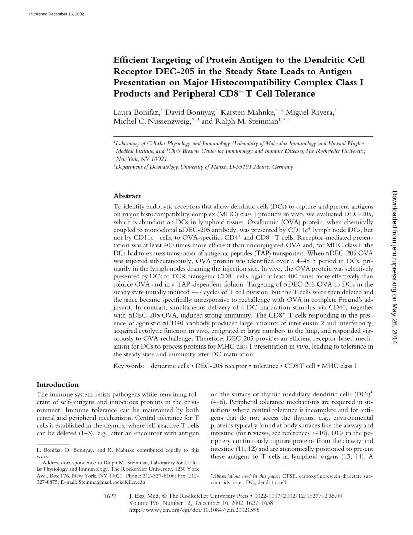
Targeting Protein to DCs In Situ via DEC-205. To ex-amine the potential role of the DEC-205 endocytic recep-tor to process proteins through the MHC class I pathway,we first chemically coupled whole OVA protein to �DEC-205 antibody. OVA is known to be presented on theH-2Kb MHC class I molecule to CD8� T cells, includingTCR transgenic OT-I T cells (36). After OVA couplingto rat anti–mouse DEC-205 antibody, free OVA wasremoved by purifying the conjugate on protein G col-umns, yielding mixtures of �1:1 IgG:OVA conjugates(MW�200 kD) and unconjugated antibody (Fig. 1 A).Based on the observed conjugation efficiency and the 4:1mass ratio of IgG to OVA, we estimated that 10% of theconjugate consisted of OVA protein.
To verify that the �DEC-205:OVA targeted DCs inmice, we injected 1 g of the conjugates subcutaneouslyand probed for OVA protein in isolated CD11c� DC-enriched (14, 37) and CD11c� DC-depleted, lymph nodecells. Prior work had demonstrated the uptake of rat�DEC-205 antibody by most DCs (32), but here we fol-lowed the capture of the OVA protein. Uptake by DCsplateaued in the draining node within 12–24 h of injection(Fig. 1, B–D). The 45 kD size of the immunoreactiveOVA in the DCs was similar to native OVA, suggestingthat most of the protein had been released from its conju-gation to the injected antibody (Fig. 1 B). Injection of 10g soluble OVA subcutaneously did not yield detectableantigen in the lymph nodes draining the site of injection(Fig. 1 C, left lane). With the �DEC-205:OVA conjugate,OVA was only found in the CD11c� DC-enriched frac-tion and not the CD11c� DC-depleted cells (Fig. 1 C).Smaller amounts of OVA were also detected in DCs in dis-tal sites like lymph nodes and spleen (Fig. 1 D). Theamount of captured OVA corresponded to �10 ng of pro-tein per 106 DCs or 105 molecules/DC (Fig. 1 B). We con-clude that protein coupled to �DEC-205 antibody is effi-ciently delivered to DCs in situ.
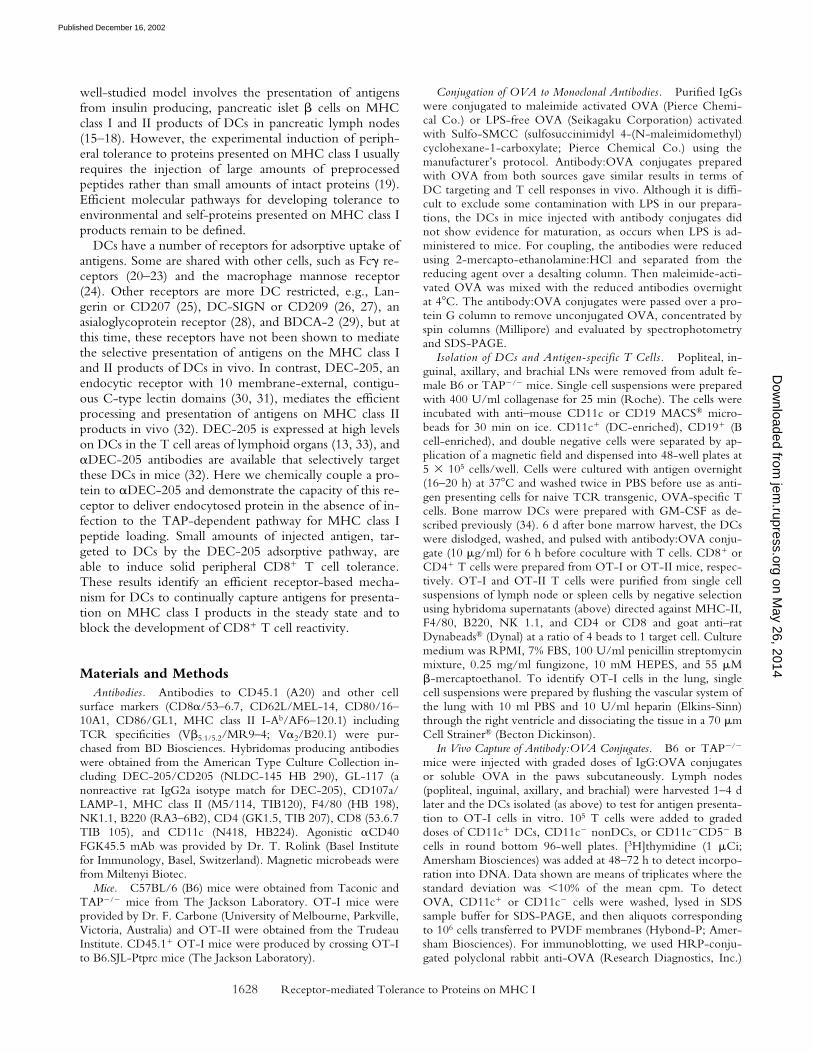
Isolated Lymph Node DCs Present �DEC-205:OVA Con-jugates on MHC Class I via TAPs. To determine whetherantigen delivered by �DEC-205 to DCs can be processedfor presentation by MHC class I, we first isolated DCs fromlymph nodes and treated them with �DEC-205:OVA invitro. The antibody:OVA conjugates were incubated withCD11c� and CD11c� lymph node cells overnight, excessconjugate was removed by washing, and then the cellswere cocultured for 2 d with OVA-specific TCR trans-genic T cells (CD8� MHC class I–restricted OT-I orCD4� MHC class II–restricted OT-II T cells). We foundthat OVA was presented by CD11c� DCs on both MHCclass I and class II products (Fig. 2 A). DCs exposed to 0.1g/ml of �DEC-205:OVA (with 10 ng/ml OVA pro-tein) were more active in presenting to MHC class I re-stricted CD8� OT-I T cells than DCs that had been ex-posed to 30 g/ml of soluble OVA (Fig. 2 A). Uptake viaDEC-205 increased the efficiency of presentation relativeto unconjugated OVA at least 1,000-fold for MHC I and
on May 26, 2014
jem.rupress.org
Dow
nloaded from
Published December 16, 2002
1630 Receptor-mediated Tolerance to Proteins on MHC I
300-fold for MHC II (Fig. 2 A). In contrast, neitherCD19� B cells nor APCs depleted of CD11c� and CD19�
cells were able to stimulate T cell proliferation above back-ground levels, even at cell doses 30-fold higher than thoserequired to observe presentation by DCs (Fig. 2 B). Isotypecontrol IgG:OVA conjugates were much less active than�DEC-205:OVA conjugates (Fig. 2 B).
Several additional controls (data not depicted) were per-formed to verify the function of the DEC-205 receptor inthe presentation of protein conjugated to the correspondingmonoclonal antibody. Because DCs can utilize Fc� recep-tors to present immune complexes on MHC class I products(20, 21), we showed that blocking these receptors (at leastCD16 and CD32) with 2.4G2 �Fc�R monoclonal antibody(38) did not reduce presentation of �DEC-205:OVA. Toevaluate another antibody:OVA conjugate that targets theendocytic system, we used LAMP-1, a lysosome-associatedmembrane protein expressed in DCs and other cells. The�LAMP-1:OVA conjugate was only comparable to isotypecontrol antibody:OVA in bringing about the presentation ofOVA peptides in the context of either MHC-I or -II byCD11c� DCs. Conjugation was essential for efficient OVAtargeting since mixtures of unconjugated �DEC-205 andsoluble OVA were only presented with a comparable effi-ciency to equivalent doses of soluble OVA, ruling out thepossibility that �DEC-205 was simply enhancing presenta-tion of nonconjugated OVA. In summary, the DEC-205 re-
ceptor mediates efficient presentation of protein antigens onboth MHC class I and II products of lymph node DCs.
To establish that MHC class I presentation by �DEC-205:OVA was TAP dependent, we performed similar ex-periments with DCs obtained from TAP-deficient mice(TAP�/�; Fig. 2 C). In the absence of TAP, proteosome-processed peptides in DCs fail to move into the endoplas-mic reticulum for association with MHC I molecules. Wefound that TAP�/� DCs were unable to present OVA onMHC class I after uptake of �DEC-205:OVA, but thesame cells presented OVA on MHC class II (Fig. 2 C).Also, the TAP�/� DCs could present preprocessed OVApeptide to MHC class I restricted OT-I T cells (data notdepicted). Thus, antigens endocytosed via DEC-205 arerouted to the MHC class I processing machinery by a path-way that requires transport of peptides from the cytoplasmto the endoplasmic reticulum.
In contrast to DCs obtained from the lymph node, DEC-205 expressing DCs generated from bone marrow progeni-tors with GM-CSF were only able to present the �DEC-205:OVA conjugates to OVA-specific CD4� OT-II T cellsbut not CD8� OT-I T cells (Fig. 2 D). Therefore, the typeof DC influences the capacity of DEC-205 to deliver anti-gens for TAP dependent MHC class I presentation.
DEC-205–mediated MHC Class I Presentation In Vivo.To determine if DCs targeted with �DEC-205:OVA alsopresent OVA peptides on MHC class I in vivo, we next
Figure 1. Conjugation and delivery ofOVA via monoclonal antibodies. (A) OVAconjugated and unconjugated IgGs re-solved by SDS-PAGE and stained withCoomassie blue. (B) Immunoblotting withanti-OVA antibody to detect OVA in theIg:OVA conjugates and in CD11c� lymphnode DCs (106/lane), the latter at 12 and24 h after subcutaneous injection of 1 g of�DEC-205:OVA or isotype control:OVA.(C) The subcutaneous injection of 1 g of�DEC-205:OVA per footpad, but not 10g of soluble OVA, targets CD11c� lymphnode DCs. 106 CD11c� cells from thedraining nodes per lane were immuno-blotted 12–72 h after injection. (D) �DEC-205:OVA uptake occurs primarily inCD11c� DCs in the draining lymph nodes.As in C, but cell fractions were preparedfrom draining lymph nodes, distal (Dist.)nodes and spleen (Spl.).
on May 26, 2014
jem.rupress.org
Dow
nloaded from
Published December 16, 2002

1631 Bonifaz et al.
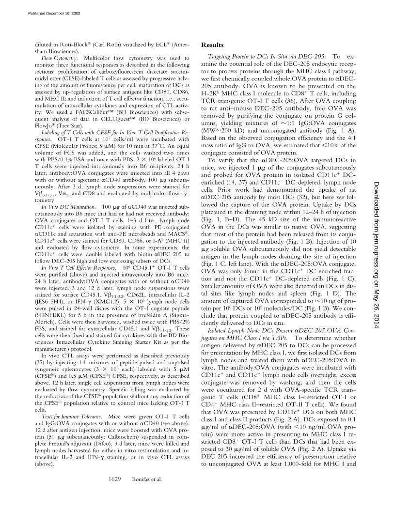
isolated CD11c� and CD11c� cells (or B cells enrichedfrom the CD11c� population by depleting CD5� T cells)from mice injected subcutaneously with conjugates or withsoluble OVA, and then we assayed for antigen presentationto OT-I cells in vitro without further addition of antigen.After injection of �DEC-205:OVA, we detected strongpresentation at 4 and 24 h after injection, but only by DCs(Fig. 3 A, left) and not by CD11c� or enriched B cells (Fig.3 A, right). The B cells also were inactive when the animalswere given �CD40 antibody (together with antibody:OVA) to enhance their costimulatory properties (data notdepicted). 100-fold higher doses of soluble OVA relative to�DEC-205:OVA (which is 10% OVA) were required todetect presentation, but again the DCs selectively presentedthe antigen. This presentation was greater when the cellswere isolated 24 h rather than 4 h after injection (Fig. 3 A).After injection of �DEC-205:OVA, presentation wasreadily detected for as long as 4 d (Fig. 3 B). The presenta-tion by DCs in vivo was TAP dependent (Fig. 3 C). Incontrast, antibody:OVA conjugates directed toward other
DC markers, e.g., MHC-II and LAMP-1, did not enhanceOVA-presentation (Fig. 3 D). In all experiments, there waslittle or no capture of the isotype-control:OVA conjugate(Figs. 3, B–D).
To verify that DCs targeted with �DEC-205:OVAcould also present antigen to T cells in situ, we transferred2 � 106 CFSE-labeled, antigen-specific T cells (CD8�
OT-I cells) 1 d before injection with �DEC-205:OVA,isotype:OVA, or soluble OVA. After 3 d, the lymph nodes(Fig. 3 E), spleen, and blood (data not depicted) were eval-uated for OT-I proliferation as assayed by CFSE dilution.Virtually all of the OT-I cells in lymph node entered cellcycle and underwent 3–7 cell divisions after a dose of just1.0 g of �DEC-205:OVA conjugate (100 ng OVA)per mouse (Fig. 3 E). For soluble OVA, at least 400-foldmore protein was required to induce comparable prolifera-tive responses, and, again, the isotype-control:OVA con-jugate elicited little or no proliferation (Fig. 3 E). Toprove that DEC-205 but not Fc� receptors were mediat-ing presentation, we verified that presentation was abol-
Figure 2. Targeting of�DEC-205:OVA to CD11c�
lymph node DCs in vitro. (A)�DEC-205:OVA elicits stron-ger presentation than OVA alonein a dose-dependent manner.CD11c� cells from C57BL/6lymph nodes were cultured 18 hin graded doses of �DEC-205:OVA or OVA alone, washed,and cocultured with OT-I orOT-II T cells before measuringuptake of [3H]thymidine at 48–72 h to assess T cell proliferation.(B) Presentation of peptides de-rived from �DEC-205:OVA isrestricted to CD11c� lymphnode cells, and not the CD19�
or CD11c�CD19� (double neg-ative) fractions. As in A, but with�DEC-205:OVA or the isotype:OVA conjugate at 10 g/ml. (C)Presentation of peptides derivedfrom �DEC-205:OVA is TAPdependent. As in B, but CD11c�
cells were prepared fromC57BL/6 or TAP�/� mice. (D)Bone marrow DCs are unable topresent �DEC-205:OVA onMHC class I products. Cellsfrom d6 cultures were harvestedand cultured with antibody:OVAconjugates for 6 h at 10 g/ml,washed, and cocultured with Tcells as in panel A. Data are rep-resentative of three experiments.
on May 26, 2014
jem.rupress.org
Dow
nloaded from
Published December 16, 2002

1632 Receptor-mediated Tolerance to Proteins on MHC I
ished with DCs from DEC-205�/� mice (data notdepicted). In summary, �DEC-205 efficiently targets anti-gens for presentation by the exogenous pathway to MHCclass I in vivo.
�DEC-205:OVA Does Not Mature DCs In Vivo. Toexamine whether �DEC-205:OVA treatment results inDC maturation in the presence or absence of OVA-specificOT-I T cells, we did FACS® studies of DCs from mice in-jected with conjugates 1 or 3 d earlier under a variety ofconditions. As illustrated in Fig. 4 A, surface expression ofCD80, CD86, as well as MHC class II products were un-changed in �DEC-205:OVA-injected mice, whether ornot they received OT-1 T cells. The number of DCs alsodid not change in mice given �DEC-205:OVA. However,coadministration of an agonistic �CD40 antibody (FGK-
45.5) as an adjuvant activated the DCs in situ over a 3 d pe-riod and increased their numbers about twofold. The ex-tent of maturation with �CD40 was similar in the absenceor presence of antigen (�DEC-205:OVA) or OT-I T cells(Fig. 4 A). Maturation was detected in CD11c� DCs thathad low and high levels of DEC-205, but the levels ofCD86 were higher in the DEC-205 high fraction (Fig. 4B). In summary, although lymph node DCs in the steadystate express molecules used in T cell activation like CD86,these DCs do not seem to differentiate further when ex-posed to �DEC-205:OVA but do differentiate in responseto agonistic �CD40 antibody.
Distinct T Cell Responses In Vivo to Antigen Presented in theSteady State and CD40-based DC Maturation. To followthe fate of the OT-I T cells that proliferated in response to
Figure 3. Targeting of �DEC-205:OVAto lymph node CD11c� DCs in vivo. (A)Only CD11c� lymph node DCs efficientlypresent exogenous �DEC-205:OVA, andto a lesser extent OVA, to OT-I T cells.C57BL/6 mice were injected with 4.0 g(1.0 g/footpad) of �DEC-205:OVA con-jugate or 400 g (100 g/footpad) of solu-ble OVA subcutaneously 4 and 24 h beforesacrifice. The CD11c� and CD11c�CD5�
(B cell) fractions were MACS® sorted fromlymph nodes and evaluated for presentationto OT-I T cells as in Fig. 2 A. Peptide con-trols were performed with the highest titra-tion of APCs (DCs, left; B cells, right) foreach group. (B) As in panel A but CD11c�
DC’s were studied 1 and 4 d after injectionof 4.0 g (1.0 g/footpad) of antibody:OVA conjugates subcutaneously. (C) Pre-sentation by DCs of OVA peptides fromC57BL/6 but not TAP�/� mice given 4.0g (1.0 g/footpad) of IgG:OVA conju-gates subcutaneously 4 d earlier. (D)�DEC-205:OVA elicits better presentationof OVA derived peptides than other DC-targeted conjugates, each injected with 4.0g (1.0 g/footpad) of IgG:OVA conju-gates subcutaneously 4 d earlier. (E)�DEC-205:OVA induces stronger in vivoproliferation of OT-I T cells than OVAalone. C57BL/6 mice were injected intra-venously with 2 � 106 CFSE-labeled OT-IT cells and then graded doses of IgG:OVAconjugates or OVA subcutaneously 24 hlater. 3 d after conjugate injection, lymphnodes were harvested and the expansion ofCD8�V�2V�5.1/5.2 cells evaluated by flowcytometry for CFSE dilution. Each panelrepresents two or more experiments.
on May 26, 2014
jem.rupress.org
Dow
nloaded from
Published December 16, 2002

1633 Bonifaz et al.
antigen targeted to DCs in vivo, we tracked the transferredT cells by flow cytometry using a combination of CD45.1and V�5.1/5.2 markers, and we compared responses in thesteady state to those following �CD40-induced DC matu-ration. At 3 d after �DEC-205:OVA injection, we foundstrong proliferative responses in the presence or absenceDC maturation (Fig. 5 A). However, �CD40 treated micealso showed greatly enhanced T cell production of IL-2and IFN-� (Fig. 5 B, bottom row).
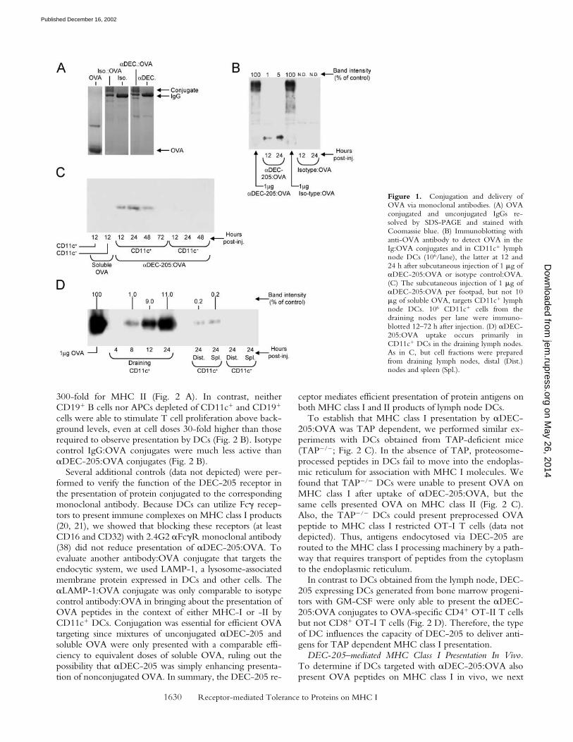
When we followed the numbers of injected OT-I Tcells at 3 d and at 12–14 d in several lymphoid tissues (Fig.6, A and B), we found that �DEC-205:OVA in the steadystate first expanded the OT-I cells, but by 12–14 d, the Tcells were virtually entirely absent from lymph nodes,spleen, or blood (Fig. 6 A, compare left and right panels,and Fig. 6 B). However, if the �DEC-205:OVA conjugatewas given with �CD40, the OT-I cells expanded relativeto the PBS control (Fig. 6, A and B) or isotype-control:
Figure 4. Maturation of DCs in vivo byagonistic �CD40 but not by �DEC-205:OVA. (A) C57BL/6 mice were injectedsubcutaneously with PBS or 4.0 g (1.0g/footpad) of �DEC-205:OVA conju-gate with or without �CD40 (100 gFGK45.5 subcutaneously), 1 and 3 d beforesacrifice. CD11c� cells were sorted byMACS® from lymph nodes and evaluatedby flow cytometry for expression of CD80,CD86, and MHC class II. Prior to injectionof the OVA conjugate and �CD40, themice were given PBS (�) or OT-I (�)cells. The bold symbols are mean fluores-cence indices of the CD11c� cells in thepresence of a maturation stimulus, while thegray-bold at day 3 indicate a significant in-crease, consistent with maturation. (B) Illus-trative FACS® data showing the maturationof the DEC-205hi CD11c� cells and DEC-205loCD11c� cells, in mice treated 3 d be-fore with PBS and �CD40 as in panel A.
Figure 5. Contrasting responses of OT-I T cells to �DEC-205:OVA in the presence or absence of �CD40-induced DC maturation. (A) �CD40 haslittle impact on �DEC-205:OVA induced proliferation of OT-I T cells. As in Fig. 3 E, but mice were or were not given �CD40 (100 g FGK45.5 sub-cutaneously). (B) Differential IL-2 and IFN-� production by OT-I T cells in response to isotype:OVA or �DEC-205:OVA with or without �CD40. Asin panel A, but lymph node suspensions were restimulated in vitro with the cognate OT-I peptide for 5 h in the presence of brefeldin A (5 g/ml) beforestaining for intracellular cytokine. Data are representative of three experiments.
on May 26, 2014
jem.rupress.org
Dow
nloaded from
Published December 16, 2002
1634 Receptor-mediated Tolerance to Proteins on MHC I
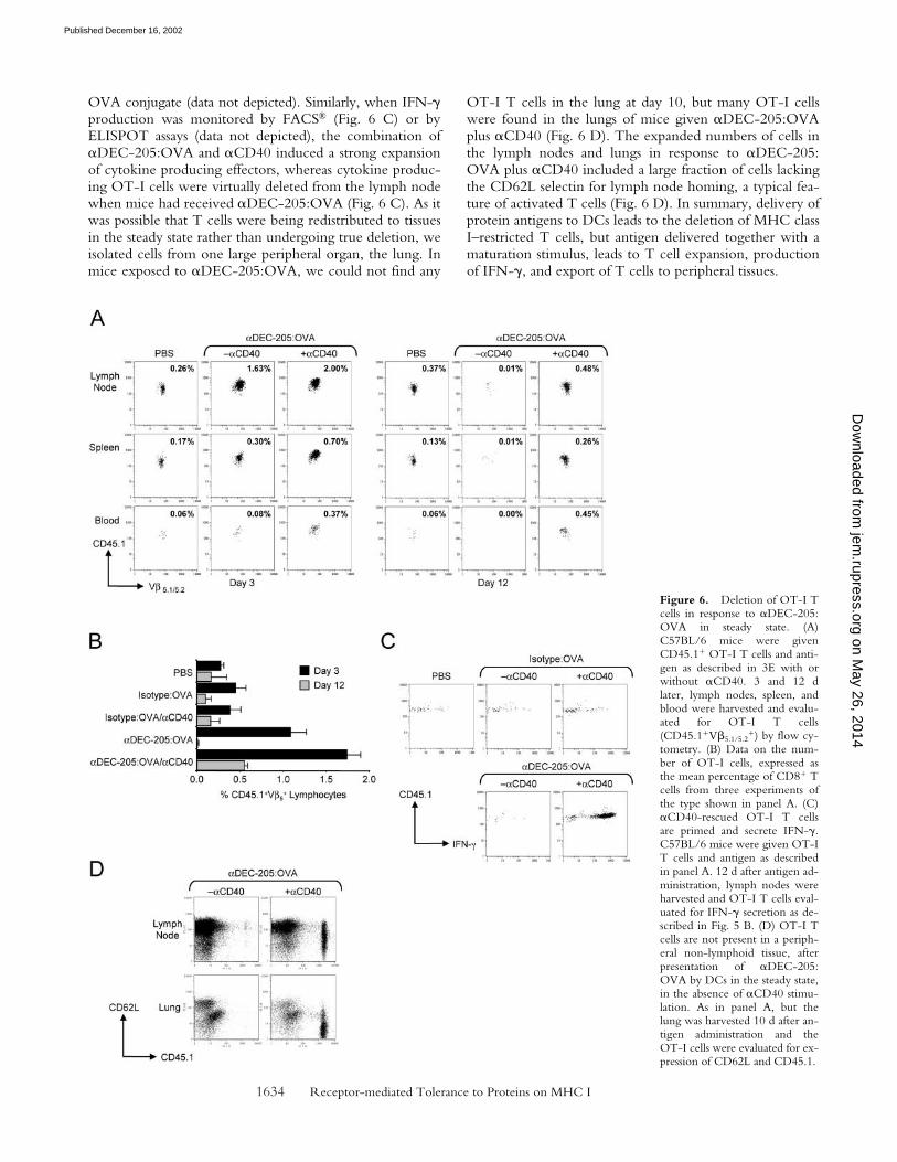
OVA conjugate (data not depicted). Similarly, when IFN-�production was monitored by FACS® (Fig. 6 C) or byELISPOT assays (data not depicted), the combination of�DEC-205:OVA and �CD40 induced a strong expansionof cytokine producing effectors, whereas cytokine produc-ing OT-I cells were virtually deleted from the lymph nodewhen mice had received �DEC-205:OVA (Fig. 6 C). As itwas possible that T cells were being redistributed to tissuesin the steady state rather than undergoing true deletion, weisolated cells from one large peripheral organ, the lung. Inmice exposed to �DEC-205:OVA, we could not find any
OT-I T cells in the lung at day 10, but many OT-I cellswere found in the lungs of mice given �DEC-205:OVAplus �CD40 (Fig. 6 D). The expanded numbers of cells inthe lymph nodes and lungs in response to �DEC-205:OVA plus �CD40 included a large fraction of cells lackingthe CD62L selectin for lymph node homing, a typical fea-ture of activated T cells (Fig. 6 D). In summary, delivery ofprotein antigens to DCs leads to the deletion of MHC classI–restricted T cells, but antigen delivered together with amaturation stimulus, leads to T cell expansion, productionof IFN-�, and export of T cells to peripheral tissues.
Figure 6. Deletion of OT-I Tcells in response to �DEC-205:OVA in steady state. (A)C57BL/6 mice were givenCD45.1� OT-I T cells and anti-gen as described in 3E with orwithout �CD40. 3 and 12 dlater, lymph nodes, spleen, andblood were harvested and evalu-ated for OT-I T cells(CD45.1�V�5.1/5.2
�) by flow cy-tometry. (B) Data on the num-ber of OT-I cells, expressed asthe mean percentage of CD8� Tcells from three experiments ofthe type shown in panel A. (C)�CD40-rescued OT-I T cellsare primed and secrete IFN-�.C57BL/6 mice were given OT-IT cells and antigen as describedin panel A. 12 d after antigen ad-ministration, lymph nodes wereharvested and OT-I T cells eval-uated for IFN-� secretion as de-scribed in Fig. 5 B. (D) OT-I Tcells are not present in a periph-eral non-lymphoid tissue, afterpresentation of �DEC-205:OVA by DCs in the steady state,in the absence of �CD40 stimu-lation. As in panel A, but thelung was harvested 10 d after an-tigen administration and theOT-I cells were evaluated for ex-pression of CD62L and CD45.1.
on May 26, 2014
jem.rupress.org
Dow
nloaded from
Published December 16, 2002
1635 Bonifaz et al.
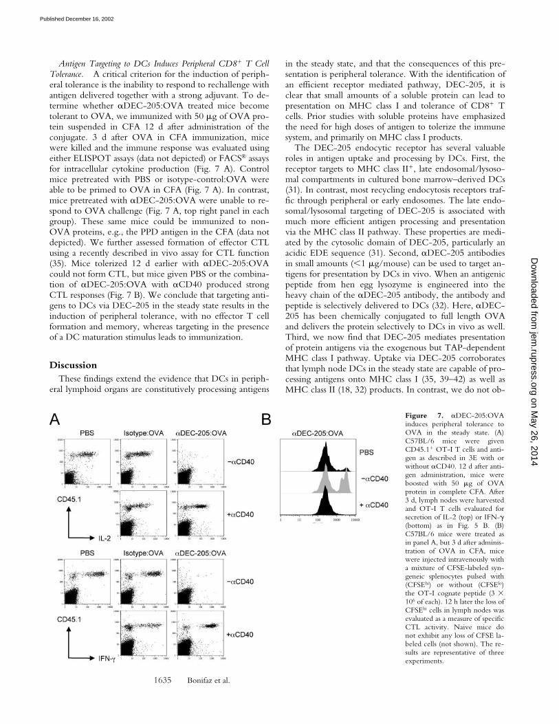
Antigen Targeting to DCs Induces Peripheral CD8� T CellTolerance. A critical criterion for the induction of periph-eral tolerance is the inability to respond to rechallenge withantigen delivered together with a strong adjuvant. To de-termine whether �DEC-205:OVA treated mice becometolerant to OVA, we immunized with 50 g of OVA pro-tein suspended in CFA 12 d after administration of theconjugate. 3 d after OVA in CFA immunization, micewere killed and the immune response was evaluated usingeither ELISPOT assays (data not depicted) or FACS® assaysfor intracellular cytokine production (Fig. 7 A). Controlmice pretreated with PBS or isotype-control:OVA wereable to be primed to OVA in CFA (Fig. 7 A). In contrast,mice pretreated with �DEC-205:OVA were unable to re-spond to OVA challenge (Fig. 7 A, top right panel in eachgroup). These same mice could be immunized to non-OVA proteins, e.g., the PPD antigen in the CFA (data notdepicted). We further assessed formation of effector CTLusing a recently described in vivo assay for CTL function(35). Mice tolerized 12 d earlier with �DEC-205:OVAcould not form CTL, but mice given PBS or the combina-tion of �DEC-205:OVA with �CD40 produced strongCTL responses (Fig. 7 B). We conclude that targeting anti-gens to DCs via DEC-205 in the steady state results in theinduction of peripheral tolerance, with no effector T cellformation and memory, whereas targeting in the presenceof a DC maturation stimulus leads to immunization.
DiscussionThese findings extend the evidence that DCs in periph-
eral lymphoid organs are constitutively processing antigens
in the steady state, and that the consequences of this pre-sentation is peripheral tolerance. With the identification ofan efficient receptor mediated pathway, DEC-205, it isclear that small amounts of a soluble protein can lead topresentation on MHC class I and tolerance of CD8� Tcells. Prior studies with soluble proteins have emphasizedthe need for high doses of antigen to tolerize the immunesystem, and primarily on MHC class I products.
The DEC-205 endocytic receptor has several valuableroles in antigen uptake and processing by DCs. First, thereceptor targets to MHC class II�, late endosomal/lysoso-mal compartments in cultured bone marrow–derived DCs(31). In contrast, most recycling endocytosis receptors traf-fic through peripheral or early endosomes. The late endo-somal/lysosomal targeting of DEC-205 is associated withmuch more efficient antigen processing and presentationvia the MHC class II pathway. These properties are medi-ated by the cytosolic domain of DEC-205, particularly anacidic EDE sequence (31). Second, �DEC-205 antibodiesin small amounts (1 g/mouse) can be used to target an-tigens for presentation by DCs in vivo. When an antigenicpeptide from hen egg lysozyme is engineered into theheavy chain of the �DEC-205 antibody, the antibody andpeptide is selectively delivered to DCs (32). Here, �DEC-205 has been chemically conjugated to full length OVAand delivers the protein selectively to DCs in vivo as well.Third, we now find that DEC-205 mediates presentationof protein antigens via the exogenous but TAP-dependentMHC class I pathway. Uptake via DEC-205 corroboratesthat lymph node DCs in the steady state are capable of pro-cessing antigens onto MHC class I (35, 39–42) as well asMHC class II (18, 32) products. In contrast, we do not ob-
Figure 7. �DEC-205:OVAinduces peripheral tolerance toOVA in the steady state. (A)C57BL/6 mice were givenCD45.1� OT-I T cells and anti-gen as described in 3E with orwithout �CD40. 12 d after anti-gen administration, mice wereboosted with 50 g of OVAprotein in complete CFA. After3 d, lymph nodes were harvestedand OT-I T cells evaluated forsecretion of IL-2 (top) or IFN-�(bottom) as in Fig. 5 B. (B)C57BL/6 mice were treated asin panel A, but 3 d after adminis-tration of OVA in CFA, micewere injected intravenously witha mixture of CFSE-labeled syn-geneic splenocytes pulsed with(CFSEhi) or without (CFSElo)the OT-I cognate peptide (3 �106 of each). 12 h later the loss ofCFSEhi cells in lymph nodes wasevaluated as a measure of specificCTL activity. Naive mice donot exhibit any loss of CFSE la-beled cells (not shown). The re-sults are representative of threeexperiments.
on May 26, 2014
jem.rupress.org
Dow
nloaded from
Published December 16, 2002

1636 Receptor-mediated Tolerance to Proteins on MHC I
serve presentation of �DEC-205:OVA on MHC class I us-ing DCs derived from bone marrow, although this DCpopulation presents exogenous proteins on MHC class IIproducts (43). In contrast, DCs from lymph node seemcompetent in both MHC class I and II presentation in thesteady state in situ, without addition of maturation stimuli.
In the steady state, DEC-205 represents a specific recep-tor for DCs to induce peripheral tolerance to soluble anti-gens for both CD4� (32) and CD8� T cells (this paper).DCs also induce tolerance to cell associated antigens invivo (35, 39–41), but the responsible receptors have notbeen identified to date. While DEC-205 greatly enhancespresentation of protein antigens, it is possible that the re-ceptor simply functions to enhance antigen uptake and notsubsequent events required for presentation on MHC classI. Also, once suitable reagents become available, it will bepossible to assess if other receptors on DCs (see Introduc-tion) function to enhance antigen presentation and periph-eral tolerance in vivo. Currently, monoclonal antibodiesare primarily available for the DEC-205 receptor and, asmentioned above, have been previously shown to targetDCs efficiently and selectively in vivo and enhance MHCclass II presentation. The key new points in this paper arefirst that DCs have efficient receptor based mechanisms toenhance presentation on MHC class I products in vivo,second that these operate in the steady state, and third, theconsequence of presentation is peripheral tolerance in theCD8� compartment by a deletional mechanism.
The existence of receptor-mediated uptake mechanismsshould allow DCs to play a valuable role in silencing reac-tivity to harmless self-antigens and environmental proteins.A single dose of 0.1 g of OVA conjugated to �DEC-205 antibody can delete and tolerize sizable numbers(�106) of injected OVA-specific CD8� T cells, corre-sponding to �1% of the �108 T cells in a mouse. Thisnumber is large, as 1/10,000 naive T cells are typicallyable to respond to a specific antigen. As a result, the capac-ity of DCs to present antigens by the exogenous pathwaygreatly exceeds the repertoire of T cells to be activated, atleast with respect to an antigen that is recognized effi-ciently, i.e., 0.1 nM peptide, as studied here. It will be im-portant to test other protein antigens and a broader reper-toire of T cells, as our studies have at this point focused onthe OT-I T cell, to obtain direct evidence on the numberand function of antigen-specific T cells. Nonetheless, ourfindings indicate that CD8� T cell tolerance (as well as im-munity) can be achieved in situ with low doses of intactprotein antigens.
DCs in lymph nodes and spleen express many featuressuggesting that they are “immature” i.e., able to captureantigens but unable to stimulate immunity (44, 45). TheDCs are active in endocytosis (40, 46) and also respond invivo to microbial, inflammatory, and T cell stimuli by pro-ducing cytokines (47) and up-regulating several costimula-tory molecules (48–50). The studies with �DEC-205:OVAcorroborate that DCs in vivo in the steady state do notstimulate an immune response even when they are effec-tively presenting antigen and inducing extensive T cell
proliferation. However, the T cells fail to differentiate andare deleted unless a maturation stimulus also is provided.To date, we have concentrated on CD40-based DC matu-ration. Nevertheless, the findings are consistent with theview that the immunogenic function of DCs, i.e., the in-duction of effector T cells and the development of mem-ory, requires that at least two sets of events take place. Oneinvolves antigen capture and successful processing to formMHC–peptide complexes; this occurs in lymph node DCsin the steady state. The other requirement entails the intri-cate process of maturation, which changes DCs in manyways, e.g., increasing T cell costimulatory molecules, in-ducing cytokines like IL-12 and IL-2, and altering the ex-pression of chemokine receptors.
Two pathways have been defined for the presentation ofantigens on MHC class I products. The classical or “endog-enous” pathway originates from newly synthesized proteinsespecially those derived from defective ribosomal initiationproducts (51). This pathway provides an elegant and estab-lished mechanism for protective immunity, guiding MHCclass I–restricted cytotoxic T lymphocytes to peptides pro-duced in infected and malignant cells. By recognizingMHC–peptide complexes displayed at the cell surface, theCD8� cytolytic response is focused on cells harboringpathogens and not on innocent bystander cells (52). A sec-ond “exogenous” pathway also exists, allowing endocy-tosed nonreplicating proteins to be presented on MHCclass I. The exogenous pathway a priori could allow nonin-fected cells to take up protein and become targets forMHC class I–specific, CD8� cytolytic T lymphocytes. Thispotentially serious problem would be averted if the exoge-nous pathway were primarily expressed in DCs. During in-fection, the processing of dying infected cells by the exoge-nous pathway provides a means for DCs to initiate CD8�
T cell immunity to pathogens that do not productively in-fect them. Likewise, during the steady state, DCs in lym-phoid organs can use the exogenous pathway to presentself-peptides on MHC class I (16, 40, 53; and this paper)and thereby induce tolerance. Therefore the induction oftolerance does not require that all cells be capable of pro-cessing antigens by the exogenous pathway; rather, thepresence of efficient receptors like DEC-205 for exogenouspresentation in vivo provides a specific mechanism for DCsto continually delete the peripheral T cell repertoire of re-activity to low levels of self and environmental proteins.
Supported by grants AI13013 and AI051573 from the National In-stitutes of Health to R.M. Steinman, by a fellowship from the Insti-tuto Mexicano del Seguro Social to L. Bonifaz, and by National In-stitutes of Health training grant CA09673 to D. Bonnyay.
Submitted: 10 September 2002Revised: 20 October 2002Accepted: 12 November 2002
References1. Kappler, J.W., N. Roehm, and P. Marrack. 1987. T cell tol-
erance by clonal elimination in the thymus. Cell. 49:273–
on May 26, 2014
jem.rupress.org
Dow
nloaded from
Published December 16, 2002
1637 Bonifaz et al.
280.2. Kisielow, P., H. Bluthmann, U.D. Staerz, M. Steinmetz, and
H. von Boehmer. 1988. Tolerance in T-cell-receptor trans-genic mice involves deletion of nonmature CD4�8� thy-mocytes. Nature. 333:742–746.
3. Bouneaud, C., P. Kourilsky, and P. Bousso. 2000. Impact ofnegative selection on the T cell repertoire reactive to a self-peptide: a large fraction of T cell clones escapes clonal detec-tion. Immunity. 13:829–840.
4. Matzinger, P., and S. Guerder. 1989. Does T-cell tolerancerequire a dedicated antigen-presenting cell? Nature. 338:74–76.
5. Zal, T., A. Volkmann, and B. Stockinger. 1994. Mechanismsof tolerance induction in major histocompatibility complexclass II–restricted T cells specific for a blood-borne self-anti-gen. J. Exp. Med. 180:2089–2099.
6. Volkmann, A., T. Zal, and B. Stockinger. 1997. Antigen-presenting cells in thymus that can negatively select MHCclass II-restricted T cells recognizing a circulating self antigen.J. Immunol. 158:693–706.
7. Stockinger, B. 1999. T lymphocyte tolerance: from thymicdeletion to peripheral control mechanisms. Adv. Immunol. 71:229–265.
8. Sakaguchi, S. 2000. Regulatory T cells: key controllers of im-munologic self-tolerance. Cell. 101:455–458.
9. Kamradt, T., and N.A. Mitchison. 2001. Tolerance and au-toimmunity. N. Engl. J. Med. 344:655–664.
10. Walker, L.S.K., and A.K. Abbas. 2002. The enemy within:keeping self-reactive T cells at bay in the periphery. Nat. Rev.Immunol. 2:11–19.
11. Vermaelen, K.Y., I. Carro-Muino, B.N. Lambrecht, andR.A. Pauwels. 2001. Specific migratory dendritic cells rapidlytransport antigen from the airways to the thoracic lymphnodes. J. Exp. Med. 193:51–60.
12. Huang, F.-P., N. Platt, M. Wykes, J.R. Major, T.J. Powell,C.D. Jenkins, and G.G. MacPherson. 2000. A discrete sub-population of dendritic cells transports apoptotic intestinalepithelial cells to T cell areas of mesenteric lymph nodes. J.Exp. Med. 191:435–442.
13. Kraal, G., M. Breel, M. Janse, and G. Bruin. 1986. Langer-hans cells, veiled cells, and interdigitating cells in the mouserecognized by a monoclonal antibody. J. Exp. Med. 163:981–997.
14. Metlay, J.P., M.D. Witmer-Pack, R. Agger, M.T. Crowley,D. Lawless, and R.M. Steinman. 1990. The distinct leuko-cyte integrins of mouse spleen dendritic cells as identifiedwith new hamster monoclonal antibodies. J. Exp. Med. 171:1753–1771.
15. Heath, W.R., and F.R. Carbone. 2001. Cross-presentationin viral immunity and self tolerance. Nat. Rev. Immunol.1:126–134.
16. Kurts, C., M. Cannarile, I. Klebba, and T. Brocker. 2001.Dendritic cells are sufficient to cross-present self-antigens toCD8 T cells in vivo. J. Immunol. 166:1439–1442.
17. Morgan, D.J., H.T. Kreuwel, and L.A. Sherman. 1999. Anti-gen concentration and precursor frequency determine therate of CD8� T cell tolerance to peripherally expressed anti-gens. J. Immunol. 163:723–727.
18. Hugues, S., E. Mougneau, W. Ferlin, D. Jeske, P. Hofman,D. Homann, L. Beaudoin, C. Schrike, M. Von Herrath, A.Lehuen, and N. Glaichenhaus. 2002. Tolerance to islet anti-gens and prevention from diabetes induced by limited apop-tosis of pancreatic � cells. Immunity. 16:169–181.
19. Aichele, P., K. Brduscha-Riem, R.M. Zinkernagel, H. Hen-gartner, and H. Pircher. 1995. T cell priming versus T celltolerance induced by synthetic peptides. J. Exp. Med. 182:261–266.
20. Regnault, A., D. Lankar, V. Lacabanne, A. Rodriguez, C.Thery, M. Rescigno, T. Saito, S. Verbeek, C. Bonnerot, P.Ricciardi-Castagnoli, and S. Amigorena. 1999. Fc� receptor-mediated induction of dendritic cell maturation and majorhistocompatibility complex class I–restricted antigen presen-tation after immune complex internalization. J. Exp. Med.189:371–380.
21. Rodriguez, A., A. Regnault, M. Kleijmeer, P. Ricciardi-Castagnoli, and S. Amigorena. 1999. Selective transport ofinternalized antigens to the cytosol for MHC class I presenta-tion in dendritic cells. Nat. Cell Biol. 1:362–368.
22. Dhodapkar, K.M., J. Krasovsky, B. Williamson, and M.V.Dhodapkar. 2002. Anti-tumor monoclonal antibodies en-hance cross-presentation of cellular antigens and the genera-tion of myeloma-specific killer T cells by dendritic cells. J.Exp. Med. 195:125–133.
23. Kalergis, A.M., and J.V. Ravetch. 2002. Inducing tumor im-munity through the selective engagement of activating Fc�receptors on dendritic cells. J. Exp. Med. 195:1653–1659.
24. Sallusto, F., M. Cella, C. Danieli, and A. Lanzavecchia. 1995.Dendritic cells use macropinocytosis and the mannose recep-tor to concentrate antigen in the major histocompatibilityclass II compartment. Downregulation by cytokines and bac-terial products. J. Exp. Med. 182:389–400.
25. Valladeau, J., O. Ravel, C. Dezutter-Dambuyant, K. Moore,M. Kleijmeer, Y. Liu, V. Duvert-Frances, C. Vincent, D.Schmitt, J. Davoust, et al. 2000. Langerin, a novel C-typelectin specific to Langerhans cells, is an endocytic receptorthat induces the formation of Birbeck granules. Immunity. 12:71–81.
26. Kwon, D.S., G. Gregario, N. Bitton, W.A. Hendrickson,and D.R. Littman. 2002. DC-SIGN mediated internalizationof HIV is required for trans-enhancement of T cell infection.Immunity. 16:135–144.
27. Engering, A., T.B. Geijtenbeek, S.J. van Vliet, M. Wijers, E.van Liempt, N. Demaurex, A. Lanzavecchia, J. Fransen,C.G. Figdor, V. Piguet, and Y. van Kooyk. 2002. The den-dritic cell-specific adhesion receptor DC-SIGN internalizesantigen for presentation to T cells. J. Immunol. 168:2118–2126.
28. Valladeau, J., V. Duvert-Frances, J.-J. Pin, M.J. Kleijmeer, S.Ait-Yahia, O. Ravel, C. Vincent, F. Vega, Jr., A. Helms, D.Gorman, et al. 2001. Immature human dendritic cells expressasialoglycoprotein receptor isoforms for efficient receptor-mediated endocytosis. J. Immunol. 167:5767–5774.
29. Dzionek, A., Y. Sohma, J. Nagafune, M. Cella, M. Colonna,F. Facchetti, G. Günther, I. Johnston, A. Lanzavecchia, T.Nagasaka, et al. 2001. BDCA-2, a novel plasmacytoid den-dritic cell-specific type II C-type lectin, mediates antigen-capture and is a potent inhibitor of interferon-�/� induction.J. Exp. Med. 194:1823–1834.
30. Jiang, W., W.J. Swiggard, C. Heufler, M. Peng, A. Mirza,R.M. Steinman, and M.C. Nussenzweig. 1995. The receptorDEC-205 expressed by dendritic cells and thymic epithelialcells is involved in antigen processing. Nature. 375:151–155.
31. Mahnke, K., M. Guo, S. Lee, H. Sepulveda, S.L. Swain, M.Nussenzweig, and R.M. Steinman. 2000. The dendritic cellreceptor for endocytosis, DEC-205, can recycle and enhanceantigen presentation via major histocompatibility complex
on May 26, 2014
jem.rupress.org
Dow
nloaded from
Published December 16, 2002
1638 Receptor-mediated Tolerance to Proteins on MHC I
class II-positive lysosomal compartments. J. Cell Biol. 151:673–683.
32. Hawiger, D., K. Inaba, Y. Dorsett, K. Guo, K. Mahnke, M.Rivera, J.V. Ravetch, R.M. Steinman, and M.C. Nussen-zweig. 2001. Dendritic cells induce peripheral T cell unre-sponsiveness under steady state conditions in vivo. J. Exp.Med. 194:769–780.
33. Witmer-Pack, M.D., W.J. Swiggard, A. Mirza, K. Inaba, andR.M. Steinman. 1995. Tissue distribution of the DEC-205protein that is detected by the monoclonal antibody NLDC-145. II. Expression in situ in lymphoid and nonlymphoid tis-sues. Cell. Immunol. 163:157–162.
34. Inaba, K., M. Inaba, N. Romani, H. Aya, M. Deguchi, S.Ikehara, S. Muramatsu, and R.M. Steinman. 1992. Genera-tion of large numbers of dendritic cells from mouse bonemarrow cultures supplemented with granulocyte/macro-phage colony-stimulating factor. J. Exp. Med. 176:1693–1702.
35. Hernandez, J., S. Aung, W.L. Redmond, and L.A. Sherman.2001. Phenotypic and functional analysis of CD8� T cellsundergoing peripheral deletion in response to cross-presenta-tion of self-antigen. J. Exp. Med. 194:707–718.
36. Hogquist, K.A., S.C. Jameson, W.R. Heath, J.L. Howard,M.J. Bevan, and F.R. Carbone. 1994. T cell receptor antago-nist peptides induce positive selection. Cell. 76:17–27.
37. Crowley, M.T., K. Inaba, M.D. Witmer-Pack, S. Gezelter,and R.M. Steinman. 1990. Use of the fluorescence activatedcell sorter to enrich dendritic cells from mouse spleen. J. Im-munol. Methods. 133:55–66.
38. Unkeless, J.C. 1979. Characterization of a monoclonal anti-body directed against mouse macrophage and lymphocyte Fcreceptors. J. Exp. Med. 150:580–596.
39. Kurts, C., H. Kosaka, F.R. Carbone, J.F.A.P. Miller, andW.R. Heath. 1997. Class I–restricted cross-presentation ofexogenous self antigens leads to deletion of autoreactiveCD8� T cells. J. Exp. Med. 186:239–245.
40. Iyoda, T., S. Shimoyama, K. Liu, Y. Omatsu, Y. Maeda, K.Takahara, Y. Akiyama, R.M. Steinman, and K. Inaba. 2002.The CD8� dendritic cell subset selectively endocytoses dyingcells in culture and in vivo. J. Exp. Med. 195:1289–1302.
41. Liu, K., T. Iyoda, M. Saternus, Y. Kimura, K. Inaba, andR.M. Steinman. 2002. Immune tolerance after delivery ofdying cells to dendritic cells in situ. J. Exp. Med. 196:1091–1097.
42. Scheinecker, C., R. McHugh, E.M. Shevach, and R.N. Ger-main. 2002. Constitutive presentation of a natural tissue au-toantigen exclusively by dendritic cells in the draining lymphnode. J. Exp. Med. 196:1079–1090.
43. Inaba, K., S. Turley, T. Iyoda, F. Yamaide, S. Shimoyama,C. Reis e Sousa, R.N. Germain, I. Mellman, and R.M.
Steinman. 2000. The formation of immunogenic MHC classII-peptide ligands in lysosomal compartments of dendriticcells is regulated by inflammatory stimuli. J. Exp. Med. 191:927–936.
44. Inaba, K., G. Schuler, M.D. Witmer, J. Valinsky, B. Atassi,and R.M. Steinman. 1986. The immunologic properties ofpurified Langerhans cells: distinct requirements for the stimu-lation of unprimed and sensitized T lymphocytes. J. Exp.Med. 164:605–613.
45. Romani, N., S. Koide, M. Crowley, M. Witmer-Pack, A.M.Livingstone, C.G. Fathman, K. Inaba, and R.M. Steinman.1989. Presentation of exogenous protein antigens by den-dritic cells to T cell clones: intact protein is presented best byimmature, epidermal Langerhans cells. J. Exp. Med. 169:1169–1178.
46. Kamath, A.T., J. Pooley, M.A. O’Keeffe, D. Vremec, Y.Zhan, A. Lew, A. D’Amico, L. Wu, D.F. Tough, and K.S.Shortman. 2000. The development, maturation, and turn-over rate of mouse spleen dendritic cell populations. J. Immu-nol. 165:6762–6770.
47. Reis e Sousa, C., S. Hieny, T. Scharton-Kersten, D. Jank-ovic, H. Charest, R.N. Germain, and A. Sher. 1997. In vivomicrobial stimulation induces rapid CD40L-independentproduction of IL-12 by dendritic cells and their re-distribu-tion to T cell areas. J. Exp. Med. 186:1819–1829.
48. De Smedt, T., B. Pajak, E. Muraille, L. Lespagnard, E. Hei-nen, P. De Baetselier, J. Urbain, O. Leo, and M. Moser.1996. Regulation of dendritic cell numbers and maturationby lipopolysaccharide in vivo. J. Exp. Med. 184:1413–1424.
49. Sparwasser, T., R.M. Vabulas, B. Villmow, G.B. Lipford,and H. Wagner. 2000. Bacterial CpG-DNA activates den-dritic cells in vivo: T helper cell–independent cytotoxic Tcell responses to soluble proteins. Eur. J. Immunol. 30:3591–3597.
50. Akbari, O., N. Panjwani, S. Garcia, R. Tascon, D. Lowrie,and B. Stockinger. 1999. DNA vaccination: transfection andactivation of dendritic cells as key events for immunity. J.Exp. Med. 189:169–178.
51. Schubert, U., L.C. Anton, J. Gibbs, C.C. Norbury, J.W.Yewdell, and J.R. Bennink. 2000. Rapid degradation of alarge fraction of newly synthesized proteins by proteasomes.Nature. 404:770–774.
52. Zinkernagel, R.M., and P.C. Doherty. 1979. MHC-restricted cytotoxic T-cells: studies on the biological role ofpolymorphic major transplantation antigens determiningT-cell restriction-specificity, function and responsiveness.Adv. Immunol. 27:51–177.
53. den Haan, J., S. Lehar, and M. Bevan. 2000. CD8� but notCD8� dendritic cells cross-prime cytotoxic T cells in vivo. J.Exp. Med. 192:1685–1696.
on May 26, 2014
jem.rupress.org
Dow
nloaded from
Published December 16, 2002