Effects of salinity levels on proteome ofSuaeda aegyptiaca leaves
13
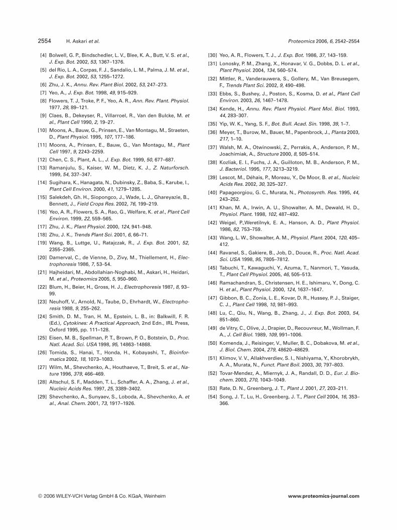
RESEARCH ARTICLE Effects of salinity levels on proteome of Suaeda aegyptiaca leaves Hossein Askari 1 , Johan Edqvist 2 , Mohsen Hajheidari 1 , Mohammad Kafi 3 and Ghasem Hosseini Salekdeh 1 1 Agricultural Biotechnology Research Institute of Iran, Karaj, Iran 2 Department of Plant Biology and Forest Genetics, SLU, Uppsala, Sweden 3 Faculty of Agriculture, Ferdowsi University of Mashhad, Iran Saline soils are the major problem of cultivated lands of Iran. Suaeda aegyptiaca is a salt-tolerant plant (halophytes) that grow naturally in salt-affected areas of Iran. We have employed proteom- ics to identify the mechanisms of salt responsiveness in leaves of S. aegyptiaca grown under dif- ferent salt concentrations. Ten-day-old plants were treated with 0, 150, 300, 450, and 600 mM NaCl. After 30 days of treatment, leaf samples were collected and analyzed using 2-D-PAGE. Out of 700 protein spots reproducible detected within replications, 102 spots showed significant re- sponse to salt treatment compared to 0 mM NaCl. We analyzed expression pattern of salt- responsive proteins using a hierarchical and two nonhierarchical (Fuzzy ART and SOM) statis- tical methods and concluded that Fuzzy ART is the superior method. Forty proteins of 12 differ- ent expression groups were analyzed using LC/MS/MS. Of these, 27 protein spots were identi- fied including proteins involved in oxidative stress tolerance, glycinebetain synthesis, cytoskele- ton remodeling, photosynthesis, ATP production, protein degradation, cyanide detoxification, and chaperone activities. The expression pattern of these proteins and their possible roles in the adaptation of S. aegyptiaca to salinity is discussed. Received: May 15, 2005 Revised: November 9, 2005 Accepted: November 14, 2005 Keywords: Halophyte / Oxidative stress / Salinity / Suaeda aegyptiaca / Two dimensional electro- phoresis 2542 Proteomics 2006, 6, 2542–2554 1 Introduction Salinity is one of the major constraints to crop productivity worldwide. Almost 77 million ha of cultivated land is affec- ted by soil salinity, which corresponds to 5% of all cultivated land. Salinity is a major problem in both dryland and irri- gated land. Although irrigation schemes cover only 15% of the cultivated land, one-third of world’s food is produced in irrigated land where one-third is being significantly affected by soil salinity [1]. High salinity causes water deficit, ion toxicity, and nutri- ent deficiency leading to molecular damage, growth arrest, and even plant death. As a consequence of salt primary effects, secondary stresses such as oxidative damage often occur. Salt stress accelerates production of reactive oxygen species (ROS) in plant cells, and the balance between for- mation and removal of ROS is a determinant factor in the severity of oxidative stress and cell damage [2, 3]. There are many potential sources of ROS production in the plant cells, such as chloroplasts, mitochondria, plasma membrane, apo- plast, and peroxisomes [4, 5]. Correspondence: Dr. Ghasem Hosseini Salekdeh, Agricultural Biotechnology Research Institute of Iran, P.O. Box 31535-1897, Karaj, Iran E-mail: [email protected] Fax: 198-261-2704539 Abbreviations: ABA, abscisic acid; AGD2, aberrant growth and death2; ASD, average SD; CCOMT, caffeoyl CoA O-methyltrans- ferase; ROS, reactive oxygen species; SAMS, S-adenosyl-L- methionine synthetase DOI 10.1002/pmic.200500328 2006 WILEY-VCH Verlag GmbH & Co. KGaA, Weinheim www.proteomics-journal.com
Transcript of Effects of salinity levels on proteome ofSuaeda aegyptiaca leaves

RESEARCH ARTICLE
Effects of salinity levels on proteome of Suaeda
aegyptiaca leaves
Hossein Askari1, Johan Edqvist2, Mohsen Hajheidari1, Mohammad Kafi3
and Ghasem Hosseini Salekdeh1
1 Agricultural Biotechnology Research Institute of Iran, Karaj, Iran2 Department of Plant Biology and Forest Genetics, SLU, Uppsala, Sweden3 Faculty of Agriculture, Ferdowsi University of Mashhad, Iran
Saline soils are the major problem of cultivated lands of Iran. Suaeda aegyptiaca is a salt-tolerantplant (halophytes) that grow naturally in salt-affected areas of Iran. We have employed proteom-ics to identify the mechanisms of salt responsiveness in leaves of S. aegyptiaca grown under dif-ferent salt concentrations. Ten-day-old plants were treated with 0, 150, 300, 450, and 600 mMNaCl. After 30 days of treatment, leaf samples were collected and analyzed using 2-D-PAGE. Outof 700 protein spots reproducible detected within replications, 102 spots showed significant re-sponse to salt treatment compared to 0 mM NaCl. We analyzed expression pattern of salt-responsive proteins using a hierarchical and two nonhierarchical (Fuzzy ART and SOM) statis-tical methods and concluded that Fuzzy ART is the superior method. Forty proteins of 12 differ-ent expression groups were analyzed using LC/MS/MS. Of these, 27 protein spots were identi-fied including proteins involved in oxidative stress tolerance, glycinebetain synthesis, cytoskele-ton remodeling, photosynthesis, ATP production, protein degradation, cyanide detoxification,and chaperone activities. The expression pattern of these proteins and their possible roles in theadaptation of S. aegyptiaca to salinity is discussed.
Received: May 15, 2005Revised: November 9, 2005
Accepted: November 14, 2005
Keywords:
Halophyte / Oxidative stress / Salinity / Suaeda aegyptiaca / Two dimensional electro-phoresis
2542 Proteomics 2006, 6, 2542–2554
1 Introduction
Salinity is one of the major constraints to crop productivityworldwide. Almost 77 million ha of cultivated land is affec-ted by soil salinity, which corresponds to 5% of all cultivated
land. Salinity is a major problem in both dryland and irri-gated land. Although irrigation schemes cover only 15% ofthe cultivated land, one-third of world’s food is produced inirrigated land where one-third is being significantly affectedby soil salinity [1].
High salinity causes water deficit, ion toxicity, and nutri-ent deficiency leading to molecular damage, growth arrest,and even plant death. As a consequence of salt primaryeffects, secondary stresses such as oxidative damage oftenoccur. Salt stress accelerates production of reactive oxygenspecies (ROS) in plant cells, and the balance between for-mation and removal of ROS is a determinant factor in theseverity of oxidative stress and cell damage [2, 3]. There aremany potential sources of ROS production in the plant cells,such as chloroplasts, mitochondria, plasma membrane, apo-plast, and peroxisomes [4, 5].
Correspondence: Dr. Ghasem Hosseini Salekdeh, AgriculturalBiotechnology Research Institute of Iran, P.O. Box 31535-1897,Karaj, IranE-mail: [email protected]: 198-261-2704539
Abbreviations: ABA, abscisic acid; AGD2, aberrant growth anddeath2; ASD, average SD; CCOMT, caffeoyl CoA O-methyltrans-ferase; ROS, reactive oxygen species; SAMS, S-adenosyl-L-methionine synthetase
DOI 10.1002/pmic.200500328
2006 WILEY-VCH Verlag GmbH & Co. KGaA, Weinheim www.proteomics-journal.com

Proteomics 2006, 6, 2542–2554 Plant Proteomics 2543
To cope with salt stress, many plants accumulate compat-ible solutes [6]. It has been suggested that their accumulationhas a significant role on plant adaptation to stress [7]. SinceNa1 inhibits many enzymes [8], accumulation of osmolytes incytosol to protect the cytoplasm from detrimental effects ofsalt and to prevent dehydration of the cytosol is a crucial partof salt tolerance in both halophytes and glycophytes.
Nowadays, there is a special attention to the methods thatenable researchers to quantitatively study expression levels ofthousands of genes in parallel, over a time course or across aseries of defined conditions. The data obtained using thesemethods provides valuable information on gene functionand interaction in cellular processes.
There are rather few examples on the use of proteomics or2-DE to study salt tolerance in plants. In one early study using aproteomic approach it was shown that salt treatment causes theaccumulation of a lectin-like protein (SalT) in rice [9]. Moonset al. [10, 11] used 2-DE to study the roles of abscisic acid (ABA)and jasmonates in salt tolerance of rice. During salt stress bothABA- and ABA-responsive proteins, such as late embryogen-esis abundant (LEA) proteins, were present at higher levels inroots of tolerant rice varieties compared to those in sensitivevarieties [10]. It was also found that ABA and jasmonatesantagonistically regulated the expression of salt-inducible pro-teins associated with water deficit or defense responses [11].Thus, six salt stress-induced proteins (peroxidase, SalT, patho-genesis-related (PR) protein-10 (PR-10), PR-1, and twounknown proteins) were also accumulated after treatment withjasmonate, while the two others (Group 3 LEA, OSR40c1)accumulated after ABA treatment. However, 2-DE analysisrevealed that salt-treated tomato plants from an ABA-deficientline showed a similar protein profile as the salt-treated normalABA-responding line, indicating that ABA may not mediate themajority of the observed changes in protein synthesis [12].Ramanjulu et al. [13] studied the effects of salt and droughtstresses on extracellular proteins in barley and identified sev-eral salt-induced proteins. A proteomic approach identified theaccumulation of the oxygen-evolving enhancer protein in themangrove Bruguiera gymnorhiza during salt stress [14]. Prote-omicswas alsoapplied to study salt-responsive proteins in rootsof the salt-tolerant rice variety Pokkali and the salt-sensitivevariety IR29 [15]. Among the salt-responsive proteins idenfiedwere an ABA- and stress-responsive protein (ASR1), ascorbateperoxidase and caffeoyl CoA O-methyltransferase (CCOMT).CCOMT was markedly up-regulated by salt stress in Pokkalibut little changed in IR29. CCOMT is involved in suberin andlignin biosynthesis, and increased lignification may help toreduce the bypass water flow that allows Na1 ions to enter riceroots via an apoplastic route [16].
It was suggested that glycophytes contain most salt toler-ance genes of halophytes and use similar regulatory pathwaysand salt tolerance effectors [17]. To test this hypothesis, salttolerance mechanisms of halophyte must be identified [18].
In the present report, we analyzed molecular response ofSuaeda aegyptiaca to different salt levels using a proteomicapproach. S. aegyptiaca is a succulent, annual halophyte,
which is native to saline soils of arid and semiarid regions ofIran. Some advantageous aspects of this plant are their highgrowth rate, high biomass production, and copious seed pro-duction in natural condition. There is no salt gland and blad-ders on its leaf surface; thus, Na ions must be sequestrated inthe vacuole spaces, similar to Suaeda salsa [19]. To our bestknowledge, this is the first report on proteome pattern of ahalophyte plant and its response to salt treatments. We ana-lyzed expression pattern of salt-responsive proteins usinghierarchical and nonhierarchical statistical methods. We eval-uated the expression pattern of proteins and discuss theirpossible roles in adaptation of S. aegyptiaca to salt treatments.
2 Materials and methods
2.1 Plant materials
Seeds of S. aegyptiaca were collected from salt plains ofsouthern regions of Iran. To decrease the natural variation,seeds of a single plant were used for this experiment. Plantswere grown in a Phytotrons of Agricultural BiotechnologyResearch Institute of Iran (ABRII) maintained at a thermoperiod of 25/197C, photoperiod 16 h, relative humidity 50%,and a Photon flux density 220 mM/m26S. The experimentwas conducted in a completely randomized design with fiveNaCl levels in three replications. Plant seeds were directlysown in polyvinylchloride (PVC) cylinders (10 cm width,50 cm length, and 10 cm depth) packed with 2 cm deep layerof gravel and then filled with acid-washed sand (washed using10% HCl). Plants were irrigated by double-distilled waterbefore NaCl application. After 10 days, plants were wateredwith full-strength hoagland’s solution containing 0, 150, 300,450, and 600 mM NaCl with 2-day intervals. After 30 days oftreatment, 40 plants were harvested from each PVC cylinderas one replication for physiological and proteomic analysis.
2.2 Physiological analysis
Leaf and stem fresh weights were measured immediatelyafter plant harvesting. Dry mass was determined after dryingfor 48 h in an oven at 607C. Total water content (TWC) wascalculated as follows: TWC = [(FW 2 DW)/FW]6100, whereFW is the leaf or stem fresh weight, and DW is leaf or stemdry weight. For determination of ion content, 300 mg finepowder from leaf samples was placed in 10 mL of 500 mMHNO3 and then incubated in 807C for 1 h; after filtering theextracts, Na and K content were assayed by flame emission.
2.3 Protein extraction
The extraction procedure was based on those of Damerval et al.[20] and Hajheidari et al. [21]. Briefly, leaf samples from eachreplication were ground in liquid nitrogen and incubated inextraction buffer (10% w/v TCA in acetone, 0.07% w/v DTT) for1 h at 2207C followed by centrifugation for 15 min at 35 000g at
2006 WILEY-VCH Verlag GmbH & Co. KGaA, Weinheim www.proteomics-journal.com

2544 H. Askari et al. Proteomics 2006, 6, 2542–2554
47C. Then, the pellets were washed using cold acetone contain-ing 0.07% DTT, incubated at 2207C for 1 h, and centrifuged for15 min at 15 000g at 47C. Washing and sedimentation of the pel-lets was repeated three times and then pellets were freeze dried.Freeze-dried powders were homogenized in lysis buffer(9 M Urea, 2% w/v CHAPS, 0.8% w/v pharmalyte pH 3–10,1% w/v DTT) in 377C for 1.5 h. After removing the pellets, pro-tein concentration was determined by Bradford assay (BioRad,Hercules, CA, USA) with BSA as the standard.
2.4 2-DE
2-DE was carried out as previously described [21]. IPG strips(18 cm, pH 4–7, linear) were loaded with sample proteinsduring rehydration for 16 h at room temperature in reswellingtray (Amersham Pharmacia Biotech, Uppsala, Sweden). Foranalytical and preparative gels, 120 mg and 1.2 mg proteinwere loaded, respectively. IEF was conducted with a Multi-phore II system (Amersham Pharmacia Biotech) for a total of70 000 Vh. IPG strips were applied on 12.5% SDS-PAGE gelsat using a PROTEAN II Multi Cell (BioRad). The protein spotsin analytical and preparative gels were visualized by silvernitrate [22] and colloidal CBB G-250 [23, 24], respectively.
2.5 Image and data analysis
The analytical gels were immediately scanned using GS-800calibrated densitometer (BioRad) at 600 dpi resolution. Mel-anie 3 software (GeneBio, Geneva, Switzerland) was used toanalyze gel images as described in the user manual. Detec-tion of spots was done using default detection parametersand spot pairing was manually performed. The molecularmass and pI of spots were calculated by standard proteinmarkers (Amersham Pharmacia Biotech) and interpolationof missing values on IPGs, respectively. Quantitative com-parison of protein spots was based on their percent volumes.One 2-D gel per sample was run and percent volume of eachspot was analyzed. The one-way analysis of variance(ANOVA) and comparison of treatment means were carriedout by SAS programs. Only those statistically significantspots (P � 0.05) were accepted and they had to be con-sistently present in all replications. The accepted spots werefiltered based on average expression level of two-fold or con-sistent and significant changes in at least three salt levels.
2.6 Protein spot classification
Classification analysis was done on protein spots significantlyaffected by salt treatments. Hierarchical clustering and selforganizing map (SOM) were performed using Cluster soft-ware version 2.11 (http://rana.lbl.gov/EisenSoftware.htm)[25]. Fuzzy Adaptive Resonance Theory (Fuzzy ART) was doneusing Fuzzy ART version 1.0 program (http://www.nubio.na-goya-u.ac.jp/proc/english/downloade.htm) [26]. Clustering ofprotein expression pattern was performed as described in soft-ware user manual. Input data for preprocessing was the
induction factor that was calculated by dividing percent volumeof each protein spot at the salt treatments by percent volume ofthe same protein spot at the control condition (0 mM NaCl).
2.7 Peptide analysis by MS
A slightly modified version of a method described by Wilmet al. [27] was used. To dry the gel pieces 100 mL of ACN wasadded to each tube followed by shaking for 15 min at roomtemperature, removal of liquid, and then vacuum cen-trifugation for 15 min. Gel pieces were rehydrated in 20 mLof digestion buffer (100 mM sodium bicarbonate, 5 mM cal-cium chloride, and 25 ng/mL trypsin (Invitrogen) at 47C for1 h. Excess liquid was removed and another 20 mL of diges-tion buffer, without trypsin, was added. Reactions wereincubated at 377C overnight. The supernatant was collectedand gel pieces were incubated in 50 mL of 25 mM sodiumbicarbonate for 15 min with shaking at 377C; this was fol-lowed by removal of liquid from the gel with 50 mL of ACN aspreviously described. Fifty microliters of 5% formic acid wasadded and the gel pieces were incubated as above. The gelpieces were again dried with ACN and stored at 2207C. Allsupernatants from the same gel pieces were collected andpooled. The liquid was evaporated in a vacuum centrifugeand the peptides were stored at 2207C. Peptides were resus-pended in 1 mL of 80% formic acid and then quickly dilutedto an 8% solution by adding 10 mL of H2O. Peptide solutionswere sonicated for 5 min before loading 5 mL of it to ananoelectrospray glass capillary (Protana Engineering A/S,Odense, Denmark) with an R2 resin (POROS 20 R2, Per-ceptive Biosystems), binding proteins by hydrophobic inter-actions. R2 capillaries were prepared by loading 1.5 mL ofR2 suspension, giving approximately 300–400 nL of resin, tothe capillary. Excess fluid was removed by centrifugation.The resin was washed twice with 5 mL of 0.5% formic acid.After the wash solution had been centrifuged through thecapillary, the peptide sample was added followed by twowashes with 5 mL of 0.5% formic acid. Peptides were elutedby adding 0.5 mL of 25% ACN in 0.5% formic acid and then0.5 mL of 50% ACN in 0.5% formic acid followed by cen-trifugation, into Au/Pd-coated nanoelectrospray glass capil-laries (Protana Engineering A/S). This method is a modifiedversion of that described by Wilm et al. [27]. To acquire pep-tide sequence data, capillaries were inserted into a quadro-pole TOF MS instrument (Micromass Q-Tof, Micromass,Manchester, UK) with a nanospray ion source. The capillaryvoltage was set to 800–900 V and the cone voltage to 40 V.Argon was used as collision gas and the kinetic energy wasset to between 20–40 eV. Peptide sequence data was analyzedusing the BioLynx program of the MassLynx NT softwarepackage (version 3.4). Peptide sequences obtained by Mass-Lynx were subjected to BLAST using blastp [28] at theNational Centre for Biotechnology Information (NCBI:http://www.ncbi.nlm.nih.gov/BLAST/) and MS BLAST [29]at the European Molecular Biology Laboratory (EMBL:http://dove.embl-heidelberg.de/Blast2/msblast.html).
2006 WILEY-VCH Verlag GmbH & Co. KGaA, Weinheim www.proteomics-journal.com

Proteomics 2006, 6, 2542–2554 Plant Proteomics 2545
Figure 1. S. aegyptiaca plants 30 days after treatment. Plantswere grown in sand and irrigated with full-strength hoagland’ssolution containing 0, 150, 300, 450, and 600 mM NaCl.
3 Results
3.1 Plant growth response
Salt treatments changed some morphological attributes ofplants and caused leaf number and size to increase (Fig. 1).Shoot dry matter increased at 150 mM NaCl, and linearlydecreased from 300 to 600 mM NaCl. Plant growth did notsignificantly change at 150–300 mM NaCl. Salt treatmentsresulted in changes in dry matter partitioning between leafand stem where stem dry weight was more sensitive to highsalinities than leaf dry weight. Leaf Na and K contents weresignificantly increased and decreased, respectively, under sali-nity conditions. Salinity of 150 mM had the greatest effect onincreasing Na and decreasing K content in leaf tissues (Fig. 2).
3.2 Salt-responsive proteins
2-D images were analyzed using software and percent vol-ume of spots were estimated and compared across the gels.About 700 protein spots showed reproducible changes
within replications. Of these, 102 spots showed significantresponse to salt treatment compared to control (0 NaCl).Protein spots with an average expression level of two-fold orsignificant changes in at least three salt levels in comparisonto control were considered as responsive proteins (Fig. 3).
Comparison of expression differences among salt levelsrevealed that along increase in salinity, up-regulation of spotssignificantly increased (Fig. 4). All of the salt-responsive pro-teins changed in abundance rather than in-gel position orpresence/absence. The majority of salt-induced changes wereincreases in abundance. Majority of changes were observed at600 mM NaCl, with 66 and 12 proteins up-regulated anddown-regulated, respectively, compared to 0 NaCl (Fig. 4).
3.3 Classification of salt-response-related proteins
In this paper, three clustering methods were performed onsalt-responsive proteins dataset including hierarchical clus-tering [25], SOM (two kinds of nonhierarchical neural net-work clustering methods) [25], and Fuzzy Adaptive Reso-nance Theory (Fuzzy ART) [26].
Unlike SOM and K-Means which use predefined clusternumber, Fuzzy ART as a kind of self-organized clusteringgroups a set of input data with accord to a similarity value:the vigilance parameter. To select vigilance values, the effectof different values of the vigilance parameters on the numberof clusters was examined (Fig. 5). As shown in Fig. 5, forvigilance value of greater than 0.74, the number of clustersincreased sharply. Therefore, to find the proper number ofthe clusters based on the threshold vigilance parameter andto get some sequential number of clusters, we tested vigi-lance parameters 0.57, 0.61, 0.65, 0.69, 0.71, and 0.74 thatcreate 9, 10, 11, 12, 13, and 14 clusters, respectively.
To examine the best number of clusters, we used averagestandard deviation (ASD) to examine the ability of the clus-tering methods to make clusters with the lowest ASD [26].The clustering results showed that the lowest ASD value wasachieved with 12 clusters (Fig. 6).
To make the methods comparable, similar cluster num-bers were selected for the other two methods.
In addition to Fuzzy ART, we used another non-hierarchical neural network clustering method, SOM, to an-alyze our data. We also applied hierarchical clustering to the
Figure 2. Effect of increasingconcentration of NaCl on Na1
and K1 content, dry matter andwater content of leaf and stem.Ten-day-old seedlings were irri-gated with full-strength hoag-land’s solution containing 0,150, 300, 450, and 600 mM NaClfor 30 days.
2006 WILEY-VCH Verlag GmbH & Co. KGaA, Weinheim www.proteomics-journal.com

2546 H. Askari et al. Proteomics 2006, 6, 2542–2554
proteome dataset. Hierarchical clustering method, as intro-duced by Eisen et al. [25], applies pair wise average-linkagealgorithm and constructs a dendrogram by which all expres-sion patterns assemble in a single tree whose branch lengthreflects the degree of similarity (Fig. 7). We used the pearsoncorrelation coefficient to define the similarity and the aver-age-linkage to assemble the items.
We also compared three clustering methods with eachother using gap area index [26]. Expression profile of each pro-tein was created by plotting induction factors against salt treat-ments. Gap area was defined as an area between each profileand its related average profile and were calculated as 0.44, 0.55,and 0.56 for Fuzzy ART, SOM, and hierarchical, respectively.
Our results showed that Fuzzy ART is the best method forclassification of our input dataset as it produced the lowest gaparea. One hundred and two responsive proteins were categor-ized in 12 expression groups (Fig. 8). Figure 8 also shows dif-ferential expression pattern of protein corresponding to12 expression groups derived from Fuzzy ART method.
3.4 Comparison of protein expression patterns under
different salt levels
In general, the least difference among the effect of salt levelson protein expression was observed between the adjacentlevels. We also classified five different salt levels using pro-
teome dataset as described by Eisen et al. [25]. The resultrevealed that five salt levels were placed in three distinctclusters. Interestingly, 0 and 150 mM NaCl, 300 mM NaCl,and 450 and 600 mM NaCl were placed in three differentclasses which represent similar trend as growth curve ofS. aegyptiaca under five salt treatments (Fig. 2).
3.5 MS-based protein identification
Forty proteins of 12 expression groups (Fig. 8) were analyzedusing LC/MS/MS after excision from CBB-stained gels anddigestion with trypsin. Of these, 27 protein spots of eightexpression groups (A, B, D, E, F, G, N, and P) were identified(Table 1). These proteins include several predominant salt-responsive proteins, a few proteins with unknown functionor without any match in database. Oxidative stress is repre-sented by cytosolic copper/zinc superoxide dismutase (35A),putative glutathione peroxidase (51A), superoxide dismuta-se (56A), peroxiredoxin-like protein (70A), dehydroascorbatereductase (128E), quinone oxidoreductase (235A), and stro-mal ascorbate peroxidase (366A).
Proteins involved in photosynthesis include ribulosebisphosphate carboxylase small chain (40B), Photosystem IID2 protein (78F), thylakoid luminal protein (79F), and sedo-heptulose-1,7 bisphosphatase (345P). We also identified pro-teins involved in protein degradation system (proteasome
(Figure 3A)
2006 WILEY-VCH Verlag GmbH & Co. KGaA, Weinheim www.proteomics-journal.com

Proteomics 2006, 6, 2542–2554 Plant Proteomics 2547
Figure 3. 2-D gel analysis of proteins extracted from S. aegyptiaca leaf. First dimension was performed using 120 mg total soluble proteinson linear gradient IPG strips with pH 4–7. In the second dimension, 12% SDS-PAGE gels were used and proteins were visualized using sil-ver nitrate. Arrows show 102 that changes reproducibly and significantly under salt treatments compared to control (0 mM NaCl). The let-ters indicate clusters to which protein spots were assigned. Clusters were produced using Fuzzy Adaptive Resonance Theory method. A, B,C, D, and E represent 2-D gels from samples treated with 0, 150, 300, 450, and 600 mM NaCl, respectively.
subunit beta type 1 and (127A) and proteasome subunit alphatype 6 (227A)), cyanide detoxification (cyanase (69A) and S-adenosylmethionine synthase (183G)), glycinebetain produc-tion (choline monooxygenase (170F and 176A)), cytoskeletonremodeling (Profilin (1A)), regulation of gene expression(transcription factor homolog BTF3-like protein (49E)), aminoacid synthesis (aminotransferase aberrant growth and death2(AGD2) (219D)), and acetyl-CoA production (pyruvate dehy-drogenase E1 component beta subunit (272N and 284A)), anda multifunctional protein (spot 71A that named immunophi-lin). Other proteins were identified as glycine-rich-RNA-bind-ing protein (18A), thylakoid luminal protein (79F), proline-rich protein (126A), and unknown proteins (72E and 80A).
4 Discussion
S. aegyptiaca is an annual halophyte, which grows rapidly atmoderate salt concentrations and can survive at extremesalinity. Halophytes have shown a tight coupling of dry mat-ter accumulation and ion uptake [30]. This provides a uniqueopportunity to compare different effects of salt on plantgrowth including beneficial, neutral, and detrimental effects.We applied a proteomic approach to analyze molecularresponses of S. aegyptiaca at these three stages.
Plants were treated with different salt levels 10 days aftersowing and leaf samples were collected after 30 days oftreatments. Our results showed that 150 mM NaCl enhanced
2006 WILEY-VCH Verlag GmbH & Co. KGaA, Weinheim www.proteomics-journal.com

2548 H. Askari et al. Proteomics 2006, 6, 2542–2554
Figure 4. Number of protein spots significantly up- or down-regulated under different salt levels compared to the controlcondition. Solid bars: proteins up-regulated after NaCl applica-tion. Open bars: proteins down-regulated after NaCl application.Major response of plant proteome to salt treatments was up-regulation of proteins. Number of salt-responsive proteins wasincreased during Na1 accumulation in leaf.
Figure 5. Effect of vigilance values on the number of the clusters.Constant values of the choice parameter were selected usingdefault parameters.
Figure 6. ASD for given cluster numbers of three clustering meth-ods and 102 spots without classification. Figure shows thatincrease in cluster number will decrease ASD with an exception forSOM clustering. With accord to the least number of cluster and theleast ASD, the proper number of clusters was determined as 12.
dry matter accumulation in the leaf tissues. The highestamount of Na and dry matter accumulation rate wasobserved at 150 mM NaCl but dry matter accumulation
Figure 7. Hierarchical display of data from differential expressionof protein spots under salt treatments. Dendrogram and coloredimage were produced as described by Eisen et al. (1998). In thismethod, all quantitative information is transmitted using a colorscale in which the color ranges from green (21) for the highestdown-regulation to red (11) for the highest up-regulation. Darkboxes (0) indicates no changes in expression pattern comparedto control condition (0 mM NaCl). Each row of colored boxes isrepresentative of a single gene and each salt treatment is repre-sented using a single column, from left (for 0 mM NaCl) to right(for 600 mM NaCl). Twelve distinguishable clusters are indicatedby white bars.
2006 WILEY-VCH Verlag GmbH & Co. KGaA, Weinheim www.proteomics-journal.com

Proteomics 2006, 6, 2542–2554 Plant Proteomics 2549Tab
le1.
Pro
tein
sid
enti
fied
by
LCM
S/M
S
Spot
No.
pI/M
rBe
stm
achi
ngge
nepr
oduc
tSp
ecie
sAc
cess
iona
lnu
mbe
rSe
quen
ce%
Iden
.Cl
uste
r
Expe
rimen
tal
Theo
retic
al
14.
66/1
44.
63/1
4.3
Prof
ilin
Lyco
pers
icon
escu
lent
umCA
D103
77VI
QGEP
QVIR
81A
184.
96/1
55.
41/1
6.8
Glyc
ine
rich-
RNA-
bind
ing
prot
ein
Hord
eum
vulg
are
CAA8
8558
QLTV
NEA
QSR
90A
355.
78/1
75.
46/1
5.2
Cyto
solic
copp
er/z
inc
supe
roxi
dedi
smut
ase
Mes
embr
yant
hem
umcr
ysta
llinu
mAA
B403
94AV
VVHA
DPDD
LGR
100
A40
6.58
/15
7.62
/20.
5Ri
bulo
sebi
spho
spha
teca
rbox
ylas
esm
allc
hain
M.c
ryst
allin
umAA
A036
98QV
QCVS
FIAY
K10
0B
QCVS
FIAY
KPT
100
Phas
eolu
svu
lgar
isCA
A426
18VP
CLEF
ELEH
AFVY
R93
M.c
ryst
allin
umAA
A330
35YW
TMW
K10
0QV
WPP
LGK
100
495.
89/2
25.
51/1
7.9
Tran
scrip
tion
fact
orho
mol
ogBT
F3-li
kepr
otei
nLo
tus
corn
icul
atus
CAE4
5592
VQAS
IAAN
TWVV
SGAP
Q10
0E
515.
87/2
08.
65/1
8.8
Phos
phol
ipid
hydr
oper
oxid
egl
utat
hion
epe
roxi
dase
Cice
rarie
tinum
CAD3
1839
QGGI
FGEG
IK90
A5.
60/1
8.9
Glut
athi
one
pero
xida
seA.
thal
iana
AAM
6351
7VE
VNGS
GTAP
LYK
7656
5.49
/17
5.46
/15.
2Su
pero
xide
dism
utas
eM
.cry
stal
linum
AAB4
0394
AVVV
HADP
DDLG
R10
0A
695.
40/1
86.
75/1
8.6
Cyan
ate
lyas
eA.
thal
iana
AAM
6325
0YD
PDLV
QEPA
LYR
69A
5.49
/18.
6A.
thal
iana
AAM
1033
2AL
PELT
DDLI
DQM
MQ
P68
6.52
/35.
5O.
sativ
aAA
G219
13AG
LTN
VYVA
QLLR
100
VVVT
FDGK
100
705.
25/2
09.
12/2
4.7
Pero
xire
doxi
nlik
epr
otei
nA.
thal
iana
AAK9
2817
DKPV
GLGV
R10
0A
7.68
/16.
1Pr
otot
heca
wic
kerh
amii
AAV6
5381
YSM
LVDD
GVVK
9071
5.20
/20
8.46
/16.
4Im
mun
ophi
linA.
thal
iana
AAM
9116
0GK
LTDG
TVFD
SSFE
R10
0A
725.
17/1
96.
19/2
8.1
Unkn
own
prot
ein
A.th
alia
naAA
M60
95VT
VLGT
SGLS
GSYV
EQR
100
E78
4.95
/17
5.08
/13.
6Ph
otos
yste
mII
prot
ein
W-li
keA.
thal
iana
AAM
6441
2FD
EPSV
FDSS
90F
794.
99/1
77.
49/2
4.5
Hypo
thet
ical
prot
ein
O.sa
tiva
AAM
9369
3AF
VGN
TLGQ
ADGV
YDK
87F
NTV
LSGS
T10
080
4.98
/19
Unkn
own
prot
ein
LQDD
ALCQ
ADLP
KA
LAPG
MLE
VDVE
VKAP
DPPM
LFK
126
5.81
/27
6.26
/33.
8Pr
olin
e-ric
hpr
otei
nH.
brev
isub
ulat
umAA
U062
29PQ
PDPQ
QNQP
VPEP
PSGY
PQCY
NQP
60A
5.57
/34.
6H.
brev
isub
ulat
umAA
U062
27QP
QMEQ
PYLP
QNQ
669.
23/2
7.1
Thin
opyr
umel
onga
tum
AAU4
3827
MEP
QPLY
PQPY
806.
71/4
0.6
H.vu
lgar
eCAA
A923
33QQ
FLPY
LPKN
QYPP
QNP
6112
75.
96/2
86.
43/2
4.3
Prot
easo
me
subu
nitb
eta
type
1O.
sativ
aBA
A282
76AL
ASSG
FQGE
V81
ADN
YTGD
NLE
IVVI
NK
866.
30/2
4.6
Petu
nia
hybr
ida
AAC3
5983
MST
GYN
ILTR
100
DAVT
PLSE
AEAL
DLVK
100
128
5.99
/29
7.59
/28.
5De
hydr
oasc
orba
tere
duct
ase
A.th
alia
naAA
M62
837
YPEP
PLAT
PPEK
100
E8.
28/2
9.9
Spin
acia
oler
acea
AAG2
4945
ESVT
TPN
K10
017
06.
12/4
16.
15/4
9.8
Chol
ine
mon
ooxy
gena
seS.
liaot
unge
nsis
AAM
4392
0AH
AFDP
SLK
100
F5.
88/4
9.7
Salic
orni
aeu
ropa
eaAA
V917
79GI
AIN
DNVQ
R10
017
66.
01/4
16.
15/4
9.8
Chol
ine
mon
ooxy
gena
seS.
liaot
unge
nsis
AAM
4392
0AT
QTTD
AQTF
DPK
100
ALG
NVE
YVVS
R10
0
2006 WILEY-VCH Verlag GmbH & Co. KGaA, Weinheim www.proteomics-journal.com

2550 H. Askari et al. Proteomics 2006, 6, 2542–2554Tab
le1.
Co
nti
nu
ed
Spot
No.
pI/M
rBe
stm
achi
ngge
nepr
oduc
tSp
ecie
sAc
cess
iona
lnu
mbe
rSe
quen
ce%
Iden
.Cl
uste
r
Expe
rimen
tal
Theo
retic
al
183
5.94
/44
5.74
/43.
2S-
aden
osyl
met
hion
ine
synt
heta
seO.
sativ
aAA
T940
53SI
VASG
LAR
100
G5.
19/4
1Pi
sum
sativ
umAA
A587
73SV
FVDS
YGTG
KI10
021
95.
62/4
37.
02/5
0.4
Amin
otra
nsfe
rase
AGD2
A.th
alia
naAA
R999
09LQ
AGYL
FPEV
GR83
DCT
PEDG
FFPD
LSAV
G86
LLYS
DGFP
VAK
100
PNSN
PNPT
GAAA
TR78
IIFFC
SPN
NPT
GAAA
TR10
08.
30/4
9.9
O.sa
tiva
AAR0
1225
GIED
SDLF
VSDG
AK78
PGSG
FGPG
GEGF
VR10
022
76.
10/3
05.
83/2
7.4
Prot
easo
me
subu
nita
lpha
type
6Gl
ycin
em
axAA
C281
35FG
YEM
PVDV
LA90
A6.
19/2
7.6
O.sa
tiva
AAO1
3479
HITI
FSPE
GR10
023
56.
30/2
76.
09/2
1.6
Quin
one
oxid
ored
ucta
seO.
sativ
aBA
D030
19W
LQVP
ETLS
DDVL
GK86
A5.
59/1
7.6
Vitis
vini
fera
AAO1
2869
SPYG
AGTF
AGDG
SR10
027
25.
09/3
65.
67/3
9.2
Pyru
vate
dehy
drog
enas
eE1
beta
subu
nit
A.th
alia
naAA
A522
25AA
EILA
EDG
ISAE
VVN
LR83
MLA
EDGI
SAEV
VNLR
835.
46/4
0Ze
am
ays
AAC7
2194
SNYM
SAGQ
ISVP
IVFR
100
VLDT
PLTE
AGFA
GLGV
GAA
855.
25/4
0O.
sativ
aBA
D131
11M
AVPQ
IEDI
VR90
TEAG
FAGL
GVG
AAY
85VL
DTPL
TEAG
FAGL
GVGA
AY85
284
5.09
/35
5.67
/39.
2Py
ruva
tede
hydr
ogen
ase
E1be
tasu
buni
tA.
thal
iana
AAA5
2225
LAEE
GISV
EVVN
LR85
A34
54.
96/3
86.
17/4
2.4
Sedo
hept
ulos
e-1,
7bi
spho
spha
tase
A.th
alia
naAA
B330
01YT
GGFV
PDVN
QILV
K86
PGI
FTN
VTSP
TAK
100
366
5.91
/33
5.88
/28
Stro
mal
asco
rbat
epe
roxi
dase
M.c
ryst
allin
umAA
C193
94FD
NSY
FK10
0A
2006 WILEY-VCH Verlag GmbH & Co. KGaA, Weinheim www.proteomics-journal.com

Proteomics 2006, 6, 2542–2554 Plant Proteomics 2551
Figure 8. Expression pattern of proteins at 0, 150, 300, 450, and 600 mM NaCl correspond to Fuzzy ART classes. Twelve clusters producedusing three different clustering algorithms (Fuzzy ART, SOM, and Hierarchical). Cluster profiles were plotted in terms of two-dimensionalarea; X-axis is salt treatment and Y-axis is induction factor. Each cluster profile is defined as an average of all profiles of its members.There is an additional column for fuzzy ART representing weight vectors drived from Fuzzy ART software.
2006 WILEY-VCH Verlag GmbH & Co. KGaA, Weinheim www.proteomics-journal.com

2552 H. Askari et al. Proteomics 2006, 6, 2542–2554
declined when NaCl level further increased. There was nosignificant difference detected on the leaf dry weights be-tween 150 and 300 mM NaCl. Finally, 450 and 600 mM NaClresulted in linear decrease of leaf dry matter. This phenom-enon is probably due to the fact that S. aegyptiaca is a truehalophyte which uses Na as an essential nutrient element.The Na concentration in leaf reached its maximum at600 mM NaCl treatment when the leaf dry matter declined tothe control level. Thus, proteomic analysis was conducted onfive salt levels that were quite comparable with respect tothree different growth stages of halophyte plants.
4.1 Cluster analysis
More than 700 proteins were resolved on 2-D gel, of which102 responded to salt compared to control (0 mM NaCl). Weevaluated hierarchical and two nonhierarchical statisticalmethods (Fuzzy ART and SOM) to analyze the pattern ofexpression of 102 protein spots. Using gap area index, wefound that Fuzzy ART method is superior to SOM and hier-archical methods. Figure 8 shows clusters into which102 proteins were assigned. Application of hierarchical andnonhierarchical (ART2 and SOM) clustering methods forproteomic data has been reported [31]. In our study, we usedsimilar methods with some modifications. We applied ASDvalues to avoid over clustering and uses gap area index toevaluate clustering methods as essentially described byTomida et al. [26]. Lonosky et al. [31] reported that non-hierarchical methods are superior to hierarchical and thatART2 is preferable to SOM. However, our results showedthat Fuzzy ART and hierarchical clusters match with eachother in many cases. It can be concluded that the nature ofdata can play a remarkable role in selecting the best cluster-ing methods.
4.2 Suaeda may use ROS as a signaling molecule for
salt acclimation
Seven out of 27 identified proteins were enzymes involved inoxidative stress tolerance. These proteins were identified ascytosolic copper/zinc superoxide dismutase (35A), putativeglutathione peroxidase (51A), superoxide dismutase (56A),peroxiredoxin-like protein (70A), dehydroascorbate reducta-se (128E), quinone oxidoreductase (235A), and stromalascorbate peroxidase (366A). Interestingly, the expressionpatterns of all these proteins but dehydroascorbate reducta-se (128E) fall into cluster A, having a moderate increase dur-ing NaCl accumulation. The concomitant increase of drymatter and oxidative stress tolerance enzymes at 150 mMNaCl may suggest different mechanisms through which theplant responses to ROS. Salt stress can accelerate productionof ROS in plant cells, and balance between production anddetoxification of ROS is a determinant factor in the oxidativestress level and cell damage [2, 3]. S. aegyptiaca, as a halo-phyte, may use ROS as signaling molecule for its acclimationto high salt level through an increase in oxidative stress tol-
erance enzymes [32]. With further increase of salt levels, thedetoxification roles of oxidative tolerance enzyme may dom-inate its signaling effects.
4.3 Cyanase as an agent for salt-responsiveness
Protein 69A expression was up-regulated during salt accu-mulation in Suaeda leaf. The protein was identified as cya-nas, an endogenous plant cyanide-detoxifying enzyme [35].Ethylene biosynthesis is considered as a major source ofcyanide production in plant cells [37, 38]. Cyanide is a potentcytotoxic agent especially as an inhibitor of metalloenzymes,which must be immediately detoxified [36]. Thus, anincrease in cellular demand for ethylene production mayindirectly increase cyanide production and a need to itsdetoxification. Cyanase is an enzyme found in plants andbacteria that catalyzes the reaction of cyanate with bicarbo-nate to produce ammonia and carbon dioxide [33]. Cyanaseand cyanate metabolism have mainly been studied in bac-teria such as Escherichia coli. Cyanate can be fairly toxic to thebacteria, probably due to its reactivity with nucleophilicgroups in proteins. However, cyanate can also serve as thesole source of nitrogen for growth of E. coli [34]. Cyanase isresponsible for detoxifying cyanate, and/or using cyanate asa nitrogen source. The up-regulation of the S. aegyptiacacyanase indicates that cyanate is formed during salt accu-mulation. The role of the plant cyanase is likely to detoxifythe cyanate, but it is also possible that the cyanase-catalyzedcatabolism of cyanate provides the plant with nitrogen andcarbon dioxide.
To predict expression pattern of Arabidopsis thaliana andOryza sativa cyanase, we analyzed their cis-acting regulatoryelements using PlantCARE [39]. Analysis of upstream non-coding sequence revealed an ethylene-responsive element(ATTTAAAA) at 2125 and 2169 in A. thaliana and O. sativa,respectively. This suggests that ethylene may control both cya-nide and cyanase production as a part of plant response to saltstress. Further experiments may reveal the interaction betweenethylene and cyanase production during salt treatments.
4.4 Up-regulation of enzymes involved in
glycinebetaine (betaine) biosynthesis
Accumulation of osmoprotectants such as glycinebetaine isone of the mechanisms associated with salt tolerance. Gly-cinebetaine is an osmoprotectant that effectively stabilizesenzymes critical to physiological functions [40]. It was shownthat the level of glycinebetaine in halophyte plants risesunder salt stress [41]. Glycinebetaine is synthesized in higherplants by two-step oxidation of choline, and the first step iscatalyzed by choline mono-oxygenase (CMO) [42]. Under salttreatment, CMO up-regulation was reported in Artiplex pros-trata, a halophyte [43]. We also found two isoforms of CMO(170F and 176A); their expression increased up to 5.9–9.3fold during Na accumulation in leaves. Protein 183G wasidentified as S-adenosyl-L-methionine synthetase (SAMS)
2006 WILEY-VCH Verlag GmbH & Co. KGaA, Weinheim www.proteomics-journal.com

Proteomics 2006, 6, 2542–2554 Plant Proteomics 2553
and was up-regulated up to 2.9 fold during salt accumula-tion. SAMS synthesizes SAM, a methyl donor which isrequired for biosynthesis of betaine and several other com-pounds [44]. It was shown that the transcript levels of SAMSare co-regulated with those of CMO to supply SAM for beta-ine synthesis in the leaves of Atriplex nummularia [45]. SAMis also involved in ethylene biosynthesis [44]. Coregulation ofSAMS and cyanase may be associated to detoxification ofextra cyanide produced by ethylene.
4.5 Profilin may be implicated in cell succulent
Profilin (71A) also falls into class A expression pattern. Profi-lins are ubiquitous and low-molecular-weight (from 12 to15 kDa) proteins, which bind actin monomer and cause eitherpolymerization or depolymerization of actin filaments.Cytoskleton remodeling resulted from polymerization, anddepolymerization of actin filaments induces plant cells tospatially and temporally respond to internal and external sig-nals [46]. It was shown that the expression level of profilin isimportant for cell elongation and cell shape in Arabidopsis [46].Up-regulation of profilin may be associated with cytoskeletonremodeling that might be required for S. aegyptiaca, a succu-lent halophyte, to adjust cellular behavior to substantial quan-tities of salt and to minimize its toxicity in plant cells.
In addition to actin, profilins also interact with severalother molecules including proline-rich proteins [47]. Inter-estingly, we also found a protein belonging to class A as pro-line-rich protein. Coregulation of profilin and proline-richprotein may also be associated with cell expansion inrespond to salt treatment.
4.6 Maintenance of photosynthetic ability can be a
part of successful halophytic strategies to face
salinity
CO2 assimilation rate of S. salsa affected by different salt levels(100, 200, 300, and 400 mM NaCl) was higher than in thecontrol plants [48]. It was shown that PSII system of halophyteplant had resistance to high salinity. Accumulation of D2 pro-tein and glycinbetaine were reported to be involved in assem-bly and stabilization of PSII [49–51]. In our study, exposure ofS. aegyptiaca plants to salt treatments resulted in more leaf drymatter accumulation compared with control condition(Fig. 2). Up-regulation of D2 protein (78F), SAMS (183G),and COM (170F and 176A) may be associated with plantmechanism to promote its photosynthetic ability.
Together with the probable increase in plant assimila-tion, up-regulation of two protein spots (272 and 284 identi-fied as pyruvate dehydrogenase), which are the major com-ponents of pyruvate dehydrogenase complex that occupystrategic position in plant catabolic and anabolic metabolism[52], could be justified.
4.7 Aminotransferase AGD2
One protein that was up-regulated during salt accumulationwas shown to be similar to AGD2 from A. thaliana. The agd2-1 mutant is known as constitutive disease-resistant showingelevated salicylic acid levels and resistance to the bacterialpathogen Pseudomonas syringae [53]. The mutant also showssome spontaneous cell death and enlarged cells. TheAGD2 protein is encoding an aminotransferase localized tothe chloroplasts. Results from in vitro aminotransfer assaysindicated that AGD2 is involved in biosynthesis of lysine [54].However, there is no significant decrease in the levels oflysine in AGD2-1, suggesting that AGD2 is not taking part inthe primary pathway for lysine biosynthesis. Rather, AGD2may be essential for the production of an amino acid-derivedmolecule produced through lysine biosynthesis. The up-reg-ulation of S. aegyptiaca AGD2 during salt accumulation sug-gests that the unknown molecule produced via AGD2 have arole in protecting the plant during abiotic stress.
5 Conclusion
To our knowledge, this is the first report on proteome analy-sis of a halophyte plant and its response to salt treatments.Quantitative analysis of protein spots on 2-DE showed a sta-tistically significant alteration in 102 leaf proteins. All of thesalt-responsive proteins changed in abundance rather thanin-gel position or presence/absence. The numbers of salt-responsive proteins were increased by an increase in saltcontent. The majority of salt-induced changes were increasesin abundance. More work is needed to determine if increasedexpression provides a growth advantage, or if higher levelsare a symptom of greater stress injury.
We applied a hierarchical and two nonhierarchical(Fuzzy ART and SOM) statistical methods to categorize theexpression pattern of responsive proteins and concludedthat Fuzzy ART is the superior method. Forty proteins of12 expression groups were analyzed using LC MS/MS lead-ing to identification of 27 proteins. This work allowed theidentification of proteins known to be associated with salttolerance such as proteins involved in glycinebetain synthe-sis and oxidative stress tolerance and also proteins thatcould be involved in new mechanisms such as cyanase.Most of salt-responsive proteins remain to be identified, butcould indicate potential candidate genes and mechanismsof salt tolerance.
6 References
[1] Munns, R., Plant Cell Environ. 2002, 25, 239–250.
[2] Borsani, O., Valpuesta, V., Botella, M. A., Plant Physiol. 2001,126, 1024–1030.
[3] Alscher, R. G., Erturk, N., Heath, L. S., J. Exp. Bot. 2002, 53,1331–1341.
2006 WILEY-VCH Verlag GmbH & Co. KGaA, Weinheim www.proteomics-journal.com
2554 H. Askari et al. Proteomics 2006, 6, 2542–2554
[4] Bolwell, G. P., Bindschedler, L. V., Blee, K. A., Butt, V. S. et al.,J. Exp. Bot. 2002, 53, 1367–1376.
[5] del Rio, L. A., Corpas, F. J., Sandalio, L. M., Palma, J. M. et al.,J. Exp. Bot. 2002, 53, 1255–1272.
[6] Zhu, J. K., Annu. Rev. Plant Biol. 2002, 53, 247–273.
[7] Yeo, A., J. Exp. Bot. 1998, 49, 915–929.
[8] Flowers, T. J, Troke, P. F., Yeo, A. R., Ann. Rev. Plant. Physiol.1977, 28, 89–121.
[9] Claes, B., Dekeyser, R., Villarroel, R., Van den Bulcke, M. etal., Plant Cell 1990, 2, 19–27.
[10] Moons, A., Bauw, G., Prinsen, E., Van Montagu, M., Straeten,D., Plant Physiol. 1995, 107, 177–186.
[11] Moons, A., Prinsen, E., Bauw, G., Van Montagu, M., PlantCell 1997, 9, 2243–2259.
[12] Chen, C. S., Plant, A. L., J. Exp. Bot. 1999, 50, 677–687.
[13] Ramanjulu, S., Kaiser, W. M., Dietz, K. J., Z. Naturforsch.1999, 54, 337–347.
[14] Sugihara, K., Hanagata, N., Dubinsky, Z., Baba, S., Karube, I.,Plant Cell Environ. 2000, 41, 1279–1285.
[15] Salekdeh, Gh. H., Siopongco, J., Wade, L. J., Ghareyazie, B.,Bennett, J., Field Crops Res. 2002, 76, 199–219.
[16] Yeo, A. R., Flowers, S. A., Rao, G., Welfare, K. et al., Plant CellEnviron. 1999, 22, 559–565.
[17] Zhu, J. K., Plant Physiol. 2000, 124, 941–948.
[18] Zhu, J. K., Trends Plant Sci. 2001, 6, 66–71.
[19] Wang, B., Luttge, U., Ratajczak, R., J. Exp. Bot. 2001, 52,2355–2365.
[20] Damerval, C., de Vienne, D., Zivy, M., Thiellement, H., Elec-trophoresis 1986, 7, 53–54.
[21] Hajheidari, M., Abdollahian-Noghabi, M., Askari, H., Heidari,M. et al., Proteomics 2005, 5, 950–960.
[22] Blum, H., Beier, H., Gross, H. J., Electrophoresis 1987, 8, 93–99.
[23] Neuhoff, V., Arnold, N., Taube, D., Ehrhardt, W., Electropho-resis 1988, 9, 255–262.
[24] Smith, D. M., Tran, H. M., Epstein, L. B., in: Balkwill, F. R.(Ed.), Cytokines: A Practical Approach, 2nd Edn., IRL Press,Oxford 1995, pp. 111–128.
[25] Eisen, M. B., Spellman, P. T., Brown, P. O., Botstein, D., Proc.Natl. Acad. Sci. USA 1998, 95, 14863–14868.
[26] Tomida, S., Hanai, T., Honda, H., Kobayashi, T., Bioinfor-matics 2002, 18, 1073–1083.
[27] Wilm, M., Shevchenko, A., Houthaeve, T., Breit, S. et al., Na-ture 1996, 379, 466–469.
[28] Altschul, S. F., Madden, T. L., Schaffer, A. A., Zhang, J. et al.,Nucleic Acids Res. 1997, 25, 3389–3402.
[29] Shevchenko, A., Sunyaev, S., Loboda, A., Shevchenko, A. etal., Anal. Chem. 2001, 73, 1917–1926.
[30] Yeo, A. R., Flowers, T. J., J. Exp. Bot. 1986, 37, 143–159.
[31] Lonosky, P. M., Zhang, X., Honavar, V. G., Dobbs, D. L. et al.,Plant Physiol. 2004, 134, 560–574.
[32] Mittler, R., Vanderauwera, S., Gollery, M., Van Breusegem,F., Trends Plant Sci. 2002, 9, 490–498.
[33] Ebbs, S., Bushey, J., Poston, S., Kosma, D. et al., Plant CellEnviron. 2003, 26, 1467–1478.
[34] Kende, H., Annu. Rev. Plant Physiol. Plant Mol. Biol. 1993,44, 283–307.
[35] Yip, W. K., Yang, S. F., Bot. Bull. Acad. Sin. 1998, 39, 1–7.
[36] Meyer, T., Burow, M., Bauer, M., Papenbrock, J., Planta 2003,217, 1–10.
[37] Walsh, M. A., Otwinowski, Z., Perrakis, A., Anderson, P. M.,Joachimiak, A., Structure 2000, 8, 505–514.
[38] Kozliak, E. I., Fuchs, J. A., Guilloton, M. B., Anderson, P. M.,J. Bacteriol. 1995, 177, 3213–3219.
[39] Lescot, M., Déhais, P., Moreau, Y., De Moor, B. et al., NucleicAcids Res. 2002, 30, 325–327.
[40] Papageorgiou, G. C., Murata, N., Photosynth. Res. 1995, 44,243–252.
[41] Khan, M. A., Irwin, A. U., Showalter, A. M., Dewald, H. D.,Physiol. Plant. 1998, 102, 487–492.
[42] Weigel, P.,Weretilnyk, E. A., Hanson, A. D., Plant Physiol.1986, 82, 753–759.
[43] Wang, L. W., Showalter, A. M., Physiol. Plant. 2004, 120, 405–412.
[44] Ravanel, S., Gakiere, B., Job, D., Douce, R., Proc. Natl. Acad.Sci. USA 1998, 95, 7805–7812.
[45] Tabuchi, T., Kawaguchi, Y., Azuma, T., Nanmori, T., Yasuda,T., Plant Cell Physiol. 2005, 46, 505–513.
[46] Ramachandran, S., Christensen, H. E., Ishimaru, Y., Dong, C.H. et al., Plant Physiol. 2000, 124, 1637–1647.
[47] Gibbon, B. C., Zonia, L. E., Kovar, D. R., Hussey, P. J., Staiger,C. J., Plant Cell 1998, 10, 981–993.
[48] Lu, C., Qiu, N., Wang, B., Zhang, J., J. Exp. Bot. 2003, 54,851–860.
[49] de Vitry, C., Olive, J., Drapier, D., Recouvreur, M., Wollman, F.A., J. Cell Biol. 1989, 109, 991–1006.
[50] Komenda, J., Reisinger, V., Muller, B. C., Dobakova, M. et al.,J. Biol. Chem. 2004, 279, 48620–48629.
[51] Klimov, V. V., Allakhverdiev, S. I., Nishiyama, Y., Khorobrykh,A. A., Murata, N., Funct. Plant Boil. 2003, 30, 797–803.
[52] Tovar-Mendez, A., Miernyk, J. A., Randall, D. D., Eur. J. Bio-chem. 2003, 270, 1043–1049.
[53] Rate, D. N., Greenberg, J. T., Plant J. 2001, 27, 203–211.
[54] Song, J. T., Lu, H., Greenberg, J. T., Plant Cell 2004, 16, 353–366.
2006 WILEY-VCH Verlag GmbH & Co. KGaA, Weinheim www.proteomics-journal.com