Doxorubicin requires the sequential activation of caspase-2 PKCδand JNK to induce apoptosis
11
Molecular Biology of the Cell Vol. 16, 3821–3831, August 2005 Doxorubicin Requires the Sequential Activation of Caspase-2, Protein Kinase C, and c-Jun NH 2 -terminal Kinase to Induce Apoptosis □ D Theocharis Panaretakis,* Edward Laane,* Katja Pokrovskaja,* Ann-Charlotte Bjo ¨ rklund,* Aristidis Moustakas, † Boris Zhivotovsky, ‡ Mats Heyman, § Maria C. Shoshan,* and Dan Grande ´r* *Department of Oncology and Pathology, Cancer Centrum Karolinska, Karolinska Hospital and Institute, S-171 76 Stockholm, Sweden; † Ludwig Institute for Cancer Research, Biomedical Center, S-752 37 Uppsala, Sweden; ‡ Division of Toxicology, Institute of Environmental Medicine, Karolinska Institute, S-171 77 Stockholm, Sweden; and § Childhood Cancer Research Unit, Astrid Lindgrens Children’s Hospital, Karolinska Hospital, SE-171 76 Stockholm, Sweden Submitted October 4, 2004; Revised May 11, 2005; Accepted May 18, 2005 Monitoring Editor: John Cleveland Here, we identified caspase-2, protein kinase C (PKC), and c-Jun NH 2 -terminal kinase (JNK) as key components of the doxorubicin-induced apoptotic cascade. Using cells stably transfected with an antisense construct for caspase-2 (AS2) as well as a chemical caspase-2 inhibitor, we demonstrate that caspase-2 is required in doxorubicin-induced apoptosis. We also identified PKC as a novel caspase-2 substrate. PKC was cleaved/activated in a caspase-2– dependent manner after doxorubicin treatment both in cells and in vitro. PKC is furthermore required for efficient doxorubicin-induced apoptosis because its chemical inhibition as well as adenoviral expression of a kinase dead (KD) mutant of PKC severely attenuated doxorubicin-induced apoptosis. Furthermore, PKC and JNK inhibition show that PKC lies upstream of JNK in doxorubicin-induced death. Jnk-deficient mouse embryo fibroblasts (MEFs) were highly resistant to doxorubicin compared with wild type (WT), as were WT Jurkat cells treated with SP600125, further supporting the importance of JNK in doxorubicin-induced apoptosis. Chemical inhibitors for PKC and JNK do not synergize and do not function in doxorubicin-treated AS2 cells. Caspase-2, PKC, and JNK were furthermore implicated in doxorubicin-induced apoptosis of primary acute lymphoblastic leukemia blasts. The data thus support a sequential model involving caspase-2, PKC, and JNK signaling in response to doxorubicin, leading to the activation of Bak and execution of apoptosis. INTRODUCTION The anthracyclin doxorubicin (DXR) is a major antitumor agent used in the treatment of a variety of human cancers. Its intracellular effects include free radical formation, inhibition of DNA topoisomerase II, and DNA intercalation, resulting in inhibition of DNA replication and strand break-related DNA damage (Gewirtz, 1999). As with many other chemo- therapeutic antitumor drugs, the ensuing induction of apo- ptosis seems to be the main antiproliferative effect. DXR- induced apoptosis typically involves the mitochondrial pathway. More specifically, we found that DXR induces Bak activation before cytochrome c (cyt c) release from mito- chondria and effector caspase activation (Panaretakis et al., 2002b). Accordingly, overexpression of the antiapoptotic Bcl-2 protein blocks DXR-induced Bak activation and apo- ptosis (Decaudin et al., 1997; Panaretakis et al., 2002b). How- ever, despite its widespread use in the clinic, and the many types of cellular damage DXR has been shown to cause, the apoptosis-inducing signaling upstream of the mitochondria is poorly characterized (Gewirtz, 1999). The mitochondrial apoptosis pathway activated by many chemotherapeutic agents commonly involves stress-trans- ducing kinases, culminating at the mitochondria. With the aid of proapoptotic Bcl-2 proteins such as Bak, Bax, and the BH3-only family members, cyt c and other apoptogenic fac- tors such as SMAC and Htr2 will be released from the mitochondrial intermembrane space to the cytoplasm (Wei et al., 2001). The release of these factors results in caspase activation (such as caspase-9 and -3) and an increase of their activity through inhibition of the inhibitors of caspases (such as IAPS). An extra level of complexity was added recently to the stress-induced apoptotic signaling by the demonstration that some DNA-damaging agents cause caspase-2 activation upstream of Bak activation and the ensuing mitochondrial events (Lassus et al., 2002; Robertson et al., 2002). The pro- apoptotic protein caspase-2L (referred to here as caspase-2) is the prevailing isoform expressed in most tissues (Wang et al., 1994), and it is the only procaspase present constitutively in the nucleus (Zhivotovsky et al., 1999). Caspase-2 thus provides an important link between DNA damage and the engagement of the mitochondrial apoptotic pathway, but the pathways induced after caspase-2 activation and before This article was published online ahead of print in MBC in Press (http://www.molbiolcell.org/cgi/doi/10.1091/mbc.E04 –10 – 0862) on May 25, 2005. □ D The online version of this article contains supplemental material at MBC Online (http://www.molbiolcell.org). Address correspondence to: Dan Grande ´r ([email protected]). © 2005 by The American Society for Cell Biology 3821 http://www.molbiolcell.org/content/suppl/2005/05/24/E04-10-0862.DC1.html Supplemental Material can be found at:
-
Upload
independent -
Category
Documents
-
view
0 -
download
0
Transcript of Doxorubicin requires the sequential activation of caspase-2 PKCδand JNK to induce apoptosis

Molecular Biology of the CellVol. 16, 3821–3831, August 2005
Doxorubicin Requires the Sequential Activation ofCaspase-2, Protein Kinase C�, and c-Jun NH2-terminalKinase to Induce Apoptosis□D
Theocharis Panaretakis,* Edward Laane,* Katja Pokrovskaja,*Ann-Charlotte Bjorklund,* Aristidis Moustakas,† Boris Zhivotovsky,‡Mats Heyman,§ Maria C. Shoshan,* and Dan Grander*
*Department of Oncology and Pathology, Cancer Centrum Karolinska, Karolinska Hospital and Institute,S-171 76 Stockholm, Sweden; †Ludwig Institute for Cancer Research, Biomedical Center, S-752 37 Uppsala,Sweden; ‡Division of Toxicology, Institute of Environmental Medicine, Karolinska Institute, S-171 77Stockholm, Sweden; and §Childhood Cancer Research Unit, Astrid Lindgrens Children’s Hospital, KarolinskaHospital, SE-171 76 Stockholm, Sweden
Submitted October 4, 2004; Revised May 11, 2005; Accepted May 18, 2005Monitoring Editor: John Cleveland
Here, we identified caspase-2, protein kinase C (PKC)�, and c-Jun NH2-terminal kinase (JNK) as key components of thedoxorubicin-induced apoptotic cascade. Using cells stably transfected with an antisense construct for caspase-2 (AS2) aswell as a chemical caspase-2 inhibitor, we demonstrate that caspase-2 is required in doxorubicin-induced apoptosis. Wealso identified PKC� as a novel caspase-2 substrate. PKC� was cleaved/activated in a caspase-2–dependent manner afterdoxorubicin treatment both in cells and in vitro. PKC� is furthermore required for efficient doxorubicin-induced apoptosisbecause its chemical inhibition as well as adenoviral expression of a kinase dead (KD) mutant of PKC� severelyattenuated doxorubicin-induced apoptosis. Furthermore, PKC� and JNK inhibition show that PKC� lies upstream of JNKin doxorubicin-induced death. Jnk-deficient mouse embryo fibroblasts (MEFs) were highly resistant to doxorubicincompared with wild type (WT), as were WT Jurkat cells treated with SP600125, further supporting the importance of JNKin doxorubicin-induced apoptosis. Chemical inhibitors for PKC� and JNK do not synergize and do not function indoxorubicin-treated AS2 cells. Caspase-2, PKC�, and JNK were furthermore implicated in doxorubicin-induced apoptosisof primary acute lymphoblastic leukemia blasts. The data thus support a sequential model involving caspase-2, PKC�, andJNK signaling in response to doxorubicin, leading to the activation of Bak and execution of apoptosis.
INTRODUCTION
The anthracyclin doxorubicin (DXR) is a major antitumoragent used in the treatment of a variety of human cancers. Itsintracellular effects include free radical formation, inhibitionof DNA topoisomerase II, and DNA intercalation, resultingin inhibition of DNA replication and strand break-relatedDNA damage (Gewirtz, 1999). As with many other chemo-therapeutic antitumor drugs, the ensuing induction of apo-ptosis seems to be the main antiproliferative effect. DXR-induced apoptosis typically involves the mitochondrialpathway. More specifically, we found that DXR induces Bakactivation before cytochrome c (cyt c) release from mito-chondria and effector caspase activation (Panaretakis et al.,2002b). Accordingly, overexpression of the antiapoptoticBcl-2 protein blocks DXR-induced Bak activation and apo-ptosis (Decaudin et al., 1997; Panaretakis et al., 2002b). How-ever, despite its widespread use in the clinic, and the many
types of cellular damage DXR has been shown to cause, theapoptosis-inducing signaling upstream of the mitochondriais poorly characterized (Gewirtz, 1999).
The mitochondrial apoptosis pathway activated by manychemotherapeutic agents commonly involves stress-trans-ducing kinases, culminating at the mitochondria. With theaid of proapoptotic Bcl-2 proteins such as Bak, Bax, and theBH3-only family members, cyt c and other apoptogenic fac-tors such as SMAC and Htr2 will be released from themitochondrial intermembrane space to the cytoplasm (Weiet al., 2001). The release of these factors results in caspaseactivation (such as caspase-9 and -3) and an increase of theiractivity through inhibition of the inhibitors of caspases (suchas IAPS).
An extra level of complexity was added recently to thestress-induced apoptotic signaling by the demonstrationthat some DNA-damaging agents cause caspase-2 activationupstream of Bak activation and the ensuing mitochondrialevents (Lassus et al., 2002; Robertson et al., 2002). The pro-apoptotic protein caspase-2L (referred to here as caspase-2)is the prevailing isoform expressed in most tissues (Wang etal., 1994), and it is the only procaspase present constitutivelyin the nucleus (Zhivotovsky et al., 1999). Caspase-2 thusprovides an important link between DNA damage and theengagement of the mitochondrial apoptotic pathway, butthe pathways induced after caspase-2 activation and before
This article was published online ahead of print in MBC in Press(http://www.molbiolcell.org/cgi/doi/10.1091/mbc.E04–10–0862)on May 25, 2005.□D The online version of this article contains supplemental materialat MBC Online (http://www.molbiolcell.org).
Address correspondence to: Dan Grander ([email protected]).
© 2005 by The American Society for Cell Biology 3821 http://www.molbiolcell.org/content/suppl/2005/05/24/E04-10-0862.DC1.htmlSupplemental Material can be found at:

the activation of the mitochondrial proapoptotic protein Bakare poorly understood.
Protein kinase C (PKC) � is a proapoptotic kinase that hasbeen associated with the response to DNA damage andapoptosis (Reyland et al., 1999; Dal Pra et al., 2000; Godboutet al., 2002). PKC� is a Ca2�-independent member of a largesuperfamily of PKC isoforms that differ based on their re-quirement for lipid cofactors and Ca2� for activation (Jaken,1996). In some cell types, PKC� overexpression can eveninduce apoptosis in the absence of additional stimuli (Emotoet al., 1996; Ghayur et al., 1996; Denning et al., 1998; Reylandet al., 1999). Furthermore, recent studies show that cellsderived from PKC�-null transgenic mice are defective inmitochondria-dependent apoptosis induced by variousagents such as tumor necrosis factor-�, UV irradiation, andH2O2 (Leitges et al., 2001). Proteolytic activation of PKC� bydownstream effector caspases, resulting in the generation ofa 40-kDa active kinase domain, occurs in response to avariety of stimuli, including some DNA-damaging agents(Emoto et al., 1996; Reyland et al., 1999) and FAS ligandstimulation (Mizuno et al., 1997; Frasch et al., 2000). Also,when a construct encoding this 40-kDa active catalytic do-main of PKC� is transiently transfected into cultured cells, itrapidly induces apoptosis (Ghayur et al., 1996; Mizuno et al.,1997; Bharti et al., 1998).
Activation of the stress-activated protein kinase (SAPK)pathways have long been associated with the apoptotic re-sponse induced by DXR and other DNA-damaging agents(Kharbanda et al., 1995; Verheij et al., 1996; Testolin et al.,1997). The proapoptotic roles of the activated c-Jun NH2-terminal kinase (JNK) isoforms are not clearly defined, butthe phosphorylation of transcription factors such as c-Jun,ATF2 as well as pro- and antiapoptotic Bcl-2 family mem-bers such as Bim and Bcl-2 has been suggested to be ofimportance (Derijard et al., 1994; Gupta et al., 1995;Yamamoto et al., 1999; Lei and Davis, 2003). Despite thatDXR leads to the activation of JNK, the signaling cascadeleading to this activation has not been delineated.
In previous studies, we identified the proapoptotic familymembers Bak and Bax as important mediators of DXR-induced apoptosis. In the present investigation, we havesought to define the key DXR-regulated upstream signalingevents as well as examine the possibility whether theseupstream signaling events belong to the same pathway orthey are part of several pathways activated by DXR. Briefly,our data demonstrate the requirement and sequential actionof caspase-2, PKC�, and JNK for the execution of DXR-induced apoptosis.
MATERIALS AND METHODS
PatientsThe study included leukemic cells from three patients diagnosed with pediatricacute lymphoblastic leukemia (ALL). The diagnosis was established according tothe World Health Organization classification. All patients were informed of theinvestigational nature of this study, and informed consent was obtained fromeach patient in accordance with the local Hospital Clinic Ethical Committee(Karolinska Hospital, Stockholm, Sweden), which also approved the presentstudy. Leukemic blasts from these three patients were isolated by density cen-trifugation on Ficoll-Hypaque (Amersham Biosciences, Uppsala, Sweden).
Cell Lines, Culture Conditions, and TreatmentThe wild-type (WT) Jurkat T-ALL cell line or Jurkat cells stably transfectedwith either a neo-vector or procaspase-2 antisense (Casp-2/AS) were culturedin RPMI 1640 medium (Invitrogen, Berlin, Germany) supplemented with 10%(vol/vol) heat-inactivated fetal calf serum (Invitrogen), 2 mM l-glutamine, 50�g/ml streptomycin, and 50 �g/ml penicillin and maintained in a humidifiedincubator under 5% CO2 at 37°C. U266, multiple myeloma cells, were culturedin the same conditions as Jurkat cells. Wild-type and Jnk- and reconstituted
mouse embryo fibroblasts (MEFs) cells were cultured in similarly supple-mented DMEM in a humidified incubator under 5% CO2 at 37°C. Cells weremaintained in a logarithmic growth phase for all experiments.
Cells were treated with 60 ng/ml DXR (Adriamycin; Pharmacia & Upjohn,Stockholm, Sweden) for the indicated time points up to 24 h. The concentra-tion of DXR was based on initial dose-response experiments and chosen to beclinically relevant (Gewirtz, 1999; Panaretakis et al., 2002a).
Adenoviral Vectors and Infection of U266 CellsGeneration of the wild-type and kinase dead recombinant PKC� adenoviruseshas been described previously (Carpenter et al., 2001). The kinase deadmutant K376R has been shown to function as an isoform-specific inhibitorykinase (Li et al., 1995). The infection protocol was a modified version of thatdescribed previously (Matassa et al., 2001). Briefly, U266 cells were infectedwith adenoviral wild-type PKC� (adPKC�-WT), dominant negative kinasedead (adPKC�-KD), and a mock vector (adMXM) for 6 h in RPMI 1640medium but with 1% serum. Coinfection of an adenoviral vector expressinggreen fluorescent protein (GFP) was used to select the U266 cells that havebeen infected. The adenoGFP construct was used at a ratio of 1:2 (1adGFP:2adPKCd). After the infection period, the cells were transferred to a largervolume in complete RPMI 1640 medium as described above. U266 cells wereincubated for an additional 36 h in the presence or absence of DXR.
Inhibitors and AntibodiesThe JNK inhibitor SP600125 (Calbiochem, San Diego, CA) was used at 10 �M.The PKC� inhibitor rottlerin (Calbiochem) was used at 2 �M. The PKC�/�inhibitor Go6976 (Sigma, St. Louis, MO) was used at 10 nM. The pancaspaseinhibitor z-VAD-FMK (25 �M), the caspase-3 inhibitor z-DEVD-FMK (5 �M),and the caspase-2 inhibitor (z-VDVAD-FMK) (10 �M) were purchased fromMP Biomedicals (Irvine, CA).
For analysis of Bak and Bax activation, we used antibodies that specificallyrecognize the active conformation of Bak (AM03, clone TC100; Calbiochem)and Bax (clone 6A7; BD Biosciences PharMingen, San Diego, CA) as describedpreviously (Panaretakis et al., 2002a). The antibody against PKC� was pur-chased from Santa Cruz Biotechnology (Santa Cruz, CA) and against phos-pho-JNK (pTpY183/185) and phosphor-c-Jun (Ser63) from Cell SignalingTechnology (Beverly, MA).
Assessment of ApoptosisRedistribution of plasma membrane phosphatidyl serine (PS) is a marker ofapoptosis and was assessed using Annexin V FLUOS (Roch Diagnostics,Mannheim, Germany) according to the manufacturer’s protocol and as de-scribed previously (Panaretakis et al., 2002a). The subsequent analysis wasperformed on a FACSCalibur flow cytometer (BD Biosciences, San Jose, CA)using the CellQuest Pro software (BD Biosciences).
Reduction in mitochondrial inner membrane potential, ��m, is a typicalfeature of apoptotic cells. To detect DXR-induced changes in ��m, cells werestained with tetramethylrhodamine ethyl ester perchlorate (Molecular Probes,Eugene, OR), and the assay was performed as described previously (Panare-takis et al., 2002a).
In Vitro Caspase-2 AssayCaspase activity was measured by cleavage of the caspase-2 substrate Ac-VDVAD-AMC (Peptide Institute, Osaka, Japan) in a fluorometric assay asdescribed previously (Garcia-Calvo et al., 1999). Briefly, aliquots containing1 � 106 cells were washed once with ice-cold phosphate-buffered saline (PBS),pelleted, resuspended in 25 �l of PBS, and then transferred to a 96-well plate.Fifty microliters of freshly prepared substrate buffer (100 mM 2-(N-morpho-lino)ethanesulfonic acid [MES], 10% polyethylene glycol [PEG], 0.1% CHAPS,and 10 mM dithiothreitol [DTT], pH 6.5) containing the respective substrateswere added per well. Cleavage of the fluorogenic peptide substrate wasmonitored by 7-amino-4-methylcoumarin liberation in a Fluoroscan II platereader (Labsystems, Stockholm, Sweden). Experiments were performed induplicates, and the activity was expressed as change in fluorescence units.
Western Blot AnalysisFor Western blots, total cell extracts were prepared by direct lysis in a hotLaemmli buffer. Samples corresponding to 1 � 105 cells per well were sepa-rated on 10 or 12% SDS-PAGE followed by electroblotting to polyvinylidenedifluoride-membranes (Roche Diagnostics) by semidry transfer. The filters werethen probed with the appropriate primary antibody in 5% milk in PBS orTris-buffered saline and 0.2% Tween 20 for 1 h at room temperature or overnightat 4°C and thereafter with a secondary antibody for 1 h. Protein bands werevisualized using a SuperSignal West Pico chemiluminescent substrate (PierceChemical, Rockford, IL) according to the manufacturer’s protocol. The imageswere captured using a LAS-l 000 from Fujifilm (Tokyo, Japan).
In Vitro Cleavage of PKC� by Caspase-2Bacterial expression plasmids containing active or inactive (mutant) recom-binant caspase-2 were obtained from Professor Emad Alnemri (Kimmell
T. Panaretakis et al.
Molecular Biology of the Cell3822

Cancer Center, Thomas Jefferson University, Philadelphia, PA) and wereoverexpressed in the Escherichia coli strain BL21 (DE3) as C- or N-terminalHis6-tagged proteins by using pET-21a or pET-28a vector (Novagen, Madi-son, WI) and purified by standard Ni2�-affinity chromatography. Recombi-nant PKC� protein was purchased from Upstate Biotechnology (Charlottesville,
VA). In total, 100 ng of PKC� was incubated with 10 ng and 20 ng of caspase-2in a final volume of 50 �l of assay buffer (1 mM MES, pH 6.5; 10% PEG, 0.1%CHAPS, and 10 mM DTT) for 2 h at 37°C. The reaction was terminated by theaddition of Laemmli buffer, and proteins were analyzed by SDS-PAGE andimmunoblotting, as described above.
Figure 1. DXR requires caspase-2 to induce apoptosis in Jurkat cells. WT and AS2 Jurkat cells were cultured in the presence or absence of60 ng/ml DXR for 24 h, and the relative activity of caspase-2 (A) toward a synthetic substrate was measured as described under Materialsand Methods. D60, Jurkat cells treated with 60 ng/ml DXR. The bars represent the mean value of three independent experiments, and the errorbars represent SD. Jurkat WT (D, i) and AS2 (D, ii) cells were treated with 60 ng/ml DXR for 24 h in the presence or absence of zVDVAD.Caspase-2 activity was measured (B) as well as two apoptotic markers: Annexin V positivity (C), D60, Jurkat cells treated with 60 ng/ml DXRfor the indicated time; and Bak activation (D). (i) Gray line, WT, control cells; black line, WT, DXR-treated cells; dotted line, WT,zVDVAD-treated cells; and dash and dot line, WT, DXR � zVDVAD-treated cells; (ii) gray line, AS2, control cells; black line, AS2,DXR-treated cells; dotted line, AS2, zVDVAD-treated cells; and dash and dot line, AS2, DXR � zVDVAD-treated cells. The bars representthe mean value of three independent experiments, and the error bars represent SD. The flow cytometry histograms are representative of threeindependent experiments. After treatment with 60 ng/ml DXR for 24 h, WT and AS2 Jurkat cells were stained for Annexin V positivity andthe activated form of Bak. Annexin V positivity (E, i) and Bak (E, ii) activation were analyzed by flow cytometry. (i) Dash and dot line, WT,control cells; black line, WT, DXR-treated cells; dotted line, AS2, control cells; and gray line, AS2, DXR-treated cells; (ii) gray line, WT, controlcells; dash and dot line, WT, DXR-treated cells; dotted line, AS2, control cells; and black line, AS2, DXR-treated cells. The results arerepresentative of three independent experiments.
DXR Induces Death via Caspase-2, PKC�, and JNK
Vol. 16, August 2005 3823

ImmunostainingCells were cytospun onto glass slides, fixed in 4% paraformaldehyde for 20min, and permeabilized using digitonin in PBS for 10 min. Stainings with theantibodies against cyt c (clone 6H2.B4; BD Biosciences PharMingen) andPKC� (Santa Cruz Biotechnology) were performed for 1 h at room tempera-ture, followed by incubation with a secondary fluorescein isothiocyanate-conjugated antibody (anti-rabbit for PKC�) and Texas Red-conjugated anti-body (horse antimouse for cytochrome c). Slides were mounted usingVectashield with 4,6-diamidino-2-phenylindole for the staining of nuclei (Vec-tor Laboratories, Burlingame, CA) before viewed with a Zeiss Axioplan 2imaging microscope.
RESULTS
DXR Requires Caspase-2 to Induce Apoptosis inJurkat CellsIt has been shown previously that caspase-2 is an initiatorcaspase for some types of DNA-damaging agents. Its role inDXR-induced apoptosis has not been investigated.
The effects of DXR treatment on caspase-2 activity wereinvestigated in Jurkat cells. Already after 6 h of culture, there
was a slight induction of caspase-2 activity, which increasedwith time (Figure 1A). To confirm the involvement ofcaspase-2, a specific caspase-2 inhibitor, zVDVAD, wasused. Jurkat cells were pretreated for 1 h with 10 �M zVD-VAD and then cotreated with 60 ng/ml DXR for up to 24 h.As expected, zVDVAD treatment effectively blockedcaspase-2 activity at all time points examined (Figure 1B).Pretreatment with the caspase-2 inhibitor blocked approxi-mately half of the DXR-induced increase in Annexin V-positive cells (Figure 1C). Similarly, DXR-induced Bak acti-vation was blocked to a similar extent by zVDVAD (Figure1D, i).
To further strengthen the notion that caspase-2 activationis important in DXR-induced apoptosis, we used Jurkat cellsstably transfected with an antisense construct for caspase-2(denoted as AS2 cells). Although these cells are not void ofcaspase-2 activity (Robertson et al., 2002), the effect of DXRon caspase-2 activation was severely attenuated (Figure 1A).Fully in line with the data obtained using chemical caspase-2inhibition, AS2 cells demonstrated a significant resistance toDXR treatment, as demonstrated by Annexin V and Bakstainings (Figure 1E, i and ii, respectively). Furthermore, thechemical caspase-2 inhibitor, as expected, had no effect onthe AS2 Jurkat cells (Figure 1, C and D, ii).
DXR-induced Apoptosis Requires PKC�
Although caspase-2 has an established role in some types ofapoptosis, the key downstream substrates are unclear. Oneof the recently identified stress-activated kinases is the PKCfamily member PKC�, which also has been shown to be acaspase-3 substrate. To determine whether PKC� is cleaved/activated in a caspase-2–dependent manner, protein lysateswere prepared from WT and AS2 Jurkat cells treated withDXR for 6, 16, and 24 h. After Western blotting, the filter wasprobed with an anti-PKC� antibody and both the full-length
Figure 2. Caspase-2 is required for protein kinase C� cleavage invitro and in vivo in Jurkat cells. (A) Immunoblot analysis of proteinkinase C� cleavage after 6, 16, and 24 h of treatment with DXR inJurkat WT and AS2 cells. C, control; D60, 60 ng/ml DXR-treatedcells; protein kinase C�-FL, protein kinase C� full length; and pro-tein kinase C�-CL, protein kinase C� cleaved. Tubulin was used asloading control. The results shown are representative of three inde-pendent experiments. (B) Immunoblot analysis of protein kinase C�cleavage by recombinant caspase-2 after 2 h at 37°C. The lanes wereloaded with the following: Ctrl, untreated WT Jurkat cells; D60, WTJurkat cells treated with DXR 60 ng/ml for 20 h; protein kinase C�,recombinant protein kinase C� alone; protein kinase C� � WTC2,protein kinase C� was incubated with 10 or 20 ng of WT recombi-nant caspase-2; and protein kinase C� � mtC2, protein kinase C�was incubated with mutant caspase-2. The results shown are rep-resentative of three independent experiments.
Figure 3. DXR induces protein kinase C� cleavage mainly throughcaspase-2. Immunoblot analysis of DXR-induced protein kinase C�cleavage in the presence or absence of caspase-2 (zVDVAD) andcaspase-3 (zDEVD) inhibitor after 24 h of treatment. The lanes wereloaded with the following: Ctrl, untreated WT Jurkat cells; D60, WTJurkat cells treated with 60 ng/ml DXR; D60 � VDVAD, WT Jurkatcells pretreated with VDVAD followed by DXR 60 ng/ml; D60 �DEVD, WT Jurkat cells pretreated with DEVD followed by DXR 60ng/ml; VDVAD, WT Jurkat cells treated with VDVAD; and DEVD,WT Jurkat cells treated with DEVD. Tubulin was used as loadingcontrol. The results shown are representative of three independentexperiments.
T. Panaretakis et al.
Molecular Biology of the Cell3824

and the cleaved fragments were thus visualized. Consider-able cleavage of the full-length PKC� to its active form couldbe clearly observed after 16 h and was further enhanced by24 h of DXR treatment of WT Jurkat cells. This active frag-ment was absent after cotreatment of WT Jurkat cells withDXR and the caspase-2 inhibitor zVDVAD and clearly de-creased in the AS2 cells, indicating that caspase-2 activity isinvolved in PKC� cleavage (Figure 2A).
To determine whether PKC� is a substrate for caspase-2,we performed an in vitro cleavage assay. RecombinantPKC� was incubated with either wild-type or mutant recom-binant caspase-2, and the samples were analyzed by West-ern blotting. PKC� was cleaved by caspase-2 in vitro (Figure2B). The cleaved fragment corresponded to the 40-kDa ac-tive PKC�, suggesting that caspase-2 is generating a PKC�-cleaved fragment similar to that cleaved by caspase-3.
It has been published previously that caspase-3 cleavesPKC� both in vitro and in cell lines. To exclude the possi-bility that caspase-3 and not caspase-2 is responsible for theobserved PKC� cleavage that we observe in Jurkat cells, wepretreated WT Jurkat cells with either zDEVD (5 �M) or
zVDVAD (10 �M) and analyzed for DXR-induced PKC�cleavage after 24 h of treatment (Figure 3). The caspase-3inhibitor blocks partially the DXR-induced PKC� cleavage,whereas the caspase-2 inhibitor blocks it completely. How-ever, the caspase-3 inhibitor did block the 20-fold DXR-induced caspase-3 activity (data not shown). These datasuggest that caspase-2 is primarily responsible for the PKC�cleavage induced by DXR in Jurkat cells without excludingthe possibility that caspase-3 also cleaves PKC� as part of asecond wave later in the apoptosis process.
To evaluate the functional significance of caspase-2–me-diated PKC� cleavage, we examined whether inhibition ofPKC� activity has any effect on the apoptosis induced byDXR. WT and AS2 Jurkat cells as well as U266 cells werepretreated with 2 �M rottlerin (PKC� inhibitor) 1 h beforethe addition of DXR. Similar to caspase-2 inhibition, rottlerinwas able to block approximately half of the Annexin Vpositivity as well as Bak activation induced by DXR in WTJurkat cells (Figure 4, A and C, i, respectively), whereasrottlerin had little effect on Annexin V positivity or Bakactivation in DXR-treated AS2 cells (Figure 4, A and C, ii,
Figure 4. Rottlerin partly blocks Annexin V positivity and Bak activation in DXR-treated WT Jurkat and U266 cells. Jurkat WT (i) and AS2(ii) cells were pretreated with rottlerin for 1 h followed by addition of 60 ng/ml DXR (D60) for 24 h and then analyzed for Annexin Vpositivity (A) and Bak activation (C) by flow cytometry: (i) gray line, WT, control cells; black line, WT, DXR-treated cells; dotted line, WT,rottlerin-treated cells; and dash and dot line, WT, DXR � rottlerin-treated cells; (ii) gray line, AS2, control cells; black line, AS2, DXR-treatedcells, dotted line, AS2, rottlerin-treated cells; and dash and dot line, AS2, DXR � rottlerin-treated cells. U266 cells were pretreated withrottlerin for 1 h followed by addition of 60 ng/ml DXR (D60) for 48 h and then analyzed for Annexin V positivity (D) by flow cytometry.WT Jurkat cells were pretreated with either rottlerin or Go6976 for 1 h followed by treatment with 60 ng/ml DXR for 24 h and then analyzedfor Annexin V positivity (B) by flow cytometry. The bars represent the mean value of three independent experiments, and the error barsrepresent SD. The flow cytometry histograms are representative of three independent experiments.
DXR Induces Death via Caspase-2, PKC�, and JNK
Vol. 16, August 2005 3825

respectively). In U266 cells, PKC� inhibition by rottlerinblocked more than half of the DXR-induced apoptosis (Fig-ure 4D). To verify that PKC� and not other PKC isoformssuch as PKC�/� are involved in the execution of apoptosisinitiated by DXR, we compared the inhibitory effect of rot-tlerin to that of Go6976, a specific inhibitor of typical PKCs,i.e., PKC� and PKC�. The 10 nM Go6976 concentration thatwe have used has been demonstrated to not inhibit PKC� orany of the other atypical PKC family members (Martiny-Baron et al., 1993). Inhibition of these typical PKCs with 10nM Go6976 even enhanced DXR-induced Annexin V posi-tivity, suggesting that the other PKCs do not play a role inthe execution of DXR-induced apoptosis (Figure 4B).
Because chemical inhibitors also may exert nonspecificeffects, we decided to further confirm the involvement ofPKC� by using an adenoviral construct that expresses adominant inhibitory form of PKC� (adPKCd-KD). Due tothe extremely low infectivity of the Jurkat cells used, weused the U266 cell line that we demonstrated behaves in a
similar manner in response to DXR. U266 cells were infectedfor 6 h with adenoviruses encoding wild-type PKC� (ad-PKC�-WT), dominant negative kinase dead PKC� (adPKC�-KD), or a mock vector (adMXM), and expression of PKC�protein was analyzed by immunoblotting (Figure 5A). Ad-PKC�-WT and adPKC�-KD were abundantly expressed inU266 cells infected with these constructs compared with thecells infected with either adGFP or with adMXM alone,which expressed endogenous levels of PKC�. AdPKCd-WT-,adPKCd-KD-, and adMXM-infected U266 cells were coin-fected with adGFP, and the GFP-positive cell populationwas assayed for mitochondrial depolarization as a marker ofDXR-induced cell death. We have previously demonstratedthat the amount of mitochondrial depolarization fully cor-relates with apoptotic cell death as measured by other mark-ers such as caspase-3, Annexin V, and cyt c release (Panare-takis et al., 2002b). In line with the effect induced by rottlerin,the U266 cells infected with the adPKC�-KD demonstrated adecrease in DXR-induced mitochondrial depolarization andapoptosis compared with mock virus-infected cells (Figure5B).
To further assess the presence of caspase-2 and PKC� inthe same pathway, we analyzed Annexin V positivity levelsafter pretreatment of WT Jurkat cells with rottlerin in com-bination with zVDVAD 1 h before the addition of DXR. Thecombination of rottlerin and zVDVAD failed to block DXR-induced Annexin V positivity any further than the inhibitorsindependently (Figure 6). The above-mentioned data sug-gest that caspase-2 and PKC� are in the same pathway andthat caspase-2 acts upstream to cleave/activate PKC�.
DXR-induced Apoptosis Requires JNKIt has been demonstrated previously that DXR activatesJNK, although the functional significance of this has notbeen determined. To investigate the involvement of JNK inDXR-induced apoptosis, a specific inhibitor of JNK,SP600125, was used. Jurkat WT and AS2 cells were pre-treated with 10 �M SP600125 for 1 h, followed by the addi-tion of 60 ng/ml DXR for up to 24 h. SP600125 was found tolargely block DXR-induced apoptosis in WT Jurkat cells,
Figure 5. Effect of the kinase dead protein kinase C� mutant onDXR-induced apoptosis. U266 cells were coinfected with adenoGFPand adPKCd-WT, adPKCD-KD, or adMXM, and the protein kinaseC� protein expression (A) as well as the effect of these mutant onDXR-induced mitochondrial depolarization as a measurement ofcell death (B) was analyzed; D60, U266 cells treated with 60 ng/mlDXR for the indicated time. The immunoblot is a representativeresult of three independent experiments; protein kinase C�-FL, pro-tein kinase C� full length. The bars represent the mean value of twoindependent experiments, and the error bars represent SD.
Figure 6. Effects of combination of caspase-2 and protein kinase C�inhibitors on DXR-treated Jurkat cells. WT and AS2 Jurkat cells weretreated with 60 ng/ml DXR (D60) for 24 h in the presence or absenceof 2 �M rottlerin and 10 �M zVDVAD. The effect of the inhibitorswas assessed as alterations in Annexin V positivity. The bars rep-resent the mean value of three independent experiments, and theerror bars represent SD.
T. Panaretakis et al.
Molecular Biology of the Cell3826

both as Annexin V positivity as well as Bak activation (Fig-ure 7, A and B, i, respectively). In contrast, SP600125 did nothave any effect on Annexin V positivity and Bak activationin AS2 Jurkat cells treated with DXR (Figure 7, A and B, ii,respectively). The importance of JNK in DXR-induced apo-ptosis was further demonstrated with the resistance to DXRof MEFs lacking both jnk1 and jnk2 (MEFs DKO) comparedwith wild-type and jnk1 reconstituted MEFs (Figure 7C).
To determine the relation between JNK-activation andcaspase-2 in this system, we also analyzed the effect of the JNKinhibitor on DXR-induced caspase-2 activity. SP600125 wasfound to have only a minor inhibitory effect on caspase-2activity, suggesting that JNK is not situated upstream ofcaspase-2 in the DXR-induced signaling cascade (Figure 7D).
To establish the relationship between JNK and PKC�cleavage/activation, we investigated the effect of SP600125on PKC� cleavage in DXR-treated WT Jurkat cells. SP600125
exerted no inhibitory effect on PKC� cleavage (Figure 12). Asexpected, in DXR-treated AS2 cells there was little PKC�cleavage, which was not affected by the addition of SP600125(Figure 8).
To further confirm that caspase-2, PKC�, and JNK areactivated upstream of the mitochondria, we analyzed theeffect of their inhibitors on the DXR-induced cyt c release.WT Jurkat cells were treated with either zVDVAD, rottlerin,or SP600125 followed by DXR for 24 h, and immunostain-ings for cyt c were made. Thereafter, the number of cells thathave released cyt c was counted (Figure 9). The data lendfurther support to the notion that caspase-2, PKC�, and JNKare active upstream of the mitochondria, because their inhi-bition leads to the significant decrease of the DXR-inducedcyt c release. Fully in line with previous studies, the per-centage of cells that have released cyt c from the mitochon-dria correlated very well with the percentage of cells that
Figure 7. SP600125 partly blocks DXR-induced apoptosis. WT and AS2 Jurkat cells were treated with 60 ng/ml DXR for 24 h in the presenceor absence of SP600125. The inhibition in DXR-induced apoptosis was assessed by flow cytometry as changes in Annexin V positivity (A) andBak activation (B, i and ii). (i) Gray line, WT, control cells; black line, WT, DXR-treated cells; dotted line, WT, SP600125-treated cells; dashand dot line, WT, DXR�SP600125-treated cells; (ii) gray line, AS2, control cells; black line, AS2, DXR-treated cells; dotted line, AS2,SP600125-treated cells; and dash and dot line, AS2, DXR � SP600125-treated cells. (C) WT and jnk1/jnk2 null- and jnk1-reconstituted MEFcells were cultured in the presence or absence of DXR 60 ng/ml (D60) for 48 h. The alterations in apoptosis were assessed as changes inAnnexin V positivity. The bars represent the mean value of three independent experiments, and the error bars represent SD. The flowcytometry histograms are representative of three independent experiments. (D) WT and AS2 Jurkat cells were treated with DXR 60 ng/ml(D60) for 24 h in the presence or absence of SP600125, and the activity of caspase-2 was measured. The bars represent the mean value of threeindependent experiments, and the error bars represent SD.
DXR Induces Death via Caspase-2, PKC�, and JNK
Vol. 16, August 2005 3827
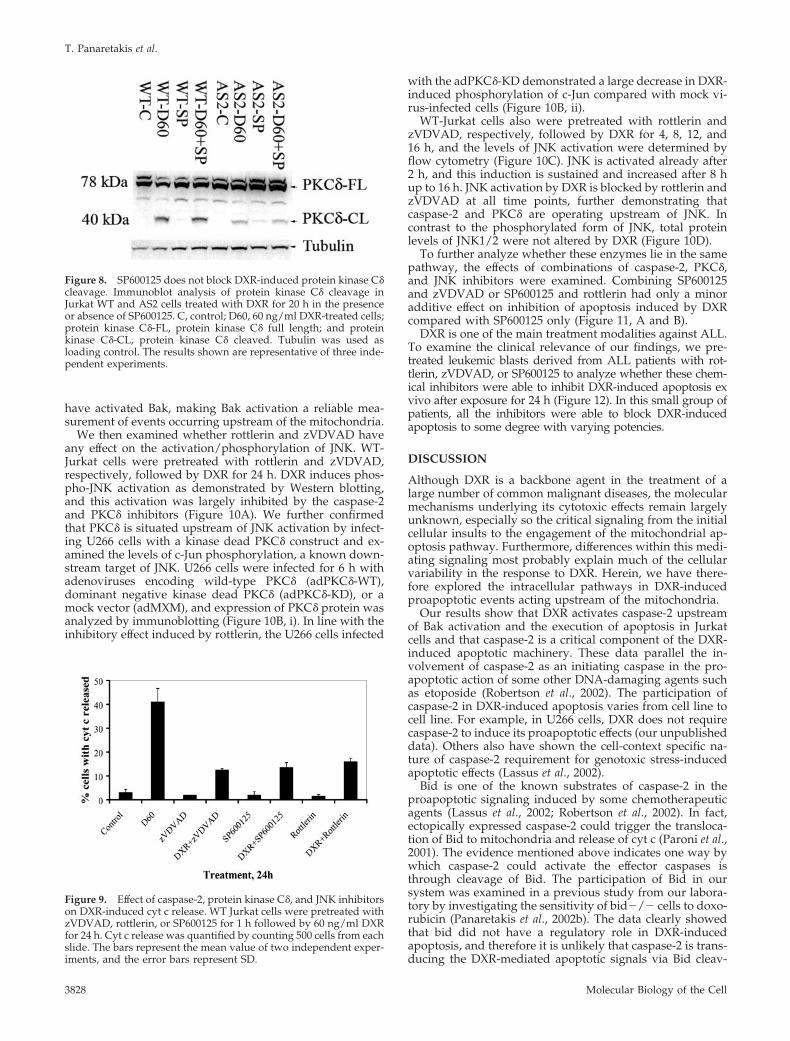
have activated Bak, making Bak activation a reliable mea-surement of events occurring upstream of the mitochondria.
We then examined whether rottlerin and zVDVAD haveany effect on the activation/phosphorylation of JNK. WT-Jurkat cells were pretreated with rottlerin and zVDVAD,respectively, followed by DXR for 24 h. DXR induces phos-pho-JNK activation as demonstrated by Western blotting,and this activation was largely inhibited by the caspase-2and PKC� inhibitors (Figure 10A). We further confirmedthat PKC� is situated upstream of JNK activation by infect-ing U266 cells with a kinase dead PKC� construct and ex-amined the levels of c-Jun phosphorylation, a known down-stream target of JNK. U266 cells were infected for 6 h withadenoviruses encoding wild-type PKC� (adPKC�-WT),dominant negative kinase dead PKC� (adPKC�-KD), or amock vector (adMXM), and expression of PKC� protein wasanalyzed by immunoblotting (Figure 10B, i). In line with theinhibitory effect induced by rottlerin, the U266 cells infected
with the adPKC�-KD demonstrated a large decrease in DXR-induced phosphorylation of c-Jun compared with mock vi-rus-infected cells (Figure 10B, ii).
WT-Jurkat cells also were pretreated with rottlerin andzVDVAD, respectively, followed by DXR for 4, 8, 12, and16 h, and the levels of JNK activation were determined byflow cytometry (Figure 10C). JNK is activated already after2 h, and this induction is sustained and increased after 8 hup to 16 h. JNK activation by DXR is blocked by rottlerin andzVDVAD at all time points, further demonstrating thatcaspase-2 and PKC� are operating upstream of JNK. Incontrast to the phosphorylated form of JNK, total proteinlevels of JNK1/2 were not altered by DXR (Figure 10D).
To further analyze whether these enzymes lie in the samepathway, the effects of combinations of caspase-2, PKC�,and JNK inhibitors were examined. Combining SP600125and zVDVAD or SP600125 and rottlerin had only a minoradditive effect on inhibition of apoptosis induced by DXRcompared with SP600125 only (Figure 11, A and B).
DXR is one of the main treatment modalities against ALL.To examine the clinical relevance of our findings, we pre-treated leukemic blasts derived from ALL patients with rot-tlerin, zVDVAD, or SP600125 to analyze whether these chem-ical inhibitors were able to inhibit DXR-induced apoptosis exvivo after exposure for 24 h (Figure 12). In this small group ofpatients, all the inhibitors were able to block DXR-inducedapoptosis to some degree with varying potencies.
DISCUSSION
Although DXR is a backbone agent in the treatment of alarge number of common malignant diseases, the molecularmechanisms underlying its cytotoxic effects remain largelyunknown, especially so the critical signaling from the initialcellular insults to the engagement of the mitochondrial ap-optosis pathway. Furthermore, differences within this medi-ating signaling most probably explain much of the cellularvariability in the response to DXR. Herein, we have there-fore explored the intracellular pathways in DXR-inducedproapoptotic events acting upstream of the mitochondria.
Our results show that DXR activates caspase-2 upstreamof Bak activation and the execution of apoptosis in Jurkatcells and that caspase-2 is a critical component of the DXR-induced apoptotic machinery. These data parallel the in-volvement of caspase-2 as an initiating caspase in the pro-apoptotic action of some other DNA-damaging agents suchas etoposide (Robertson et al., 2002). The participation ofcaspase-2 in DXR-induced apoptosis varies from cell line tocell line. For example, in U266 cells, DXR does not requirecaspase-2 to induce its proapoptotic effects (our unpublisheddata). Others also have shown the cell-context specific na-ture of caspase-2 requirement for genotoxic stress-inducedapoptotic effects (Lassus et al., 2002).
Bid is one of the known substrates of caspase-2 in theproapoptotic signaling induced by some chemotherapeuticagents (Lassus et al., 2002; Robertson et al., 2002). In fact,ectopically expressed caspase-2 could trigger the transloca-tion of Bid to mitochondria and release of cyt c (Paroni et al.,2001). The evidence mentioned above indicates one way bywhich caspase-2 could activate the effector caspases isthrough cleavage of Bid. The participation of Bid in oursystem was examined in a previous study from our labora-tory by investigating the sensitivity of bid�/� cells to doxo-rubicin (Panaretakis et al., 2002b). The data clearly showedthat bid did not have a regulatory role in DXR-inducedapoptosis, and therefore it is unlikely that caspase-2 is trans-ducing the DXR-mediated apoptotic signals via Bid cleav-
Figure 8. SP600125 does not block DXR-induced protein kinase C�cleavage. Immunoblot analysis of protein kinase C� cleavage inJurkat WT and AS2 cells treated with DXR for 20 h in the presenceor absence of SP600125. C, control; D60, 60 ng/ml DXR-treated cells;protein kinase C�-FL, protein kinase C� full length; and proteinkinase C�-CL, protein kinase C� cleaved. Tubulin was used asloading control. The results shown are representative of three inde-pendent experiments.
Figure 9. Effect of caspase-2, protein kinase C�, and JNK inhibitorson DXR-induced cyt c release. WT Jurkat cells were pretreated withzVDVAD, rottlerin, or SP600125 for 1 h followed by 60 ng/ml DXRfor 24 h. Cyt c release was quantified by counting 500 cells from eachslide. The bars represent the mean value of two independent exper-iments, and the error bars represent SD.
T. Panaretakis et al.
Molecular Biology of the Cell3828

age. In this study, we suggest that an alternative pathwayexists, namely via the caspase-2–dependent cleavage andactivation of PKC� to lead to the activation of Bak, cyt crelease, and effector caspase-activation
We initially assumed that the partial nature of the resis-tance to DXR in the AS2 Jurkat cells is due to incompletedown-regulation of caspase-2 protein levels as demon-strated in previous investigations by using these cells (Rob-ertson et al., 2002). However, the very efficient chemicalinhibition of caspase-2 activity by zVDVAD in WT Jurkatcells had a similar incomplete effect. Moreover, chemicalinhibitors of the other two enzymes involved, JNK andPKC�, led to a similar incomplete inhibition of DXR-inducedapoptosis, suggesting that several proapoptotic pathwaysare induced simultaneously by DXR, which would be in linewith the multitarget nature of this drug (Gewirtz, 1999).
In this study, we have discovered another major down-stream target for caspase-2, namely, PKC�. Its cleavage isattenuated in the antisense caspase-2 Jurkat cells and iscompletely blocked by the caspase-2 inhibitor zVDVAD.Furthermore, caspase-2 was found to directly cleave PKC�in vitro, in a similar manner to caspase-3 (Ghayur et al.,1996). PKC� is a known caspase-3 substrate and in thisstudy, we demonstrate for the first time that PKC� also iscleaved by caspase-2. The optimal recognition motif for thegroup II caspases (caspase-2, caspase-3, and caspase-7) isDEXD (Mancini et al., 2000). In fact, it has been shown beforethat caspase-2 and caspase-3 have similar substrates speci-
ficities. �II Spectrin and Golgin are two proteins that arecleaved on the same site by caspase-2 and caspase-3 (Man-cini et al., 2000; Rotter et al., 2004).
We have demonstrated previously that DXR is a potentinducer of apoptosis as well as Bak activation in U266 cells(Panaretakis et al., 2002b). Furthermore, inhibition of PKC� orJNK in U266 cells had similar effects compared with Jurkatcells. The similarities between Jurkat and U266 cells allowedus to use U266 for the adenoviral experiments because theJurkat cells used in this study were not possible to infect.
Inhibition of PKC� activity either by chemical inhibitionor by using a kinase dead mutant furthermore resulted in aconsiderable resistance to DXR-induced cell death. Thesedata indicate that PKC� is an important regulator in theexecution of the DXR-induced apoptotic program.
The mechanism of activation of PKC� by various pro-apoptotic and antiapoptotic stimuli involves a series of post-translational modifications (phosphorylations and cleavage)(for reviews on PKC�, see Basu, 2003; Jackson and Foster,2004). Although distinct tyrosine and serine/threoninephosphorylations can lead to either an anti- or proapoptoticresponse, cleavage of PKC� to its 40-kDa, fully active cata-lytic fragment is the only definite modification that leads toa proapoptotic response as demonstrated by Emoto et al.(1996). Data supporting this hypothesis come mainly fromexperiments in which overexpression of this fragment byitself induces chromatin condensation and DNA fragmenta-tion (Ghayur et al., 1996).
Figure 10. Effect of rottlerin and zVDVADon DXR-induced JNK activation. (A) Immu-noblot analysis of JNK phosphorylation inJurkat WT pretreated with VDVAD or rot-tlerin for 1 h followed by the addition ofDXR for 24 h. WT-C, control; WT-D60, 60ng/ml DXR-treated cells; WT-D60 � VD-VAD, WT Jurkat cells pretreated with VD-VAD and 60 ng/ml DXR; WT-D60 � rot-tlerin, WT Jurkat cells pretreated withrottlerin and 60 ng/ml DXR; VDVAD, WTJurkat cells treated with VDVAD; and rot-tlerin, WT Jurkat cells treated with rottlerin.Tubulin was used as loading control. Theresults shown are representative of two in-dependent experiments. (B) U266 cells wereinfected with adPKCd-WT, adPKCD-KD,and adMXM, and the protein kinase C� pro-tein expression (i) as well as the effect ofthese mutant on DXR-induced phosphory-lation of c-Jun (ii) was analyzed. D60, U266cells treated with 60 ng/ml DXR for theindicated time. The immunoblot is a repre-sentative result of two independent experi-ments; protein kinase C�-FL, protein kinaseC� full length. (C) WT-Jurkat cells weretreated with 60 ng/ml DXR (D60) for theindicated time points in the presence or ab-sence of rottlerin or zVDVAD, and the phos-pho-JNK levels were assessed by flow cy-tometry. The bars represent the mean valueof two independent experiments, and theerror bars represent SD. (D) Total proteinlevels of JNK were assessed by Westernblotting in WT-Jurkat cells treated with 60ng/ml DXR for the indicated time points.The result shown is representative of twoindependent experiments.
DXR Induces Death via Caspase-2, PKC�, and JNK
Vol. 16, August 2005 3829

The previous proposition that caspase-3 is the onlycaspase that could cleave PKC� posed a problem as to howsuch a downstream effector caspase could activate an up-stream regulator of apoptosis. Furthermore, expression of akinase dead mutant inhibited both caspase-9 and caspase-3activation, further suggesting that an alternate, upstreamcaspase could be responsible for the cleavage of PKC�(Matassa et al., 2001). In this study, we demonstrate that theinitiator caspase-2 has the ability to cleave, and thereforeactivate, PKC�.
The downstream proapoptotic targets of PKC� includec-Abl, Lamin B, and lipid scramblase and PKC� activationcauses disassembly of nuclear lamins and inhibition ofDNA-PK expression (Bharti et al., 1998; Cross et al., 2000;Frasch et al., 2000; Godbout et al., 2002). Another putativedownstream target in response to DNA-damage is JNK(Yoshida et al., 2002).
We found that JNK is activated in response to DXR as wasdescribed for cells exposed to various DNA-damagingagents (Kharbanda et al., 2000; Yoshida et al., 2002; Besirliand Johnson, 2003). However, the mechanism of activationof either JNK directly or of the SAPK cascade is poorlydescribed. The fact that DXR-induced apoptosis and Bakactivation are severely attenuated in cells treated with the
JNK inhibitor SP600125 supports a key role of JNK activa-tion also in DXR-induced apoptosis. jnk1 and jnk2 DKOMEFs were almost completely resistant to DXR-induced ap-optosis, further emphasizing the importance of JNK in thisrespect. The relationship between JNK activation and theother proapoptotic enzymes analyzed in this study wasevaluated. The fact that there is no clear effect of the JNKinhibitor SP600125 in AS2 cells, or cells cotreated with in-hibitors to caspase-2 or PKC�, strongly indicates that JNKacts in the same pathway. Furthermore, JNK inhibition hadno effect on DXR-induced PKC� cleavage or phosphoryla-tion, whereas both rottlerin and zVDVAD inhibit the DXR-induced activating phosphorylation of JNK. In addition, theDXR-induced phosphorylation of c-Jun, a known down-stream target of JNK was partially blocked by the dominantnegative form of PKC�, further demonstrating that proteinkinase C� is upstream of JNK phosphorylation and activa-tion. These data further indicate that JNK is important at astep downstream of caspase-2 and protein kinase C�. It isnot known whether protein kinase C� directly activates JNKby phosphorylation or by activating the SAPK cascade. Thisneeds to be established in future studies.
A minor decrease in caspase-2 activity was noted, how-ever, after SP600125 treatment. One likely explanation forthis could be that the amplification of initiator caspases byeffector caspases, such as caspase-3, is diminished by JNKinhibition.
Primary ALL samples were used to confirm the clinicalimportance of protein kinase C�, caspase-2, and JNK in theexecution of DXR-induced apoptosis. It is conceivable thatthese proteins are required in vivo for DXR to exert itsantineoplastic effects, and deactivating mutations in theseproteins may reflect one of the mechanisms of resistanceagainst this drug.
In summary, the present investigation proposes a modelfor DXR-induced cytotoxicity, where a significant part of theapoptosis-inducing capacity of this drug is exerted by asequential activation of caspase-2, protein kinase C�, andJNK, leading to Bak activation and apoptosis. This charac-terization of the molecular background to DXR-induced ap-
Figure 11. Effects of combination of SP600125-zVDVAD andSP600125-rottlerin on DXR-induced apoptosis. WT and AS2 Jurkatcells were treated with 60 ng/ml DXR (D60) for 24 h in the presenceor absence of SP600125 � rottlerin (A) and SP600125 � zVDVAD(B). Changes in DXR-induced apoptosis were assessed by flowcytometry as alterations in Annexin V positivity. The bars representthe mean value of three independent experiments, and the errorbars represent SD.
Figure 12. Effects of rottlerin, zVDVAD and SP600125 in DXR-treated patient samples. Leukemic blasts from three different ALLpatients were pretreated for 1 h with rottlerin, zVDVAD, andSP600125 followed by 100 ng/ml DXR (D100) for 24 h. The inhibi-tion in DXR-induced apoptosis was assessed by flow cytometry aschanges in Annexin V positivity.
T. Panaretakis et al.
Molecular Biology of the Cell3830

optosis will ultimately lead to an optimized clinical use ofthis important chemotherapeutic compound in terms of se-lection of sensitive patients, combination with other drugs,and overcoming resistance as well as greater understandingof the major apoptosis pathways.
ACKNOWLEDGMENTS
We thank Dr. Eric Solary for Casp-2/AS and neo-Jurkat cells. We thank Prof.Emad Alnemri for bacterial plasmids and Dr. Andrej Kropotov for purification ofrecombinant caspase-2 proteins. We thank Dr. Trevor Biden for generouslyproviding adenoviral constructs adPKCd-wt, adPKCd-KD, and adMXM. Wethank Dr. Kanaga Sabapathy for jnk- and jnk1-reconstituted MEF cells. Thisstudy was supported by the Swedish and Stockholm Cancer societies.
REFERENCES
Basu, A. (2003). Involvement of protein kinase C-� in DNA damage-inducedapoptosis. J. Cell Mol. Med. 7, 341–350.
Besirli, C. G., and Johnson, E. M., Jr. (2003). JNK-independent activation ofc-Jun during neuronal apoptosis induced by multiple DNA-damaging agents.J. Biol. Chem. 278, 22357–22366.
Bharti, A., et al. (1998). Inactivation of DNA-dependent protein kinase byprotein kinase C�: implications for apoptosis. Mol. Cell. Biol. 18, 6719–6728.
Carpenter, L., Cordery, D., and Biden, T. J. (2001). Protein kinase C� activationby interleukin-1� stabilizes inducible nitric-oxide synthase mRNA in pancre-atic �-cells. J. Biol. Chem. 276, 5368–5374.
Cross, T., Griffiths, G., Deacon, E., Sallis, R., Gough, M., Watters, D., and Lord,J. M. (2000). Protein kinase C-� is an apoptotic lamin kinase. Oncogene 19,2331–2337.
Dal Pra, I., Whitfield, J. F., Chiarini, A., and Armato, U. (2000). Increasedactivity of the protein kinase C-[�] holoenzyme in the cytoplasmic particulatefraction precedes the activation of caspases in polyomavirus-transformedpyF111 rat fibroblasts exposed to calphostin C or topoisomerase-II inhibitors.Exp. Cell Res. 255, 171–183.
Decaudin, D., Geley, S., Hirsch, T., Castedo, M., Marchetti, P., Macho, A.,Kofler, R., and Kroemer, G. (1997). Bcl-2 and Bcl-XL antagonize the mitochon-drial dysfunction preceding nuclear apoptosis induced by chemotherapeuticagents. Cancer Res 57, 62–67.
Denning, M. F., Wang, Y., Nickoloff, B. J., and Wrone-Smith, T. (1998). Proteinkinase C� is activated by caspase-dependent proteolysis during ultravioletradiation-induced apoptosis of human keratinocytes. J. Biol. Chem. 273,29995–30002.
Derijard, B., Hibi, M., Wu, I. H., Barrett, T., Su, B., Deng, T., Karin, M., and Davis,R. J. (1994). JNK 1, a protein kinase stimulated by UV light and Ha-Ras thatbinds and phosphorylates the c-Jun activation domain. Cell 76, 1025–1037.
Emoto, Y., Kisaki, H., Manome, Y., Kharbanda, S., and Kufe, D. (1996).Activation of protein kinase C� in human myeloid leukemia cells treated with1-�-d-arabinofuranosylcytosine. Blood 87, 1990–1996.
Frasch, S. C., Henson, P. M., Kailey, J. M., Richter, D. A., Janes, M. S., Fadok,V. A., and Bratton, D. L. (2000). Regulation of phospholipid scramblaseactivity during apoptosis and cell activation by protein kinase C�. J. Biol.Chem. 275, 23065–23073.
Garcia-Calvo, M., Peterson, E. P., Rasper, D. M., Vaillancourt, J. P., Zamboni,R., Nicholson, D. W., and Thornberry, N. A. (1999). Purification and catalyticproperties of human caspase family members. Cell Death Differ. 6, 362–369.
Gewirtz, D. A. (1999). A critical evaluation of the mechanisms of actionproposed for the antitumor effects of the anthracycline antibiotics Adriamycinand daunorubicin. Biochem. Pharmacol. 57, 727–741.
Ghayur, T., et al. (1996). Proteolytic activation of protein kinase C� by anICE/CED 3-like protease induces characteristics of apoptosis. J. Exp. Med.184, 2399–2404.
Godbout, J. P., Pesavento, J., Hartman, M. E., Manson, S. R., and Freund, G. G.(2002). Methylglyoxal enhances cisplatin-induced cytotoxicity by activatingprotein kinase C�. J. Biol. Chem. 277, 2554–2561.
Gupta, S., Campbell, D., Derijard, B., and Davis, R. J. (1995). Transcriptionfactor ATF2 regulation by the JNK signal transduction pathway. Science 267,389–393.
Jackson, D. N., and Foster, D. A. (2004). The enigmatic protein kinase C�:complex roles in cell proliferation and survival. FASEB J. 18, 627–636.
Jaken, S. (1996). Protein kinase C isozymes and substrates. Curr. Opin. CellBiol. 8, 168–173.
Kharbanda, S., Pandey, P., Ren, R., Mayer, B., Zon, L., and Kufe, D. (1995).c-Abl activation regulates induction of the SEK1/stress-activated proteinkinase pathway in the cellular response to 1-�-d-arabinofuranosylcytosine.J. Biol. Chem. 270, 30278–30281.
Kharbanda, S., et al. (2000). Translocation of SAPK/JNK to mitochondria andinteraction with Bcl-x(L) in response to DNA damage. J. Biol. Chem. 275, 322–327.
Lassus, P., Opitz-Araya, X., and Lazebnik, Y. (2002). Requirement forcaspase-2 in stress-induced apoptosis before mitochondrial permeabilization.Science 297, 1352–1354.
Lei, K., and Davis, R. J. (2003). JNK phosphorylation of Bim-related membersof the Bcl2 family induces Bax-dependent apoptosis. Proc. Natl. Acad. Sci.USA 100, 2432–2437.
Leitges, M., Mayr, M., Braun, U., Mayr, U., Li, C., Pfister, G., Ghaffari-Tabrizi,N., Baier, G., Hu, Y., and Xu, Q. (2001). Exacerbated vein graft arteriosclerosisin protein kinase C�-null mice. J. Clin. Investig. 108, 1505–1512.
Li, W., Yu, J. C., Shin, D. Y., and Pierce, J. H. (1995). Characterization of aprotein kinase C-� (protein kinase C-�) ATP binding mutant. An inactiveenzyme that competitively inhibits wild type protein kinase C-� enzymaticactivity. J. Biol. Chem. 270, 8311–8318.
Mancini, M., Machamer, C. E., Roy, S., Nicholson, D. W., Thornberry, N. A.,Casciola-Rosen, L. A., and Rosen, A. (2000). Caspase-2 is localized at the Golgicomplex and cleaves golgin-160 during apoptosis. J. Cell Biol. 149, 603–612.
Martiny-Baron, G., Kazanietz, M. G., Mischak, H., Blumberg, P. M., Kochs, G.,Hug, H., Marme, D., and Schachtele, C. (1993). Selective inhibition of proteinkinase C isozymes by the indolocarbazole Go 6976. J. Biol. Chem. 268, 9194–9197.
Matassa, A. A., Carpenter, L., Biden, T. J., Humphries, M. J., and Reyland,M. E. (2001). PKC� is required for mitochondrial-dependent apoptosis insalivary epithelial cells. J. Biol. Chem. 276, 29719–29728.
Mizuno, K., Noda, K., Araki, T., Imaoka, T., Kobayashi, Y., Akita, Y., Shi-monaka, M., Kishi, S., and Ohno, S. (1997). The proteolytic cleavage of proteinkinase C isotypes, which generates kinase and regulatory fragments, corre-lates with Fas-mediated and 12-O-tetradecanoyl-phorbol-13-acetate-inducedapoptosis. Eur. J. Biochem. 250, 7–18.
Panaretakis, T., Pokrovskaja, K., Shoshan, M. C., and Grander, D. (2002a).Activation of Bak, Bax and BH3-only proteins in the apoptotic response todoxorubicin. J. Biol. Chem. 277, 44317–44326.
Panaretakis, T., Pokrovskaja, K., Shoshan, M. C., and Grander, D. (2002b).Activation of Bak, Bax, and BH3-only proteins in the apoptotic response todoxorubicin. J. Biol. Chem. 277, 44317–44326.
Paroni, G., Henderson, C., Schneider, C., and Brancolini, C. (2001). Caspase-2-induced apoptosis is dependent on caspase-9, but its processing during UV-or tumor necrosis factor-dependent cell death requires caspase-3. J. Biol.Chem. 276, 21907–21915.
Reyland, M. E., Anderson, S. M., Matassa, A. A., Barzen, K. A., and Quissell,D. O. (1999). Protein kinase C� is essential for etoposide-induced apoptosis insalivary gland acinar cells. J. Biol. Chem. 274, 19115–19123.
Robertson, J. D., Enoksson, M., Suomela, M., Zhivotovsky, B., and Orrenius,S. (2002). Caspase-2 acts upstream of mitochondria to promote cytochrome crelease during etoposide-induced apoptosis. J. Biol. Chem. 277, 29803–29809.
Rotter, B., Kroviarski, Y., Nicolas, G., Dhermy, D., and Lecomte, M. C. (2004).�II-Spectrin is an in vitro target for caspase-2, and its cleavage is regulated bycalmodulin binding. Biochem. J. 378, 161–168.
Testolin, L., Carson, C., Wang, Y., Walker, P. R., Armato, U., and Sikorska, M.(1997). Jun and JNK kinase are activated in thymocytes in response to VM26and radiation but not glucocorticoids. Exp. Cell Res. 230, 220–232.
Verheij, M., et al. (1996). Requirement for ceramide-initiated SAPK/JNK sig-nalling in stress-induced apoptosis. Nature 380, 75–79.
Wang, L., Miura, M., Bergeron, L., Zhu, H., and Yuan, J. (1994). Ich-1, anIce/ced-3-related gene, encodes both positive and negative regulators ofprogrammed cell death. Cell 78, 739–750.
Wei, M. C., Zong, W. X., Cheng, E. H., Lindsten, T., Panoutsakopoulou, V.,Ross, A. J., Roth, K. A., MacGregor, G. R., Thompson, C. B., and Korsmeyer,S. J. (2001). Proapoptotic BAX and BAK: a requisite gateway to mitochondrialdysfunction and death. Science 292, 727–730.
Yamamoto, K., Ichijo, H., and Korsmeyer, S. J. (1999). BCL-2 is phosphory-lated and inactivated by an ASK1/Jun N-terminal protein kinase pathwaynormally activated at G(2)/M. Mol. Cell Biol. 19, 8469–8478.
Yoshida, K., Miki, Y., and Kufe, D. (2002). Activation of SAPK/JNK signaling byprotein kinase C� in response to DNA damage. J. Biol. Chem. 277, 48372–48378.
Zhivotovsky, B., Samali, A., Gahm, A., and Orrenius, S. (1999). Caspases: theirintracellular localization and translocation during apoptosis. Cell Death Dif-fer. 6, 644–651.
DXR Induces Death via Caspase-2, PKC�, and JNK
Vol. 16, August 2005 3831