A phosphatase cascade by which rewarding stimuli control nucleosomal response
Upload
independentCategory
view
2download
0
DNA damage in transcribed genes induces apoptosis via the JNK pathway
and the JNK-phosphatase MKP-1
Mohamed Hamdi1,4, Jaap Kool1,2,4,5, Paulien Cornelissen-Steijger1,2, Francoise Carlotti1,Herman E Popeijus1, Corina van der Burgt1, Josephine M Janssen1, Akira Yasui3,Rob C Hoeben1, Carrol Terleth1,2, Leon H Mullenders2 and Hans van Dam*,1
1Department of Molecular Cell Biology, Leiden University Medical Center, Wassenaarseweg 72, 2333AL Leiden, The Netherlands;2Department of Toxicogenetics, Leiden University Medical Center, Wassenaarseweg 72, 2333AL Leiden, The Netherlands;3Department of Molecular Genetics, Institute of Development, Aging and Cancer, Tohoku University, 980-8575 Sendai, Japan
The nucleotide excision repair (NER) system consists oftwo subpathways, global genome repair (GGR) andtranscription-coupled repair (TCR), which exhibit distinctfunctions in the cellular response to genotoxic stress.Defects in TCR result in prolonged UV light-inducedstalling of RNA polymerase II and hypersensitivity toapoptosis induced by UV and certain chemotherapeuticdrugs. Here, we show that low doses of UV triggerdelayed activation of the stress-induced MAPkinase JNKand its proapoptotic targets c-Jun and ATF-3 in TCR-deficient primary human fibroblasts from XerodermaPigmentosum (XP) and Cockayne syndrome (CS)patients. This delayed activation of the JNK pathway isnot observed in GGR-deficient TCR-proficient XP cells, isindependent of functional p53, and is established throughrepression of the JNK-phosphatase MKP-1 rather than byactivation of the JNK kinases MKK4 and 7. Enzymaticreversal of UV-induced cyclobutane pyrimidine dimers(CPDs) by CPD photolyase abrogated JNK activation,MKP-1 repression, and apoptosis in TCR-deficient XPAcells. Ectopic expression of MKP-1 inhibited DNA-damage-induced JNK activity and apoptosis. Theseresults identify both MKP-1 and JNK as sensors anddownstream effectors of persistent DNA damage intranscribed genes and suggest a link between the JNKpathway and UV-induced stalling of RNApol II.Oncogene (2005) 24, 7135–7144. doi:10.1038/sj.onc.1208875;published online 25 July 2005
Keywords: AP-1; DNA damage; MKP-1; JNK; trans-cription-coupled repair
Introduction
Mammalian cells exhibit multiple biological responsesto genotoxic stress, including cell cycle checkpoints,
DNA repair, and apoptosis. Inactivation of theseresponses may result in genomic instability and celltransformation, as well as modifications of therapeuticsensitivity. Bulky DNA-helix distorting lesions such asUV-induced photolesions and cisplatin-induced DNAadducts can be removed by the nucleotide excisionrepair (NER) system. Deficiencies in NER lead toenhancement of DNA-damage-induced cell killing andmutagenesis. Two distinct NER subpathways can bedistinguished: global genome repair (GGR), whichrepairs DNA damage throughout the genome, andtranscription-coupled repair (TCR), which repairs DNAlesions in the transcribed strand of active genes. TheDNA repair factors XPC and XPE are exclusivelyinvolved in GGR, whereas the CSA and CSB proteinsonly play a role in TCR. Other NER proteins, such asXPA, XPB, and XPD, function in both TCR and GGR(reviewed by Berneburg and Lehmann, 2001; Mitchellet al., 2003). Genetic defects in these NER genes cancause the human diseases Xeroderma pigmentosum (XP)and Cockayne Syndrome (CS). XP patients are verysensitive to UV light, undergo progressive degenerationof the skin, and show a 1000-fold increased risk for skincancer, mainly on sun-exposed parts of the body. CSpatients, who die at young age, show more pleiotropicfeatures, including neurological degradation, but notincreased skin cancer (reviewed by van Steeg andKraemer, 1999; de Boer and Hoeijmakers, 2000).Importantly, in contrast to TCR-proficient cells, TCR-deficient cells already undergo apoptosis at very lowdoses of UV light, and, in addition, are sensitive to thechemotherapeutic drugs cisplatin and illudin/Irofulven(McKay et al., 1998, 2001; van Oosten et al., 2000;Jaspers et al., 2002; Spivak et al., 2002). This sensitivity ofTCR-deficient cells is associated with prolonged UV-induced inhibition of transcription due to prolongedstalling of RNA polymerase II (RNApol II) (Rockx et al.,2000; Berneburg and Lehmann, 2001; Svejstrup, 2002).In addition to DNA lesions, UV light and other
genotoxic agents can generate reactive oxygen species(ROS), which result in lipid peroxidation, proteinoxidation, and glutathione depletion (Mercurio andManning, 1999). The cell can respond to these types ofstress by activating various signaling cascades, including
Received 4 March 2005; revised 3 May 2005; accepted 24 May 2005;published online 25 July 2005
*Correspondence: H van Dam; E-mail: [email protected] authors contributed equally to this paper5Current address: Netherlands Cancer Institute, Division of MolecularGenetics, Plesmanlaan 121, 1066CX, Amsterdam, The Netherlands
Oncogene (2005) 24, 7135–7144& 2005 Nature Publishing Group All rights reserved 0950-9232/05 $30.00
www.nature.com/onc
the p53, NF-kB, ATR, and MAPkinase (MAPK)pathways. Depending on the cell type and the genotoxicstress, the combined action of these signaling pathwayscan trigger transient or prolonged cell cycle arrest orapoptosis (Martindale and Holbrook, 2002; Shaulianand Karin, 2002; Brancho et al., 2003; Todd et al.,2004). The MAPK family member JNK/SAPK is able tocontrol apoptosis both positively and negatively, de-pending on the cell type, cellular context, and the stresssignal. Proapoptotic substrates of JNK comprise bothtranscription factors, like the transcription factor AP-1component c-Jun, and components of the apoptosis-executing machinery, like the proapoptotic Bcl-2-relatedproteins (Lei et al., 2002; Shaulian and Karin, 2002;Weston and Davis, 2002; Lei and Davis, 2003).Antiapoptotic JNK targets include JunD and thesurvival gene cIAP-2 (Lamb et al., 2003). In additionto c-Jun and JunD, JNK can activate and/or induce theexpression of various other AP-1 components involvedin cell proliferation and survival, like ATF-2, ATF-3,and c-Fos (Hai et al., 1999; Shaulian and Karin, 2001;van Dam and Castellazzi, 2001). Interestingly, recentstudies indicate that c-Jun and ATF-2 can control thecellular response to DNA damage by regulating theexpression of various DNA repair genes, includingERCC3, XPA, RAD23B, and MSH2 (Hayakawa et al.,2003, 2004; MacLaren et al., 2004).JNK activity in the cell is tightly controlled by both
protein kinases and protein phosphatases. Various typesof stimuli activate JNK through phosphorylation by thedual specificity kinases MKK4 or MKK7 (Morrisonand Davis, 2003; Wada et al., 2004). In contrast,mitogens and stress stimuli can inactivate JNK throughinduction of the expression of JNK phosphatases, whichinclude serine/threonine phosphatases, tyrosine phos-phatases, and dual specificity (threonine/tyrosine) phos-phatases (Camps et al., 2000; Keyse, 2000; Farooq andZhou, 2004). Activation of the dual specificity JNK-phosphatase MKP-1 seems to be involved in the cellulardefense against H2O2- and UV-induced apoptosis(Franklin et al., 1998; Xu et al., 2004), whereasinhibition of MKP-1 can potentiate JNK-related apop-tosis (Guo et al., 1998; Desbois-Mouthon et al., 2000;Sanchez-Perez et al., 2000; Mizuno et al., 2004).In this study, we investigated the signaling route by
which UV-induced DNA damage induces apoptosis inTCR-deficient human fibroblasts. We identified ATF-3,c-Jun, and JNK as important sensors of DNA damagein the transcribed strand of active genes, at UV dosesthat are cytotoxic for TCR-deficient fibroblasts, but notfor TCR-proficient fibroblasts. Activation of ATF-3, c-Jun, and JNK at these UV doses occurs with delayedkinetics and is mediated by inhibition of MKP-1expression rather than by activation of the JNK kinasesMKK4 and 7. Importantly, removal of UV-inducedcyclobutane dimers (CPDs) by CPD photolyase abro-gated both the inhibition of MKP-1 and the activationof JNK, c-Jun, and ATF-3, while inhibition of JNKactivation by ectopic expression of MKP-1 stronglysuppressed DNA-damage-induced apoptosis. Theseresults reveal a new functional link between the MKP-
1–JNK pathway and persistent DNA damage intranscribed genes.
Results
Low doses of UV induce ATF-3, c-Jun, and JNK activityin TCR-deficient primary human fibroblasts
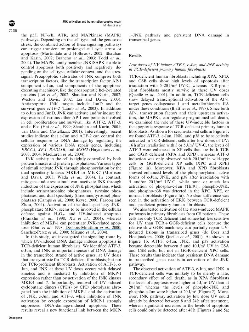
TCR-deficient human fibroblasts including XPA, XPD,and CSB cells show high levels of apoptosis afterirradiation with 5–20 J/m2 UV-C, whereas TCR-profi-cient fibroblasts mostly survive at these UV doses(Queille et al., 2001). In addition, TCR-deficient cellsshow delayed transcriptional activation of the AP-1target genes collagenase 1 and metallothionein IIAunder these conditions (Blattner et al., 1998). Since bothAP-1 transcription factors and their upstream activa-tors, the MAPKs, can regulate programmed cell death,we examined the role of these UV-inducible factors inthe apoptotic response of TCR-deficient primary humanfibroblasts. As shown for serum-starved cells in Figure 1,we found ATF-3, c-Jun, JNK, and p38 to be selectivelyactivated in TCR-deficient cells after low doses of UV:16 h after irradiation with 3 or 5 J/m2 UV-C, the levels ofATF-3 were enhanced in XP cells that are both TCRand GGR deficient (XPA and XPD), whereas ATF-3induction was only observed with 20 J/m2 in wild-typecells or GGR-deficient XP cells (XPC and XPE)(Figure 1a). Moreover, XPA and XPD fibroblastsshowed enhanced levels of the phosphorylated, activeforms of c-Jun, JNK, and p38 after irradiation with5 and/or 20 J/m2 UV-C, while none or much lessactivation of phospho-c-Jun (Thr91), phospho-JNK,and phospho-p38 was detected in the XPC, XPE, ornormal fibroblasts (Figure 1a). No clear difference wasseen in the activation of ERK between TCR-deficientand -proficient primary human fibroblasts.We also tested activation of the stress-induced MAPK
pathways in primary fibroblasts from CS patients. Thesecells are only TCR deficient and somewhat less sensitivefor UV than TCRþGGR-deficient cells because therelative slow GGR machinery can partially repair UV-induced lesions in transcribed genes (de Boer andHoeijmakers, 2000; Queille et al., 2001). As shown inFigure 1b, ATF3, c-Jun, JNK, and p38 activationbecame detectable between 5 and 10 J/m2 UV in CSAand CSB cells, but not in GGR-deficient XPC cells.These results thus indicate that persistent DNA damagein transcribed genes results in activation of the JNKpathway.The observed activation of ATF-3, c-Jun, and JNK in
TCR-deficient cells was unlikely to be merely a late,secondary effect of cell death, as in XPA fibroblaststhe levels of apoptosis were higher at 5 J/m2 UV than at20 J/m2 whereas the levels of phospho-JNK andphospho-c-Jun were higher at 20 J/m2 (Figure 2). More-over, JNK pathway activation by low dose UV couldalready be detected between 8 and 24 h after treatment,whereas significant increases in the levels of apoptoticcells could only be detected after 48 h (Figures 2 and 5a;
JNK activation and transcription-coupled repairM Hamdi et al
7136
Oncogene
data not shown). The relatively low levels of apoptosisin XPA cells after 20 J/m2 UV are presumably due tostrong inhibition of transcription at this UV dose(Queille et al., 2001), preventing the expression ofproapoptotic genes. In line with this, XPA cells containrelatively low amounts of ATF-3 and c-Jun protein andmRNA after high doses of UV (Figures 2 and 3).In summary, these results suggest that the JNK
pathway is involved in the apoptotic response of TCR-deficient cells.
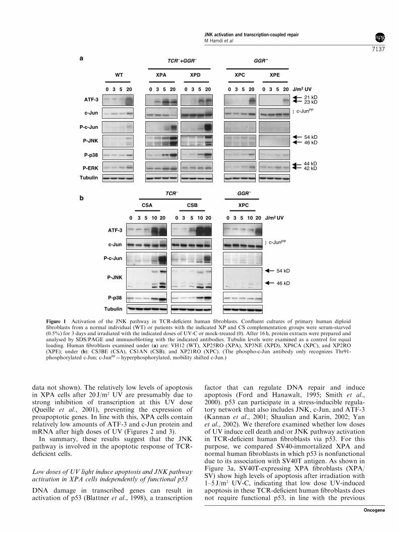
Low doses of UV light induce apoptosis and JNK pathwayactivation in XPA cells independently of functional p53
DNA damage in transcribed genes can result inactivation of p53 (Blattner et al., 1998), a transcription
factor that can regulate DNA repair and induceapoptosis (Ford and Hanawalt, 1995; Smith et al.,2000). p53 can participate in a stress-inducible regula-tory network that also includes JNK, c-Jun, and ATF-3(Kannan et al., 2001; Shaulian and Karin, 2002; Yanet al., 2002). We therefore examined whether low dosesof UV induce cell death and/or JNK pathway activationin TCR-deficient human fibroblasts via p53. For thispurpose, we compared SV40-immortalized XPA andnormal human fibroblasts in which p53 is nonfunctionaldue to its association with SV40T antigen. As shown inFigure 3a, SV40T-expressing XPA fibroblasts (XPA/SV) show high levels of apoptosis after irradiation with1–5 J/m2 UV-C, indicating that low dose UV-inducedapoptosis in these TCR-deficient human fibroblasts doesnot require functional p53, in line with the previous
WT
0 3 5 20 J/m2 UV
c-Jun
P-c-Jun
ATF-3
0 3 5 20
XPC XPE
0 3 5 200 3 5 20
GGR -
XPDXPA
0 3 5 20
P-JNK54 kD46 kD
P-ERK
P-p38
Tubulin
] c-JunPP
21 kD23 kD
44 kD42 kD
c-Jun
P-c-Jun
ATF-3
P-JNK
P-p38
Tubulin
54 kD
46 kD
] c-JunPP
CSA CSB
0 3 5 10 20 0 3 5 10 20
XPC
0 3 5 10 20 J/m2 UV
TCR - GGR -
TCR -+GGR -a
b
Figure 1 Activation of the JNK pathway in TCR-deficient human fibroblasts. Confluent cultures of primary human diploidfibroblasts from a normal individual (WT) or patients with the indicated XP and CS complementation groups were serum-starved(0.5%) for 3 days and irradiated with the indicated doses of UV-C or mock-treated (0). After 16 h, protein extracts were prepared andanalysed by SDS/PAGE and immunoblotting with the indicated antibodies. Tubulin levels were examined as a control for equalloading. Human fibroblasts examined under (a) are: VH12 (WT), XP25RO (XPA), XP3NE (XPD), XP8CA (XPC), and XP2RO(XPE); under (b): CS3BE (CSA), CS1AN (CSB), and XP21RO (XPC). (The phospho-c-Jun antibody only recognizes Thr91-phosphorylated c-Jun; c-JunPP¼hyperphosphorylated, mobility shifted c-Jun.)
JNK activation and transcription-coupled repairM Hamdi et al
7137
Oncogene
studies (McKay et al., 2001). Moreover, after irradiationwith 1 and 5 J/m2 UV-C, high levels of phosphorylatedactive JNK, c-Jun, and, to a lesser extent, p38 weredetected in the XPA/SV cells but not in wild-type cells(WT/SV; Figure 3b). Likewise, at 1 J/m2 the amounts ofATF-3 protein and mRNA were only induced in theXPA/SV cells (Figure 3b and c). These data show that inXPA cells both the apoptotic response and activation ofthe JNK pathway by low dose UV do not requirefunctional p53.
Apoptosis and JNK pathway activation via accumulationof UV-induced cyclobutane pyrimidine dimers
About 75% of the total amount of UV-inducedDNA damage comprises CPDs (de Boer and Hoeij-makers, 2000). To verify that UV-induced DNA damagerather than effects on other cellular targets like
cell membrane receptors triggers the apoptosis-asso-ciated JNK pathway activation in UV-irradiated XPAcells, we examined immortalized human XPA cellsectopically expressing CPD photolyase (Nakajimaet al., 2004). This enzyme can revert CPDs uponillumination with visible light (photoreactivation(PR)). As depicted in Figure 4a, removal of UV-inducedCPDs by photoreactivation strongly reduced the induc-tion of apoptosis by low doses of UV in XPA cells:3.9-fold at 1 J/m2 and 3.8-fold at 3 J/m2. (The residual
WT XPA
0
10
20
30
40
50
510 20 510 20
60
70
UV (J/m2) UV (J/m2)
24h48h72h
WT
24h48h72h
XPA
% a
po
pto
sis
0
10
20
30
40
50
60
70
c-Jun
P-c-Jun
ATF-3
P-JNK
P-MKK4
Tubulin
0 1 5 20 O.S
.
0 1 5 20 O.S
.J/m2 UV J/m2 UV
] c-JunPP
a
b
Figure 2 Apoptosis and JNK-c-Jun pathway activation inXPA and control primary human fibroblasts. (a) Subconfluent,proliferating cultures of XPA and normal human fibroblastswere irradiated with the indicated doses of UV-C or mock-treated(0) and harvested for FACS analysis after 24, 48, and 72 h.The level of apoptosis represents the percentage of cells in sub-G1.(b) Western analysis of the experiment described under (a). Proteinextracts were prepared 24 h after UV irradiation and analysedby SDS/PAGE and immunoblotting with the indicated anti-bodies. Tubulin levels were examined as a control forequal loading. As a control for MKK4 activation, parallel cultureswere treated for 15min with 0.5M sodium chloride (osmoticstress: O.S.)
P-JNK
P-p38
Tubulin
0 1 5 10 0 1 5 10 J/m2 UV
WT/SV XPA/SV
c-Jun
ATF-3
] c-JunPP
% a
po
pto
sis
WT/SV
UV (J/m2) UV (J/m2)
0
10
20
30
40
50
60
70
0 3 5 10
XPA/SV
0
10
20
30
40
50
60
70
3 5 10
J/m2 UV
XPA/SV
atf-3
c-jun
hef
0 1 5 10 0 1 5 10
WT/SV
1 0 1
a
b
c
Figure 3 Apoptosis and JNK pathway activation in SV40T-immortalized XPA and control human fibroblasts. (a) Subcon-fluent, proliferating cultures of SV40T-immortalized humandiploid fibroblasts from a control individual (WT/SV) or an XPApatient (XPA/SV) were irradiated with increasing doses of UV-Cand harvested for FACS analysis 48 h later. The level of apoptosisrepresents the percentage of cells in sub-G1. (b, c) The immorta-lized XPA and control fibroblasts analysed under (a) wereirradiated with 1, 5 or 10 J/m2 UV-C or mock-treated (0). After24 h, protein extracts and mRNA were prepared for immunoblot(b) and Northern blot (c) analysis. hef-1 mRNA expression wasdetermined to verify equal loading (c)
JNK activation and transcription-coupled repairM Hamdi et al
7138
Oncogene

apoptosis after photoreactivation is most likelydue to UV-induced 6-4-photoproducts, which arenot converted by CPD photolyase.) Importantly,CPD removal almost completely abrogated the activa-tion of ATF-3, JNK, and p38 (Figure 4b, rightpanel).These results thus indicate that both apoptosis and
JNK pathway activation can be triggered by prolongedpresence of CPDs on transcribed genes and subsequentstalling of RNA pol II.
DNA-damage-induced JNK activation througha CPD-mediated decrease of MAPK phosphatase-1
We next examined the mechanism by which DNAdamage induces JNK activation in XPA cells. Amultitude of stimuli including osmotic stress activateJNK through phosphorylation and activation of theJNK kinases MKK4 (SEK) and MKK7 (Morrisonand Davis, 2003; Wada et al., 2004). However, we didnot observe MKK4 or MKK7 activation by low dosesof UV, neither in primary nor in SV40T-immortalizedcells (Figures 2 and 5a, data not shown), althoughactivation of MKK4 and 7 activity by osmotic stresscould be detected (Figure 2, data not shown). Incontrast, as depicted in Figure 5a, we observed asignificant decrease in the expression of MKP-1, adual-specificity MAPK phosphatase that preferablydephosphorylates p38 and JNK (Franklin and Kraft,1997; Li et al., 2001). Importantly, reduction in theprotein levels of MKP-1 in SV40-immortalized XPAcells could already be observed 8 h after 1 J/m2 UV, atime point at which JNK phosphorylation was onlystarting to increase.
-+ PR
% a
po
pto
sis
UV (J/m2)
P-p38
++ ++- - - -
CPD phlcontrol
P-JNK
PR
1J0J 1J0J
ATF-3
c-Jun
Tubulin
XPA / SV
CPD phl
XPA/SV XPA/SV
] c-JunPP
0
10
20
30
40
50
60
70
0 31
a
b
Figure 4 UV-induced apoptosis and JNK pathway activationthrough nonrepaired cyclobutane pyrimidine dimers (CPD). (a)SV40T-immortalized XPA cells expressing CPD photolyase (XPA/SV CPD phl) were irradiated with 1 or 3 J/m2 UV-C or mock-treated (0). Subsequently, cells were treated for 1 h with photo-reactivating light or nontreated (7PR) and harvested for FACSanalysis 47 h later. The level of apoptosis represents the percentageof cells in sub-G1. (b) Immortalized XPA cells expressing CPDphotolyase (XPA/SV CPD phl) or control cells only containing theempty vector (XPA/SV control) were irradiated with 1 J/m2 UV-C(1J) or mock treated (0J), and subsequently treated for 1 h withphotoreactivating light or non-treated (7PR). After 23 h, proteinextracts were prepared for analysis by SDS/PAGE and immuno-blotting with the indicated antibodies
0 0 1 0 1 J/m2 UV
8 hrs 11 hrs
P-JNK
MKP-1
P-JNK
MKP-1
0 1 0 1 0 1 J/m2 UV
cont
rol
MKP1
C/Sm
MKP1
TubulinTubulin
0 1 J/m2 UV
mkp-1
hef
P-MKK4
- -
CPD phl
PR
0J 1J
++
MKP-1
Tubulin
XPA / SV
XPA / SVXPA / SV
XPA / SV
a b
dc
Figure 5 Low dose UV activates JNK in XPA cells via inhibitionof MKP-1 expression. (a, c) SV40T-immortalized XPA fibroblastswere irradiated with 1 J/m2 UV-C or mock-treated (0), andharvested after 8 and 11 h for immunoblot analysis (a), or after8 h for Northern blot analysis (c). HEF-1 mRNA expression wasdetermined to verify equal loading (c). (b) SV40T-immortalizedXPA cells were transduced with lentiviruses containing wild-typeMKP-1, the catalytically inactive dominant-negative mutant C/SmMKP-1, or an empty control virus. Transduction efficiency was95–100% as verified by FACS determination. Next, the threecultures were irradiated with 1 J/m2 UV-C or mock-treated (0), andharvested after 16 h for immunoblot analysis. Please note that inthe control cells the endogenous MKP-1 is not visible because arelatively short exposure is shown. (d) SV40T-immortalized XPAcells expressing CPD photolyase (XPA CPD phl) were irradiatedwith 1 J/m2 UV-C (1J) or mock treated (0J), and subsequentlytreated for 1 h with photoreactivating light or nontreated (7PR).After 7 h, protein extracts were prepared for immunoblot analysis
JNK activation and transcription-coupled repairM Hamdi et al
7139
Oncogene

We next examined whether inactivation of MKP-1activity in XP-A cells can cause JNK activation in theabsence of UV-induced DNA damage. For this purpose,we inhibited the function of the endogenous MKP-1protein via ectopic expression of the catalytically inactivedominant-negative MKP-1 mutant C/SmMKP1, intro-duced by a lentiviral vector (Bazuine et al., 2004; Slacket al., 2001). As depicted in Figure 5b, expression of thedominant-negative MKP-1 mutant resulted in increasedlevels of active phospho-JNK, but did not interfere withthe activation of JNK by low dose UV. In contrast,overexpression of wild-type MKP-1 did not increase thebasal activity of JNK and strongly inhibited JNKactivation by UV. From these results we conclude thatinhibition of JNK-phosphatase activity can cause JNKactivation in the XPA cells. Subsequent studies showedthat irradiation with low doses of UV results in adecrease in the levels of mkp-1 mRNA (Figure 5c) andthat enzymatic removal of CPDs by CPD photolyaseabrogates the UV-induced decrease in MKP-1 expres-sion (Figure 5d).In summary, accumulation of UV-induced cyclobu-
tane dimers causes a decrease in the expression of theJNK-phosphatase MKP-1 in XPA cells, which subse-quently results in enhancement of JNK activity.
Ectopic expression of MAPK phosphatase-1 inhibits bothDNA-damage-induced JNK activity and DNA-damage-induced apoptosis in XPA cells
The JNK pathway can trigger and/or enhance pro-grammed cell death in various cell types (Davis, 2000).We therefore examined whether inhibition of JNKaffects low dose UV-induced apoptosis in XPA cells.For this purpose, we ectopically expressed either wild-type MKP-1, which can dephosphorylate both JNK,p38, and ERK, or an MKP-1 mutant that onlyefficiently binds to JNK (m(JNK)MKP-1) (Slack et al.,2001). As shown in Figure 6a, we found the JNK-restricted MKP-1 mutant to be more efficient ininhibiting UV-induced JNK activation than wild-typeMKP-1, whereas the (weak) activation of p38 and ERKby UV was not significantly inhibited by either of thetwo MKP-1 constructs. Moreover, the JNK-specificMKP-1 mutant did not have any effect on theproliferation of nonirradiated XPA cells, while, asreported previously (Li et al., 1999; Engelbrecht et al.,2003), wild-type MKP-1 slightly reduced the prolifera-tion of these cells, also after low dose UV irradiation(data not shown). Importantly, the JNK-restrictedm(JNK)MKP-1 efficiently inhibited the induction ofapoptosis by 1 J/m2 UV in the XPA cells (Figure 6b),showing that JNK is an essential component of theDNA-damage-induced apoptotic program in these cells.In line with this, expression of the catalytically inactivedominant-negative MKP-1 mutant C/SmMKP1 notonly induced JNK activity but also further enhancedthe apoptotic response of XPA cells to low doses of UV.In conclusion, our results demonstrate that upon
DNA damage in transcribed genes down-regulation ofMKP-1 causes activation of the JNK pathway, which
subsequently plays an important functional role inDNA-damage-induced apoptosis.
Discussion
DNA repair and DNA-damage-induced signaling playkey protective roles in cancer development and cancertherapy (Mitchell et al., 2003). The NER system, arepair pathway for DNA-helix distorting lesionslike UV-induced photolesions exhibits a critical functionin both processes, as deficiencies in NER lead toenhancement of DNA-damage-induced mutagenesisand cell killing. Several studies indicate that the twoNER subpathways, GGR and TCR, differentiallycontrol mutagenesis and programmed cell death, be-cause TCR-deficient cells are much more sensitive toUV-induced apoptosis and certain chemotherapeuticdrugs than TCR-proficient XP cells (McKay et al., 1998,2001; Jaspers et al., 2002; Spivak et al., 2002). In thisstudy, we identified the JNK pathway as one of thecritical sensors and downstream effectors of persistentDNA damage in transcribed genes. We showed that lowdoses of UV light activate JNK, c-Jun, and ATF-3 inTCR-deficient human fibroblasts (XPA, XPD, CSA,and CSB), but not in TCR-proficient cells (XPE andXPC). Moreover, inhibition of JNK activity was foundto strongly reduce low dose UV-induced apoptosis inXPA cells.Previous studies showed that JNK signaling can
regulate apoptosis both positively and negatively,depending on the cell type, cellular context, and thenature and dose of treatment (Davis, 2000; Chang andKarin, 2001). Strong and sustained JNK activation ispredominantly associated with induction or enhance-ment of apoptosis, whereas transient JNK activationcan result in cell survival (Chang and Karin, 2001; Lambet al., 2003). In line with the presumed proapoptoticfunction of sustained JNK activation, we found the lowdose UV-induced apoptotic response in XPA fibroblaststo be preceded by delayed and long-lasting JNKactivation. Moreover, this sustained JNK activationwas accompanied by prolonged phosphorylation andactivation of the endogenous JNK substrate c-Jun, acomponent of transcription factor AP-1 that can act as aproapoptotic effector of JNK (Bossy-Wetzel et al., 1997;Behrens et al., 1999).The delayed phosphorylation and activation of JNK
in XPA and other TCR-defient cells is accompanied byinhibition of the JNK-phosphatase MKP-1 (this manu-script, our unpublished observations). Various studiesindicate that MKP-1 mainly exhibits antiapoptoticactivity. For instance, conditional over expression ofMKP-1 can repress the rapid activation of JNK and p38in response to high doses of UV and subsequentlyimpede apoptosis (Franklin et al., 1998). Moreover, thenonapoptotic DNA-damaging agent trans-platin in-duces MKP-1 expression and activates JNK onlytransiently, while its apoptotic isomer cis-platin whichcannot activate MKP-1 induces more sustained JNKactivation (Sanchez-Perez et al., 1998, 2000).
JNK activation and transcription-coupled repairM Hamdi et al
7140
Oncogene
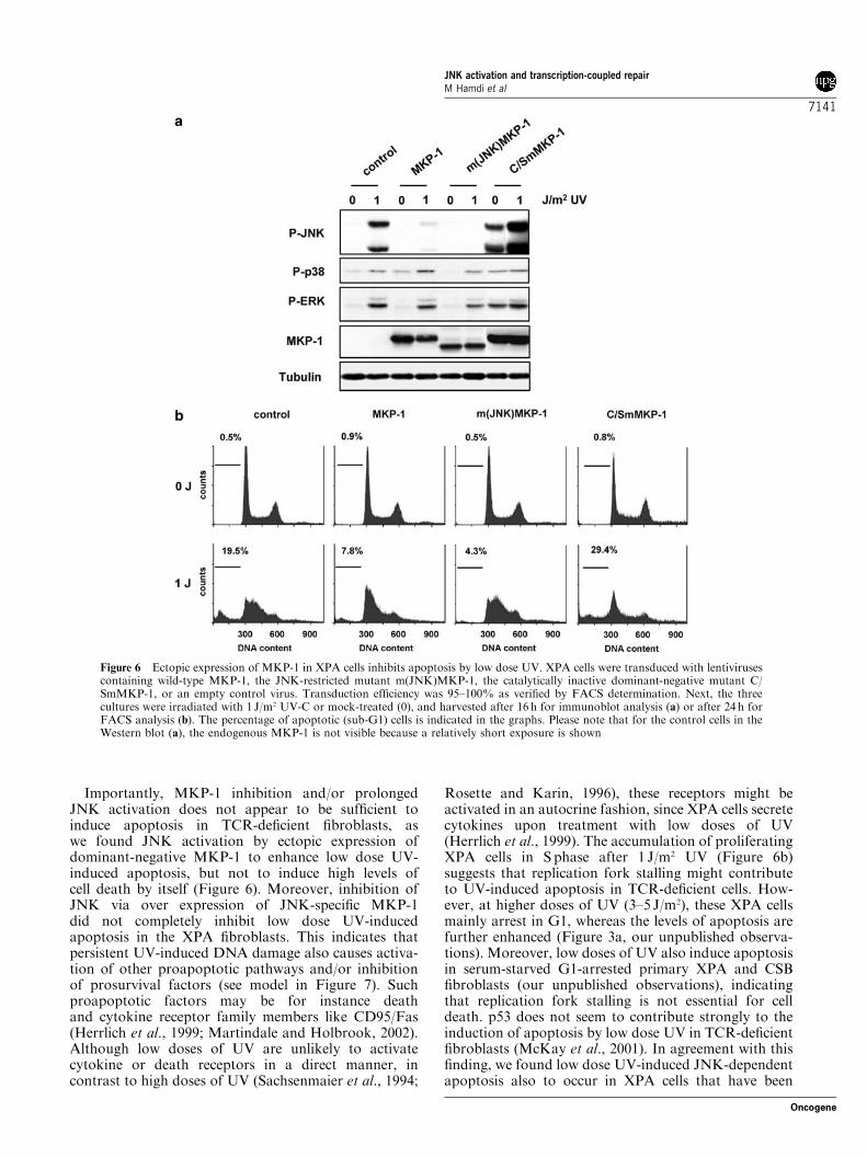
Importantly, MKP-1 inhibition and/or prolongedJNK activation does not appear to be sufficient toinduce apoptosis in TCR-deficient fibroblasts, aswe found JNK activation by ectopic expression ofdominant-negative MKP-1 to enhance low dose UV-induced apoptosis, but not to induce high levels ofcell death by itself (Figure 6). Moreover, inhibition ofJNK via over expression of JNK-specific MKP-1did not completely inhibit low dose UV-inducedapoptosis in the XPA fibroblasts. This indicates thatpersistent UV-induced DNA damage also causes activa-tion of other proapoptotic pathways and/or inhibitionof prosurvival factors (see model in Figure 7). Suchproapoptotic factors may be for instance deathand cytokine receptor family members like CD95/Fas(Herrlich et al., 1999; Martindale and Holbrook, 2002).Although low doses of UV are unlikely to activatecytokine or death receptors in a direct manner, incontrast to high doses of UV (Sachsenmaier et al., 1994;
Rosette and Karin, 1996), these receptors might beactivated in an autocrine fashion, since XPA cells secretecytokines upon treatment with low doses of UV(Herrlich et al., 1999). The accumulation of proliferatingXPA cells in S phase after 1 J/m2 UV (Figure 6b)suggests that replication fork stalling might contributeto UV-induced apoptosis in TCR-deficient cells. How-ever, at higher doses of UV (3–5 J/m2), these XPA cellsmainly arrest in G1, whereas the levels of apoptosis arefurther enhanced (Figure 3a, our unpublished observa-tions). Moreover, low doses of UV also induce apoptosisin serum-starved G1-arrested primary XPA and CSBfibroblasts (our unpublished observations), indicatingthat replication fork stalling is not essential for celldeath. p53 does not seem to contribute strongly to theinduction of apoptosis by low dose UV in TCR-deficientfibroblasts (McKay et al., 2001). In agreement with thisfinding, we found low dose UV-induced JNK-dependentapoptosis also to occur in XPA cells that have been
Figure 6 Ectopic expression of MKP-1 in XPA cells inhibits apoptosis by low dose UV. XPA cells were transduced with lentivirusescontaining wild-type MKP-1, the JNK-restricted mutant m(JNK)MKP-1, the catalytically inactive dominant-negative mutant C/SmMKP-1, or an empty control virus. Transduction efficiency was 95–100% as verified by FACS determination. Next, the threecultures were irradiated with 1 J/m2 UV-C or mock-treated (0), and harvested after 16 h for immunoblot analysis (a) or after 24 h forFACS analysis (b). The percentage of apoptotic (sub-G1) cells is indicated in the graphs. Please note that for the control cells in theWestern blot (a), the endogenous MKP-1 is not visible because a relatively short exposure is shown
JNK activation and transcription-coupled repairM Hamdi et al
7141
Oncogene
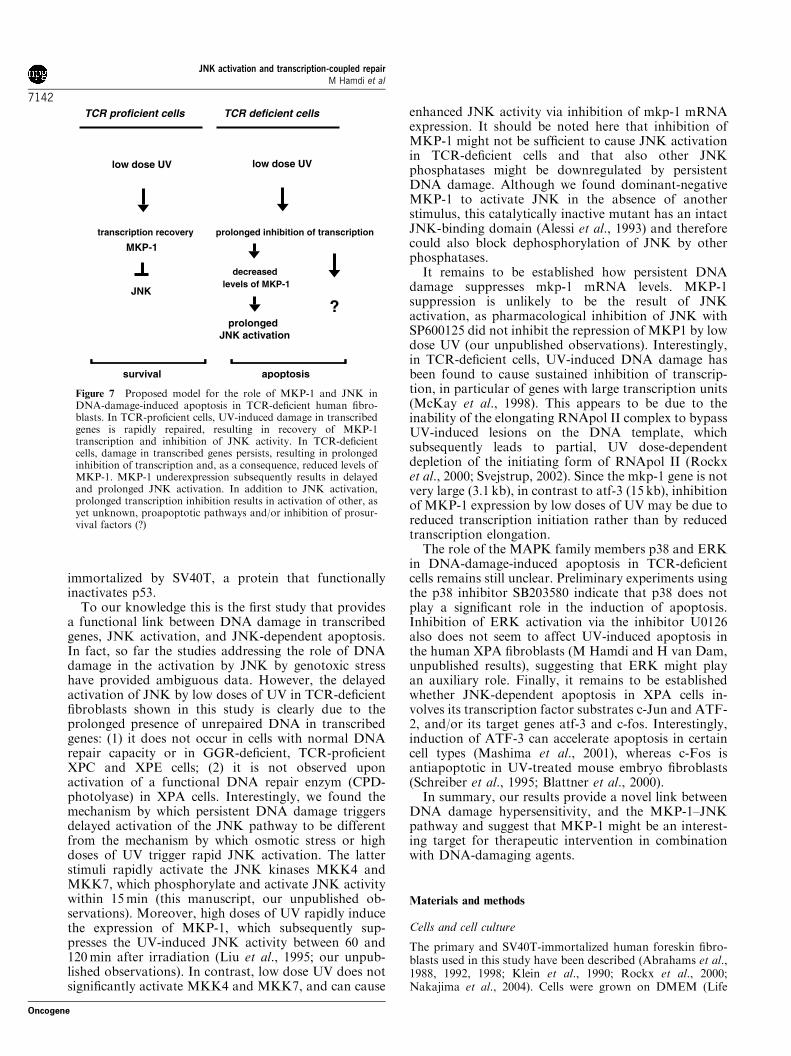
immortalized by SV40T, a protein that functionallyinactivates p53.To our knowledge this is the first study that provides
a functional link between DNA damage in transcribedgenes, JNK activation, and JNK-dependent apoptosis.In fact, so far the studies addressing the role of DNAdamage in the activation by JNK by genotoxic stresshave provided ambiguous data. However, the delayedactivation of JNK by low doses of UV in TCR-deficientfibroblasts shown in this study is clearly due to theprolonged presence of unrepaired DNA in transcribedgenes: (1) it does not occur in cells with normal DNArepair capacity or in GGR-deficient, TCR-proficientXPC and XPE cells; (2) it is not observed uponactivation of a functional DNA repair enzym (CPD-photolyase) in XPA cells. Interestingly, we found themechanism by which persistent DNA damage triggersdelayed activation of the JNK pathway to be differentfrom the mechanism by which osmotic stress or highdoses of UV trigger rapid JNK activation. The latterstimuli rapidly activate the JNK kinases MKK4 andMKK7, which phosphorylate and activate JNK activitywithin 15min (this manuscript, our unpublished ob-servations). Moreover, high doses of UV rapidly inducethe expression of MKP-1, which subsequently sup-presses the UV-induced JNK activity between 60 and120min after irradiation (Liu et al., 1995; our unpub-lished observations). In contrast, low dose UV does notsignificantly activate MKK4 and MKK7, and can cause
enhanced JNK activity via inhibition of mkp-1 mRNAexpression. It should be noted here that inhibition ofMKP-1 might not be sufficient to cause JNK activationin TCR-deficient cells and that also other JNKphosphatases might be downregulated by persistentDNA damage. Although we found dominant-negativeMKP-1 to activate JNK in the absence of anotherstimulus, this catalytically inactive mutant has an intactJNK-binding domain (Alessi et al., 1993) and thereforecould also block dephosphorylation of JNK by otherphosphatases.It remains to be established how persistent DNA
damage suppresses mkp-1 mRNA levels. MKP-1suppression is unlikely to be the result of JNKactivation, as pharmacological inhibition of JNK withSP600125 did not inhibit the repression of MKP1 by lowdose UV (our unpublished observations). Interestingly,in TCR-deficient cells, UV-induced DNA damage hasbeen found to cause sustained inhibition of transcrip-tion, in particular of genes with large transcription units(McKay et al., 1998). This appears to be due to theinability of the elongating RNApol II complex to bypassUV-induced lesions on the DNA template, whichsubsequently leads to partial, UV dose-dependentdepletion of the initiating form of RNApol II (Rockxet al., 2000; Svejstrup, 2002). Since the mkp-1 gene is notvery large (3.1 kb), in contrast to atf-3 (15 kb), inhibitionof MKP-1 expression by low doses of UV may be due toreduced transcription initiation rather than by reducedtranscription elongation.The role of the MAPK family members p38 and ERK
in DNA-damage-induced apoptosis in TCR-deficientcells remains still unclear. Preliminary experiments usingthe p38 inhibitor SB203580 indicate that p38 does notplay a significant role in the induction of apoptosis.Inhibition of ERK activation via the inhibitor U0126also does not seem to affect UV-induced apoptosis inthe human XPA fibroblasts (M Hamdi and H van Dam,unpublished results), suggesting that ERK might playan auxiliary role. Finally, it remains to be establishedwhether JNK-dependent apoptosis in XPA cells in-volves its transcription factor substrates c-Jun and ATF-2, and/or its target genes atf-3 and c-fos. Interestingly,induction of ATF-3 can accelerate apoptosis in certaincell types (Mashima et al., 2001), whereas c-Fos isantiapoptotic in UV-treated mouse embryo fibroblasts(Schreiber et al., 1995; Blattner et al., 2000).In summary, our results provide a novel link between
DNA damage hypersensitivity, and the MKP-1–JNKpathway and suggest that MKP-1 might be an interest-ing target for therapeutic intervention in combinationwith DNA-damaging agents.
Materials and methods
Cells and cell culture
The primary and SV40T-immortalized human foreskin fibro-blasts used in this study have been described (Abrahams et al.,1988, 1992, 1998; Klein et al., 1990; Rockx et al., 2000;Nakajima et al., 2004). Cells were grown on DMEM (Life
low dose UV
prolonged inhibition of transcription
survival
prolongedJNK activation
apoptosis
?
transcription recovery
MKP-1
JNK
decreasedlevels of MKP-1
TCR proficient cells
low dose UV
TCR deficient cells
Figure 7 Proposed model for the role of MKP-1 and JNK inDNA-damage-induced apoptosis in TCR-deficient human fibro-blasts. In TCR-proficient cells, UV-induced damage in transcribedgenes is rapidly repaired, resulting in recovery of MKP-1transcription and inhibition of JNK activity. In TCR-deficientcells, damage in transcribed genes persists, resulting in prolongedinhibition of transcription and, as a consequence, reduced levels ofMKP-1. MKP-1 underexpression subsequently results in delayedand prolonged JNK activation. In addition to JNK activation,prolonged transcription inhibition results in activation of other, asyet unknown, proapoptotic pathways and/or inhibition of prosur-vival factors (?)
JNK activation and transcription-coupled repairM Hamdi et al
7142
Oncogene
technologies, Inc., Paisley, UK) supplemented with 9% FBS(Life technologies, Inc.), penicillin, and streptomycin.For UV-C irradiation, a 30W germicidal lamp (TUV;
Philips Electronic Instruments Inc., Eindhoven, The Nether-lands) was used. Prior to UV treatment, culture medium wascollected, dishes were washed once with PBS (10mM Na2H-PO4/0.14mM NaCl, pH 7.4), and the PBS was removed beforeirradiation at room temperature. After irradiation, cells wererefed with the collected medium. For photoreactivation, cellswere irradiated for 1 h with a 30W 450 nm emitting light tube(Philips Electronic Instruments, Inc.) at RT, while mock-treated cells were kept in dark at RT for the same period.
FACS analysis
For quantitation of sub-G1 apoptotic cells, adhering andfloating cells were collected via trypsinization and centrifuga-tion. After resuspending in PBS, cells were fixed in 70%ethanol, washed in PBS, stained with 7.5 mM propidiumiodide(PI) containing 50mg/ml RNAse A, and analysed by flowcytometry (FACS-Calibur, Becton Dickenson). FACS resultswere analysed with WinMDI 2.8 software.
Western analysis and antibodies
For preparation of protein extracts, cells were washed oncewith ice-cold PBS and subsequently lysed on ice in ice-coldbuffer containing 10mM Tris pH 7.5, 150mM NaCl, 1%NP40, 1% sodium deoxycholate, and 0.1% sodium dodecylsulfate (SDS) containing protease and phosphatase inhibitors(van Dam et al., 1993). Lysates were cleared by centrifugation.Routinely either 20 or 30mg of protein were loaded on SDS–PAGE gels. Antibodies used are c-Jun-H79, ATF3-C19, andMKP-1-M18 from Santa Cruz Biotechnology, Inc. (SantaCruz, CA, USA), ERK-Thr202/Tyr204, SEK/MKK-4-Thr223, and PhosphoPluss p38 MAP kinase (Thr180/Tyr182) from Cell Signalling Technology, Inc. (Beverly, MA,USA), Anti-ACTIVEs JNK pAb pTPpY from PromegaCorporation, and tubulin from Sigma. The monoclonalantibody against threonine 91-phosphorylated c-Jun waskindly provided by Dr A Behrens, Cancer UK, London(Nateri et al., 2004).
RNA isolation and Northern analysis
RNA was isolated with the SV Total RNA isolation system(Promega) followed by an additional acid phenol extraction.Northern analysis including the c-jun, atf-3, and hef-1 probeshas been described previously (van Dam et al., 1989, 1990;Kool et al., 2003). As mkp-1 probe, the EcoRI insert ofp3CH134 containing the mouse mkp-1 cDNA (Lau andNathans, 1985) was used.
MKP-1 constructs and lentivirus transduction
cDNAs of MKP-1, C/SmMKP1, and m(JNK)MKP-1 (Keyseand Emslie, 1992; Alessi et al., 1993; Slack et al., 2001) were akind gift of Dr S Keyse (Dundee, Scotland, UK) and reclonedinto the lentiviral vector pRRL-cPPT-CMV-IRES-GFP-PRE-SIN (also named pLV-CMV-IRES-GFP; Carlotti et al., 2004).The viruses were produced as described previously (Carlottiet al., 2004). Briefly, the lentiviral cDNA construct wascotransfected overnight with three helper plasmids encodingHIV1 gag/pol, HIV1 rev, and VSV-G envelope into 293T cells.The medium was refreshed and viruses were harvested after 48and 72 h, passed through 0.45 mm filters and stored at �801C.Virus was quantitated by antigen capture ELISA measuringHIV p24 levels (ZeptoMetrix Corporation, New York, USA).For transduction, viral supernatants were overnight added tosubconfluent cell cultures in fresh medium supplemented with8 mg/ml polybrene (Sigma). The next day, the medium wasreplaced. Transduction efficiency was 95–100% as verified byFACS analysis (IRES-GFP).
Acknowledgements
We thank Martijn Rabelink for assisting with lentivirusproduction, Axel Behrens for kindly providing the anti-phospho-Thr91-cJun antibody, and Jan Hoeijmakers, HarryVrieling, Bert van Zeeland, Davy Rockx, Marcel Volker,Merlijn Bazuine, Peter Abrahams, and Lex van der Eb forhelpful discussions. This work was supported by grants fromthe Netherlands Organisation for Scientific Research (NWO),the Dutch Cancer Society (KWF), and the RadiationProtection, Biomed, TMR, and RTN Programs of theEuropean Community.
References
Abrahams PJ, Houweling A, Cornelissen-Steijger PDM,Jaspers NGJ, Darroudi F, Meijers CM, Mullenders LHF,Filon R, Arwert F, Pinedo HM, Natarajan APT, Terleth C,van Zeeland AA and van der Eb AJ. (1998). Mut. Res-DNARepair, 407, 189–201.
Abrahams PJ, Houweling A and van der Eb AJ. (1992).Cancer Res., 52, 53–57.
Abrahams PJ, van der Kleij AAM, Schouten R and van der EbAJ. (1988). Cancer Res., 48, 6054–6057.
Alessi DR, Smythe C and Keyse SM. (1993). Oncogene, 8,
2015–2020.Bazuine M, Carlotti F, Tafrechi RSJ, Hoeben RC andMaassen JA. (2004). Mol. Endocrinol., 18, 1697–1707.
Behrens A, Sibilia M and Wagner EF. (1999). Nat. Genet., 21,326–329.
Berneburg M and Lehmann AR. (2001). Adv. Genet., 43,
71–102.Blattner C, Bender K, Herrlich P and Rahmsdorf HJ. (1998).
Oncogene, 16, 2827–2834.Blattner C, Kannouche P, Litfin M, Bender K, Rahmsdorf HJ,Angulo JF and Herrlich P. (2000). Mol. Cell. Biol., 20,
3616–3625.
Bossy-Wetzel E, Bakiri L and Yaniv M. (1997). EMBO J., 16,
1695–1709.Brancho D, Tanaka N, Jaeschke A, Ventura JJ, Kelkar N,Tanaka Y, Kyuuma M, Takeshita T, Flavell RA and DavisRJ. (2003). Genes Dev., 17, 1969–1978.
Camps M, Nichols A and Arkinstall S. (2000). FASEB J., 14,
6–16.Carlotti F, Bazuine M, Kekarainen T, Seppen J, Pognonec P,Maassen JA and Hoeben RC. (2004). Mol. Ther., 9,
209–217.Chang LF and Karin M. (2001). Nature, 410, 37–40.Davis RJ. (2000). Cell, 103, 239–252.de Boer J and Hoeijmakers JHJ. (2000). Carcinogenesis, 21,
453–460.Desbois-Mouthon C, Cadoret A, Blivet-Van Eggelpoel MJ,Bertrand F, Caron M, Atfi A, Cherqui G and Capeau J.(2000). Endocrinology, 141, 922–931.
Engelbrecht Y, de Wet H, Horsch K, Langeveldt CR, HoughFS and Hulley PA. (2003). Endocrinology, 144, 412–422.
Farooq A and Zhou MM. (2004). Cell. Signal., 16, 769–779.Ford JM and Hanawalt PC. (1995). Proc. Natl. Acad. Sci.
USA, 92, 8876–8880.
JNK activation and transcription-coupled repairM Hamdi et al
7143
Oncogene
Franklin CC and Kraft AS. (1997). J. Biol. Chem., 272,
16917–16923.Franklin CC, Srikanth S and Kraft AS. (1998). Proc. Natl.
Acad. Sci. USA, 95, 3014–3019.Guo YL, Kang BB and Williamson JR. (1998). J. Biol. Chem.,
273, 10362–10366.Hai T, Wolfgang CD, Marsee DK, Allen AE and SivaprasadU. (1999). Gene Expression, 7, 321–335.
Hayakawa J, Depatie C, Ohmichi M and Mercola D. (2003).J. Biol. Chem., 278, 20582–20592.
Hayakawa J, Mittal S, Wang Y, Korkmaz KS, Adamson E,English C, Omichi M, McClelland M and Mercola D.(2004). Mol. Cell, 16, 521–535.
Herrlich P, Bender K, Knebel A, Bohmer FD, Gross S,Blattner C, Rahmsdorf HJ and Gottlicher M. (1999). CRAcad. Sci. III, 322, 121–125.
Jaspers NGJ, Raams A, Kelner MJ, Ng JMY, Yamashita YM,Takeda S, McMorris TC and Hoeijmakers JHJ. (2002).DNA Repair, 1, 1027–1038.
Kannan K, Amariglio N, Rechavi G, Jakob-Hirsch J, Kela I,Kaminski N, Getz G, Domany E and Givol D. (2001).Oncogene, 20, 2225–2234.
Keyse SM. (2000). Curr. Opin. Cell Biol., 12, 186–192.Keyse SM and Emslie EA. (1992). Nature, 359, 644–647.Kool J, Hamdi M, Cornelissen-Steijger PDM, van der Eb AJ,Terleth C and van Dam H. (2003). Oncogene, 22, 4235–4242.
Klein B, Pastink A, Odijk H, Westerveld A and van der Eb AJ.(1990). Exp. Cell Res., 191, 256–262.
Lamb JA, Ventura JJ, Hess P, Flavell RA and Davis RJ.(2003). Mol. Cell, 11, 1479–1489.
Lau LF and Nathans D. (1985). EMBO J., 4, 3145–3151.Lei K and Davis RJ. (2003). Proc. Natl. Acad. Sci. USA, 100,
2432–2437.Lei K, Nimnual A, Zong WX, Kennedy NJ, Flavell RA,Thompson CB, Bar-Sagi D and Davis RJ. (2002). Mol. Cell.Biol., 22, 4929–4942.
Li CH, Hu YH, Mayr M and Xu QB. (1999). J. Biol. Chem.,274, 25273–25280.
Li J, Gorospe M, Hutter D, Barnes J, Keyse SM and Liu YS.(2001). Mol. Cell. Biol., 21, 8213–8224.
Liu YS, Gorospe M, Yang CL and Holbrook NJ. (1995).J. Biol. Chem., 270, 8377–8380.
MacLaren A, Black EJ, Clark W and Gillespie DAF. (2004).Mol. Cell. Biol., 24, 9006–9018.
Martindale JL and Holbrook NJ. (2002). J. Cell. Physiol., 192,
1–15.Mashima T, Udagawa S and Tsuruo T. (2001). J. Cell. Physiol,
188, 352–358.Mckay BC, Becerril C and Ljungman M. (2001). Oncogene, 20,
6805–6808.Mckay BC, Ljungman M and Rainbow AJ. (1998). Oncogene,
17, 545–555.Mercurio F and Manning AM. (1999). Oncogene, 18,
6163–6171.Mitchell JR, Hoeijmakers JHJ and Niedernhofer LJ. (2003).
Curr. Opin. Cell Biol., 15, 232–240.Mizuno R, Oya M, Shiomi T, Marumo K, Okada Y andMurai M. (2004). J. Urol., 172, 723–727.
Morrison DK and Davis RJ. (2003). Ann. Rev. Cell Dev. Biol.,19, 91–118.
Nakajima S, Lan L, Kanno S, Takao M, Yamamoto K,Eker APM and Yasui A. (2004). J. Biol. Chem., 279,46674–46677.
Nateri AS, Riera-Sans L, Da Costa C and Behrens A. (2004).Science, 303, 1374–1378.
Queille S, Drougard C, Sarasin A and Daya-Grosjean L.(2001). J. Invest. Dermatol., 117, 1162–1170.
Rockx DAP, Mason R, van Hoffen A, Barton MC, Citterio E,Bregman DB, van Zeeland AA, Vrieling H and MullendersLHF. (2000). Proc. Natl. Acad. Sci. USA, 97, 10503–10508.
Rosette C and Karin M. (1996). Science, 274, 1194–1197.Sachsenmaier C, Radlerpohl A, Zinck R, Nordheim A,Herrlich P and Rahmsdorf HJ. (1994). Cell, 78, 963–972.
Sanchez-Perez I, Martinez-Gomariz M, Williams D, KeyseSM and Perona R. (2000). Oncogene, 19, 5142–5152.
Sanchez-Perez I, Murguia JR and Perona R. (1998). Oncogene,16, 533–540.
Schreiber M, Baumann B, Cotten M, Angel P and Wagner EF.(1995). EMBO J., 14, 5338–5349.
Shaulian E and Karin M. (2001). Oncogene, 20, 2390–2400.Shaulian E and Karin M. (2002). Nat. Cell Biol., 4,
E131–E136.Slack DN, Seternes OM, Gabrielsen M and Keyse SM. (2001).
J. Biol. Chem., 276, 16491–16500.Smith ML, Ford JM, Hollander MC, Bortnick RA, Amund-son SA, Seo YR, Deng CX, Hanawalt PC and Fornace AJ.(2000). Mol. Cell. Biol., 20, 3705–3714.
Spivak G, Itoh T, Matsunaga T, Nikaido O, Hanawalt P andYamaizumi M. (2002). DNA Repair, 1, 629–643.
Svejstrup JQ. (2002). Mol. Cell, 9, 1151–1152.Todd DE, Densham RM, Molton SA, Balmanno K, NewsonC, Weston CR, Garner AP, Scott L and Cook SJ. (2004).Oncogene, 23, 3284–3295.
van Dam H and Castellazzi M. (2001). Oncogene, 20,
2453–2464.van Dam H, DuyndamM, Rottier R, Bosch A, De Vries-SmitsL, Herrlich P, Zantema A, Angel P and van der Eb AJ.(1993). EMBO J., 12, 479–487.
van Dam H, Offringa R, Meijer I, Stein B, Smits AM, HerrlichP, Bos JL and van der Eb AJ. (1990). Mol. Cell. Biol., 10,
5857–5864.van Dam H, Offringa R, Smits AMM, Bos JL, Jones NC andvan der Eb AJ. (1989). Oncogene, 4, 1207–1212.
van Oosten M, Rebel H, Friedberg EC, van Steeg H, van derHorst GTJ, van Kranen HJ, Westerman A, van Zeeland AA,Mullenders LHF and de Gruijl FR. (2000). Proc. Natl. Acad.Sci. USA, 97, 11268–11273.
van Steeg H and Kraemer KH. (1999). Mol. Med. Today, 5,
86–94.Wada T, Joza N, Cheng HYM, Sasaki T, Kozieradzki I,Bachmaier K, Katada T, Schreiber M, Wagner EF, NishinaH and Penninger JM. (2004). Nat. Cell Biol., 6, 215–226.
Weston CR and Davis RJ. (2002). Curr. Opin. Genet Dev., 12,
14–21.Xu QH, Konta T, Nakayama KJ, Furusu A, Moreno-Manzano V, Lucio-Cazana J, Ishikawa Y, Fine LG, Yao Jand Kitamura M. (2004). Free Radic Biol. Med., 36,985–993.
Yan CH, Wang H and Boyd DD. (2002). J. Biol. Chem., 277,
10804–10812.
JNK activation and transcription-coupled repairM Hamdi et al
7144
Oncogene
Copyright © 2022 FDOKUMEN